Enhanced External Counterpulsation Inhibits Intimal Hyperplasia by Modifying Shear Stress-Responsive...
10
Hui, Zhensheng Zheng and Guifu Wu Yafei Jin, Yan Xiong, Jiangui He, Dianqiu Fang, Kuijian Wang, William E. Lawson, John C.K. Yan Zhang, Xiaohong He, Xiaolin Chen, Hong Ma, Donghong Liu, Jinyun Luo, Zhimin Du, Responsive Gene Expression in Hypercholesterolemic Pigs - Stress Enhanced External Counterpulsation Inhibits Intimal Hyperplasia by Modifying Shear Print ISSN: 0009-7322. Online ISSN: 1524-4539 Copyright © 2007 American Heart Association, Inc. All rights reserved. is published by the American Heart Association, 7272 Greenville Avenue, Dallas, TX 75231 Circulation doi: 10.1161/CIRCULATIONAHA.106.647248 2007;116:526-534; originally published online July 9, 2007; Circulation. http://circ.ahajournals.org/content/116/5/526 World Wide Web at: The online version of this article, along with updated information and services, is located on the http://circ.ahajournals.org/content/suppl/2007/07/11/CIRCULATIONAHA.106.647248.DC1.html Data Supplement (unedited) at: http://circ.ahajournals.org//subscriptions/ is online at: Circulation Information about subscribing to Subscriptions: http://www.lww.com/reprints Information about reprints can be found online at: Reprints: document. Permissions and Rights Question and Answer this process is available in the click Request Permissions in the middle column of the Web page under Services. Further information about Office. Once the online version of the published article for which permission is being requested is located, can be obtained via RightsLink, a service of the Copyright Clearance Center, not the Editorial Circulation in Requests for permissions to reproduce figures, tables, or portions of articles originally published Permissions: by guest on March 7, 2014 http://circ.ahajournals.org/ Downloaded from by guest on March 7, 2014 http://circ.ahajournals.org/ Downloaded from
-
Upload
independent -
Category
Documents
-
view
0 -
download
0
Transcript of Enhanced External Counterpulsation Inhibits Intimal Hyperplasia by Modifying Shear Stress-Responsive...

Hui, Zhensheng Zheng and Guifu WuYafei Jin, Yan Xiong, Jiangui He, Dianqiu Fang, Kuijian Wang, William E. Lawson, John C.K.
Yan Zhang, Xiaohong He, Xiaolin Chen, Hong Ma, Donghong Liu, Jinyun Luo, Zhimin Du,Responsive Gene Expression in Hypercholesterolemic Pigs−Stress
Enhanced External Counterpulsation Inhibits Intimal Hyperplasia by Modifying Shear
Print ISSN: 0009-7322. Online ISSN: 1524-4539 Copyright © 2007 American Heart Association, Inc. All rights reserved.
is published by the American Heart Association, 7272 Greenville Avenue, Dallas, TX 75231Circulation doi: 10.1161/CIRCULATIONAHA.106.6472482007;116:526-534; originally published online July 9, 2007;Circulation.
http://circ.ahajournals.org/content/116/5/526World Wide Web at:
The online version of this article, along with updated information and services, is located on the
http://circ.ahajournals.org/content/suppl/2007/07/11/CIRCULATIONAHA.106.647248.DC1.htmlData Supplement (unedited) at:
http://circ.ahajournals.org//subscriptions/
is online at: Circulation Information about subscribing to Subscriptions:
http://www.lww.com/reprints Information about reprints can be found online at: Reprints:
document. Permissions and Rights Question and Answer this process is available in the
click Request Permissions in the middle column of the Web page under Services. Further information aboutOffice. Once the online version of the published article for which permission is being requested is located,
can be obtained via RightsLink, a service of the Copyright Clearance Center, not the EditorialCirculationin Requests for permissions to reproduce figures, tables, or portions of articles originally publishedPermissions:
by guest on March 7, 2014http://circ.ahajournals.org/Downloaded from by guest on March 7, 2014http://circ.ahajournals.org/Downloaded from
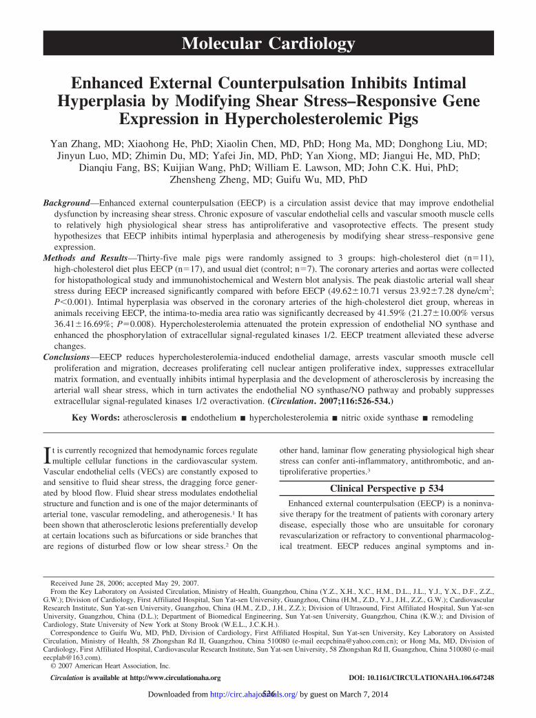
Enhanced External Counterpulsation Inhibits IntimalHyperplasia by Modifying Shear Stress–Responsive Gene
Expression in Hypercholesterolemic PigsYan Zhang, MD; Xiaohong He, PhD; Xiaolin Chen, MD, PhD; Hong Ma, MD; Donghong Liu, MD;
Jinyun Luo, MD; Zhimin Du, MD; Yafei Jin, MD, PhD; Yan Xiong, MD; Jiangui He, MD, PhD;Dianqiu Fang, BS; Kuijian Wang, PhD; William E. Lawson, MD; John C.K. Hui, PhD;
Zhensheng Zheng, MD; Guifu Wu, MD, PhD
Background—Enhanced external counterpulsation (EECP) is a circulation assist device that may improve endothelialdysfunction by increasing shear stress. Chronic exposure of vascular endothelial cells and vascular smooth muscle cellsto relatively high physiological shear stress has antiproliferative and vasoprotective effects. The present studyhypothesizes that EECP inhibits intimal hyperplasia and atherogenesis by modifying shear stress–responsive geneexpression.
Methods and Results—Thirty-five male pigs were randomly assigned to 3 groups: high-cholesterol diet (n�11),high-cholesterol diet plus EECP (n�17), and usual diet (control; n�7). The coronary arteries and aortas were collectedfor histopathological study and immunohistochemical and Western blot analysis. The peak diastolic arterial wall shearstress during EECP increased significantly compared with before EECP (49.62�10.71 versus 23.92�7.28 dyne/cm2;P�0.001). Intimal hyperplasia was observed in the coronary arteries of the high-cholesterol diet group, whereas inanimals receiving EECP, the intima-to-media area ratio was significantly decreased by 41.59% (21.27�10.00% versus36.41�16.69%; P�0.008). Hypercholesterolemia attenuated the protein expression of endothelial NO synthase andenhanced the phosphorylation of extracellular signal-regulated kinases 1/2. EECP treatment alleviated these adversechanges.
Conclusions—EECP reduces hypercholesterolemia-induced endothelial damage, arrests vascular smooth muscle cellproliferation and migration, decreases proliferating cell nuclear antigen proliferative index, suppresses extracellularmatrix formation, and eventually inhibits intimal hyperplasia and the development of atherosclerosis by increasing thearterial wall shear stress, which in turn activates the endothelial NO synthase/NO pathway and probably suppressesextracellular signal-regulated kinases 1/2 overactivation. (Circulation. 2007;116:526-534.)
Key Words: atherosclerosis � endothelium � hypercholesterolemia � nitric oxide synthase � remodeling
It is currently recognized that hemodynamic forces regulatemultiple cellular functions in the cardiovascular system.
Vascular endothelial cells (VECs) are constantly exposed toand sensitive to fluid shear stress, the dragging force gener-ated by blood flow. Fluid shear stress modulates endothelialstructure and function and is one of the major determinants ofarterial tone, vascular remodeling, and atherogenesis.1 It hasbeen shown that atherosclerotic lesions preferentially developat certain locations such as bifurcations or side branches thatare regions of disturbed flow or low shear stress.2 On the
other hand, laminar flow generating physiological high shearstress can confer anti-inflammatory, antithrombotic, and an-tiproliferative properties.3
Clinical Perspective p 534Enhanced external counterpulsation (EECP) is a noninva-
sive therapy for the treatment of patients with coronary arterydisease, especially those who are unsuitable for coronaryrevascularization or refractory to conventional pharmacolog-ical treatment. EECP reduces anginal symptoms and in-
Received June 28, 2006; accepted May 29, 2007.From the Key Laboratory on Assisted Circulation, Ministry of Health, Guangzhou, China (Y.Z., X.H., X.C., H.M., D.L., J.L., Y.J., Y.X., D.F., Z.Z.,
G.W.); Division of Cardiology, First Affiliated Hospital, Sun Yat-sen University, Guangzhou, China (H.M., Z.D., Y.J., J.H., Z.Z., G.W.); CardiovascularResearch Institute, Sun Yat-sen University, Guangzhou, China (H.M., Z.D., J.H., Z.Z.); Division of Ultrasound, First Affiliated Hospital, Sun Yat-senUniversity, Guangzhou, China (D.L.); Department of Biomedical Engineering, Sun Yat-sen University, Guangzhou, China (K.W.); and Division ofCardiology, State University of New York at Stony Brook (W.E.L., J.C.K.H.).
Correspondence to Guifu Wu, MD, PhD, Division of Cardiology, First Affiliated Hospital, Sun Yat-sen University, Key Laboratory on AssistedCirculation, Ministry of Health, 58 Zhongshan Rd II, Guangzhou, China 510080 (e-mail [email protected]); or Hong Ma, MD, Division ofCardiology, First Affiliated Hospital, Cardiovascular Research Institute, Sun Yat-sen University, 58 Zhongshan Rd II, Guangzhou, China 510080 ([email protected]).
© 2007 American Heart Association, Inc.
Circulation is available at http://www.circulationaha.org DOI: 10.1161/CIRCULATIONAHA.106.647248
526
Molecular Cardiology
by guest on March 7, 2014http://circ.ahajournals.org/Downloaded from

creases exercise capacity in the majority of patients undergo-ing treatment.4,5 Growing evidence suggests thatimprovement in endothelial function represents an importantmechanism for the clinical benefits of EECP.6,7 However, theexact molecular mechanism remains elusive, and no athero-sclerotic animal model other than the use of an acute EECPtreatment protocol has been reported.
The present study was designed to investigate the effect ofEECP on vascular endothelial function, vascular remodeling,and atherogenesis, as well as VEC gene expression in aporcine experimental model of hypercholesterolemia. Wehypothesized that EECP could increase shear stress in vivo,improve both structure and function of the vascular endothe-lium, inhibit intimal hyperplasia, and therefore arrest athero-genesis through modulating shear stress–responsive geneexpression such as upregulating vascular endothelial NOsynthase (eNOS) expression and probably downregulatingextracellular signal-regulated kinases 1/2 (ERK1/2) activity.
MethodsAnimal ModelThirty-five male Yorkshire (male) and Landrance (female) crossbredpigs (Swine Plant of South China University, Guangzhou, China)with an average weight of 7.4 kg were randomized to 3 groups(7.6�0.7 kg in the control group; 7.2�0.8 kg in the high-cholesteroldiet [CHOL] group; and 7.4�0.8 kg in the high-cholesteroldiet�EECP [CHOL�EECP] group). All procedures were in accor-dance with the Guide for the Care and Use of Laboratory Animalspublished by the US National Institutes of Health. Seven pigs werefed a usual diet (control group) over the course of 15 weeks.Twenty-eight pigs were fed a high-cholesterol atherogenic dietcontaining 4% cholesterol, 10% yolk powder, 8% lard, and 1.2%salts for 15 weeks. After 8 weeks of high-cholesterol feeding, whilestill on the atherogenic diet, 17 hypercholesterolemic pigs weresubjected to EECP intervention (CHOL�EECP group). The remain-ing 11 pigs served as hypercholesterolemic controls (CHOL group).All pigs were anesthetized with midazolam 5 to 10 mg IM and 3%pentobarbital 10 mg/kg per hour IV infusion. Blood samples werecollected at weeks 0, 8, 12, and 15 of study for the measurement oflipid profiles (Hitachi 7170A Tokyo, Japan) and blood viscosity(Rheocalc V2.1, Brookfield Engineering Laboratory, Inc, Middle-boro, Mass). At week 15, all pigs were euthanized by an overdosageinjection of 10% potassium chloride into the heart.
EECP ProtocolPigs in the CHOL�EECP cohort received a total of 34�2 hours ofEECP treatment, 2-hour sessions every other day over a 7-weekperiod, imitating the standard clinical EECP protocol. With the useof a clinical system (Shuangshan EECP-MCI, Guangzhou, China),EECP was performed by laying the pigs on their left side andwrapping 2 sets of cuffs modified to closely fit the low extremitiesand the hips of the pigs. The cuffs were sequentially inflated withcompressed air from distal to proximal in early diastole and rapidlydeflated right before systole. The pressure applied to the cuffs wasset at 0.035 to 0.040 mPa/cm2. Effective hemodynamic changes ofEECP were demonstrated by achieving a diastolic-to-systolic ratio�1.2 with the use of the plethysmographic technique.
Hemodynamic MeasurementsAt week 15, Doppler flow examination was performed with the useof a color Doppler ultrasound system (ATL-HDI-5000, Phillip ComAmerica) equipped with an ECG-triggered instrument and a 5- to10-MHz multifrequency high-resolution linear probe, as previouslydescribed.8 For the calculation of wall shear stress, internal diameter
and blood flow velocity in the right brachial artery were measuredjust before and during EECP treatment. Peak diastolic velocity wasrecorded as the mean of 3 cardiac cycles.
The intensity of wall shear stress (�) was calculated according tothe formula9 � (dyne/cm2)�4�V/ID, where � is the viscosityexpressed in poise, V is the blood flow velocity expressed incentimeters per second, and ID is the internal diameter expressed incentimeters.
Morphological EvaluationAt the termination of the experiment, a 1-cm segment of the leftanterior descending coronary artery (LAD), 0.5 cm distal to thebifurcation of the left main coronary artery, was harvested and fixedand then cut into 3 segments: proximal, middle, and distal. Theparaffin-embedded segments were sectioned into 4-�m-thick crosssections and stained with hematoxylin-eosin or Gomori’s aldehydefuchsin elastic stains. Parameters were measured at �40 magnifica-tion with the use of a Zeiss-KONTRON IBAS 2.5 Automatic ImageAnalysis System (Zeiss, Munich, Germany). The intimal-to-medialratio and wall-to-lumen ratio were calculated for evaluation ofvascular remodeling. To quantify collagen deposition in the coronaryartery wall, van Gieson–stained sections were scanned to assess thepercentage of the intimal and partial medial area occupied bycollagen. For each LAD, at least 3 sections from different segmentswere analyzed. Segments of the LADs were treated for scanningelectron microscopy (Hitachi S520). Morphological evaluation andWestern blot band densitometry and immunohistochemical analyseswere performed by technicians who were blinded to the groupassignment.
Immunohistochemical StudySerial paraffin-embedded cross sections of LAD were used forimmunostaining according to the streptavidin-biotin complex or theElivision immunohistochemical technique.10 They were incubatedwith rabbit polyclonal phospho-ERK antibody (at 1:100 dilution;Cell Signaling Technology, Danvers, Mass), rabbit polyclonal eNOSantibody (at 1:70 dilution; Santa Cruz Biotechnology, Inc, SantaCruz, Calif), mouse anti–proliferating cell nuclear antigen (PCNA)antibody (at 1:100 dilution; Dako, Glostrup, Denmark), or mousemonoclonal smooth muscle �-actin antibody (at 1:100 dilution;Boster Biological Technology, Inc, Wuhan, China). The prolifera-tion index was defined as the percentage of PCNA-positive cellnumber against the total nucleated cell number. The percentage ofthe intimal area occupied by the �-actin–positive area was assessed.Controls in the absence of primary antibodies were also performed.
Western Blot AnalysisThe arch of the aorta was snap-frozen and homogenized in T-PERtissue protein extraction reagent (Pierce Chemical Co, Rockford, Ill)at 0°C to 4°C. The protein concentration was determined with aprotein assay system according to the manufacturer (Bio-Rad Dc;Bio-Rad Laboratories, Hercules, Calif). Equal amounts of totalprotein, 30 �g each, were subjected to electrophoresis on SDS-PAGE with the use of a 6% gel for eNOS and a 10% gel forphospho-ERK1/2. The separated proteins were electrophoreticallytransferred to Hybond-PVDF membranes (Amersham PharmaciaBio). After blocking, the membranes were incubated with eNOSantibody (at 1:200 dilution) or phospho-ERK1/2 antibody (at 1:1000dilution; phosphoPlus kit, Cell Signaling) overnight at 30 V and 4°C,then incubated with a horseradish peroxidase–conjugated secondaryantibody (1:2000) and exposed to an x-ray film with the use of theenhanced chemiluminescence immunoblotting detection system. Theamount of protein was quantified by densitometry.
The membranes were then stripped in stripping buffer(100 mmol/L �-ME, 2% SDS, 62.5 mmol/L Tris-HCl, pH 6.8) at48°C for 30 minutes, then blocked, incubated with a total ERK1/2antibody (similar to the manner described above, at 1:1000 dilution),and detected as internal control.
Zhang et al EECP Inhibits Intimal Hyperplasia 527
by guest on March 7, 2014http://circ.ahajournals.org/Downloaded from
Statistical AnalysisData are presented as mean�SD unless indicated otherwise. Whenhomogeneity of variance was confirmed by the Levene variancehomogeneity test, statistical comparison among 3 groups was per-formed by 1-way ANOVA. If the F test results were �0.05, post hoccomparisons were performed with the Bonferroni test. A value ofP�0.05 was considered statistically significant. When variablesviolated the assumptions for variance homogeneity, the Kruskal-Wallis ranking test was alternatively used, with P�0.05 representingsignificant differences. Post hoc comparisons were performed by theMann-Whitney test for 2 independent samples recording exact 2-tailed significance. To avoid an accumulation of errors due tomultiple comparisons, the levels of significance were adjusted byHolm’s method. It was used, for example, in the results presented inFigure 5: P�0.017 was considered statistically significant for com-parison of the CHOL group versus the control group, P�0.025 forthe CHOL group versus the CHOL�EECP group, and P�0.05 forthe control group versus the CHOL�EECP group.
When multiple time point measurements were taken over time(cholesterol and low-density lipoprotein measurements), repeated-measures analysis was performed with the general linear modelsgeneral factorial ANOVA, followed by the aforementioned methods.SPSS 13.0 software was used for all statistical calculations.
The authors had full access to and take full responsibility for theintegrity of the data. All authors have read and agreed to themanuscript as written.
ResultsCharacteristics of Cholesterol-Fed EECP PigsAt the end of the study at week 15, the body weight of theCHOL�EECP group was slightly but not significantly lessthan that of the CHOL group (P�0.05) (Table). The athero-genic diet resulted in a 4.79-fold increase in serum cholesterollevel and 6.66-fold increase in low-density lipoprotein levelin the CHOL group. The cholesterol levels of theCHOL�EECP group were slightly lower than those of theCHOL group, but this difference was not statistically signif-icant (P�0.05).
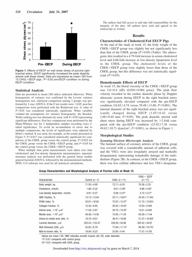
Hemodynamic Effects of EECPAt week 15, the blood viscosity of the CHOL�EECP groupwas 3.8�0.4 mPa (0.038�0.004 poise). The peak flowvelocity recorded in the cardiac diastolic phase by Dopplerultrasonic system during EECP in the right brachial arterywas significantly elevated compared with the pre-EECPcondition (24.62�4.74 versus 59.48�13.60; P�0.001). Theinternal diameter of the right brachial artery was not signif-icantly changed during EECP (1.65�0.42 versus1.68�0.44 mm; P�0.05). The peak diastolic arterial wallshear stress during EECP was increased by �2-fold com-pared with the pre-EECP condition (23.92�7.28 versus49.62�10.71 dyne/cm2; P�0.001), as shown in Figure 1.
Morphological Studies
Scanning Electron Microscopic AnalysisThe luminal surface of coronary arteries of the CHOL groupwas covered with a considerable amount of adherent cells,and the VECs were also irregularly arrayed and markedlydesquamated, representing remarkable damage of the endo-thelium (Figure 2B). In contrast, in the CHOL�EECP group,there was less cellular adherence and less VECs desquama-
Figure 1. Effects of EECP on wall shear stress of porcine rightbrachial artery. EECP significantly increased the peak diastolicarterial wall shear stress. Data are expressed as mean�SD from13 CHOL�EECP pigs. *P�0.05 pre-EECP condition vs duringEECP condition.
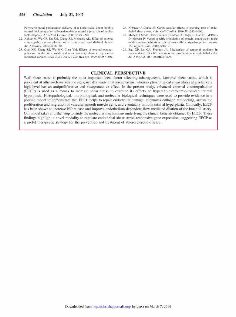
Group Characteristics and Morphological Analysis of Porcine LADs at Week 15
Characteristic Control (n�7) CHOL (n�11)CHOL� EECP
(n�17)
Body weight, kg 71.50�4.80 72.71�6.35 70.38�2.32
Cholesterol, mmol/L 1.88�0.32 9.01�2.89* 6.98�2.85*
Low-density lipoprotein, mmol/L 0.91�0.27 6.06�2.47* 4.72�2.57*
SMC fraction, % 15.13�13.84 53.11�16.61* 24.54�17.45†
PCNA index, % 42.01�18.65 75.07�12.47* 51.72�12.62†
Collagen fraction, % 12.35�6.85 26.55�10.05* 12.03�2.94†
Intimal area, �103 �m2 11.58�5.67 28.72�14.25* 13.61�6.84†
Medial area, �103 �m2 59.38�14.56 73.92�11.29 65.28�11.64
Intima-to-media area ratio, % 19.70�9.01 36.41�16.69 21.27�10.00†
Luminal diameter, �m 355.43�123.47 339.50�42.88 355.42�44.87
Wall thickness (OD), �m 55.62�6.79 77.84�11.15* 60.39�10.75†
Wall-to-lumen ratio, % 18.04�8.23 23.95�4.44 17.57�4.70†
Values are mean�SD. SMC indicates smooth muscle cell; OD, outer diameter.*P�0.01 compared with control group.†P�0.01 compared with CHOL group.
528 Circulation July 31, 2007
by guest on March 7, 2014http://circ.ahajournals.org/Downloaded from

tion, and the VECs tended to align in parallel to the directionof blood flow, as shown in Figure 2C.
Intimal HyperplasiaThe LAD of the CHOL group showed marked intimalthickening and significant atherosclerotic lesions, as dem-onstrated in Figure 3B and 3E. Morphometric analysisrevealed that the intimal area (P�0.004�0.025 versuscontrol group) and wall thickness (P�0.001 versus controlgroup) were significantly larger in the CHOL group than inthe normal control group, as shown in the Table. In theCHOL�EECP pigs, the intimal area was significantly
smaller (P�0.001�0.017 versus CHOL) and the LAD wallthickness was significantly thinner (P�0.001�0.05 versusCHOL) than in the CHOL group, as demonstrated inFigure 3C and 3F. Meanwhile, the differences of medialarea, medial thickness, and area within the internal elasticlamina and external elastic lamina among the 3 groups didnot reach statistical significance (P�0.05). EECP treat-ment resulted in a significantly reduced intima-to-mediaarea ratio (P�0.008�0.017 versus CHOL) and wall-to-lumen ratio (P�0.004�0.05 versus CHOL), showing anantihyperplastic effect (Table).
Figure 2. Scanning electron micro-graphic characteristics of the porcineLADs (magnification �500). A, Represen-tative micrograph of normal controlgroup. B, Representative micrograph ofCHOL group. The luminal surface wascovered with many adherent cells; seri-ous endothelial cell disarrangement anddesquamation were observed. C, Repre-sentative micrograph of CHOL�EECPgroup. Less cellular adherence could beobserved. VECs aligned parallel to thedirection of blood flow.
Figure 3. Effect of EECP on the histomorphology and intimal hyperplasia of LADs of hypercholesterolemic pigs. A, B and C, Represen-tative photomicrographs (magnification �40) of elastic-stained (dark purple) LAD. D, E and F, Representative photomicrographs (magni-fication �400) of LAD sections stained with hematoxylin-eosin (HE). A and D, Control group. B and E, CHOL group. C and F,CHOL�EECP group. G, Effect of EECP on intima-media thickness ratio of porcine LAD. Significant intimal thickening could beobserved in the CHOL group. EECP treatment resulted in diminished intimal hyperplasia in the CHOL�EECP group. Arrows indicateinternal elastic lamina. Values are mean�SD. *P�0.007�0.05; †P�0.002�0.05 vs CHOL group, 1-way ANOVA and Bonferroni test.
Zhang et al EECP Inhibits Intimal Hyperplasia 529
by guest on March 7, 2014http://circ.ahajournals.org/Downloaded from

Vascular Smooth Muscle Cell ProliferationIn hypercholesterolemic pigs, vascular smooth muscle cells(VSMCs) proliferated and migrated from the media into theintima through the broken and disarranged internal elasticlamina (Figure 4B). VSMC volume fraction, as representedby the percentage of intima area occupied by smooth musclecells in the intima,11 was significantly elevated in the CHOLgroup compared with the normal control group(P�0.005�0.05), as shown in the Table. EECP treatmentreduced the smooth muscle cell fraction by 53.79% comparedwith the CHOL group (P�0.003�0.05). These results indi-cated that the reduced intimal thickness of the LADs of theCHOL�EECP pigs was accompanied by reduced VSMCs inthe intima (Figure 4C).
Proliferation IndexAs shown in Figure 4, PCNA-positive cells (mainly VECsand VSMCs) were detected in the intima and the medial layerof the LADs. The PCNA-positive index was significantlygreater in the coronary arteries of the CHOL group than inthose of the normal control group (P�0.001 versus control),
as shown in Figure 4E and the Table. The decreased numberof PCNA-positive cells in the coronary arteries of theCHOL�EECP group (P�0.005�0.05 versus CHOL group)indicated that the proliferative changes induced by hypercho-lesterolemia were significantly restored by EECP interven-tion (Figure 4F).
Collagen RemodelingAccompanying thinning and breakage of the internal elasticlamina, elastic fibrin proliferation and disarrangement couldbe observed in the hyperplastic intima of the LAD (Figure3B). There also appeared to be a marked increase in collagenaccumulation in the hyperplastic intima and partial media(P�0.001�0.025 versus control group) (Figure 4H). Thepercentage of collagen-positive area in the CHOL�EECPgroup was significantly lower than that in the CHOL group(P�0.001). These results indicate that the proliferation ofvascular connective tissue and extracellular matrix caused byhypercholesterolemia was inhibited by EECP treatment, asshown in the Table and Figure 4I.
Figure 4. Effect of EECP on vascular remodeling in hypercholesterolemic pigs. A, B, and C, Representative photomicrographs of thesmooth muscle–specific �-actin immunohistochemical staining of LADs (brown, magnification �40). In the CHOL group, VSMCs prolif-erated and migrated from the media into the intima. In the CHOL�EECP group, reduced intimal thickness was accompanied withreduced VSMCs in the intima. D, E, and F, Immunohistochemical staining against PCNA of LADs (brown, magnification �400). A, D,and G, Control group. B, E, and H, CHOL group. C, F, and I, CHOL�EECP group. A marked increase in PCNA-positive cell number ofthe CHOL group was seen. In animals receiving EECP, the proliferative changes were significantly inhibited. G, H, and I, The V-G colla-gen staining of LADs (red, magnification �400). A marked increase in collagen accumulation was observed in the CHOL group but wasinhibited by EECP treatment.
530 Circulation July 31, 2007
by guest on March 7, 2014http://circ.ahajournals.org/Downloaded from

Effect of EECP on Level of eNOS Protein ExpressionMicrographs of porcine LADs with eNOS antibody immuno-histochemical staining are shown in Figure 5. The eNOSprotein was localized mainly in VECs. In the CHOL group, aremarkably weaker eNOS expression was observed (Figure5B). In the CHOL�EECP group, the reduction of eNOSexpression was not significant (Figure 5C).
Western blotting showed that the amount of eNOS proteinwas significantly decreased in the CHOL group comparedwith the control group (P�0.009�0.017). The eNOS proteinlevel in the CHOL�EECP group was 3.16 times that of theCHOL group (P�0.023�0.025 versus CHOL). These resultssuggest that EECP treatment significantly elevates eNOSprotein expression and alleviates the severe inhibition ofeNOS expression induced by hypercholesterolemia. Thechanges observed with Western blot correlated strongly withimmunohistochemistry results.
Effect of EECP on Phospho-ERK1/2 ActivityAs shown by immunohistochemical staining with phosph-ERK1/2 in Figure 6, phosphorylated (activated) ERK1/2, asassessed by brown staining, was detected mainly in the nucleiof VECs and smooth muscle cells. In atherosclerotic lesions,phospho-ERK1/2 protein was abundant in hyperplastic intima(Figure 6B). There were not as many positively stained nucleias in the CHOL�EECP group.
As shown by Western blot analysis, the ERK1/2 activity(expressed as the ratio of phospho-ERK1/2 to total ERK1/2)was higher in the CHOL group (P�0.035�0.017 for ERK1and P�0.001 for ERK2 versus control). In the CHOL�EECPgroup, the ERK1/2 activity showed a descending tendency(P�0.036�0.025 for ERK1 and P�0.026�0.025 for ERK2versus CHOL), as shown in Figure 6E. These results indi-cated that EECP intervention presumably inhibited the phos-phorylation of ERK1/2 induced by hypercholesterolemia. Thechanges observed with Western blot also correlated stronglywith immunohistochemistry results.
DiscussionWall shear stress is probably the most important local factor ableto influence atherogenesis. In large arteries, the magnitude ofshear stress is in the range of 10 to 70 dyne/cm2. Arterial levelshear stress (�15 dyne/cm2) induces endothelial quiescence andan atheroprotective gene expression profile, whereas low shearstress (�4 dyne/cm2), which is prevalent at atherosclerosis-prone sites, stimulates an atherogenic phenotype.2 Shear stressregulates endothelial structure and function by regulating theexpression of mechanosensitive genes. VECs subjected to a longduration of laminar shear stress at the relatively high levels havea lower rate of DNA synthesis than those under static condi-tions.12 In animals with vessel grafts13 or stented vessels,14
increased local shear stress induces regression of intimal hyper-
Figure 5. Effect of EECP on eNOS protein expression. A, B, and C, Immunohistochemistry (magnification �400) of eNOS in LADs of 3groups (brown-staining VECs). The expression of eNOS protein was decreased remarkably in the CHOL group but restored significantlyin the CHOL�EECP group. A, Control group. B, CHOL group. C, CHOL�EECP group. D, Representative Western blot bands of eNOSin porcine aortas. Bands of actin served as control for equal protein loading. E, Histograms show fluorescence intensity of bandsexpressed as the ratio of control. Results are expressed as mean�SEM of 5 independent experiments from 27 porcine protein sam-ples. eNOS protein expression was inhibited in the CHOL group and was elevated in CHOL�EECP group. *P�0.009�0.017, CHOLgroup vs control group; †P�0.023�0.025, CHOL�EECP group vs CHOL group; Mann-Whitney test adjusted by Holm’s method.
Zhang et al EECP Inhibits Intimal Hyperplasia 531
by guest on March 7, 2014http://circ.ahajournals.org/Downloaded from

plasia. In humans, it has also been demonstrated that augmen-tation of wall shear stress inhibits neointimal hyperplasia afterstent implantation.15,16
The systolic deflation/diastolic inflation sequence of EECPleads to systolic unloading and diastolic augmentation, result-ing in increased blood flow in a pulsatile manner. In thepresent study, the data showed that during EECP, the peakdiastolic arterial wall shear stress increased �2-fold. EECPhas the definite effect of elevating arterial wall shear stress invivo, whereas most drugs have failed to demonstrate suchbeneficial hemodynamic effects.
Using histopathological and morphological investigations,we have provided evidence in a porcine model that EECPimproves hypercholesterolemia-induced endothelial damage,arrests collagen remodeling, and inhibits the proliferation andmigration of VSMCs, findings supported by the reduction ofthe cellular proliferative index. The significant effects ofEECP on these main components of atherosclerotic lesionsprovide support for the hypothesis that EECP can inhibitintimal hyperplasia induced by hypercholesterolemia.
Acute increase in shear stress can cause acute robust NOproduction, which plays a critical role in vessel relaxation,whereas chronic NO production due to the increased laminal
shear stress may serve as an antiatherogenic and anti-inflammatory molecule.17 Being the rate-limiting enzyme essen-tial for NO synthesis, and with shear stress–responsive elementsin its gene promotor region, eNOS may serve as a mechanosen-sor coupling NO release to long-term hemodynamic changes.18
Previous studies have shown that a primary defect in the NOsynthase/NO pathway can accelerate neointimal formation andimpair endothelial function.19 In contrast, strategies aimed atincreasing local NO production, such as eNOS gene transfer20 ordelivery of NO donor,21 inhibit neointimal thickening afterartery injury. Both clinical investigation and animal studies fromour laboratory have shown that EECP treatment increasedplasma NO levels and eNOS gene expression.22,23 In the presentstudy, our results indicate that hypercholesterolemia markedlydecreased eNOS protein level, whereas EECP significantlyelevated eNOS protein expression.
It is well known that regular physical exercise improvesendothelial function by increasing blood flow and shearstress, which in turn enhances the eNOS/NO pathway.24 Inthis aspect, the mechanism of EECP is similar to physicalexercise. Because most cardiac patients cannot exercisesufficiently to achieve a similar degree of increase in arterialshear stress, EECP can help to provide a vascular protectivebenefit similar to that of vigorous exercise.
Figure 6. Effect of EECP on hypercholesterolemia-induced activation of ERK1/2. A, B, and C, Representative photomicrographs (mag-nification �400) of immunohistochemical staining against phospho-ERK1/2 (brown staining, nucleus localized) of porcine LADs. Theexpression of phospho-ERK1/2 was increased remarkably in the CHOL group but inhibited in the CHOL�EECP group. A, Controlgroup. B, CHOL group. C, CHOL�EECP group. D, Representative Western blot bands of phospho-ERK1/2 and total ERK1/2 (afterstripping) of porcine aorta. E, Histograms showing the fluorescence intensity of bands expressed as the ratio of phospho/total ERK1/2.The hypercholesterolemia-induced ERK1/2 phosphorylation was inhibited in the CHOL�EECP group. Results are expressed asmean�SEM of 6 independent experiments from 32 porcine protein samples. #P�0.035�0.017 for ERK1 CHOL group vs control group;*P�0.001�0.017 for ERK2 CHOL group vs control group; †P�0.036�0.025 for ERK1 CHOL�EECP group vs CHOL group;§P�0.026�0.025 for ERK2 CHOL�EECP group vs CHOL group; Mann-Whitney test adjusted by Holm’s method.
532 Circulation July 31, 2007
by guest on March 7, 2014http://circ.ahajournals.org/Downloaded from

Inhibition of ERK1/2 phosphorylation has been shown to becritical in NO inhibition of cellular proliferation.25 NO superox-ide and peroxynitrite induced by fluid shear stress appear tofunction as signaling molecules in flow-dependent activation ofmitogen-activated protein kinases. A previous study has docu-mented that exogenous NO mediates both temporal gradientshear-induced activation and steady shear-induced downregula-tion of phospho-ERK1/2.26 On the other hand, NOS blockadestimulates protein synthesis by enhancing ERK1/2 phosphory-lation in large arteries.25 As the central element of growthsignaling pathway, ERK1/2 is thought to be directly involved intransmitting signals from growth factor receptors to the nucleusto regulate gene transcription and protein synthesis, leading toproliferation, differentiation, or apoptosis. In the present study,ERK1/2 phosphorylation was upregulated in the hypercholester-olemic control pigs, whereas it was downregulated in the EECPgroup. ERK1/2 phosphorylation was inversely related to eNOSexpression, consistent with previous studies.25,26 These resultssuggest that EECP therapy helps VECs to maintain an athero-protective gene profile and resist the deleterious effects ofhypercholesterolemia.
One limitation of the present study is the relatively smallnumber of samples, which may influence the power ofstatistical tests. ERK activity showed a downregulating ten-dency in animals receiving EECP compared with hypercho-lesterolemic controls, but differences did not reach statisticalsignificance because the sample size may have been under-powered. Another limitation refers to the probable systematicbias in Doppler hemodynamic measurements performed bythe same ultrasound technician, who was not blinded to thegroup assignment. In addition, the hemodynamic parameterswere measured in a peripheral artery and therefore cannot beused directly to draw any conclusion about shear stress in thecoronary circulation. Still another limitation refers to the lackof a control group that used uncoordinated EECP.
In conclusion, the present study demonstrates a correlationbetween EECP-mediated increased shear stress and the inhi-bition of intimal hyperplasia, supporting the antiatherogeniceffect of chronic exposure to shear stress, and suggests thatEECP should be considered a therapeutic strategy for thetreatment of atherosclerotic occlusive disease.
AcknowledgmentsWe thank Ruide Hu, Huixi Wu, Lihong Che, Jinjun Zhang, GuifangLin, Gang Dai, Luguang Liang, Minzhe Feng, Qiang Xie, andYuetao Qian for excellent technical assistance.
Sources of FundingThe present study was supported by the Special Fund for BasicResearch on Scientific Instruments, National Natural Science Foun-dation, China (No. 50113-4103048); the Tenth National “Five-Year”Key Project (No. 2001BA706B07); and the 2003 Guangdong Pro-vincial Project of Science and Technology (No. 2003C30601).
DisclosuresDr Lawson served on the Speakers’ Bureau of Vasomedical. Dr Huiserved as chief technology officer and senior vice president ofVasomedical. The other authors report no conflicts.
References1. Cunningham KS, Gotlieb AI. The role of shear stress in the pathogenesis
of atherosclerosis. Lab Invest. 2005;85:9–23.2. Malek AM, Alper SL, Izumo S. Hemodynamic shear stress and its role in
atherosclerosis. JAMA. 1999;282:2035–2042.3. Resnick N, Yahav H, Shay-Salit A, Shushy M, Schubert S, Zilberman
LCM, Wofovitz E. Fluid shear stress and the vascular endothelium: forbetter and for worse. Prog Biophys Mol Biol. 2003;81:177–199.
4. Arora RR, Chou TM, Jain D, Fleishman B, Crawford L, McKiernan T,Nesto RW. The Multicenter Study of Enhanced External Counterpul-sation (MUST-EECP): effect of EECP on exercise-induced myo-cardial ischemia and angina episodes. J Am Coll Cardiol. 1999;33:1833–1840.
5. Urano H, Ikeda H, Ueno T, Matsumoto T, Murohara T, Imaizumi T.Enhanced external counterpulsation improves exercise tolerance, reducesexercise-induced myocardial ischemia and improves left ventricular dia-stolic filling in patients with coronary artery disease. J Am Coll Cardiol.2001;37:93–99.
6. Bonetti PO, Barsness GW, Keelan PC, Schnell TI, Pumper GM, KuvinJT, Schnall RP, Holmes DR, Higano ST, Lerman A. Enhanced externalcounterpulsation improves endothelial function in patients with symptom-atic coronary artery disease. J Am Coll Cardiol. 2003;41:1761–1768.
7. Shechter M, Matetzky S, Feinberg MS, Chouraqui P, Rotstein Z, Hod H.External counterpulsation therapy improves endothelial function inpatients with refractory angina pectoris. J Am Coll Cardiol. 2003;42:2090–2095.
8. Irace C, Cortese C, Fiaschi E, Carallo C, Farinaro E, Gnasso A. Wallshear stress is associated with intima-media thickness and carotid athero-sclerosis in subjects at low coronary heart disease risk. Stroke. 2004;35:464–468.
9. Gnasso A, Carallo C, Irace C, Spagnulo V, Novara GD, Mattioli PL, PujiaA. Association between intima-media thickness and wall shear stress incommon carotid arteries in healthy male subjects. Circulation. 1996;94:3257–3262.
10. Nan KJ, Guo H, Ruan ZP, Jing Z, Liu SX. Expression of p57kip2 and itsrelationship with clinicopathology, PCNA and p53 in primary hepato-cellular carcinoma. World J Gastroenterol. 2005;11:1237–1240.
11. Mattsson EJR, Kohler TR, Vergel SM, Clowes AW. Increased blood flowinduces regression of intimal hyperplasia. Arterioscler Thromb Vasc Biol.1997;17:2245–2249.
12. Lin K, Hsu PP, Chen BP, Yuan S, Usami S, Shyy JYJ, Li YS, Chien S.Molecular mechanism of endothelial growth arrest by laminal shearstress. Proc Natl Acad Sci U S A. 2000;97:9385–9389.
13. Wentzel JJ, Gijsen FJH, Stergiopulos N, Serruys PW, Slager CJ, KramsR. Shear stress, vascular remodeling and neointimal formation.J Biomech. 2003;36:681–688.
14. Carlier SG, Damme LCA, Blommerde CP, Wentzel JJ, Langehove GV,Verheye S, Kockx MM, Knaapen MWM, Cheng C, Gijsen F, DunckerDJ, Stergiopulos N, Slager CJ, Serruys PW, Krams R. Augmentation ofwall shear stress inhibits neointimal hyperplasia after stent implantation.Circulation. 2003;107:2741–2746.
15. Stone PH, Coskun AU, Kinlay S, Clark ME, Sonka M, Wahle A, IlegbusiOJ, Yeghiazarians Y, Popma JJ, Orav J, Kuntz RE, Feidman CL. Effectof endothelial shear stress on the progression of coronary artery disease,vascular remodeling, and in-stent restenosis in humans: in vivo 6-monthfollow-up study. Circulation. 2003;108:438–444.
16. Boo YC, Jo H. Flow-dependent regulation of endothelial nitric oxidesynthase: role of protein kinases. Am J Physiol. 2003;285:C499–C508.
17. Klein-Nulend J, Helfrich MH, Sterck JGH, MacPherson H, Joldersma M,Ralston SH, Semeins CM, Burger EH. Nitric oxide response to shearstress by human bone cell cultures is endothelial nitric oxide synthasedependent. Biochem Biophys Res Commun. 1998;250:108–114.
18. Rudic RD, Shesely EG, Maeda N, Smithies O, Segal SS, Sessa WC.Direct evidence for the importance of endothelium-derived nitric oxide invascular remodeling. J Clin Invest. 1998;101:731–736.
19. Cayatte AJ, Palacino JJ, Horten K, Cohen RA. Chronic inhibition of nitricoxide production accelerates neointimal formation and impairs endothe-lial function in hypercholesterolemic rabbits. Arterioscler Thromb. 1994;14:753–759.
20. Varenne O, Pislaru S, Gillijns H. Local adenovirus-mediated transfer ofhuman endothelial nitric oxide synthase reduces luminal narrowing aftercoronary angioplasty in pigs. Circulation. 1998;98:919–926.
21. Kaul S, Cercek B, Rengstrom J, Xu XP, Molloy MD, Dimayuga P, ParikhAK, Fishbein MC, Nilsson J, Rajavashisth TB, Shah PK.
Zhang et al EECP Inhibits Intimal Hyperplasia 533
by guest on March 7, 2014http://circ.ahajournals.org/Downloaded from
Polymeric-based perivascular delivery of a nitric oxide donor inhibitsintimal thickening after balloon denudation arterial injury: role of nuclearfactor-kappaB. J Am Coll Cardiol. 2000;35:493–501.
22. Akhtar M, Wu GF, Du ZM, Zheng ZS, Michaels AD. Effect of externalcounterpulsation on plasma nitric oxide and endothelin-1 levels.Am J Cardiol. 2006;98:28–30.
23. Qian XX, Zheng ZS, Wu WK, Chen YM. Effects of external counter-pulsation on the nitric oxide and nitric oxide synthase in myocardialinfarction canines. Acad J Sun Yat-sen Uni Med Sci. 1999;20:257–260.
24. Niebauer J, Cooke JP. Cardiovascular effects of exercise: role of endo-thelial shear stress. J Am Coll Cardiol. 1996;28:1652–1660.
25. Martens FMAC, Demeilliers B, Girardot D, Daigle C, Dao HH, deBloisD, Moreau P. Vessel-specific stimulation of protein synthesis by nitricoxide synthase inhibition: role of extracellular signal-regulated kinases1/2. Hypertension. 2002;39:16–21.
26. Bao XP, Lu CA, Frangos JA. Mechanism of temporal gradients inshear-induced ERK1/2 activation and proliferation in endothelial cells.Am J Physiol. 2001;281:H22–H29.
CLINICAL PERSPECTIVEWall shear stress is probably the most important local factor affecting atherogenesis. Lowered shear stress, which isprevalent at atherosclerosis-prone sites, usually leads to atherosclerosis, whereas physiological shear stress at a relativelyhigh level has an antiproliferative and vasoprotective effect. In the present study, enhanced external counterpulsation(EECP) is used as a means to increase shear stress to examine its effects on hypercholesterolemic-induced intimalhyperplasia. Histopathological, morphological, and molecular biological techniques were used to provide evidence in aporcine model to demonstrate that EECP helps to repair endothelial damage, attenuates collagen remodeling, arrests theproliferation and migration of vascular smooth muscle cells, and eventually inhibits intimal hyperplasia. Clinically, EECPhas been shown to increase NO release and improve endothelium-dependent flow-mediated dilation of the brachial artery.Our model takes a further step to study the molecular mechanisms underlying the clinical benefits obtained by EECP. Thesefindings highlight a novel modality to regulate endothelial shear stress–responsive gene expression, suggesting EECP asa useful therapeutic strategy for the prevention and treatment of atherosclerotic disease.
534 Circulation July 31, 2007
by guest on March 7, 2014http://circ.ahajournals.org/Downloaded from