Microglandular hyperplasia: a model for the de novo emergence and evolution of endocervical reserve...
8
Original contributions Microglandular hyperplasia: a model for the de novo emergence and evolution of endocervical reserve cells Agnieszka K. Witkiewicz MD a , Jonathan L. Hecht MD, PhD b , Aida Cviko MD a , Frank D. McKeon PhD c , Tan A. Ince MD, PhD a , Christopher P. Crum MD a, * a Division of Women’s and Perinatal Pathology, Department of Pathology, Brigham and Women’s Hospital, Boston, MA 02115, USA b Department of Pathology, Beth Israel Deaconess Medical Center, Boston, MA 02115, USA c Department of Cell Biology, Harvard Medical School, Boston, MA 02115, USA Received 13 April 2004; accepted 29 October 2004 Summary Background: Microglandular hyperplasia (MGH) of the cervix in human beings is associated early with gland proliferation and terminates in mature squamous metaplasia. Using antibodies to basal cell markers, we analyzed biopsies with MGH to profile the distribution and evolution of reserve cells and their relationship to these epithelial components. Design: Serial sections of 24 MGHs were subdivided into (1) early MGH with microacinar proliferation, abundant subnuclear vacuoles, and a paucity of supporting stroma and (2) late MGH with more prominent supporting stroma and/or squamous metaplasia. Serial sections were stained with antibodies to p63, bcl-2, and keratin-5. Results: Three patterns of p63 staining were observed corresponding to the age of the MGH: (1) scattered staining of columnar cells, (2) focal subcolumnar staining in a reserve cell distribution, and (3) linear subcolumnar arrays of p63-positive reserve cells that in some MGHs expanded into a squamous metaplasia. Early acinar proliferations showed weak and focal columnar cell staining followed by focal subcolumnar p63-positive cells. In late lesions, p63 staining was compartmentalized to the extraglandular (or subcolumnar) areas. Stainings of p63, bcl-2, and keratin-5 were concordant. Staining for keratin 14, which localizes to squamous cells, was variable. Conclusions: The immunohistochemical profile in MGH indicates that reserve cells are created in adulthood during specialized columnar proliferations. This columnar to reserve cell transition may produce a stable population of reserve cells or a transition to squamous metaplasia. Similar patterns are seen in cervical neoplasia, suggesting a link between benign and neoplastic cervical epithelial differentiation. D 2005 Elsevier Inc. All rights reserved. 0046-8177/$ – see front matter D 2005 Elsevier Inc. All rights reserved. doi:10.1016/j.humpath.2004.10.017 * Corresponding author. E-mail address: [email protected] (C.P. Crum). Keywords: Microglandular hyperplasia; p63; bcl-2; Keratin; Cervical neoplasms; Reserve cells Human Pathology (2005) 36, 154–161 www.elsevier.com/locate/humpath
Transcript of Microglandular hyperplasia: a model for the de novo emergence and evolution of endocervical reserve...

www.elsevier.com/locate/humpath
Original contributions
Microglandular hyperplasia: a model for the de novoemergence and evolution of endocervical reserve cells
Agnieszka K. Witkiewicz MDa, Jonathan L. Hecht MD, PhDb, Aida Cviko MDa,Frank D. McKeon PhDc, Tan A. Ince MD, PhDa, Christopher P. Crum MDa,*
aDivision of Women’s and Perinatal Pathology, Department of Pathology, Brigham and Women’s Hospital, Boston,
MA 02115, USAbDepartment of Pathology, Beth Israel Deaconess Medical Center, Boston, MA 02115, USAcDepartment of Cell Biology, Harvard Medical School, Boston, MA 02115, USA
Received 13 April 2004; accepted 29 October 2004
0046-8177/$ – see front matter D 2005
doi:10.1016/j.humpath.2004.10.017
* Corresponding author.
E-mail address: [email protected]
Keywords:Microglandular
hyperplasia;
p63;
bcl-2;
Keratin;
Cervical neoplasms;
Reserve cells
SummaryBackground: Microglandular hyperplasia (MGH) of the cervix in human beings is associated early with
gland proliferation and terminates in mature squamous metaplasia. Using antibodies to basal cell
markers, we analyzed biopsies with MGH to profile the distribution and evolution of reserve cells and
their relationship to these epithelial components.
Design: Serial sections of 24 MGHs were subdivided into (1) early MGH with microacinar
proliferation, abundant subnuclear vacuoles, and a paucity of supporting stroma and (2) late MGH
with more prominent supporting stroma and/or squamous metaplasia. Serial sections were stained with
antibodies to p63, bcl-2, and keratin-5.
Results: Three patterns of p63 staining were observed corresponding to the age of the MGH: (1)
scattered staining of columnar cells, (2) focal subcolumnar staining in a reserve cell distribution, and (3)
linear subcolumnar arrays of p63-positive reserve cells that in some MGHs expanded into a squamous
metaplasia. Early acinar proliferations showed weak and focal columnar cell staining followed by focal
subcolumnar p63-positive cells. In late lesions, p63 staining was compartmentalized to the
extraglandular (or subcolumnar) areas. Stainings of p63, bcl-2, and keratin-5 were concordant. Staining
for keratin 14, which localizes to squamous cells, was variable.
Conclusions: The immunohistochemical profile in MGH indicates that reserve cells are created in
adulthood during specialized columnar proliferations. This columnar to reserve cell transition may
produce a stable population of reserve cells or a transition to squamous metaplasia. Similar patterns are
seen in cervical neoplasia, suggesting a link between benign and neoplastic cervical epithelial
differentiation.
D 2005 Elsevier Inc. All rights reserved.
Human Pathology (2005) 36, 154–161
Elsevier Inc. All rights reserved.
(C.P. Crum).

Microglandular hyperplasia: a model for the de novo emergence and evolution of endocervical reserve cells 155
1. Introduction
Reserve cells are an important component of the uterine
cervical transformation zone and are most conspicuous
beneath the columnar epithelium of the endocervix just
cephalad to the squamocolumnar junction [1]. This cell
population is considered important in the evolution of the
transformation zone during reproductive years, during
which reserve cells expand in number and undergo
squamous differentiation. Reserve cells presumably origi-
nate during development via the induction of basal cells
within preexisting mqllerian epithelium [2,3]. Cells analo-
gous to basal cells are also seen in the salivary gland, breast,
and prostate and can be distinguished from the overlying
columnar epithelium by expression of the p53 homolog, p63
[4,5]. Similarly, induction of basal cells in the urogenital
sinus epithelium may mediate the transition to urothelium,
which is also intensely p63-positive [3].
Precisely whether and how reserve cells are generated
during reproductive life has not been evaluated in detail.
However, studies have established the following scenario
which includes the following steps: (1) induction of p63
occurs in the columnar epithelium lining the vagina and
cervix during reproductive life, giving rise to subcolumnar
cells identical to reserve cells in the adult cervix [Fig. 1(A)],
(2) these cells do not emerge in the p63-null mouse, and (3)
administration of estrogens to mice at 1-month age—or the
onset of estrus—results in the conversion of these cells to
squamous epithelium [Fig. 1(B)] (C.P. Crum and F.D.
McKeon, unpublished data). The implication from these
observations is that reserve cells can be induced from
mqllerian epithelium and convert to squamous cells. The
importance of these observations lies in the fact that such
cells may be integral to changes in cell differentiation in
both benign and neoplastic states [5,6].
Fig. 1 (A) Vaginal epithelium in the newborn mouse consists of mqllerthe reserve cells convert to mature squamous mucosa with loss of the c
Although experimental studies of cervical transformation
zone in human beings are not feasible, evidence exists for the
coexistence of glandular proliferation and squamous differ-
entiation. A common morphological entity in the cervix of
reproductive-age women is microglandular hyperplasia
(MGH) [7]. Microglandular hyperplasia may present as a
predominately microacinar proliferation in its early phase
and ultimately undergoes conversion into squamous meta-
plasia. Presumably, the transition from a purely columnar to
a squamous mucosa entails the presence of reserve cells.
The purpose of this study was to explore the relationship
between glandular proliferation in MGH, reserve cells, and
the onset of squamous metaplasia using p63, keratin, and bcl-
2 staining to localize reserve cells in these conditions. The
implications of this process for the varied cell phenotypes
seen in normal and abnormal cervix are discussed.
2. Methods
2.1. Case selection and histological classification
With the approval of the Brigham and Women’s Hospital
institutional review board, cervical samples, including cone
biopsies containing foci of MGH (24 cases), were identified
during the routine surgical pathology practice in our
departments.
2.2. Immunohistochemistry
2.2.1. Antibodies2.2.1.1. p63. Preparation of a monoclonal antibody to p63
was described in detail previously [8]. Briefly, a cDNA
fragment containing the N-terminal portion (amino acids
1-205) of DNp63 was overexpressed as a glutathione
S-transferase fusion protein and used for subsequent
ian epithelium with subcolumnar cells. (B) After estrogenic stimuli,
olumnar cells.

Fig. 3 Localization of p63 in the transformation zone from a
cervical conization specimen. In the upper part of the field, p63-
positive cells are distributed at the base of small acini and between
acini of immature columnar epithelium. Below, p63-positive cells
are discretely arranged beneath mature columnar epithelial cells.
Fig. 2 (A) Type 1 MGH with microacinar architecture and prominent subnuclear vacuoles, (B) type 2 MGH with supporting stroma and
conspicuous reserve cells, and (C) type 2 MGH with developing squamous metaplasia.
A.K. Witkiewicz et al.156
monoclonal antibody production. The antibody generated
by this construct reacted with all p63 proteins, including the
TA-p53 targets (F. McKeon, unpublished data). However,
the dominant p63 protein detected in epithelium is DNp63
(C.P. Crum and F.D. McKeon, unpublished data).
Immunostaining for p63 was performed on 4-lm depar-
affinized sections using horseradish peroxidase–conjugated
goat antimouse IgG antibody as previously described [8]. In
each case, stromal cells were used as an internal negative
control. Previously described nuclear staining in basal and
parabasal cells of the ectocervix and maturing transforma-
tion zone served as an internal positive control [5].
2.2.1.2. Keratin 5/6. Keratin 5/6 is a previously described
marker of reserve cells [9] and was detected using a mouse
monoclonal antibody to a conjugated synthetic peptide
derived from the carboxy terminus (clone D5/16B4, dilution
1:10, Zymed Laboratories, San Francisco, Calif). Stains
were performed on 4-lm deparaffinized sections using
diaminobenzidine detection. Antigen retrieval involved
microwave treatment for 30 minutes at 1998F in 1mmol/L
EDTA buffer pH 8. The section of normal skin was used as
a positive control.
2.2.1.3. Keratin 14. Keratin 14 (CK14) is a type I
intermediate filament distinguishing stratifying epithelial
cells from simple epithelium and is strongly associated with
squamous differentiation [9]. This protein was detected
using a monoclonal antibody (clone, dilution 1:50, Research
Diagnostics, Flanders, NJ) to a conjugated synthetic peptide
derived from the carboxy terminus. Stains were performed
on 4-lm deparaffinized sections using diaminobenzidine
detection after 30-minute microwave treatment for antigen
retrieval.
2.2.1.4. Bcl-2. Bcl-2 plays a central role in the inhibition of
apoptosis, has been localized to basal squamous and
subcolumnar reserve cells, was detected using an antibody
to a synthetic peptide composed of amino acids 41 to 54 of
the BCL-2 protein (clone 124, dilution 1:25 Dako Corp,
Carpinteria, Calif) [5]. Stains were performed on 4-lmdeparaffinized sections using mouse secondary antibodies
and diaminobenzidine detection after antigen retrival (mi-
crowave, 30 minutes).
3. Results
3.1. Cases
Twenty-four cases of MGH were obtained via a search of
pathology diagnostic records with the approval of the
institutional review board at Brigham and Women’s

Fig. 4 Distribution of p63 in type 1 MGH. (A) Low cuboidal (immature) columnar cells line small acini, with weak or negative nuclear
staining for p63. (B) Focal and linear staining of microacini at variable intensity. Small groups or single nuclei are present beneath or between
acini. (C) Another field from this biopsy depicts areas with minimal or no staining (lower left), staining confined principally to basal
columnar epithelial cells (lower right) and subcolumnar cells (middle and middle right).
Microglandular hyperplasia: a model for the de novo emergence and evolution of endocervical reserve cells 157
Hospital. The mean age of 24 patients was 33 (range, 27-57)
years. The primary histological diagnosis was high-grade
squamous intraepithelial lesion in 16 cases, low-grade
squamous intraepithelial lesion in 3 cases, endocervical
polyp in 2, and endometrial polyp in 3 cases. Twelve
patients underwent cone procedure, 8 biopsies, and 4
polypectomy. The cases were not selected based on the
primary diagnosis and were incidental to and spatially
separated from the squamous intraepithelial lesions for
which the procedures were performed.
Cases of MGH were identified using established criteria
and were subdivided into 2 categories: (1) early MGH with
microacinar architecture, abundant subnuclear vacuoles, and
a paucity of supporting stroma [Fig. 2(A)] and (2) late or
Fig. 5 Distribution of p63 in type 2 MGH. (A) Linear arrays of p63-p
(B) Immature (above) and mature (below) columnar cells maturation wi
normal cervix.
evolving hyperplasias with more prominent supporting
stroma, with or without squamous metaplasia [Fig. 2(B)
and (C)]. Early MGHs were present in 12 cases and were
characterized by proliferation of closely packed glands of
varying size often with cystic dilatation with little interven-
ing stroma [Fig. 2(A)]. Cuboidal or columnar cells
commonly exhibiting subnuclear and supranuclear vacuoles
lined the glands. The nuclei of the lesional cells were
uniform. The intraglandular mucinous secretions often
contained inflammatory cells, predominantly neutrophils.
Of 12 cases of late MGH, 3 exhibited transition from early
to late changes, and 9 had more prominent supporting
stroma, with various degree of squamous metaplasia, with
stromal hyalinization present in 1 case [Fig. 2(B) and (C)].
ositive reserve cells are now extensively distributed beneath acini.
th prominent subnuclear p63-positive cells, similar to that seen in

Fig. 6 (A) Early squamous metaplasia in an expanding population of subcolumnar cells. (B) Squamous differentiation is now more
conspicuous, obscuring some acini.
Fig. 7 Endocervical columnar epithelium with reserve cells (A) showing coexpression of p63, (B) keratin 5 (C), and bcl-2 (D).
A.K. Witkiewicz et al.158
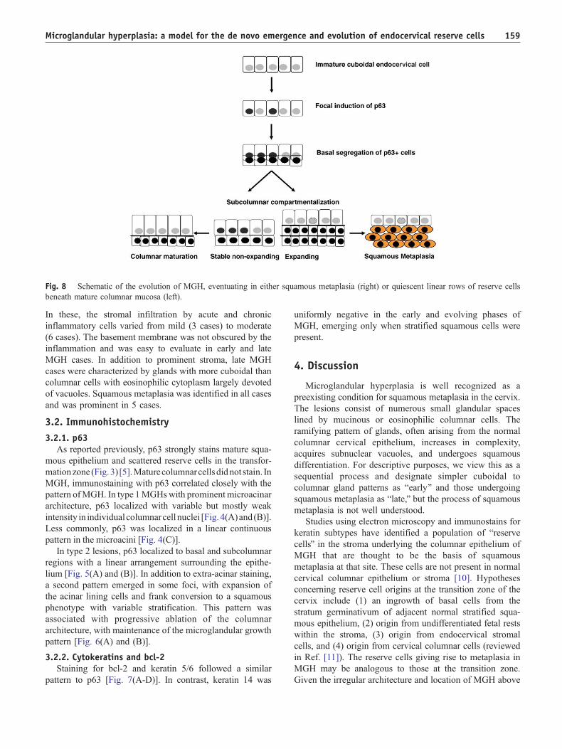
Fig. 8 Schematic of the evolution of MGH, eventuating in either squamous metaplasia (right) or quiescent linear rows of reserve cells
beneath mature columnar mucosa (left).
Microglandular hyperplasia: a model for the de novo emergence and evolution of endocervical reserve cells 159
In these, the stromal infiltration by acute and chronic
inflammatory cells varied from mild (3 cases) to moderate
(6 cases). The basement membrane was not obscured by the
inflammation and was easy to evaluate in early and late
MGH cases. In addition to prominent stroma, late MGH
cases were characterized by glands with more cuboidal than
columnar cells with eosinophilic cytoplasm largely devoted
of vacuoles. Squamous metaplasia was identified in all cases
and was prominent in 5 cases.
3.2. Immunohistochemistry
3.2.1. p63As reported previously, p63 strongly stains mature squa-
mous epithelium and scattered reserve cells in the transfor-
mationzone (Fig.3) [5].Maturecolumnarcellsdidnot stain. In
MGH, immunostaining with p63 correlated closely with the
pattern ofMGH. In type 1MGHswith prominent microacinar
architecture, p63 localized with variable but mostly weak
intensity in individual columnarcell nuclei [Fig. 4(A)and (B)].
Less commonly, p63 was localized in a linear continuous
pattern in the microacini [Fig. 4(C)].
In type 2 lesions, p63 localized to basal and subcolumnar
regions with a linear arrangement surrounding the epithe-
lium [Fig. 5(A) and (B)]. In addition to extra-acinar staining,
a second pattern emerged in some foci, with expansion of
the acinar lining cells and frank conversion to a squamous
phenotype with variable stratification. This pattern was
associated with progressive ablation of the columnar
architecture, with maintenance of the microglandular growth
pattern [Fig. 6(A) and (B)].
3.2.2. Cytokeratins and bcl-2Staining for bcl-2 and keratin 5/6 followed a similar
pattern to p63 [Fig. 7(A-D)]. In contrast, keratin 14 was
uniformly negative in the early and evolving phases of
MGH, emerging only when stratified squamous cells were
present.
4. Discussion
Microglandular hyperplasia is well recognized as a
preexisting condition for squamous metaplasia in the cervix.
The lesions consist of numerous small glandular spaces
lined by mucinous or eosinophilic columnar cells. The
ramifying pattern of glands, often arising from the normal
columnar cervical epithelium, increases in complexity,
acquires subnuclear vacuoles, and undergoes squamous
differentiation. For descriptive purposes, we view this as a
sequential process and designate simpler cuboidal to
columnar gland patterns as bearly Q and those undergoing
squamous metaplasia as blate,Q but the process of squamous
metaplasia is not well understood.
Studies using electron microscopy and immunostains for
keratin subtypes have identified a population of breservecellsQ in the stroma underlying the columnar epithelium of
MGH that are thought to be the basis of squamous
metaplasia at that site. These cells are not present in normal
cervical columnar epithelium or stroma [10]. Hypotheses
concerning reserve cell origins at the transition zone of the
cervix include (1) an ingrowth of basal cells from the
stratum germinativum of adjacent normal stratified squa-
mous epithelium, (2) origin from undifferentiated fetal rests
within the stroma, (3) origin from endocervical stromal
cells, and (4) origin from cervical columnar cells (reviewed
in Ref. [11]). The reserve cells giving rise to metaplasia in
MGH may be analogous to those at the transition zone.
Given the irregular architecture and location of MGH above

A.K. Witkiewicz et al.160
the transition zone, however, an origin from basal cells of
stratified squamous epithelium is excluded.
Patterns of cytokeratin expression described in the
transition zone support a columnar cell origin. Reserve
cells express keratins CK5, 8, 17, 18, and 19. Expression of
CK14 is variable. Columnar epithelium away from the
transition zone expresses CK7, 8, 18, and 19. However,
columnar cells overlying reserve cells in the transition zone
also express CK5 and 17, similar to reserve cells, indicating
that these cells may undergo a low level of squamous
differentiation, a concept supported by the distribution of
p63 immunostaining in this study [9]. Mature ectocervical
squamous epithelia express CK4, 13, and 14, but lack CK8,
18, and 19. Thus, reserve cells are taken either to have a
tendency to bidirectional differentiation or to be an
intermediate step in the transition from columnar to
squamous cells [12].
The keratin content of reserve cells is complex and
argues against their stromal origin because endocervical
stroma contains no cytoskeletal keratin proteins. It is
unlikely that stromal cells migrating through the basal
membrane would initiate expression of multiple keratin
types. Also, vimentin, so characteristic of stromal cells, is
not expressed in reserve cells [9,13]. The stroma of MGH is
morphologically distinct from cervical stroma with a more
epithelioid phenotype and the presence of acute and chronic
inflammation.
The p63 staining patterns we observed are consistent
with the emergence of reserve cells during the evolution of
MGH and before squamous metaplasia. The relative paucity
of reserve cells in type 1 microacinar lesions and their
abundance in lesions with multilayered epithelium and
squamous differentiation are consistent with an evolution
from microacinar/columnar MGH to more complex MGH
with metaplasia. We have observed similar phenomena in
the endometrium, where tubal and mucinous metaplasias
may harbor prominent p63-positive basal or subcolumnar
cells [14]. The endometrium normally does not contain
reserve cells, and their emergence suggests their origin from
preexisting components of the epithelium [2,3].
Our data also support a columnar cell origin of the reserve
cells. In early lesions, weak immunoreactivity for p63
localized to single cells and progressed to a single layer
pattern of stronger staining columnar cells. In more mature
lesions, more intense staining was seen, localizing to basal
and subcolumnar regions. With squamous metaplasia,
staining was present in the multilayered metaplastic cells
filling the microacini. Staining for other reserve cell markers
including keratin 5/6 and bcl-2 [5,9] showed a similar pattern
to p63, whereas CK14, a mature squamous cell marker, was
only present in epithelia exhibiting squamous differentiation.
A columnar cell origin for reserve cells is also consistent
with previous observations at the transition zone where there
is a correlation between p63 and CK14 staining in immature
squamous epithelium; p63 tended to extend beyond the
CK14 zone into the adjacent columnar cells [5].
Why MGH is not more frequently associated with human
papillomavirus (HPV)–related lesions remains unclear. Pos-
sible explanations include lack of receptors in MGH for HPV
virion attachment, inability to sustain HPV infection in a
reserve cell population not undergoing squamous differenti-
ation (and therefore not promoting viral replication), and
protection of acini from infection by the overlying epitheli-
um. Nonetheless, the ability of the cervical epithelium to
generate reserve cells is mirrored in the range of intra-
epithelial neoplasms seen in the cervix. They include the
following: (1) poorly differentiated adenocarcinomas in situ
(stratified mucin-producing intraepithelial lesions) in which
p63-positive cells are focally present in the basal cell layers
[5] [Fig. 3(O)], (2) rare p63-positive subcolumnar mucin-
producing precursor lesions that correspond to the transition
from columnar to reserve cell [6] [Fig. 3(B)], (3) immature
bmetaplasticQ squamous intraepithelial lesions (SILs) that
follow the growth pattern of MGH-associated squamous
metaplasia, and (4) a range of metaplastic growth patterns in
SIL variously termed atypical immature metaplasia, meta-
plastic SIL, and recently, beosinophilic dysplasiaQ[5,6,15,16]. A simplified explanation for these disparate
phenotypes is that epithelial differentiation is governed by 2
switch points in endocervical epithelial cell fate, including the
transition from columnar to reserve cell, and reserve cell to
squamous differentiation (Fig. 8). The same molecular
pathways governing these switches in MGH might explain
the multiplicity of neoplastic phenotypes observed.
References
[1] Richart RM. Cervical intraepithelial neoplasia. Pathol Annu
1973;8:301 -28.
[2] Kurita T, Cunha GR. Roles of p63 in differentiation of mqllerian duct
epithelial cells. Ann N Y Acad Sci 2001;948:9 -12.
[3] Ince TA, Cviko AP, Quade BJ, Yang A, McKeon FD, Mutter GL, et al.
p63 Coordinates anogenital modeling and epithelial cell differentiation
in the developing female urogenital tract. Am J Pathol 2002;161:
1111 -7.
[4] Yang A, Schweitzer R, Sun D, KaghadM,Walker N, Bronson RT, et al.
p63 Is essential for regenerative proliferation in limb, craniofacial and
epithelial development. Nature 1999;398(6729):714 -8.
[5] Quade BJ, Yang A, Wang Y, Sun D, Park J, Sheets EE, et al.
Expression of the p53 homologue p63 in early cervical neoplasia.
Gynecol Oncol 2001;80:24 -9.
[6] Park JJ, Sun D, Quade BJ, Flynn C, Sheets EE, Yang A, et al. Stratified
mucin-producing intraepithelial lesions of the cervix: adenosquamous
or columnar cell neoplasia? Am J Surg Pathol 2000;24:1414-9.
[7] Jones MW, Silverberg SG. Cervical adenocarcinoma in young
women: possible relationship to microglandular hyperplasia and use
of oral contraceptives. Obstet Gynecol 1989;73:984-9.
[8] Yang A, Kaghad M, Wang Y, Gillett E, Fleming MD, Dotsch V, et al.
p63, A p53 homolog at 3q27-29, encodes multiple products with
transactivating, death-inducing, and dominant-negative activities. Mol
Cell 1998;2:305-16.
[9] Smedts F, Ramaekers F, Troyanovsky S, Pruszczynski M, Robben H,
Lane B, et al. Basal-cell keratins in cervical reserve cells and a
comparison to their expression in cervical intraepithelial neoplasia.
Am J Pathol 1992;140:601-12.

Microglandular hyperplasia: a model for the de novo emergence and evolution of endocervical reserve cells 161
[10] Wilkinson E, Dufour DR. Pathogenesis of microglandular hyperplasia
of the cervix uteri. Obstet Gynecol 1976;47:189 -95.
[11] Henry MR. The beginnings of analytic gynecologic cytology: the
Patten and Reagan legacy. Cancer 1997;81:322 -4.
[12] Elson DA, Riley RR, Lacey A, Thordarson G, Talamantes FJ, Arbeit
JM. Sensitivity of the cervical transformation zone to estrogen-
induced squamous carcinogenesis. Cancer Res 2000;60:1267-75.
[13] Weikel W, Wagner R, Moll R. Characterization of subcolumnar
reserve cells and other epithelia of human uterine cervix. Demonstra-
tion of diverse cytokeratin polypeptides in reserve cells. Virchows
Arch B Cell Pathol Incl Mol Pathol 1987;54:98 -110.
[14] O’Connell JT, Mutter GL, Cviko A, Nucci M, Quade BJ, Kozakewich
HP, et al. Identification of a basal/reserve cell immunophenotype in
benign and neoplastic endometrium: a study with the p53 homologue
p63. Gynecol Oncol 2001;80:30 -6.
[15] Geng L, Connolly DC, Isacson C, Ronnett BM, Cho KR. Atypical
immature metaplasia (AIM) of the cervix: is it related to high-grade
squamous intraepithelial lesion (HSIL)? Hum Pathol 1999;30:345 -51.
[16] Ma L, Fisk JM, Zhang RR, Ulukus EC, Crum CP, Zheng W.
Eosinophilic dysplasia of the cervix: a newly recognized variant of
cervical squamous intraepithelial neoplasia. Am J Surg Pathol 2004;
28:1474-84.