INtimal hyPerplasia evAluated by oCT in de novo COROnary lesions treated by drug-eluting balloon and...
8
STUDY PROTOCOL Open Access INtimal hyPerplasia evAluated by oCT in de novo COROnary lesions treated by drug-eluting balloon and bare-metal stent (IN-PACT CORO): study protocol for a randomized controlled trial Francesco Burzotta * , Marta Francesca Brancati, Carlo Trani, Italo Porto, Antonella Tommasino, Gianluigi De Maria, Giampaolo Niccoli, Antonio Maria Leone, Valentina Coluccia, Giovanni Schiavoni and Filippo Crea Abstract Background: Neointimal hyperplasia plays a pivotal role in the pathogenesis of in-stent restenosis in patients undergoing percutaneous coronary interventions. Drug-eluting balloons are a promising tool to prevent restenosis after coronary angioplasty. Moreover, an increased knowledge of the pathophysiology of restenosis my help improve therapeutic strategies. Methods/Design: We present the design of an open-label, randomized three-arm clinical trial aimed to assess whether a strategy of bare-metal stent implantation with additional use of drug-eluting balloons, either before (pre-dilation) or after stenting (post-dilation), reduces the primary endpoint of in-stent neointimal hyperplasia area as compared with a strategy of bare-metal stent implantation alone. This primary endpoint will be assessed by optical coherence tomography at follow-up. Secondary endpoints will be the percentage of uncovered struts, and the percentage of struts with incomplete apposition. An ancillary study investigating the relation between systemic levels of endothelial progenitors cells and neointimal hyperplasia, and the interaction between endothelial progenitors cell levels and drug-eluting balloons has been planned. Thirty consecutive patients undergoing percutaneous coronary intervention will be randomized with a 1:1:1 design to bare-metal stent implantation alone (n = 10); bare-metal stent implantation after pre-dilation with a drug-eluting balloon (n = 10); or bare-metal stent implantation followed by post-dilation with a drug-eluting balloon (n = 10). Six-month follow-up coronary angiography with optical coherence tomography imaging of the stented segment will be performed in all patients. Blood samples for the assessment of endothelial progenitors cell levels will be collected on admission and at 6 months. Discussion: Experimental and early clinical data showed that inhibition of neointimal hyperplasia may be obtained by local administration of antiproliferative drugs loaded on the surface of angioplasty balloons. The INtimal hyPerplasia evAluated by oCT in de novo COROnary lesions treated by drug-eluting balloon and bare-metal stent (IN-PACT CORO) trial was conceived to test the superiority of a strategy of bare-metal stent implantation with additional drug-eluting balloon use (either before or after stenting) versus a strategy of bare-metal stent implantation alone for the reduction of neointimal hyperplasia. We also planned an ancillary study to assess the role of endothelial progenitors cells in the pathophysiology of neointimal hyperplasia. Trial registration: Clinicaltrials.gov NCT01057563. Keywords: Bare-metal stent, Drug-eluting balloon, Percutaneous coronary interventions * Correspondence: [email protected] Institute of Cardiology, Catholic University of the Sacred Heart, L.go Gemelli, 8 00168 Rome, Italy Full list of author information is available at the end of the article © 2012 Burzotta et al.; licensee BioMed Central Ltd. This is an Open Access article distributed under the terms of the Creative Commons Attribution License (http://creativecommons.org/licenses/by/2.0), which permits unrestricted use, distribution, and reproduction in any medium, provided the original work is properly cited. Burzotta et al. Trials 2012, 13:55 TRIALS http://www.trialsjournal.com/content/13/1/55
-
Upload
independent -
Category
Documents
-
view
4 -
download
0
Transcript of INtimal hyPerplasia evAluated by oCT in de novo COROnary lesions treated by drug-eluting balloon and...

Burzotta et al. Trials 2012, 13:55
TRIALShttp://www.trialsjournal.com/content/13/1/55
STUDY PROTOCOL Open Access
INtimal hyPerplasia evAluated by oCT in de novoCOROnary lesions treated by drug-eluting balloonand bare-metal stent (IN-PACT CORO): studyprotocol for a randomized controlled trialFrancesco Burzotta*, Marta Francesca Brancati, Carlo Trani, Italo Porto, Antonella Tommasino, Gianluigi De Maria,Giampaolo Niccoli, Antonio Maria Leone, Valentina Coluccia, Giovanni Schiavoni and Filippo Crea
Abstract
Background: Neointimal hyperplasia plays a pivotal role in the pathogenesis of in-stent restenosis in patientsundergoing percutaneous coronary interventions. Drug-eluting balloons are a promising tool to prevent restenosisafter coronary angioplasty. Moreover, an increased knowledge of the pathophysiology of restenosis my helpimprove therapeutic strategies.
Methods/Design: We present the design of an open-label, randomized three-arm clinical trial aimed to assesswhether a strategy of bare-metal stent implantation with additional use of drug-eluting balloons, either before(pre-dilation) or after stenting (post-dilation), reduces the primary endpoint of in-stent neointimal hyperplasia areaas compared with a strategy of bare-metal stent implantation alone. This primary endpoint will be assessed byoptical coherence tomography at follow-up. Secondary endpoints will be the percentage of uncovered struts, andthe percentage of struts with incomplete apposition. An ancillary study investigating the relation between systemiclevels of endothelial progenitors cells and neointimal hyperplasia, and the interaction between endothelialprogenitors cell levels and drug-eluting balloons has been planned. Thirty consecutive patients undergoingpercutaneous coronary intervention will be randomized with a 1:1:1 design to bare-metal stent implantation alone(n = 10); bare-metal stent implantation after pre-dilation with a drug-eluting balloon (n = 10); or bare-metal stentimplantation followed by post-dilation with a drug-eluting balloon (n = 10). Six-month follow-up coronaryangiography with optical coherence tomography imaging of the stented segment will be performed in all patients.Blood samples for the assessment of endothelial progenitors cell levels will be collected on admission and at6 months.
Discussion: Experimental and early clinical data showed that inhibition of neointimal hyperplasia may be obtainedby local administration of antiproliferative drugs loaded on the surface of angioplasty balloons. The INtimalhyPerplasia evAluated by oCT in de novo COROnary lesions treated by drug-eluting balloon and bare-metal stent(IN-PACT CORO) trial was conceived to test the superiority of a strategy of bare-metal stent implantation withadditional drug-eluting balloon use (either before or after stenting) versus a strategy of bare-metal stentimplantation alone for the reduction of neointimal hyperplasia. We also planned an ancillary study to assess the roleof endothelial progenitors cells in the pathophysiology of neointimal hyperplasia.
Trial registration: Clinicaltrials.gov NCT01057563.
Keywords: Bare-metal stent, Drug-eluting balloon, Percutaneous coronary interventions
* Correspondence: [email protected] of Cardiology, Catholic University of the Sacred Heart, L.go Gemelli,8 00168 Rome, ItalyFull list of author information is available at the end of the article
© 2012 Burzotta et al.; licensee BioMed CentraCommons Attribution License (http://creativecreproduction in any medium, provided the or
l Ltd. This is an Open Access article distributed under the terms of the Creativeommons.org/licenses/by/2.0), which permits unrestricted use, distribution, andiginal work is properly cited.

Burzotta et al. Trials 2012, 13:55 Page 2 of 8http://www.trialsjournal.com/content/13/1/55
BackgroundRestenosis due to neointimal hyperplasia causes repeattarget vessel revascularization in a relevant number ofpatients undergoing percutaneous coronary interventions(PCI).
Drug-eluting stents (DES) are currently widely adoptedto reduce the restenosis rate and repeat revasculariza-tions [1]. However, DES technology is associated with aprofound inhibition of stent strut endothelialization,which may lead to the presence of uncovered stent strutsand to the persistence of polymer inducing inflammatoryreactions in the vessel wall [2,3]. Such factors may in-crease the risk of stent thrombosis so that prolongeddual antiplatelet therapy is recommended after DES im-plantation [4].Experimental data [5] and early clinical experiences
showed that inhibition of neointimal hyperplasia may beobtained by local administration of antiproliferativedrugs (like paclitaxel) loaded on the surface of angio-plasty balloons [6-8]. Accordingly, drug-eluting balloons(DEBs) are a promising tool to prevent restenosis andavoid the undesirable persistence of DES polymers in thevessel wall, thus potentially increasing the safety of PCI.Most of the scientific evidence regarding DEB efficacy isactually concentrated in the treatment of patients within-stent restenosis; there are some data about use ofDEBs for de novo lesions and for bifurcation lesions [9].For de novo lesions the only registry available is thePaclitaxel-Eluting PTCA-Balloon Catheter to Treat SmallVessel Coronary Artery Disease (PEPCAD I), which con-cluded that DEBs are associated with a high proceduralsuccess rate in small de novo lesions, while DEBs in con-junction with a bare-metal stent (BMS) remains a con-cern because of high restenosis rate [10]. Until now,DEBs failed to show equivalence to DES regarding angio-graphic endpoints during PCI of small coronary arteries[11].Optical coherence tomography (OCT) is a novel im-
aging modality with high resolution to assess neointimalcoverage and stent strut apposition [12-14]. It was usedin a recent study [15] evaluating the effect of sequentialapplication of DEBs and BMS (for de novo coronarylesions) on neointimal hyperplasia; the sequence of appli-cation (DEB first versus BMS first) did not influence out-come, except for better apposition when applying theBMS first. However, in this study there was no controlgroup (that is, BMS alone).Thus, we designed an open-label, single center, rando-
mized trial to evaluate whether additional DEB use inpatients undergoing BMS implantation, either before orafter the BMS implantation, improves neointimal forma-tion assessed by OCT and affects the process of strutcoverage at follow-up, compared with BMS implantationalone.
Moreover, recent clinical observations have suggestedthat endothelial progenitor cells (EPC), may play a rolein the process of in-stent restenosis [16,17]. We thereforeplanned an ancillary study, aiming at assessing the rela-tion between levels of systemic EPC and neointimalhyperplasia at follow-up.
Aims of the main studyTo assess whether a strategy of BMS implantation withadditional DEB use, either at the time of pre-dilation orof post-dilation:
1. reduces neointimal hyperplasia, as compared to astrategy of BMS implantation alone;
2. affects strut coverage and strut malapposition.
Aims of endothelial progenitor cell ancillary studyTo evaluate possible correlations of EPC levels with neoin-timal hyperplasia, stent coverage and stent malapposition.
Methods/DesignStudy designThe INtimal hyPerplasia evAluated by oCT in de novoCOROnary lesions treated by drug-eluting balloon andbare-metal stent (IN-PACT CORO) trial is a single cen-ter, open-label, randomized trial enrolling 30 consecutivepatients undergoing PCI with BMS implantation.Recruited patients will be randomized 1:1:1 to threearms:
1. BMS implantation (BMS group).2. BMS implantation after lesion pre-dilation with DEB
(PRE-DEB group).3. BMS implantation followed by post-dilation with
DEB (POST-DEB group).
Clinical follow-up will be performed at 1, 6 and12 months. Enrolled patients will undergo a 6-month fol-low-up coronary angiography with OCT evaluation ofthe stented segment using the C7-XRTM Coronary Im-aging System (LightLab Imaging Inc., Westford, MA,USA). OCT analysis will be performed off-line by expertOCT analysts blinded to the treatment assignment.The study protocol was conceived in March 2009, con-
formed to the Declaration of Helsinki, and was approvedby the Ethical Committee of our center. We prepared awritten informed consent which patients will be asked tosign to be enrolled in the protocol.The full study flow-chart is represented in Figure 1.
Study endpointsThe primary endpoint is 6-month in-stent neointimalhyperplasia area assessed by OCT.
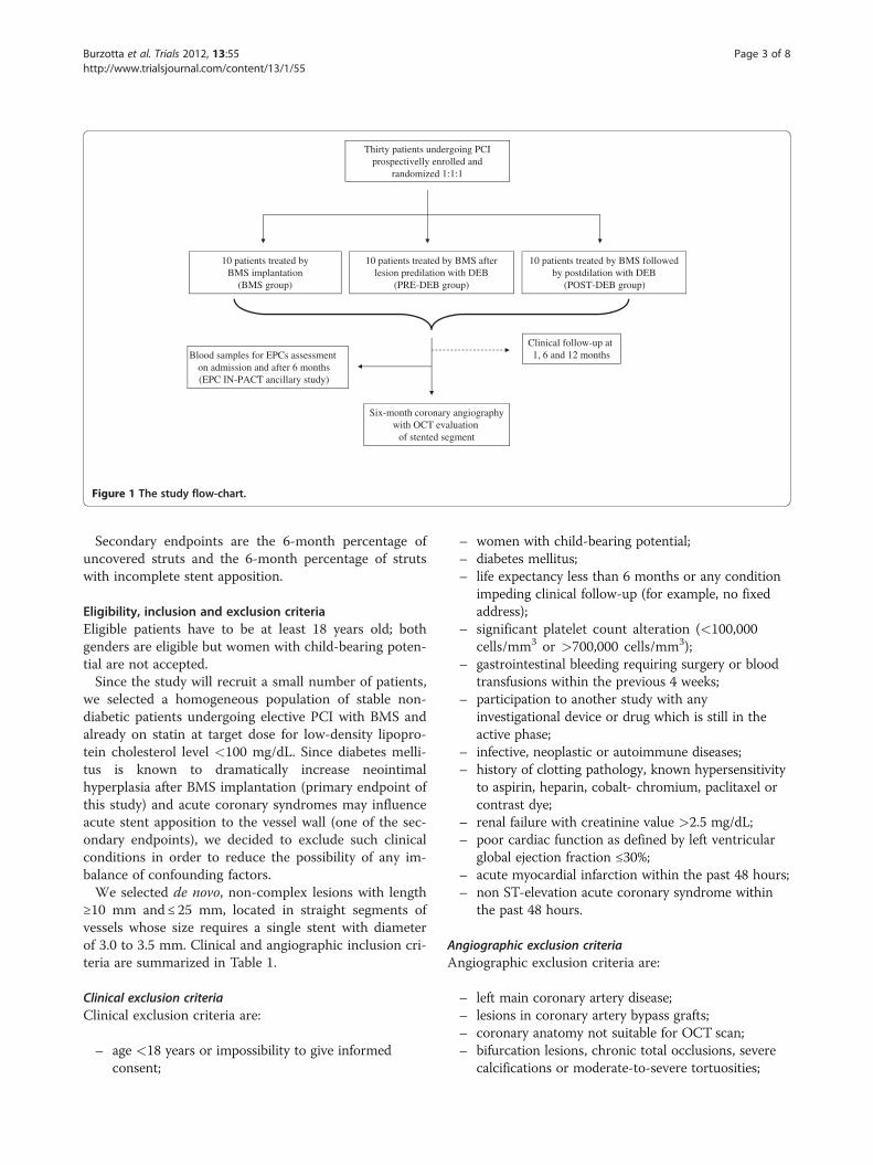
Thirty patients undergoing PCI prospectivelly enrolled and
randomized 1:1:1
10 patients treated byBMS implantation
(BMS group)
10 patients treated by BMS after lesion predilation with DEB
(PRE-DEB group)
10 patients treated by BMS followed by postdilation with DEB
(POST-DEB group)
Six-month coronary angiographywith OCT evaluation
of stented segment
Clinical follow-up at 1, 6 and 12 monthsBlood samples for EPCs assessment
on admission and after 6 months(EPC IN-PACT ancillary study)
Figure 1 The study flow-chart.
Burzotta et al. Trials 2012, 13:55 Page 3 of 8http://www.trialsjournal.com/content/13/1/55
Secondary endpoints are the 6-month percentage ofuncovered struts and the 6-month percentage of strutswith incomplete stent apposition.
Eligibility, inclusion and exclusion criteriaEligible patients have to be at least 18 years old; bothgenders are eligible but women with child-bearing poten-tial are not accepted.Since the study will recruit a small number of patients,
we selected a homogeneous population of stable non-diabetic patients undergoing elective PCI with BMS andalready on statin at target dose for low-density lipopro-tein cholesterol level <100 mg/dL. Since diabetes melli-tus is known to dramatically increase neointimalhyperplasia after BMS implantation (primary endpoint ofthis study) and acute coronary syndromes may influenceacute stent apposition to the vessel wall (one of the sec-ondary endpoints), we decided to exclude such clinicalconditions in order to reduce the possibility of any im-balance of confounding factors.We selected de novo, non-complex lesions with length
≥10 mm and≤ 25 mm, located in straight segments ofvessels whose size requires a single stent with diameterof 3.0 to 3.5 mm. Clinical and angiographic inclusion cri-teria are summarized in Table 1.
Clinical exclusion criteriaClinical exclusion criteria are:
– age <18 years or impossibility to give informedconsent;
– women with child-bearing potential;– diabetes mellitus;– life expectancy less than 6 months or any condition
impeding clinical follow-up (for example, no fixedaddress);
– significant platelet count alteration (<100,000cells/mm3 or >700,000 cells/mm3);
– gastrointestinal bleeding requiring surgery or bloodtransfusions within the previous 4 weeks;
– participation to another study with anyinvestigational device or drug which is still in theactive phase;
– infective, neoplastic or autoimmune diseases;– history of clotting pathology, known hypersensitivity
to aspirin, heparin, cobalt- chromium, paclitaxel orcontrast dye;
– renal failure with creatinine value >2.5 mg/dL;– poor cardiac function as defined by left ventricular
global ejection fraction ≤30%;– acute myocardial infarction within the past 48 hours;– non ST-elevation acute coronary syndrome within
the past 48 hours.
Angiographic exclusion criteriaAngiographic exclusion criteria are:
– left main coronary artery disease;– lesions in coronary artery bypass grafts;– coronary anatomy not suitable for OCT scan;– bifurcation lesions, chronic total occlusions, severe
calcifications or moderate-to-severe tortuosities;

Table 1 Inclusion criteria
Clinical inclusion criteria Non-diabetic patients with a stable coronary artery disease, undergoing elective PCI with BMS
patients already on statin, at target cholesterol level (low-density lipoprotein cholesterol <100 mg/dL)
Angiographic inclusion criteria De novo non-complex lesions located in straight coronary segments
lesion length ≥10 mm and ≤25 mm
vessel size requiring a single stent with diameter between 3.0 and 3.5 mm
BMS: bare-metal stent; PCI: percutaneous coronary interventions.
Burzotta et al. Trials 2012, 13:55 Page 4 of 8http://www.trialsjournal.com/content/13/1/55
– presence of additional non-target lesions requiringtreatment, within and outside the target vessel,which are not successfully treated (non-target lesionsshould be treated prior to the target lesion).
Technical details of the percutaneous coronaryinterventions procedureGeneral considerations on drug-eluting balloon usageA DEB is mainly intended to serve as drug delivery tothe vessel wall and should therefore always cover thestenotic area as well as adjacent vessel segments coveredby a stent or dilated by a balloon catheter, incidentally orby intention.DEB length and positioning within the target lesion
will therefore be carefully chosen to avoid geographicmiss between DEB and treated vessel segments.
Characteristics of the drug-eluting balloon used in the studyIN.PACTTM FalconTM is a paclitaxel-eluting PCI ballooncatheter manufactured by Invatec Technology CenterGmbH (Hungerbüelstrasse 12, 8500 Frauenfeld –Switzerland). Model mix includes Rapid Exchange (RX)and, for some sizes, Over The Wire (OTW) design. Theballoon is coated with FreePacTM, a paclitaxel-elutingformulation, at the dose of 3.0 μg per mm2 of the bal-loon surface.The IN.PACTTM FalconTM RX dilatation balloon
catheter is composed of a proximal single-lumenshaft, a dual-lumen distal shaft and a balloon closeto the catheter tip. The proximal shaft consists of astainless steel hypotube with a proximal end luer-lock connector (hub) for balloon inflation. At the op-posite side, a special transition construction guaran-tees an optimal push-torque transmission throughthe full catheter length. The first lumen of the distalshaft is dedicated to guide wire passage while theother, which continues through the proximal shaft upto the hub, is dedicated to the inflation of the bal-loon. Maximum guide wire diameter is 0.014 inches(0.36 mm). A flushing needle with a luer port is pro-vided in the sterile packaging to facilitate the flush-ing of the guide wire lumen prior to usage.The IN.PACTTM FalconTM OTW dilatation catheter
consists of a dual-lumen shaft ending proximally with a
Y connector (hub) and distally with a balloon closed tothe catheter tip. The straight port of the Y connector isthe guide wire entrance and the side port is used to in-flate and deflate the balloon. Both lumens run throughthe entire shaft length. The guide wire lumen permitsthe use of guide wires to facilitate advancement of thecatheter to and through the stenosis to be dilated and itends at the tip of the catheter. Maximum guide wirediameter is 0.014 inches (0.36 mm).The balloon is designed to reach specific diameters
at specific pressures. A single central radiopaquemarker and/or two radiopaque markers are availablein order to correctly position the balloon underfluoroscopy. IN.PACTTM FalconTM is available indifferent balloon sizes.The majority of the drug is released within the first
30 seconds of balloon inflation. The duration of theinflation should therefore be between 30 seconds and1 minute for optimal drug release.
Percutaneous coronary interventions proceduredescription according to randomization
1. BMS group procedure:
– Lesion pre-dilation with an undersized semi-compliant balloon (balloon to artery ratio, 0.5:1).– BMS implantation (stent to artery ratio, 1.1:1).– Post-dilation of the stented segment with a non-
compliant balloon at high pressure (16 to 18 atm).
2. PRE-DEB group procedure:
a. Pre-dilation– Pre-dilation of the target lesion with anundersized semi-compliant standardpercutaneous transluminal coronaryangioplasty (PTCA) balloon (balloon to arteryratio, 0.5:1).
b. DEB dilation– DEB diameter and pressure: nominal DEB
diameter must be chosen to guarantee fullvessel wall contact at a pressure close to orslightly higher than the DEB nominal pressure(balloon to artery ratio, 1:1).

Burzotta et al. Trials 2012, 13:55 Page 5 of 8http://www.trialsjournal.com/content/13/1/55
– DEB length: nominal DEB length must exceed10 mm (5 mm per edge), the length of thestent which is planned to be deployed.
– DEB inflation time: 45 seconds.c. BMS implantationd. Post-dilation
– Post-dilation of the stented segment must beperformed with a non-compliant PTCAballoon.
– Balloon diameter: nominal PTCA balloondiameter must be chosen to reach a balloon tostent ratio of 1:1 at high pressure (16 to18 atm).
– Balloon length and positioning: PTCA balloonlength should be shorter than the length of thedeployed stent. In case of post-stent edgeresidual stenosis, post-dilation balloon must fallwithin are outside the stent (5 mm per edge)which was the previously dilated by the DEB.
3. POST-DEB group procedure:
a. Pre-dilation– Pre-dilation of the target lesion must beperformed with an undersized semi-compliantstandard PTCA balloon (balloon to arteryratio, 0.5:1).
b. BMS implantation– Stent to artery ratio, 1.1:1.– Stent length must allow full coverage of the
target lesion with a single stent as well as be10 mm shorter than the DEB which theoperator is planning to use next.
c. Post-dilation– Post-dilation of the stented segment must be
performed with a non-compliant PTCAballoon.
– Balloon diameter: nominal PTCA balloondiameter must be chosen to reach a balloon tostent ratio of 1:1 at high pressure (16 to18 atm).
d. DEB dilation– DEB length and positioning: DEB length must
be 10 mm longer than the previously deployedstent (or than the extended pre-treated area incase of former post-dilation outside the stentedges) and centered within such pre-treatedlength (5 mm per edge).
– DEB inflation time: 45 seconds.– Balloon to stent ratio: 1.1:1 at a pressure close
or slightly higher of the DEB nominal pressure.
The angiographic results of the procedure will beassessed by three-dimensional quantitative coronaryangiography (QCA) using the Paieon Cardi-Op system
(Paieon, Inc., 747 Third Avenue, New York 10017-2803,United States).
Post-procedural managementCardiac damage markers (creatine-kinase-MB and tropo-nin I) will be assessed before the procedure, and 6 hoursand 24 hours after PCI. Thereafter, further blood sampleswill be performed only if clinically indicated.After PCI, patients will be given aspirin (75 to
100 mg/d) and life-long clopidogrel (75 mg/d) for≥3 months (according to the on-label prescription forDEB-treated patients).
Follow-upClinical follow-up will take place at 1 month (±1 week),6 months (±2 weeks) and 1 year (±30 days) by clinicalvisit or phone interview.At the 6-month (± 2 weeks) follow-up, all patients will
undergo a coronary angiography (with three-dimensionalQCA) and an OCT study.We anticipate a patient drop-out rate of 10%.
Rationale for optical coherence tomography selection anddescription of optical coherence tomography analysisFor many years, QCA has been used to assess regressionand progression of coronary obstructions in pharmaco-logical interventions, to assess the efficacy of PCI andstenting, and for vessel sizing. However, QCA late lumenloss is the difference between two minimal lumen diam-eter measurements at two different times, and the axiallocation of this diameter is variable at each time point.An angiogram only examines the lumen, while the dis-ease is in the vessel wall. These limitations have spurredthe search for new intravascular diagnostic imaging tech-niques. A meta-analysis of QCA versus intravascularultrasound (IVUS) parameters for assessing stent resten-osis [18] showed by regression analysis that QCA latelumen loss and percentage of diameter stenosis corre-lates only moderately with IVUS evaluation of neointimalhyperplasia. A recent work [19] evaluated the correlationof angiographic late loss with the degree of in-stentneointimal proliferation assessed by OCT, demonstratinga poor correlation of angiographic late loss with OCT atlow degrees of neointimal proliferation (R = 0.38).The detection of the post-stent implantation intima by
OCT and IVUS has also been compared. After a medianfollow-up time of 6 months, Matsumoto et al. examinedthe reaction of the intima by OCT and found that 64% ofthe struts had become covered with an intima <100 μmthick (below the resolution of IVUS) [20]. The correl-ation between OCT and histology measurements(r = 0.980, P <0.001 for lumen area; r = 0.978, P <0.001for stent area; and r = 0.961, P <0.001 for neointimalarea) was stronger than the correlation between IVUS

Burzotta et al. Trials 2012, 13:55 Page 6 of 8http://www.trialsjournal.com/content/13/1/55
and histology (r = 0.803, P <0.001 for lumen area;r = 0.817, P <0.001 for stent area; and r = 0.776, P <0.001for neointimal area). In addition, the diagnostic accuracyfor detecting a small degree of neointimal proliferationby OCT (area under the curve = 0.967, 95% confidenceinterval 0.914 to 1.019) was higher than that by IVUS(area under the curve = 0.781, 95% confidence interval0.621 to 838) [21].The frequency of detection of stent malapposition is
greater with OCT than with IVUS. In a recent in vivostudy by Kubo et al., the detection rate with OCT was47% in a sample of 55 patients, while with IVUS it wasonly 18% (P <0.001) [22]. Similar results were reportedby Bouma et al. five years earlier in a study involving ex-perimental animals [23].So OCT represents a useful clinical tool to study
endothelialization of stents and abnormal tissueresponses and to detect the presence of delayed healingor incomplete apposition of stents to the arterial wall asa possible mechanism of in stent thrombosis [24,25].In our study, OCT will be performed with the imaging
system C7 XR (LightLab Imaging Inc.), using a non-occlusive technique, with automated intracoronary injec-tion of iso-osmolar contrast. This Fourier domain-OCTsystem achieves 10 times higher resolution than IVUS, ap-proximately 15 microns, with pullback speed of 2 cm/sec,and an acquisition frame rate of 100 frames/sec.The entire stent length will be assessed and cross-
sectional images will be analyzed every 0.4 mm.The struts will be classified as uncovered if a tissue
layer on the endoluminal surface is not visible or coveredin the presence of visible tissue between the endoluminalsurface and the lumen.The tissue coverage thickness will be measured in each
strut as the distance from the strut endoluminal surfaceto the lumen. In each cross-section analyzed, a series ofparameters will be calculated.
Endpoint assessmentThe primary endpoint is in-stent neointimal hyperplasiaarea, that is stent area minus lumen area [26] and itspercentage (tissue coverage area/stent area × 100) evalu-ated by OCT. The other related parameters of tissuecoverage thickness (μm), tissue volume coverage (tissuecoverage area × stent length) and its percentage (tissuecoverage volume/stent volume × 100) will also be evalu-ated. To assess the pattern of coverage, the ratio betweenthe difference of maximum and the minimum tissuethickness coverage (minimum tissue thickness coverage/
Table 2 Secondary endpoints
Percentage of uncovered struts
Percentage of struts with incomplete strut apposition (ISA)
maximum tissue thickness coverage) will be calculated ineach frame. A ratio close to 1 indicates an asymmetrictissue coverage, on the opposite a ratio close to 0 indi-cates a symmetric tissue coverage.The secondary OCT endpoints are described in Table 2.
Incomplete strut apposition (ISA) will be defined as a dis-tance between strut endoluminal surface and the vessel wallhigher than strut thickness. ISA will be considered presentif at least one single strut is incompletely apposed to thevessel wall. In each OCT frame analyzed, the number ofstruts with ISA and the maximum distance from the endo-luminal stent strut to the vessel wall will be measured.
Sample size calculation and statistical analysisThe IN-PACT CORO trial is open-label, randomizedclinical trial in which patients will be randomly assigned,with a 1:1:1 design, to BMS implantation alone, BMS im-plantation with pre-DEB use, and BMS implantationwith post-DEB use. The primary aim is to test the super-iority of a strategy of BMS implantation with pre- orpost-DEB use versus a strategy of BMS implantationalone on the primary endpoint of in-stent neointimalhyperplasia area, assessed by OCT at follow-up. Second-ary endpoints will be the percentage of uncovered struts,and the percentage of struts with ISA.Little information is available on neointimal prolifera-
tion after BMS implantation: two small non-randomizedstudies [13,27] reported maximal and minimal neointi-mal thickness (mm) at 7.3 month follow-up (first study)and mean neointimal thickness at 8 month follow-up(latter study) being more than four-fold higher in theBMS group compared with the sirolimus (rapamycin)-eluting stent group, although data on neointimal area arenot available. In the Harmonizing Outcomes WithRevascularization and Stents in Acute Myocardial Infarc-tion (HORIZONS-AMI) OCT sub-study, a mean neointi-mal area of 2.8 ± 1.4 was reported 13 months after aBMS implantation [28].A recent randomized study comparing 12 polymer-
coated rapamycin-eluting stents to 12 non- polymerrapamycin-eluting stents [29] reported a neointimal areaof 0.3 ± 0.2 mm2 in the polymer stent versus1.2 ± 0.8 mm2 in the non-polymer stent, with a differenceof 0.9 mm2 (95% confidence interval, 0.3 to 1.4).We have hypothesized that additional DEB use (either
before or after BMS implantation) will yield a neointimalarea similar to that reported in the non-polymer rapamy-cin-eluting stent (that is, 1.2 mm2) and that this valuecorresponds to an approximately 50% reduction of mean
Number of uncovered struts/total number of struts
Number of struts with ISA/total number of struts
Burzotta et al. Trials 2012, 13:55 Page 7 of 8http://www.trialsjournal.com/content/13/1/55
neointimal area in the group receiving BMS implantationalone (hypothesizing a control group hyperplasia betterthan that reported in the BMS arm of the HORIZONS-AMI OCT sub-study). As two co-primary endpoints arepre-specified: that is, either pre- or post-DEB use reducesneointimal hyperplasia as compared to BMS alone, atype I error of 2.5% will be allocated to each endpoint topreserve the total type I error at the 5% level. Power isset to 85% for each primary endpoint. To detect suchdifference, 10 patients will be required in each group.Since we anticipate a drop-out rate of 10%, we will re-cruit another patient for each group in case of loss to fol-low-up or unanalyzable studies.Continuous variables will be reported as mean and
standard deviation or as median and interquartile range(according to their distribution) and comparisons be-tween two groups will be made with unpaired t-test orMann Whitney U-test, as appropriate. Categorical vari-ables will be presented as numbers and frequenciesand comparisons will be made using Chi-square test orFisher’s exact test, as appropriate.
Endothelial progenitor cell ancillary study designA correlation analysis will be performed to ascertainwhether EPC contribute to neointimal regrowth in suchpatients.Peripheral blood samples of all patients enrolled in the
IN-PACT CORO protocol will be obtained on admissionand after 6 months of follow-up (Figure 1).
Endothelial progenitor cells levels assessment forendothelial progenitor cells ancillary studyThe cytofluorimetric assays will be performed on admis-sion and after 6 months’ follow-up. Peripheral blood willbe drawn and buffered using sodium citrate. One hundredmicroliters of blood will be incubated with 5 μL of phyco-erythrin-conjugated monoclonal antibody against CD34,with 5 μL of fluorescein isothiocyanate-conjugated mono-clonal antibody against kinase insert domain receptor(KDR) and with 5 μL of monoclonal antibody againstCD45. Fluorescence-activated cell sorting assay will beperformed according to the specific log gain of forwardand side scatter. EPC will be detected as cells CD34+KDR+CD45- The negativity for CD45 is fundamental todistinguish EPC from other nucleated cells in peripheralblood, represented by leukocytes [30].
Study limitationsThe major anticipated limitation of the present study isthat it is an open-label and not a double-blinded trial. Inorder to increase the feasibility of this spontaneous, non-sponsored study, we decided to perform an open-labeltrial. The treating physicians cannot be blinded since the
DEB device is fairly different from a standard balloondue to its thick white covering material.However, OCT analyses will be performed by an expert
OCT image analyzer who is not involved in patient re-cruitment and blinded to patient randomization andclinical status.
Trial statusPatient recruitment is ongoing.
AbbreviationsBMS: Bare-metal stent; DEB: Drug-eluting balloon; DES: Drug-eluting stent;EPC: Endothelial progenitor cells; ISA: Incomplete strut apposition;IVUS: Intravascular ultrasound; KDR: Kinase insert domain receptor;OCT: Optical coherence tomography; OTW: Over The Wire; PCI: Percutaneouscoronary intervention; POST-DEB: Post-dilation with drug-eluting balloon;PRE-DEB: Pre-dilation with drug-eluting balloon; PTCA: Percutaneoustransluminal coronary angioplasty; QCA: Quantitative coronary angiography;RX: Rapid Exchange.
Competing interestsThe authors declare that they have no competing interests.
Author detailsInstitute of Cardiology, Catholic University of the Sacred Heart, L.go Gemelli 800168 Rome, Italy.
Authors’ contributionsFB conceived the main study and drafted the manuscript. CT madesubstantial contribution in study design and coordination. IP conceived theEPC ancillary study and helped to draft the manuscript. MFB helped to draftthe manuscript and participated in study coordination and acquisition ofdata. AT, GN and AML revised the manuscript with important intellectualcontribution. GDM and VC participated in acquisition of data. GS and FChave given final approval of the version to be published. All authors readand approved the final manuscript.
FundingThe authors declare that there is no funding source for this study.
Received: 8 May 2011 Accepted: 6 May 2012Published: 6 May 2012
References1. Waugh J, Wagstaff AJ: The paclitaxel (TAXUS)-eluting stent: a review of its
use in the management of de novo coronary artery lesions. Am JCardiovasc Drugs 2004, 4:257–268.
2. Finn AV, Joner M, Nakazawa G, Kolodgie F, Newell J, John MC, Gold HK,Virmani R: Pathological correlates of late drug-eluting stent thrombosis:strut coverage as a marker of endothelialization. Circulation 2007,115:2435–2441.
3. Joner M, Finn AV, Farb A, Mont EK, Kolodgie FD, Ladich E, Kutys R, Skorija K,Gold HK, Virmani R: Pathology of drug-eluting stents in humans: delayedhealing and late thrombotic risk. J Am Coll Cardiol 2006, 48:193–202.
4. King SB 3rd, Smith SC Jr, Hirshfeld JW Jr, Jacobs AK, Morrison DA, WilliamsDO, Feldman TE, Kern MJ, O’Neill WW, Schaff HV, Whitlow PL, ACC/AHA/SCA,Adams CD, Anderson JL, Buller CE, Creager MA, Ettinger SM, Halperin JL,Hunt SA, Krumholz HM, Kushner FG, Lytle BW Nishimura R, Page RL, RiegelB, Tarkington LG, Yancy CW: 2007 focused update of the ACC/AHA/SCAI2005 guideline update for percutaneous coronary intervention: a reportof the American College of Cardiology/American Heart Association TaskForce on Practice guidelines. J Am Coll Cardiol 2008, 51:172–209.
5. Scheller B, Speck U, Abramjuk C, Bernhardt U, Böhm M, Nickenig G:Paclitaxel balloon coating, a novel method for prevention and therapy ofrestenosis. Circulation 2004, 110:810–814.
6. Unverdorben M, Vallbracht C, Cremers B, Heuer H, Hengstenberg C,Maikowski C, Werner GS, Antoni D, Kleber FX, Bocksch W, Leschke M,Ackermann H, Boxberger M, Speck U, Degenhardt R, Scheller B: Paclitaxel-
Burzotta et al. Trials 2012, 13:55 Page 8 of 8http://www.trialsjournal.com/content/13/1/55
coated balloon catheter versus paclitaxel-coated stent for the treatmentof coronary in-stent restenosis. Circulation 2009, 119(23):2986–2994.
7. Scheller B, Hehrlein C, Bocksch W, Rutsch W, Haghi D, Dietz U, Böhm M,Speck U: Treatment of coronary in-stent restenosis with a paclitaxel-coated balloon catheter. N Engl J Med 2006, 355:2113–2124.
8. Scheller B, Hehrlein C, Bocksch W, Rutsch W, Haghi D, Dietz U, Böhm M,Speck U: Two year follow-up after treatment of coronary in-stentrestenosis with a paclitaxel-coated balloon catheter. Clin Res Cardiol 2008,97:773–781.
9. Waksman R, Pakala R: Drug-eluting balloon. The comeback kid? CircCardiovasc Interven 2009, 2:352–358.
10. Maier LS, Maack C, Ritter O, Bohm M: Hotline update of clinical trials andregistries presented at the German Cardiac Society meeting 2008(PEPCAD LokalTax, INH, German ablation registry, German deviceregistry, DESDE registry, DHR, Reality, SWEETHEART registry, ADMA,GERSHWIN). Clin Res Cardiol 2008, 97:356–363.
11. Cortese B, Micheli A, Picchi A, Coppolaro A, Bandinelli L, Severi S, Limbruno U:Paclitaxel-coated balloon versus drug-eluting stent durino PCI of smallcoronary vessels, a prospective randomised clinical trial. The PICCOLETOstudy. Heart 2010, 96:1291–1296.
12. Prati F, Cera M, Ramazzotti V, Imola F, Giudice R, Albertucci M: Safety andfeasibility of a new non-occlusive technique for facilitated intracoronaryoptical coherence tomography (OCT) acquisition in various clinical andanatomical scenarios. EuroInterv 2007, 3:365–370.
13. Chen BX, Ma FY, Luo W, Ruan JH, Xie WL, Zhao XZ, Sun SH, Guo XM, Wang F,Tian T, Chu XW: Neointimal coverage of bare-metal and sirolimus-elutingstents evaluated with optical coherence tomography. Heart 2008, 94:566–570.
14. Prati F, Regar E, Mintz GS, Arbustini E, Di Mario C, Jang IK, Akasaka T, CostaM, Guagliumi G, Grube E, Ozaki Y, Pinto F, Serruys PW, Expert’s OCT ReviewDocument: Expert review document on methodology, terminology, andclinical applications of optical coherence tomography: physicalprinciples, methodology of image acquisition, and clinical application forassessment of coronary arteries and atherosclerosis. Eur Heart J 2010,31:401–415.
15. Gutierrez-Chico JL, van Geuns RJ, Koch KT, Koolen J, Duckers H, Regar E,Serruys PW: Paclitaxel-coated balloon in combination with bare metalstent for treatment of de novo coronary lesions: an optical coherencetomography first-in-human randomised trial, balloon first vs. stent first.Eurointervention 2011, 7(6):711–722.
16. George J, Herz I, Goldstein E, Abashidze S, Deutch V, Finkelstein A,Michowitz Y, Miller H, Keren G: Number and adhesive properties ofcirculating endothelial progenitor cells in patients with in-stentrestenosis. Arterioscler Thromb Vasc Biol 2003, 23:e57–e60.
17. Pelliccia F, Cianfrocca C, Rosano G, Mercuro G, Speciale G, Pasceri V: Role ofendothelial progenitor cells in restenosis and progression of coronaryatherosclerosis after percutaneous coronary intervention: a prospectivestudy. JACC Cardiovasc Interv 2010, 3:78–86.
18. Escolar E, Mintz GS, Popma J, Michalek A, Kim SW, Mandinov L, Koglin J,Stone G, Ellis SG, Grube E, Dawkins KD, Weissman NJ: Meta-analysis ofangiographic versus intravascular ultrasound parameters of drug-elutingstent efficacy (from TAXUS IV, V and VI). Am J Cardiol 2007, 10:621–626.
19. Kim J, Wallace-Bradley D, Alviar C, Conditt G, Milewski K, Afari ME, Cheng Y,Gallego C, Tellez A, Stone GW, Kaluza GL, Granada JF: Correlation ofangiographic late loss with neointimal proliferation in stents evaluatedby OCT and histology in porcine coronary arteries. J Am Coll Cardiol Img2011, 4:1002–1010.
20. Matsumoto D, Shite J, Shinke T, Otake H, Tanino Y, Ogasawara D, Sawada T,Paredes OL, Hirata K, Yokoyama M: Neointimal coverage of sirolimus-eluting stents at 6-month follow-up: evaluated by optical coherencetomography. Eur Heart J 2007, 28:961–967.
21. Suzuki Y, Ikeno F, Koizumi T, Tio F, Yeung AC, Yock PG, Fitzgerald PJ, FearonWF: In vivo comparison between optical coherence tomography andintravascular ultrasound for detecting small degrees of in-stentneointima after stent implantation. JACC Cardiovasc Interv 2008, 1:168–173.
22. Kubo T, Imanishi T, Kitabata H, Kuroi A, Ueno S, Yamano T, Tanimoto T,Matsuo Y, Masho T, Takarada S, Tanaka A, Nakamura N, Mizukoshi M,Tomobuchi Y, Akasaka T: Comparison of vascular response after sirolimuseluting stent implantation between patients with unstable and stableangina pectoris: a serial optical coherence tomography study. J Am CollCardiol Img 2008, 1:475–484.
23. Bouma BE, Tearney GJ, Yabushita H, Shishkov M, Kauffman CR, DeJosephGauthier D, MacNeill BD, Houser SL, Aretz HT, Halpern EF, Jang IK:Evaluation of intracoronary stenting by intravascular optical coherencetomography. Heart 2003, 89:317–320.
24. Cook S, Wenaweser P, Togni M, Billinger M, Morger C, Seiler C, Vogel R, Hess O,Meier B, Windecker S: Incomplete stent apposition and very late stentthrombosis after drug-eluting stent implantation. Circulation 2007, 115:2426–2434.
25. Guagliumi G, Sirbu V: Optical coherence tomography: high resolutionintravascular imaging to evaluate vascular healing after coronarystenting. Catheter Cardiovasc Interv 2008, 72:237–247.
26. Maehara A, Minz GS, Lansky AJ, Witzenbichler B, Guagliumi G, Brodie B,Kellett MA Jr, Parise H, Mehran R, Stone GW: Volumetric intravascularultrasound analysis of paclitaxel-eluting and bare metal stents in acutemyocardial infarction: the Harmonizing Outcomes with Revascularizationand Stents in Acute Myocardial Infarction Intravascular UltrasoundSubstudy. Circulation 2009, 120:1875–1882.
27. Tatsuya I, Mitsuyasu T, Yoshihiro T: Optical coherence tomography analysisof neointimal stent coverage in sirolimus eluting stents compared withbare metal stents. American College of Cardiology Scientific Sessions2006. J Am Coll Cardiol 2006, 47(Suppl):A2906–A2973.
28. Guagliumi G, Costa MA, Sirbu V, Musumeci G, Bezerra HG, Suzuki N, MatiashviliA, Lortkipanidze N, Mihalcsik L, Trivisonno A, Valsecchi O, Mintz GS, Dressler O,Parise H, Maehara A, Cristea E, Lansky AJ, Mehran R, Stone GW: Strut coverageand late malapposition with paclitaxel-eluting stents compared with baremetal stents in acute myocardial infarction: optical coherence tomographysubstudy of the harmonizing outcomes with revascularization and stents inacute myocardial infarction (HORIZONS-AMI) trial. Circulation 2011,123:274–281.
29. Moore P, Barlis P, Spiro J, Ghimire G, Roughton M, Di Mario C, Wallis W, Ilsley C,Mitchell A, Mason M, Kharbanda R, Vincent P, Sherwin S, Dalby M: Arandomized optical coherence tomography study of coronary stent strutcoverage and luminal protrusion with rapamycin-eluting stents. JACCCardiovasc Interv 2009, 2:437–444.
30. Porto I, Di Vito L, De Maria GL, Dato I, Tritarelli A, Leone AM, Niccoli G,Capogrossi MC, Biasucci LM, Crea F: Comparison of the effects of ramiprilversus telmisartan on high-sensitivity C-reactive protein and endothelialprogenitor cells after acute coronary syndrome. Am J Cardiol 2009,103:1500–1505.
doi:10.1186/1745-6215-13-55Cite this article as: Burzotta et al.: INtimal hyPerplasia evAluated by oCTin de novo COROnary lesions treated by drug-eluting balloon andbare-metal stent (IN-PACT CORO): study protocol for a randomizedcontrolled trial. Trials 2012 13:55.
Submit your next manuscript to BioMed Centraland take full advantage of:
• Convenient online submission
• Thorough peer review
• No space constraints or color figure charges
• Immediate publication on acceptance
• Inclusion in PubMed, CAS, Scopus and Google Scholar
• Research which is freely available for redistribution
Submit your manuscript at www.biomedcentral.com/submit