The Direct Effect of Graft Compliance Mismatch Per Se on Development of Host Arterial Intimal...
13
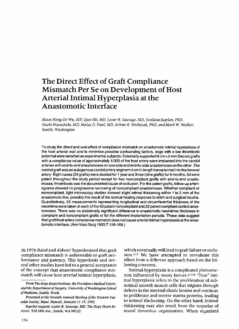
The Direct Effect of Graft Compliance Mismatch Per Se on Development of Host Arterial Intimal Hyperplasia at the Anastomotic Interface Moses Hong-De Wu, MD, Qun Shi, MD, Lester R. Sauvage, MD, Svetlana Kaplan, PhD, Naoki Hayashida, MD, Malay D. Patel, MD, Arlene R. Wechezak, PhD, and Mark W. Walker, Seattle, Washington To study the directand sole effect of compliance mismatch on anastomotic intimal hyperplasia of the host arterial wall and to minimize possible confounding factors, dogs with a low thrombotic potential were selected as experimental subjects. Externally supported 6 cm x 5 mm Dacron grafts with a compliance value of approximately 1/300 of the host artery were implanted into the carotid arteries with end-to-end anastomoses on one side and end-to-side anastomoses on the other. The control graft was an autogenous carotid artery segment 4 cm in length transplanted into the femoral artery. Eight cases (24 grafts) were studied for 1 year and three (nine grafts) for 6 months. All were patent throughout the study period except for two noncompliant grafts with end-to-end anasto- moses; thrombosis was the documented cause of occlusion. For the patent grafts, follow-up arteri- ograms showed no progressive narrowing of noncompliant anastomoses. Whether compliant or noncompliant, light microscopy studies showed slight intimal thickening within 1 to 2 mm of the anastomotic line, possibly the result of the normal healing response to stitch and surgical trauma. Quantitatively, 22 measurements representing longitudinal and circumferential thickness of the neointima were taken at each of the 40 patent noncompliant and 22 patent compliant control anas- tomoses. There was no statistically significant difference in anastomotic neointimal thickness in compliant and noncompliant grafts or for the different implantation periods. These data suggest that graft/host artery compliance mismatch does not cause artedal intimal hyperplasia at the anas- tomotic interface. (Ann Vasc Surg 1993;7:156-168.) In 1976 Baird and Abbott ~hypothesized that graft compliance mismatch is unfavorable to graft per- formance and patency. This hypothesis and sev- eral other studies have led to a general acceptance of the concept that anastomotic compliance mis- match will cause host arterial intimal hyperplasia, From The Hope Heart Institute, the ProvidenceMedical Center, and the Department of Surgery, University of Washington School ofMedicine, Seattle, Wash. Presented at the Seventh Annual Meeting of the Western Vas- cular Society,MauL Hawaii, January 11-15, 1992~ Reprint requests: Lester R. Sauvage, MD, The Hope Heart In- stitute, 52818th Ave., Seattle, WA 98122. which eventually will lead to graft failure or occlu- sionJ 43 We have attempted to reevaluate this effect from a different approach based on the fol- lowing concerns. Intimal hyperplasia is a complicated phenome- non influenced by many factors. 4,7,~4 "True" inti- mal hyperplasia refers to the proliferation of sub- intimal smooth muscle cells that migrate through defects in the internal elastic lamina and continue to proliferate and secrete matrix proteins, leading to intirnat thickening. On the other hand, intimal thickening may also result from the sequelae of mural thrombus organization. When olganized 156
Transcript of The Direct Effect of Graft Compliance Mismatch Per Se on Development of Host Arterial Intimal...
The Direct Effect of Graft Compliance Mismatch Per Se on Deve lopment of Host Arterial Intimal Hyperplasia at the Anastomotic Interface
Moses Hong-De Wu, MD, Qun Shi, MD, Lester R. Sauvage, MD, Svetlana Kaplan, PhD, Naoki Hayashida, MD, Malay D. Patel, MD, Arlene R. Wechezak, PhD, and Mark W. Walker, Seattle, Washington
To study the directand sole effect of compliance mismatch on anastomotic intimal hyperplasia of the host arterial wall and to minimize possible confounding factors, dogs with a low thrombotic potential were selected as experimental subjects. Externally supported 6 cm x 5 mm Dacron grafts with a compliance value of approximately 1/300 of the host artery were implanted into the carotid arteries with end-to-end anastomoses on one side and end-to-side anastomoses on the other. The control graft was an autogenous carotid artery segment 4 cm in length transplanted into the femoral artery. Eight cases (24 grafts) were studied for 1 year and three (nine grafts) for 6 months. All were patent throughout the study period except for two noncompliant grafts with end-to-end anasto- moses; thrombosis was the documented cause of occlusion. For the patent grafts, follow-up arteri- ograms showed no progressive narrowing of noncompliant anastomoses. Whether compliant or noncompliant, light microscopy studies showed slight intimal thickening within 1 to 2 mm of the anastomotic line, possibly the result of the normal healing response to stitch and surgical trauma. Quantitatively, 22 measurements representing longitudinal and circumferential thickness of the neointima were taken at each of the 40 patent noncompliant and 22 patent compliant control anas- tomoses. There was no statistically significant difference in anastomotic neointimal thickness in compliant and noncompliant grafts or for the different implantation periods. These data suggest that graft/host artery compliance mismatch does not cause artedal intimal hyperplasia at the anas- tomotic interface. (Ann Vasc Surg 1993;7:156-168.)
In 1976 Baird and Abbott ~ hypothesized that graft compliance mismatch is unfavorable to graft per- formance and patency. This hypothesis and sev- eral other studies have led to a general acceptance of the concept that anastomotic compliance mis- match will cause host arterial intimal hyperplasia,
From The Hope Heart Institute, the Providence Medical Center, and the Department of Surgery, University of Washington School of Medicine, Seattle, Wash.
Presented at the Seventh Annual Meeting of the Western Vas- cular Society, MauL Hawaii, January 11-15, 1992~
Reprint requests: Lester R. Sauvage, MD, The Hope Heart In- stitute, 52818th Ave., Seattle, WA 98122.
which eventually will lead to graft failure or occlu- sionJ 43 We have at tempted to reevaluate this effect f rom a different approach based on the fol- lowing concerns.
Intimal hyperplasia is a complicated phenome- non influenced by m a n y factors. 4,7,~4 "True" inti- mal hyperplasia refers to the proliferation of sub- intimal smooth muscle cells that migrate through defects in the internal elastic lamina and continue to proliferate and secrete matrix proteins, leading to intirnat thickening. On the other hand, intimal thickening m a y also result f rom the sequelae of mural thrombus organization. When olganized
156

Vol. 7, No. 2 1993 Direct effect of graft compliance mismatch per se 157
luminal thrombus in an advanced stage is present on a flow surface, in many instances it is difficult to determine by gross and microscopic studies whether the thickened myofibroblastic tissue orig- inated from primary hyperplasia ("true" intimal hyperplasia) or was secondary to organization of thrombus. 15-19 Although immunocytochemical la- beling using antibodies to ~-actin can specifically identify smooth muscle cells, this cell type is a component of both forms of intimaI thickening and thus not a distinguishing marker for the origin of thickened intima. This ambiguity has been a confounding factor in many previous compliance studies that used small-caliber synthetic grafts im- planted in carotid, iliac, or femoral arteries in ani- mal models. The hypothetical advantage of these arteries is that they are muscular and therefore may be prone to smooth muscle ceil proliferation if there is a cause-and-effect relationship be tween the vascular wall response and graft compliance mismatch. Unfortunately, all k n o w n small-caliber synthetic grafts and their anastomoses tend to show some degree of thrombus formation fol- lowed by organization. These grafts also have poor patency, which prevents the long-term study nec- essary to observe the development of intimal hy- perplasia. In fact, most reported studies have re- quired anticoagulant or antiplatelet regimens to improve graft patency. This is undesirable because the two most commonly used agents, heparin and aspirin, tend to decrease intimal hyperplasia. 2°-24 Thus an experimental model is needed in which factors that may influence the experimental re- suits, such as platelet adhesion, thrombosis, orga- nization of thrombus, and medicinal treatment, can be minimized as much as possible so that the direct and sole effect of compliance mismatch, that is, the long-term mechanical stimulation of the arterial wall alone, can be studied.
We have previously demonstra ted that dogs with low thrombotic potential were able to achieve a satisfactory patency with minimal or no thrombus formation on small-caliber grafts w h e n no medicinal agents were used. 25 This suggested that in this type of subject the above-described concerns can be minimized. Therefore we have selected dogs with low thrombotic potential for this study.
M A T E R I A L A N D M E T H O D S
Care of the dogs used in this study complied with the "Principles of Laboratory Animal Care" and the "Guide for the Care and Use of Laboratory Ani-
mals" (National Institutes of Health Publication No. 80-23, revised I985).
Criteria and Screening Tests for Exper imenta l Subjects
Screening tests to identify the subjects with stable low thrombotic potential were performed on male mongrel dogs in our laboratory's animal pool. The tests included evaluation of platelet aggregation induced with adenosine diphosphate and deter- mination of the ratio of thromboxane A2 to prosta- cyclin; the actual measurement was based on the ratio of their metabolites, thromboxane B2 (TxB2) and 6-keto-prostaglandin Fic ~ (6-keto-PGFi~). The methods have been described in previous publica- tions. 25-27 A platelet aggregation response of <25% and a ratio of <1.2 were considered the identifying criteria for subjects with low thrombotic potential. If the first screening identified a dog that met the crite- ria, the tests were repeated in 2 weeks for confirma- tion.
The stability of low thrombotic potential status was rechecked on 3 of the 1 1 study subjects prior to their sacrifice. Complete blood cell counts were also performed on three subjects who met the low thrombotic potential criteria.
M e a s u r e m e n t of the Compl iance Value o f the Test Grafts, Control Grafts, and Their Host Arteries
An almost completely noncompliant prosthesis, the preclotted, 5 ram, externally supported EXS Dacron graft (Bard Vascular Systems, C.R. Bard, Inc., Billerica, Mass.) was used as the test graft, which was interposit ioned in the canine carotid artery. The very compliant autogenous carotid ar- tery was used as the control graft and transplanted into the femoral artery. The compliance values of the following materials were measured with the conventional methods and equipment described in our previous publications. 2s,29 Five different segments of the externally supported study graft were p redo t t ed with canine blood to impervious- ness by the same method used for implantation. 3° Five canine carotid and five canine femoral arter- ies were obtained from subjects sacrificed in our facility. The physiologic length of the arteries was marked before removal and maintained during the in vitro measurement . Ten measurements were taken for each sample. Thus the average compliance value of each material was obtained from 50 measurements . The compliance ratio of graft-to-host artery was then calculated.

Annals of 158 W u et al. Vascular Surgery
Surgical Procedures
Eleven male dogs with a confirmed low thrombot- ic potential and weighing from 21 to 38 kg (aver- age 28.2 + 5.5 kg) were used for this study. After the subjects were premedicated with 0.25 mg/kg acepromazine and 0.01 mg/kg atropine, anesthe- sia was induced with 5 to 10 ml of 4% thiamylal intravenously and maintained with a combinat ion of 0.5% to 1.0% halothane and a mixture of ni- trous oxide and oxygen in a 2:1 ratio via a transoral endotracheal tube connected with a closed-circuit respirator. Microsurgicai techniques aided by x4.5 optical magnification were used for the surgeries.
Bilateral carotid arteries were freed through a midline incision of the neck. A segment of the sup- ported Dacron s tudy graft 15 cm long and 5 m m in diameter was preclotted with autogenous blood drawn from the external jugular vein. 30 A 4.5 cm length of the right carotid was excised and pre- served in cold heparinized blood for later trans- plantation. A 6 cm segment of the p redo t t ed sup- ported Dacron graft was then implanted with end- to-end anastomoses of 8-0 Prolene running su- tures both proximally and distally (Fig. 1, A). Suture stitches crossed the graft's support coil so that the anastomoses were virtually noncompli- ant. Another 6 cm length of the supported Dacron graft obtained from the remaining part of the above-ment ioned preclotted segment was im-
planted into the left carotid in the same manner, but using end-to-side anastomoses proximally and distally with 8-0 Prolene sutures, again creating a marked discrepancy be tween the compliance of the host vessel and the graft with its rigid support rings (Fig. 1, B). The carotid artery was then ligated be tween the proximal and distal anastomotic heels. The left femoral artery was freed and a 4 cm length of preserved autogenous carotid artery, which served as the compliant control graft, was interposit ioned with end- to-end anastomoses at both ends with 8-0 Prolene running sutures.
The caliber and flow rates were measured and recorded for both carotid arteries and the left femoral artery with a caliper and a square-wave electromagnetic f lowmeter (model 501, Carolina Medical Electronics, King, N.C.). The neck and femoral wounds were then dosed in a routine manner. No anticoagulant or antiplatelet agents were given before, during, or after surgery. Anti- biotics were given for the first 5 postoperative days. Eight of the 11 subjects were studied for 12 months and three for 6 months.
Graft Fo l low-Up
Periodic arteriograms were obtained to monitor the patency and anastomotic and luminal appear- ance of the supported grafts. For all subjects, arte-
Fig. 1. Implanted grafts. A, End-to-end anastomoses; sutures cross the noncompliant support coil. B, End-to-side anastomoses; inset shows inner side of anastomosis.

Vol. 7, No. 2 1993 Direct effect of graft compliance mismatch per se 159
riography was performed at 2 months and also prior to specimen retrieval. For the eight subjects in the 1 -year group, an additional arteriogram was taken 6 months after the first one (8 months post- operatively).
Arteriographicprocedure. Under the same anes- thesia used for surgery, a No. 7 F angiocatheter was introduced through the distal part of the right femoral artery or its side branch and positioned at the base of the left or right carotid artery with the aid of fluoroscopy and a small amoun t of injected Conray (60% iothalamate meglumine, Malinckrodt, St. Louis, Mo.). Arteriograms were then taken at the time of contrast injection. For repeat arteriography, the catheter again was in- serted from the right femoral artery but proximal to the site where it was previously introduced. The patency of the autogenous carotid graft trans- planted into the femoral artery also was checked by palpation and Doppler examinat ion at these times.
Specimen Retrieval a n d A na ly s i s
After the last arteriogram was taken, the dog was given 10,000 IU heparin and the implanted grafts were gently exposed and harvested. Meanwhile, the wound-heal ing and perigraft tissue reaction were assessed grossly. (An in vivo fixation tech- nique could not be applied because procurement of a fresh sample for the immunocytochemis t ry studies described below was required.)
After the graft lumen was gently flushed with phosphate-buffered solution, the specimen was carefully opened longitudinally, pinned out (with proper tension matched to the in vivo length marked before it was severed), and photographed. The nature of the graft flow surface and the tissue crossing the anastomoses was examined under the stereomicroscope. A map of the surface was drawn on 1 m m 2 grid paper and the thrombus-free sur- face (TFS) score was calculated, expressing the percentage of the total surface area of the graft that was free of thrombus. 3~
Before fixation, while the specimen was fresh, tissue blocks across anastomoses were taken for scanning electron microscopy (SEM), transmis- sion electron microscopy (TEM), and immunocy- tochemistry studies, which included factor VIII/ yon Willebrand factor (FVIII/vWF) staining for verification of endothelial cell presence and a- actin staining for verifying smooth m u s d e cells.
The methods used for SEM, TEM, and FVIIIt vWF have been described previously. 32 Smooth muscle cells were identified by staining with
a monoclonal antibody diluted 1:500 against ~-actin (ICN Biomedicals, Costa Mesa, Calif.), fol- lowed by the secondary ant iserum (1:100), goat ant imouse IgG conjugated to fluorescein (Calbio- chem, La Jolla, Calif.). The staining procedure was the same used for FVIII/vWF staining.
The most important quantitative evaluation in this study was measurement of the host arterial intimal thickness in the anastomotic zone. Tissue samples were taken after the specimen was fixed in 10% buffered formalin at least 24 hours. To determine the mean intimal thickness as accurate- ly as possible, a multiple measuring method was used. TwO longitudinal tissue blocks crossing each anastomosis and located approximately 6 to 7 m m apart along the suture line (about half the distance of the graft circumference) were taken; each block included at least an 8 m m length of host arterial wall (Fig. 2, A and B). After the hematoxyl in and eosin-stained slides were made, the intimal thick- ness was measured under a light microscope with an ocular micrometer. The sites measured on every slide were from the anastomotic junct ion to 7 m m onto the host artery and at 0.25 m m inter- vals within the first 1 rnm and at 1 rnm intervals thereafter (Fig. 2, C), so that the host arterial inti- real thickness of each anastomosis was evaluated by 22 measurements (11 measurements for each of the two tissue samples from every anastomosis). After the data were collected and calculated, sta- tistical comparisons were made of the different groups according to the anastomotic nature (com- pliant or noncompliant) , implant time, and loca- tions (proximal or distal). The comparison was made with BBN Company RS/1 software, which
B
C I1 I 0 signifies suture line
3 4 6 7mm
Fig. 2. Areas from which tissue blocks were taken across the anastomotic interface for study. A, End-to-end anas- tomoses. B, End-to-side anastomoses. C, Points where intimal thicknesses were measured on each tissue block.

Annals of 160 W u et al. Vascular Surgery
applied either the t test or robust test after analyz- ing the pattern of data distribution for the groups being compared and selecting the most appropri- ate method.
R E S U L T S T h r o m b o t i c P o t en t ia l Status a n d C o m p l e t e B lood Cell Counts of S t u d y Subjects
The range of platelet aggregation response for the subjects screened was 15 % to 23 %, with an aver- age value of 19.4 + 3.0%. The range of the ratio of TxBfl6-keto-PGFl~ was 0.76 to 1.20, with an aver- age value of 0.95 _+ 0.21. The thrombotic potential status, reevaluated prior to sacrifice for the three subjects in the 6 -month group, demonstrated that their low thrombotic potential status was stable. The complete blood cell counts on three randomly selected study subjects showed all parameters to be within the normal range.
C o m p l i a n c e Values of the S t u d y Grafts a nd C o m p l i a n c e Ratios B e t w e e n t h e Grafts a n d Host Arteries
The compliance value (percentage change in di- ame te r /mm Hg) of 5 m m preclotted supported study grafts was (0.093 _+ 0.019) x 10-2; the canine carotid artery was 0.29 _+ 0.022 and the canine femoral artery was 0.247 _+ 0.039. The compliance ratio of graft-to-host artery in the noncompliant graft group (externally supported graft to canine carotid artery) was i:300.6 = 0.003; in the compli- ant control group (canine carotid to femoral ar- tery) i twas 1:0.84= 1.190.
Graft F l o w Rate, Patency, General Per formance , a nd F o l l o w - U p Arter iograms
The average flow rate recorded during surgery in the noncompliant end-to-end anastomosis group was 143 + 32 ml /min and in the compliant control group it was I 18 +_ 24 ml/min. Twenty of the 22 noncompliant grafts and all 11 compliant control grafts were patent th roughout the study period. One noncompliant graft with end-to-end anasto- moses in both the 6 -month and 1-year groups was found to be occluded on the arteriogram taken prior to sacrifice. Grossly, red and mixed thrombus filled and adhered to the lumens of the two occlud- ed grafts, with a short distance of semiorganization at the ends. About half of the original anastomotic sutures still could be seen and were not covered by newly built-up tissue. Microscopically the host ar- terial intima at the suture line where there was no
thrombus adhesion seemed normal without re- markable proliferation or thickening, and the inti- ma extended across the anastomoses for about 1 cm to form a layer of "neointimal pannus" on the graft surface. In the areas where there was throm- bus adhesion, thickened intima was fused with myofibroblastic tissue resulting from the organiza- tion of thrombus.
Among the 20 patent noncompliant grafts, seven with end-to-end anastomoses and eight with end-to-side anastomoses had I00% TFS scores (Fig. 3, A and B) and the remaining five grafts had TFS scores of 72%, 84%, 92%, 93%, and 97%. All the thrombotic areas were located at the middle portions of the synthetic grafts and there was no thrombus in the anastomotic areas. The average TFS score for patent noncompliant grafts with end-to-end anastomoses was 96.1%, and for end-to-side anastomoses the average was 97.5 %. All the 11 compliant control grafts had TFS scores of 100% (Fig. 3, O- The follow-up arterio- grams on all patent noncompliant grafts with either end-to-end or end-to-side anastomoses showed stable, patent anastomoses with no signs of progressive narrowing ( Fig. 4).
Observat ion and M e a s u r e m e n t of the Hos t Arterial In t imal R e s p o n s e at the A n a s t o m o t i c Interface
Grossly, all the patent noncomptiant and compli- ant control anastomoses had a smooth anastomot- ic junct ion and interface. There were no gross signs of intimal hyperplasia at the anastomotic site to cause luminal narrowing, and all the original 8- 0 Prolene suture material at the anastomoses could be clearly identified (Fig. 3, A and B). Light microscopy studies showed that in 123 of the total 124 histologic slides studied from both the patent noncompliant grafts and the compliant controls there was very little intimal thickening of the host artery within 1 to 2 m m of the anastomotic line; only one histologic slide from the compliant con- trol group showed intimal thickening beyond that distance, with thicknesses of 45 and 10 ~m at 3 and 4 ram, respectively, from the anastomotic line. Table I shows the average thickness of the host arterial intima at corresponding points within 5 m m of the anastomotic zone for each group. (The actual measurements were taken to 7 mm, but the findings were negative after 5 mm.) The data show that the thickened tissue generally followed a slope-like pattern and the degree of thickening was detectable only at the microscopic level. Most of the increase was limited to the area within I mm

Vol. 7, No. 2 1993 Direct effect of graft compliance mismatch per se 161
~ / : : | i . . . . '
Fig. 3. One-year spedmens from same subject. A, Noncompliant end-to-end specimens with close- up view of anastomotic junction on left. B, Noncompliant end-to-side spedmen with dose-up view of anastomotic junction on left (this view corresponds to the inset in Fig. 1, B). t2, Compliant control.
Fig. 4. Follow-up arteriograms of noncompliant study grafts. End- to-end anastomoses at 2 months (A) and 1 year (B). End-to-side anastomoses at 2 months (C) and 1 year (D).

t~
Tab
le I
. Th
ick
nes
s (g
m)
of th
e h
ost
art
eria
l in
tim
a at
th
e an
asto
mo
tic
inte
rfac
e ar
ea
Stu
dy g
roup
0
Dis
tanc
e fr
om a
nast
omot
ic s
utur
e li
ne (
ram
)
0.25
0.
5 0.
75
1 2
3 4
5
1 y
ear
afte
r im
plan
t N
on
com
pli
ant,
en
d to
end
, p
rox
imal
(n
= 14
) N
on
com
pli
ant,
en
d t
o en
d, d
ista
l (n
= 1
4)
No
nco
mp
lian
t, e
nd
to
side
, p
rox
imal
(n
= 16
) N
on
com
pli
ant,
en
d t
o si
de,
dist
al (
n =
16)
Co
mp
lian
t co
ntro
l, p
rox
imal
(n
= 16
) C
om
pli
ant
cont
rol,
dis
tal
(n =
16)
6
mo
nth
s af
ter
imp
lan
t N
on
com
pli
ant,
en
d t
o en
d, p
rox
imal
(n
= 4)
N
on
com
pli
ant,
end
to
end,
dis
tal
(n =
4)
No
nco
mp
lian
t, e
nd t
o si
de,
pro
xim
al (
n =
6)
No
nco
mp
lian
t, e
nd t
o si
de,
dist
al (
n =
6)
Co
mp
lian
t co
ntro
l, p
rox
imal
(n
= 6)
C
om
pli
ant
cont
rol,
dis
tal
(n =
6)
53.2
-+ 1
5.7
22
.7+
15.
7 0.
6_+
2.4
0 0
50.1
_+24
.8
17.1
__+2
5.0
9.6-
+22
.7
8.4_
_+17
.8
1.9+
7.
2 47
.1-+
30.0
2
8.5
+2
0.0
18
.6-+
17.2
13
.6_+
16.7
9
.6+
14
.6
71.4
__+3
7.9
57.1
-+35
.2
53.4
-+38
.5
32.8
_+32
.6
21.4
_+28
.2
51.0
-+19
.1
36.0
:t:2
3.7
19.3
+-2
1.0
16.1
-+20
.4
5.1_
_+10
.9
87.1
_+50
.6
67.0
-+66
.7
57.0
_+78
.6
35.4
_+61
.1
12.4
_+30
.0
45.0
_+ 4
0.9
78.7
-+ 3
2.3
63.0
_+
30.6
96
.0 +
46.
8 48
.0-+
31.
0 40
.5 -
+ 7.
5
9.0+
_ 10
.3
40.5
_+ 2
7.9
42.0
_+ 3
3.5
67.5
_+39
.7
34.5
_+
35.7
33
.0+
-21.
0
0 0
0 0
0 0
0 0
1.1_
+4,
5 0
0 0
1.5_
+4.
2 0
0 0
0 0
0 0
8.4_
+33
.7
2.8_
+11
.2
0.6_
+2.
5 0
0 0
0 0
13.5
_+27
.0
6.7_
+ 13
.5
0 0
28.5
_+34
.8
18.0
-+35
.5
3.0-
+
7.3
0 31
.5+
_30.
5 24
.0-+
31.0
12
.0_+
21.0
4.
5_+
4.9
19.5
_+18
.3
9.0_
+11
:3
3.0-
+
7.3
1.5_
+3.
6 13
.5_+
15.
8 4.
5_+
7.5
0 0
0 0
0 0
0 0
0 0
0 0
0 0
0 0
0 0
0 0
2

vol. 7, No. 2 1993 Direct effect of grafl compliance mismatch per se 163
of the anastomoses. Right at the graft-host ar tery junct ion the thicknesses ranged from 40 to 96 ~tm; at 1 m m from the junct ion the thickness de- creased, ranging from almost no thickening to 21 lira. The microscopic observations suggest that this slope-like thickening served the funct ion of smoothing the short distance from the anastomot- ic line or neoint ima on the graft surface along the host artery. In the instances where the junct ion of the anastomoses was smooth or fiat, there was even less or almost no microscopic intimal thick-
ening. Histologically, the limited thickening con- sisted of multiple layers of fibroblasts and smooth muscle cells covered by endothelial cells, as dem- onstrated by SEM, TEM, FVIII/vWE and u-actin studies in addition to light microscopy. Beyond the 2 m m area the original host arterial internal elastic lamina with its endothel ial cells serving as the flow surface could easily be discerned and there was no substantial proliferation of smooth muscle cells (Figs. 5 and 6).
The average thickness of the host arterial inti-
Fig. 5. Histologic views of anastomotic interface from 1-year specimens; circles identify areas on host artery from which enlarged views on fight were taken. A, Noncompliant end-to-end anasto- moses. B, Noncompliant end-to-side anastomoses. C, Compliant control. (Light microscopy mag- nification x45 [enlarged views x 112]; hematoxylin and eosin stain.)

~ ~:C ~i~ ~̧̧¸̧II̧̧ ~ ' ~i ~ ' ~,~
Fig, 6. Studies of host arterial walls at the end-to-end anastomotic interface 1 year after implanta- tion of noncompliant grafts~ A, SEM (×100) showing suture line covered by endothelial cells. B, TEM (×2500) within I to 2 mm of the anastomotic line showing normal-appearing host arterial surface covered by endothelial cells. There is no intimal thickening above the internal elastic lam- ina. C, ~-Actin staining within 1 mm of the anastomotic line identifying smooth muscle cells pre- sent in the limited intimal thickening (x225). D, An area within 1 mm of the anastomotic line showing limited intimal thickening covered by endothelial cells as identified by FVIIUvWF stain- ing (×450). E, Same specimen as D showing an area approximately 1 mm farther front the anasto- motic line. Vascular surface has a normal appearance; internal elastic lamina covered by endothe- lial cells identified by FVIIUvWF staining (×450).
Fig. 7. Intimal thickening of canine carotid artery in response to endarterectomy. Subjects had low (A), medium (B), and high (C) thrombotic potentials,

Vol. 7, No. 2 1993 Direct effect of graft compliance mismatch per se 165
Table II. Average thickness (Bm) of the host arterial intima within 5 m m of anastomotic interface
Noncompl ian t anas tomoses
Implant Compl iant anas tomoses
period Proximal Distal Proximal Distal Proximal Distal
End to end End to side
1 year 8.5 ± 18.4 9.6 ± 16.3 13.2 ± 16.1 26.4 ± 28.4 14.2 ± 17.4 30.1 ± 32.9 (n = 126) (n = 126) (n = 144) (n = 144) (n = 144) (n = 144)
6months 6.0 ± 14.9 15.5 ± 27.1 I7.2 ± 22.9 26.2 ± 34.1 12.8 ± 17.6 10.2 ± 15.6 (n = 36) (n = 36) (n = 54) (n = 54) (n = 54) (n = 54)
NOTE: n represents the number of measurements taken on the tissue samples from which the average value was derived.
ma in the area within 5 mIn of the anastomotic line for the groups described in Table I is presented in Table II. For all noncompliant anastomotic groups it ranged from 6 to 26 Bm, and for compliant con- trol groups it ranged from 10 to 30 gm.
Statistical comparisons made between the study groups and based on the data in Table II de- tected no significant difference between the non- compliant and compliant groups either proximal- ly, distally, or overall. Comparisons made between noncompliant 6-month and 1-year groups, be- tween noncompliant proximal and distal groups, between compliant 6-month and 1-year groups, and between compliant proximal and distal groups also did not show significant differences. However, there was a significant difference be- tween noncompliant end-to-end and end-to-side groups.
D I S C U S S I O N
The rationale for using dogs with low thrombotic potential has been stated in the introduction. However, one question may be raised: "Do these dogs lack the ability of randomly selected dogs to respond with intimal hyperplasia or thickening?" A review of the literature indicates that this matter has not been addressed. To answer this question, experimental balloon denudat ion and endarter- ectomy, respectively, were performed on one carot- id artery in each of six dogs. Among them, three had low thrombotic potential, two had medium potential (which represents the majority popula- tion), and one had high thrombotic potential. Specimens were evaluated 8 weeks after surgery. Histologically, similar degrees of vascular hyper- plastic or thickening response (150 to 200 gm) to the injury were observed in the dogs with the three different types of thrombotic potential (Fig. 7).
The approach of this study was to minimize those possible factors, with the exception of com-
pIiance mismatch, that may influence develop- ment of intimal hyperplasia or thickening. The re- sults showed that the "natural" patency of small- caliber synthetic grafts wi thout medicinal treat- ment was satisfactorily high (83% for the 6- mon th group and 94% for the 1-year group), the patent specimens had a high TFS score, and all anastomoses were free of thrombus. This suggests that the objectives of this experimental design basically have been achieved through the use of dogs with low thrombotic potential.
There was an extreme difference between the compliance ratio of the noncompliant anastomo- sis (1:300) and the compliant control anastomosis (near l : l ) . The time periods studied extended to 1 year, an implant period that has rarely been reported in other experimental compliance stud- ies. 3,8,~°-13,33 Therefore both the graft material and observation times provided sufficient conditions to show any effects of compliance mismatch.
Although it would have been more appropriate if both the test and control grafts could be studied in the carotid position, we had observed during previous years that the caliber and flow rates of carotid and femoral arteries in the same dog are quite similar. Therefore we chose the femoral site for the compliant control graft, which allowed us to study noncompliant grafts with both end-to- side and end-to-end anastomoses in each subject's carotid arteries. In this series the diameters and flow rates of carotid and femoral arteries were comparable (4.12 _+ 0.25 m m and 143 _+ 32 ml/min, respectively, for the carotid arteries; 3.99 _+ 0.29 m m and 118 ± 24 mI/min, respectively, for the femoral arteries). Thus, theoretically, the velocities and shear stress at the two sites would also have been comparable. There may be some inherent hemodynamic difference between the carotid and femoral arteries, given the fact that the carotid circulation has more diastolic flow than the femoral artery, and this might affect development of intimal hyperplasia at the control site. However,

Annals of 166 W u et al. Vascular Surgery
even if a control had not been included in this study, the absolute values of intimal thickness ob- tained from the noncompliant grafts (Tables I and II) demonstrate that the degree of thickening was insufficient to influence graft performance and long-term patency.
There were two occlusions of noncompliant grafts in this study. Both the gross and microscopic studies clearly demonstra ted that the occlusion was caused by thrombosis associated with the in- herent thrombogenici ty of small-caliber synthetic grafts rather than primary intimal hyperplasia. It is possible that if the two specimens had been re- trieved after a longer implant period the thrombus wou ld have appeared to be more organized, mak- ing it difficult to distinguish be tween hyperplasia and thrombosis as the cause of occlusion.
Almost all of the microscopically observed thickened intima on the specimens was limited to within 1 to 2 m m of the anastomotic line. Because the distribution pattern of the thickened intima was so similar be tween the noncompliant and compliant control groups with no significant dif- ferences (Tables I and II), this limited thickening was apparently not caused by compliance mis- match but rather might be associated with tissue response to surgical trauma, organization of mi- crothrombi at the anastomotic line and stitch holes, and "self-smoothing" of the uneven anasto- motic junct ion (Fig. 5). Al though these three fac- tors can be greatly minimized by careful microsur- gical techniques, they are still, to some degree, un- avoidable.
LoGerfo et al. 34 reported the p h e n o m e n o n of downs t ream anastomotic hyperplasia. Our study did not show significant differences be tween proximal and distal intimal thicknesses. However, the measured values of the distal intima were slightly thicker than in the proximal intima.
To avoid as much manipulat ion as possible of the harvested specimens, compliance values of the test grafts were not measured after removal. However, stereomicroscopic examinat ion of gross specimens did not reveal any deterioration or breakage of the external support coils of the test grafts; in addition, a previous study demonstra ted that there is no statistically different change in compliance values of porous Dacron grafts 6 months after implantation. 29
The time factor is always a concern in studies of the development of intimal hyperplasia. The fol- low-up arteriograms (Fig. 4) and measurement of the intimal thickness (Tables I and II) demonstrat- ed that anastomotic intima was not progressively thickened during longer implantation despite the
noncompliant anastomoses. In addition, two specimens implanted 5 years in one dog with a low thrombotic potential from an early pilot study were evaluated. These were 4 m m EXS grafts bilat- erally implanted in the carotid arteries with end- to-side anastomoses incorporating the rigid sup- port rings as in this study. Neither in the series of follow-up arteriograms, in gross specimens, nor in the histologic studies were there positive signs that intimal hyperplasia had developed on these ex- ceptionally long-term noncompliant anastomoses (Fig. 8).
Murabayashi et al. 33 presented an approach for evaluating the effects of graft compliance by inde- pendent ly varying the mechanical characteristics of the graft substrates without affecting the blood compatibility of the graft surface. The purpose of this design was to control the influence of the thrombogenic factors of the test graft on compli- ance effect. Untortunately, the patency rate was low, and only two grafts were reported patent be- yond a I -mon th implant period. Their 1-month semicompliant specimen (compliance = 3.5 x 1 0 .2 to 4.0 x 10 -2) showed no intimal hyperplasia, and for the sole 13-month specimen they observed only 10 to 150 g m of intimal thickening. The small number of specimens studied limited the conclu- sions that could be made.
Okuhn et al. 35 created compliance mismatch by applying external bands on native arteries and reached conclusions similar to this study. Because there was no surigcal t rauma through the vascular wall or directly on the flow surface, it is possible that confounding factors that may cause intimal thickening or hyperplasia, such as thrombosis, organization of thrombus, and platelet adhesion, were minimized in their experimental model.
Recently, Bassiouny et al.36 reported an associa- tion with end-to-side anastomoses of two differ- ent sites of intimal thickening - - at the suture line and on the anastomotic floor. We did not observe intimat thickening of the anastomotic floor in our end-to-side anastomosis specimens. This probably is due to the difference in flow patterns and turbu- lence. In their model the flow runs into proximal and distal exits with a remarkable stagnation zone at the floor, whereas in our end-to-side anastomo- sis model (Fig. 1) the host artery was ligated be- tween the two heels of the anastomosis so that the entire flow runs distally. Regarding intimal thick- ening of the suture line, we have suggested that this may be associated with surgical trauma. Al- though the amoun t of the thickening was very limited on both the end- to-end and end-to-side anastomotic areas in our study, the neointima of

Vol. 7, No. 2 1993 Direct effect of graft compliance mismatch per se 167
Fig. 8. Noncompliant grafts implanted with end-to-side anastomoses in a dog for 5 years. A and B, Gross specimens; inset is a close-up of one of the inner anastomoses. C, Light micrograph (x45) of an anastomotic junction. D, One-month postoperative arteriogram. E, Five-year presacrifice ar- teriogram taken of same graft shown in D.
the end- to-s ide anas tomosis g roup was signifi- cant ly th icker t h a n the e n d - t o - e n d anas tomos is g roup (Tables I and II). This d i f fe rence m a y be re- lated to two factors: (1) technica l ly it is slightly more difficult to m a k e a per fec t ly s m o o t h and eve r t ed j u n c t i o n at the end- to -s ide anas tomos i s t han at the e n d - t o - e n d anas tomos is and (2) t he re is a more c o m p l e x s econda ry f low pa t t e rn at the end- to- side anastomosis . 36
It is possible tha t compl i ance mi sma tch m a y p roduce secondary effects such as a t e n d e n c y for
platelets to adhe re to the vascular wall as a resul t of f low t u r b u l e n c e or separa t ion , wh ich t h e n s t imu- lates in t imal hyperplas ia . However , this type of effect was no t the focus of this study. This exper i - m e n t a l m o d e l was des igned to s tudy on ly the di- rect mechan ica l effect of compl iance m i s m a t c h on in t imal hyperplas ia . The data in Tables I and II d e m o n s t r a t e tha t t he re was no significant differ- ence in in t imal th ickness b e t w e e n n o n c o m p l i a n t and compl i an t con t ro l g roups w h e n compar i sons w e r e m a d e f rom th r ee d i f ferent aspects.

Annals of 168 Wu et al. Vascular Surgery
We conclude that the mechanical factor of graft compliance mismatch does not cause host arterial intimal hyperplasia at the anastomotic interface,
We thank David W. Criss, medical photographer, Mary Ann Sedgwick Harvey, research medical editor, and Mary-Ann Nelson, medical illustrator, for their assistance.
REFERENCES
1. Baird RN, Abbott WM. Pulsatile blood-flow in arterial grafts. Lancet 1976;2:948-949.
2. Clark RE, Apostolou S, Kardos JL. Mismatch of mechanical properties as a cause of arterial prostheses thrombosis. Surg Forum 1976;27:208.
3. Kidson IG, Abbott WM. Low compliance and arterial graft occlusion. Circulation 1978; 58 (Suppl 1 ):11 - 14.
4. Kinley CE, Marble AE. Compliance: A continuing problem with vascular grafts. J Cardiovasc Surg 1980;21:163-170.
5. Walden R, L'Italien G J, Megerman J, et aL Matched elastic properties and successful arterial grafting. Arch Surg 1980;115:1166-1169.
6. Madras PN, Ward CA, Johnson WR, et al. Anastomotic hyper- plasia. Surgery 198t;90:922-923.
7. Abbutt WM, Cambria RP. Control of physical characteristics (elasticity and compliance) of vascular graft. In Stanley JC, ed. Biologic and Synthetic Vascular Prostheses. New York: Grune & Stratton, 1982, pp 189-220.
8. Hunter GC, Carson SN, Wong HN, et al. Experimental small- diameter graft patency: Effect of compliance, porosity, and graft healing potential. Curr Probl Surg 1980;37:439-441.
9. Megernran J, Abbott WM. Compliance in vascular grafts. In Wright C, ed. Vascular Grafting. Littleton, Mass.: Wright-PSG, pp 344-364.
10. White R, Goldberg L, Hirose F, et al, Effect of healing on small internal diameter arterial graft compliance. Biomater Med Devices Artif Organs 1983; 11:21-29.
11. Kidson IG. The effect of wall mechanical properties on paten- cy of arterial grafts. Ann R Coil Surg Engl 1983;65:24-29.
12. Pomposelli F, Schoen F, Cohen R, et al. Conformational stress and anastomotic hyperplasia. J Vasc S u rg 1984; 1 : 525- 535.
13. Abbott WM, Megerman J, Hasson JE, et al. Effect of compli- ance mismatch on vascular graft patency. J Vasc Surg 1987;5:376-382.
14. DeWeese JA, Green RM. Control of anastomotic neointimal fibrous hyperplasia in vascular grafts. In Stanley JC, ed. Biological and synthetic vascular prostheses, New York: Grune & Stratton, 1982, pp 653-659.
15. DeWeese JA. Anastonn)tic intimal hyperplasia. In Stanley JC, ed. Biologic and Synthetic Vascular Prostheses. New York: Grune & Stratton, 1982, pp 147-152,
16. Kuwano H, Hashizume M, Yang Y, et al. Patterns of pannus growth of the expanded polytetrafluoroethylene vascular graft with special attention to the intimal hyperplasia forma- tion. Am Surg 1986;52:663-666.
17. Abbott WM, Megerman J. Does compliance mismatch alone cause neointimal hyperplasia? [letter] J Vasc Surg 1989; 9:507.
18. Bulkley BH, Hutchins GM. Accelerated "atherosclerosis:" A morphologic study of 97 saphenous vein coronary artery bypass grafts. Circulation 1977;55:163-169.
19. Haudenschild CC. Morphology of intimal hyperplasia [spe- cial communication]. J Vasc Surg 1989; 10:591-592.
20. Hagen P-O, Wang Z-G, Mikat EM, et al. Antiplatelet therapy reduces aortic intimal hyperplasia distal to small diameter vascular prostheses (PTFE) in nonhuman primates. Ann Surg 1982;195:328-339,
21, Bush HL Jr, Jakubowski JA, Sentissi JM. Early healing after carotid endarterectomy: Effect of high- and low-dose aspirin on thrombosis and early neointimal hyperplasia in a nonhu- man primate model. J Vasc Snrg 1988;7:275-283.
22. McCann RL, Hagen P-O, Fuchs JCA. Aspirin and dipyri- damole decrease intirnaI hyperplasia in experimental vein grafts. Ann Surg 1980; 191:238-243.
23, Clowes AW, Clowes MM. Inhibition by heparin of smooth muscle hyperplasia [special communication]. J Vasc Surg 1989;10:589-591.
24. Dryjski M, Mikat E, Bjornsson TD. Inhibition of intimal hyperplasia after arterial injury by heparins and heparinoid. J Vasc Surg 1988;8:623-633.
25, Kaplan S, Marcoe KF, Sauvage LR, et al, The effect of prede- termined thrombotic potential of the recipient on small-cal- iber graft performance. J Vasc Surg 1986; 3:311 -321.
26. Zammit M, Kaplan S, Sauvage LR, et al. Aspirin therapy in small-caliber arterial prostheses: Long-term experimental observations. J Vase Surg 1984; 1:839-851.
27. Kaplan S, Sauvage LR, Marcoe KF, et al. A new combination therapy for selective and prolonged antiplatelet effect: Results in the dog. Stroke 1986; 17:450-454.
28. Cengiz M, Sauvage LR, Berger K, et al, Effects of compliance alteration on healing of a porous Dacron prosthesis in the thoracic aorta of the dog. Surg Gynecol Obstet 1984; 158:145- 151.
29. Usui Y, Golf GG, Sauvage LR, et al. Effect of healing on com- pliance of porous Dacron grafts. Ann Vasc Surg 1988;2:20- 27.
30. Yates SG, Barros D'Sa AAB, Berger K, et al. The preclotting of porous arterial prostheses. Ann Surg 1978:188:611-622.
31. Kenny DA, Berger K, Walker MW, et at. Experimental com- parison of the thrombogenicity of fibrin and PTFE flow sur- faces. Ann Surg 1980; 191 : 355- 361,
32. Mathisen SR, Wu H-D, Sauvage LR, et al. An experimental study of eight current arterial prostheses. J Vasc Surg 1986; 4:33-41.
33. Murabayashi S, Kambic H, Harasaki H, et al. Fabrication and Iong-ternr implantation of semi-compliant small vascular prosthesis. Trans Am Soc Artif Intern Organs 1985;31:50- 54.
34, LoGerlo FW, Quist WC, Nowak MD, et al. Downstreanr anas- tomotic hyperplasia: A mechanism of failure in Dacron arter- ial grafts. Ann Surg 1983; 197:479-483.
35. Okuhn SP, Connelty DP, Calakos N, et at. Does compliance mismatch alone cause neointimal hyperplasia? J Vasc Surg 1989;9:35-45.
36. Bassiouny HS, White S, Glagov S, et al. Anastomotic intimal hyperplasia: Mechanical injury or flow induced. J Vasc Surg 1992;15:708-717.