Effect of Sodium Silicate and Salicylic Acid ... - Research Square
Upload
independentCategory
view
1download
0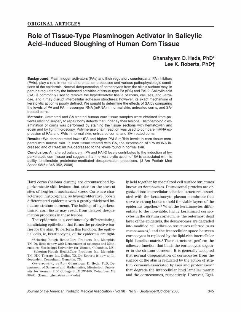
Journal of the American Podiatric Medical Association • Vol 98 • No 5 • September/October 2008 345
Hard corns (heloma durum) are circumscribed hy-perkeratotic skin lesions that arise on the toes atsites of long-term mechanical stress. Corns are char-acterized, histologically, as hyperproliferative, poorlydifferentiated epidermis with a greatly thickened im-mature stratum corneum. The buildup of hyperkera-tinized corn tissue may result from delayed desqua-mation processes in these lesions.
The epidermis is a continuously differentiating,keratinizing epithelium that forms the protective bar-rier for the skin. To perform this function, the epithe-lial cells, ie, keratinocytes, of the epidermis are tight-
ly held together by specialized cell surface structuresknown as desmosomes. Desmosomal proteins are or-ganized into intercellular adhesion structures associ-ated with the keratinocyte plasma membrane thatserve as strong bonds to hold the viable layers of theepidermis together.1, 2 When the keratinocytes differ-entiate to the nonviable, highly keratinized corneo-cytes in the stratum corneum, ie, the outermost deadlayer of the epidermis, the desmosomes are degradedinto modified cell adhesion structures referred to ascorneosomes,3 and the intercellular space betweencorneocytes is replaced by the lipid-rich intercellularlipid lamellae matrix.4 These structures perform theadhesive function that binds the corneocytes togeth-er in the stratum corneum. It is generally acceptedthat normal desquamation of corneocytes from thesurface of the skin is regulated by the action of stra-tum corneum-associated lipases and proteinases5-7
that degrade the intercellular lipid lamellar matrixand the corneosomes, respectively. However, Egel-
Background: Plasminogen activators (PAs) and their regulatory counterparts, PA inhibitors(PAIs), play a role in normal differentiation processes and various pathophysiologic condi-tions of the epidermis. Normal desquamation of corneocytes from the skinʼs surface may, inpart, be regulated by the balanced activities of tissue-type PA (tPA) and PAI-2. Salicylic acid(SA) is commonly used to remove the hyperkeratotic tissue of corns, calluses, and verru-cae, and it may disrupt intercellular adhesion structures; however, its exact mechanism ofkeratolytic action is poorly defined. We sought to determine the effects of SA by comparingthe levels of PA and PAI messenger RNA (mRNA) in normal skin, untreated corns, and SA-treated corns.
Methods: Untreated and SA-treated human corn tissue samples were obtained from pa-tients electing surgery to repair bony defects that underlay their lesions. Histopathologic ex-amination of corns was performed by staining the tissue sections with hematoxylin andeosin and by light microscopy. Polymerase chain reaction was used to compare mRNA ex-pression of PAs and PAIs in normal skin, untreated corns, and SA-treated corns.
Results: We demonstrated lower tPA and higher PAI-2 mRNA levels in corn tissue com-pared with normal skin. In corn tissue treated with SA, the expression of tPA mRNA in-creased and of PAI-2 mRNA decreased to the levels found in normal skin.
Conclusion: An altered balance in tPA and PAI-2 levels contributes to the induction of hy-perkeratotic corn tissue and suggests that the keratolytic action of SA is associated with itsability to stimulate proteinase-meditated desquamation processes. (J Am Podiatr MedAssoc 98(5): 345-352, 2008)
*Schering-Plough HealthCare Products Inc, Memphis,TN. Dr. Heda is now with Department of Sciences and Math-ematics, Mississippi University for Women, Columbus, MS.
†Schering-Plough HealthCare Products Inc, Memphis,TN; ODC Therapy Inc, Dallas, TX. Dr. Roberts is now an In-dependent Consultant, Memphis, TN.
Corresponding author: Ghanshyam D. Heda, PhD, De-partment of Sciences and Mathematics, Mississippi Univer-sity for Women, 1100 College St, MUW-100, Columbus, MS39701. (E-mail: [email protected])
Role of Tissue-Type Plasminogen Activator in SalicylicAcid–Induced Sloughing of Human Corn Tissue
Ghanshyam D. Heda, PhD*Lee K. Roberts, PhD†
ORIGINAL ARTICLES

346 September/October 2008 • Vol 98 • No 5 • Journal of the American Podiatric Medical Association
rud et al8 suggested that complete dissociation of thestratum corneum is induced by trypsinization ratherthan by lipid solvents, which indicates that proteolyt-ic degradation of the corneosomes is absolutely re-quired to break the corneocyte intercellular bonds.
Plasminogen activators (tissue type [tPA] andurokinase type [uPA]) are serine proteinases that bylimited proteolysis convert plasminogen to the activetrypsin-like, broad-specificity proteinase plasmin. Ac-tivity of these proteinases is regulated by two types ofPA inhibitors (PAIs), PAI-1 and PAI-2.9 Levels of PAsare altered during differentiation, migration, and me-tastasis and in some skin diseases.9-12 The type of PAinvolved in these processes, however, varies from celltype to cell type, reflecting the complexity of the pro-teolytic mechanisms involved in these cellular andtissue processes. For example, more differentiatedcells and detached squame-like cells have higher lev-els of PA than do less differentiated cells.13, 14 Plasminis required to dissolve the fibrin-fibronectin matrixafter reepithelialization during the process of woundhealing.15 In skin diseases, such as pemphigus andpsoriasis, where epidermal cellular adhesion is al-tered to form blisters or surface scales, a higher levelof PA has been reported.16-18 Based on the observedhigh content of PAIs in tumor extracts combined withuPA, their role in tumor cell growth and metastasishas been postulated.19-21
No information is available on the role of PAs andPAIs in either the pathogenesis or the keratolytic re-moval of hyperkeratotic skin lesions, such as cornsand calluses. Salicylic acid (SA) in various concentra-tions is used for removing lentigines, pigmented ker-atoses, actinically damaged skin,22 warts, corns, andcalluses.23 Our study was designed to determine therole of PAs and PAIs in the formation of corns and todetermine whether the keratolytic action of SA is as-sociated with its ability to stimulate tissue dissocia-tion through the activation of proteinase pathways.Comparisons of PA and PAI messenger RNA (mRNA)levels in normal skin, untreated corns, and SA-treatedcorns demonstrated a role for tPA and PAI-2 in thepathogenesis of corns and their removal by SA.
Materials and Methods
Materials
Cesium chloride, guanidine isothiocyanate, and sar-cosine were purchased from Fluka Chemika-Bio-chemika (Ronkonkoma, New York). DNA molecularweight marker was obtained from BioVentures Inc(Murfreesboro, Tennessee). Recombinant RNasin ri-
bonuclease inhibitor, AMV reverse transcriptase,oligo (dT)15 primer, deoxynucleotide triphosphates,recombinant Taq DNA polymerase, and all otherreagents for polymerase chain reaction (PCR) werepurchased from Promega Corp (Madison, Wisconsin).NuSieve 3:1 agarose was purchased from FMC Bio-Products (Rockland, Maine). All of the other chemi-cals were of reagent grade and were obtained fromFisher Scientific (Pittsburgh, Pennsylvania) and Sigma-Aldrich Chemical Co (St Louis, Missouri).
Procurement of Human Tissues
Untreated and SA-treated corns were obtained frompatients undergoing elective hammer toe surgery at alocal podiatric medical clinic. Before they were ex-cised as part of the surgical procedure, the corns weretreated for 2 or 3 consecutive days with topical appli-cations of a commercial preparation of SA formulatedas 12.6% wt/wt in a flexible collodion-like vehicle. Thefinal application of SA occurred 48 hours before thescheduled surgery. A licensed, board-certified podi-atric physician performed all of the SA applicationsand surgical procedures. The study was conductedunder a protocol approved by Western InstitutionalReview Board (Olympia, Washington), and all of theparticipants signed an informed consent agreement.Patients had already agreed to undergo the describedelective surgery before they were recruited and en-rolled in the SA treatment phase of the study. Aftertheir removal, the corn specimens were placed insterile phosphate-buffered saline. The nonlesionalends of untreated corns were excised with a sterilescalpel. These tissues served as the control skin inthis study. As soon as possible after collection, all ofthe samples were frozen in liquid nitrogen and werestored dry at –70°C until analysis.
Extraction of RNA
Total RNA from normal tissue and human corn tissuewas prepared according to the procedure of Chirgwinet al.24 Frozen tissue was pulverized with a Ther-movac pulverizer (Thermovac Industries, Coupague,New York) and was resuspended in 4 M of guanidineisothiocyanate buffer to prepare cell lysates using acold Dounce homogenizer. Crude cell lysates werefurther processed by passing them through a syringefitted with needles of increasing gauge size (18 to 23to 26). The final suspension was applied to a 4.7-M ce-sium chloride gradient and was centrifuged at 35,000rpm in a swinging bucket rotor SW 41 in a SorvallOTD55B ultracentrifuge for 18 hours at 20°C. The quan-

Journal of the American Podiatric Medical Association • Vol 98 • No 5 • September/October 2008 347
Results
Histopathologic Examination of Corns
Tissue samples were processed and stained withhematoxylin and eosin for routine light microscopicexamination.31 Human hard corns are characterizedby marked hypertrophy of the horny and viable layersof the epidermis; loss of the defined pattern of epider-mal papillae and dermal rete ridges, especially in thearea underlying the thickest portion of the corn; andan increased number of cell layers in the stratumspinosum and stratum corneum. Hyperproliferationof the keratinocytes in corns has also been reported.10
In addition, we observed marked retention of nuclearmaterial by the corneocytes and an obvious disruptionor alteration of the stratum granulosum layer in mostcorn specimens at the junction demarking normal skinand the lesion (Fig. 1). At higher magnification, alter-ation of the granulosum layer is readily apparent (Fig.2). Rather than the compact, darkly stained kerato-hylin granules in the two- to three-cell-thick stratumgranulosum of normal toe skin (Fig. 1), the kerato-hylin granules are lightly stained, poorly formed, anddiffusely distributed throughout the five or six layersof cells in the corn. Treatment of corn tissue with SAbefore excision resulted in marked differentiation ofthe tissue (Fig. 3). Furthermore, the histologic fea-tures observed in the SA-treated corn tissue were sim-ilar to those of normal skin (Fig. 3). Collectively, thesehistologic features suggest that corn tissue arises atskin sites where epidermal differentiation is signifi-cantly delayed and that the keratolytic action of SAmay involve the induction of normal differentiation oflesional epidermal tissue.
Previous studies32-34 have shown that PAs and PAIsplay an important regulatory role in the normal differ-entiation processes of the epidermis. Given the al-tered differentiation patterns observed in corn tissue,we hypothesized that the epidermal-associated serineproteinases and their regulatory proteins may also bealtered in corns. To evaluate the level of expression ofmRNA for different PAs and PAIs, total RNA was ex-tracted from the thick, central portion of untreatedand SA-treated corn specimens (the area left of theblack arrow in Fig. 1). RNA was also extracted fromthe trimmed edge pieces of untreated corns (the arearight of the black arrow in Fig. 1) that are histological-ly characteristic of normal toe skin; it was used as acontrol for PA and PAI mRNA expression. Analysis ofRNA was chosen for this study because it is more sen-sitive than measurements of low levels of protein, andit was assumed that mRNA expression in these tissueswould correlate with translation to functional protein.
titative and qualitative measurements of RNA sampleswere obtained by measuring their absorption at opti-cal density at 260 and 280 nm and comparing the ab-sorption ratio for the two wavelengths. The quality ofRNA samples was routinely checked by electrophores-ing the samples on formaldehyde/agarose gels.25
Analysis of mRNA Expression
Reverse transcription (RT) of total RNA and amplifi-cation of specific complementary DNA fragments byPCR were performed according to previously pub-lished procedures.20, 21 Briefly, 1 μg of total RNA wastransferred to a microcentrifuge tube containing 20 μLof total reaction volume consisting of 5 mM of magne-sium chloride; 10 mM of Tris hydrochloride (pH 8.8);50 mM of potassium chloride; 0.1% Triton X-100; 1 mMeach of dATP, dGTP, dTTP, and dCTP; 1 U of rRNasinribonuclease inhibitor, 15 U of AMV reverse transcrip-tase; and 0.5 mg of oligo (dT)15 primer. The RT reac-tion was performed for 20 minutes at 42°C. The PCRamplification was performed in the same tube with atotal volume of 100 μL of reaction buffer containing 10mM of Tris hydrochloride (pH 8.8); 50 mM of potassi-um chloride; 1.5 mM of magnesium chloride; 200 mMeach of dATP, dGTP, dTTP, and dCTP; 1 μM each ofsense and antisense oligonucleotide primers (see the“PCR Primers” subsection); and 2.5 U of recombinantTaq DNA polymerase. Reverse transcription PCR con-trol reactions contained all of the ingredients exceptreverse transcriptase. The mixtures were coveredwith 50 μL of mineral oil and were transferred to aPerkinElmer Cetus Thermal Cycler (PerkinElmer Lifeand Analytical Sciences, Waltham, Massachusetts) forPCR. Each cycle of PCR consisted of 2 min of denatu-ration (95°C), 3 min of annealing (55°C), and 1.5 minof polymerization (72°C). After 35 cycles of PCR, thefinal product was extended for 7 min at 72°C. The RT-PCR products were routinely analyzed by means ofelectrophoresis on agarose gel (NuSieve 3:1) in thepresence of ethidium bromide.
PCR Primers
Oligonucleotide primers of PAs and PAIs used forPCR were derived from published sequences (Table1). Primers were designed to flank one or more of theintron regions to ensure the amplification of mRNAtranscripts. All of the primers were synthesized bySigma-Aldrich Chemical Co and were obtained aslyophilized, reverse-phase, column-purified powder.A 20-μM stock solution was prepared by resuspend-ing the lyophilized primers in filtered, sterile distilledwater and was stored in small aliquots at –20°C.

348 September/October 2008 • Vol 98 • No 5 • Journal of the American Podiatric Medical Association
mRNA Expression of PAs and PAIs in NormalSkin, Untreated Corns, and SA-Treated Corns
The PCR was used to determine the level of mRNAexpression for PAs and PAIs in normal human skinand in untreated and SA-treated human corns. Com-pared with normal skin, untreated corns expressed a
higher level of mRNA for PAI-2 and a decreased level ofmRNA for tPA (Fig. 4). The SA-treated corns showedan increase in tPA expression and a concomitant de-crease in PAI-2 expression compared with the un-treated corn samples. The levels of tPA and PAI-2mRNA expression in SA-treated corns were equiva-lent to those observed in normal skin. Treatment with
Table 1. PCR Primers Used for RT-PCR Analyses
Primer Sequence cDNA Location Exp. PCR RestrictionGene (5′ to 3′) (bp) Product Size (bp) Enzymes Source
tPA TGCAGAGATGAAAAAACGCAG (s) 205–225 Eco RI (2)GACCACCCGG TATGTTCTGCC (as) 1201–1221 1016 Pst 1 (1) Pennica et al26
uPA GAGATGAAGTTTGAGGT GG (s) 1152–1170 Eco RI (1)CAGGCCATT CTCTTCC (as) 1688–1673 521 Acc I (1) Verde et al27
PAI-1 ATGCTCCAGCTGACAACAGGA (s) 202–222 Sma I (4)TGAAAGAC TCGTGAAGTCAGC (as) 979–999 797 Eco RI (1) Ginsburg et al28
PAI-2 TGGGAGTTCTAGAA TCTGAGC (s) 143–163 Acc I (2)CGAGTCTAAGATC TTGAGGGT (as) 782–802 659 Rsa I (2) Schleuning et al29
Actin-γ AAGAAC GTGCTTGTG GAAGAC (s) 2402–2422 Rsa I (1)GGTTACGGCAGCA CTTTTAT (as) 2812–2832 430 Dsa I (1) Erba et al30
Abbreviations: as, antisense; bp, base pairs; cDNA, complementary DNA; PAI, plasminogen activator inhibitor; PCR, poly-merase chain reaction; RT, reverse transcription; s, sense; tPA, tissue-type plasminogen activator; uPA, urokinase-type plas-minogen activator.
Figure 1. Photomicrograph of corn tissue at the mar-gin with normal skin (H&E, ×40). Corn tissue was re-moved during elective hammer toe surgery and wasstained with hematoxylin and eosin for routine histo-logic examination by means of light microscopy. Theblack arrow marks the margin between normal skin(right of arrow) and the corn tissue (left of arrow). Atthis magnification, the marked thickening of the epi-dermis (e) in the corn tissue is readily apparent. Otherhistologic differences in the corn tissue include theelongation and disorganization of the rete ridges (r),the diffuse and poorly differentiated stratum granulo-sum layer (sg), and the immature, poorly differentiatedcorneocytes (retention of hematoxylin-staining rem-nants of the nucleus) in the stratum corneum (sc).
Figure 2. Photomicrograph of corn tissue (H&E,×100). At higher magnification, the diffuseness andpoorly differentiated granulosum layer (sg) are appar-ent in the corn tissue. At this magnification, the nor-mally dark hematoxylin staining of compact granulesin the cells of this layer are lightly stained and diffusedwithin the cells. Furthermore, the granulosum celllayer is thickened 2- to 3-fold.

Journal of the American Podiatric Medical Association • Vol 98 • No 5 • September/October 2008 349
SA had no detectable effect on uPA mRNA expres-sion, which seemed to be equivalent to that of normalskin and untreated corns. The level of mRNA expres-sion for PAI-1 was slight and was detectable only inuntreated corns (Fig. 4). Expression of PAI-1 mRNAwas not detected in either normal skin or SA-treatedcorns. The amount of mRNA for the cytoskeletal pro-tein actin-γ was constant for all of the samples, whichsuggests that under the given PCR conditions, modu-lation of tPA, PAI-1, and PAI-2 mRNA levels resultedfrom a gene-targeted increase or decrease in tran-scription and was not due to a general increase or de-crease in transcription rates of all genes.
Discussion
The presence of serine proteinases in the epidermisand their role in the degradation of desmosomal andcertain other epidermal adhesion structures that leadsto desquamation of the plantar stratum corneum has
been demonstrated by several groups.7, 8, 35-40 Plasmino-gen activators are serine proteinases that convert azymogen form of plasminogen to plasmin. Plasminplays a key role in fibrinolysis, epidermal differentia-tion, and desquamation.9, 32-34 Because normal fibri-nolytic activities are observed in differentiating ep-ithelial layers but not in terminally differentiatedcornified layers or in the basal membrane,41 we initi-ated this study to detect the levels of PA and PAI ex-pression in normal and cornified human skin andtheir relationship with the keratolytic action of SA.These results demonstrate that human corn tissue isdeprived of tPA mRNA, perhaps by the virtue of high-er levels of its inhibitor, PAI-2. Although the level ofPAI-1 mRNA was also increased, but to a lesser ex-tent, it too may play a role in the formation of hyper-keratotic lesions. This increase suggests that overex-pression of PAI-2 with concomitant underexpressionof tPA leads to reduced differentiation and desquama-tion rates in corns, thereby resulting in developmentof the thickened hyperkeratotic tissue. The increasedlevels of PAI-1 mRNA in untreated corns could alsobe associated with other hyperkeratotic tissues. Stud-ies with PAI-1 transgenic mice provide further sup-port of this hypothesis in that thickening of the stra-tum corneum at various body sites in these animalsand delayed desquamation processes in cornified lay-ers seem to be associated with abnormally high ex-pression of PAI-1.34 Furthermore, in situ hybridizationstudies have shown higher distribution of PAI-2 butnot of tPA in cornified tissue layers,33 which suggestsa role for PAI-2 in cornified envelope formation by
Figure 3. Salicylic acid (SA)-treated corn tissue (×100).After treatment with SA (before surgical removal), thehistologic features of the SA-treated corn tissue aresimilar to those of normal skin. That is, the granulosumlayer (sg) is now well demarcated, with darkly staining,compact granules in the cells. In the stratum corneum(sc), the corneocytes directly above the stratum granu-losum are now well differentiated. Furthermore, thethickness of the tissue is greatly reduced in that the fullthickness of the epidermis can be seen in this pho-tomicrograph compared with only a portion of the epi-dermis being present in the untreated corn tissue inFigure 2, which was taken at the same magnification.
Figure 4. Levels of messenger RNA (mRNA) expres-sion for plasminogen activators (PAs) and PA in-hibitors (PAIs) in normal human skin, untreated corns,and salicylic acid-treated corns. Polymerase chain re-action–generated products were analyzed on 1% agar-ose gel to measure the relative amounts of mRNA fortissue-type PA (tPA), urokinase-type PA (uPA), PAI-1,and PAI-2 in normal human skin (lane 1), untreatedcorn (lane 2), and salicylic acid-treated corn (lane 3).Actin-γ was used as mRNA control. Abbreviation: bp,base pairs.

350 September/October 2008 • Vol 98 • No 5 • Journal of the American Podiatric Medical Association
preventing tPA function in the normal desquamationprocess.
The histologic evidence and molecular biologicalanalyses presented herein demonstrate the role thatserine proteinases and their regulators play in the for-mation of hyperkeratotic tissues, such as corns, andthe ability of the keratolytic SA to alter PA levels,thus driving normal differentiation of these tissues.
The higher levels of PAI-2 mRNA in normal skinwere not surprising because this is the most commonform of PAI in the epidermis.42 Urokinase-type PA isexpressed at very low levels in the epidermis of nor-mal skin and is mainly confined to the basal layer.32, 42
This may explain why uPA remained uninvolved inthe desquamation of SA-treated hyperkeratotic tissue.Conversely, tPA mRNA, although not detectable in aprevious investigation43 using the conventional North-ern blot hybridization procedure, was found to bepresent in normal epidermis using the PCR techniquedescribed herein. Not only did we demonstrate tPAmRNA in the epidermis, but these data also indicatedits role in SA-induced keratolysis of corn tissue.
The role of proteinases in epidermal cell detach-ment (acantholysis) was originally described bySchiltz et al44 in 1978, in which cultured human skinepidermal cell monolayers can be detached from thesurface of plastic tissue culture dishes when treatedwith antisera from patients with pemphigus. Thisprocess could be reversed by treating the cells withgeneral serine proteinase inhibitors, such as α2-macro-globulin or soybean trypsin inhibitor,44, 45 which sug-gests the higher levels of serine proteinases associat-ed with acantholysis. Similar findings have beenreported in experiments performed with mouse epi-dermal cells46 and organ-cultured human skin.47 Inter-estingly, PAI-2, a naturally occurring PAI in humanepidermis, is capable of blocking pemphigus IgG-in-duced acantholysis in organ-cultured human skin,45 afurther indication of the involvement of PAs in acan-tholysis. The high levels of serine proteinases thathave been observed in sera from patients with pem-phigus12, 18 are now known to be mostly composed ofPAs.39, 48 Similarly, higher tPA antigen16, 49 and mRNA12, 40
levels and higher uPA mRNA43 levels have been foundin psoriatic lesions.
Although there is abundant information availableon the role of the PA system in epithelial cell differen-tiation, until now, nothing was known about how thissystem operates within hyperkeratotic tissues, suchas corn lesions. Because desmoglein and desmoplakinare retained in the immature stratum corneum ofcorns, the corneocytes are likely held tightly togetherby functional desmosomes, also described as cor-neodesmosomes,50 in the absence of plasmin. The
most commonly used keratolytic agent,51 SA is ap-proved for sale over the counter for corn and callusremoval in the United States. It is reported to causedegradation of cell adhesion molecules.51, 52 Despitethe widespread use of SA as a keratolytic agent, verylittle work has been done to define a mechanism bywhich it disrupts the cellular adhesion molecules.The results of our study indicate that SA normalizestPA, PAI-1, and PAI-2 mRNA levels in hyperkeratoticlesions and suggest that SA triggers desquamation byPA-induced plasmin activation. In vitro studies usingnormal human epidermal keratinocytes and SCC-9 (ahuman tongue squamous carcinoma) cell lines haveshown little or no effect of SA on either PA or PAImRNA levels (data not shown). This result suggeststhat SA selectively affects the expression of thesegenes only in hyperkeratotic tissue, such as corns.The observations in our study regarding the gene reg-ulatory activity of SA are not unique to hyperkera-tinized corn tissue of the skin. The most recent studiesin HeLa cells have shown that SA is capable of down-regulating a variety of genes when cells are exposedto ultraviolet light.53 The genes activated by SA in-crease cell survival after ultraviolet light exposure viasuppression of p53-independent apoptosis.54 Collec-tively, these data demonstrate the ability of SA to dif-ferentially regulate gene expression in cells in variousstages of differentiation.
Although PAs are major activators of plasminogen,there are certain other serine proteinases that can ac-tivate plasminogen, such as kallikrein and blood co-agulation factors XI and XII.8 On the other hand, theproduct of plasminogen activation, plasmin, can beinactivated by α2-antiplasmin, α2-macroglobulin, α1-antitrypsin, antithrombin III, and C1 inhibitor.9, 54 It is,therefore, possible that similar to PAI-2, these addi-tional inhibitors of plasmin may contribute to cornifi-cation of the suprabasal cell layer by decreasing theexpression of serine proteinases other than PAs thatare capable of activating plasminogen.
Conclusion
These data provide the first evidence of a possiblekey relationship for enhanced protease activity and aconcomitant decrease in proteinase inhibitor activityin keratolytic agent-induced desquamation of corntissue. Transcriptional analysis of PAs and PAIs innormal, cornified, and SA-treated cornified skin willcontribute to the further understanding of the post-transcriptional events associated with normal tissuedifferentiation. Studies on the in situ distribution ofthese genes in corn tissue and their relative levels ofprotein expression and studies of other serine pro-

Journal of the American Podiatric Medical Association • Vol 98 • No 5 • September/October 2008 351
teinases and their inhibitors may contribute to a bet-ter understanding of the keratolytic mechanisms ofSA and other potential disease-mitigating keratolyticagents. Use of the analytical approach describedherein can help facilitate the selection and formula-tion of efficacious products to alleviate and improvevarious hyperkeratotic skin conditions.
Acknowledgments: Donald R. Skwor, DPM, Mem-phis, for providing untreated and SA-treated humancorn tissues; Mary A. Chryssanthis Montgomery andDiane Hood for assisting with tissue preparation andhistologic examinations; and Frank Anthony, PhD, forhelpful review and comments during the drafting ofthe manuscript and for assisting with compiling datafor the preparation of the manuscript.Financial Disclosure: Drs. Heda and Roberts wereemployees of Schering-Plough HealthCare ProductsInc at the time of study and received financial sup-port to conduct this study.Conflict of Interest: None reported.
References
1. BIZZOZERO G: Sulla struttura degli epiteli pavimentosistratificati [abstract]. Zentral Medizinisches Wochen-schrift 9: 482, 1871.
2. STAEHELIN LA: Structure and function of intercellularjunctions. Int Rev Cytol 39: 191, 1974.
3. CHAPMAN SJ, WALSH A: Desmosomes, corneosomes anddesquamation: an ultra structural study of adult pig epi-dermis. Arch Dermatol Res 282: 304, 1990.
4. ELIAS PM, GRAYSON S, LAMPE MA, ET AL: “The Intercor-neocyte Space,” in Stratum Corneum, ed by R Marks,G Plewig, p 53, Springer-Verlag, Berlin-Heidelberg, 1986.
5. BIDMEAD MC, RODGER ML: The effect of enzymes on stra-tum corneum. J Soc Cosmet Chem 24: 493, 1973.
6. BJELLAND S, GILDBERG A, VOLDEN G: Degradation ofhuman epidermal keratin by fish pepsin. Dermatol Res280: 119, 1988.
7. LUNDSTROM A, EGELRUD T: Stratum corneum chymotrypticenzyme: a proteinase which may be generally present inthe stratum corneum and with a possible involvement indesquamation. Acta Derm Venereol 71: 471, 1991.
8. EGELRUD T, HOFER P-A, LUNDSTROM A: Proteolytic degra-dation of desmosomes in plantar stratum corneum leadsto cell dissociation in vitro. Acta Derm Venereol 68: 93,1988.
9. DANO K, ANDREASEN J, GRONDAHL-HANSEN J, ET AL: Plas-minogen activators, tissue degradation, and cancer. AdvCancer Res 44: 140, 1985.
10. MCCARTHY DJ, HABOWSKY JE: Histochemistry and anthro-pometric analysis of heloma durum. JAPA 65: 4, 1975.
11. HASHIMOTO K, WUN TC, BAIRD J, ET AL: Keratinocyte in-hibitors of plasminogen activator: characterization andability to prevent pemphigus IgG induced acantholysis.J Invest Dermatol 92: 310, 1989.
12. SINGER KH, HASHIMOTO K, LAZARUS GS: Antibody-inducedproteinase activation: a proposed mechanism for pem-phigus. Springer Semin Immunopathol 4: 17, 1981.
13. JENSEN PJ, JOHN M, BAIRD J: Urokinase and tissue typeplasminogen activators in human keratinocyte culture.Exp Cell Res 187: 162, 1990.
14. ISSEROFF RR, FUSENIG NE, RIFKIN DB: Plasminogen acti-vator in differentiating mouse keratinocytes. J InvestDermatol 80: 217, 1983.
15. CLARK RAF, LANIGAN JM, DELLAPELLE P, ET AL: Fibronectinand fibrin provide a provisional matrix for epidermalcell migration during wound reepithelialization. J InvestDermatol 79: 264, 1982.
16. GRONDAHL-HANSEN J, OTTEVANGER V: Tissue-type plas-minogen activator concentrations in plasma from pa-tients with psoriasis. Acta Derm Venereol 69: 391, 1989.
17. LOTTI T, BONAN P, PANCONESI E: Plasminogen activationin psoriasis. Acta Derm Venereol Suppl 146: 36, 1989.
18. GRANDO SA: Decompensation in proteinase-inhibitor sys-tem and application of proteinase inhibitors in pemphi-gus and pemphigoid. J Dermatol Sci 4: 95, 1992.
19. SIER CFM, VERSPAGET HW, GRIFFIOEN G, ET AL: Imbalanceof plasminogen activators and their inhibitors in humancolorectal neoplasia. Gastroenterology 101: 1522, 1991.
20. REILLY D, CHRISTENSEN L, DUCH M, ET AL: Type-1 plas-minogen activator inhibitor in human breast carcino-mas. Int J Cancer 50: 208, 1992.
21. NAGAYAMA M, SATO A, HAYAKAWA H, ET AL: Plasminogenactivators and their inhibitors in non-small cell lungcancer: low content of type 2 plasminogen activator in-hibitor associated with tumor dissemination. Cancer 71:
1398, 1994.22. SWINEHART JM: Salicylic acid ointment peeling of the
hands and forearms. Effective nonsurgical removal ofpigmented lesions and actinic damage. J Dermatol SurgOncol 18: 495, 1992.
23. PARISH LC, MONROE E, REX IH: Treatment of commonwarts with high-potency (26%) salicylic acid. Clin Ther10: 462, 1988.
24. CHIRGWIN JM, PRZYBYLA AE, MACDONALD RJ, ET AL: Isola-tion of biologically active ribonucleic acid from sourcesenriched in ribonuclease. Biochemistry 18: 5249, 1979.
25. SAMBROUK J, FRITCH EF, MANIATIS T: Molecular Cloning:
A Laboratory Manual, p 7.43, Cold Spring Harbor Lab-oratory Press, Cold Spring Harbor, NY, 1989.
26. PENNICA D, HOLMES WE, KOHR WJ, ET AL: Cloning and ex-pression of human tissue-type plasminogen activatorcDNA in E. coli. Nature 301: 214, 1983.
27. VERDE P, STOPPELLI MP, GALEFFI P, ET AL: Identificationand primary sequence of an unspliced human urokinasepoly (A)+ RNA. Proc Natl Acad Sci U S A 81: 4727, 1984.
28. GINSBURG D, ZAHEB R, YANG AY, ET AL: cDNA cloning ofhuman plasminogen activator-inhibitor from endothelialcells. J Clin Invest 78: 1673, 1986.
29. SCHLEUNING WD, MEDCALF RL, HESSION C, ET AL: Plasmino-gen activator inhibitor-2: regulation of gene transcriptionduring phorbol ester-mediated differentiation of U-937human histiocytic lymphoma cells. Mol Cell Biol 7: 4564,1987.
30. ERBA HP, EDDY R, SHOWS T, ET AL: Structure, chromo-some location, and expression of the human γ-actingene: differential evolution, location, and expression ofthe cytoskeletal β- and γ-actin genes. Mol Cell Biol 8:
1775, 1988.31. LUNA LG: “Manual of Histologic Staining,” in Methods of
the Armed Forces Institute of Pathology, 3rd Ed, Mc-Graw-Hill, New York, 1968.
352 September/October 2008 • Vol 98 • No 5 • Journal of the American Podiatric Medical Association
32. LAZARUS GS, JENSEN PJ: Plasminogen activators in ep-ithelial biology. Semin Thromb Hemost 17: 210, 1991.
33. CHEN CS, LYONS-GIORDANO B, LAZARUS GS, ET AL: Differ-ential expression of plasminogen activators and theirinhibitors in an organotypic skin coculture system. JCell Sci 106: 45, 1993.
34. LYONS-GIORDANO B, LAZARUS GS: Skin abnormalities inmice transgenic for plasminogen activator inhibitor 1:implications for the regulation of desquamation and fol-licular neogenesis by plasminogen activator enzymes.Dev Biol 170: 289, 1995.
35. LUNDSTROM A, EGELRUD T: Evidence that cell sheddingfrom plantar stratum corneum in vitro involves endoge-nous proteolysis of the desmosomal protein desmogleinI. J Invest Dermatol 94: 216, 1990.
36. LUNDSTROM A, EGELRUD T: Cell shedding from humanplantar skin in vitro: evidence that two different typesof protein structures are degraded by a chymotrypsin-like enzyme. Arch Dermatol Res 282: 234, 1990.
37. EGELRUD T, LUNDSTROM A: A chymotrypsin-like pro-teinase that may be involved in desquamation in plan-tar stratum corneum. Arch Dermatol Res 283: 108, 1991.
38. SUZUKI Y, NOMURA J, HORI J, ET AL: Detection and char-acterization of endogenous protease associated withdesquamation of stratum corneum. Arch Dermatol Res285: 372, 1993.
39. IMAI S: The effect of the proteolytic enzyme savinase onplantar skin in vitro. Arch Dermatol Res 283: 377, 1991.
40. KASHIMA M, FUKUYAMA K, KIKUCHI M, ET AL: Limited pro-teolysis of high molecular weight histidine-rich proteinof rat epidermis by epidermal proteinases. J Invest Der-matol 90: 829, 1988.
41. ASTRUP T: “Cell Induced Fibrinolysis: A FundamentalProcess,” in Cold Spring Harbor Conference on Cell Pro-
liferation, Vol 2, ed by E Reich, DB Rifkin, E Shaw, p 343,Cold Spring Harbor Press, Cold Spring Harbor, NY, 1975.
42. HIBINO T, IZAKI S, OHKUMA M, ET AL: Epidermal plasmino-gen activator inhibitor (PAI) is immunologically identi-cal to placental-type PAI-2. FEBS Lett 231: 202, 1988.
43. SPIERS EM, LAZARUS GS, LYONS-GIORDANO B: Expression
of plasminogen activator enzymes in psoriatic epider-mis. J Invest Dermatol 102: 333, 1994.
44. SCHILTZ JR, HU C, MICHEL B: Pemphigus antibody inter-action with human epidermal cells in culture: a pro-posed mechanism for pemphigus acantholysis. J ClinInvest 62: 778, 1978.
45. HASHIMOTO K, SHAFRAN KM, WEBBER PS, ET AL: Anti-cellsurface pemphigus auto antibody stimulates plasmino-gen activator activity of human epidermal cells: a mech-anism for the loss of epidermal cohesion and blisterformation. J Exp Med 157: 259, 1983.
46. FARB RM, DYKES R, LAZARUS GS: Anti-epidermal-cell-sur-face pemphigus antibody detaches viable epidermalcells from culture plates by activation of proteinase.Proc Natl Acad Sci U S A 75: 459, 1978.
47. MORIOKA S, NAITO K, OGAWA H: The pathogenic role ofpemphigus antibodies and proteinase in epidermal acan-tholysis. J Invest Dermatol 76: 337, 1981.
48. BAIRD J, LAZARUS GS, BELIN D, ET AL: mRNA for tissue-type plasminogen activator is present in lesional epi-dermis from patients with psoriasis, pemphigus, bullouspemphigoid, but is not detected in normal epidermis. JInvest Dermatol 95: 548, 1990.
49. GISSLER HM, FRANK R, KRAMER MD: Immunohistochemi-cal characterization of the plasminogen activator sys-tem in psoriatic epidermis. Br J Dermatol 128: 612, 1993.
50. SERRE G, MILS V, HAFTEK M, ET AL: Identification of latedifferentiation antigens of human cornified epithelia,expressed in re-organized desmosomes and bound tocross-linked envelope. J Invest Dermatol 97: 106, 1991.
51. DAVIES M, MARKS M: Studies on the effect of salicylicacid on normal skin. Br J Dermatol 95: 187, 1976.
52. HUBER C, CHRISTOPHER E: “Keratolytic” effect of salicylicacid. Arch Derm Res 257: 293, 1977.
53. PAUNESKU T, CHANG-LIU CM, SHEARIN-JONES P, ET AL: Iden-tification of genes regulated by UV/salicylic acid. Int JRadiat Biol 76: 189, 2000.
54. ROBBINS KC: “Fibrinolysis,” in Thrombosis, ed by HCKwaan, EJW Bowie, p 23, WB Saunders, Philadelphia,1982.
Copyright © 2022 FDOKUMEN