Urokinase-type Plasminogen Activator-like Proteases in Teleosts Lack Genuine Receptor-binding...
12
Urokinase-type Plasminogen Activator-like Proteases in Teleosts Lack Genuine Receptor-binding Epidermal Growth Factor-like Domains * □ S Received for publication, April 4, 2012, and in revised form, June 12, 2012 Published, JBC Papers in Press, June 25, 2012, DOI 10.1074/jbc.M112.369207 René Bager ‡§ , Thomas K. Kristensen ‡ , Jan K. Jensen ‡§ , Agnieszka Szczur ‡§ , Anni Christensen ‡§ , Lisbeth M. Andersen ‡§ , Jesper S. Johansen ‡ , Niels Larsen ‡ , Erik Baatrup ¶ , Mingdong Huang § , Michael Ploug § **, and Peter A. Andreasen ‡§1 From the ‡ Department of Molecular Biology and Genetics and the § Danish–Chinese Centre for Proteases and Cancer, Aarhus University, 10 Gustav Wieds Vej, 8000 Aarhus C, Denmark, the ¶ Department of Bioscience, Zoophysiology, Aarhus University, Building 1131, 3 C.F. Moellers Allé, 8000 Aarhus C, Denmark, the Fujian Institute of Research on the Structure of Matter, Chinese Academy of Sciences, Fuzhou, China, and the **Finsen Laboratory, Rigshospitalet and Biotech Research and Innovation Center, Copenhagen Biocenter, 5 Ole Maaloes Vej, 2200 Copenhagen N, Denmark Background: In mammals, extracellular proteolysis initiated by urokinase-type plasminogen activator (uPA) is associated with cell surfaces via the uPA-receptor uPAR. Results: Fish have two uPAs, one lacking the uPAR-binding domain, another having a uPAR-binding domain lacking two cysteines and a genuine uPAR-binding sequence. Conclusion: In fish, uPA-dependent proteolysis occurs without uPAR. Significance: Evolutionary studies provide a more comprehensive understanding of extracellular proteolysis. Plasminogen activation catalyzed by urokinase-type plasmin- ogen activator (uPA) plays an important role in normal and pathological tissue remodeling processes. Since its discovery in the mid-1980s, the cell membrane-anchored urokinase-type plasminogen activator receptor (uPAR) has been believed to be central to the functions of uPA, as uPA-catalyzed plasminogen activation activity appeared to be confined to cell surfaces through the binding of uPA to uPAR. However, a functional uPAR has so far only been identified in mammals. We have now cloned, recombinantly produced, and characterized two zebrafish proteases, zfuPA-a and zfuPA-b, which by several cri- teria are the fish orthologs of mammalian uPA. Thus, both pro- teases catalyze the activation of fish plasminogen efficiently and both proteases are inhibited rapidly by plasminogen activator inhibitor-1 (PAI-1). But zfuPA-a differs from mammalian uPA by lacking the exon encoding the uPAR-binding epidermal growth factor-like domain; zfuPA-b differs from mammalian uPA by lacking two cysteines of the epidermal growth factor- like domain and a uPAR-binding sequence comparable with that found in mammalian uPA. Accordingly, no zfuPA-b bind- ing activity could be found in fish white blood cells or fish cell lines. We therefore propose that the current consensus of uPA- catalyzed plasminogen activation taking place on cell surfaces, derived from observations with mammals, is too narrow. Fish uPAs appear incapable of receptor binding in the manner known from mammals and uPA-catalyzed plasminogen activa- tion in fish may occur mainly in solution. Studies with nonmam- malian vertebrate species are needed to obtain a comprehensive understanding of the mechanism of plasminogen activation. Plasminogen activators are serine proteases that catalyze the conversion of the extracellular zymogen plasminogen to the active serine protease plasmin. Plasmin in turn catalyzes degra- dation of extracellular matrix proteins like fibrin and laminin. In mammals, there are two types of plasminogen activators, urokinase-type plasminogen activator (uPA) 2 and tissue-type plasminogen activator (tPA). Plasminogen activation is strictly controlled temporally and spatially. Thus, secretion of tPA from endothelial cells can initiate intravascular fibrinolysis, securing blood vessel patency (for a review, see Ref. 1). Cell- specific uPA production can initiate localized extracellular matrix turnover (for a review, see Ref. 2). uPA-catalyzed plas- minogen activation is believed to take place mainly on cell sur- faces, where uPA accumulates due to high-affinity binding by the receptor, uPAR. uPAR also functions as an adhesion recep- tor due to its affinity to the extracellular matrix protein vit- ronectin. It may also regulate the activity of integrins. uPAR is a three-domain protein bound to cell surfaces by a glycosylphos- phatidylinositol anchor (for a review, see Ref. 3). Plasminogen activator inhibitor-1 (PAI-1) is a fast and specific inhibitor of * This work was supported by Danish Medical Research Council Grant 2107– 040003, Danish National Research Foundation Grant 26-331-6, and Novo- Nordisk Foundation Grant R114-A11382 (to P. A. A.). □ S This article contains supplemental Figs. S1–S6. The nucleotide sequence(s) reported in this paper has been submitted to the GenBank TM /EBI Data Bank with accession number(s) FJ660930 and HQ403602. 1 To whom correspondence should be addressed: Dept. of Molecular Biology and Genetics, Aarhus University, 10 Gustav Wieds Vej, 8000 Aarhus C, Den- mark. Tel.: 45-87155456; E-mail: [email protected]. 2 The abbreviations used are: uPA, urokinase-type plasminogen activator; ef1, elongation factor 1-; PAI-1, plasminogen activator inhibitor-1; S-2288, H-D-Ile-Pro-Arg-p-nitroanilide; S-2251, H-D-Val-Leu-Lys-p-nitroani- lide; S-2444, pyroGlu-Gly-Arg-p-nitroanilide; tPA, tissue-type plasminogen activator; uPAR, urokinase-type plasminogen activator receptor; zfuPA-a, zebrafish urokinase-type plasminogen activator-like protease-a; zfuPA-b, zebrafish urokinase-type plasminogen activator-like protease-b; qPCR, quantitative PCR. THE JOURNAL OF BIOLOGICAL CHEMISTRY VOL. 287, NO. 33, pp. 27526 –27536, August 10, 2012 © 2012 by The American Society for Biochemistry and Molecular Biology, Inc. Published in the U.S.A. 27526 JOURNAL OF BIOLOGICAL CHEMISTRY VOLUME 287 • NUMBER 33 • AUGUST 10, 2012 by guest on July 20, 2015 http://www.jbc.org/ Downloaded from
-
Upload
independent -
Category
Documents
-
view
0 -
download
0
Transcript of Urokinase-type Plasminogen Activator-like Proteases in Teleosts Lack Genuine Receptor-binding...
Urokinase-type Plasminogen Activator-like Proteases inTeleosts Lack Genuine Receptor-binding Epidermal GrowthFactor-like Domains*□S
Received for publication, April 4, 2012, and in revised form, June 12, 2012 Published, JBC Papers in Press, June 25, 2012, DOI 10.1074/jbc.M112.369207
René Bager‡§, Thomas K. Kristensen‡, Jan K. Jensen‡§, Agnieszka Szczur‡§, Anni Christensen‡§,Lisbeth M. Andersen‡§, Jesper S. Johansen‡, Niels Larsen‡, Erik Baatrup¶, Mingdong Huang§�, Michael Ploug§**,and Peter A. Andreasen‡§1
From the ‡Department of Molecular Biology and Genetics and the §Danish–Chinese Centre for Proteases and Cancer, AarhusUniversity, 10 Gustav Wieds Vej, 8000 Aarhus C, Denmark, the ¶Department of Bioscience, Zoophysiology, Aarhus University,Building 1131, 3 C.F. Moellers Allé, 8000 Aarhus C, Denmark, the �Fujian Institute of Research on the Structure of Matter, ChineseAcademy of Sciences, Fuzhou, China, and the **Finsen Laboratory, Rigshospitalet and Biotech Research and Innovation Center,Copenhagen Biocenter, 5 Ole Maaloes Vej, 2200 Copenhagen N, Denmark
Background: In mammals, extracellular proteolysis initiated by urokinase-type plasminogen activator (uPA) is associatedwith cell surfaces via the uPA-receptor uPAR.Results: Fish have two uPAs, one lacking the uPAR-binding domain, another having a uPAR-binding domain lacking twocysteines and a genuine uPAR-binding sequence.Conclusion: In fish, uPA-dependent proteolysis occurs without uPAR.Significance: Evolutionary studies provide a more comprehensive understanding of extracellular proteolysis.
Plasminogen activation catalyzed by urokinase-type plasmin-ogen activator (uPA) plays an important role in normal andpathological tissue remodeling processes. Since its discovery inthe mid-1980s, the cell membrane-anchored urokinase-typeplasminogen activator receptor (uPAR) has been believed to becentral to the functions of uPA, as uPA-catalyzed plasminogenactivation activity appeared to be confined to cell surfacesthrough the binding of uPA to uPAR. However, a functionaluPAR has so far only been identified inmammals.We have nowcloned, recombinantly produced, and characterized twozebrafish proteases, zfuPA-a and zfuPA-b, which by several cri-teria are the fish orthologs of mammalian uPA. Thus, both pro-teases catalyze the activation of fish plasminogen efficiently andboth proteases are inhibited rapidly by plasminogen activatorinhibitor-1 (PAI-1). But zfuPA-a differs from mammalian uPAby lacking the exon encoding the uPAR-binding epidermalgrowth factor-like domain; zfuPA-b differs from mammalianuPA by lacking two cysteines of the epidermal growth factor-like domain and a uPAR-binding sequence comparable withthat found in mammalian uPA. Accordingly, no zfuPA-b bind-ing activity could be found in fish white blood cells or fish celllines. We therefore propose that the current consensus of uPA-catalyzed plasminogen activation taking place on cell surfaces,derived from observations with mammals, is too narrow. FishuPAs appear incapable of receptor binding in the manner
known from mammals and uPA-catalyzed plasminogen activa-tion in fishmay occurmainly in solution. Studieswith nonmam-malian vertebrate species are needed to obtain a comprehensiveunderstanding of the mechanism of plasminogen activation.
Plasminogen activators are serine proteases that catalyze theconversion of the extracellular zymogen plasminogen to theactive serine protease plasmin. Plasmin in turn catalyzes degra-dation of extracellular matrix proteins like fibrin and laminin.In mammals, there are two types of plasminogen activators,urokinase-type plasminogen activator (uPA)2 and tissue-typeplasminogen activator (tPA). Plasminogen activation is strictlycontrolled temporally and spatially. Thus, secretion of tPAfrom endothelial cells can initiate intravascular fibrinolysis,securing blood vessel patency (for a review, see Ref. 1). Cell-specific uPA production can initiate localized extracellularmatrix turnover (for a review, see Ref. 2). uPA-catalyzed plas-minogen activation is believed to take place mainly on cell sur-faces, where uPA accumulates due to high-affinity binding bythe receptor, uPAR. uPAR also functions as an adhesion recep-tor due to its affinity to the extracellular matrix protein vit-ronectin. It may also regulate the activity of integrins. uPAR is athree-domain protein bound to cell surfaces by a glycosylphos-phatidylinositol anchor (for a review, see Ref. 3). Plasminogenactivator inhibitor-1 (PAI-1) is a fast and specific inhibitor of
* This work was supported by Danish Medical Research Council Grant 2107–040003, Danish National Research Foundation Grant 26-331-6, and Novo-Nordisk Foundation Grant R114-A11382 (to P. A. A.).
□S This article contains supplemental Figs. S1–S6.The nucleotide sequence(s) reported in this paper has been submitted to the
GenBankTM/EBI Data Bank with accession number(s) FJ660930 andHQ403602.
1 To whom correspondence should be addressed: Dept. of Molecular Biologyand Genetics, Aarhus University, 10 Gustav Wieds Vej, 8000 Aarhus C, Den-mark. Tel.: 45-87155456; E-mail: [email protected].
2 The abbreviations used are: uPA, urokinase-type plasminogen activator;ef1�, elongation factor 1-�; PAI-1, plasminogen activator inhibitor-1;S-2288, H-D-Ile-Pro-Arg-p-nitroanilide; S-2251, H-D-Val-Leu-Lys-p-nitroani-lide; S-2444, pyroGlu-Gly-Arg-p-nitroanilide; tPA, tissue-type plasminogenactivator; uPAR, urokinase-type plasminogen activator receptor; zfuPA-a,zebrafish urokinase-type plasminogen activator-like protease-a; zfuPA-b,zebrafish urokinase-type plasminogen activator-like protease-b; qPCR,quantitative PCR.
THE JOURNAL OF BIOLOGICAL CHEMISTRY VOL. 287, NO. 33, pp. 27526 –27536, August 10, 2012© 2012 by The American Society for Biochemistry and Molecular Biology, Inc. Published in the U.S.A.
27526 JOURNAL OF BIOLOGICAL CHEMISTRY VOLUME 287 • NUMBER 33 • AUGUST 10, 2012
by guest on July 20, 2015http://w
ww
.jbc.org/D
ownloaded from
both plasminogen activators (for a review, see Ref. 4). uPAR-bounduPA�PAI-1 complex can be internalized and degraded byreceptors of the low density lipoprotein receptor family (forreviews, see Refs. 4 and 5).As listed from the N terminus, uPA from mammals has an
epidermal growth factor (EGF)-like domain (residues 7–47 inhuman uPA), a kringle domain (residues 50–131 in humanuPA), an interdomain linker (residues 132–147 in human uPA),and a C-terminal catalytic domain (in human uPA, residues148–411 or 1–251 in the chymotrypsin template numbering).It is secreted as a single-chain proform or zymogen with anactivity at least 250-fold lower than that of the fully active two-chain form, which is produced by cleavage of the Lys158/15–Ile159/16 bond (throughout the text, when double numbers sep-arated by a slash are used to describe an amino acid position, thefirst number refers to the actual amino acid number while thesecond number refers to number of the corresponding aminoacid in chymotrypsionogen.) After cleavage, residues 1–158/15remain bound to the catalytic domain by a Cys148/1–Cys279/122disulfide bond. No function has yet been assigned to the kringledomain (for a review, see Ref. 2). It is the EGF-like domain thatbinds to uPAR (6).Plasminogen exists in all vertebrates (7, 8), but so far, plas-
minogen activation has been studied almost exclusively inmammals and birds, whereas reports describing direct charac-terization of plasminogen activators from lower vertebrateshave been sparse. Takahashi et al. (9) reported that two anti-gens from platyfish (Xiphophorus maculatus) withMr values of50,000 and 55,000, respectively, cross-reacted with two mono-clonal antibodies against human uPA; two major plasminogenactivators, as detected by SDS-PAGE and fibrin-agarosezymography, migrated with similarMr values. In goldfish, SDS-PAGE and casein zymography of homogenates of injured opticnerve revealed plasminogen activation activitymigrating as twobands withMr between 60,000 and 65,000 (10).The sequencing of the genomes of vertebrate species beyond
mammals and birds now allows more detailed biochemicalstudies into plasminogen activation in reptiles, amphibians,and bony fishes. Recently, both uPA-like and uPAR-like, but asyet unverified, sequences were found in the genome of the liz-ard Anolis carolinensis. There is no knowledge about uPA oruPAR in amphibians. For zebrafish (Danio rerio), the databasescontain evidence for one plasminogen gene, one zebrafish tPA(zftPA) gene, and one zebrafish PAI-1 (zfPAI-1) gene, neither ofwhich have any obviously striking differences from their mam-malian counterparts. In contrast, we have foundno evidence fora uPAR-like protein in teleosts in the sequence databases, andfound two genes encoding proteins resembling mammalianuPA, which we will refer to as zebrafish urokinase-type plas-minogen activator-like protease-a (zfuPA-a) and zebrafishurokinase-type plasminogen activator-like protease-b (zfuPA-b). Here, we present experimentally verified full-length cDNAsequences of zfuPA-a and zfuPA-b. These proteins have theunusual feature of either lacking of a growth factor domain orhaving a growth factor domain with an unusual sequence andan unusual disulfide bond connectivity. Accordingly, we wereunable to identify binding activity on autologous cells forzebrafish uPAs. We therefore hypothesize that uPA-catalyzed
plasminogen activation in fishes functions without an uPARlike that known from mammals.
EXPERIMENTAL PROCEDURES
RNAExtraction andReverse Transcription—Sexuallymaturemale and female zebrafish (D. rerio) were sacrificed by chillingon ice and dissected. Testis, ovary, liver, heart, gill, eye, andbrain tissues were subsequently stored in RNAlater (Ambion,Austin, TX) to protect the RNA from degradation. Total RNAwas isolated from the RNAlater preserved samples using theQiagen RNeasy Mini Kit (Qiagen). This protocol included aDNase treatment step according to the manufacturer’s recom-mendations to eliminate genomic DNA contamination. Forcloning purposes, cDNA was generated from an mRNA poolextracted from testes of 16 adult zebrafish.Derivatization of the zfuPA-a cDNA—Full-length zfuPAa
cDNA was isolated and assembled from a total of 15 overlap-ping partial cDNA fragments. Six fragments were PCR ampli-fied using primers designed from a published predicted partialzfuPA-a cDNA sequence (GenBank accession numberXM_693024.2) that was identified by searching the NCBI pro-tein database using human uPA. Although this sequence in itsentirety was incorrectly predicted as demonstrated in the pres-ent study, part of the 3�-end of the sequence was correct andtherefore useful for primer design. Six fragments were PCRamplified using primers that were designed from the publishedgenomic sequence containing the putative zfuPA-a gene(GenBank accession number CR762430). A fragment includingthe entire coding region was finally PCR amplified to experi-mentally verify the sequence assembled from the fragmentsdescribed above. The 5�- and 3�-ends were isolated using theSMART RACE cDNA Amplification Kit from Clontechtogether with the Advantage2 PCR Enzyme System (Clontech).For 5�- and 3�-RACE, nested PCR was used to amplify discreteRACE PCR products.PCR fragments were isolated from agarose gels using the
E.Z.N.A. Gel Extraction Kit (Omega Bio-tek) or the QIAquickGel Extraction Kit (Qiagen) and cloned into the pCR2.1-TOPOor pCR4-TOPO vector using the TOPO TA Cloning systemwith the electrocompetent TOP10 Escherichia coli strain(Invitrogen). Sequencing reactions were performed using theBigDye Terminator version 3.1 Cycle Sequencing Kit (AppliedBiosystems, Warrington, UK) with standard M13 forward andreverse primers and sequencing was performed on anABI 3130(Applied Biosystems) or an ABI 3730 XL (Applied Biosystems)by AGOWA (AGOWA, Berlin, Germany). The full cDNAsequence was deposited at GenBank (GenBank accession num-ber FJ660930).Derivatization of the zfuPA-b cDNA—Primers were designed
to amplify most of the predicted coding sequence (GenBankaccession number XM_001346681.2). The sequence wascloned and verified by sequencing and the full cDNA sequencewas established using 3�- and 5�-RACEPCR as described above.In all cases, several individual clones were sequenced. The fullcDNA sequence was deposited at GenBank (GenBank acces-sion number HQ403602).Sequence Analysis—Intron-exon structures of the zfuPA-a
and zfuPA-b genes were determined from the genomic
uPA-like Proteases in Teleosts
AUGUST 10, 2012 • VOLUME 287 • NUMBER 33 JOURNAL OF BIOLOGICAL CHEMISTRY 27527
by guest on July 20, 2015http://w
ww
.jbc.org/D
ownloaded from
sequences containing the genes (GenBank accession numbersNC_007124 and NC_007123, respectively) using Spidey (11).Unless otherwise stated, sequences were aligned usingMUSCLE (12) with default parameters.Quantitative Real-time PCR—Tissue levels of the zfuPA-a
and zfuPA-b mRNAs were measured by real-time quantitativePCR. Real-time qPCR primers for use in SYBR Green assayswere designed following the general recommendations onqPCR primer design with respect to base composition, second-ary structures, amplicon lengths, and melting temperatures.Quantification of zfuPA-a mRNA was performed using the
primers CAGCACACTGGTTTGCCTAGA and TGCAG-GCTGAAACATTACCCT (forward and reverse, respectively)spanning bases 1288–1393. Quantification of zfuPA-b mRNAwas performed using the primers GGCAACATGATCAC-CGAGAAandGCATAAACACCAGGCCGAAA (forward andreverse, respectively) spanning base pairs 1295–1470.PCR amplifications were performed in 96-well optical plates
using the iQ SYBRGreen Supermix (Bio-Rad) and relative geneexpressionwas quantifiedwith theMyiQReal-timePCRDetec-tion System (Bio-Rad) using the standard curve method. Real-time qPCR thermocycling conditions for zfuPA-a consisted ofan initial denaturation at 95 °C for 3 min and 40 cycles of 95 °Cfor 10 s and 60 °C annealing/DNA synthesis for 45 s. ForzfuPA-b, the conditions were 95 °C for 3 min and 40 cycles of95 °C for 15 s and 62 °C for 45 s. The amplificationwas followedby a determination of amplicon melting temperature by dena-turing at 95 °C for 1 min, reannealing at 55 °C for 1 min, andincreasing the temperature 0.5 °C for every 10 s from 55 to95 °C. All primer pairs amplified a single PCR product of theexpected length when visualized on an agarose gel and pro-duced a single well defined peak on amplicon melting curvesand were therefore considered specific. To normalize the sam-ples for variability in reverse transcription efficiency and RNAquality and quantity, relative zfuPA-a and zfuPA-bmRNAs lev-els were divided by relative elongation factor 1-� (ef1�; primersequences in Ref. (13)) mRNA levels that showed very littlevariation across tissues.Recombinant Production of zfuPA-a and zfuPA-b Protein—The
full coding sequences of zfuPA-a and zfuPAb were cloned withC-terminal His6 tags to the pTT5 vector, a derivative of the pTTvector (14). Suspension growing HEK293–6E cells (vector andcells kindly provided by Yves Durocher, NCR BiotechnologyResearch Institute, Montreal, Canada) were cultured in F17medium (Invitrogen) supplemented with 0.1% Pluronic F-68(Invitrogen), 4 mM L-Gln (Lonza), and 25 �g/ml of G418 (Invitro-gen).Cellswere transfectedusingMr�25,000 linearpolyethyleni-mine (Polysciences), essentially as described elsewhere (15).Twenty-four hours post-transfection, TryptoneN1 (Organotech-nie SAS) was added to a final concentration of 0.5% (w/v). Condi-tioned medium was collected 96 h post-transfection and therecombinant proteins were purified using immobilized metal ionaffinity chromatography followed by size-exclusion chromatogra-phy. The purified proteins were at least 95% pure, as judged byCoomassie-stainedSDS-PAGE (supplemental Fig. S1).The active,two-chain enzymes were produced by incubation with human orsalmon plasmin followed by immobilized metal ion affinity chro-matography. The activated proteins were at least 80% active as
judged by Coomassie-stained SDS-PAGE after reaction withzebrafish PAI-1 (data not shown). Protein concentrations weredetermined from absorbance measurements using extinctioncoefficients calculated by the EXPASY ProtParam tool (16).We introduced a K15A (chymotrypsinogen template num-
bering) mutation in zfuPA-a to ensure that it could be purifiedin the noncleaved form. As judged by silver staining after SDS-PAGE under reducing conditions, the purified material con-tained no detectable two-chain zfuPA-a (data not shown).zfuPA-b was purified exclusively in the noncleaved form, asjudged by SDS-PAGE (data not shown) without the need forany mutations. 125I-Labeled human uPA or zfuPA-b were pre-pared as described (17).Recombinant PAI-1—Primers were designed to amplify the
full coding sequence of zfPAI-1, using an available databasesequence (GenBank accession number NM_001114559.1).The sequence was cloned and several individual clones weresequenced. The sequence was identical to the databasesequence except for the stop codon that was located 24 bplater, corresponding to a C-terminal extension of the proteinby 8 amino acid residues. The full coding sequence wascloned into the pTT5 vector and expressed in HEK293-6E asdescribed above, and purified using ion-exchange chroma-tography followed by size exclusion chromatography. Thepurified protein was at least 95% pure as judged by Coomass-ie-stained SDS-PAGE (data not shown). Human PAI-1 (withthe N-terminal His tag and heart muscle kinase recognitionsite) was expressed in E. coli as described (18). Active PAI-1was purified from nonactive PAI-1 by affinity chromatogra-phy on immobilized �-anhydrotrypsin (19).Fish Plasminogen—Fish plasminogen was purified from
salmon plasma (Meridian Life Science, Inc., Saco,ME) by affin-ity chromatography on lysine-Sepharose 4B (GE Healthcare)according to Deutsch and Mertz (20). In Coomassie Blue-stained gels from nonreducing SDS-PAGE, the purified proteinmigrated as a Mr of �85,000 band (data not shown). Humanplasminogen was purified from human plasma by the samemethod.Chromogenic Assays for uPA—The amidolytic activities of
human uPA, zfuPA-a, and zfuPA-b were assayed using thechromogenic substrates S-2288 (Ile-Pro-Arg-p-nitroanilide) orS-2444 (pyro-Glu-Gly-Arg-p-nitroanilide) (Chromogenix).Assays were performed in 200-�l reaction volumes in a bufferconsisting of 10 mM Hepes, 140 mM NaCl, pH 7.4, 0.1% BSA,incubated at 37 °C. The substrate turnover was followed bymonitoring the optical density (OD) at 405 nm. For Km meas-urements a dilution series of substrate ranging from 6 mM to 6�M was incubated with 4 nM zfuPA-a, 2 nM zfuPA-b, or 4 nMhuman uPA. Initial reaction velocities (v0) were calculated fromthe progress curves by linear regression. Km and Vmax weredetermined by fitting to the equation: v0 � (Vmax � [S])/(Km �[S]), where [S] represents the substrate concentration. kcat val-ues were calculated by dividing Vmax with the enzyme concen-tration and using a conversion factor of 2.8854 OD/mM (deter-mined from a standard curve of fully converted substrate).Ki values were determined on the basis of assumption of sim-
ple Michaelis-Menten kinetics and competitive inhibition,using the equation: V� (Vmax � [S])/([S]�Km � (1� [I]0/Ki)),
uPA-like Proteases in Teleosts
27528 JOURNAL OF BIOLOGICAL CHEMISTRY VOLUME 287 • NUMBER 33 • AUGUST 10, 2012
by guest on July 20, 2015http://w
ww
.jbc.org/D
ownloaded from

where [I]0 is the initial inhibitor concentration. The Ki valueswere estimated in either of two ways. The first way consisted inmeasuring apparent Km values (Km,obs) in the presence of dif-ferent inhibitor concentrations and fitting to the equationKm,obs �Km � (1� [I]0/Ki). The secondway consisted inmeas-uring the apparent Ki values (Ki,obs) in the presence of differentsubstrate concentrations and fitting to the equation: Ki,obs �Km � (1 � [I]0/Ki).
Second-order rate constants for inhibition by PAI-1 weredetermined essentially as described elsewhere (21) using sub-strates S-2444 and S-2288. Concentrations used were 2 nMhuman uPA, 25 nM human PAI-1, and 3 mM S-2444; 2 nMzfuPA-a, 75 nM human PAI-1, 3mM S-2288; 1 nM zfuPA-b, 12.5nM human PAI-1 and 3 mM S-2288; 2 nM human uPA, 75 nMzfPAI-1, 3 mM S-2444; 2 nM zfuPA-a, 25 nM zfPAI-1, and 6 mM
S-2888; or 1 nM zfuPA-b, 12.5 nM zfPAI-1, 6 mM S-2288.Analysis of Plasminogen Activation by SDS-PAGE—1 �M
human or salmon plasminogen was incubated with 100 nMzfuPA-a, zfuPA-b, or human uPA at 37 °C in the presence of 1�M aprotinin. After 0, 5, 10, 15, 20, 30, 60, and 120min, samplescorresponding to 3 �g of plasminogen were withdrawn and lefton ice until analyzed by reducing SDS-PAGE (4–16%). Theconversion of plasminogen to plasmin was quantified by usingscanning densitometry, where the intensity of a highmolecularweight band represented plasminogen and the combined inten-sity of two lower molecular weight bands represented plasmin.uPA Binding Assays with Adherent Cell Lines—The zebrafish
fibroblast-like cell lines ZF4 (22) and PAC2 (23) (both derivedfrom zebrafish embryos; both kindly provided by Saskia Rueb,Leiden University, The Netherlands) were cultured at 28 °C inL-15 medium supplemented with 15% fetal bovine serum.Standard cell culture techniques and materials were used.PAC2 cells were detached using trypsin-EDTA, whereas ZF4cells were only treated with trypsin. Cells were grown to con-fluence in 24-well multiwell plates and washed with PBS andincubated with 5 pM 125I-labeled zfuPA-b in ice-cold bindingbuffer (10 mMHepes, 140 mMNaCl, 2 mM CaCl2, 1 mMMgCl2,1% BSA, pH 7.4) with or without 200 nM unlabeled zfuPA-b.After a 16-h incubation at 4 °C, the medium was collected. Thecells werewashed and solubilized in 1MNaOH. Radioactivity inthe medium and radioactivity associated with the cells wasquantified using a �-counter.uPA Binding Assays with White Blood Cells—Human white
blood cells were purified from �50 ml of fresh, EDTA-treatedblood, collected from two healthy male donors. Blood wasdiluted at a 1:1 ratio with phosphate-buffered saline (PBS) andoverlaid on Ficoll-Paque Plus (GE Healthcare). After centrifu-gation (45min, 400� g, 20 °C), white blood cells were collectedandwashed twice in PBS before resuspension in PBS containing1% bovine serum albumin (BSA). Cells were counted using ahemocytometer.White blood cells from trout were purified from �45 ml of
fresh heparin and citrate-treated blood collected from one �4kg rainbow trout. Collected blood was stored on ice immedi-ately after extraction anduntil centrifugation.White blood cellswere isolated using a method modified from those previouslypublished (24, 25). Blood were diluted 10:1 with 20 mM sodiumphosphate, pH7.2, 190mMNaCl and interlaid in a discontinued
Percoll gradient (density top layer 1.060 g/ml, and bottom layer1.075 g/ml). Following centrifugation (40 min, 400 � g, 4 °C),the layer containing white blood cells was identified in a lightmicroscope, collected, and the cells were counted.For binding experiments, 1� 106 cells were incubated with 3
pM 125I-labeled zfuPA-b or human uPA. For competition, thesingle-chain forms of zfuPA-b or humanuPAwere used at a 200nM concentration. The cells and the ligand were incubated inPBS containing 1% BSA for 3 h at 4 °C. Cells were collected bycentrifugation and radioactivity in the medium and radioactiv-ity associated with the cells was quantified using a �-counter.Homology Modeling—Homology models of the serine prote-
ase domains of zfuPA-a and zfuPA-b were generated by Swiss-Model (26) and docked onto the structure of human PAI-1(PDB entry 3PB1).Bioinformatic Search for uPAR Homologues—To find new
uPAR representatives in the public databases, the most con-served uPA/uPAR sequence signatures were isolated and thesearch motifs created shown in Fig. S6. The UniProt (27) (July2011 version) and Refseq (28) (version 48) were downloadedand indexed. The datasetswere searched using Patscan (29) andPatscan output was converted to match tables with taxonomytaken from the databases. Less sensitive BLAST searches werealso done against the nonredundant protein data banks atNCBI.
RESULTS
Plasminogen and Plasminogen Activator-like Genes in theZebrafish Genome—A search of the databases using BLASTrevealed one zebrafish plasminogen gene, one zebrafish tPAgene, and one zebrafish PAI-1 gene, which displayed no imme-diately striking differences from theirmammalian counterparts(data not shown). In contrast, there are two genes similar tomammalian uPA, both containing a genuine serine proteasecatalytic domain and an N-terminal extension. We decided toverify that the two genes encode the fish counterparts of uPAbycloning full-length cDNAs and expressing the proteins forboth. We will refer to them as zfuPA-a and zfuPA-b.Sequence Analysis of zfuPA-a—The zfuPA-a mRNA is 2408
base pairs long and is comprised of 11 exons. The gene spans7923 base pairs (introns and exons) on zebrafish chromosome13. The coding sequence starts in exon 2 and stops within exon11. The predicted translation start codon corresponds to a 402-amino acid long open reading frame before a stop codon isencountered. Upstream from the predicted translation startcodon, an in-frame stop codon can be found, ruling out anyother potential translation start codons upstream of the pre-dicted one.Most exons of the fish gene coincided with the correspond-
ing exons in human uPA gene (PLAU), except for two notablefeatures. First, the linker between the kringle and the catalyticdomain was encoded by an extra exon in zfuPA-a; second, theexon corresponding to the exon encoding the EGF-like domainin human uPA is lacking in zfuPA-a (Fig. 1). Knowing that thecDNA sequence is full-length rules out the possibility of anEGF-like domain-encoding exon further upstream.Another unusual feature of zfuPA-a is the occurrence of cys-
teines in the kringle. Compared with the human uPA kringle,
uPA-like Proteases in Teleosts
AUGUST 10, 2012 • VOLUME 287 • NUMBER 33 JOURNAL OF BIOLOGICAL CHEMISTRY 27529
by guest on July 20, 2015http://w
ww
.jbc.org/D
ownloaded from
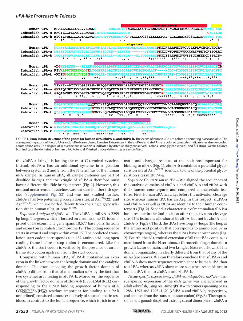
the zfuPA-a kringle is lacking the most C-terminal cysteine.Instead, zfuPA-a has an additional cysteine in a positionbetween cysteines 2 and 3 from the N terminus of the humanuPA kringle. In human uPA, all kringle cysteines are part ofdisulfide bridges and the kringle of zfuPA-a therefore musthave a different disulfide bridge pattern (Fig. 1). However, thisunusual occurrence of cysteines was not seen in other fish spe-cies (supplemental Fig. S2) and was not studied further.zfuPA-a has two potential glycosylation sites, at Asn76/227 andAsn97/248, which are both different from the single glycosyla-tion site in human uPA, at Asn145/302.Sequence Analysis of zfuPA-b—The zfuPA-b mRNA is 2299
bp long. The gene, which is located on chromosome 12, is com-prised of 14 exons. The gene spans 18,577 base pairs (intronsand exons) on zebrafish chromosome 12. The coding sequencestarts in exon 4 and stops within exon 15. The predicted trans-lation start codon corresponds to a 432-amino acid long openreading frame before a stop codon is encountered. Like forzfuPA-b, the start codon is verified by the presence of an in-frame stop codon upstream from the start codon.Compared with human uPA, zfuPA-b contained an extra
exon in the linker between the kringle domain and the catalyticdomain. The exon encoding the growth factor domain ofzfuPA-b differs from that of mammalian uPA by the fact thattwo cysteines are missing in zfuPA-b. Moreover, the sequenceof the growth factor domain of zfuPA-b (LSSSLSGHRLL) cor-responding to the uPAR binding sequence of human uPA(VSNKYFSNIHW; residues important for binding to uPARunderlined) consisted almost exclusively of short aliphatic res-idues, in contrast to the human sequence, which is rich in aro-
matic and charged residues at the positions important forbinding to uPAR (Fig. 1). zfuPA-b contained a potential glyco-sylation site at Asn76/227, identical to one of the potential glyco-sylation sites in zfuPA-a.Sequence Comparison to tPA—We aligned the sequences of
the catalytic domains of zfuPA-a and zfuPA-b and zftPA withtheir human counterparts and compared characteristic fea-tures. First, human uPA has a Lys before the activation cleavagesite, whereas human tPA has an Arg. In this respect, zfuPA-aand zfuPA-b as well as zftPA are identical to their human coun-terparts (Fig. 2). Second, a characteristic of mammalian tPA is abasic residue in the 2nd position after the activation cleavagesite. This feature is also shared by zftPA, but not by zfuPA-a orzfuPA-b (Fig. 2). Third, the tPAs have long 37-loops (the loop atthe amino acid position that corresponds to amino acid 37 inchymotrypsinogen), whereas the uPAs have shorter ones (Fig.2). Fourth, the N-terminal extension of all the tPAs contain, asmentioned from the N terminus, a fibronectin finger domain, agrowth factor domain, and two kringles (data not shown). Thisdomain organization is clearly different from that of any of theuPAs (see above). We can therefore conclude that zfuPA-a andzfuPA-b showmore sequence resemblance to human uPA thanto zftPA, whereas zftPA show more sequence resemblance tohuman tPA than to zfuPA-a and zfuPA-b.Tissue-specific Expression of zfuPA-aand zfuPA-bmRNA—Tis-
sue-specific expression of the uPA genes was characterized inadult zebrafish, using real-timeqPCRwithprimers spanningbases1288–1393 and 1295–1470 (zfuPA-a and zfuPA-b, respectively,and counted fromthe translation start codon) (Fig. 3). The expres-sion in the gonads displayed a strong sexual dimorphism, zfuPA-a
FIGURE 1. Exon-intron structure of the genes for human uPA, zfuPA-a, and zfuPA-b. The exons of human uPA are colored alternating black and blue. Thecorresponding exons of the zfuPA-a and zfuPA-b are colored likewise. Extra exons in the zfuPA-a and zfuPA-b are colored green. Red indicates residues encodedacross splice sites. The degree of sequence conservation is indicated by asterisks (fully conserved), colons (strongly conserved), and full stops (weak). Coloredbars indicate the domains of human uPA. Potential N-linked glycosylation sites are underlined.
uPA-like Proteases in Teleosts
27530 JOURNAL OF BIOLOGICAL CHEMISTRY VOLUME 287 • NUMBER 33 • AUGUST 10, 2012
by guest on July 20, 2015http://w
ww
.jbc.org/D
ownloaded from
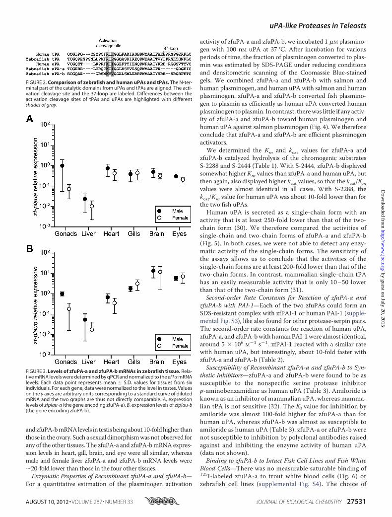
andzfuPA-bmRNAlevels in testisbeingabout10-foldhigher thanthose in theovary. Such a sexual dimorphismwasnot observed forany of the other tissues. The zfuPA-a and zfuPA-bmRNAexpres-sion levels in heart, gill, brain, and eye were all similar, whereasmale and female liver zfuPA-a and zfuPA-b mRNA levels were�20-fold lower than those in the four other tissues.Enzymatic Properties of Recombinant zfuPA-a and zfuPA-b—
For a quantitative estimation of the plasminogen activation
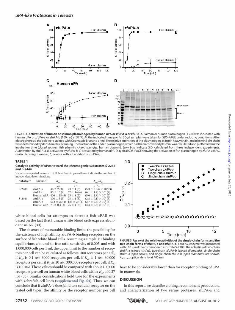
activity of zfuPA-a and zfuPA-b, we incubated 1 �M plasmino-gen with 100 nM uPA at 37 °C. After incubation for variousperiods of time, the fraction of plasminogen converted to plas-min was estimated by SDS-PAGE under reducing conditionsand densitometric scanning of the Coomassie Blue-stainedgels. We combined zfuPA-a and zfuPA-b with salmon andhuman plasminogen, and human uPA with salmon and humanplasminogen. zfuPA-a and zfuPA-b converted fish plasmino-gen to plasmin as efficiently as human uPA converted humanplasminogen to plasmin. In contrast, therewas little if any activ-ity of zfuPA-a and zfuPA-b toward human plasminogen andhuman uPA against salmon plasminogen (Fig. 4). We thereforeconclude that zfuPA-a and zfuPA-b are efficient plasminogenactivators.We determined the Km and kcat values for zfuPA-a and
zfuPA-b catalyzed hydrolysis of the chromogenic substratesS-2288 and S-2444 (Table 1). With S-2444, zfuPA-b displayedsomewhat higher Km values than zfuPA-a and human uPA, butthen again, also displayed higher kcat values, so that the kcat/Kmvalues were almost identical in all cases. With S-2288, thekcat/Km value for human uPA was about 10-fold lower than forthe two fish uPAs.Human uPA is secreted as a single-chain form with an
activity that is at least 250-fold lower than that of the two-chain form (30). We therefore compared the activities ofsingle-chain and two-chain forms of zfuPA-a and zfuPA-b(Fig. 5). In both cases, we were not able to detect any enzy-matic activity of the single-chain forms. The sensitivity ofthe assays allows us to conclude that the activities of thesingle-chain forms are at least 200-fold lower than that of thetwo-chain forms. In contrast, mammalian single-chain tPAhas an easily measurable activity that is only 10–50 lowerthan that of the two-chain form (31).Second-order Rate Constants for Reaction of zfuPA-a and
zfuPA-b with PAI-1—Each of the two zfuPAs could form anSDS-resistant complex with zfPAI-1 or human PAI-1 (supple-mental Fig. S3), like also found for other protease-serpin pairs.The second-order rate constants for reaction of human uPA,zfuPA-a, and zfuPA-bwith humanPAI-1were almost identical,around 5 � 106 M�1 s�1. zfPAI-1 reacted with a similar ratewith human uPA, but interestingly, about 10-fold faster withzfuPA-a and zfuPA-b (Table 2).Susceptibility of Recombinant zfuPA-a and zfuPA-b to Syn-
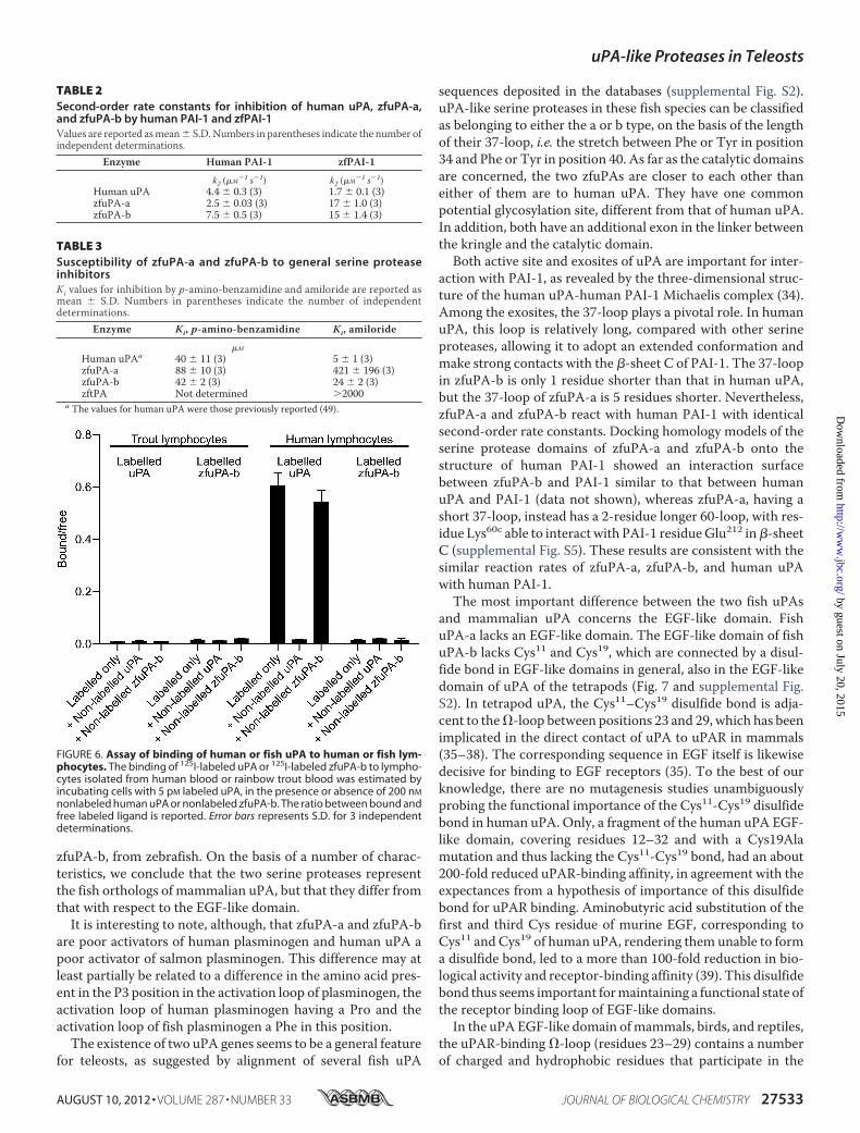
thetic Inhibitors—zfuPA-a and zfuPA-b were found to be assusceptible to the nonspecific serine protease inhibitorp-aminobenzamidine as human uPA (Table 3). Amiloride isknown as an inhibitor of mammalian uPA, whereas mamma-lian tPA is not sensitive (32). The Ki value for inhibition byamiloride was almost 100-fold higher for zfuPA-a than forhuman uPA, whereas zfuPA-b was almost as susceptible toamiloride as human uPA (Table 3). zfuPA-a or zfuPA-b werenot susceptible to inhibition by polyclonal antibodies raisedagainst and inhibiting the enzyme activity of human uPA(data not shown).Binding to zfuPA-b to Intact Fish Cell Lines and Fish White
Blood Cells—There was no measurable saturable binding of125I-labeled zfuPA-a to trout white blood cells (Fig. 6) orzebrafish cell lines (supplemental Fig. S4). The choice of
FIGURE 2. Comparison of zebrafish and human uPAs and tPAs. The N-ter-minal part of the catalytic domains from uPAs and tPAs are aligned. The acti-vation cleavage site and the 37-loop are labeled. Differences between theactivation cleavage sites of tPAs and uPAs are highlighted with differentshades of gray.
FIGURE 3. Levels of zfuPA-a and zfuPA-b mRNAs in zebrafish tissue. Rela-tive mRNA levels were determined by qPCR and normalized to the ef1� mRNAlevels. Each data point represents mean � S.D. values for tissues from sixindividuals. For each gene, data were normalized to the level in testes. Valueson the y axes are arbitrary units corresponding to a standard curve of dilutedmRNA and the two graphs are thus not directly comparable. A, expressionlevels of zfplau-a (the gene encoding zfuPA-a). B, expression levels of zfplau-b(the gene encoding zfuPA-b).
uPA-like Proteases in Teleosts
AUGUST 10, 2012 • VOLUME 287 • NUMBER 33 JOURNAL OF BIOLOGICAL CHEMISTRY 27531
by guest on July 20, 2015http://w
ww
.jbc.org/D
ownloaded from
white blood cells for attempts to detect a fish uPAR wasbased on the fact that human white blood cells express abun-dant uPAR (33).The absence of measurable binding limits the possibility for
the existence of high affinity zfuPA-b binding receptors on thesurface of fish white blood cells. Assuming a simple 1:1 bindingequilibrium, a bound-to-free ratio sensitivity of 0.005, and with1,000,000 cells per 1ml, the upper limit to the number of recep-tors per cell can be calculated as follows: 300 receptors per cell,if KD is 0.1 nM; 3000 receptors per cell, if KD is 1 nM; 30,000receptors per cell, ifKD is 10 nM; 300,000 receptors per cell, ifKDis 100 nM. These values should be comparedwith about 100,000receptors per cell on human white blood cells with a KD of 0.27nM (33). Similar considerations hold true for the experimentswith zebrafish cell lines (supplemental Fig. S4). Thus, we canconclude that if zfuPA-b does bind to a cellular receptor on thetested cell types, the affinity or the receptor number per cell
have to be considerably lower than for receptor binding of uPAin mammals.
DISCUSSION
In this report, we describe cloning, recombinant production,and characterization of two serine proteases, zfuPA-a and
FIGURE 4. Activation of human or salmon plasminogen by human uPA or zfuPA-a or zfuPA-b. Salmon or human plasminogen (1 �M) was incubated withhuman uPA or zfuPA-a or zfuPA-b (100 nM) at 37 °C. At the indicated time points, 30-�l samples were taken for SDS-PAGE under reducing conditions. Afterelectrophoresis, the gels were stained with Coomassie Blue and dried. The relative intensities of the plasminogen, plasmin heavy chain, and plasmin light chainwere determined by densitometric scanning. The fraction of the added plasminogen, which had been converted plasmin, was calculated and plotted versus theincubation time (closed squares, fish plasmin; closed triangles, human plasmin). Error bars indicate S.D. calculated from three independent experiments.A, activation by zfuPA-a. B, activation by zfuPA-b. C, activation by human uPA. D, typical SDS-PAGE showing the activation of fish plasminogen by zfuPA-a (MW,molecular weight marker; C, control without addition of zfuPA-a).
TABLE 1Catalytic activity of uPAs toward the chromogenic substrates S-2288and S-2444Values are reported asmean� S.D. Numbers in parentheses indicate the number ofindependent determinations.
Substrate Enzyme Km kcat kcat/Km
�M s�1 s�1 M�1
S-2288 zfuPA-a 44 � 2 (3) 23 � 1 (3) (5.3 � 0.04) � 105 (3)zfuPA-b 85 � 15 (4) 52 � 14 (4) (6.1 � 1.4) � 105 (4)Human uPA 406 � 18 (5) 23 � 8 (5) (5.6 � 1.9) � 104 (5)
S-2444 zfuPA-a 100 � 3 (3) 28 � 1 (3) (2.8 � 0.1) � 105 (3)zfuPA-b 512 � 25 (4) 136 � 27 (4) (2.7 � 0.6) � 105 (4)Human uPA 73 � 0.4 (3) 25 � 4 (3) (3.4 � 0.5) � 105 (3)
FIGURE 5. Assay of the relative activities of the single-chain forms and thetwo-chain forms of zfuPA-a and zfuPA-b. Four nM enzyme was incubatedwith 100 �M of the chromogenic substrate S-2288. The activities of two-chainzfuPA-a (closed circles), two-chain zfuPA-b (closed diamonds), single-chainzfuPA-a (open circles), and single-chain zfuPA-b (open diamonds) are shown.A405 nm, optical density at 405 nm.
uPA-like Proteases in Teleosts
27532 JOURNAL OF BIOLOGICAL CHEMISTRY VOLUME 287 • NUMBER 33 • AUGUST 10, 2012
by guest on July 20, 2015http://w
ww
.jbc.org/D
ownloaded from
zfuPA-b, from zebrafish. On the basis of a number of charac-teristics, we conclude that the two serine proteases representthe fish orthologs of mammalian uPA, but that they differ fromthat with respect to the EGF-like domain.It is interesting to note, although, that zfuPA-a and zfuPA-b
are poor activators of human plasminogen and human uPA apoor activator of salmon plasminogen. This difference may atleast partially be related to a difference in the amino acid pres-ent in the P3 position in the activation loop of plasminogen, theactivation loop of human plasminogen having a Pro and theactivation loop of fish plasminogen a Phe in this position.The existence of two uPA genes seems to be a general feature
for teleosts, as suggested by alignment of several fish uPA
sequences deposited in the databases (supplemental Fig. S2).uPA-like serine proteases in these fish species can be classifiedas belonging to either the a or b type, on the basis of the lengthof their 37-loop, i.e. the stretch between Phe or Tyr in position34 and Phe or Tyr in position 40. As far as the catalytic domainsare concerned, the two zfuPAs are closer to each other thaneither of them are to human uPA. They have one commonpotential glycosylation site, different from that of human uPA.In addition, both have an additional exon in the linker betweenthe kringle and the catalytic domain.Both active site and exosites of uPA are important for inter-
action with PAI-1, as revealed by the three-dimensional struc-ture of the human uPA-human PAI-1 Michaelis complex (34).Among the exosites, the 37-loop plays a pivotal role. In humanuPA, this loop is relatively long, compared with other serineproteases, allowing it to adopt an extended conformation andmake strong contacts with the �-sheet C of PAI-1. The 37-loopin zfuPA-b is only 1 residue shorter than that in human uPA,but the 37-loop of zfuPA-a is 5 residues shorter. Nevertheless,zfuPA-a and zfuPA-b react with human PAI-1 with identicalsecond-order rate constants. Docking homology models of theserine protease domains of zfuPA-a and zfuPA-b onto thestructure of human PAI-1 showed an interaction surfacebetween zfuPA-b and PAI-1 similar to that between humanuPA and PAI-1 (data not shown), whereas zfuPA-a, having ashort 37-loop, instead has a 2-residue longer 60-loop, with res-idue Lys60c able to interact with PAI-1 residueGlu212 in�-sheetC (supplemental Fig. S5). These results are consistent with thesimilar reaction rates of zfuPA-a, zfuPA-b, and human uPAwith human PAI-1.The most important difference between the two fish uPAs
and mammalian uPA concerns the EGF-like domain. FishuPA-a lacks an EGF-like domain. The EGF-like domain of fishuPA-b lacks Cys11 and Cys19, which are connected by a disul-fide bond in EGF-like domains in general, also in the EGF-likedomain of uPA of the tetrapods (Fig. 7 and supplemental Fig.S2). In tetrapod uPA, the Cys11–Cys19 disulfide bond is adja-cent to the�-loop between positions 23 and 29, which has beenimplicated in the direct contact of uPA to uPAR in mammals(35–38). The corresponding sequence in EGF itself is likewisedecisive for binding to EGF receptors (35). To the best of ourknowledge, there are no mutagenesis studies unambiguouslyprobing the functional importance of the Cys11-Cys19 disulfidebond in human uPA. Only, a fragment of the human uPA EGF-like domain, covering residues 12–32 and with a Cys19Alamutation and thus lacking the Cys11-Cys19 bond, had an about200-fold reduced uPAR-binding affinity, in agreement with theexpectances from a hypothesis of importance of this disulfidebond for uPAR binding. Aminobutyric acid substitution of thefirst and third Cys residue of murine EGF, corresponding toCys11 and Cys19 of human uPA, rendering them unable to forma disulfide bond, led to a more than 100-fold reduction in bio-logical activity and receptor-binding affinity (39). This disulfidebond thus seems important formaintaining a functional state ofthe receptor binding loop of EGF-like domains.In the uPA EGF-like domain ofmammals, birds, and reptiles,
the uPAR-binding �-loop (residues 23–29) contains a numberof charged and hydrophobic residues that participate in the
TABLE 2Second-order rate constants for inhibition of human uPA, zfuPA-a,and zfuPA-b by human PAI-1 and zfPAI-1Values are reported asmean� S.D. Numbers in parentheses indicate the number ofindependent determinations.
Enzyme Human PAI-1 zfPAI-1
k2 (�M�1 s�1) k2 (�M�1 s�1)Human uPA 4.4 � 0.3 (3) 1.7 � 0.1 (3)zfuPA-a 2.5 � 0.03 (3) 17 � 1.0 (3)zfuPA-b 7.5 � 0.5 (3) 15 � 1.4 (3)
TABLE 3Susceptibility of zfuPA-a and zfuPA-b to general serine proteaseinhibitorsKi values for inhibition by p-amino-benzamidine and amiloride are reported asmean � S.D. Numbers in parentheses indicate the number of independentdeterminations.
Enzyme Ki, p-amino-benzamidine Ki, amiloride
�M
Human uPAa 40 � 11 (3) 5 � 1 (3)zfuPA-a 88 � 10 (3) 421 � 196 (3)zfuPA-b 42 � 2 (3) 24 � 2 (3)zftPA Not determined 2000
a The values for human uPA were those previously reported (49).
FIGURE 6. Assay of binding of human or fish uPA to human or fish lym-phocytes. The binding of 125I-labeled uPA or 125I-labeled zfuPA-b to lympho-cytes isolated from human blood or rainbow trout blood was estimated byincubating cells with 5 pM labeled uPA, in the presence or absence of 200 nM
nonlabeled human uPA or nonlabeled zfuPA-b. The ratio between bound andfree labeled ligand is reported. Error bars represents S.D. for 3 independentdeterminations.
uPA-like Proteases in Teleosts
AUGUST 10, 2012 • VOLUME 287 • NUMBER 33 JOURNAL OF BIOLOGICAL CHEMISTRY 27533
by guest on July 20, 2015http://w
ww
.jbc.org/D
ownloaded from
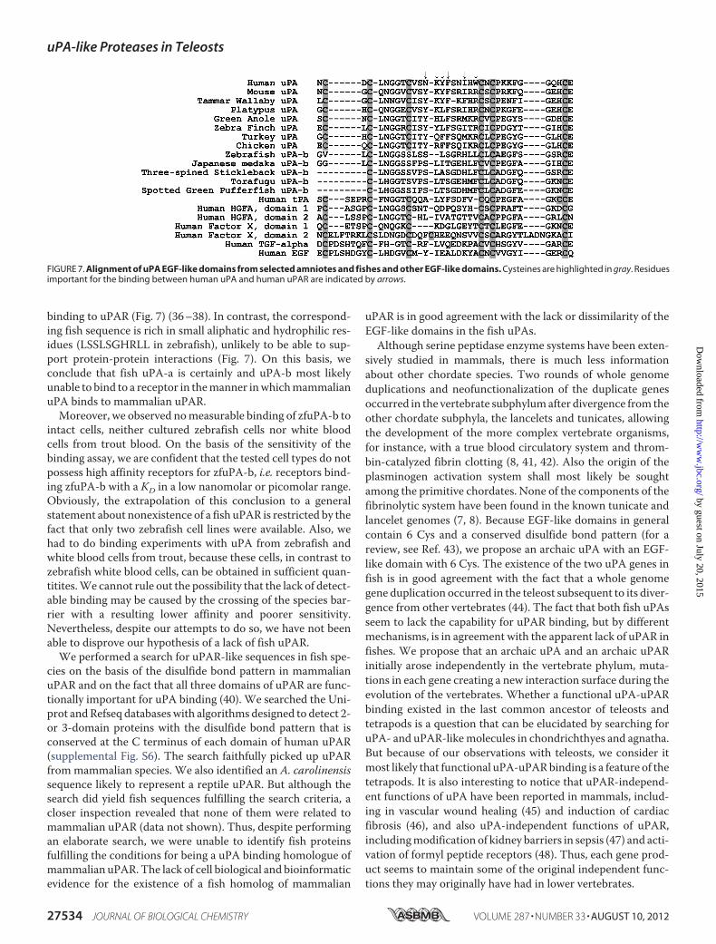
binding to uPAR (Fig. 7) (36–38). In contrast, the correspond-ing fish sequence is rich in small aliphatic and hydrophilic res-idues (LSSLSGHRLL in zebrafish), unlikely to be able to sup-port protein-protein interactions (Fig. 7). On this basis, weconclude that fish uPA-a is certainly and uPA-b most likelyunable to bind to a receptor in themanner inwhichmammalianuPA binds to mammalian uPAR.Moreover, we observed nomeasurable binding of zfuPA-b to
intact cells, neither cultured zebrafish cells nor white bloodcells from trout blood. On the basis of the sensitivity of thebinding assay, we are confident that the tested cell types do notpossess high affinity receptors for zfuPA-b, i.e. receptors bind-ing zfuPA-b with a KD in a low nanomolar or picomolar range.Obviously, the extrapolation of this conclusion to a generalstatement about nonexistence of a fish uPAR is restricted by thefact that only two zebrafish cell lines were available. Also, wehad to do binding experiments with uPA from zebrafish andwhite blood cells from trout, because these cells, in contrast tozebrafish white blood cells, can be obtained in sufficient quan-titites.We cannot rule out the possibility that the lack of detect-able binding may be caused by the crossing of the species bar-rier with a resulting lower affinity and poorer sensitivity.Nevertheless, despite our attempts to do so, we have not beenable to disprove our hypothesis of a lack of fish uPAR.We performed a search for uPAR-like sequences in fish spe-
cies on the basis of the disulfide bond pattern in mammalianuPAR and on the fact that all three domains of uPAR are func-tionally important for uPA binding (40). We searched the Uni-prot andRefseq databaseswith algorithms designed to detect 2-or 3-domain proteins with the disulfide bond pattern that isconserved at the C terminus of each domain of human uPAR(supplemental Fig. S6). The search faithfully picked up uPARfrommammalian species. We also identified an A. carolinensissequence likely to represent a reptile uPAR. But although thesearch did yield fish sequences fulfilling the search criteria, acloser inspection revealed that none of them were related tomammalian uPAR (data not shown). Thus, despite performingan elaborate search, we were unable to identify fish proteinsfulfilling the conditions for being a uPA binding homologue ofmammalian uPAR. The lack of cell biological and bioinformaticevidence for the existence of a fish homolog of mammalian
uPAR is in good agreement with the lack or dissimilarity of theEGF-like domains in the fish uPAs.Although serine peptidase enzyme systems have been exten-
sively studied in mammals, there is much less informationabout other chordate species. Two rounds of whole genomeduplications and neofunctionalization of the duplicate genesoccurred in the vertebrate subphylumafter divergence from theother chordate subphyla, the lancelets and tunicates, allowingthe development of the more complex vertebrate organisms,for instance, with a true blood circulatory system and throm-bin-catalyzed fibrin clotting (8, 41, 42). Also the origin of theplasminogen activation system shall most likely be soughtamong the primitive chordates. None of the components of thefibrinolytic system have been found in the known tunicate andlancelet genomes (7, 8). Because EGF-like domains in generalcontain 6 Cys and a conserved disulfide bond pattern (for areview, see Ref. 43), we propose an archaic uPA with an EGF-like domain with 6 Cys. The existence of the two uPA genes infish is in good agreement with the fact that a whole genomegene duplication occurred in the teleost subsequent to its diver-gence from other vertebrates (44). The fact that both fish uPAsseem to lack the capability for uPAR binding, but by differentmechanisms, is in agreement with the apparent lack of uPAR infishes. We propose that an archaic uPA and an archaic uPARinitially arose independently in the vertebrate phylum, muta-tions in each gene creating a new interaction surface during theevolution of the vertebrates. Whether a functional uPA-uPARbinding existed in the last common ancestor of teleosts andtetrapods is a question that can be elucidated by searching foruPA- and uPAR-likemolecules in chondrichthyes and agnatha.But because of our observations with teleosts, we consider itmost likely that functional uPA-uPARbinding is a feature of thetetrapods. It is also interesting to notice that uPAR-independ-ent functions of uPA have been reported in mammals, includ-ing in vascular wound healing (45) and induction of cardiacfibrosis (46), and also uPA-independent functions of uPAR,includingmodification of kidney barriers in sepsis (47) and acti-vation of formyl peptide receptors (48). Thus, each gene prod-uct seems to maintain some of the original independent func-tions they may originally have had in lower vertebrates.
FIGURE 7. Alignment of uPA EGF-like domains from selected amniotes and fishes and other EGF-like domains. Cysteines are highlighted in gray. Residuesimportant for the binding between human uPA and human uPAR are indicated by arrows.
uPA-like Proteases in Teleosts
27534 JOURNAL OF BIOLOGICAL CHEMISTRY VOLUME 287 • NUMBER 33 • AUGUST 10, 2012
by guest on July 20, 2015http://w
ww
.jbc.org/D
ownloaded from
The results presented here suggest that the focus on the func-tions of plasminogen activation in mammals, due to its rele-vance to human pathophysiology and the availability of tar-geted gene deletions in mice, may have given a too narrowpicture of the mechanism of action of the system. Studies innonmammalian species are needed to obtain a comprehensiveunderstanding of the biochemistry and cell biology of plasmin-ogen activation. The two fish gene products described fulfill aminimum requirement for having a biological function, namelyby being expressed in the fish. Obviously, establishment of theirexact biological functions will have to await functional studieswith specific gene deletions and exact determinations of celltypes expressing them.REFERENCES1. Collen, D. (1999) The plasminogen (fibrinolytic) system. Thromb. Hae-
most. 82, 259–2702. Andreasen, P. A., Kjøller, L., Christensen, L., and Duffy, M. J. (1997) The
urokinase-type plasminogen activator system in cancer metastasis. A re-view. Int. J. Cancer 72, 1–22
3. Kjaergaard, M., Hansen, L.V., Jacobsen, B., Gardsvoll, H., and Ploug, M.(2008) Structure and ligand interactions of the urokinase receptor (uPAR).Front. Biosci. 13, 5441–5461
4. Dupont, D. M., Madsen, J. B., Kristensen, T., Bodker, J.S., Blouse, G. E.,Wind, T., and Andreasen, P. A. (2009) Biochemical properties of plas-minogen activator inhibitor-1. Front. Biosci. 14, 1337–1361
5. Gliemann, J., Nykjaer, A., Petersen, C. M., Jørgensen, K. E., Nielsen, M.,Andreasen, P. A., Christensen, E. I., Lookene, A., Olivecrona, G., andMoestrup, S. K. (1994) The multiligand �2-macroglobulin receptor/lowdensity lipoprotein receptor-related protein (�2MR/LRP). Ann. N.Y.Acad. Sci. 737, 20–38
6. Appella, E., Weber, I. T., and Blasi, F. (1988) Structure and function ofepidermal growth factor-like regions in proteins. FEBS Lett. 231, 1–4
7. Jiang, Y., and Doolittle, R. F. (2003) The evolution of vertebrate bloodcoagulation as viewed from a comparison of pufferfish and sea squirtgenomes. Proc. Natl. Acad. Sci. U.S.A. 100, 7527–7532
8. Doolittle, R. F. (2009) Step-by-step evolution of vertebrate blood coagula-tion. Cold Spring Harbor Symp. Quant. Biol. 74, 35–40
9. Takahashi, K., Wakamatsu, Y., Ozato, K., and Wakayama, Y. (1987) Fishplasminogen activators, their identification and characterization. CellStruct. Funct. 12, 11–22
10. Sallés, F. J., Schechter, N., and Strickland, S. (1990) A plasminogen-acti-vator is induced during goldfish optic nerve regeneration. EMBO J. 9,2471–2477
11. Wheelan, S. J., Church, D. M., and Ostell, J. M. (2001) Spidey. A tool formRNA to genomic alignments. Genome Res. 11, 1952–1957
12. Edgar, R. C. (2004) MUSCLE, multiple sequence alignment with highaccuracy and high throughput. Nucleic Acids Res. 32, 1792–1797
13. Alberti,M., Kausch,U.,Haindl, S., Leibiger, R., Budczies, J., Seifert,M., andHock, B. (2005) Gene expression patterns, a tool for bioanalysis. Int. J.Environ. Anal. Chem. 85, 589–608
14. Durocher, Y., Perret, S., and Kamen, A. (2002) High-level and high-throughput recombinant protein production by transient transfection ofsuspension-growing human 293-EBNA1 cells. Nucleic Acids Res. 30, E9
15. Tom, R., Bisson, L., and Durocher, Y. (2008) Transfection of HEK293-EBNA1 cells in suspension with linear PEI for production of recombinantproteins. CSH Protoc. 2008, 10.1101/pdb.prot4977
16. Gasteiger, E., Hoogland, C., Gattiker, A., Duvaud, S.,Wilkins,M. R., Appel,R. D., and Bairoch, A. (2005) Protein identification and analysis tools onthe ExPASy server. The Proteomics Protocols Handbook, pp. 571–607,Humana Press, Inc., NJ
17. Jensen, P. H., Christensen, E. I., Ebbesen, P., Gliemann, J., and Andreasen,P. A. (1990) Lysosomal degradation of receptor-bound urokinase-typeplasminogen activator is enhanced by its inhibitors in human trophoblas-tic choriocarcinoma cells. Cell Regul. 1, 1043–1056
18. Madsen, J. B., Dupont, D.M., Andersen, T. B., Nielsen, A. F., Sang, L., Brix,
D. M., Jensen, J. K., Broos, T., Hendrickx, M. L., Christensen, A., Kjems, J.,and Andreasen, P. A. (2010) RNA aptamers as conformational probes andregulatory agents for plasminogen activator inhibitor-1. Biochemistry 49,4103–4115
19. Blouse, G. E., Perron, M. J., Kvassman, J. O., Yunus, S., Thompson, J. H.,Betts, R. L., Lutter, L. C., and Shore, J. D. (2003) Mutation of the highlyconserved tryptophan in the serpin breach region alters the inhibitorymechanism of plasminogen activator inhibitor-1. Biochemistry 42,12260–12272
20. Deutsch, D. G., and Mertz, E. T. (1970) Plasminogen. Purification fromhuman plasma by affinity chromatography. Science 170, 1095–1096
21. Futamura, A., and Gettins, P. G. (2000) Serine 380 (P14) 3 glutamatemutation activates antithrombin as an inhibitor of factor Xa. J. Biol. Chem.275, 4092–4098
22. Driever, W., and Rangini, Z. (1993) Characterization of a cell line derivedfromzebrafish (Brachydanio rerio) embryos. InVitroCell. Dev. Biol. Anim.29, 749–754
23. Culp, P. A. (1994) RandomDNA integrations as an approach to insertionalmutagenesis in the zebrafish (Brachydanio rerio). Ph.D. thesis, Massachu-setts Institute of Technology
24. Braun-Nesje, R., Bertheussen, K., Kaplan, G., and Seljelid, R. (1981) Sal-monid macrophages. Separation, in vitro culture, and characterization. J.Fish Dis. 4, 141–151
25. Pettersen, E. F., Bjerknes, R., andWergeland, H. I. (2000) Studies of Atlan-tic salmon (Salmo salar L.) blood, spleen, and head kidney leucocytesusing specific monoclonal antibodies, immunohistochemistry and flowcytometry. Fish Shellfish Immunol. 10, 695–710
26. Arnold, K., Bordoli, L., Kopp, J., and Schwede, T. (2006) The SWISS-MODEL workspace. A web-based environment for protein structure ho-mology modeling. Bioinformatics 22, 195–201
27. UniProt, C. (2011) Ongoing and future developments at the UniversalProtein Resource. Nucleic Acids Res. 39, D214–219
28. Pruitt, K. D., Tatusova, T., and Maglott, D. R. (2005) NCBI referencesequence (RefSeq). A curated nonredundant sequence database of ge-nomes, transcripts, and proteins. Nucleic Acids Res. 33, D501–504
29. Dsouza, M., Larsen, N., and Overbeek, R. (1997) Searching for patterns ingenomic data. Trends Genet. 13, 497–498
30. Petersen, L. C., Lund, L. R., Nielsen, L. S., Danø, K., and Skriver, L. (1988)One-chain urokinase-type plasminogen activator from human sarcomacells is a proenzyme with little or no intrinsic activity. J. Biol. Chem. 263,11189–11195
31. Andreasen, P. A., Petersen, L. C., andDanø, K. (1991) Diversity in catalyticproperties of single chain and two chain tissue-type plasminogen activa-tor. Fibrinolysis 5, 207–215
32. Vassalli, J. D., and Belin, D. (1987) Amiloride selectively inhibits the uroki-nase-type plasminogen activator. FEBS Lett. 214, 187–191
33. Ploug, M., Ostergaard, S., Hansen, L. B., Holm, A., and Danø, K. (1998)Photoaffinity labeling of the human receptor for urokinase-type plas-minogen activator using a decapeptide antagonist. Evidence for a com-posite ligand-binding site and a short interdomain separation. Bio-chemistry 37, 3612–3622
34. Lin, Z., Jiang, L., Yuan, C., Jensen, J. K., Zhang, X., Luo, Z., Furie, B. C.,Furie, B., Andreasen, P. A., and Huang, M. (2011) Structural basis forrecognition of urokinase-type plasminogen activator by plasminogen ac-tivator inhibitor-1. J. Biol. Chem. 286, 7027–7032
35. Appella, E., Robinson, E. A., Ullrich, S. J., Stoppelli, M. P., Corti, A., Cas-sani, G., and Blasi, F. (1987) The receptor-binding sequence of urokinase.A biological function for the growth factor module of proteases. J. Biol.Chem. 262, 4437–4440
36. Huai, Q.,Mazar, A. P., Kuo, A., Parry, G. C., Shaw, D. E., Callahan, J., Li, Y.,Yuan, C., Bian, C., Chen, L., Furie, B., Furie, B. C., Cines, D. B., and Huang,M. (2006) Structure of human urokinase plasminogen activator in com-plex with its receptor. Science 311, 656–659
37. Lin, L., Gårdsvoll, H., Huai, Q., Huang, M., and Ploug, M. (2010) Struc-ture-based engineering of species selectivity in the interaction betweenurokinase and its receptor. Implication for preclinical cancer therapy.J. Biol. Chem. 285, 10982–10992
38. Barinka, C., Parry, G., Callahan, J., Shaw, D. E., Kuo, A., Bdeir, K., Cines,
uPA-like Proteases in Teleosts
AUGUST 10, 2012 • VOLUME 287 • NUMBER 33 JOURNAL OF BIOLOGICAL CHEMISTRY 27535
by guest on July 20, 2015http://w
ww
.jbc.org/D
ownloaded from
D. B., Mazar, A., and Lubkowski, J. (2006) Structural basis of interactionbetween urokinase-type plasminogen activator and its receptor. J. Mol.Biol. 363, 482–495
39. Barnham, K. J., Torres, A. M., Alewood, D., Alewood, P. F., Domagala, T.,Nice, E. C., andNorton, R. S. (1998) Role of the 6–20 disulfide bridge in thestructure and activity of epidermal growth factor. Protein Sci. 7,1738–1749
40. Behrendt, N., Ronne, E., and Dano, K. (1996) Domain interplay in theurokinase receptor. Requirement for the third domain in high affinityligand binding and demonstration of ligand contact sites in distinct recep-tor domains. J. Biol. Chem. 271, 22885–22894
41. Pébusque, M. J., Coulier, F., Birnbaum, D., and Pontarotti, P. (1998) An-cient large-scale genome duplications. Phylogenetic and linkage analysesshed light on chordate genome evolution.Mol. Biol. Evol. 15, 1145–1159
42. Putnam, N. H., Butts, T., Ferrier, D.E., Furlong, R. F., Hellsten, U., Ka-washima, T., Robinson-Rechavi, M., Shoguchi, E., Terry, A., Yu, J. K.,Benito-Gutiérrez, E. L., Dubchak, I., Garcia-Fernàndez, J., Gibson-Brown,J. J., Grigoriev, I. V., Horton, A. C., de Jong, P. J., Jurka, J., Kapitonov, V. V.,Kohara, Y., Kuroki, Y., Lindquist, E., Lucas, S., Osoegawa, K., Pennacchio,L. A., Salamov, A. A., Satou, Y., Sauka-Spengler, T., Schmutz, J., Shin-I, T.,Toyoda, A., Bronner-Fraser, M., Fujiyama, A., Holland, L. Z., Holland,P.W., Satoh,N., andRokhsar, D. S. (2008)The amphioxus genome and theevolution of the chordate karyotype. Nature 453, 1064–1071
43. Wouters, M. A., Rigoutsos, I., Chu, C. K., Feng, L. L., Sparrow, D. B., andDunwoodie, S. L. (2005) Evolution of distinct EGF domains with specificfunctions. Protein Sci. 14, 1091–1103
44. Jaillon, O., Aury, J. M., Brunet, F., Petit, J. L., Stange-Thomann, N.,Mauceli, E., Bouneau, L., Fischer, C.,Ozouf-Costaz, C., Bernot, A.,Nicaud,S., Jaffe, D., Fisher, S., Lutfalla, G., Dossat, C., Segurens, B., Dasilva, C.,Salanoubat, M., Levy, M., Boudet, N., Castellano, S., Anthouard, V., Jubin,
C., Castelli, V., Katinka, M., Vacherie, B., Biémont, C., Skalli, Z., Cattolico,L., Poulain, J., De Berardinis, V., Cruaud, C., Duprat, S., Brottier, P., Cou-tanceau, J. P., Gouzy, J., Parra, G., Lardier, G., Chapple, C.,McKernan, K. J.,McEwan, P., Bosak, S., Kellis, M., Volff, J. N., Guigó, R., Zody, M. C.,Mesirov, J., Lindblad-Toh, K., Birren, B., Nusbaum, C., Kahn, D., Robin-son-Rechavi, M., Laudet, V., Schachter, V., Quetier, F., Saurin, W., Scar-pelli, C.,Wincker, P., Lander, E. S., Weissenbach, J., and Roest Crollius, H.(2004) Genome duplication in the teleost fish Tetraodon nigroviridis re-veals the early vertebrate proto-karyotype. Nature 431, 946–957
45. Carmeliet, P., Moons, L., Dewerchin, M., Rosenberg, S., Herbert, J. M.,Lupu, F., and Collen, D. (1998) Receptor-independent role of urokinase-type plasminogen activator in pericellular plasmin andmatrixmetallopro-teinase proteolysis during vascularwoundhealing inmice. J. Cell Biol.140,233–245
46. Stempien-Otero, A., Plawman, A., Meznarich, J., Dyamenahalli, T., Ot-suka, G., and Dichek, D. A. (2006)Mechanisms of cardiac fibrosis inducedby urokinase plasminogen activator. J. Biol. Chem. 281, 15345–15351
47. Wei, C., Möller, C. C., Altintas, M. M., Li, J., Schwarz, K., Zacchigna, S.,Xie, L., Henger, A., Schmid, H., Rastaldi, M. P., Cowan, P., Kretzler, M.,Parrilla, R., Bendayan, M., Gupta, V., Nikolic, B., Kalluri, R., Carmeliet, P.,Mundel, P., andReiser, J. (2008)Modification of kidney barrier function bythe urokinase receptor. Nat. Med. 14, 55–63
48. Rivière, S., Challet, L., Fluegge, D., Spehr, M., and Rodriguez, I. (2009)Formyl peptide receptor-like proteins are a novel family of vomeronasalchemosensors. Nature 459, 574–577
49. Bøtkjaer, K. A., Byszuk, A. A., Andersen, L. M., Christensen, A., Andrea-sen, P.A., and Blouse, G. E. (2009) Nonproteolytic induction of catalyticactivity into the single-chain form of urokinase-type plasminogen activa-tor by dipeptides. Biochemistry 48, 9606–9617
uPA-like Proteases in Teleosts
27536 JOURNAL OF BIOLOGICAL CHEMISTRY VOLUME 287 • NUMBER 33 • AUGUST 10, 2012
by guest on July 20, 2015http://w
ww
.jbc.org/D
ownloaded from
Michael Ploug and Peter A. AndreasenHuang,Niels Larsen, Erik Baatrup, Mingdong
Lisbeth M. Andersen, Jesper S. Johansen,Jensen, Agnieszka Szczur, Anni Christensen, René Bager, Thomas K. Kristensen, Jan K. Factor-like DomainsReceptor-binding Epidermal GrowthProteases in Teleosts Lack Genuine Urokinase-type Plasminogen Activator-likeEnzymology:
doi: 10.1074/jbc.M112.369207 originally published online June 25, 20122012, 287:27526-27536.J. Biol. Chem.
10.1074/jbc.M112.369207Access the most updated version of this article at doi:
.JBC Affinity SitesFind articles, minireviews, Reflections and Classics on similar topics on the
Alerts:
When a correction for this article is posted•
When this article is cited•
to choose from all of JBC's e-mail alertsClick here
Supplemental material:
http://www.jbc.org/content/suppl/2012/06/25/M112.369207.DC1.html
http://www.jbc.org/content/287/33/27526.full.html#ref-list-1
This article cites 47 references, 19 of which can be accessed free at
by guest on July 20, 2015http://w
ww
.jbc.org/D
ownloaded from