
Formation of oral and pharyngeal dentition in teleosts depends on differential recruitment of...
12
The FASEB Journal • Research Communication Formation of oral and pharyngeal dentition in teleosts depends on differential recruitment of retinoic acid signaling Yann Gibert,* Laure Bernard,* Melanie Debiais-Thibaud, † Franck Bourrat, ‡ Jean-Stephane Joly, ‡ Karen Pottin, § Axel Meyer, ¶ Sylvie Retaux, § David W. Stock, William R. Jackman, # Pawat Seritrakul, # Gerrit Begemann, ¶ and Vincent Laudet* ,1 *Molecular Zoology Group, Institut de Ge ´nomique Fonctionnelle de Lyon, Universite ´ de Lyon, Centre National de la Recherche Scientifique (CNRS), Institut National de la Recherche Agronomique (INRA), UCB Lyon 1, Ecole Normale Supe ´rieure de Lyon, Lyon France; † Equipe Genome Developpement et Evolution UPR 9034, CNRS, Gif-sur-Yvette, France; ‡ INRA U1126 Group and § Equipe DECA, UPR 3294, Institut de Neurobiologie A. Fessard, CNRS, Gif-Sur-Yvette, France; ¶ Zoology and Evolutionary Biology, Department of Biology, University of Konstanz, Konstanz, Germany; Department of Ecology and Evolutionary Biology, University of Colorado, Boulder, Colorado, USA; and # Biology Department, Bowdoin College, Brunswick, Maine, USA ABSTRACT One of the goals of evolutionary devel- opmental biology is to link specific adaptations to changes in developmental pathways. The dentition of cypriniform fishes, which in contrast to many other teleost fish species possess pharyngeal teeth but lack oral teeth, provides a suitable model to study the development of feeding adaptations. Here, we have examined the involvement of retinoic acid (RA) in tooth development and show that RA is specifically required to induce the pharyngeal tooth developmen- tal program in zebrafish. Perturbation of RA signal- ing at this stage abolished tooth induction without affecting the development of tooth-associated cera- tobranchial bones. We show that this inductive event is dependent on RA synthesis from aldh1a2 in the ventral posterior pharynx. Fibroblast growth factor (FGF) signaling has been shown to be critical for tooth induction in zebrafish, and its loss has been associated with oral tooth loss in cypriniform fishes. Pharmacological treatments targeting the RA and FGF pathways revealed that both pathways act inde- pendently during tooth induction. In contrast, we find that in Mexican tetra and medaka, species that also possess oral teeth, both oral and pharyngeal teeth are induced independently of RA. Our analyses suggest an evolutionary scenario in which the gene network controlling tooth development obtained RA dependency in the lineage leading to the cyprini- forms. The loss of pharyngeal teeth in this group was cancelled out through a shift in aldh1a2 expression, while oral teeth might have been lost ultimately due to deficient RA signaling in the oral cavity.—Gibert, Y., Bernard, L., Debiais-Thibaud, M., Bourrat, F., Joly, J.-S., Pottin, K., Meyer, A., Retaux, S., Stock, D. W., Jackman, W. R., Seritrakul, P., Begemann, G., Laudet, V. Formation of oral and pharyngeal denti- tion in teleosts depends on differential recruitment of retinoic acid signaling. FASEB J. 24, 3298 –3309 (2010). www.fasebj.org Key Words: tooth Astyanax FGF zebrafish Teeth are a vertebrate innovation that not only display a huge variability in terms of localization, num- ber, shape, and size but also exhibit a conserved mode of development in most vertebrates (1). It is believed that the teeth of teleost fish have the same dual epithelio-mesenchymal origin as mammals and that neural crest cells (NCCs) are the source of the mesen- chyme implicated in tooth development (2). The function and expression of genes required for tooth development have been intensively studied in the mouse (3, 4). In contrast to mammals, the study of actinopterygian fish allows a better understanding of the general mechanisms controlling the development of teeth at additional sites within the oral cavity (re- viewed in ref. 5). In actinopterygian fishes, there is extensive variation in tooth location, i.e., teeth may be present in upper and lower jaw margins, palate, floor of the mouth, as well as upper and lower pharyngeal surfaces (5). The dentition of actinopterygian fish therefore provides a unique model system to decipher the mechanisms controlling tooth location and its variation during evolution. For example, the 3 main developmental models for tooth development (the zebrafish Danio rerio, a cypriniform; the Mexican tetra 1 Correspondence: Molecular Zoology Group, Institut de Ge ´nomique Fonctionnelle de Lyon, Universite ´ de Lyon, CNRS, INRA, UCB Lyon 1, Ecole Normale Supe ´rieure de Lyon, 69364 Lyon Cedex 07, France. E-mail: vincent. [email protected] doi: 10.1096/fj.09-147488 3298 0892-6638/10/0024-3298 © FASEB
Transcript of Formation of oral and pharyngeal dentition in teleosts depends on differential recruitment of...
The FASEB Journal • Research Communication
Formation of oral and pharyngeal dentition in teleostsdepends on differential recruitment of retinoic acidsignaling
Yann Gibert,* Laure Bernard,* Melanie Debiais-Thibaud,† Franck Bourrat,‡
Jean-Stephane Joly,‡ Karen Pottin,§ Axel Meyer,¶ Sylvie Retaux,§ David W. Stock,�
William R. Jackman,# Pawat Seritrakul,# Gerrit Begemann,¶ and Vincent Laudet*,1
*Molecular Zoology Group, Institut de Genomique Fonctionnelle de Lyon, Universite de Lyon,Centre National de la Recherche Scientifique (CNRS), Institut National de la RechercheAgronomique (INRA), UCB Lyon 1, Ecole Normale Superieure de Lyon, Lyon France; †EquipeGenome Developpement et Evolution UPR 9034, CNRS, Gif-sur-Yvette, France; ‡INRA U1126 Groupand §Equipe DECA, UPR 3294, Institut de Neurobiologie A. Fessard, CNRS, Gif-Sur-Yvette, France;¶Zoology and Evolutionary Biology, Department of Biology, University of Konstanz, Konstanz,Germany; �Department of Ecology and Evolutionary Biology, University of Colorado, Boulder,Colorado, USA; and #Biology Department, Bowdoin College, Brunswick, Maine, USA
ABSTRACT One of the goals of evolutionary devel-opmental biology is to link specific adaptations tochanges in developmental pathways. The dentition ofcypriniform fishes, which in contrast to many otherteleost fish species possess pharyngeal teeth but lackoral teeth, provides a suitable model to study thedevelopment of feeding adaptations. Here, we haveexamined the involvement of retinoic acid (RA) intooth development and show that RA is specificallyrequired to induce the pharyngeal tooth developmen-tal program in zebrafish. Perturbation of RA signal-ing at this stage abolished tooth induction withoutaffecting the development of tooth-associated cera-tobranchial bones. We show that this inductive eventis dependent on RA synthesis from aldh1a2 in theventral posterior pharynx. Fibroblast growth factor(FGF) signaling has been shown to be critical fortooth induction in zebrafish, and its loss has beenassociated with oral tooth loss in cypriniform fishes.Pharmacological treatments targeting the RA andFGF pathways revealed that both pathways act inde-pendently during tooth induction. In contrast, wefind that in Mexican tetra and medaka, species thatalso possess oral teeth, both oral and pharyngealteeth are induced independently of RA. Our analysessuggest an evolutionary scenario in which the genenetwork controlling tooth development obtained RAdependency in the lineage leading to the cyprini-forms. The loss of pharyngeal teeth in this group wascancelled out through a shift in aldh1a2 expression,while oral teeth might have been lost ultimately dueto deficient RA signaling in the oral cavity.—Gibert,Y., Bernard, L., Debiais-Thibaud, M., Bourrat, F.,Joly, J.-S., Pottin, K., Meyer, A., Retaux, S., Stock, D.W., Jackman, W. R., Seritrakul, P., Begemann, G.,Laudet, V. Formation of oral and pharyngeal denti-tion in teleosts depends on differential recruitment
of retinoic acid signaling. FASEB J. 24, 3298 –3309(2010). www.fasebj.org
Key Words: tooth � Astyanax � FGF � zebrafish
Teeth are a vertebrate innovation that not onlydisplay a huge variability in terms of localization, num-ber, shape, and size but also exhibit a conserved modeof development in most vertebrates (1). It is believedthat the teeth of teleost fish have the same dualepithelio-mesenchymal origin as mammals and thatneural crest cells (NCCs) are the source of the mesen-chyme implicated in tooth development (2).
The function and expression of genes required fortooth development have been intensively studied in themouse (3, 4). In contrast to mammals, the study ofactinopterygian fish allows a better understanding ofthe general mechanisms controlling the developmentof teeth at additional sites within the oral cavity (re-viewed in ref. 5). In actinopterygian fishes, there isextensive variation in tooth location, i.e., teeth may bepresent in upper and lower jaw margins, palate, floor ofthe mouth, as well as upper and lower pharyngealsurfaces (5). The dentition of actinopterygian fishtherefore provides a unique model system to decipherthe mechanisms controlling tooth location and itsvariation during evolution. For example, the 3 maindevelopmental models for tooth development (thezebrafish Danio rerio, a cypriniform; the Mexican tetra
1 Correspondence: Molecular Zoology Group, Institut deGenomique Fonctionnelle de Lyon, Universite de Lyon,CNRS, INRA, UCB Lyon 1, Ecole Normale Superieure deLyon, 69364 Lyon Cedex 07, France. E-mail: [email protected]
doi: 10.1096/fj.09-147488
3298 0892-6638/10/0024-3298 © FASEB
Astyanax mexicanus, a characiform that is closely relatedto cypriniforms inside the Otophysi; and the medakaOryzias latipes, a beloniform that is located in a less basalposition in the actinopterygian tree, inside the perco-morph) bear teeth in different locations (6).
The adult zebrafish, like all cypriniforms, lacksteeth in the oral cavity but retains a set of 11 teeth oneach side of the fifth ceratobranchial arch (7). Theprecise pattern by which teeth are formed in thepharyngeal cavity and are then replaced has beendescribed in zebrafish (8). All cypriniforms lack anupper pharyngeal jaw. While other fish species possesboth upper and pharyngeal jaws, in cypriniforms theteeth grind against a chewing pad on the base of theskull (7). The first pair of pharyngeal teeth (called4V1) is induced �48 hours postfertilization (hpf) inzebrafish and rapidly starts to undergo differentia-tion events, such as matrix deposition, and becomesattached to the fifth ceratobranchial arch at 3 dayspostfertilization (dpf). Research on mutants that lackposterior pharyngeal arches, yet develop the first pairof teeth (i.e., howler), has shown that tooth develop-ment in zebrafish is independent of pharyngeal archformation (9). Furthermore, an fgf3 morphant devel-ops the first pharyngeal teeth despite the absence ofthe posterior-most ceratobranchials (10).
In contrast to the zebrafish, the medaka and theMexican tetra possess both oral and pharyngeal teeth(6, 11). The nature of the differences in inductivesignals that explain the loss of oral teeth in cyprini-forms remains unknown. Given that oral teeth havenever been regained in the �3000 species of cyprini-forms (12), it has been proposed that a developmentalconstraint in the ability to form teeth in the oral cavityexists in these fishes, as a consequence of a complexseries of genetic modifications (6).
In zebrafish, depletion of fibroblast growth factor(FGF) signaling by the use of SU5402, an inhibitor ofFgf receptors, abolishes the induction of the first pair ofpharyngeal teeth (10). Interestingly, inhibition of FGFsignaling in A. mexicanus produces a partial phenocopyof the zebrafish oral cavity, in that oral teeth and theexpression of markers of the tooth epithelium areabsent (6). Therefore, it has been hypothesized that thespecific loss of FGF signaling in the oral epithelium ofcypriniforms led to the associated loss of oral teeth (6).
Given the numerous examples where signalingthrough Fgfs is linked to or antagonized by retinoicacid (RA) signaling during development (13–17) andsince, in zebrafish, teeth are localized in a pharyngealregion that is patterned by RA, we scrutinized the roleof the RA-signaling pathway during pharyngeal toothdevelopment. RA is derived from vitamin A and acts asa signaling molecule in a variety of processes duringvertebrate embryonic development (18). Its effect istransduced by RA receptors (RARs) encoded by 3different genes in mammals, the product of whichregulates the expression of specific target genes (re-viewed in refs. 19, 20). In zebrafish mutants for aldh1a2,the main RA-producing enzyme during mouse develop-
ment (21) have been identified, and loss of RA signal-ing during gastrulation leads to apoptosis of postoticNCCs, resulting in a near complete loss of pharyngealarches 3–7 (22–24). Similarly, Aldh1a2�/� mice fail toform pharyngeal arch 3 (21). A lack of RA signalingduring gastrulation leads to the absence of all branchialarches (22, 23). During somitogenesis, a lack of RAresults in the failure of the formation of the posteriorpharyngeal pouches leading to the absence of theposterior-most pharyngeal arches (24). Furthermore, alack of RA during somitogenesis leads to a loss of the 4posterior most branchial arches by massive cell death ofNCC derivatives in the ventral posterior pharynx (24).However, a late role of RA signaling in the developmentof pharyngeal dentition, after NCC migration andpharyngeal arch development, has not been addresseduntil now.
Here, we show that RA signaling is required duringpharyngeal tooth development in the zebrafish andcharacterize the underlying signaling components. Wedemonstrate that tooth development in other teleostssuch as medaka and Astyanax that possess oral teeth inaddition to the pharyngeal ones is independent of RAsignaling, and we propose an evolutionary scenario forthe loss of oral teeth in cypriniforms.
MATERIALS AND METHODS
Fish husbandry
Zebrafish, medaka, and Astyanax were reared and staged asdescribed previously (25–27). Zebrafish strains of AB/Tu andKonstanz wild-type, nlsi26 (22), were reared and staged at28.5°C according to Kimmel et al. (27). Medaka embryos ofthe Carbio strain were raised at 28.5°C and staged accordingto Iwamatsu (25). Astyanax embryos were obtained fromsurface fish as described previously (26), reared at 22.5°C,and staged by comparing their development to the zebrafishembryo (27). Incubating Astyanax embryos at 22.5°C led to amuch slower developmental rate than what authors reportedwhere their Astyanax embryos are incubated at 25°C or higher(6). This temperature difference explains the difference oftiming in teeth formation (e.g., onset of dlx2b expression inthe first pharyngeal teeth at 60 hpf in our case and at 42 hpfin Stock et al. (6).
Pharmacological treatments
Embryos were incubated in the dark at 28.5°C in all-trans RA(Sigma, St. Louis, MO, USA) at various concentrations (from10�9 M to 10�6 M) diluted with embryo medium from a 10�4
M stock solution in ethanol. The pan-RA receptor antagonistBMS493 (a kind gift of Hinrich Gronemeyer; Institut Genet-ique Biologie Moleculaire Cellulaire, Illkirch, France) wasdiluted to 5 � 10�6 M from a 10�2 M stock solution inethanol. 4-Diethylaminobenzaldehyde (DEAB; Fluka, Buchs,Switzerland) was applied at a concentration of 10�5 M from a10�2 M stock in DMSO, without shielding from daylight. TheRAR� and RAR� antagonists and agonists (BMS614, CD2665,BMS641, and BMS961, respectively; a kind gift of PierreGermain; Centre de Biochimie Structurale, Montpellier,France) were used at a concentration of 5 � 10�7 M, derivedfrom a 10�2 M stock solution in DMSO. SU5402, an Fgfr
3299RETINOIC ACID AND TOOTH INDUCTION IN TELEOSTS
inhibitor (Sigma), was diluted to 5 � 10�7 M from a 2.5 �10�3 M stock in DMSO. As controls, wild-type embryos weretreated with equivalent concentrations of ethanol or DMSO.
For pulse treatments using DEAB, embryos were washed 3times in E3 medium after treatment to remove DEAB bywashing out. Embryos where then allowed to develop undernormal condition in E3 medium until fixation.
Injection of an inducible zebrafish fgf10 construct
A heat shock-inducible DNA construct (hsp70:fgf10a-GFP) wasused to activate zebrafish fgf10a expression during develop-ment. This construct is composed of the zebrafish hsp70promoter (28), the coding sequence of zebrafish fgf10a withGFP fused to the C-terminal end, and the SV40 polyadenyla-tion signal, with all of these flanked by I-SceI homing endo-nuclease sequences. Details of the construction will be de-scribed elsewhere. Before injection, plasmid DNA wasdigested with I-SceI meganuclease (New England Biolabs,Beverly, MA, USA) for 1 h at 37°C, mixed 1:1 with 0.2 M KCland 0.2% phenol red, and stored at �20°C. One-cell-stageembryos were injected with �1 nl (25 pg) of the digestedDNA solution. Heat shocks of 30 min at 40°C were performedonce or several times during development to induce hsp70:fgf10a-GFP expression.
In situ hybridization
Whole-mount in situ hybridization was performed as de-scribed previously for aldh1a2 (23), using the followingadditional probes: dlx2a, dlx2b, dlx3b (29), pitx2a (10), andeve1 (30) for zebrafish; dlx3b (11) for the medaka; and dlx2b(6) and aldh1a2 (this study) for the Mexican tetra.
Histology
Cartilage staining with Alcian blue was performed as de-scribed by Schilling et al. (9) at 82 or 112 hpf. Mineralizedtissue staining with Alizarin red was performed as described(31) at 120 hpf for zebrafish and 12 dpf for medaka. Forsectioning, embryos were embedded in paraffin and sec-tioned at 7 �m.
Cloning of A. mexicanus aldh1a2
RT-PCR was carried out on total cellular RNA isolated fromA. mexicanus surface fish 24 hpf embryos. RT-PCR wascarried out on total cellular RNA isolated from A. mexicanussurface fish 24 hpf embryos using the following degener-ated primers 5�-ATCAARGAGGCTGGITTTCCACC-3� and5�-CCATTGCCRGACATTTTGWAICC-3� (PCR condition:94°C for 30 s and 58°C for 30 s 72°C for 2 min with 35cycles). The resulting PCR product was cloned into PCRII-Topo (Invitrogen, Carlsbad, CA, USA) and sequenced. Thepartial sequence of the A. mexicanus aldh1a2 gene has beendeposited in the NCBI database under accension no.FJ360898.
RESULTS
RA signaling during late development is necessary fortooth induction
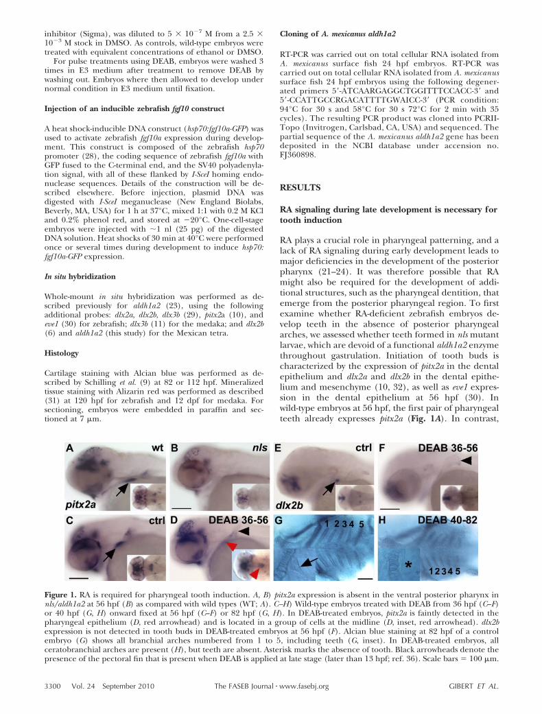
RA plays a crucial role in pharyngeal patterning, and alack of RA signaling during early development leads tomajor deficiencies in the development of the posteriorpharynx (21–24). It was therefore possible that RAmight also be required for the development of addi-tional structures, such as the pharyngeal dentition, thatemerge from the posterior pharyngeal region. To firstexamine whether RA-deficient zebrafish embryos de-velop teeth in the absence of posterior pharyngealarches, we assessed whether teeth formed in nls mutantlarvae, which are devoid of a functional aldh1a2 enzymethroughout gastrulation. Initiation of tooth buds ischaracterized by the expression of pitx2a in the dentalepithelium and dlx2a and dlx2b in the dental epithe-lium and mesenchyme (10, 32), as well as eve1 expres-sion in the dental epithelium at 56 hpf (30). Inwild-type embryos at 56 hpf, the first pair of pharyngealteeth already expresses pitx2a (Fig. 1A). In contrast,
Figure 1. RA is required for pharyngeal tooth induction. A, B) pitx2a expression is absent in the ventral posterior pharynx innls/aldh1a2 at 56 hpf (B) as compared with wild types (WT; A). C–H) Wild-type embryos treated with DEAB from 36 hpf (C–F)or 40 hpf (G, H) onward fixed at 56 hpf (C–F) or 82 hpf (G, H). In DEAB-treated embryos, pitx2a is faintly detected in thepharyngeal epithelium (D, red arrowhead) and is located in a group of cells at the midline (D, inset, red arrowhead). dlx2bexpression is not detected in tooth buds in DEAB-treated embryos at 56 hpf (F). Alcian blue staining at 82 hpf of a controlembryo (G) shows all branchial arches numbered from 1 to 5, including teeth (G, inset). In DEAB-treated embryos, allceratobranchial arches are present (H), but teeth are absent. Asterisk marks the absence of tooth. Black arrowheads denote thepresence of the pectoral fin that is present when DEAB is applied at late stage (later than 13 hpf; ref. 36). Scale bars � 100 �m.
3300 Vol. 24 September 2010 GIBERT ET AL.The FASEB Journal � www.fasebj.org
pitx2a expression is not detected in the ventral poste-rior pharynx of nls larvae (Fig. 1B). More generally,neither of the other tooth markers mentioned above(dlx2a, dlx2b, or eve1) are expressed in the ventralposterior pharynx in nls at 56 hpf but are present in thetooth buds of wild types (not shown). To avoid dealingwith developmental delay, nls embryos were able todevelop until 72 hpf and tooth development was visu-alized using dlx2b staining. Although the first tooth budwas still detected in control embryo, nls embryos neverexpress dlx2b in the tooth bud even at 72 hpf (notshown). Moreover, teeth are never detected by Alcianblue or Alizarin red staining in nls mutant embryos at120 hpf (not shown; refs. 22, 23). This confirms that theloss of early RA signaling, starting with zygotic expres-sion, which has pleiotropic defects on pharyngeal de-velopment (24), leads to a failure of pharyngeal toothinduction.
To study whether RA plays a role specifically duringpharyngeal tooth induction, we blocked RA signalingby 2 complementary strategies, i.e., inhibiting RA syn-thesis and blocking the signaling pathway at the level ofRARs. We treated wild-type embryos with 10�5 MDEAB, a competitive, reversible inhibitor of retinalde-hyde dehydrogenases (33), or with 5 � 10�6 MBMS493, a specific antagonist that inhibits the activa-tion of all RARs. Embryos were treated from differentdevelopmental stages onward and assayed for molecu-lar markers of tooth induction (Table 1). Overall, theexpression of pitx2a was always abolished when embryoswere incubated before 36 hpf and left in DEAB or BMS493 until fixation at 56 hpf. In contrast, when treatmentcommenced within this developmental window (36–56hpf), faint pitx2a expression was detected (Fig. 1D, redarrowhead). pitx2a is the earliest known indicator ofzebrafish tooth development and starting at 36 hpf isexpressed in the pharyngeal epithelium of wild-typeembryo, with the strongest expression prefiguring the
first tooth germ (10). Therefore, when RA signaling isinhibited from 36 hpf onward, a low level of pitx2aexpression remains in the pharyngeal epithelium (Fig.1D). Of note, the later RA signaling is inhibited, thestronger pitx2a expression remains (not shown). Inter-estingly, its expression is confined to 1 patch of cells atthe midline rather than to the paired but distinct toothbuds of wild types (Fig. 1C, D). dlx2b expression in thetooth epithelium, however, is absent from embryostreated with DEAB from 36 hpf onward, until fixation at56 hpf (Fig. 1F). This also holds true for dlx2a and eve1,which are not detected in DEAB-treated embryos (notshown). Here again to avoid a simple developmentaltooth induction delay induced by DEAB treatment, wetreated wild-type embryos with DEAB form 36 hpfonward and fixed them at 72 hpf. DEAB treatmentscommencing at 36 hpf display very little developmentaldelay compare with the nls mutant. DEAB-treated em-bryos from 36 to 72 hpf do not express dlx2b in theposterior ventral pharynx (Supplemental Fig. S1B).
To confirm that late DEAB treatment does notinterfere with pharyngeal patterning and, conse-quently, with the development of posterior branchialarches, we blocked RA synthesis from 40 hpf onward,after pitx2a is detected in DEAB-treated embryos, fixedthe embryos at 82 hpf, and found that the cartilaginousskeleton of all 5 branchial arches developed normally(Fig. 1H). At the same time, this DEAB treatmentregime was effective at specifically inhibiting pharyn-geal tooth induction, as judged by Alcian blue staining(Fig. 1H, inset).
To find the developmental timing at which RA is notrequired anymore to induce the first pair of teeth, weperformed DEAB treatment starting at different timespostfertilization. All treatments that commenced be-fore 43 hpf resulted in the absence of tooth inductionas monitored by either dlx2a or dlx2b expression (Table1). To confirm that 43 hpf is a crucial time in RA
TABLE 1. Timing of RA and FGF for the induction of the first pair of teeth in zebrafish
Time of treatment (hpf) pitx2a dlx2a dlx2b eve1
DEAB 32-56 Absent (100%, n�6) Absent (100%, n�6) Absent (100%, n�6) Absent (100%, n�6)BMS 493 32-56 Absent (100%, n�6) Absent (100%, n�6) Absent (100%, n�6) Absent (100%, n�6)DEAB 36-56 Present (100%, n�6) Absent (100%, n�6) Absent (100%, n�6) Absent (100%, n�6)BMS 493 36-56 Present (100%, n�6) Absent (100%, n�6) Absent (100%, n�6) Absent (100%, n�6)DEAB 38-56 Present (100%, n�6) Absent (100%, n�6) Absent (100%, n�6) Absent (100%, n�6)DEAB 40-56 Present (100%, n�6) Absent (100%, n�6) Absent (100%, n�6) NADEAB 42-56 Present (100%, n�6) Absent (100%, n�6) Absent (100%, n�6) NADEAB 43-56 Present (100%, n�6) Absent (85%, n�14) Absent (83%, n�6) NADEAB 44-56 Present (100%, n�6) Present (85%, n�14) Present (100%, n�6) NADEAB 45-56 Present (100%, n�6) Present (100%, n�6) Present (100%, n�6) NASU5402 32-56 Present (100%, n�6) Absent (100%, n�6) Absent (100%, n�6) NASU5402 36-56 NA Absent (100%, n�6) Absent (100%, n�6) NASU5402 40-56 NA Absent (100%, n�6) Absent (100%, n�6) NASU5402 44-56 NA Absent (100%, n�6) Absent (100%, n�6) NASU5402 47-56 NA Absent (100%, n�6) Absent (100%, n�6) NASU5402 48-56 NA Present (100%, n�12) Present (100%, n�6) NASU5402 49-56 NA Present (100%, n�6) Present (100%, n�6) NA
DEAB, BMS 493, or SU5402 was applied at different time points in development. Tooth induction was scored by the presence or absenceof specific tooth markers in the tooth bud. NA, nonapplicable.
3301RETINOIC ACID AND TOOTH INDUCTION IN TELEOSTS
requirement for tooth induction, we treated wild-typeembryos with DEAB at either 40 hpf (before the crucialtime point) or 45 hpf (after the crucial time point) andmonitored tooth presence by Alcian blue at 112 hpf orAlizarin red at 120 hpf. In all embryos treated at 40 hpf,teeth are always absent, while teeth are always presentwhen treatment starts at 45 hpf (Supplemental Fig.S1C–F). This is true with both staining methods usedfor tooth visualization. We further confirmed on trans-verse sections that no expression can be found for dlx2bin the tooth anlage when DEAB is applied before 43hpf (Supplemental Fig. S2E). In summary, these phar-macological treatments convincingly show that RA sig-naling is required during pharyngeal tooth inductionup to 43 hpf in zebrafish.
aldh1a2 expression is required for the induction ofthe first pair of pharyngeal teeth
To determine the location and source of RA requiredfor pharyngeal tooth induction, we examined the ex-pression of the RA-synthesizing enzymes known inzebrafish. The last enzymatic step of RA production iscatalyzed by the retinaldahyde dehydrogenases of theAldha1 protein family, which in mammals are encodedby 3 genes, aldh1a1–3. Two orthologs of the mamma-lian aldh1a2 and aldh1a3 genes have been characterizedin zebrafish (22, 23, 34, 35), whereas the aldh1a1homologue is absent from the genomes of fish (35, 36).Another gene thought to be a retinaldehyde dehydro-genase, originally named raldh4, was identified and
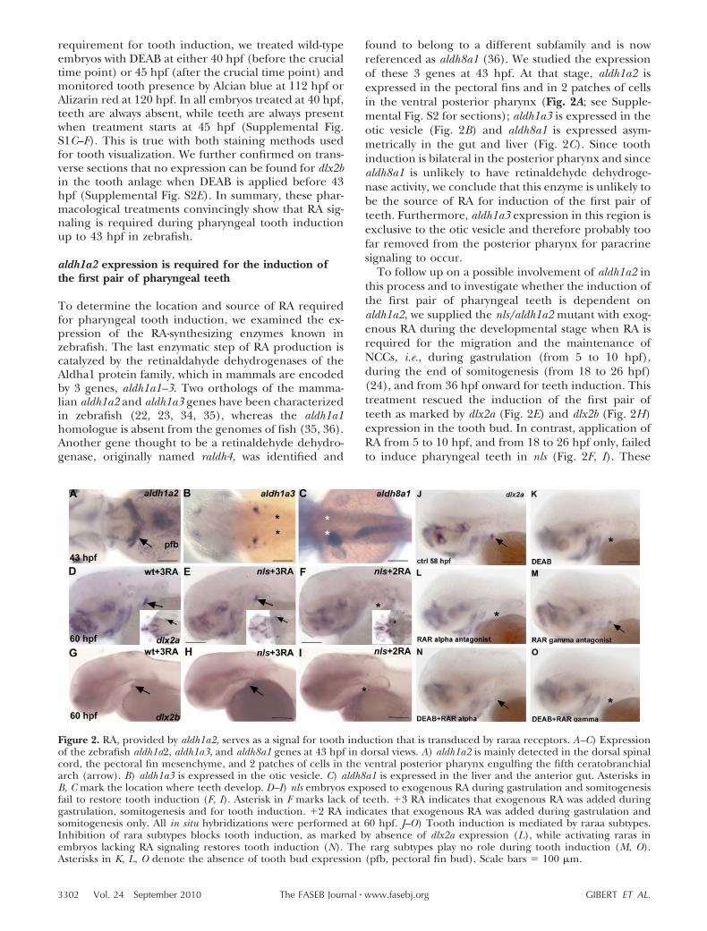
found to belong to a different subfamily and is nowreferenced as aldh8a1 (36). We studied the expressionof these 3 genes at 43 hpf. At that stage, aldh1a2 isexpressed in the pectoral fins and in 2 patches of cellsin the ventral posterior pharynx (Fig. 2A; see Supple-mental Fig. S2 for sections); aldh1a3 is expressed in theotic vesicle (Fig. 2B) and aldh8a1 is expressed asym-metrically in the gut and liver (Fig. 2C). Since toothinduction is bilateral in the posterior pharynx and sincealdh8a1 is unlikely to have retinaldehyde dehydroge-nase activity, we conclude that this enzyme is unlikely tobe the source of RA for induction of the first pair ofteeth. Furthermore, aldh1a3 expression in this region isexclusive to the otic vesicle and therefore probably toofar removed from the posterior pharynx for paracrinesignaling to occur.
To follow up on a possible involvement of aldh1a2 inthis process and to investigate whether the induction ofthe first pair of pharyngeal teeth is dependent onaldh1a2, we supplied the nls/aldh1a2 mutant with exog-enous RA during the developmental stage when RA isrequired for the migration and the maintenance ofNCCs, i.e., during gastrulation (from 5 to 10 hpf),during the end of somitogenesis (from 18 to 26 hpf)(24), and from 36 hpf onward for teeth induction. Thistreatment rescued the induction of the first pair ofteeth as marked by dlx2a (Fig. 2E) and dlx2b (Fig. 2H)expression in the tooth bud. In contrast, application ofRA from 5 to 10 hpf, and from 18 to 26 hpf only, failedto induce pharyngeal teeth in nls (Fig. 2F, I). These
Figure 2. RA, provided by aldh1a2, serves as a signal for tooth induction that is transduced by raraa receptors. A–C) Expressionof the zebrafish aldh1a2, aldh1a3, and aldh8a1 genes at 43 hpf in dorsal views. A) aldh1a2 is mainly detected in the dorsal spinalcord, the pectoral fin mesenchyme, and 2 patches of cells in the ventral posterior pharynx engulfing the fifth ceratobranchialarch (arrow). B) aldh1a3 is expressed in the otic vesicle. C) aldh8a1 is expressed in the liver and the anterior gut. Asterisks inB, C mark the location where teeth develop. D–I) nls embryos exposed to exogenous RA during gastrulation and somitogenesisfail to restore tooth induction (F, I). Asterisk in F marks lack of teeth. 3 RA indicates that exogenous RA was added duringgastrulation, somitogenesis and for tooth induction. 2 RA indicates that exogenous RA was added during gastrulation andsomitogenesis only. All in situ hybridizations were performed at 60 hpf. J–O) Tooth induction is mediated by raraa subtypes.Inhibition of rara subtypes blocks tooth induction, as marked by absence of dlx2a expression (L), while activating raras inembryos lacking RA signaling restores tooth induction (N). The rarg subtypes play no role during tooth induction (M, O).Asterisks in K, L, O denote the absence of tooth bud expression (pfb, pectoral fin bud). Scale bars � 100 �m.
3302 Vol. 24 September 2010 GIBERT ET AL.The FASEB Journal � www.fasebj.org
results demonstrate that aldh1a2 is required to inducethe first pair of pharyngeal teeth at 43 hpf.
At 43 hpf, aldh1a2 is strongly expressed in 2 patchesof cells in the ventral posterior pharynx (Fig. 2A, arrow,and Supplemental Fig. S2A) but also in the pectoral finbud mesenchyme (Supplemental Fig. S3A, B; arrow-head; ref. 37). Due to its proximity with the tooth bud,we examined the possibility that the pectoral fin couldbe a source of RA for the induction of the first pair ofteeth. To this end, we analyzed pharyngeal tooth for-mation in embryos that lack pectoral fins, in spade-tail/no tail double mutants (37), and in tbx5 morphants(38). We found that the absence of pectoral fin budsdoes not prevent the induction of the first pair of teeth(Supplemental Fig. S3C–H). Given the expression pat-tern of aldh1a2 and the rescue of pharyngeal teeth innls/aldh1a2 mutants by RA, it is therefore most likelythat the source of RA must be the 2 groups of cellslocated in the ventral posterior pharynx (SupplementalFig. S2A). A transverse section of a 43 hpf wild-typeembryo stained for aldh1a2 showed that the cells ex-pressing aldh1a2 at this stage coincide with the expres-sion of the tooth bud as marked by pitx2a at 56 hpf(Supplemental Fig. S2).
RA acts via the rara receptors for the induction ofthe first pair of teeth
RA regulates gene expression through ligand-inducedactivation of RARs, which in zebrafish are encoded by 4different genes: 2 rara paralogues (raraa and -b) and 2rarg paralogues (rarga and -b). Orthologs of mammalianRAR have been shown to be missing from the ze-brafish genome (39). At 43 hpf, expression of all RARtranscripts can be detected in the ventral pharynx (39,40). To determine which RARs are required for theinduction of the first pair of pharyngeal teeth, we useda pharmacological approach with compounds whoseselectivity for zebrafish RARs had been characaterizedin vitro (41). First, we selectively blocked the rarasubtypes from 36 hpf onward until fixation at 58 hpfwith the selective antagonist BMS614 (42) and assayedfor dlx2a expression as an indication of tooth inductionat 58 hpf. Inhibition of the raras resulted in a failure oftooth induction (Fig. 2L), whereas specific inhibition ofthe rarg subtypes using the selective antagonist CD2665did not inhibit the induction of dlx2a expression in thetooth bud at 58 hpf (Fig. 2M). To confirm that toothinduction requires rara genes, we simultaneouslyblocked RA biosynthesis with DEAB before tooth induc-tion (from 36 hpf onward) and activated the rara orrarg subtypes with selective agonists. We found that inthe absence of endogenous RA activation of the rarasusing the selective agonist BMS641 was sufficient torescue the induction of the first pair of teeth (Fig. 2N),while activation of the rargs using the selective agonistBMS961 cannot restore dlx2a expression in DEAB-treated embryos (Fig. 2O). These results strongly sug-gest that activation of raras is required and sufficient forthe induction of the first pair of pharyngeal teeth. Due
to the unavailability of paralogue-specific compounds,it was not possible to discriminate between the 2 raraparalogues in zebrafish. At the time of the induction ofthe first pair of teeth, raraa is expressed specifically inthe posterior most branchial arches, while rarab isfound in a more diffuse pattern along the entirepharyngeal arches (39, 43). At the same time of devel-opment, rarga is strongly expressed in the entire phar-ynx, while rargb is strongly detected in all but the lastpharyngeal arch (39, 43). The expression patterns ofraraa and raraab thus do not provide a clear indicationabout which of these genes is responsible for mediatingthe effect of RA on tooth induction, as they are bothdetected in the ventral posterior pharynx at the time oftooth induction by RA. Given that morpholino knock-down experiments have shown that rargs are necessaryfor pharyngeal patterning (44), our data showing that itis rather the rara paralogs that are critical for pharyn-geal tooth induction further reinforce the notion thatpharyngeal patterning and pharyngeal tooth inductionare unlinked events during zebrafish development.
RA and FGF signaling act independently of eachother during tooth induction
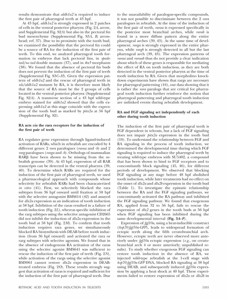
The induction of the first pair of pharyngeal teeth isFGF dependent in teleosts, but a lack of FGF signalingdoes not impair pitx2a expression in the tooth bud(10). To understand the relationship between FGF andRA signaling in the process of tooth induction, wedetermined the developmental time during which FGFsignaling is required to induce the pharyngeal teeth bytreating wild-type embryos with SU5402, a compoundthat has been shown to bind to FGF receptors and toconcomitantly block signaling (45, 46), for differentperiods of development. We observed that blockingFGF signaling at any stage before 48 hpf abolishedtooth induction, while later treatments had no effect onthe onset of dlx2a and dlx2b expression in the tooth bud(Table 1). To investigate the epistatic relationshipbetween the RA and the FGF signaling pathways, weconcomitantly activated the RA pathway and inhibitedthe FGF signaling pathway. We found that exogenousRA, applied from 32 to 56 hpf, fails to rescue theexpression of dlx2 genes in the tooth buds at 56 hpfwhen FGF signaling has been inhibited during thesame developmental interval (Fig. 3A–F).
Expression of fgf10a, using a heat-inducible construct(hsp70:fgf10a-GFP), leads to widespread formation ofectopic teeth along the fifth ceratobranchial arch.However, ectopic teeth are never observed more ante-riorly under fgf10a ectopic expression (e.g., on cerato-branchial arch 4 or more anteriorly; unpublished re-sults). To study whether exogenous FGF signaling canrestore tooth induction in the absence of RA, weinjected wild-type zebrafish at the 1-cell stage withhsp70:fgf10a-GFP DNA, blocked RA signaling at 38 hpfusing DEAB, and subsequently induced fgf10a expres-sion by applying a heat shock at 40 hpf. These experi-ments failed to restore expression of dlx2a or dlx2b in
3303RETINOIC ACID AND TOOTH INDUCTION IN TELEOSTS
the tooth bud (Fig. 3H, J), and mineralized teeth couldnot be detected at 100 hpf (not shown). We concludefrom these experiments that RA and fgf are bothrequired in independent pathways to induce teeth inzebrafish.
Induction of oral and pharyngeal teeth in medakaand Astyanax is independent of RA signaling
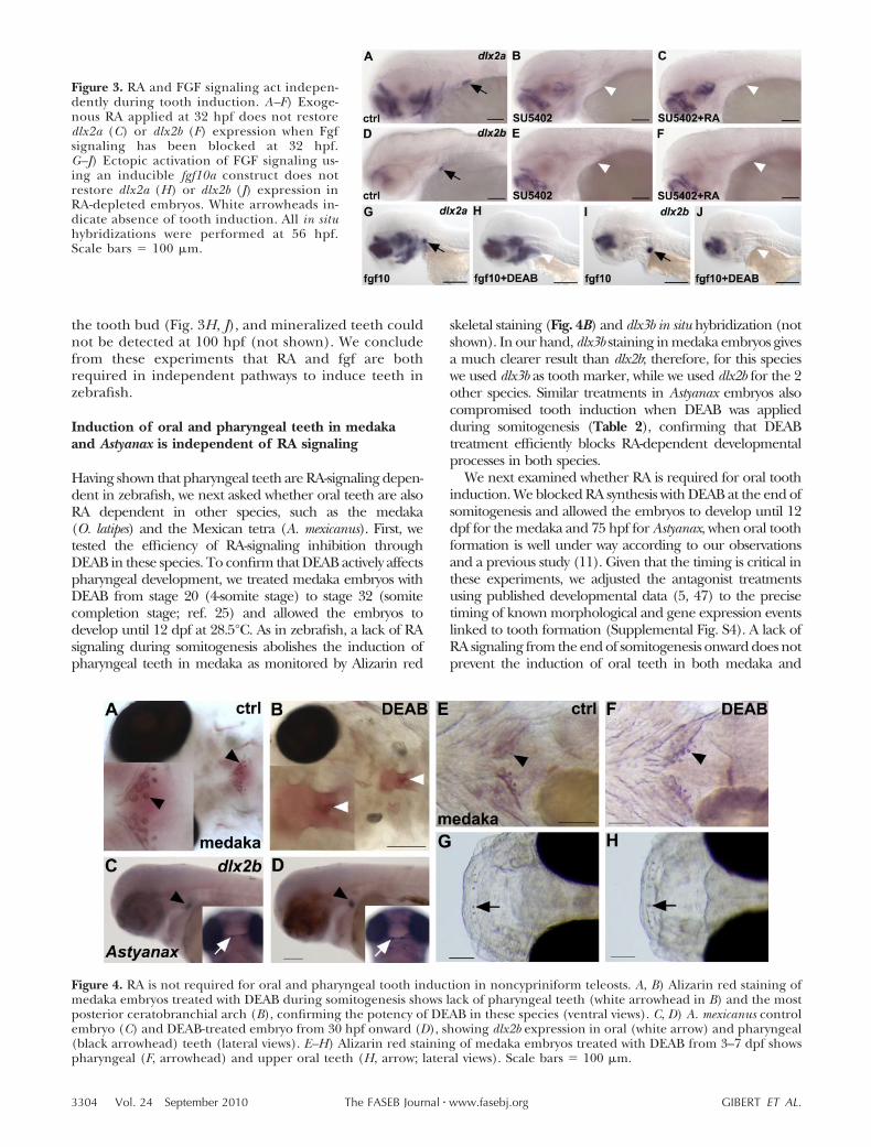
Having shown that pharyngeal teeth are RA-signaling depen-dent in zebrafish, we next asked whether oral teeth are alsoRA dependent in other species, such as the medaka(O. latipes) and the Mexican tetra (A. mexicanus). First, wetested the efficiency of RA-signaling inhibition throughDEAB in these species. To confirm that DEAB actively affectspharyngeal development, we treated medaka embryos withDEAB from stage 20 (4-somite stage) to stage 32 (somitecompletion stage; ref. 25) and allowed the embryos todevelop until 12 dpf at 28.5°C. As in zebrafish, a lack of RAsignaling during somitogenesis abolishes the induction ofpharyngeal teeth in medaka as monitored by Alizarin red
skeletal staining (Fig. 4B) and dlx3b in situ hybridization (notshown). In our hand, dlx3b staining in medaka embryos givesa much clearer result than dlx2b; therefore, for this specieswe used dlx3b as tooth marker, while we used dlx2b for the 2other species. Similar treatments in Astyanax embryos alsocompromised tooth induction when DEAB was appliedduring somitogenesis (Table 2), confirming that DEABtreatment efficiently blocks RA-dependent developmentalprocesses in both species.
We next examined whether RA is required for oral toothinduction. We blocked RA synthesis with DEAB at the end ofsomitogenesis and allowed the embryos to develop until 12dpf for the medaka and 75 hpf for Astyanax, when oral toothformation is well under way according to our observationsand a previous study (11). Given that the timing is critical inthese experiments, we adjusted the antagonist treatmentsusing published developmental data (5, 47) to the precisetiming of known morphological and gene expression eventslinked to tooth formation (Supplemental Fig. S4). A lack ofRA signaling from the end of somitogenesis onward does notprevent the induction of oral teeth in both medaka and
Figure 3. RA and FGF signaling act indepen-dently during tooth induction. A–F) Exoge-nous RA applied at 32 hpf does not restoredlx2a (C) or dlx2b (F) expression when Fgfsignaling has been blocked at 32 hpf.G–J) Ectopic activation of FGF signaling us-ing an inducible fgf10a construct does notrestore dlx2a (H) or dlx2b (J) expression inRA-depleted embryos. White arrowheads in-dicate absence of tooth induction. All in situhybridizations were performed at 56 hpf.Scale bars � 100 �m.
Figure 4. RA is not required for oral and pharyngeal tooth induction in noncypriniform teleosts. A, B) Alizarin red staining ofmedaka embryos treated with DEAB during somitogenesis shows lack of pharyngeal teeth (white arrowhead in B) and the mostposterior ceratobranchial arch (B), confirming the potency of DEAB in these species (ventral views). C, D) A. mexicanus controlembryo (C) and DEAB-treated embryo from 30 hpf onward (D), showing dlx2b expression in oral (white arrow) and pharyngeal(black arrowhead) teeth (lateral views). E–H) Alizarin red staining of medaka embryos treated with DEAB from 3–7 dpf showspharyngeal (F, arrowhead) and upper oral teeth (H, arrow; lateral views). Scale bars � 100 �m.
3304 Vol. 24 September 2010 GIBERT ET AL.The FASEB Journal � www.fasebj.org
Astyanax [Fig. 4D (inset), H]. We conclude that oral toothinduction is independent of RA signaling in these teleosts.
To examine whether RA is required for pharyngeal toothinduction, medaka and Astyanax embryos were treated withDEAB from the end of somitogenesis onward, i.e., after RA isrequired for the maintenance of NCC derivates. In theseexperiments, where RA inhibition commences before toothmarker gene expression, induction of pharyngeal teethsurprisingly was not compromised in either species (Fig. 4D,F). Because we did not anticipate a difference in RA require-ments for pharyngeal dentition between teleosts species, weexamined whether higher doses of DEAB, up to 160 �M forthe medaka and 75 �M for Astyananx at different develop-mental stages, would affect pharyngeal tooth development.Neither of these treatments led to an absence of pharyngealteeth (Table 2). However, earlier treatments lead to anabsence of pharyngeal tooth development: for example,when DEAB was applied at 24 hpf in Astyanax, no pharyngealteeth were visible at 75 hpf (n�8), while 50% of embryoshad teeth when DEAB was applied at 27 hpf (n�8), and allembryos developed teeth when DEAB was added at 30 hpfor later (n�16) (Table 2). However, a lack of RA duringearly stage of embryonic development (i.e., 24 hpf in Asty-anax) will affect the maintenance of posterior NCC derivates(24), leading to an absence of posterior structure in thepharynx including teeth. In our experimental settings, pha-ryngeal teeth are induced at �60 hpf in Astyanax (Supple-mental Fig. S4), meaning that DEAB treatment from 30 hpfonward will definitively target this developmental stage. Webelieve that 27 hpf is the end of RA requirement formaintenance of the posterior NCCs in Astyanax; that is whyDEAB treatments starting at this stage or earlier preventpharyngeal tooth induction by suppressing posterior NCC
derivatives, while later treatments (after 27 hpf) only targetpharyngeal tooth induction without any effect. Notably,typical defects that accompany the loss of RA signaling, likepericardial edema and a failure of swim bladder inflation,were always observed after DEAB treatments at the higherdoses. The inhibition experiments showed that pharyngealtooth induction is not dependent on RA in medaka andAstyanax, even though in zebrafish teeth clearly fail to beinduced in the absence of RA signaling.
Differential requirements for RA signaling in theposterior pharynx correlate with distinct aldh1a2expression patterns in teleosts
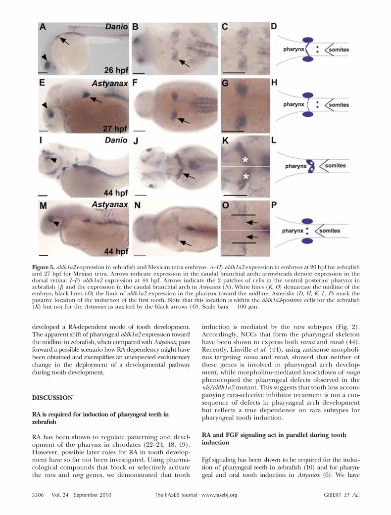
To examine whether the lack of RA requirement for pha-ryngeal tooth induction in medaka and Astyanax might belinked to differences in RA synthesis, we compared aldh1a2expression with that in zebrafish. Since heterochronic shiftsin pharyngeal tooth induction are more pronounced be-tween zebrafish and medaka (Supplemental Fig. S4), wecompared the gene expression patterns of aldh1a2 ortho-logues of Astyanax and zebrafish, which develop at moresimilar rates. At 26 hpf in zebrafish, aldh1a2 is detected in thedorsal retina, the caudal part of the branchial arch primor-dium, and in the somitic mesoderm (Fig. 5A–C; ref. 23). Asimilar pattern is observed for the Astyanax orthologue ofaldh1a2 (Fig. 5E–G), suggesting an overall evolutionary con-servation of aldh1a2 gene expression in Ostariophysi, thetaxon containing the orders Characiformes and Cyprini-formes.
At the time of pharyngeal tooth induction, however,aldh1a2 expression in the pharyngeal region differs mark-edly between zebrafish and Astyanax. In zebrafish, expres-sion is detected in 2 patches of cells in the ventral posteriorpharynx engulfing the fifth ceratobranchial arch (Fig. 5I–K).While the overall Astyanax aldh1a2 expression resembles theexpression pattern observed in zebrafish (e.g., expression indorsal retina and the dorsal anterior spinal cord; see Fig.5M–O; see also Supplemental Fig. S5 for aldh1a2 expressionat 44 hpf), expression in the pharyngeal region showsdistinct differences between the 2 species. In Astyanax,aldh1a2-expressing cells are located further laterally than inzebrafish and exclude the pharyngeal region (Fig. 5J–L,N–P). In contrast, in zebrafish aldh1a2 expression is locatedin the ventral posterior pharynx reaching the midline. Basedon dlx2b expression in the tooth bud at 56 hpf in zebrafishand 75 hpf in Astyanax, the tooth primordia in zebrafish arelocated in a domain overlapping with aldh1a2 expression,whereas in Astyanax induction of the first teeth (marked byarrows in Fig. 5O) takes place outside the aldh1a2 expressiondomain.
Based on RA inhibition experiments in medaka andAstyanax and on the expression pattern of aldh1a2 in Asty-anax, we conclude that the tooth primordia are not in closeproximity to RA at the stage when tooth induction occurs. InAstyanax and medaka, therefore, the induction of both oraland pharyngeal dentition is independent of RA signaling.Because oral tooth loss in zebrafish is thought to be a derivedtrait (5), this suggests that primitively in teleost fishes toothinduction was RA independent and that cypriniform fish
TABLE 2. DEAB treatment in zebrafish, medaka, and Mexicantetra at different time points
Species DEAB treatmentTooth
induction
D. rerio 5–11 hpf (5 �m) �D. rerio 12–24 hpf (10 �m) �D. rerio 18–30 hpf (10 �m) �D. rerio 23–30 hpf (10 �m) �D. rerio 20–28 hpf (10 �m) �D. rerio 28–32 hpf (10 �m) �D. rerio 40–56 hpf (10 �m) �O. latipes Stage 20–stage 32 (30 �m) �O. latipes 4–8 dpf (30 �m) �O. latipes 3–7 dpf (60 �m) �O. latipes 5–8 dpf (60 �m) �O. latipes 4–7 dpf (100 �m) �O. latipes 4–8 dpf (120 �m) �O. latipes 4–8 dpf (160 �m) �A. mexicanus 18–75 hpf (20 �m) �A. mexicanus 24–75 hpf (20 �m) �A. mexicanus 27–75 hpf (50 �m) �/�A. mexicanus 30–75 hpf (50 �m) �A. mexicanus 30–75 hpf (75 �m) �A. mexicanus 40–75 hpf (50 �m) �
The presence of teeth was monitored by in situ hybridizationwith dlx2a and or dlx2b for the zebrafish and the Mexican tetra and byAlizarin red staining and dlx3b for the medaka.
3305RETINOIC ACID AND TOOTH INDUCTION IN TELEOSTS
developed a RA-dependent mode of tooth development.The apparent shift of pharyngeal aldh1a2 expression towardthe midline in zebrafish, when compared with Astyanax, putsforward a possible scenario how RA dependency might havebeen obtained and exemplifies an unexpected evolutionarychange in the deployment of a developmental pathwayduring tooth development.
DISCUSSION
RA is required for induction of pharyngeal teeth inzebrafish
RA has been shown to regulate patterning and devel-opment of the pharynx in chordates (22–24, 48, 49).However, possible later roles for RA in tooth develop-ment have so far not been investigated. Using pharma-cological compounds that block or selectively activatethe rara and rarg genes, we demonstrated that tooth
induction is mediated by the rara subtypes (Fig. 2).Accordingly, NCCs that form the pharyngeal skeletonhave been shown to express both raraa and rarab (44).Recently, Linville et al. (44), using antisense morpholi-nos targeting raraa and rarab, showed that neither ofthese genes is involved in pharyngeal arch develop-ment, while morpholino-mediated knockdown of rargaphenocopied the pharyngeal defects observed in thenls/aldh1a2 mutant. This suggests that tooth loss accom-panying rara-selective inhibitor treatment is not a con-sequence of defects in pharyngeal arch developmentbut reflects a true dependence on rara subtypes forpharyngeal tooth induction.
RA and FGF signaling act in parallel during toothinduction
Fgf signaling has been shown to be required for the induc-tion of pharyngeal teeth in zebrafish (10) and for pharyn-geal and oral tooth induction in Astyanax (6). We have
Figure 5. aldh1a2 expression in zebrafish and Mexican tetra embryos. A–H) aldh1a2 expression in embryos at 26 hpf for zebrafishand 27 hpf for Mexian tetra. Arrows indicate expression in the caudal branchial arch; arrowheads denote expression in thedorsal retina. I–P) aldh1a2 expression at 44 hpf. Arrows indicate the 2 patches of cells in the ventral posterior pharynx inzebrafish (J) and the expression in the caudal branchial arch in Astyanax (N). White lines (K, O) demarcate the midline of theembryo; black lines (O) the limit of aldh1a2 expression in the pharynx toward the midline. Asterisks (D, H, K, L, P) mark theputative location of the induction of the first tooth. Note that this location is within the aldh1a2-positive cells for the zebrafish(K) but not for the Astyanax as marked by the black arrows (O). Scale bars � 100 �m.
3306 Vol. 24 September 2010 GIBERT ET AL.The FASEB Journal � www.fasebj.org
shown that in zebrafish RA signaling is required in additionto FGF signaling for tooth induction. The temporal require-ments for both signaling pathways appear to be slightlydistinct, RA signaling is only required before 43 hpf toinduce tooth markers, while inhibition of Fgf signaling up to48 hpf results in loss of dlx gene expression at 56 hpf (Table1). However, we were not able to derive a clear epistaticrelationship between both pathways, as exogenous RA wasunable to rescue early tooth markers in the absence of FGFsignaling, and overexpression of fgf10 alone, which is suffi-cient to induce ectopic teeth on the fifth ceratobranchialarches (unpublished results), was ineffective at rescuingtooth development when RA signaling had been blocked.Jackman et al. (10) showed that inhibition of FGF signalingdoes not impair the expression of pitx2a, the earliest knowngene expressed in pharyngeal teeth. Our finding that a lackof RA signaling reduces and changes the expression domainof pitx2a in the dental epithelium suggests that RA and Fgfsignaling both participate in the tooth developmental pro-gram. RA is required before 36 hpf for induction of pitx2a,whose expression is normally induced at 36 hpf in theepithelium of the first pharyngeal teeth, and up to 43 hpf forcontinued expression of pitx2a. In contrast, Fgf signaling isneither required for induction nor maintenance of pitx2aexpression but is necessary to induce dlx gene expression.Since RA cannot overcome a lack of Fgf signaling, Fgf mightplay a more specific role in tooth formation after the dentalepithelium has been specified. Given that we observe amisplacement of the pitx2a expression domains when RAsignaling is reduced after pitx2a induction, it is likely that RAis also involved in patterning events at the midline of theventral pharynx. We conclude from these observations thatRA and FGF signaling are required in parallel for successiveevents during tooth induction (Fig. 6).
RA is not involved in tooth induction in other teleosts
Our experiments in medaka and Astyanax show that RA isdispensable for oral tooth induction and, more surprisingly,suggest that RA signaling is also not required for the induc-tion of pharyngeal teeth. In addition, expression of aldh1a2in the pharynx of zebrafish extends more toward the mid-line and thus toward the region of pharyngeal tooth forma-tion than in Astyanax at the time of tooth induction. Togather these observations into a coherent model, it is impor-tant to recall that cypriniforms have lost oral teeth early onduring evolution and that this event has not been reverted inany of the 3000 known species in this order (5, 6). Thus,medaka and Astyanax probably provide a better glimpse onthe ancestral situation occurring in fishes, whereas the ze-brafish corresponds to the derived situation (Fig. 6B). Ourdata suggest that pharyngeal teeth in cypriniforms obtainedRA regulation for induction, while in other taxa this induc-tive phase is RA independent. It is tempting to correlate thisRA dependency of tooth induction with the observed shift inaldh1a2 expression, suggesting that a relocation of RA syn-thesis that occurred specifically in cypriniforms acted as aprecondition for this event. It is likely that dependency onRA arose at the level of a specific enhancer of one of theearly genes controlling the tooth induction process. One
candidate gene could be pitx2a, as it is the only gene in thetooth development cascade whose expression is affected byRA but not Fgf signaling.
Once pharyngeal tooth induction became RA depen-dent, it is likely that this pleiotropic signal also affected thedevelopmental program of oral teeth. Our model there-fore provides an explanation for oral tooth loss in thelineage leading to the cypriniforms. Indeed, the lack ofexpression of aldh1a2 and aldh1a3 indicates that theanterior region of the embryo and mostly the oral regionare devoid of RA (14). This model is interesting toconsider in light of recent results showing that the signal-ing pathways implicated in tooth induction in the oral andpharyngeal regions are largely identical (6, 11).
Cypriniforms were unlikely to lose oral teeth due tostructural mutations in the gene repertoire requiredfor tooth formation in general, as pleiotropic defectselsewhere would have been deleterious. Our empiri-cally deduced scenario, in which tooth developmentacquires RA dependency, thus provides an alternativeand plausible hypothesis that provides a functional linkbetween the loss of oral teeth and the gain of RAregulation in pharyngeal teeth in cypriniforms. Thissituation is reminiscent to the diastema, a region de-void of teeth between the incisor and molar fields,found in rodents (50). It has been observed that theexpression of natural inhibitors of the FGF pathway,encoded by the sprouty genes, in the diastema preventsfreshly induced tooth buds to further engage in the
Figure 6. Proposed model of genetic pathways involved inpharyngeal tooth induction in zebrafish. A) aldh1a2 from theposterior ventral pharynx generates RA. This signal activatesthe RAR subtype �. RA signaling is required for a properexpression of pitx2a; however, in absence of this signal, pitx2ais still detected at a lower level as a spot at the midline. Dottedlines represent an RA requirement for proper expression ofpitx2a. pitx2a, in turn, regulates the expression of dlx2a, dlx2b,and eve1. FGF signaling is required after RA signaling (48 vs.43 hpf). Moreover we know from previous study that a lack ofFGF signaling does not abolish pitx2a expression in the toothbud (10). B) Phylogenetic tree of the main fish modelsshowing the presence of absence of a particular set of teeth(oral vs. pharyngeal). Red branch represents the hijack of RAfor tooth induction in zebrafish.
3307RETINOIC ACID AND TOOTH INDUCTION IN TELEOSTS
tooth developmental program (51). We believe thesituation in zebrafish to be rather different, as noindication of tooth formation is apparent in the oralregion, which suggests a blockade at a very early step(6); and the obligatory inclusion of an additionalsignaling pathway into the tooth developmental pro-gram that is otherwise inactive in the oral region mayexplain the loss of oral teeth.
This work was supported by an ARC fellowship (to Y.G.),funding from the Deutsche Forschungsgemeinschaft (toG.B.), funding from the ENS Lyon, the ANR program and theFrench Ministry of Research (to V.L.), an ANR-Neuro (toS.R.), INRA, CNRS, the ANR Grant Choregnet, the MarineGenomics Center of Excellence, the FP6 STREP Plurigenes(to J.-S.J.), and NIH 5F32DE015029 and P20RR016463 (toW.R.J.), and a NSF IOS-0446720 (to D.W.S.). The authorsthank Pierre Germain (Centre de Biochimie Structurale,Montpellier, France) for his kind gift of the RARa and RARgselective agonists and antagonists. Thanks to Matthieu Simionand Laurent Legendre for expert animal care in Gif.
REFERENCES
1. Smith, M. M., Fraser, G. J., and Mitsiadis, T. A. (2009) Dentallamina as source of odontogenic stem cells: evolutionary originsand developmental control of tooth generation in gnathos-tomes. J. Exp. Zoolog. B Mol. Dev. Evol. 312B, 260–280
2. Miletich, I., and Sharpe, P. T. (2004) Neural crest contributionto mammalian tooth formation. Birth Defects Res. C Embryo Today72, 200–212
3. Mitsiadis, T. A., and Smith, M. M. (2006) How do genes maketeeth to order through development? J. Exp. Zoolog. B Mol. Dev.Evol. 306, 177–182
4. Tompkins, K. (2006) Molecular mechanisms of cytodifferentia-tion in mammalian tooth development. Connect. Tissue Res. 47,111–118
5. Stock, D. W. (2007) Zebrafish dentition in comparative context.J. Exp. Zoolog. B Mol. Dev. Evol. 308, 523–549
6. Stock, D. W., Jackman, W. R., and Trapani, J. (2006) Develop-mental genetic mechanisms of evolutionary tooth loss in cypri-niform fishes. Development 133, 3127–3137
7. Huysseune, A., Van der heyden, C., and Sire, J. Y. (1998) Earlydevelopment of the zebrafish (Danio rerio) pharyngeal dentition(Teleostei, Cyprinidae). Anat. Embryol. (Berl.) 198, 289–305
8. Van der Heyden, C., Huysseune, A., and Sire, J. Y. (2000)Development and fine structure of pharyngeal replacementteeth in juvenile zebrafish (Danio rerio) (Teleostei, Cyprinidae).Cell Tissue Res. 302, 205–219
9. Schilling, T. F., Piotrowski, T., Grandel, H., Brand, M., Heisen-berg, C. P., Jiang, Y. J., Beuchle, D., Hammerschmidt, M., Kane,D. A., Mullins, M. C., van Eeden, F. J., Kelsh, R. N., Furutani-Seiki, M., Granato, M., Haffter, P., Odenthal, J., Warga, R. M.,Trowe, T., and Nusslein-Volhard, C. (1996) Jaw and branchialarch mutants in zebrafish I: branchial arches. Development 123,329–344
10. Jackman, W. R., Draper, B. W., and Stock, D. W. (2004) Fgfsignaling is required for zebrafish tooth development. Dev. Biol.274, 139–157
11. Debiais-Thibaud, M., Borday-Birraux, V., Germon, I., Bourrat,F., Metcalfe, C. J., Casane, D., and Laurenti, P. (2007) Develop-ment of oral and pharyngeal teeth in the medaka (Oryziaslatipes): comparison of morphology and expression of eve1gene. J. Exp. Zoolog. B Mol. Dev. Evol. 308, 693–708
12. Nelson, J. S. (2006) Fishes of the World, 4th Ed., John Wiley &Sons, Hoboken, NJ, USA
13. Hernandez, R. E., Rikhof, H. A., Bachmann, R., and Moens,C. B. (2004) vhnf1 integrates global RA patterning and localFGF signals to direct posterior hindbrain development in ze-brafish. Development 131, 4511–4520
14. White, R. J., Nie, Q., Lander, A. D., and Schilling, T. F. (2007)Complex regulation of cyp26a1 creates a robust retinoic acidgradient in the zebrafish embryo. PLoS Biol. 5, e304
15. Sirbu, I. O., and Duester, G. (2006) Retinoic-acid signalling innode ectoderm and posterior neural plate directs left-rightpatterning of somitic mesoderm. Nat. Cell Biol. 8, 271–277
16. Hamade, A., Deries, M., Begemann, G., Bally-Cuif, L., Genet, C.,Sabatier, F., Bonnieu, A., and Cousin, X. (2006) Retinoic acidactivates myogenesis in vivo through Fgf8 signalling. Dev. Biol.289, 127–140
17. Ribes, V., Wang, Z., Dolle, P., and Niederreither, K. (2006)Retinaldehyde dehydrogenase 2 (RALDH2)-mediated retinoicacid synthesis regulates early mouse embryonic forebrain devel-opment by controlling FGF and sonic hedgehog signaling.Development 133, 351–361
18. Mark, M., Ghyselinck, N. B., and Chambon, P. (2006) Functionof retinoid nuclear receptors: lessons from genetic and pharma-cological dissections of the retinoic acid signaling pathwayduring mouse embryogenesis. Annu. Rev. Pharmacol. Toxicol. 46,451–480
19. Germain, P., Chambon, P., Eichele, G., Evans, R. M., Lazar,M. A., Leid, M., De Lera, A. R., Lotan, R., Mangelsdorf, D. J.,and Gronemeyer, H. (2006) International Union of Pharma-cology. LX. Retinoic acid receptors. Pharmacol. Rev. 58,712–725
20. Morriss-Kay, G. M., and Ward, S. J. (1999) Retinoids andmammalian development. Int. Rev. Cytol. 188, 73–131
21. Niederreither, K., Subbarayan, V., Dolle, P., and Chambon, P.(1999) Embryonic retinoic acid synthesis is essential for earlymouse post-implantation development. Nat. Genet. 21, 444 –448
22. Begemann, G., Schilling, T. F., Rauch, G. J., Geisler, R., andIngham, P. W. (2001) The zebrafish neckless mutation reveals arequirement for raldh2 in mesodermal signals that pattern thehindbrain. Development 128, 3081–3094
23. Grandel, H., Lun, K., Rauch, G. J., Rhinn, M., Piotrowski, T.,Houart, C., Sordino, P., Kuchler, A. M., Schulte-Merker, S.,Geisler, R., Holder, N., Wilson, S. W., and Brand, M. (2002)Retinoic acid signalling in the zebrafish embryo is necessaryduring pre-segmentation stages to pattern the anterior-posterioraxis of the CNS and to induce a pectoral fin bud. Development129, 2851–2865
24. Kopinke, D., Sasine, J., Swift, J., Stephens, W. Z., and Piotrowski,T. (2006) Retinoic acid is required for endodermal pouchmorphogenesis and not for pharyngeal endoderm specification.Dev. Dyn. 235, 2695–2709
25. Iwamatsu, T. (2004) Stages of normal development in themedaka Oryzias latipes. Mech. Dev. 121, 605–618
26. Menuet, A., Alunni, A., Joly, J. S., Jeffery, W. R., and Retaux, S.(2007) Expanded expression of sonic hedgehog in Astyanaxcavefish: multiple consequences on forebrain development andevolution. Development 134, 845–855
27. Kimmel, C. B., Ballard, W. W., Kimmel, S. R., Ullmann, B., andSchilling, T. F. (1995) Stages of embryonic development of thezebrafish. Dev. Dyn. 203, 253–310
28. Halloran, M. C., Sato-Maeda, M., Warren, J. T., Su, F., Lele, Z.,Krone, P. H., Kuwada, J. Y., and Shoji, W. (2000) Laser-inducedgene expression in specific cells of transgenic zebrafish. Devel-opment 127, 1953–1960
29. Akimenko, M. A., Ekker, M., Wegner, J., Lin, W., and Wester-field, M. (1994) Combinatorial expression of three zebrafishgenes related to distal-less: part of a homeobox gene code forthe head. J. Neurosci. 14, 3475–3486
30. Laurenti, P., Thaeron, C., Allizard, F., Huysseune, A., and Sire,J. Y. (2004) Cellular expression of eve1 suggests its requirementfor the differentiation of the ameloblasts and for the initiationand morphogenesis of the first tooth in the zebrafish (Daniorerio). Dev. Dyn. 230, 727–733
31. Fisher, S., and Halpern, M. E. (1999) Patterning the zebrafishaxial skeleton requires early chordin function. Nat. Genet. 23,442–446
32. Borday-Birraux, V., Van der Heyden, C., Debiais-Thibaud, M.,Verreijdt, L., Stock, D. W., Huysseune, A., and Sire, J. Y. (2006)Expression of Dlx genes during the development of the ze-brafish pharyngeal dentition: evolutionary implications. Evol.Dev. 8, 130–141
3308 Vol. 24 September 2010 GIBERT ET AL.The FASEB Journal � www.fasebj.org
33. Begemann, G., Marx, M., Mebus, K., Meyer, A., and Bastmeyer,M. (2004) Beyond the neckless phenotype: influence of reducedretinoic acid signaling on motor neuron development in thezebrafish hindbrain. Dev. Biol. 271, 119–129
34. Liang, D., Zhang, M., Bao, J., Zhang, L., Xu, X., Gao, X., andZhao, Q. (2008) Expressions of Raldh3 and Raldh4 duringzebrafish early development. Gene Expr. Patterns 8, 248–253
35. Pittlik, S., Domingues, S., Meyer, A., and Begemann, G. (2008)Expression of zebrafish aldh1a3 (raldh3) and absence ofaldh1a1 in teleosts. Gene Expr. Patterns 8, 141–147
36. Canestro, C., Postlethwait, J. H., Gonzalez-Duarte, R., andAlbalat, R. (2006) Is retinoic acid genetic machinery a chordateinnovation? Evol. Dev. 8, 394–406
37. Gibert, Y., Gajewski, A., Meyer, A., and Begemann, G. (2006)Induction and prepatterning of the zebrafish pectoral fin budrequires axial retinoic acid signaling. Development 133, 2649–2659
38. Ahn, D. G., Kourakis, M. J., Rohde, L. A., Silver, L. M., and Ho,R. K. (2002) T-box gene tbx5 is essential for formation of thepectoral limb bud. Nature 417, 754–758
39. Bertrand, S., Thisse, B., Tavares, R., Sachs, L., Chaumot, A.,Bardet, P. L., Escriva, H., Duffraisse, M., Marchand, O., Safi, R.,Thisse, C., and Laudet, V. (2007) Unexpected novel relationallinks uncovered by extensive developmental profiling of nuclearreceptor expression. PLoS Genet. 3, e188
40. Hale, L. A., Tallafuss, A., Yan, Y. L., Dudley, L., Eisen, J. S., andPostlethwait, J. H. (2006) Characterization of the retinoic acidreceptor genes raraa, rarab and rarg during zebrafish develop-ment. Gene Expr. Patterns 6, 546–555
41. Escriva, H., Bertrand, S., Germain, P., Robinson-Rechavi, M.,Umbhauer, M., Cartry, J., Duffraisse, M., Holland, L., Gronem-eyer, H., and Laudet, V. (2006) Neofunctionalization in verte-brates: the example of retinoic acid receptors. PLoS Genet. 2,e102
42. de Lera, A. R., Bourguet, W., Altucci, L., and Gronemeyer, H.(2007) Design of selective nuclear receptor modulators: RARand RXR as a case study. Nat. Rev. Drug Discov. 6, 811–820
43. Thisse, C., and Thisse, B. (2008) High-resolution in situ hybrid-ization to whole-mount zebrafish embryos. Nat. Protoc. 3, 59–69
44. Linville, A., Radtke, K., Waxman, J. S., Yelon, D., and Schilling,T. F. (2009) Combinatorial roles for zebrafish retinoic acidreceptors in the hindbrain, limbs and pharyngeal arches. Dev.Biol. 325, 60–70
45. Furthauer, M., Reifers, F., Brand, M., Thisse, B., and Thisse, C.(2001) sprouty4 acts in vivo as a feedback-induced antagonist ofFGF signaling in zebrafish. Development 128, 2175–2186
46. Mandler, M., and Neubuser, A. (2001) FGF signaling is neces-sary for the specification of the odontogenic mesenchyme. Dev.Biol. 240, 548–559
47. Debiais-Thibaud, M., Germon, I., Laurenti, P., Casane, D., andBorday-Birraux, V. (2008) Low divergence in Dlx gene expres-sion between dentitions of the medaka (Oryzias latipes) versushigh level of expression shuffling in osteichtyans. Evol. Dev. 10,464–476
48. Escriva, H., Holland, N. D., Gronemeyer, H., Laudet, V., andHolland, L. Z. (2002) The retinoic acid signaling pathwayregulates anterior/posterior patterning in the nerve cord andpharynx of amphioxus, a chordate lacking neural crest. Develop-ment 129, 2905–2916
49. Matt, N., Ghyselinck, N. B., Wendling, O., Chambon, P., andMark, M. (2003) Retinoic acid-induced developmental defectsare mediated by RARbeta/RXR heterodimers in the pharyngealendoderm. Development 130, 2083–2093
50. Peterkova, R., Lesot, H., and Peterka, M. (2006) Phylogeneticmemory of developing mammalian dentition. J. Exp. Zoolog. BMol. Dev. Evol. 306, 234–250
51. Klein, O. D., Minowada, G., Peterkova, R., Kangas, A., Yu, B. D.,Lesot, H., Peterka, M., Jernvall, J., and Martin, G. R. (2006)Sprouty genes control diastema tooth development via bidirec-tional antagonism of epithelial-mesenchymal FGF signaling.Dev. Cell 11, 181–190
Received for publication October 13, 2009.Accepted for publication April 15, 2010.
3309RETINOIC ACID AND TOOTH INDUCTION IN TELEOSTS


![4* • Pharingeal grooves/cleft : 4 • [Pharyngeal membrane]](https://static.fdokumen.com/doc/165x107/6334ea00b9085e0bf5093ec7/4-pharingeal-groovescleft-4-pharyngeal-membrane.jpg)

