Novel mutant-enriched sequencing identified high frequency ofPIK3CA mutations in pharyngeal cancer
14
Novel mutant-enriched Sequencing Identified High Frequency of PIK3CA Mutations in Pharyngeal Cancer Wanglong Qiu 1 , Guo-Xia Tong 2 , Spiros Manolidis 1 , Lanny G. Close 1 , Adel M. Assaad 2 , and Gloria H. Su 1,2 1 Departments of Otolaryngology and Head and Neck Surgery, Columbia University College of Physicians and Surgeons, New York, NY 10032 2 Department of Pathology, Columbia University College of Physicians and Surgeons, New York, NY 10032 Abstract We previously reported four PIK3CA mutations in 38 head and neck cancer samples; three of which were identified in six pharyngeal cancer samples. To determine the mutation frequency of PIK3CA in pharyngeal cancer, we studied 24 additional cases of pharyngeal squamous cell carcinoma in this study. Using both direct genomic DNA sequencing and novel mutant-enriched sequencing methods developed specifically for the three hot-spot mutations (H1047R, E545K and E452K) of PIK3CA, we detected five mutations of PIK3CA in the 24 pharyngeal cancers (20.8%). Three of the five mutations had been missed by the conventional sequencing method and were subsequently detected by novel mutant-enriched sequencing methods. We showed that the mutant-enriched sequencing method for the H1047R hot-spot mutation can identify the mutation in a mixed population of mutant and wild-type DNA sequences at 1:360 ratios. These novel mutant-enriched sequencing methods allow the detection of the PIK3CA hot-spot mutations in clinical specimens which often contain limited tumor tissues (i.e. biopsy specimens). The data further supports that oncogenic PIK3CA may play a critical role in pharyngeal carcinogenesis, and the mutant-enriched sequencing methods for PIK3CA are sensitive and reliable ways to detect PIK3CA mutations in clinical samples. Because PIK3CA and its pathway are potential targets for chemotherapy and radiation therapy, and frequent somatic mutation of PIK3CA has been identified in many human cancer types (e.g. breast cancer, colorectal cancer), the abilities to detect PIK3CA mutations with enhanced sensitivities have great potential impacts on target therapies for many cancer types. Keywords mutant-enriched sequencing method; PIK3CA oncogene; hot-spot mutation; pharyngeal cancer; HNSCC INTRODUCTION Oral and pharyngeal cancer accounts for over half a million new cancer cases and 2,710,000 deaths worldwide per annum. It is the sixth most common malignant tumors in the world. In the United States alone, there were 30,990 estimated new cancer cases and 7,430 new deaths To whom requests for reprints should be addressed: Dr. Su at the Department of Otolaryngology/Head and Neck Surgery, Columbia University College of Physicians and Surgeons, 1130 St. Nicholas Ave. ICRC 10-04, New York, NY 10032. Phone: 212-851-4624; [email protected]. NIH Public Access Author Manuscript Int J Cancer. Author manuscript; available in PMC 2009 December 14. Published in final edited form as: Int J Cancer. 2008 March 1; 122(5): 1189–1194. doi:10.1002/ijc.23217. NIH-PA Author Manuscript NIH-PA Author Manuscript NIH-PA Author Manuscript
Transcript of Novel mutant-enriched sequencing identified high frequency ofPIK3CA mutations in pharyngeal cancer
Novel mutant-enriched Sequencing Identified High Frequency ofPIK3CA Mutations in Pharyngeal Cancer
Wanglong Qiu1, Guo-Xia Tong2, Spiros Manolidis1, Lanny G. Close1, Adel M. Assaad2, andGloria H. Su1,21 Departments of Otolaryngology and Head and Neck Surgery, Columbia University College ofPhysicians and Surgeons, New York, NY 100322 Department of Pathology, Columbia University College of Physicians and Surgeons, New York,NY 10032
AbstractWe previously reported four PIK3CA mutations in 38 head and neck cancer samples; three of whichwere identified in six pharyngeal cancer samples. To determine the mutation frequency of PIK3CAin pharyngeal cancer, we studied 24 additional cases of pharyngeal squamous cell carcinoma in thisstudy. Using both direct genomic DNA sequencing and novel mutant-enriched sequencing methodsdeveloped specifically for the three hot-spot mutations (H1047R, E545K and E452K) of PIK3CA,we detected five mutations of PIK3CA in the 24 pharyngeal cancers (20.8%). Three of the fivemutations had been missed by the conventional sequencing method and were subsequently detectedby novel mutant-enriched sequencing methods. We showed that the mutant-enriched sequencingmethod for the H1047R hot-spot mutation can identify the mutation in a mixed population of mutantand wild-type DNA sequences at 1:360 ratios. These novel mutant-enriched sequencing methodsallow the detection of the PIK3CA hot-spot mutations in clinical specimens which often containlimited tumor tissues (i.e. biopsy specimens).
The data further supports that oncogenic PIK3CA may play a critical role in pharyngealcarcinogenesis, and the mutant-enriched sequencing methods for PIK3CA are sensitive and reliableways to detect PIK3CA mutations in clinical samples. Because PIK3CA and its pathway are potentialtargets for chemotherapy and radiation therapy, and frequent somatic mutation of PIK3CA has beenidentified in many human cancer types (e.g. breast cancer, colorectal cancer), the abilities to detectPIK3CA mutations with enhanced sensitivities have great potential impacts on target therapies formany cancer types.
Keywordsmutant-enriched sequencing method; PIK3CA oncogene; hot-spot mutation; pharyngeal cancer;HNSCC
INTRODUCTIONOral and pharyngeal cancer accounts for over half a million new cancer cases and 2,710,000deaths worldwide per annum. It is the sixth most common malignant tumors in the world. Inthe United States alone, there were 30,990 estimated new cancer cases and 7,430 new deaths
To whom requests for reprints should be addressed: Dr. Su at the Department of Otolaryngology/Head and Neck Surgery, ColumbiaUniversity College of Physicians and Surgeons, 1130 St. Nicholas Ave. ICRC 10-04, New York, NY 10032. Phone: 212-851-4624;[email protected].
NIH Public AccessAuthor ManuscriptInt J Cancer. Author manuscript; available in PMC 2009 December 14.
Published in final edited form as:Int J Cancer. 2008 March 1; 122(5): 1189–1194. doi:10.1002/ijc.23217.
NIH
-PA Author Manuscript
NIH
-PA Author Manuscript
NIH
-PA Author Manuscript

in the oral cavity and pharynx in 2006 1. Despite recent advances in surgical techniques andimprovement in radiation therapy, the median survival of patients with head and neck cancerhas changed little over the past few decades 2. Thus, a better understanding of molecular andgenetic feature of head and neck cancer would be critically helpful for the development of newmethods for early diagnosis, monitoring, and targeting therapy, and the eventual improvementin the survival rate. Recently, significant progress has been achieved in the understanding ofthe molecular genetic events underlying the development of oral squamous cell carcinoma,which includes inactivation of multiple tumor suppressor genes (p16, p53, p14, and FHIT) andactivation of oncogenes (cyclin D1and EGFR) 3, 4. However, the molecular genetic profile ofpharyngeal carcinogenesis is relatively less understood.
PIK3CA, which is a member of phosphatidylinositol 3-kinases (PI3Ks) family, has beendemonstrated to function as an oncogene in some human cancer types in vivo and in vitro 5–8. Recent studies have revealed a high frequency of somatic mutations at the PIK3CA locus inseveral human cancers 7, 9–12. Approximately 80% of these somatic mutations clustered in thehelical domain (exon 9) and kinase domain (exon 20) of PIK3CA 7. The three most frequentmutational spots of the PIK3CA gene, named H1047R, E545K, and E542K, are located at itsexons 9 and 20. These mutations have been shown to elevate the PIK3CA oncogenic activitiesvia the Akt signaling pathway 8, 13. Increasing evidence has shown that PIK3CA plays animportant oncogenic role in the tumorigenesis of many human cancer types, including humanhead and neck cancer 11.
The phosphatidylinositol 3′-kinase (PI3K) pathway is frequently activated in head and neckcancer through genomic amplification or activating mutation of the PIK3CA gene, or the lossof expression and/or function of the inhibitory Pten protein, that results in the activation of theAkt oncogenic signaling pathway 11, 14–17. We have previously reported PIK3CA mutationsin head and neck squamous cell carcinoma (4/38, 11%), and three of the reported PIK3CAmutations were identified in six pharyngeal squamous cell carcinoma samples in the study11. Due to the small sample size in that study, the true mutation frequency of PIK3CA inpharyngeal cancer could not be determined. To address that issue, in this present study, weaimed to investigate the mutation frequency of the PIK3CA gene in additional 24 pharyngealcancer specimens.
MATERIAL AND METHODSPatients and tissue samples
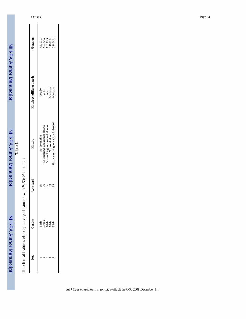
Twenty-four cases of paraffin-embedded pharyngeal cancer blocks were obtained from theDepartment of Pathology of the Columbia University Medical Center (CUMC). Theacquisition of the tissue specimens was approved by the Institutional Review Board andperformed in accordance with Health Insurance Portability and Accountability Act (HIPAA)regulations. The 24 cases studied included five surgical resection specimens and nineteen casesof small biopsy specimens. They came from five female and nineteen male patients, with agesranging from 38 to 78 years-old (average 57.9 ± 12.2 years-old). Eleven were heavy smokers(more than 40 packs per year), two were moderate smokers, two had no smoking and had onlyoccasional alcohol use, and the remaining nine patients’ history was not available. Two of the11 heavy smokers also had heavy alcohol consumption and one abused cocaine. One patientwas a HIV carrier. All patients were diagnosed as squamous cell carcinomas of the pharynx.The grade of cancer in these patients was four well-, 18 moderately-, and two poorly-differentiated. The cases were reviewed by two pathologists and the diagnosis confirmed.
Five 10μm thickness sections were cut for each case and genomic DNA was extracted fromthe tumor tissues using QIAmp DNA Kit (QIAGEN Inc., Valencia, CA). The procedures were
Qiu et al. Page 2
Int J Cancer. Author manuscript; available in PMC 2009 December 14.
NIH
-PA Author Manuscript
NIH
-PA Author Manuscript
NIH
-PA Author Manuscript
performed according to the manufacturer’s instructions for purification of genomic DNA fromparaffin-embedded tissue.
Conventional genomic sequencingExons 9 and 20 of PIK3CA gene were analyzed by PCR amplification of genomic DNA (40ngeach) and direct sequencing of the PCR products. New PCR primers were designed for thisstudy to allow more efficient amplifications of genomic DNA from paraffin-embedded tissues.Primers for exon 9 were also designed to avoid interference from a homologous pseudogenelocated on chromosome 22q11.2 cat eye syndrome region 11. The primers for the PIK3CAexons 9 and 20 are PIK-E9F: CCAGAGGGGAAAAATATGACA; PIK-E9R:CATTTTAGCACTTACCTGTGAC; PIK-E20F: CATTTGCTCCAAACTGACCA; PIK-E20R: TGAGCTTTCATTTTCTCAGTTATCTTTTC. Before sequencing, PCR productswere purified using the Geneclean Turbo Nucleic Acid purification Kit (Qbiogene, Irvine, CA).Finally, purified DNA fragments were sequenced using the corresponding forward PCRprimers. Samples found to have a genetic alteration in the target gene were subsequentlysequenced in the reverse direction to confirm the mutation using the reverse PCR primers. Themutation was then further verified by sequencing of a second PCR product derivedindependently from the original template. All sequencings were performed with ABI’s 3100capillary automated sequencers at the DNA facility of the CUMC 11.
Mutant-enriched sequencing for detecting PIK3CA mutations, H1047R, E545K, and E542KTo detect the PIK3CA hot-spot mutation A3140G (H1047R), each sample (40ng of genomicDNA) was first amplified using outer primers PIK-E20OF(GACATTTGAGCAAAGACCTGAA) and PIK-E20OR (ATCAAACCCTGTTTGCGTTT)for 30 cycles. After this first round of PCR, 2μl from each PCR product were digested with2μl of restriction enzyme BsaBI (10U/ul, New England BioLabs, Ipswich, MA) in a total of50 μl volume at 60°C overnight. Then 2μl of the digest were used for the second round of PCRfor 40 cycles. The primers for the second PCR are PIK-E20IF(CATTTGCTCCAAACTGACCA) and PIK-E20IR(TGAGCTTTCATTTTCTCAGTTATCTTTTC). Each PCR product with the correct size waspurified for DNA sequencing using the same primers as for the second PCR (PIK-E20IF orPIK-E20IR) (Fig. 1A).
For mutant-enriched sequencing of PIK3CA exon 9 hot-spot mutation, G1633A (E545K), theprocedure is similar to the one described above for the hot-spot mutation A3140G, except forthe enzyme and primers used. A mismatch primer PIK-E9MF(TCTACACGAGATCCTCTCTCTGTAATCTC) was used as the forward primer for bothrounds of PCR. The reverse primers for the first and second PCR were respectively PIK-E9OR(GCATTTAATGTGCCAACTACCA) and PIK-E9IR(CTGAGATCAGCCAAATTCAGTTATTTTTTC). The restriction enzyme digestion wasperformed with Hpy188I at 37°C overnight. The reverse PCR primer PIK-E9R was also usedas the DNA sequencing primer (Fig. 1B).
For the hot-spot mutation at exon 9, G1624A (E542K), the PCR strategy of mutant-enrichedsequencing is the same as the one described above for the hot-spot mutation G1633A (E545K),in which a mismatch primer is designed to create a unique restriction enzyme site EcoRI in thePIK3CA exon 9 region. The mismatch primer PIK-2E9MR(CATAGAAAATCTTTCTCCTGCTCAGTGAAT) was used as the reverse primer for bothrounds of PCR. The forward primers for the first and second PCR were respectivelyPIK-2E9OF (GATTGGTTCTTTCCTGTCTCTG) and PIK-2E9IF(TTGCTTTTTCTGTAAATCATCTGTG). The restriction enzyme EcoRI digestion was
Qiu et al. Page 3
Int J Cancer. Author manuscript; available in PMC 2009 December 14.
NIH
-PA Author Manuscript
NIH
-PA Author Manuscript
NIH
-PA Author Manuscript
performed at 37°C overnight. The forward PCR primer PIK-2E9IF was also used as the DNAsequencing primer (Fig. 1C).
The PCR condition for all the PCR reactions is 94°C, 2 minutes; (94°C, 30 seconds; 60°C, 30seconds; 72°C, 30 seconds) × 40 cycles; 72°C, 5 minutes.
RESULTSConventional DNA sequencing detected PIK3CA mutations only in surgically resected butnot in biopsy specimens of pharyngeal cancer
We initially screened for PIK3CA mutation in 24 cases of pharyngeal squamous cell carcinomasamples using the direct genomic sequencing method that we had applied to our previous studyon HNSCC 11. Because most pharyngeal cancers are treated non-surgically (i.e. concurrentchemoradiation), 19 of our specimens were biopsy tissues. Two mutations of PIK3CA wereidentified in the five resection samples and none in the biopsy specimens. One was PIK3CAhot-spot mutation G1633A (E545K) (Fig. 2A). The other mutation, was a missense mutationin exon 20 nucleotide 3127 A→ G, led to a codon 1043 ATG (Met)→GTG (Val) substitution(Fig. 2B). This missense mutation of PIK3CA has been reported previously 18. Both mutationswere not detected in the surrounding normal tissues, thus both were somatic mutations.
Mutant-enriched sequencing identified PIK3CA hotspot mutation H1047R in samplesscreened negative by conventional DNA sequencing approach
Conventional DNA sequencing method only recognizes the mutant DNA if it is present in morethan 10 percent of a mutant/wild-type mixed population in primary tumor tissue 19, 20.Therefore we hypothesized that the low tumor to normal cell ratio in our biopsy specimensmay have generated some false negative results and contributed to the lower frequency ofPIK3CA mutation observed in the current study (2/24, 8.3%) than our previous report (3/6,50%) 11. The unexpectedly low mutation frequency of PIK3CA in the biopsy samples mightalso have been caused by the overall low or poor DNA contents available in these tissues. Weset out to develop a mutant-enriched sequencing method to detect hot-spot mutations ofPIK3CA in specimens with low tumor DNA contribution, such as biopsy samples.
Mutant-enriched sequencing methods generally involve a first-round PCR, restriction enzymedigest, and a second-round PCR. They selectively amplify the mutant copy of a target gene inthe second round of PCR by reducing the wild-type copy number via a restriction enzymedigestion that is specific for the wild-type sequence after the first round PCR. Thus, mutant-enriched DNA sequencing is particularly valuable when the ratio of mutant DNA is expectedto be low. In our method of mutant-enriched sequencing for the detection of PIK3CA exon 20hotspot mutation A3140G (H1047R), we took advantage of a natural restriction enzyme siteon the wild-type sequence that can be recognized and digested by enzyme BsaBI (cutsGATNNNNATC). This enzyme site is destroyed when the DNA is mutated, thus enzymeBsaBI exclusively digests the wild-type PIK3CA exon 20 DNA, but not the mutant A3140G(H1047R) sequence (Fig. 1A).
The feasibility of our method was first investigated in a head and neck cancer cell line, Detroit562, which harbors the H1047R mutation 11. As shown in Figure 3A1–2, both the mutant andwild-type peaks were observed at 1:1 ratio as expected using conventional genomic sequencing(Fig. 3A1), and the wild-type peak was entirely eliminated when our mutant-enrichedsequencing method was applied (Fig. 3A2). To determine the sensitivity of our assay, we madea series of dilutions of the genomic DNA from mutant cell line Detroit 562 with DNA fromanother cell line that has two wild-type PIK3CA alleles. When the ratio of mutant and wild-type DNA copies reached beyond 1: 360, the mutant peak could still be recognized by mutant-
Qiu et al. Page 4
Int J Cancer. Author manuscript; available in PMC 2009 December 14.
NIH
-PA Author Manuscript
NIH
-PA Author Manuscript
NIH
-PA Author Manuscript
enriched sequencing (Fig. 3A2). In contrast, with conventional PCR-sequencing, the mutantpeak disappeared when the ratio of mutant and wild-type DNA reached 1:18. This indicatedthat mutant-enriched sequencing was at least twenty fold more sensitive than direct genomicsequencing.
Using this mutant-enriched sequencing method, two additional mutations of the PIK3CA genewere identified in these 24 pharyngeal cancer samples (data not shown and Table 1). This resultsupported our hypothesis that the low frequency of PIK3CA mutation detected in clinicalpharyngeal cancer biopsy samples by the conventional DNA sequencing method was partiallycaused by the contamination of normal cells.
Mutant-enrich sequencing for PIK3CA exon 9 hotspot mutation E545KFor mutant-enriched sequencing of PIK3CA exon 9 hotspot mutation, G1633A (E545K),mismatch primer (PIK-E9MF) was designed to introduce two A→ T nucleotide mismatchesin the forward primer to create a unique restriction enzyme site Hpy188I (TCNGA) (Fig. 2B),because there is no natural unique restriction enzyme site specific for the wild-type but not themutant DNA sequences at this hot-spot. Using a patient’s tumor DNA with known PIK3CAE545K mutation (patient No. 4, Table 1), we showed that using our mutant-enrichedsequencing method, only the mutant peak remained while the more prominent wild-type peakobserved in the conventional sequencing assay disappeared (Fig. 3B1–2). Applying thispowerful method, we screened the 24 pharyngeal cancer samples. We did not uncover anyadditional cases at this hot-spot mutation.
Mutant-enrich sequencing for PIK3CA exon 9 hotspot mutation E542K (G1624A) and a non-hot-spot mutation E542G (A1625G)
We applied the same mismatch PCR strategy to enrich the mutant allele of PIK3CA hotspotmutation E542K. A restriction enzyme EcoRI site was introduced by the mismatch primerPIK-2E9R. EcoRI digestion disrupted the wild-type PIK3CA DNA, but not the mutant ofPIK3CA E542K sequences (Fig. 1C).
This mutant-enriched sequencing method was tested in a previously reported patient samplewith a known PIK3CA E542K mutation 11. We showed that only the mutant peak remainedwhile the corresponding wild-type peak completely vanished when the mutant-enrichedsequencing was applied (Fig. 3C1–2). Interestingly, this method can detect not only the E542K(G1624A) mutation, but also a previously described non-hot-spot E542G (A1625G) mutation13 (data not shown). We screened the 24 specimens of pharyngeal cancer and an additionalcase of PIK3CA E542K (G1624A) mutation was identified (Fig. 3C3–4).
Five mutations of the PIK3CA gene were identified in the 24 pharyngeal cancer samplesFive mutations of PIK3CA were identified in the 24 cases of pharyngeal cancer by thecombination of conventional sequencing and mutant-enriched sequencing methods (Table 1).Four of the five mutations could have been identified by the mutant-enriched sequencingmethods alone because they were hot-spot mutations (4/5, 80%), but only two were detectableby the conventional sequencing method (2/5, 40%). Two patients with PIK3CA mutations werenot smokers and only drank alcohol occasionally, while one was a heavy smoker and drankalcohol occasionally (Table 1). There is no apparent association between PIK3CA mutationand smoking or alcohol consumption. There is also no apparent association betweenPIK3CA mutation and the degree of tumor cell differentiation (Table 1).
Qiu et al. Page 5
Int J Cancer. Author manuscript; available in PMC 2009 December 14.
NIH
-PA Author Manuscript
NIH
-PA Author Manuscript
NIH
-PA Author Manuscript
DISCUSSIONPreviously we reported PIK3CA mutations in head and neck cancers (4/38, 10%), three of thefour were identified in pharyngeal cancer samples (3/6, 50%) 11. However, whether PIK3CAcan be used as a potential biomarker for diagnosis and molecular target therapy in pharyngealcancer was unclear, due to the small sample number available at the time. Therefore, we decidedto investigate the frequency of PIK3CA mutation in pharyngeal cancer using 24 additionalsamples.
Initially, only two mutations of PIK3CA, including a hotspot mutation E545K and a missensemutation in exon 20 nucleotide 3127 A→ G, were found in five surgically resection specimensand none in 19 biopsy specimens by the conventional genomic sequencing method. Weattributed this low mutation frequency to the quality of clinical biopsy samples. Clinical biopsysamples often contain small numbers of tumor cells mixed with a large population of normalcells. The mutant DNA is often missed by the conventional PCR- sequencing method. Toincrease the sensitivity of detecting PIK3CA gene mutation, we developed novel mutant-enriched DNA sequencing methods for its three hot-spot mutations, H1047R, E545K andE542K. The three hot-spot mutations account for 78.6% of all PIK3CA mutations reported 7,21. We were able to show that mutant-enriched sequencing can identify the H1047R mutantDNA in a mixed population with wild-type DNA at sensitivity of 0.0028 (1 mutant: 360 wild-type DNA copies). Using this mutant-enriched sequencing method for H1047, we found twoadditional mutations in these pharyngeal cancer samples. An additional mutation was identifiedby the mutant-enriched sequencing protocol for hotspot mutation H542K (G1624A). Thus, atotal of five PIK3CA mutations were identified in 24 cases of pharyngeal cancer in combinationof regular DNA sequencing and mutant-enriched sequencing. It is important to note that fourof the five mutations were hot-spot mutations and could have been identified by the mutant-enriched sequencing methods alone (4/5, 80%), but only two were detectable by theconventional sequencing method (2/5, 40%). This means that the ability to detect PIK3CAmutations increased by 200% when the mutant-enriched sequencing methods are utilized. The80% detection rate is within the expectation, because the three hot-spot mutations account for~80% of total PIK3CA mutations. Two patients with PIK3CA mutations did not have a historyof smoking or alcohol-abuse. This suggests that PIK3CA mutation might be a critical cause forthose pharyngeal cancer patients without history of smoking and alcohol consumption.PIK3CA mutation was not found associated with the degree of differentiation in the currentstudy.
The primers for the mutant-enriched sequencing analyses of the PIK3CA exon 9 hotspotmutations were designed to avoid interference from a homologous pseudogene located onchromosome 22q11.2 cat eye syndrome region 11. At least one of the two primers for eachPCR amplification contains mismatched base pairs to the pseudogene. Those mismatchednucleotides in either the forward and/or reverse primers seemed sufficient to prevent PCRamplification of the pseudogene in our mutant-enriched sequencing analyses. This wassupported by the fact that we never observed the A1634C change in our samples. The A1634Cchange was mistaken as a mutation in previous literatures, when in fact it is only one of thedissimilarities between the pseudogene and PIK3CA at Exon 9, nucleotide 1634 (it’s A forPIK3CA and C for the pseudogene) 11. We also did not observe the frameshift change that hadalso been associated with the amplification of the pseudogene 11.
Comparing to mutant-selective PCR and restriction fragment length polymorphism (RFLP)analysis 19, 20, we believe that our mutant-enriched sequencing method is more sensitive andspecific because: 1) there is the possibility of the non-specific digestion by the restrictionenzyme when confirming the mutation by PCR-RFLP; 2) small amount of mutant DNA afterdigestion may not be enough to be visualized on an agarose gel in the PCR-RFLP analysis.
Qiu et al. Page 6
Int J Cancer. Author manuscript; available in PMC 2009 December 14.
NIH
-PA Author Manuscript
NIH
-PA Author Manuscript
NIH
-PA Author Manuscript
The mutant-enriched sequencing method directly displays the exact nucleotide sequence; 3)the mutant-enrich sequencing method is not limited by available restriction enzyme sites. Aunique enzyme site could be introduced by mismatch PCR, as we have done for the detectionof PIK3CA exon 9 hot-spot mutations E545K and E542K. Thus, the mutant-enrichedsequencing method is more superior to both the PCR-RFLP analysis and the conventionalgenomic sequencing assay for detecting hot-spot mutations. This method is particularlyvaluable in clinical applications where tumor samples are often mixed with a large populationof normal cells.
Our current study concluded that PIK3CA mutation frequency in pharyngeal cancer is ~21%(5/24). A recent study of PIK3CA mutation in nasopharyngeal carcinoma reported twomutations in six cell lines and but none was identified in 40 clinical samples (4.3% or 2/46)15. It is possible that the low tumor to normal DNA ratio in their clinical samples might havecontributed to the negative finding in that study. Thus, a more sensitive assay could haveimproved the detection of PIK3CA mutations in such clinical samples where limited amountsof tumor DNA were available.
In conclusion, sensitive mutant-enriched sequencing methods were developed to detect thethree hotspot mutations of the PIK3CA gene in clinical tumor samples. This novel detectionmethod can detect approximately 80% of all PIK3CA mutations 21 and is valuable in clinicalapplications particularly in samples with little tumor cell contribution (i.e. biopsy samples) orwith tumor subclones that usually go undetected by the conventional sequencing methodbecause of their minor cellular populations. Several studies have shown that PIK3CA and itspathway are potential targets for chemotherapy or radiation therapy, including target therapiesfor EGFR, Her-2, mTOR, and Akt 22, 23. Therefore, the clinical applications of the mutant-enriched sequencing methods would have great potential impacts on early detection and targettherapy for many cancer types harboring frequent PIK3CA mutations (e.g. breast cancer,colorectal cancer).
AcknowledgmentsThis work was supported by the NCI Temin Award CA095434 and the NCI R01 CA109525.
The abbreviations used are
HNSCC Head and neck squamous cell carcinoma
PCR polymerase chain reaction
PI3K phosphatidylinositol 3-kinase
References1. Jemal A, Siegel R, Ward E, Murray T, Xu J, Smigal C, Thun MJ. Cancer statistics. CA Cancer J Clin
2006;56:106–30. [PubMed: 16514137]2. Carvalho AL, Nishimoto IN, Califano JA, Kowalski LP. Trends in incidence and prognosis for head
and neck cancer in the United States: a site-specific analysis of the SEER database. Int J Cancer2005;114:806–16. [PubMed: 15609302]
3. Forastiere A, Koch W, Trotti A, Sidransky D. Head and neck cancer. N Engl J Med 2001;345:1890–900. [PubMed: 11756581]
4. Williams HK. Molecular pathogenesis of oral squamous carcinoma. Mol Pathol 2000;53:165–72.[PubMed: 11040937]
5. Ma YY, Wei SJ, Lin YC, Lung JC, Chang TC, Whang-Peng J, Liu JM, Yang DM, Yang WK, ShenCY. PIK3CA as an oncogene in cervical cancer. Oncogene 2000;19:2739–44. [PubMed: 10851074]
Qiu et al. Page 7
Int J Cancer. Author manuscript; available in PMC 2009 December 14.
NIH
-PA Author Manuscript
NIH
-PA Author Manuscript
NIH
-PA Author Manuscript

6. Shayesteh L, Lu Y, Kuo WL, Baldocchi R, Godfrey T, Collins C, Pinkel D, Powell B, Mills GB, GrayJW. PIK3CA is implicated as an oncogene in ovarian cancer. Nat Genet 1999;21:99–102. [PubMed:9916799]
7. Samuels Y, Wang Z, Bardelli A, Silliman N, Ptak J, Szabo S, Yan H, Gazdar A, Powell SM, RigginsGJ, Willson JK, Markowitz S, et al. High frequency of mutations of the PIK3CA gene in humancancers. Science 2004;304:554. [PubMed: 15016963]
8. Kang S, Bader AG, Vogt PK. Phosphatidylinositol 3-kinase mutations identified in human cancer areoncogenic. Proc Natl Acad Sci U S A 2005;102:802–7. [PubMed: 15647370]
9. Lee JW, Soung YH, Kim SY, Lee HW, Park WS, Nam SW, Kim SH, Lee JY, Yoo NJ, Lee SH. PIK3CAgene is frequently mutated in breast carcinomas and hepatocellular carcinomas. Oncogene2005;24:1477–80. [PubMed: 15608678]
10. Campbell IG, Russell SE, Choong DY, Montgomery KG, Ciavarella ML, Hooi CS, Cristiano BE,Pearson RB, Phillips WA. Mutation of the PIK3CA gene in ovarian and breast cancer. Cancer Res2004;64:7678–81. [PubMed: 15520168]
11. Qiu W, Schonleben F, Li X, Ho DJ, Close LG, Manolidis S, Bennett BP, Su GH. PIK3CA mutationsin head and neck squamous cell carcinoma. Clin Cancer Res 2006;12:1441–6. [PubMed: 16533766]
12. Schonleben F, Qiu W, Ciau NT, Ho DJ, Li X, Allendorf JD, Remotti HE, Su GH. PIK3CA mutationsin intraductal papillary mucinous neoplasm/carcinoma of the pancreas. Clin Cancer Res2006;12:3851–5. [PubMed: 16778113]
13. Samuels Y, Velculescu VE. Oncogenic mutations of PIK3CA in human cancers. Cell Cycle2004;3:1221–4. [PubMed: 15467468]
14. Garcia-Rostan G, Costa AM, Pereira-Castro I, Salvatore G, Hernandez R, Hermsem MJ, Herrero A,Fusco A, Cameselle-Teijeiro J, Santoro M. Mutation of the PIK3CA gene in anaplastic thyroid cancer.Cancer Res 2005;65:10199–207. [PubMed: 16288007]
15. Or YY, Hui AB, To KF, Lam CN, Lo KW. PIK3CA mutations in nasopharyngeal carcinoma. Int JCancer 2006;118:1065–7. [PubMed: 16114017]
16. Pedrero JM, Carracedo DG, Pinto CM, Zapatero AH, Rodrigo JP, Nieto CS, Gonzalez MV. Frequentgenetic and biochemical alterations of the PI 3-K/AKT/PTEN pathway in head and neck squamouscell carcinoma. Int J Cancer 2005;114:242–8. [PubMed: 15543611]
17. Woenckhaus J, Steger K, Werner E, Fenic I, Gamerdinger U, Dreyer T, Stahl U. Genomic gain ofPIK3CA and increased expression of p110alpha are associated with progression of dysplasia intoinvasive squamous cell carcinoma. J Pathol 2002;198:335–42. [PubMed: 12375266]
18. Kozaki K, Imoto I, Pimkhaokham A, Hasegawa S, Tsuda H, Omura K, Inazawa J. PIK3CA mutationis an oncogenic aberration at advanced stages of oral squamous cell carcinoma. Cancer Sci2006;97:1351–8. [PubMed: 17052259]
19. Levi S, Urbano-Ispizua A, Gill R, Thomas DM, Gilbertson J, Foster C, Marshall CJ. Multiple K-rascodon 12 mutations in cholangiocarcinomas demonstrated with a sensitive polymerase chain reactiontechnique. Cancer Res 1991;51:3497–502. [PubMed: 1675933]
20. Nakahori S, Yokosuka O, Ehata T, Chuang WL, Imazeki F, Ito Y, Ohto M. Detection of hepatitis Bvirus precore stop codon mutants by selective amplification method: frequent detection of precoremutants in hepatitis B e antigen positive healthy carriers. J Gastroenterol Hepatol 1995;10:419–25.[PubMed: 8527708]
21. Bader AG, Kang S, Zhao L, Vogt PK. Oncogenic PI3K deregulates transcription and translation. NatRev Cancer 2005;5:921–9. [PubMed: 16341083]
22. Rogers SJ, Box C, Harrington KJ, Nutting C, Rhys-Evans P, Eccles SA. The phosphoinositide 3-kinase signalling pathway as a therapeutic target in squamous cell carcinoma of the head and neck.Expert Opin Ther Targets 2005;9:769–90. [PubMed: 16083342]
23. Kim DW, Huamani J, Fu A, Hallahan DE. Molecular strategies targeting the host component of cancerto enhance tumor response to radiation therapy. Int J Radiat Oncol Biol Phys 2006;64:38–46.[PubMed: 16377414]
Qiu et al. Page 8
Int J Cancer. Author manuscript; available in PMC 2009 December 14.
NIH
-PA Author Manuscript
NIH
-PA Author Manuscript
NIH
-PA Author Manuscript

Fig. 1. The schematic of the mutant-enriched sequencing methods for detecting PIK3CA mutations,H1047R, E545K and E542KA. The protocol for detectingPIK3CA mutation H1047R. Enzyme BsaBI specifically cuts thewild-type sequences of the exon 20, but not the mutant copies with A3140G nucleotidealteration. After digesting the first PCR product with the enzyme BsaBI, the second PCRselectively amplifies the mutant copies. B. A unique restriction enzyme site Hpy188I wasintroduced by mismatch PCR for detecting PIK3CA mutation E545K. The mismatch primer(PIK-E9MF) has two A→ T nucleotide substitutions in the forward primer to create a uniqueenzyme site Hpy188I in the wild-type sequences of the PIK3CA exon 9, but not the mutantsequences. C. The mismatch primer PIK-2E9MR was designed to create a unique restriction
Qiu et al. Page 9
Int J Cancer. Author manuscript; available in PMC 2009 December 14.
NIH
-PA Author Manuscript
NIH
-PA Author Manuscript
NIH
-PA Author Manuscript

enzyme site EcoRI for enriching PIK3CA mutation E542K (G1624A) and E542G (A1625G)with the similar strategy for the hot-spot mutation E545K.
Qiu et al. Page 10
Int J Cancer. Author manuscript; available in PMC 2009 December 14.
NIH
-PA Author Manuscript
NIH
-PA Author Manuscript
NIH
-PA Author Manuscript
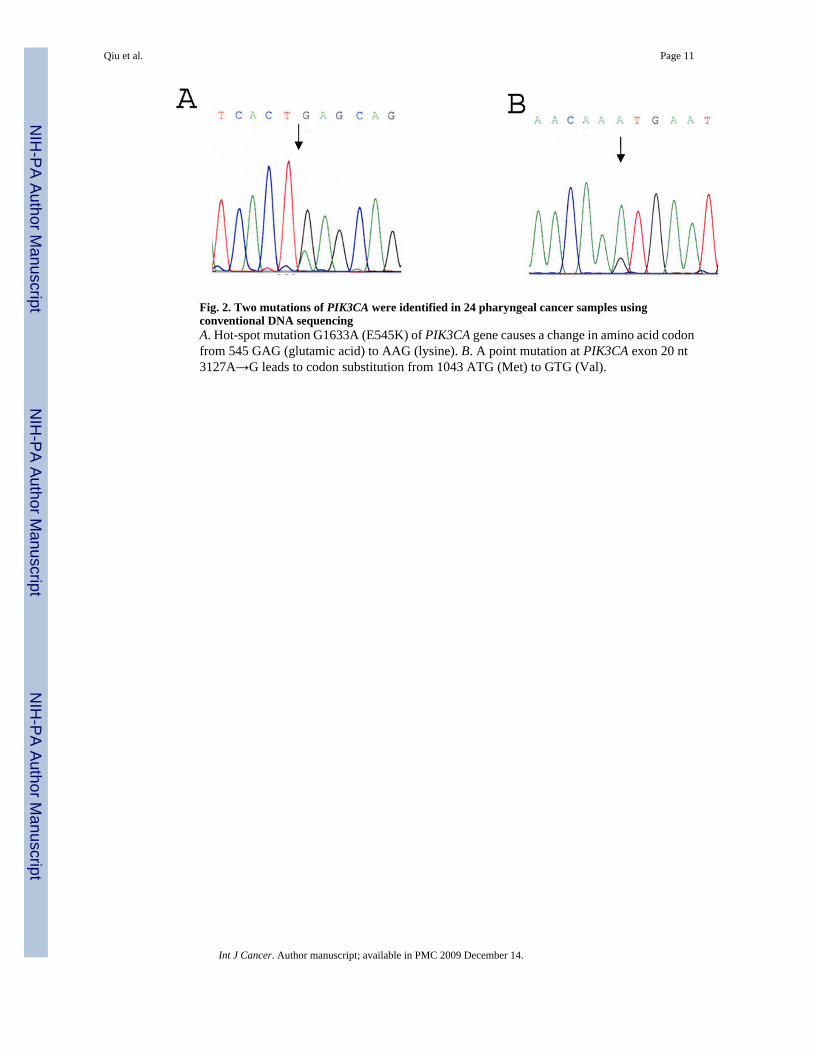
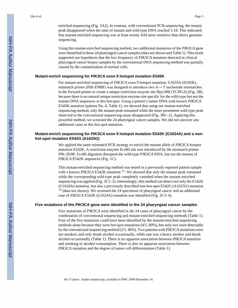
Fig. 2. Two mutations of PIK3CA were identified in 24 pharyngeal cancer samples usingconventional DNA sequencingA. Hot-spot mutation G1633A (E545K) of PIK3CA gene causes a change in amino acid codonfrom 545 GAG (glutamic acid) to AAG (lysine). B. A point mutation at PIK3CA exon 20 nt3127A→G leads to codon substitution from 1043 ATG (Met) to GTG (Val).
Qiu et al. Page 11
Int J Cancer. Author manuscript; available in PMC 2009 December 14.
NIH
-PA Author Manuscript
NIH
-PA Author Manuscript
NIH
-PA Author Manuscript
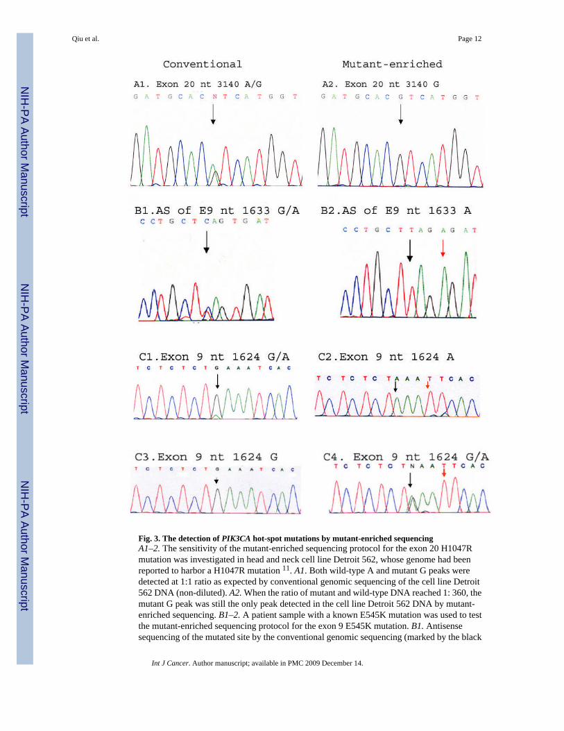
Fig. 3. The detection of PIK3CA hot-spot mutations by mutant-enriched sequencingA1–2. The sensitivity of the mutant-enriched sequencing protocol for the exon 20 H1047Rmutation was investigated in head and neck cell line Detroit 562, whose genome had beenreported to harbor a H1047R mutation 11. A1. Both wild-type A and mutant G peaks weredetected at 1:1 ratio as expected by conventional genomic sequencing of the cell line Detroit562 DNA (non-diluted). A2. When the ratio of mutant and wild-type DNA reached 1: 360, themutant G peak was still the only peak detected in the cell line Detroit 562 DNA by mutant-enriched sequencing. B1–2. A patient sample with a known E545K mutation was used to testthe mutant-enriched sequencing protocol for the exon 9 E545K mutation. B1. Antisensesequencing of the mutated site by the conventional genomic sequencing (marked by the black
Qiu et al. Page 12
Int J Cancer. Author manuscript; available in PMC 2009 December 14.
NIH
-PA Author Manuscript
NIH
-PA Author Manuscript
NIH
-PA Author Manuscript

arrow) detected both the wild-type and the mutant alleles. B2. The peak representing the wild-type allele in the same sample disappeared when the mutant-enriched sequencing method wasapplied (indicated by the black arrow). The red arrow marks the nucleotide A1630T changethat was introduced by mismatch primer (PIK-E9MF) in order to generate the restrictionenzyme Hpy188I (TCNGA) site. C1–4. The mutant-enriched sequencing method identified anundetected PIK3CA hotspot mutation E542K (G1624A) by the conventional sequencing.C1. Forward sequencing of a clinical sample with a known E542K mutation by the conventionalgenomic sequencing method (marked by the black arrow) displayed a dominant presence ofthe wild-type allele over the mutant allele. C2. The wild-type allele in the same sampledisappeared when the mutant-enriched sequencing method was applied (indicated by the blackarrow). The red arrow marks the nucleotide A1627T change that was introduced by mismatchprimer (PIK-2E9MF) to generate a unique restriction enzyme EcoRI (GAATTC) site. The caseof pharyngeal cancer that had been tested negative for a mutation by the conventional genomicsequencing method (C3), but was subsequently identified with a PIK3CA E542K mutationusing the mutant-enriched sequencing method (C4).
Qiu et al. Page 13
Int J Cancer. Author manuscript; available in PMC 2009 December 14.
NIH
-PA Author Manuscript
NIH
-PA Author Manuscript
NIH
-PA Author Manuscript
NIH
-PA Author Manuscript
NIH
-PA Author Manuscript
NIH
-PA Author Manuscript
Qiu et al. Page 14Ta
ble
1
The
clin
ical
feat
ures
of f
ive
phar
ynge
al c
ance
rs w
ith P
IK3C
A m
utat
ion.
No.
Gen
der
Age
(yea
r)H
isto
ryH
isto
logy
(diff
eren
tiate
d)M
utat
ion
1M
ale
59N
ot A
vaila
ble
Poor
lyA
3127
G2
Fem
ale
70N
o sm
okin
g, o
ccas
iona
l alc
ohol
Wel
lA
3140
G3
Mal
e66
No
smok
ing,
occ
asio
nal a
lcoh
olW
ell
A31
40G
4M
ale
43N
ot A
vaila
ble
Mod
erat
eG
1633
A5
Mal
e64
Hea
vy sm
okin
g, o
ccas
iona
l alc
ohol
Mod
erat
eG
1624
A
Int J Cancer. Author manuscript; available in PMC 2009 December 14.