Lung volume-related ch~nges in the pharyngeal area of ...
8
Eur Respir J 1989, 2, Lung volume-related in the pharyngeal area of obese females with and without obstructive sleep apnoea I. Rubinstein, V. Hoffstein, T.D. Bradley Lung volume-related changes in the pharyngeal area of obese females with and without obstructive sleep apnoea. I. Rubinstein, V. Hoffstein, T.D. Brad fey. ABSTRACT: The majority or male patients with obstructive sleep apnoea (OSA) have an abnormal pharyngeal structure and function, with epl· sodlc complete airway occlusion during sleep. Since OSA Is less common In females than In males, lt Is possible that other abnormalities are active In female patients with OSA. Consequently, we measured pharyn- geal area and its lung volume-related changes (LVRC) from functional residual capacity (FRC) to residual volume (RV) in overweight females, 14 with OSA and 14 without OSA. Pharyngeal areas were measured using the acoustic renection technique. While there were no signincant dl"erences In pharyngeal area between the OSA and control groups at either FRC (mean±so, 3.49±0.46 cm 2 vs 3.08±0.63 cm 1 ) or RV (2.86±0.47 cm 2 I'S 2.67±0.49 cm 1 ), the reduction In pharyngeal area between FRC and RV was si&:nlncantly greater In the OSA group (0.63±0.23 cm 1 I'S 0.33±0.32 cm 1 , pd>.OS). Furthermore, although the expiratory reserve volume (ERV) was not slgnlncantly different between the two groups (0.4±0.2 l I'S 0.4±0.3 1), LVRC, defined as the reduction In pharyngeal area normalized by ERV, was significantly higher 1!1 the females with OSA than In the non-apnoeic controls (2.68±2.24 cm 2 ·t 1 vs 1.17±1.23 cm 2 ·t 1 , p<0.02). We conclude that females with OSA have abnormal pha- ryngeal mechanics similar to males with OSA. Eur Respir J., 1989, 2, 344-351. Departments of Medicine, Toronto Hospital and St. Michael's Hospital, and search Laboratory, Queen Eliubeth Ho'J!ltal versity of Toronto, Canada. ' Correspondence: Dr T. D. Bradley, !Ja: lll rttn- Mcdicine, ENI0-212, Toronto 200 Eliubeth Stred, Toronto, Ontario MSG 2C4. ' Keywords: Acoustic reflection; airway area; obesit.M. pharynl!; sleep. Supported by the Medical Research Canada operating grant MA 6669, ians Service of Ontario, Inc., and by Institute of the Queen Elizabeth I. Rubinstein is a feUow of the Canadian Association. The pathophysiology of obstructive sleep apnoea (OSA) has been well studied in males, who are affcc!ed far more frequently than females by this disorder [1, 2]. While the pathogenesis of recurrent upper airway obslruction in males is multifactorial, pharyngeal nar- rowing and increased pharyngeal collapsibility have been identified as important factors rendering the phar· ynx susceptible to occlusion during sleep In contrast. little is known about the pathogenesis of OSA in females. Whereas obesity is a predisposing factor for the development of OSA, the much lower preva- lence of OSA in females as compared to males fl) is surprising in view of the higher incidence of obesity among women [7]. This raises the question as to whether pharyngeal abnormalities observed in males with OSA arc also present in apnoeic females. females with and without OSA using !he reflection technique [3, 5, 6, 8). Our findings s that in overweight females, just as in overweight abnormalities of pharyngeal function may playr important role in the pathogenesis of OSA. Nevertheless, since pharyngeal collapse is the critical event in OSA, we hypothesized th at pharyngeal dys- function may play a role in the pathogenesis of OSA in females as is the case in males. In order to test this hypothesis. pharyngeal cross-sectional area and its changes with lung volume during continuous slow expiration from functional residual capacity (FRC) to residual volume (RV) were measured in overweight Methods Subjects Subjects consisted of 14 consecutive female patienlt willl OSA and 14 female control subjects All patients and controls were overweight. as by a body mass index (BMI) greater than 24 (9), All patients were referred to the sleep laboratory W cause of clinical suspicion of sleep apnoea. Among .: 14 control subjects, 10 were referred because of cal suspicion of OSA, while the other 4 were refornu for routine evaluation prior to gastroplasty fo.r wel s: reduc ti on. None of the patients or control subJects .h any clinical or laboratory evidence of All apnoeic and non-apnoeic patients had a h1stoiY habitual snoring.
-
Upload
khangminh22 -
Category
Documents
-
view
3 -
download
0
Transcript of Lung volume-related ch~nges in the pharyngeal area of ...
Eur Respir J 1989, 2, 3~4~51
Lung volume-related ch~nges in the pharyngeal area of obese females with and without obstructive sleep apnoea
I. Rubinstein, V. Hoffstein, T.D. Bradley
Lung volume-related changes in the pharyngeal area of obese females with and without obstructive sleep apnoea. I. Rubinstein, V. Hoffstein, T.D. Brad fey. ABSTRACT: The majority or male patients with obstructive sleep apnoea (OSA) have an abnormal pharyngeal structure and function, with epl· sodlc complete airway occlusion during sleep. Since OSA Is less common In females than In males, lt Is possible that other abnormalities are active In female patients with OSA. Consequently, we measured pharyngeal area and its lung volume-related changes (LVRC) from functional residual capacity (FRC) to residual volume (RV) in overweight females, 14 with OSA and 14 without OSA. Pharyngeal areas were measured using the acoustic renection technique. While there were no signincant dl"erences In pharyngeal area between the OSA and control groups at either FRC (mean±so, 3.49±0.46 cm2 vs 3.08±0.63 cm1) or RV (2.86±0.47 cm2 I'S 2.67±0.49 cm1), the reduction In pharyngeal area between FRC and RV was si&:nlncantly greater In the OSA group (0.63±0.23 cm1 I'S
0.33±0.32 cm1, pd>.OS). Furthermore, although the expiratory reserve volume (ERV) was not slgnlncantly different between the two groups (0.4±0.2 l I'S 0.4±0.3 1), L VRC, defined as the reduction In pharyngeal area normalized by ERV, was significantly higher 1!1 the females with OSA than In the non-apnoeic controls (2.68±2.24 cm2·t1 vs 1.17±1.23 cm2·t1, p<0.02). We conclude that females with OSA have abnormal pharyngeal mechanics similar to males with OSA. Eur Respir J., 1989, 2, 344-351.
Departments of Medicine, Toronto Hospital and St. Michael's Hospital, and search Laboratory, Queen Eliubeth Ho'J!ltal versity of Toronto, Canada. '
Correspondence: Dr T. D. Bradley, !Ja:lllrttnMcdicine, ENI0-212, Toronto 200 Eliubeth Stred, Toronto, Ontario MSG 2C4. '
Keywords: Acoustic reflection; airway area; obesit.M. pharynl!; sleep.
Supported by the Medical Research Canada operating grant MA 6669, ians Service of Ontario, Inc., and by Institute of the Queen Elizabeth I. Rubinstein is a feUow of the Canadian Association.
The pathophysiology of obstructive sleep apnoea (OSA) has been well studied in males, who are affcc!ed far more frequently than females by this disorder [1, 2]. While the pathogenesis of recurrent upper airway obslruction in males is multifactorial, pharyngeal narrowing and increased pharyngeal collapsibility have been identified as important factors rendering the phar· ynx susceptible to occlusion during sleep [3~]. In contrast. little is known about the pathogenesis of OSA in females. Whereas obesity is a predisposing factor for the development of OSA, the much lower prevalence of OSA in females as compared to males fl) is surprising in view of the higher incidence of obesity among women [7]. This raises the question as to whether pharyngeal abnormalities observed in males with OSA arc also present in apnoeic females.
females with and without OSA using !he reflection technique [3, 5, 6, 8). Our findings s that in overweight females, just as in overweight abnormalities of pharyngeal function may playr important role in the pathogenesis of OSA.
Nevertheless, since pharyngeal collapse is the critical event in OSA, we hypothesized that pharyngeal dysfunction may play a role in the pathogenesis of OSA in females as is the case in males. In order to test this hypothesis. pharyngeal cross-sectional area and its changes with lung volume during continuous slow expiration from functional residual capacity (FRC) to residual volume (RV) were measured in overweight
Methods
Subjects
Subjects consisted of 14 consecutive female patienlt willl OSA and 14 female control subjects without~~ All patients and controls were overweight. as de~nw by a body mass index (BMI) greater than 24 kg·m·~ (9), All patients were referred to the sleep laboratory W cause of clinical suspicion of sleep apnoea. Among.: 14 control subjects, 10 were referred because of o~ cal suspicion of OSA, while the other 4 were refornu for routine evaluation prior to gastroplasty fo.r wels: reduction. None of the patients or control subJects .h any clinical or laboratory evidence of hypoth~roid•~ All apnoeic and non-apnoeic patients had a h1stoiY habitual snoring.
PHARYNX IN OBESE FEMALES WITii SLEEP APNOEA 345
sleep studies were performed in all 28 using standard polysomnographic techniques
Respiratory movements of the rib cage and abdoas well as tidal volume were measured using a
inductance plethysmograph (RespiLracc, Monitoring, Ardsley, NY) calibrated by the equations method. An car oximeter (model
Hcwlell-Packard Corp., Waltham, MA) was ror continuous monitoring or anerial oxygen satu(Saol). All variables were recorded continuously
polygraph (Model 78, Grass Instruments, Quincy, at a paper speed of 15 mm·s·1•
stages were scored according to slandard [10). ObstruCtive apnoeas were identified as the of tidal volume excursions for at least 10 s
which l.here were paradoxical movements of the and abdomen. Obstructive hypopnoeas were
reductions in tidal volume at least 50%
below the baseline, lasting greater than 10 s and accompanied by paradoxical movements of the rib cage and abdomen (2, 6]. The numbe.r of apnoeas plus hypopnocas per hour of sleep was calculated and termed the apnoea-hypopnoea index (AfD). The diagnosis of OSA was cslablished if the ARl was greater than 10. The lowest nocrumal arterial oxygen saturation (low Sao
2)
was also recorded.
Pulmonary function
Lung volumes and maximum expiratory flow rates were measured in all subjects by body plethysmography and spirometry.
Pharyngeal areas
These were determined in all 28 subjects using the acoustic reflection technique as described previously [5]. The basic principles of the technique, experimental
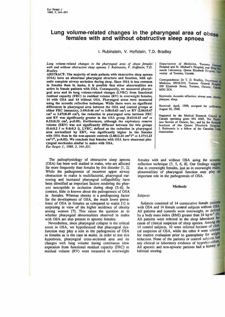
Table 1. -Anthropometric data, menopausal status, and sleep data
Subj~t Age Weight BMI Menopausal AHI Low Sao2 yrs kg kg·m·2 starus %
OSA
1 56 102 40 post 45 37 2 50 149 54 post 49 51 3 56 83 37 post 18 64 4 ss 81 33 post 23 81 5 40 91 33 pre 56 62 6 29 127 51 pre 50 81 7 61 109 43 post 33 55 8 53 81 34 post 39 82 9 46 121 46 pre 13 72 10 51 110 46 pre 14 67 11 46 99 39 pre 21 92 12 39 102 51 pre 21 50 13 42 117 44 pre 52 76 14 58 100 37 post 21 58
Mean 49 105 42 33 67 ±so 9 19 7 16 15
Non-OSA
1 60 84 35 post 7 40 2 36 96 37 pre 6 86 3 37 129 38 pre 0 87 4 40 135 47 pre 2 91 5 49 152 60 pre 9 65 6 28 135 48 pre 6 82 7 42 78 32 pre 2 95 8 55 132 53 post 7 80 9 50 121 49 post 0 91 10 56 133 53 post 0 85 11 45 122 44 pre 4 90 12 52 117 43 post 2 91 13 58 68 27 post 4 90 14 45 122 45 pre 3 88
Mean 47 116 44 4 83 ±so 9 25 9 3 14
---p NS NS NS <0.001 <0.005
----OSA: obstructive sleep apnoea; BMI: body ma.ss index; AHI: apnoea-hypopnoea index; Low Sao2: lowest nocrumal arterial oxygen saturation; NS: not sigruficant.
346 I. RUBINST£liN, V. HOFFSTEIN, T.D. BRADLEY
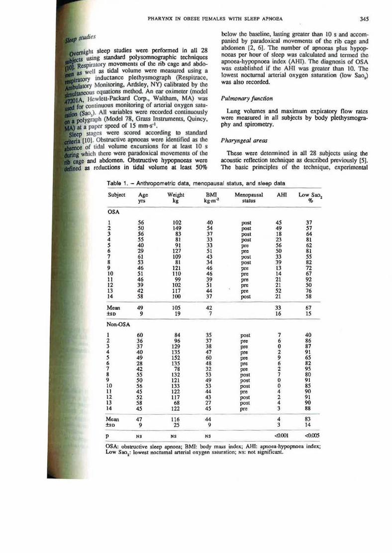
Table 2. - Pulmonary function data
Subject ffiC RV ERV FEY/PVC % pred % pred 1 ratio % pred
OSA
1 3.1 (119) 2.8 (165) 0.3 0.53 (71) 2 2.5 (78) 2.3 (121) 0.2 0 .59 (92) 3 2.5 (106) 2.2 (145) 0.3 0.80 (108) 4 2 .1 (84) 1.9 (119) 0.2 0.71 (95) 5 3.0 (100) 2.3 (135) 0.7 0.78 (102) 6 1.9 (70) 1.3 (93) 0.6 0.75 (95) 7 2.7 (108) 2.2 (129) 0 .5 0 .84 (115) 8 2.2 (88) 1.5 (94) 0.7 0.75 (100) 9 2.2 (69) 2.1 (117) 0 .1 0.88 (116) 10 1.9 (76) 1.6 (100) 0.3 0.83 (110) 11 2.6 (105) 2.3 (163) 0.3 0.77 (101) 12 1.9 (110) 1.8 (178) 0.1 0.90 (116) 13 2.8 (91) 2.5 (141) 0.3 0.77 (101) 14 2 .6 (83) 2.1 (105) 0.5 0.83 (115)
Mean 2.4 (92) 2.1 (128) 0.4 0.77 (103) ·±sO 0.4 (16) 0.4 (26) 0.2 0.09 (12)
Non-OSA
1 3.2 (139) 3.0 (200) 0.2 0.65 (88) 2 2.2 (85) 1.9 (136) 0.3 0.86 (110) 3 2.0 (51) 1.6 (76) 0.4 0.80 (102) 4 2.5 (80) 2.0 (115) 0.5 0.78 (102) 5 2.7 (96) 2.6 (153) 0.1 0.86 (114) 6 2.2 (71) 1.5 (94) 0.7 0 .83 (105) 7 1.9 (73) 0.8 (53) 1.1 0.85 (111) 8 3.4 (126) 3.2 (192) 0.2 0.77 (103) 9 2.3 (78) 2.0 (114) 0.3 0.85 (113) 10 3.2 (111) 3 .0 (165) 0.2 0.71 (96) 11 1.6 (63) 1.4 (92) 0.2 0.88 (115) 12 1.8 (60) 1.4 (78) 0.4 0.73 (98) 13 2 .3 (85) 1.7 (100) 0.6 0.70 (95) 14 3.2 (106) 2.8 (160) 0.4 0.63 (82)
Mean 2.5 (87) 2.1 (123) 0.4 0.78 (102) ±so 0.6 (25) 0.7 (45) 0.3 0.08 (10)
FRC: functionaJ rc.siduol capacity; RV: TC$idunl volume; ERV: expiratory reserve volume; FEY 1: rorced expiratory volume in one second; FVC: forced vitaJ capi!City; OSA: obslrucllvo sleep apnoCll; 1: lilte.~. No significant di rrcrcnees bcawecn variables for OSA or nonOSA.
set-up, mourhpiece construction, and software for data processing are identical to that described previously, and will be summarized here only briefly [3, 5, 6). The technique is based on measurements of high fre<Juency sound waves which are aimed at !he mouth and reflected as they propagate along the respiratory tract. From !he knowledge of reflected intensities and the Lime of arrival of reflections, the area at a given distance from the sensing microphone is computed, and a plot of airway cross-sectional area as a function of distance into the airway is constructed. We shall refer to such plots as the area-distance plots. The apparatus used in the present study consists of an acoustic emitter (loudspeaker), an acoustic transducer (microphone), and a 2 m long stainless steel wavetube. Appropriate amplifiers and filters condition the signal which is then processed to calculate and display the changes in airway area on the computer terminal screen. Simullancous
measurements of lung volume are obtained from the po· tentiometer attachment of the spirometer coupled to the wavetube. The system allows for the acquisition of 64 consecutive uninterrupted area-distance measurements as quickly as 5 times·s·•.
Experimental design
Acoustic reflection studies were performed with an awake subject in the seated position. wearing a noseclip, and coupled to the wavetube by means of a custom-made mouthpiece constructed from soft dental wax. Care was taken to ensure that the subject was comfortable and maintained the same neutral head position and neclc configuration during all measurements. First, the subject comfortably breathed an 80% helium-20% oxygen mixture for 2- 3 min from a
PHARYNX IN OBESE FllMAlBS WlTII SLBBP APNOEA 347
bag attached 10 the proximal end of the by means of a three-way valve. During this equiJibmtion, three slow vital capacity ma
perfonned in order 10 s tandardize lung . The measurements began by changing the three-way valve, thereby enabling the
breathe through the wavetube into the spiwhich was also filled with the same gas In each individual we made simultaneous
ts of lung volume and pharyngeal crossarea. Measurements were performed while the
awake subject exhaled slowly from total lung (TLC) 10 residual volume (RV) and, in a
during quiet tidal breathing at FRC. manoeuvre, 64 measurements of pharyngeal lung volume were obtained at intervals of
s, and two separate slow expiratory manoeuvres pedormcd. Mean pharyngeal cross-sectional area detennined at FRC (AFRc) and at RV (ARv) by
integrating the total area between the mouthpiece and the glottis and dividing by the distance of integration [5, 6). Up to 128 measurements of area were acquired at FRC and 12-24 measurements were available at RV . We chose to measure pharyngeal area at FRC and RV because these are the lung volumes between which patients would be breathing in the recumbent position during sleep when lung volume is at or below the upright FRC. The difference between the pharyngeal area at FRC and RV (i.e. Av,.c-~v) was calculated in all patients and control subjects.
In order to account for the differences in expiratory reserve volume (ERV), we normalized the difference in mean pharyngeal cross-sectional area <Anc-ARv) by ERV to obtain the lung volume-related change (L VRC) of the pharyngeal area. A two-tailed, unpaired Student's t-test analysis was used 10 compare variables between OSA and non-OSA groups. Statistical significance was established if p<0.05.
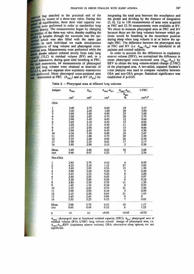
Table 3. - Pharyngeal area at different lung volumes
Subject APRC A..v APRc-A..v An.c·A..v LVRC
~c cm1 cm1 cm1 % cm1·t1
OSA
1 3.40 2.75 0.65 19 2.17 2 3.40 2.40 1.00 29 5.00 3 3 .90 3.00 0.90 23 3.00 4 3.80 3.05 0.15 20 3.75 5 3.90 3.45 0.45 12 0.64 6 3.65 3.10 0.55 15 0.92 1 3.50 3.10 0.50 11 0.80 8 3.35 2.90 0.45 13 0.64 9 2.90 2.45 0.45 16 4.50 10 4.30 3.50 0.80 19 2.67 11 2.55 1.80 0 .15 29 2.50 12 3 .25 2.40 0 .85 26 8.50 13 3.90 3 .25 0.65 17 2.17 14 3.05 2.90 0.15 . 5 0.30
Mean 3.49 2.86 0.63 18 2.68 ±so 0.46 0.47 0.23 1 2.24
Non-OSA
1 2.85 2.75 0.10 4 0.50 2 4.25 3.10 1.15 27 3.83 3 3.60 3.05 0.55 15 1.38 4 3 .80 3.60 0.20 5 0.40 5 2.45 2.25 0.20 8 2.00 6 2.90 2.85 0.05 2 0.07 1 3.20 3.00 0.20 6 0.18 8 2.80 2.50 0.30 11 1.50 9 1.65 1.55 0.10 6 0.33 10 3.35 2.65 0.70 2 1 3.50 11 3.00 2.90 0.10 3 0.50 12 3.15 2.50 0.65 21 1.63 13 2.65 2.65 0.00 0 0 14 3.50 3.25 0.25 7 0.63
Mean 3.08 2.76 0.33 10 1.17 ±so 0.63 0.49 0.32 8 1.23
p NS NS <0.05 <0.05 <0.02
AFRC: pharyngeal area at functional residual capacity (FRC); ARv; pharyngeal area at residual volume (RV); LVRC: lung volume related change of pharyngeal area, i.e. AFRC-ARvfERV (expiratory reserve volume); OSA: obstructive sleep apnoea; NS: not significant.
348 l. RUBINSTEIN, V. IIOFfoSTI!JN, T.O. BRADLEY
Results Discussion
The anthropometric data. menopausal status. pulmo· nary function tests, and sleep data for all subjects with and without OSA are shown in tables I and 2. There were no significant differences in age, BM[, FRC, RV, ERV, or forced expiratory volume in one second (FEV1)/forced vital capacity (FVC) between the two groups. The lowest nocturnal oxygen satumtion was significantly lower in the OSA group (67± 15% vs 83±14%, p<0.005).
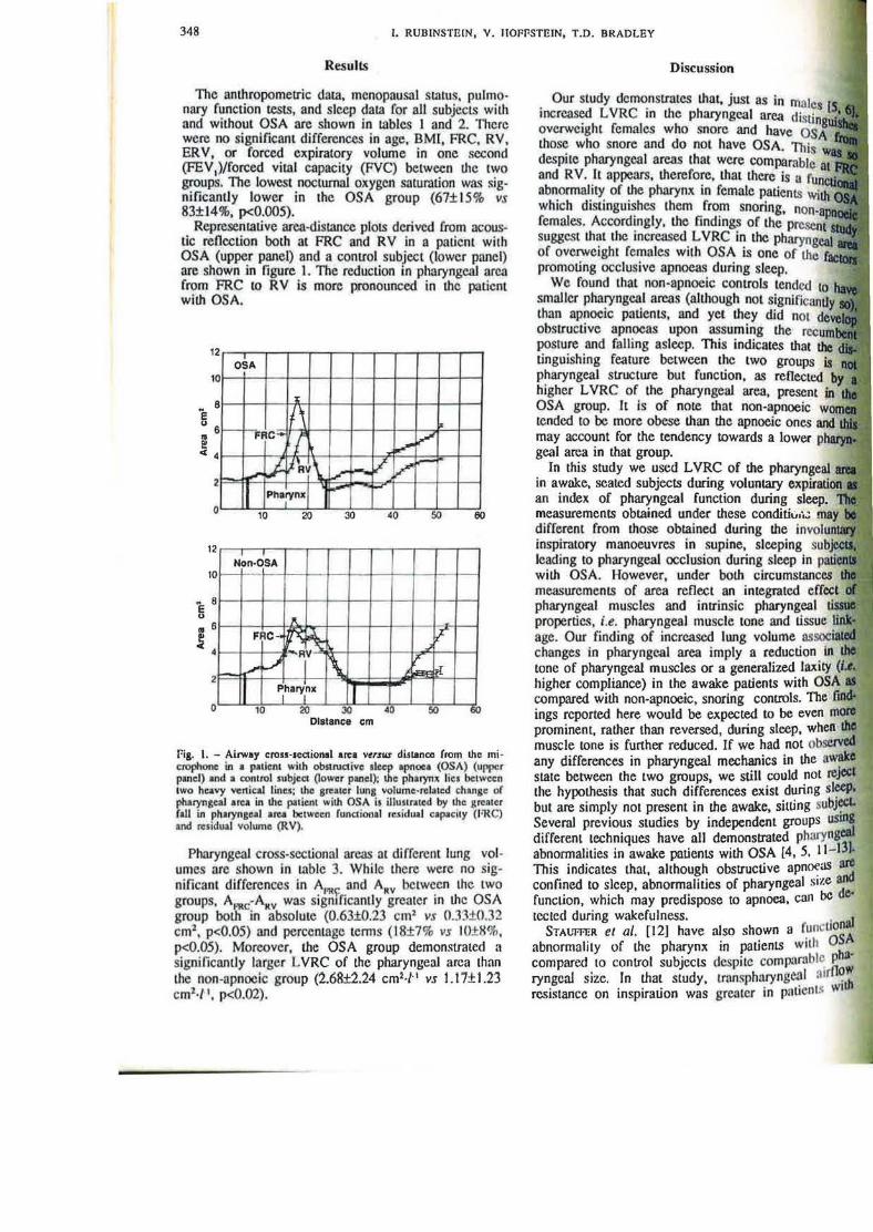
Representative area-distance plots derived from acoustic reOection both at FRC and RV in a patient with OSA (upper panel) and a conttol subject (lower panel) are shown in figure 1. The reduction in pharyngeal area from FRC to RV is more pronounced in the patient with OSA.
2 OSA
0
8 :z:
I{\ - 1-;:Rc- rr,. i-~ V. ~'Ay\ / f-.
11 Pharvnx 1 ...... 2
0
12
10
" 8 E 0
.. 6
s 4
2
10 20 30
Non·OSA f- -
"' FRC .... ~~ <'J RI \ >---
I ~harytx I
~ ~
V V -I-'"
40 50
.,.-IF
iPr .... ..a.:l
0 10 20 30 40 50 Distance em
60
-
60
foig. I. - Airway cross·seaio1111l area vtrsus di!lanc.e from !he microphone in a patient with obstructive alcep apnoea (OSA) (uppc:r panel) and a control subject (lower paoel): !he pharynx lies between two heavy ver1ic:al line&; !he greater luns volume·relatcd change 1>f pharyngeal area in !he p:uient with OSA it illustnated by !he grentcr faU in pharyngeal area between funalonal rC$ idu•l capacuy (FRC) and res idual volume (RV).
Pharyngeal cross-sectional areas at differom lung volumes are shown in table 3. While there were no significant differences in APR~ and ARv between the two groups. A c·Attv was sigruficantly greater in the OSA group bot~ in absolute (0.63±0.23 cm1 vs 0.33±0.32 cm1 , p<0.05) and percentage tenns {18±7% vs IO±X%, p<0.05). Moreover, the OSA group demonstrated a significantly l:trger L VRC of the pharyngeal area than the non-apnoeic group (2.68±2.24 cm1·/' 1 vs t .17± 1.23 cm1·/'1, p<0.02).
Our study demonstrates that, just as in males (S increased L VRC in Ute pharyngeal area d • overweight females who snore and have those who snore and do not have OSA. Thi~ despite pharyngeal areas that were comparable 81 and RV. ll appears, therefore, that there is a abnormality of the pharynx in female patients wilh which distinguishes !.hem from snoring, nm•--·--" females. Accordingly, the findings of the suggC$t that the increased L VRC in the omtrv•~.,. .... , of overweight females with OSA is one of the promoting occlusive apnoeas during sleep.
We found that non-apnoeic controls tended 10 smaller pharyngeal areas {aiUmugh not .,,5 ,uu.•~am than apnoeic patients, and yet. they did not obstructive apnoeas upon assuming the posture and falling asleep. This indicates that l.bc tinguishing feature between the two groups is pharyngeal structure but function, as reflected higher LVRC of the pharyngeal area, present in OSA group. It is of note that non-apnoeic tended to be more obese than the apnoeic ones may account for the tendency towards a lower pn&II'YQoill geal area in that group.
In this study we used LVRC of the nnarvnaP.RI
in awake, seated subjects during voluntary ex~ltcattton an index of pharyngeal function during measurements obtained under these conditii.J,;.; different from those obtained during the inspiratory manoeuvres in supine, sleeping leading to pharyngeal occlusion during sleep in with OSA. However, under both circumstances measurements of area reflect an integrated effect pharyngeal muscles and imrinsic pharyngeal •iuMi~•
properties, i.e. pharyngeal muscle tone and tissue 1iJ1t.! age. Our finding of increased lung volume nssoei.aled changes in pharyngeal area imply a reduction In lJio; tone of pharyngeal muscles or a generalized laxity (1.1.. higher compliance) in the awake patients with OSA at compared with non-apnoeic, snoring controls. The find. ings reported here would be expected to be even mote prominent, rather than reversed, during sleep, wheiJ IIIo muscle tone is further reduced. If we had not o~ any differences in pharyngeal mechanics in the awake state between the two groups, we still could not rejcc& the hypothesis that such differences exist during si~ but are simply not present in the awake, sitting subJ~ Several previous studies by independent groups USinl differenL techniques have all demonstrated phuryngeel
31 abnormalities in awake patients with OSA [4, 5, 11-1 • This indicates that, although obstructive apnoeas aro confined to sleep, abnormalities of pharyngeal stze alldd function, which may predispose to apnoea, can be erected during wakefulness. . aJ
STAUfl'ER et al. [12] have also shown a funcuon abnormality of the pharynx in patients with O~A compared to control subjects despite comparable P 4" ryngeal size. In that study, t.rnnsph~ryng~l :Jtrll?~ resistance on inspiration was greater tn pnuents W1
PHARYNX IN OBESE FEMALES WITH SLEEP APNOEA 349
in controls, even in the presence of similar cross-sectional area as measured at PRC by
tomography scanning. They concluded inspiratory pharyngeal resjstance may be
10 an impaired ability to dilate the pharyngeal during inspiration in patients with OSA. Pre-
l.his would predispose them to pharyngeal on inspiratlon during sleep. These findings, with the present data, emphasize that an ab
function of Lhe pharynx may be of equal or Importance than anatomical pharyngeal narrow
the pathophysiology of upper airway collapse in Further evidence in support of this view is that airWay collapse is state-related and occurs only sleep. presumably because of the superimposia sleep-related reduction in pharyngeal dilator tone upon an inltinsically collapsible pharynx
of sleep state, recumbency is also with a further reduction in pharyngeal area
as well as a concomitant fall in ERV [19). of posture on pharyngeal area and distensi
patients with and without OSA has been addressed in a recent study [18). It was assuming the supine posture results in a
··ret:tuctlon in pharyngeal area in apnoeic and individuals. Furthennore, pharyngeal <listen
is significantly higher in the apnoeic patients, the non-apnoeic controls, independent of the Thus. it appears that the abnormalities in phastructure and function present in the sitting are preserved, and, in fact, are further accen-
in the supine position. It is possible that at some a critical narrowing is reached so that the balance
begins to favour pharyngeal collapse upon to negative inspiratory pressure during sleep
mrtir•• II•J'Iv in the presence of increased pressure in the pharyngeal area [3, 5, 6]. Although was not measured in the present study,
measurements in males with OSA have deman association between L VRC and increased
changes of pharyngeal area [3, 20]. that this relationship is also present in reresults indicate that females with OSA have
pharynxes than conltol subjects. Clearly, this is an approximation since we have no measof intra-pharyngeal pressures to calculate
10\rn .... n, distensibility. Consequently. while the absoum•rvr'n"" ' area did not differ between the two
L VRC and higher pharyngeal colamong patients with OSA would favour
~~'""""' collapse during sleep with the generation of intra-pharyngeal pressure at the onset of
""'~~~talion (15] . we found that females with OSA have
. pharyngeal mechanics, just as in males with d•sorder [5, 6], this is probably not the sole
.!IIIIIJOmn<>l:ty responsible for airway occlusion during Closer examination of our results (table 3) reveals
among 14 females with OSA there were 5 with '"-"""'"'Y low LVRC (below 1.00 cm2·l'1); nevertheless,
these patients had severe sleep apnoea with a mean AHI of 40. It is possible that abnonnalities at other anatomical sites - bOlh proximal and distal to the pharynx • are responsible for promoting airway obslruction in these patients. For example, iL .is known that upper airway collapse in males may occur at the level of the nasopharynx [21 ). However. nasopharyngeal area cannot be measured by acoustic reflection Yia the mouth. The results of acoustic rencction measurements of the pharynx described here and elsewhere [3, 5, 6, 20] are the mean of measurements made between the end of the mouthpiece (i.e. at the end of the hard palate) and the glottis. Accordingly, whereas we found no difference in mean pharyngeal area at either FRC or RV between the two groups, it is possible that there were differences in nasopharyngeal area that we could not detect. Furthermore, RUDINSTl!lN et al. (221 have recently demonstrated that patients with severe OSA may have paradoxjcal inspiratory narrowing of the glottis as the only abnormality of the upper airway. Although we did not examine nasopharyngeal function or glottic movements in our patients, it is possible that abnormalities at these sites may have contributed to their sleep apnoea.
IL is possible that structural differences in the pharyngeal airway. between OSA and non-OSA females do exist, but cannot be resolved by the acoustic method. In this study we measured the average pharyngeal area, obtained by integrating l cm sections. Any differences in pharyngeal area occurring over a distance less than I cm cannot be resolved by this technique. GUJLLEMfNAULT et al. [23] used X-ray cephalometry and found subtle, but definite narrowing of the posterior airway space in females with OSA as compared to the non-apnoeic controls.
Factors involved in L VRC of the pharyngeal area have not been precisely determined. Possible mechanisms include reflex activation of pharyngeal dilator muscles via pulmonary sltetch receptors during inspiration as described for the larynx, trachea, and l.he upper airway muscles [24-26]. The expira10ry fall in pharyngeal area may be either passive and related to the intrinsic muscle-tissue linkage or compliance of the pharyngeal walJs [3J, or actjve and related to the expiratory activation of the pharyngeal constrictors. Active pharyngeal constriction, rather than a passive rcnex related to changes in lung volume, is favoured by a recent study of FoUKE and SnouL ( 171 which fai led to demonstrate any changes in pharyngeal area with passive increases in lung volume produced by a negative pressure ventilator. Why some obese females but not others should demonstrate greater L VRC in the pharyngeal area is not clear. Differences in the dis tribution of the adipose tissue in the neck and pharyngeal walls with fatty infiltration rendering the pharynx more "noppy'' is one possible explanation.
While previous studies have suggested a role for the menopause in the pathogcnesis of OSA in some fcmnles. the bulk of the evidence indicates that the menopause per se is not of major pathophysiological significance [7, 27- 301. G uuJ..HMINAvt.T et al. (23] found no difference in sl:ltic upper airwuy dimensions between
350 I. RUBINSTEIN, V, JIOFFSTEJN, T.D. BRAOLEY
premenopausal and posunenopausal women with OSA. In the present study, OSA and control women were very closely matched for both age and weight, and it is ve.ry unlikely that differences in menopausal status accounted fo.r Lhe presence or absence of OSA or for the observed differences in pharyngeal function between the two groups.
In summary, the present findings indicate that OSA in overweight females is associated with an abnormality of pharyngeal function which is similar to that seen in overweight males suffering from this disorder. This functional abnormality may be chamcteri?.ed by increased LVRC of the pharyngeal area, which implies n greater tendency of the pharynx to collapse at low lung volumes, presumably in response to normal negative inspiratory pressures generated during sleep. Although the precise mechanism responsible for these abnormalities cannot be de-duced from our measurements, they imply that high L VRC in the pharyngeal area is an important factor in the palhogenesis of OSA in overweight women.
ttcknowkdgtttt6ffll: The authon gratefully ae· knowledge the secretarial assisl8nce o( I. Lewis in preparation oC the manuscript.
References
1. Guilleminault C, Tilkian A, Dement WC. - The sleep apnea syndromes. Ann Rev Med, 1976, 27, 465-484. 2. Bradley TD, Rulherford R, Grossman RP, et al. -Role of daytime hypoxemia in the palhogenesis of right heart failure in lhe obstructive sleep apnea syndrome. Am Rev Respir Dis, 1985, 131, 835-839. 3. Brown IG, Bradley TO, Phillipson EA, Zamel N, Hoffstein V. - Pharyngeal compliance in snoring subjects wilh and without obstructive sleep apnea. Am Rev Respir Dis, 1985, 132, 211-215. 4. Surau PM, Dee P, Atkinson RL, Armstrong P, Wilhoit SC. - Fluoroscopic and computed tomographic features of the pharyngeal airway in obstructive sleep apnea. Am Rev Respir Dis. 1983, 127, 487-492. 5. Hoffstein V, Zamel N, Phillipson EA. - Lung volume dependence of pharyngeal cross-sectional area in patients with obstructive sleep apnea. Am Rev Respir Dis, 1984, 130. 175-178. 6. Bradley TO, Brown IG, Grossman RP, et al. - Pharyngeal size in snorers, non-snorers and patients wilh obstructive sleep apnea. N Engl J Med, 1986, 315, 1327-1331. 7. Wittels EH. - Obesity and hormonal factors in sleep and sleep apnea. Med Clin North Am, 1985, 69, 1265- 1280. 8. Fredbcrg JJ, Wohl MEB, Glass GM, Dorkin HL. -Airway area by acoustic reflections measured ar lhe mouth. J Appl Physiol. 1980, 41!, 749-758. 9. Thomas AE, McKay DA, Cutlip MR. - A monograph melhod for assessing body weight Am J Clin Nutr, 1976, 29. 302-304. 10. Rechtschafen A, Kales A. - In: A manual of standardized terminology, techniques and scoring system for sleep stages of human subjects. Nation3l Institute of Neurological Diseases and Blindness Neurological Information Network, Belhesda, 1968. 11. Phillipson EA, Bowcs G. - Sleep disorders. In : Update:
pulmonary discnses and di.wrdcrs. A.P. Pishman ed Hill, New York, 1982. pp. 256-273. ., 12. Stauffcr IL. Zwill ich CW, Cadiewt RJ, Bi:(Jer P.O. A. Varnno LA, White D P. - Pharyngettl size 1\nd in obstructive sleep ap:nea. Am RBV R!.rpir Dis 623-627. . 1.3. Haponik EJ, Smith PL. Bohlm3n MB, Goldman SM, Bleccker ER. - Computerized 10n10
._.
obstructive sleep apn.ea: correlation of airway size ology during sleep and wa.kefulnes.s. Am Rev 1983, 127, 221- 226. 14. Suratt PM, Wilhoit SC, Atkinson RL. - Blcvalocl flow resistance In awake obese subjects with obslrUI.lJN apnea. Am Rev Respir Dis, 1983, 127, 1625. • 15. Remmer~ JE, De Gr~ot WJ, Sauerland EK, And\ - Palhogenes1s of upper auway occlusion during sleep Physiol, 1978, 44, 931-938. • 16. Bradley TO, Phillipson EA. - Palhogenesis and physiology of lhe obstructive sleep apnea syndrome. North Am, 1985, 69, 1169-1185. 17. Foukc JM, Strohl KP. - Effect of position aut volume on upper airway geometry. J Appl Physiol, 1987 375-380. • 18. Brown IG, McLean PA, Bouchcr R, Zamel N V. - Changes in pharyngeal cross-sectional area ' and application of continuous positive airway patients wilh obstructive sleep apnea. Am Rev Rupir 1987, 135, 387-391. 19. Hudgel DW, Devadatta P. - Decrease in residual capacity during sleep in normal humans. I Physiol, 1984, 57, 1319- 1322. 20. Bradley TO, Brown IG, Zamel N, Phiru.-on Hoffstein V. - Differences in pharyngeal properties bWMJM snorers with predominantly central sleep apnea and without sleep apnea. Am Rev Respir Dis, 1981, 387-391. 21. Hudgel DW. - Variable site of airway narrowing obstructive sleep apnea patients. J Appl Physwl, 1986, 1403-1409. 22. Rubinstein I, Slutsky AS, Zamel N, Hoffstein V. Paradoxical inspiratory glottic narrowi.ng in patienta_ severe obstructive sleep apnca. J Clin Invest, 1988, 1051- 1055. 23. Guilleminault C, Quera-Salva MA, Paninen Jammieson A. - Women and the obstructive sleep ..... syndrome. Chest, 1988, 93, 104-109. 24. Stransky A, Szereda-Przestaszewska M, Widdicombe JO. - The effect of lung reflexes on laryngeal resistance .S motoneurone discharge. J Physiol (Lond), 1973, 231, 417-431; 25. Widdicombe JG, Nadel IA. - Reflex effect of 111111 inflation on tracheal volume. J Appl Physiol, !963, IS. 681- 686. 26. Van Lunreran E, Strohl KP, Parker OM, Bruce EN, v•. de Graff WB, Chemiak NS. - Phasic volume related feedback on upper airway muscle activity. J App/ Physiol, 1984, 56, 730-736. 27. Johnson MW, Anch AM, Rcmmers JE. - [nJuclio~ of the obsrrucrive sleep apnea syndrome in a . wor~1an9:: exogenous androgen administration. Am RBV RespiT o~.~·. I • 129, 1023-1025. . 28. Block AJ, Wynne JW, Boyscn PG, Lindsay S. M!Uti11 C. Cantor B. - Menopause, medroxyprogesterone nnd l)(ath-ing during sleep. Am J Med, 1981, 70, 506-510. CW 29. White DP. Lombard RM. Cadicux RJ, Zwillkl~ ;,; Pharyngeal rcsis1ance in normal humans: influence ol 1>011 '
age and obesity. J Appl Physiol, 1985, 58, 365-371.
PHARYNX IN OBESE PI!MALI!S WITH SLBEP APNOEA 351
J JR. Calverley PMA. Shapiro CM, Flenley DC, . - Breathing and oxygenation during sleep are
nozmal men and normal women. Am Rev Respir 132. 86-88.
de la surface pharyngee en relati011. avec les ftulmll.nmres chez les femmes obeses avec ou sans
d'nprtie du sommeil (SAS). I. Rubinstein, T.D. Brad/ey. majorit~ des patients de sexe masculin aueints d'apn~e obstructive du sommeiJ on! une fonc
structure pharyngees anormales, permettant des completes episodiques de leur voie a~rienne
sommeil. Comme le SAS est moins frequent chez que chez les hommes, il est possible que des
6trangcres aux proprietes pharyngiennes intervienlcs femmes atteintes de SAS. Nous avons done
surface pharyngee et ses variations en rapport avec pulmonaires, depuis la CRP jusqu'au volume 14 femmes obeses avec SAS et chez 14 femmes
SAS. Les surfaces pharyngees ont ete mcsurees
par la technique de nHiexion acoustique. Alors que la surface pharyngee etait similaire dans le groupe SAS et le groupe contrOie ~ la CRF (moyenne±so 3.49±0.46 cm2 contre 3.08±0.63 cm2) et au VR (2.86±0.47 cm1 contre 2.76±0.49 cml), la r&iuction de surface pharyngee entte la CRF et le VR etait significativement plus marquee dans le groupe SAS que chez les conll'oles (0.63±0.23 cm2 contre 0.33±0.32 cml,
p<0.05). De plus. quoique le volume de reserve expiratoire ne soit pas significativement different enlre les deux groupes (0.4±0.2 I contre 0.4±0.3 1), la modification de la surface pharyngee en rapport avec Jes volumes definie commc la difference de surface pharyng~e entre la capacit6 r~iduelle fonctionnelle et le volume residue! norrnalis~ par le volume de r~ervc cxpiratoire, s'avcre plus 6levee che.z les femmes attcimes de syndrome d'apnt!e du sommeil, par comparaison avec les controles non apn~Yques (2.68±2.24 cm2·1"1 vs l.l7± 1.23 cm2-t 1• p<0.02). Nous concluons que les femmes aueintcs de syndrome d'apnee du sommeil ont une mecanique pbaryngee anormale scmblable A cclle des hom.mcs, et que ccllc-ci peut contribuer a la physiopathog~nie du syndrome d'apn6e du sommcil. Eur Respir J., 1989, 2. 344-351.


![Ch 6 Trusses[1]](https://static.fdokumen.com/doc/165x107/631285ddb033aaa8b20fad21/ch-6-trusses1.jpg)