Landolt et al (2007) Alaskan Yellowjackets: The good, the bad, and the not so bad
LETTERS TO THE EDITOR
FUNCTIONAL DENTAL OCCLUSION MAY PREVENTFALLS IN ELDERLY INDIVIDUALS WITH DEMENTIA
To the Editor: More than one-third of persons aged 65 andolder fall each year, and in half, falls are recurrent.1,2 Theprevention of falls in this growing population is a primaryconcern for maintaining an adequate quality of life. Elderlypersons with cognitive impairment and dementia have twotimes the risk for falls as cognitively normal elderly per-sons.3 It has been demonstrated that multifactorial assess-ment and intervention focused on these risk factors,including impaired gait and balance (postural instability),environmental hazards, drugs, and cardiovascular diag-noses, has been successful in preventing falls in cognitivelynormal elderly persons but not in the elderly with cognitiveimpairment and dementia.4,5 It can be hypothesized thatprimitive reflexes such as postural stability are more im-portant for demented elderly. When in an upright position,permanent oscillations are generated to maintain balance.Sensorial afferents are provided from proprioceptive, tac-tile, vestibular, and visual receptors. Proprioception of themandibular system arises from the masticatory muscularsystem and dentoalveolar ligaments.6 Poor or absent dentalocclusion may decrease proprioception in this area, inter-fering with the stability of head posture. In this study, theincidence of falls in individuals with adequate dental oc-clusion is compared with that of those with inadequatedental occlusion. In addition, in a smaller pilot study, theeffect of improved dental occlusion on occurrence of fallswas investigated in subjects who had previously fallen re-currently.
Subjects for this study were 146 ambulatory elderly per-sons who had been admitted to a special geriatric hospital forindividuals with behavior disorders who were also sufferingfrom severe dementia. Inclusion criteria limited subjects tothose who could walk, with or without a cane, for all of theirambulation. Multifactorial assessments of each patient’s func-tion, including the Mini-Mental State Examination (MMSE),and pharmacotherapeutic status were performed, and occu-pational therapists conducted daily group rehabilitations tomaintain physical ability. One dentist (MY) classified eachsubject’s occlusion into one of three categories: GroupAFnatural dentition with adequate function, Group BFpar-tially or fully edentulous but maintaining functional occlusionwith dentures in either or both jaws, and Group CFfunc-tionally inadequate occlusion with no dentures. Subjects werealso divided into two groups: the high falls group (�2 fallsduring the previous year), and the low falls group (�1 falls).In a smaller pilot study, denture treatment was provided for asmall subgroup of high falls patients who gave informed con-sent. After denture delivery, the incidence of falls was recordedover 1 year.
During the 1-year investigative period, 41 of 146 pa-tients had recurrent falls. No differences between the high
and low falls groups in proportion of women (75.6% vs69.5%), mean age (83.1 � 6.4 vs 81.9 � 6.9), use of psy-choneurotic medications (56.1% vs 61.9%), or meanMMSE score (9.6 � 6.7 vs 11.8 � 6.3) were found. Den-tal status was significantly different between the high andlow groups (Po.001) (Table 1). In the high falls group, 10patients (2 men and 8 women, mean age � standard devi-ation 81.0 � 8.0, mean MMSE score 12.1 � 4.0) who gaveinformed consent received denture treatment. Conventionaldenture treatments were provided, and all patients useddentures after treatment. During the investigative periodafter denture delivery, three patients were excluded becausethey were comatose or dead. All of the seven remainingpatients experienced decreased frequency of falls, with fivepatients experiencing one or no falls during 1 year of fol-low-up.
Within the limits of this study, it was found thatpatients with functionally inadequate dental status hadsignificantly more-frequent falls than those with function-ally adequate occlusion composed of natural teeth,dentures, or both. Because tooth loss may result in adecrease in proprioception in the masticatory musclesor dentoalveolar ligaments, an associated perturbation ofvisual stabilization and postural imbalances may be in-duced.6 One study7 indicated that dental occlusal conditionis associated with reduced lower extremity dynamicstrength, agility, and balance function in elderly people.These findings suggest that functional occlusion of naturalor artificial teeth may play an important role in generatingan adequate posture reflex through mandibular stability,preventing falls. The current small intervention pilot study,which reconstructed occlusion with dentures, supportsthese finding.
In conclusion, poor or inadequate dental occlusion maybe a predisposing factor for falls in older people, and im-proving occlusion deserves attention as an approach toprevention of falls in elderly patients. A dental examinationis recommended for inclusion in the standard health exam-ination for elderly persons, especially those with symptomsof dementia.
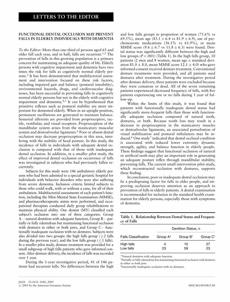
Table 1. Relationship Between Dental Status and Frequen-cy of Falls
Falls Classification
Dentition Status, n
Group A* Group Bw Group Cz
High falls 4 10 27Low falls 23 59 23
*Natural dentition with adequate function.wPartially or fully edentulous but maintaining functional occlusion with denturesin either or both jaws.zFunctionally inadequate occlusion with no dentures.
JAGS 53:1631–1642, 2005r 2005 by the American Geriatrics Society 0002-8614/05/$15.00
Mitsuyoshi Yoshida, DDS, PhDHidehiko Morikawa, DDS
Yayoi Kanehisa, MATsuyoshi Taji, DDS, PhD
Kazuhiro Tsuga, DDS, PhDYasumasa Akagawa, DDS, PhD
Department of Advanced ProsthodonticsGraduate School of Biomedical Science
Hiroshima UniversityHiroshima, Japan
ACKNOWLEDGMENT
Financial Disclosure: Mitsuyoshi Yoshida, Kazuhiro Tsuga,and Yasumasa Akagawa: supported by a grant-in-aid ofMinistry of Health, Labour and Welfare (tyoujyu-14–020).Hidehiko Morikawa and Tsuyoshi Taji: none. Yayoi Kane-hisa is a member of the geriatric hospital in this study.
Author Contributions: Mitsuyoshi Yoshida: study con-cept and design. Hidehiko Morikawa: acquisition of data.Yayoi Kanehisa: acquisition of subjects and data. TsuyoshiTaji: oral examination. Kazuhiro Tsuga: data analysis.Yasumasa Akagawa: preparation of manuscript.
Sponsor’s Role: Dr. Hideo Nakamura, president ofNakamura Geriatric Hospital, allowed data collection inthis hospital.
REFERENCES
1. Tinetti ME, Speechley M, Ginter SF. Risk factors for falls among elderly persons
living in the community. N Engl J Med 1988;319:1701–1707.
2. Nevitt MC, Cummings SR, Kidd S et al. Risk factors for recurrent nonsyncopal
falls: A prospective study. JAMA 1989;261:2663–2668.
3. Van Dijk PTM, Meulenberg OGRM, van De Sande HJ et al. Falls in dementia
patients. Gerontologist 1993;33:200–2004.
4. Close J, Ellis M, Hooper R et al. Prevention of falls in the elderly trial (PROF-
ET): A randomized controlled trial. Lancet 1999;353:93–97.
5. Shaw FE, Bond J, Richardson DA et al. Multifactorial intervention after a fall in
older people with cognitive impairment and dementia presenting to the accident
and emergency department: Randomised controlled trial. BMJ 2003;326:73.
6. Gangloff P, Louis JP, Perrin P. Dental occlusion modifies gaze and posture stab-
ilization in human subjects. Neurosci Lett 2000;3:203–206.
7. Yamaga T, Yoshihara A, Yoshitake Y et al. Relationship between dental occlu-
sion and physical fitness in an elderly population. J Gerontol A Biol Sci Med Sci
2002;57A:M616–M620.
BAD DENTITION AND RISK OF JEJUNALPERFORATION BY FISH BONE
To the Editor: Despite the frequency and variety of foreignbodies ingested, secondary intestinal perforation is a rareoccurrence. Preoperative diagnosis using computed tomo-graphy (CT) scan is rarely made because of the lack of ra-diographic contrast of nonmetallic foreign objects. A caseof jejunal perforation by a fish bone is reported. Diagnosiswas made using abdominal helical CT scan, and surgerywas immediately performed. This clinical case allows us todiscuss the several fish bone perforation sites and the patientprecipitating factors.
Coloenteric perforation by foreign bodies is a rare oc-currence and therefore a difficult preoperative diagnosis. Acase of jejunal perforation by a fish bone in an old man with
bad dentition is reported. Diagnosis was made using com-puted tomography (CT) scan before operating.
CASE REPORT
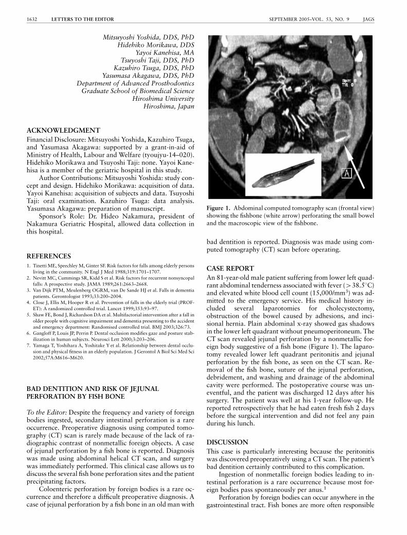
An 81-year-old male patient suffering from lower left quad-rant abdominal tenderness associated with fever (438.51C)and elevated white blood cell count (15,000/mm3) was ad-mitted to the emergency service. His medical history in-cluded several laparotomies for cholecystectomy,obstruction of the bowel caused by adhesions, and inci-sional hernia. Plain abdominal x-ray showed gas shadowsin the lower left quadrant without pneumoperitoneum. TheCT scan revealed jejunal perforation by a nonmetallic for-eign body suggestive of a fish bone (Figure 1). The laparo-tomy revealed lower left quadrant peritonitis and jejunalperforation by the fish bone, as seen on the CT scan. Re-moval of the fish bone, suture of the jejunal perforation,debridement, and washing and drainage of the abdominalcavity were performed. The postoperative course was un-eventful, and the patient was discharged 12 days after hissurgery. The patient was well at his 1-year follow-up. Hereported retrospectively that he had eaten fresh fish 2 daysbefore the surgical intervention and did not feel any painduring his lunch.
DISCUSSION
This case is particularly interesting because the peritonitiswas discovered preoperatively using a CT scan. The patient’sbad dentition certainly contributed to this complication.
Ingestion of nonmetallic foreign bodies leading to in-testinal perforation is a rare occurrence because most for-eign bodies pass spontaneously per anus.1
Perforation by foreign bodies can occur anywhere in thegastrointestinal tract. Fish bones are more often responsible
Figure 1. Abdominal computed tomography scan (frontal view)showing the fishbone (white arrow) perforating the small boweland the macroscopic view of the fishbone.
1632 LETTERS TO THE EDITOR SEPTEMBER 2005–VOL. 53, NO. 9 JAGS

for perforation of the esophagus, complicated by media-stinitis.1 Other parts of the upper digestive tract, such as thestomach or the duodenum, may be perforated.2 Fish bonesmay also pass through the gastrointestinal tract and enterother organs of the abdominal cavity such as the liver and thepancreas. One case of a fish bone totally integrated into thehepatic parenchyma has been recently reported.3 The smallbowel4 or the large bowel5,6 may be perforated too, and casesof Meckel’s diverticulum7 perforation have been described.
The clinical manifestations of gastrointestinal per-foration by foreign bodies are variable. Symptoms maybe acute, showing signs of peritonitis associated withpneumoperitoneum visible on plain abdominal x-ray,7
although the patient may present more-indolent symp-toms attributable to carcinoma or chronic disease. Onestudy reported a clinical case of an 80-year-old womanpresenting a painless, hard epigastric mass on physical ex-amination 8 days after fish ingestion.5 She received an an-tibiotic treatment for abdominal pain secondary to theinitially unsuspected perforation of the large bowel by a fishbone. Diagnosis was made at laparotomy. In the case of latediagnosis, a fish bone located inside the duodenum andpancreas was found several months after ingestion.2
As with the current patient, elderly persons are more atrisk because of the bad quality of their dentition, which isresponsible for a lack of mastication.5,8
Some cases of intestinal perforation by fish bone dis-covered using helical CT scan have been previously de-scribed.1,4,6
Ileal or jejunal perforation by fish bone is a rare occur-rence of gastroenteric perforation. Preoperative diagnosismay be made using abdominal helical CT scan in an emer-gency. It is necessary to carefully remove the bones from fishthat elderly persons eat because of the lack of masticationand the bad quality of dentition.
Brice Dugas, MDBenedicte Bernard, MD
Emmanuel Odet, MDAlain Naouri, MD, FACS
Pierre Bernard, MDDepartment of General Surgery
Hopital des ChanauxMacon, France
Pierre-Jean Valette, MD, PhDDepartment of Digestive Radiology
Hopital Edouard HerriotLyon, France
ACKNOWLEDGMENT
Financial Disclosure: None.Author Contributions: Brice Dugas: preparation of
manuscript. Benedicte Bernard: preparation of manuscript.Pierre-Jean Valette: acquisition of subjects and data andanalysis and interpretation of data. Emmanuel Odet: prep-aration of manuscript. Alain Naouri: preparation of man-uscript. Pierre Bernard: acquisition of subjects and data,analysis and interpretation of data, and preparation ofmanuscript.
Sponsor’s Role: none.
REFERENCES
1. Nozoe T, Kitamura M, Adachi Y et al. Successful conservative treatment for
esophageal perforation by a fish bone associated with mediastinitis. Hepato-
gastroenterology 1998;45:2190–2192.
2. Bourgaux JF, Fafet P, Balmes JL et al. Perforation duodenale par arete de thon:
Evolution tardive. Presse Med 1996;25:85.
3. Kessler AT, Kourtis AP. Images in clinical medicine. Liver abscess due to
Eikenella corrodens from a fish bone. N Engl J Med 2001;345:5.
4. lannelli A, Siou P, Spinelli R et al. Perforation of the ileum due to foreign body
treated laparoscopically. Surg Endosc 2002;16:538.
5. Ben Rejeb A, Gammoudi A, Ben Alaya M. Perforation intestinale par arete de
poisson. A propos d’une observation avec revue de la litterature. Ann Chir
1993;47:68–70.
6. Takada M, Kashiwagi R, Sakane M et al. 3D-CT diagnosis for ingested foreign
bodies. Am J Emerg Med 2000;18:192–193.
7. Bruyere F, Agathe-Nerine J, Baste JM. Diverticule de Meckel et arete de poisson
font mauvais menage. J Chir (Paris) 2002;139:165.
8. Read TE, Jacono F, Prakash C. Coloenteric fistula from chicken-bone perfo-
ration of the sigmoid colon. Surgery 1999;125:354–356.
CHRONIC HYPERCALCEMIA AS A REVERSIBLECAUSE OF COGNITIVE IMPAIRMENT:IMPROVEMENT AFTER A SINGLEADMINISTRATION OF PAMIDRONATE
To the Editor: Persistent elevations in serum calcium levelssecondary to primary hyperparathyroidism cause neuro-psychiatric symptoms, including confusion, alterations inmental status, delirium, and dementia.1–3 In fact, hyper-calcemia has been considered a reversible cause of demen-tia2 that can be corrected after parathyroidectomy.1
Although recent reports support the medical approach to-ward primary hyperparathyroidism as an alternative tosurgery,4 there are no reports that demonstrate cognitiveimprovement after medical correction of this disorder.
The case of a 79-year-old woman with known primaryhyperparathyroidism (adjusted serum calcium levels of 2.80–2.87 mmol/L for at least 3 years) who presented with a6-month history of progressive cognitive impairment, self-isolation, confusion, and generalized functional deteriora-tion, including severe limitations in performing instrumentalactivities of daily living (IADLs) is reported. Upon admis-sion, the patient was found to be anxious and disoriented asto time and space; no focal neurological deficits were found.Her general physical examination was otherwise normal.
Chronic hypercalcemia was considered to be the likelycause of her clinical findings. The patient was prescribed60 mg intravenous pamidronate to be administered month-ly. No other medications that might have modified hercognitive status were administered. The Mini-Mental StateExamination (MMSE) and the Clock-Drawing Test (CDT)(Figure 1) were administered 1 day before and 7 days afterthe initial pamidronate dose. Furthermore, occupationaltherapy conducted a global functional assessment using theglobal Structured Assessment of Independent Living Skills,a standardized assessment of functional motor, cognitive,instrumental, and social performance.5
Serum calcium levels declined to within normal limits(2.80 mmol/L at baseline, 2.69 mmol/L immediately afterpamidronate administration, 2.46 mmol/L 3 days afterpamidronate administration). The only adverse effect ofthe treatment was paresthesias in the fingers and toes, likelydue to the relatively low serum calcium levels after a pro-longed period of elevated serum calcium. MMSE score im-
LETTERS TO THE EDITOR 1633JAGS SEPTEMBER 2005–VOL. 53, NO. 9
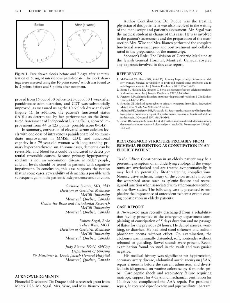
proved from 15 out of 30 before to 23 out of 30 1 week afterpamidronate administration, and CDT was substantiallyimproved, as measured using the 10 o’clock draw analysis6
(Figure 1). In addition, the patient’s functional status(IADL) as determined by her performance on the Struc-tured Assessment of Independent Living Skills, showed im-provement from 44 to 125 points (possible score 0–145).
In summary, correction of elevated serum calcium lev-els with one dose of intravenous pamidronate led to imme-diate improvement in MMSE, CDT, and functionalcapacity in a 79-year-old woman with long-standing pri-mary hyperparathyroidism. In some cases, dementia can bereversible, and blood tests could be indicated to detect po-tential reversible causes. Because primary hyperparathy-roidism is not an uncommon disease in older people,calcium levels should be tested in patients with cognitiveimpairment. In conclusion, this case supports the notionthat, in some cases, reversibility of dementia is possible withsubsequent gain in the patient’s independence and function.
Gustavo Duque, MD, PhDDivision of Geriatric Medicine
McGill UniversityMontreal, Quebec, Canada
Center for Bone and Periodontal ResearchMcGill University
Montreal, Quebec, Canada
Robert Segal, BcScFelice Wise, MOT
Division of Geriatric MedicineMcGill University
Montreal, Quebec, Canada
Judy Bianco BScN, 6NC(c)Department of Nursing
Sir Mortimer B. Davis Jewish General HospitalMontreal, Quebec, Canada
ACKNOWLEDGMENTS
Financial Disclosure: Dr. Duque holds a research grant fromMerck USA. Mr. Segal, Mrs. Wise, and Mrs. Bianco: none.
Author Contributions: Dr. Duque was the treatingphysician of this patient; he was also involved in the writingof the manuscript and patient’s assessment. Mr. Segal wasthe medical student in charge of this case. He was involvedin the patient’s assessment and the preparation of the man-uscript. Mrs. Wise and Mrs. Bianco performed the completefunctional assessment pre- and posttreatment and collabo-rated in the preparation of the manuscript.
Sponsor’s Role: The Division of Geriatric Medicine atthe Jewish General Hospital, Montreal, Canada, coveredany expenses involved in this case report.
REFERENCES
1. McDonald CA, Bruce DG, Smith DJ. Primary hyperparathyroidism in an eld-
erly woman. Surgical reversibility of profound mental status problems due to
mild hypercalcaemia. Int J Geriatr Psychiatry 2002;17:601–603.
2. Byrne EJ, Hosking DJ, Jameson C. Serial assessment of serum calcium correlates
with mental state. Int J Geriatr Psychiatry 1987;2:163–168.
3. Peterson P. Psychiatric disorders in primary hyperparathyroidism. J Clin Endocr
1968;28:1491–1495.
4. Strewler GJ. Medical approaches to primary hyperparathyroidism. Endocrinol
Metab Clin North Am 2000;29:523–539.
5. Mahurin RK, Bettignies BH, Pirozzolo FJ. Structured assessment of independent
living skills: Preliminary report of a performance measure of functional abilities
in dementia. J Gerontol 1991;46:58–M66.
6. Libon DJ, Swenson R, Sands LP et al. Further analysis of clock drawing among
demented and non-demented older subjects. Arch Clin Neuropsychol 1996;11:
193–205.
RECTOSIGMOID STRICTURE PROBABLY FROMISCHEMIA PRESENTING AS CONSTIPATION IN ANELDERLY PATIENT
To the Editor: Constipation in an elderly patient may be apresenting symptom of an underlying etiology. If the symp-toms are overlooked and are treated symptomatically, itmay lead to potentially life-threatening complications.Nonocclusive ischemic injury of the colon usually involvesthe watershed areas such as splenic flexure and rectos-igmoid junction when associated with atheromatous embolior low-flow states. The following case is presented to em-phasize the importance of antecedent ischemia events caus-ing constipation in elderly patients.
CASE REPORT
A 76-year-old man recently discharged from a rehabilita-tion facility presented to the emergency department com-plaining of constipation of 5 days duration and no passageof flatus for the previous 24 hours. He denied nausea, vom-iting, or diarrhea. He had tried stool softeners and sodiumphosphate enema without effect. On examination, theabdomen was minimally distended, soft, nontender withoutrebound or guarding. Bowel sounds were present. Rectalexamination found no stool in the vault and was guaiacnegative.
His medical history was significant for hypertension,coronary artery disease, abdominal aortic aneurysm (AAA)repair 2 months before the current admission, and divert-iculosis (diagnosed on routine colonoscopy 6 months pri-or). Cardiogenic shock and respiratory failure requiringinotropic support for 4 days and mechanical ventilation for11 days had complicated the AAA repair. For presumedsepsis, he received ciprofloxacin and piperacillin/sulbactam.
Figure 1. Free-drawn clocks before and 7 days after adminis-tration of 60 mg of intravenous pamidronate. The clock draw-ings were assessed using the 10-point score,5 which was found tobe 2 points before and 8 points after treatment.
1634 LETTERS TO THE EDITOR SEPTEMBER 2005–VOL. 53, NO. 9 JAGS
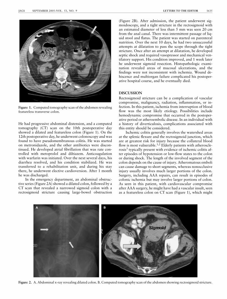
He had progressive abdominal distension, and a computedtomography (CT) scan on the 10th postoperative dayshowed a dilated and featureless colon (Figure 1). On the12th postoperative day, he underwent colonoscopy and wasfound to have pseudomembranous colitis. He was startedon metronidazole, and the other antibiotics were discon-tinued. He developed atrial fibrillation that was rate con-trolled with metoprolol and diltiazem. Anticoagulationwith warfarin was initiated. Over the next several days, hisdiarrhea resolved, and his condition stabilized. He wastransferred to a rehabilitation unit, and during his staythere, he underwent elective cardioversion. After 1 monthhe was discharged.
In the emergency department, an abdominal obstruc-tive series (Figure 2A) showed a dilated colon, followed by aCT scan that revealed a narrowed sigmoid colon with arectosigmoid stricture causing large-bowel obstruction
(Figure 2B). After admission, the patient underwent sig-moidoscopy, and a tight stricture in the rectosigmoid withan estimated diameter of less than 5 mm was seen 20 cmfrom the anal canal. There was intermittent passage of liq-uid stool and flatus. The patient was started on parenteralnutrition. Over the next 10 days, he had two unsuccessfulattempts at dilatation to pass the scope through the tightstricture. Once after an attempt at dilatation, he developedseptic shock and required vasopressor and mechanical ven-tilatory support. His condition improved, and 1 week laterhe underwent sigmoid resection. Histopathologic exami-nation revealed areas of mucosal ulcerations, and thefindings were not inconsistent with ischemia. Wound de-hiscence and multiorgan failure complicated his postoper-ative hospital course, and he eventually died.
DISCUSSION
Rectosigmoid stricture can be a complication of vascularcompromise, malignancy, radiation, inflammation, or in-fection. In this patient, ischemia from interruption of bloodflow was the most likely etiology. Possibilities includehemodynamic compromise that occurred in the postoper-ative period or atheroembolic disease. In an individual witha history of diverticulosis, complications associated withthis entity should be considered.
Ischemic colitis generally involves the watershed areasat the splenic flexure and the rectosigmoid junction, whichare at greatest risk for injury because the collateral bloodflow is most vulnerable.1,2 Elderly patients with atheroscle-rosis3 typically present with evidence of ischemic colitis af-ter episodes of hypotension or low-flow states to the colonor during shock. The length of the involved segment of thecolon depends on the cause of injury. Atheromatous embolican cause damage to short segments, whereas nonocclusiveinjury usually involves much larger portions of the colon.Surgery, including AAA repairs, can result in episodes ofcolonic ischemia but may involve larger portions of colon.As seen in this patient, with cardiovascular compromiseafter AAA surgery, he might have had a vascular insult, seenas a featureless colon on CT scan (Figure 1), which might
Figure 1. Computed tomography scan of the abdomen revealingfeatureless transverse colon.
Figure 2. A. Abdominal x-ray revealing dilated colon. B. Computed tomography scan of the abdomen showing rectosigmoid stricture.
LETTERS TO THE EDITOR 1635JAGS SEPTEMBER 2005–VOL. 53, NO. 9
have resolved but left residual effect on the watershed rec-tosigmoid region, causing a stricture. This stricture in turncaused the patient to have constipation, causing him tocome to the emergency department.
In conclusion, the clinical presentation of elderly pa-tients with constipation should be clinically correlated withtheir recent medical and surgical history to minimize themorbidity and mortality associated with this condition.
Ravi K. Bobba, MDEdward L. Arsura, MD, FACP
Khalil A. Amir, MDDepartment of Internal Medicine
Mohammad Naseem, MDDepartment of Radiology
Veterans Affairs Medical CenterUniversity of Virginia
Roanoke-Salem, VA
ACKNOWLEDGMENT
Financial Disclosure: None.Author Contributions: Ravi K. Bobba: patient care,
manuscript preparation, editing. Edward L. Arsura: man-uscript preparation, editing. Mohammad Naseem: patientcare, manuscript preparation, editing. Khalil A. Amir: man-uscript preparation, editing.
Sponsor’s Role: None.
REFERENCES
1. Greenwald DA, Brandt LJ. Colonic ischemia. J Clin Gastroenterol
1998;27:122–128.
2. Saegesser F, Loosli H, Robinson JWet al. Ischemic diseases of the large intestine.
Int Surg 1981;66:103–117.
3. Bharucha AE, Tremaine WJ, Johnson CD. Ischemic proctosigmoiditis. Am J
Gastroenterol 1996;91:2305–2309.
LONGEVITY AND CHRONIC BACK PAIN IN OLDERPEOPLE
To the Editor: Chronic back pain (CBP) is a common com-plaint of older people. Estimates of prevalence range from18% to 68% in the population aged 65 and older,1 reflect-ing the numerous definitions based on self-reported symp-toms. A number of consistent associations with functionaldisability,1 increased medical burden,2,3 and predictive fac-tors4 have been described. Although the consequences ofchronic pain are a subject of much focus, nonetheless thelong-term effect of CBP on mortality in older people is un-known. The single study identified5 with an age range from53 to 98 observed no interaction between CBP and mor-tality over 23 years of follow-up, a finding consistent withmortality data in younger CBP populations,6,7 which failedto show increased mortality specifically from cardiovascu-lar disease.
The Jerusalem Longitudinal Study8,9 addressed this is-sue, drawing data from an age-homogenous, representativesample of 458 West Jerusalem residents (209 women and249 men), comprehensively interviewed and examined at
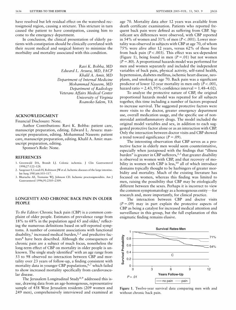
age 70. Mortality data after 12 years was available fromdeath certificate examination. Patients who reported fre-quent back pain were defined as suffering from CBP. Sig-nificant sex differences were observed, with CBP reportedin 54% of women and 31% of men (Po.001). Lower mor-tality was observed in subjects with CBP at age 70, of whom75% were alive after 12 years, versus 62% of those freefrom back pain (Po.003). This effect was sex-dependent(Figure 1), being found in men (P 5.01) but not women(P 5.80). A proportional hazards model was performed formen and women separately and included the independentvariables of back pain, physical activity, self-rated health,hypertension, diabetes mellitus, ischemic heart disease, neo-plasm, and smoking at age 70. Back pain was a significantpredictor of lower 12-year mortality in men only (Po.001,hazard ratio 5 2.43, 95% confidence interval 5 1.48–4.02).
To analyze the protective nature of CBP, the originalproportional hazards model was repeated for all subjectstogether, this time including a number of factors proposedto increase survival. The suggested protective factors weremore visits to the doctor, greater emergency departmentuse, overall medication usage, and the specific use of non-steroidal antiinflammatory drugs. The model included theoriginal model variables and sex, in addition to each sug-gested protective factor alone or as an interaction with CBP.Only the interaction between doctor visits and CBP showeda trend toward significance (P 5.09).
The interesting observation that CBP serves as a pro-tective factor in elderly men would seem counterintuitive,especially when juxtaposed with the findings that ‘‘illnessburden’’ is greater in CBP sufferers,2,3 that greater disabilityis observed in women with CBP, and that recovery of mo-bility in women with CBP is less,10 all of which introducedomains typically thought to be harbingers of greater mor-bidity and mortality. Much of the existing literature hasfocused on women, whereas this finding was limited tomen, raising the possibility that CBP may be etiologicallydifferent between the sexes. Perhaps it is incorrect to viewthe common symptomatology as a homogenous entityFforresearch and, more importantly, for clinical practice.
The interaction between CBP and doctor visits(P 5.09) may in part explain the protective aspects ofCBP as being a catalyst for increased medical attention andsurveillance in this group, but the full explanation of thisenigmatic finding remains elusive.
Survival Rates-Men
0
0.2
0.4
0.6
0.8
1
0 3 6 9 12
Years Follow-Up
Su
rviv
al P
rob
abili
ty
no pain painP = .01
71%
54%
C
Figure 1. Twelve-year survival data comparing men with andwithout chronic back pain.
1636 LETTERS TO THE EDITOR SEPTEMBER 2005–VOL. 53, NO. 9 JAGS
Jeremy M. Jacobs, MB, BSRobert Hammerman-Rozenberg, MD
Jochanan Stessman, MDDepartment of Geriatrics and Rehabilitation
Hadassah Hebrew University Medical CenterMount Scopus, Jerusalem, Israel
ACKNOWLEDGMENTS
Financial Disclosure: None.Author Contributions: Jeremy M. Jacobs: concept and
design, data analysis, and primary author. Robert Ham-merman-Rozenberg: concept and design, criticism and sug-gested improvement of data analysis, and content of letter.Jochanan Stessman: concept and design, data presentation,and suggested outline of letter with subsequent commentsand suggestions for improvement.
REFERENCES
1. Leveille SG, Guralnik JM, Hochberg M. Low back pain and disability in older
women: Independent association with difficulty but not inability to perform
daily activities. J Gerontol A Biol Sci Med Sci 1999;54A:M487–M493.
2. Vogt MT, Lauerman WC, Chirumbole M et al. A community-based study of post-
menopausal white women with back and leg pain; health status and limitations in
physical activity. J Gerontol A Biol Sci Med Sci 2002;57A:M544–M550.
3. Fanuele JC, Birkmeyer NJ, Abdu WA et al. The impact of spinal problems on
health status of patients. Spine 2000;25:1509–1514.
4. Kopec JA, Sayre EC, Esdaile JM. Predictors of back pain in a general pop-
ulation cohort. Spine 2003;29:70–78.
5. Kareholt I, Brattberg G. Pain and mortality risk among elderly persons in
Sweden. Pain 1998;77:271–278.
6. Heliovaara M, Makela M, Aromaa A et al. Low back pain and subsequent
cardiovascular mortality. Spine 1995;20:2109–2111.
7. Penttinen J. Back pain and risk of fatal ischaemic heart disease: 13 year follow
up of Finnish farmers. BMJ 1994;309:1267–1268.
8. Stessman J, Cohen A, Ginsberg GM et al. The Jerusalem seventy-year olds
longitudinal study. I. Description of the cross-sectional survey. Eur J Epidemiol
1995;11:675–684.
9. Cohen A, Stessman J, Ginsberg GM et al. The Jerusalem seventy-year olds
longitudinal study. II. Background results of the initial home interview. Eur J
Epidemiol 1995;11:685–692.
10. Leveille SG, Penninx BJ, Melzer D et al. Sex differences in the prevalence of
mobility disability in old age: The dynamics of incidence, recovery, and mor-
tality. J Gerontol B Psychol Sci Soc Sci 2000;55B:S41–S50.
WHO SHOULD DECIDE DRIVING COMPETENCE?
To the Editor: The increasing attention being paid to theevaluation of driving ability in patients with early dementiais well deserved. Being able to predict this ability, as Brownet al. attempt in their recently published study, would be avaluable tool in the care of patients and for the safety of thegeneral public.1 Their findings suggest that a neurologistwho is a dementia expert is a better predictor than the pa-tient or the patient’s family, but as the authors state, a pro-fessional driving instructor was more than five times betterthan the neurologist at predicting driving ability. Given thatthe driving instructor was much more stringent in his eval-uation and the excellent interrater reliability of the drivinginstructors, I would propose a different conclusion fromthat of the authors.
Rather than calling for more investigation to evaluateother types of doctors to see whether they could do as‘‘well’’ as the neurologist, those most qualified to performthis evaluationFprofessional driving instructorsFshouldbe encouraged to do so. Because driving ability is an em-
inently measurable skill, why not test it directly instead oftrying to find surrogates who may predict this skill? If weare truly interested in patients’ and the public’s safety, weshould be advocating for accessible and mandatory profes-sional driving evaluations for all patients with early de-mentia who wish to continue to drive.
Daniel J. Brauner, MDDepartment of Medicine/Geriatrics
University of Chicago,Chicago, IL
ACKNOWLEDGMENT
Financial Disclosure: None.Author Contributions: Daniel J. Brauner: sole author.Sponsor’s Role: None.
REFERENCES
1. Brown LB, Ott BR, Papandonatos GD et al. Prediction of on-road driving
performance in patients with early dementia. J Am Geriatr Soc 2005;53:94–98.
RESPONSE LETTER TO DR. BRAUNER
To the Editor: We appreciate the interest Dr. Brauner has inour paper. He also makes an excellent recommendation:optimally, mandatory driving evaluations conducted byprofessional driving instructors would assess driving abil-ities in patients with mild dementia (rather than relying onphysician assessments), but there are practical concerns re-garding this recommendation. Given the ever-increasingnumbers of individuals with dementia, due to our growingaging population and the limited number of driving in-structors who are experienced with elderly drivers, it simplyis not currently possible to have all demented drivers pro-fessionally evaluated. There also are concerns regarding thecost of these evaluations and who should be responsible forthem. Moreover, research suggests that many patients andfamilies look to their physicians for advice on this matterand that physician recommendations often play a key rolein the decision to stop driving.1
We believe that a two-tiered system is necessary; validscreening measures are needed to assess patients on a large-scale basis, followed by subsequent referral for on-roadtesting by a professional driving instructor for those whothe screening measures flag. Our results suggest that theassessment of a physician who is knowledgeable about de-mentia and driving may be helpful in the process of screen-ing for unsafe driving, although this is an ongoing area ofinvestigation, and neuropsychological tests of executive andvisuospatial function or driving simulators may prove to bemore valid and reliable at screening individuals than thephysician.
Detection of the unsafe demented driver remains achallenge for many communities, and we thank Dr. Braunerfor underscoring the need for formal assessments.
Laura B. Brown, PhDDepartment of Psychiatry and Human Behavior
Brown Medical School
LETTERS TO THE EDITOR 1637JAGS SEPTEMBER 2005–VOL. 53, NO. 9
Brian R. Ott, MDDepartment of Clinical Neurosciences
Brown Medical SchoolGeorge D. Papandonatos, PhD
Center for Statistical SciencesBrown University
Providence, RI
Rebecca E. Ready, PhDDepartment of Psychology
University of Massachusetts at AmherstAmherst, MA
John C. Morris, MDDepartment of Neurology
Washington University School of MedicineSt. Louis, MO
ACKNOWLEDGMENT
Financial Disclosure: None.Sponsor’s Role: None.
REFERENCES
1. Adler G, Kuskowski M. Driving cessation in older men with dementia. Alz Dis
Assoc Disord 2003;17:68–71.
DROWSINESS AND DISORIENTATION IN AWOMANWITH DEPRESSION
To the Editor: A 57-year-old woman with a history ofmanic-depressive psychosis presented to the emergency de-partment with altered mental status of a week’s duration.She was a lifelong nonsmoker and consumed alcoholoccasionally. Her medications included sodium valproate(VPA) 250 mg twice daily and aspirin 81 mg once daily.On examination, she was afebrile and hemodynamicallystable, with oxygen saturation of 100% on room air.There was no pallor, icterus, or stigmata of chronic liverdisease. She had a prominent flapping tremor of her upperextremity (asterixis).
She was drowsy but arousable and had a markedly di-minished attention span. Her pupils were reactive, moto-sensory examination was unremarkable, and the plantarresponse was flexor bilaterally. Her neck was supple, andthe rest of the examination was noncontributory.
Laboratory values, including complete blood count,liver function tests, and coagulation profile, were normal, aswere her arterial blood gases, serum and urine toxicologyscreen, electrocardiogram, chest radiograph, and computedtomography (CT) scan of the brain.
The clinical presentation of altered sensorium and re-active pupils, absent focal neurological deficits, suggestedglobal cerebral dysfunction with clinical diagnosis of met-abolic or toxic encephalopathy. Asterixis, defined as a lapsein voluntarily sustained posture, commonly occurs in thesetting of hyponatremia, uremia, hepatic encephalopathy,and carbon dioxide narcosis, none of which could be in-voked as a cause in this patient. The negative CT scan madea structural cause such as a cerebrovascular accident/space-
occupying lesion less likely. The negative toxicologicalscreen excluded a wide variety of drug-induced toxic states.
In patients undergoing chronic VPA therapy and with-out hepatic dysfunction, hyperammonemia should be rec-ognized as an important reversible cause of altered mentalstatus. Hence, plasma ammonia levels were measured inthis case. (A nonhemolyzed, heparinized venous sample ispreferred.)1 The plasma ammonia level was significantlyelevated at 198mmol/L (normal range 6–47 mmol/L). Al-though most patients with valproate-associated hyper-ammonemia (VAH) have plasma ammonia levels in excessof 150mmol/L, the correlation between ammonia levels andthe degree of encephalopathy is poor. CT and magneticresonance imaging may show cerebral edema in some cases,and electroencephalography may show diffuse slow-wavechanges and epileptiform activity. Cerebrospinal fluidglutamine levels may be elevated in VAH.1
DISCUSSION
Since its approved use as a mood stabilizer in 1995, ger-iatricians and geriatric psychiatrists have increasingly usedVPA in nursing homes and in behavioral institutions. Theneurological adverse effects of this drug are postural tremor(seen in about 6%), headache, nystagmus, dizziness, diplo-pia, incoordination, and a reversible dementia-like syn-drome.2 Hyperammonemia, defined as plasma ammoniagreater than 47 mmol/L (80mg/dL), has been reported in asmany as 20% to 50% of patients.2,3 Hyperammonemia inclinical practice more commonly occurs in the setting ofliver disease, although many conditions, including VAH,produce the syndrome of nonhepatic hyperammonemia:1
� Inherited urea cycle enzyme deficiency/transport defectdisorders
� Primary carnitine deficiency and abnormalities of fattyacid oxidation
� Organic acidurias� Portosystemic shunts with normal liver function� Urinary diversion-ureterosigmoidostomy and ileal con-
duit� Subureteric injection for vesicoureteric reflux and uri-
nary infection with urease splitting organisms� Multiple myeloma and myeloid leukemia� Reye’s syndromeFexcluding inborn errors of metabo-
lism such as medium chain acyl coenzyme A deficiency� Drugs such as halothane, enflurane, 5-fluorouracil, as-
paraginase, and sodium valproate� Metabolic-hyperinsulinemic hypoglycemia, distal renal
tubular acidosis
VPA is a branched-chain carboxylic acid, whose bioavail-ability after oral ingestion is excellent. It is more than 90%protein bound and undergoes extensive metabolism in theliver via glucoronidation, mitochondrial b-oxidation, and toa lesser extent, cytosolic o/o1-oxidation.2,3 In VAH, the in-creased ammonia level is predominantly hepatic in origin,from impaired urea cycle function and subsequent inabilityto metabolize nitrogen loads. The renal cortex plays a minorpart through enhanced glutamine uptake and increasedglutaminase activity.3 L-carnitine deficiency has also beenshown to be associated with VAH. VPA is esterified withcarnitine to form a valprolyl-carnitine ester that helps in
1638 LETTERS TO THE EDITOR SEPTEMBER 2005–VOL. 53, NO. 9 JAGS
translocating the drug across the mitochondrial membrane.This complex then diffuses into the cytosol and is eventuallyexcreted, producing a relative carnitine deficiency. This re-sults in impaired b-oxidation of long-chain fatty acid andproducts of valproate o-oxidation accumulate in the blood,producing hyperammonemia.3 Also, VPA metabolites re-duce levels of acetyl-CoA and glutamate, which results inimpaired urea synthesis and increased ammoniagenesis.3
VAH often improves with drug discontinuation. Short-term dietary protein restriction, use of nonabsorbable di-saccharides such as lactulose and lactilol,1 and use of a highoral carbohydrate diet or intravenous 10% or 20% dex-trose with insulin may reduce nitrogen load. L-carnitinesupplementation (100 mg/kg) as intravenous infusion overan hour is currently recommended for valproate-inducedliver damage and possibly VAH.2 For patients with orni-thine transcarbamylase deficiency, sodium benzoate andsodium phenylacetate therapy in a specialist-care settingmay be appropriate.1 In resistant cases, hemodialysis willcorrect the biochemical disorder.
Because VPA levels correlate poorly with VAH, a highindex of clinical suspicion is required to make the diagnosisof VAH and is potentially curable.
Shanmugam Uthamalingam, MBBS, MRCP (UK)Nacogdoches Medical Specialties
Nacogdoches, TX
Sajal Kumar, MBBS, MDDivision of Nephrology
Department of MedicineUniversity of Texas, Medical Branch
Galveston, TX
Abu R. Vasudevan, MBBS, MD, MRCP (UK)Center for Cardiovascular Prevention
Lipids and Atherosclerosis SectionDepartment of Medicine
Baylor College of MedicineHouston, TX
William G. Winters, MDDepartment of Medicine
Sound Shore Medical Center of WestchesterNew York Medical College
New Rochelle, NY
ACKNOWLEDGMENT
Financial Disclosure: The authors have no financial supportfor research, consultantships, or speakers forums, and haveno company holdings (e.g., stocks) or patents on this article.
Author Contributions: Shanmugam Uthamalingam:first author, wrote case presentation, and searched the ref-erences for the discussion part of the manuscript. SajalKumar: second author, helped in the formulation of thediscussion part of the manuscript. Abu R Vasudevan: thirdauthor, helped in the formulation of the discussion part ofthe manuscript. Williams G Winters: attending physician onthe case who helped in the differential diagnosis and gavefeedback on the writing of the manuscript.
Sponsor’s Role: None.
REFERENCES
1. Hawkes ND, Thomas GA, Jurewicz A et al. Non-hepatic hyperammonaemia.
An important, potentially reversible cause of encephalopathy. Postgrad Med J
2001;77:717–722.
2. Perucca E. Pharmacological and therapeutic properties of valproate: A
summary after 35 years of clinical experience. CNS Drugs 2002;16:695–714.
3. Sztajnkrycer MD. Valproic acid toxicity. Overview and management. J Toxicol
Clin Toxicol 2002;40:789–801.
TUNING INTO SILENCE BUT WHISTLING: AN OLDMAN WITH APHASIA
To the Editor: A 76-year-old right-handed man was admit-ted to our center for a poststroke rehabilitation program.His medical history included gait and speech problems since1997. He also suffered from recurrent ischemic attacks be-tween 1997 and 2001 without any significant functionalsequelae. In 2001, a magnetic resonance angiography wasperformed that showed atheromatous plaques bilaterally ininternal and common carotid arteries with severe luminalocclusions. He has been followed conservatively till 2004,and in the interim, he has also encountered a few moreischemic attacks with mild impairment in cognition andspeech and weakness of the extremities. However, he couldspeak and walk independently with a cane. An endovascu-lar stent was implanted into the left vertebral and rightcommon carotid arteries in August 2004. Two weeks later,he had a right temporoparietal hemorrhagic infarct and washospitalized with the complaints of severe gait and speechdisturbances. After he had been followed in the neurosur-gery unit for acute care, he was transferred to our center forrehabilitation.
The physical examination was consistent with aphasiawithout any cooperation and generalized decrease in mus-cle strength without overt spasticity. He could be wheeled ina wheelchair or could walk with the aid of two people.During the examination, it was observed that he continu-ously whistled or attempted to whistle. His whistle was notmelodic but was rather in truncated soundsFmore or lessof the same tone. His relatives declared that he had beenwhistling the same way for a year and with a higher pitchbefore the recent episode. They added that, when they hadasked him the reason for his whistling, he explained that hewhistled to impose authority on the people nearby. Theyalso described the premorbid character of the patient asauthoritarian.
He was followed on a rehabilitation protocol ofstrengthening exercises and mobilization. Significant im-provement was not achieved after 2 months because of hisfluctuating, uncooperative mood. Occasionally, he wouldbegin to say individual words or sentences, especially whenhe read them on signs or when his mood was favorable, andhis whistling decreased. A control cranial magnetic reso-nance imaging revealed a (small) resolving infarct, severecerebral atrophy, small-vessel ischemic disease, and chronicinfarcts in the right frontal and occipital lobes. The psychi-atry department provided a consultation, and his whistlingwas attributed to a regressive behavior that could be seen inpatients with cerebral damage due to cerebrovascular ac-cidents and his explanation for it to be a defense mecha-nism. Vascular dementia was also noted to have contributedto his general psychiatric mood.
LETTERS TO THE EDITOR 1639JAGS SEPTEMBER 2005–VOL. 53, NO. 9
In our clinical experience, we have not seen such a pa-tient since, and we could find only one similar report in thepertinent literature mentioning a musically naive patientwith dominant frontotemporal and anterior parietal infarctwho had developed spontaneous compulsive whistling inaddition to hypermusia and musicophilia.1 The problem offinding correspondence between a particular neuronal or-ganization and a specific function of the human brain re-mains a central question of neuroscience. Despite the ideathat language and music are two sides of the same intellec-tual coin, research on brain-damaged patients has shownthat loss of musical ability does not necessarily accompanyloss of verbal function. Further scientific data are awaitedto shed light on the possible correlations between language,music, syntax, and brain dysfunction.
In closing, far from elucidating the intricate underlyingmechanism in our patient, what we intended to underscorein this report was to orient physicians toward a rare chal-lenge in clinical practice. We also advocate an initial con-servative approach, especially in elderly patients, in whomsuppressive medications might hamper recovery from braindamage.
Murat Kara, MDLevent Ozcakar, MDErkan Ozguclu, MDZafer Hascelik, MD
Department of Physical Medicine and RehabilitationSemra Ulusoy Kaymak, MD
Department of PsychiatryHacettepe University Medical School
Ankara, Turkey
ACKNOWLEDGMENT
Financial Disclosure: None.Author Contributions: Murat Kara: concept and de-
sign, preparation of the manuscript. Levent Ozcakar: con-cept and design, preparation of the manuscript. ErkanOzguclu: concept and design, preparation of the manu-script. Semra Ulusoy Kaymak: concept and design. ZaferHascelik: concept and design.
Sponsor’s Role: None.
REFERENCES
1. Jacome DE. Aphasia with elation, hypermusia, musicophilia and compulsive
whistling. J Neurol Neurosurg Psychiatry 1984;47:308–310.
EVIDENCE OF ACETYLCHOLINESTERASEINHIBITORS IN TREATING ALZHEIMER’S DISEASEIN VERY OLD PATIENTS IS UNCERTAIN
To the Editor: In his editorial, Dr. Royall discussed severalimportant issues in assessing the evidence of acetylcholin-esterase inhibitors (AChEIs) in treating Alzheimer’s disease(AD) but not the generalizability of AChEI trials.1 It wasreported that dementia research including therapeutic trialswas systemically biased toward younger patients.2 For ex-ample, the mean age was 72 in one meta-analysis of done-pezil trials in treating AD.3 Because the response to AChEIs,
neuropathology, and apolipoprotein E genotype in patientswith AD and the incidence and prevalence of dementia wereall age dependent,2 applying the results of the current pos-itive AChEI trials to adults aged 75 and older should not beconsidered as evidence based but an extrapolation-basedapproach. AD 2000 was the largest study of donepezil inperson-years of placebo-controlled treatment.4,5 The meanage of the participants was 76. It had a lower attrition rateand fewer missing data than any previous study. It has beenthe only non-industry-funded study. It is the only reportednegative trial.4,5 This should further increase the currentskepticism of medical professionals and the public as tousing AChEIs. One ideal solution would be to recruit moreparticipants who are similar to our general population withAD in a future AChEIs trial.
In the face of such a difficult situation, our choice ofusing AChEIs, just like other medical decisions, should bebased on the following five distinct areas:6 empirical evi-dence (clinical research including systemic reviews, ran-domized, controlled trials, other controlled trials, andcohort studies), experiential evidence (personal or expertclinical experience), physiological rationale (an understand-ing of physiological principles), values of patient, family,and professionals, and system features (economic, logic,legal, and cultural facilitators or barriers to the provision ofmedical care). The relative weight given to each of theseareas is dependent on individual cases.6
In contrast to Dr. Royall’s literature review, I found threehead-to-head randomized, controlled trials comparing twoAChEIs.7 Unfortunately, the several serious limitations ofthese three head-to-head trials based on the Consolidation ofthe Standards of Reporting Trials criteria make the initialdrug choice of AChEIs for patients with AD uncertain.7
Huai Yong Cheng, MD, MPHColumbia University Medical Center
New York, NY
ACKNOWLEDGMENTS
Financial disclosure: Huai Yong Cheng has no conflict ofinterest in support for research, consultantships, speakersforum, or any company holdings.
Author’s contribution: Huai Yong Cheng was the soleauthor and was fully responsible for this letter.
Sponsor’s role: N/A.
REFERENCES
1. Royall DR. The emperor has no clothes. Dementia treatment on the eve of the
aging era. J Am Geriatr Soc 2005;53:163–164.
2. Schoenmaker N, Van Gool WA. The age gap between patients in clinical studies
and in the general population: A pitfall for dementia research. Lancet Neurol
2004;3:627–630.
3. Whitehead A, Perdomo C, Pratt RD et al. Donepezil for the symptomatic
treatment of patients with mild to moderate Alzheimer’s disease: A meta-anal-
ysis of individual patient data from randomized controlled trials. Int J Geriatr
Psychiatry 2004;19:624–633.
4. Courtney C, Farrell D, Gray R et al. Long-term donepezil treatment in 565
patients with Alzheimer’s disease (AD 2000): Randomized double-blind trial.
Lancet 2004;363:2105–2115.
5. Gray R, Bentham P, Hills R. Authors’ reply. Lancet 2004;364:1216.
6. Tonelli MR. The limits of evidence-based medicine. Respir Care 2001;46:
1435–1440.
7. Hogan DB, Goldlist B, Naglie G et al. Comparison studies of cholinesterase
inhibitors for Alzheimer’s disease. Lancet Neurol 2004;3:622–626.
1640 LETTERS TO THE EDITOR SEPTEMBER 2005–VOL. 53, NO. 9 JAGS
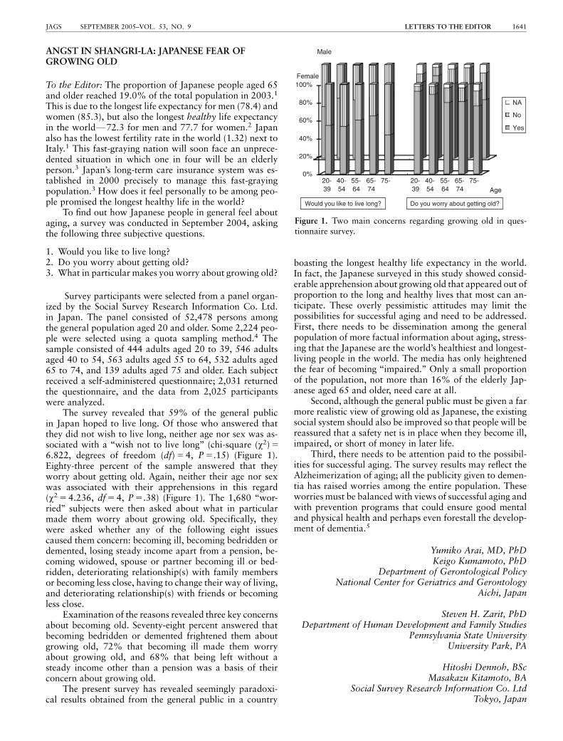
ANGST IN SHANGRI-LA: JAPANESE FEAR OFGROWING OLD
To the Editor: The proportion of Japanese people aged 65and older reached 19.0% of the total population in 2003.1
This is due to the longest life expectancy for men (78.4) andwomen (85.3), but also the longest healthy life expectancyin the worldF72.3 for men and 77.7 for women.2 Japanalso has the lowest fertility rate in the world (1.32) next toItaly.1 This fast-graying nation will soon face an unprece-dented situation in which one in four will be an elderlyperson.3 Japan’s long-term care insurance system was es-tablished in 2000 precisely to manage this fast-grayingpopulation.3 How does it feel personally to be among peo-ple promised the longest healthy life in the world?
To find out how Japanese people in general feel aboutaging, a survey was conducted in September 2004, askingthe following three subjective questions.
1. Would you like to live long?2. Do you worry about getting old?3. What in particular makes you worry about growing old?
Survey participants were selected from a panel organ-ized by the Social Survey Research Information Co. Ltd.in Japan. The panel consisted of 52,478 persons amongthe general population aged 20 and older. Some 2,224 peo-ple were selected using a quota sampling method.4 Thesample consisted of 444 adults aged 20 to 39, 546 adultsaged 40 to 54, 563 adults aged 55 to 64, 532 adults aged65 to 74, and 139 adults aged 75 and older. Each subjectreceived a self-administered questionnaire; 2,031 returnedthe questionnaire, and the data from 2,025 participantswere analyzed.
The survey revealed that 59% of the general publicin Japan hoped to live long. Of those who answered thatthey did not wish to live long, neither age nor sex was as-sociated with a ‘‘wish not to live long’’ (chi-square (w2) 5
6.822, degrees of freedom (df) 5 4, P 5.15) (Figure 1).Eighty-three percent of the sample answered that theyworry about getting old. Again, neither their age nor sexwas associated with their apprehensions in this regard(w2 5 4.236, df 5 4, P 5.38) (Figure 1). The 1,680 ‘‘wor-ried’’ subjects were then asked about what in particularmade them worry about growing old. Specifically, theywere asked whether any of the following eight issuescaused them concern: becoming ill, becoming bedridden ordemented, losing steady income apart from a pension, be-coming widowed, spouse or partner becoming ill or bed-ridden, deteriorating relationship(s) with family membersor becoming less close, having to change their way of living,and deteriorating relationship(s) with friends or becomingless close.
Examination of the reasons revealed three key concernsabout becoming old. Seventy-eight percent answered thatbecoming bedridden or demented frightened them aboutgrowing old, 72% that becoming ill made them worryabout growing old, and 68% that being left without asteady income other than a pension was a basis of theirconcern about growing old.
The present survey has revealed seemingly paradoxi-cal results obtained from the general public in a country
boasting the longest healthy life expectancy in the world.In fact, the Japanese surveyed in this study showed consid-erable apprehension about growing old that appeared out ofproportion to the long and healthy lives that most can an-ticipate. These overly pessimistic attitudes may limit thepossibilities for successful aging and need to be addressed.First, there needs to be dissemination among the generalpopulation of more factual information about aging, stress-ing that the Japanese are the world’s healthiest and longest-living people in the world. The media has only heightenedthe fear of becoming ‘‘impaired.’’ Only a small proportionof the population, not more than 16% of the elderly Jap-anese aged 65 and older, need care at all.
Second, although the general public must be given a farmore realistic view of growing old as Japanese, the existingsocial system should also be improved so that people will bereassured that a safety net is in place when they become ill,impaired, or short of money in later life.
Third, there needs to be attention paid to the possibil-ities for successful aging. The survey results may reflect theAlzheimerization of aging; all the publicity given to demen-tia has raised worries among the entire population. Theseworries must be balanced with views of successful aging andwith prevention programs that could ensure good mentaland physical health and perhaps even forestall the develop-ment of dementia.5
Yumiko Arai, MD, PhDKeigo Kumamoto, PhD
Department of Gerontological PolicyNational Center for Geriatrics and Gerontology
Aichi, Japan
Steven H. Zarit, PhDDepartment of Human Development and Family Studies
Pennsylvania State UniversityUniversity Park, PA
Hitoshi Dennoh, BScMasakazu Kitamoto, BA
Social Survey Research Information Co. LtdTokyo, Japan
Male
NA
No
Yes
0%
20%
40%
60%
80%
100%
20-39
40-54
55-64
65-74
75- 20-39
40-54
55-64
65-74
75-Age
Female
Would you like to live long? Do you worry about getting old?
Figure 1. Two main concerns regarding growing old in ques-tionnaire survey.
LETTERS TO THE EDITOR 1641JAGS SEPTEMBER 2005–VOL. 53, NO. 9

ACKNOWLEDGMENTS
Financial Disclosure: There are no conflicts of interest orfunding to declare.
Author Contributions: All authors contributed to writ-ing the manuscript and worked together as a group withregard to the study concept, design, acquisition of surveyquestionnaire subjects, and/or data, analysis and interpre-tation of data.
REFERENCES
1. Ministry of Health Labor and Welfare. Trends in Public Health in Japan (Ko-
kumin eisei no doko) [in Japanese]. Tokyo: Health Welfare Statistics Associ-
ation, 2004.
2. The World Health Report. Annex Table 4. Health Life Expectancy (HALE) in
All WHO Member States (estimates for 2002). Geneva, Switzerland: World
Health Organization, 2004.
3. Arai Y. Japan’s new long-term health care insurance. Lancet 2001;357:1713.
4. Moser CA, Kalton G, eds. Survey Methods in Social Investigation. Hants,
England, Gower, 1989: pp 127–137.
5. Blazer DG. Self-efficacy and depression in late life: A primary prevention ap-
proach. Aging Ment Health 2002;6:315–324.
ECONOMIC IMPLICATIONS OF DISCHARGE OFELDERLY FROM THE EMERGENCY DEPARTMENTTRIAL RESULTS
To the Editor: We enjoyed reading the article by Caplanet al. detailing the results of the Discharge of Elderly fromthe Emergency Department (DEED II) randomized con-trolled trial.1 The trial highlights the many benefits of com-prehensive geriatric assessment (CGA)Fmultidisciplinarycare for older adults sent home from the emergency de-partment (ED). We wish to highlight important economicimplications of this well-conducted trial. Economic evalu-ation of healthcare interventions can help to set prioritiesfor scarce healthcare resources.2,3 Economic evaluation al-so influences the policymakers who make funding decisionsthat can allow the implementation of innovative programssuch as the one described in the DEED II trial. Below, asimple cost-benefit analysis based on data from this trial isprovided.
In the DEED II trial, patients randomized to receiveCGA, multidisciplinary team support, had a lower rate ofhospital admission in the first month after the initial EDvisit than those who did not. The absolute risk reductionwas 5.7% (22.2% – 16.5%). Thus, the number needed totreat to prevent one hospital admission was 17.54 (1/0.057).4 In other words, if 17.54 older adults are managedwith CGA for up to 28 days, one hospital admission will beprevented. If it is assumed that this intervention costs $500per patient, then the cost to prevent one hospitalization is$8,770 (17.54 � $500). This cost must be weighed againstthe cost of the avoided hospitalization. If it is assumed thatthe cost to hospitalize one frail older adult is $1,500 per dayand that the average length of stay for such a patient is 7days, then the average cost per hospitalization is $10,500($1,500 � 7). Thus, implementation of CGA offsets ap-proximately $1,730 in hospital costs for the average frail
older adult ($10,500 – $8,770). These simple calculationsdo not account for the savings in terms of potential down-stream costs, consequences of hospitalizing frail olderadults (e.g., pressure ulcers, delirium, functional decline).5
These calculations also ignore the benefits seen in the trial interms of fewer ED visits.1 Thus, the potential savings maybe substantial.
The costs listed above are purely hypothetical but arelikely to be reasonable reflections of the relative costs of thevarious options regardless of the healthcare setting (e.g.,Australia, United States, Canada). Straightforward substi-tutions of costs could be made in the above model to per-form sensitivity analyses of the financial merits of linkingCGA to ED care. Investigators should consider collectingcost data alongside clinical outcomes when conducting fu-ture trials that evaluate innovative models of geriatric care.2
In addition to the impressive clinical benefits seen in theDEED II trial (reductions in ED visits and hospital admis-sions), healthcare funders may be especially interested to seethe cost benefits of implementing CGA alongside usual EDcare for frail older adults.
Sudeep S. Gill, MD, MSCMichelle Gibson, MD
Division of Geriatric MedicineAbdullah Al-Hammadi, MD
Department of PsychiatryQueen’s University
Kingston, Ontario, Canada
ACKNOWLEDGMENTS
Financial Disclosure: Drs. Sudeep S. Gill, Michelle Gibson,and Abdullah Al-Hammadi attest to the fact that there wasno financial support involved in the preparation of thismanuscript; none of the authors has a conflict of interest(financial or otherwise) related to the content of this sub-mission for publication.
Author Contributions: Drs. Sudeep S. Gill, MichelleGibson, and Abdullah Al-Hammadi were involved in theinitial conceptualization of this manuscript. Dr. Gill pre-pared the calculations, initial draft of the manuscript. Allthree authors helped to revise the manuscript and approvedthe final version. Dr. Gill will act as guarantor.
REFERENCES
1. Caplan GA, Williams AJ, Daly B et al. A randomized, controlled trial of
comprehensive geriatric assessment and multidisciplinary intervention after
discharge of elderly from the emergency departmentFthe DEED II Study. J Am
Geriatr Soc 2004;52:1417–1423.
2. Drummond MF, O’Brien B, Stoddart GL et al., eds. Methods for the Economic
Evaluation of Health Care Programmes, 2nd Ed. Oxford: Oxford University
Press, 2001.
3. Detsky AS, Naglie IG. A clinician’s guide to cost-effectiveness analysis. Ann
Intern Med 1990;113:147–154.
4. Guyatt GH, Sackett DL, Cook DJ. Users’ guides to the medical literature: II.
How to use an article about therapy or prevention: B. What are the results and
will they help me in caring for my patients? JAMA 1994;271:59–63.
5. Creditor MC. Hazards of hospitalization of the elderly. Ann Intern Med
1993;118:219–223.
1642 LETTERS TO THE EDITOR SEPTEMBER 2005–VOL. 53, NO. 9 JAGS
Copyright © 2022 FDOKUMEN