Behavioral deficits in sepsis-surviving rats induced by cecal ligation and perforation
Upload
khangminh22Category
view
4download
0
EDUCATIONAL REVIEW Open Access
Gastrointestinal perforation: clinical andMDCT clues for identification of aetiologyStyliani Pouli1, Androniki Kozana1, Ioanna Papakitsou2, Maria Daskalogiannaki1 and Maria Raissaki1*
Abstract
Gastrointestinal tract (GIT) perforation is a common medical emergency associated with considerable mortality,ranging from 30 to 50%. Clinical presentation varies: oesophageal perforations can present with acute chest pain,odynophagia and vomiting, gastroduodenal perforations with acute severe abdominal pain, while colonicperforations tend to follow a slower progression course with secondary bacterial peritonitis or localised abscesses. Asubset of patients may present with delayed symptoms, abscess mimicking an abdominal mass, or with sepsis.Direct multidetector computed tomography (MDCT) findings support the diagnosis and localise the perforation sitewhile ancillary findings may suggest underlying conditions that need further investigation following primary repairof ruptured bowel. MDCT findings include extraluminal gas, visible bowel wall discontinuity, extraluminal contrast,bowel wall thickening, abnormal mural enhancement, localised fat stranding and/or free fluid, as well as localisedphlegmon or abscess in contained perforations.The purpose of this article is to review the spectrum of MDCT findings encountered in GIT perforation andemphasise the MDCT and clinical clues suggestive of the underlying aetiology and localisation of perforation site.
Keywords: Gastrointestinal perforation, Emergency, Aetiology, Multidetector computed tomography
Key points
� GIT perforation manifests with extraluminal gas,wall discontinuity or thickening, fat stranding.
� Pneumoperitoneum and extraluminal oral contrastrange from abundant to absent.
� Ancillary findings comprise foreign bodies,masses, excessive wall thickening, ischemia, faecalimpaction.
� Supramesocolic pneumoperitoneum andhyperenhancing gastroduodenal wall suggestperforated peptic ulcer disease.
� Persistent or increasing free air and/or ascitespostoperatively indicate iatrogenic perforation.
BackgroundDespite progress in emergency medicine, gastrointestinaltract perforation remains a condition associated withconsiderable mortality, ranging from 30 to 50% [1, 2].Clinical presentation varies: oesophageal perforationscan present with non-specific symptoms such as acutechest pain, odynophagia and vomiting [2], gastroduo-denal perforations typically present with acute abdom-inal pain [1, 3], whereas colonic perforations tend tofollow a slower progression course, presenting withsecondary bacterial peritonitis or localised abscess for-mation [1, 4]. A subset of patients exhibits delayedsymptoms, abscess formation that mimics an abdom-inal mass, or with sepsis [3]. Evaluation of patientswith abdominal, chest or neck pain comprises a thor-ough medical history, inquiring about prior bouts ofsimilar pain and predisposing conditions such as priorsurgery or instrumentation, abdominal trauma,ingested foreign bodies, medical conditions includingpeptic ulcer disease and medications, especially non-steroid anti-inflammatory drugs (NSAIDs). Manage-ment may entail a short-term treatment of the cause,i.e. retrieval of an ingested foreign body or a long-
© The Author(s). 2020 Open Access This article is distributed under the terms of the Creative Commons Attribution 4.0International License (http://creativecommons.org/licenses/by/4.0/), which permits unrestricted use, distribution, andreproduction in any medium, provided you give appropriate credit to the original author(s) and the source, provide a link tothe Creative Commons license, and indicate if changes were made.
* Correspondence: [email protected]; [email protected] of Radiology, University Hospital of Heraklion, Faculty ofMedicine-University of Crete, Stavrakia, Voutes 21110, Heraklion, Crete,GreeceFull list of author information is available at the end of the article
Insights into ImagingPouli et al. Insights into Imaging (2020) 11:31 https://doi.org/10.1186/s13244-019-0823-6
term treatment of a medical condition such asCrohn’s disease (CD) complicated by perforation.Multidetector computed tomography (MDCT) is the
modality of choice for the evaluation of suspected per-foration, due to its high sensitivity in detecting extralum-inal gas and ability to localise the perforation site, withan accuracy ranging from 82 to 90% [2, 5–8].In this review, we discuss the spectrum of MDCT find-
ings encountered in GIT perforation and emphasise theimaging and clinical clues that may be important forprompt diagnosis of the aetiology of perforation and forlocalisation of the perforation site.
CT techniqueIn suspected pharyngeal and oesophageal perforation,the thorax should be scanned from the oropharynx andthoracic inlet, respectively, to the upper abdomen [2].For suspected gastroduodenal, small or large bowel per-foration, scanning should extend from the lung bases tothe pubic symphysis [1, 7–10].Axial images of 2-mm slice thickness should be ob-
tained at a non-contrast and a portal phase (100 mL,70–80 s following intravenous administration of low-osmolarity iodinated contrast medium) [1, 6–12]. In sus-pected ischemic infarction, a biphasic technique in thearterial and portal phases, following 120–150 mL of con-trast, is required for the detection of vascular changesand perfusion abnormalities [3]. In blunt trauma, anadded late 3–5-min phase is needed to exclude low-flowactive bleeding [3, 13]. Review of images in multiple win-dow settings is indispensable, since extraluminal air andforeign bodies are better visualised in lung and bonewindow settings, respectively [2, 6–12, 14–18]. Multipla-nar reformations can greatly increase the accuracy in lo-calisation of perforation site [2, 6–12, 14–18].The administration of water-soluble iodinated oral
contrast remains debatable. Oral contrast can signifi-cantly delay management, obscure radiopaque foreignbodies and may be poorly tolerated [5, 7–9, 11, 16,19, 20]. When present, oral contrast leakage is ahighly specific sign for perforation site localisation;however, it carries a low sensitivity (19–42%) [2, 10,12–14, 20].Evaluation of colorectal perforation following con-
trast per os requires a preparation time of 3 h, espe-cially in patients with diminished bowel motility [11];thus, some institutions recommend administration ofintrarectal contrast [10, 11, 13]. This should be per-formed cautiously and in small quantities to avoidfurther rupture of a friable colonic wall (i.e. in in-flammatory, ischemic or neoplastic conditions). Inthe presence of a known low colorectal or coloanalanastomosis, the use of a rectal tube is advised
instead of a balloon catheter, to avoid disruption ofthe anastomosis [10].In our institution oral contrast is generally avoided
when perforation is strongly suspected. Oral contrast isconsidered in patients with good clinical conditionwhich allows the delay in performing the scan related tobowel preparation, especially in atypical clinical findingswith a wide differential diagnosis, in a complicated his-tory such as suspected abscess, operated or oncologicpatients and when the initial non-contrast scan providesinconclusive findings that could be better characterisedusing oral contrast. The above are particularly applicablefor oesophageal and gastroduodenal perforations. Intrar-ectal contrast is reserved for challenging occasions, suchas in post-operative and post-radiotherapy patients.
CT findingsDirect CT signs independent of perforation site includeextraluminal gas and extraluminal oral contrast, as wellas bowel wall discontinuity from which contrast, air orluminal contents are spilled out [2, 5–8, 10, 12, 15].Bowel discontinuity does not invariably cause pneumo-peritoneum and may result into a localised phlegmon oran abscess [2, 7, 8, 10, 12]. Indirect signs include seg-mental bowel wall thickening, abnormal wall enhance-ment, localised fat stranding and/or free fluid [2, 5–8,10, 12, 15]. It is important, when possible, to distinguish“contained” from “free” perforations because in the lattercase emergent surgery is required [3]. Aetiology-specificsigns are analysed in upcoming sections.
Oesophageal perforationsOesophageal perforation represents a rare but life-threatening condition [21–23]. Overall mortality ap-proaches 13.3% but rises significantly after 24 h fromsymptom onset [21, 23, 24].Most patients present with significant distress accom-
panied by non-specific symptomatology which comprisessudden-onset pain, fever, dysphagia, dyspnea, hoarse-ness, dysphonia and subcutaneous emphysema in vari-ous combinations [2, 21, 23].CT findings include oesophageal wall thickening,
mural gas, mural defect(s), mediastinal or cervical fatstranding, free gas/contrast/fluid in the mediastinum,subcutaneous emphysema, pleural effusion and foreignbodies [2, 12]. In cases of complicated tumours or stric-tures, an oesophago-respiratory fistula, lung abscess orrecurrent aspiration pneumonia may develop [2]. Loweroesophageal perforations may result in extraluminal gasthat dissects inferiorly into the abdomen and can mimicgastric perforation [7, 10].The most frequent causes of oesophageal perforation
are iatrogenic, followed by spontaneous rupture, foreignbody ingestion, trauma, tumour and less commonly
Pouli et al. Insights into Imaging (2020) 11:31 Page 2 of 19
caustic ingestion, pill oesophagitis, and infectious ulcersin patients with AIDS [2, 21, 23–26].Iatrogenic manipulations constitute the leading cause
of oesophageal perforation [1, 21, 24] (Fig. 1). The riskof oesophageal injury is highest with rigid endoscopies(0.11%), escalating up to 10–15% with the addition oftreatment manipulations, such as stricture dilatation andstent placement [2, 21]. Endoscopic retrograde cholan-giopancreatography (ERCP) carries low perforation rates(0.03–0.3%); the oesophagus is the most common associ-ated perforation site, affected in approximately half ofperforations [2].Spontaneous oesophageal perforation (Boerhaave syn-
drome) is caused by incomplete cricopharyngeal relax-ation during severe vomiting, resulting in abruptlyincreased intraluminal pressure with subsequent rupture[2, 27, 28]. Rupture occurs usually at the distal left pos-terior oesophageal wall, accompanied by pneumomedias-tinum and left pleural effusion (Fig. 2). Vomiting, chestpain and subcutaneous emphysema comprise the Mack-ler triad which is indicative of this syndrome [2, 23]. Anintermediate form of oesophageal injury referred to assubmucosal dissection or intramural rupture causingintramural air or contrast leak is rare (Fig. 3), associatedwith instrumentation, foreign body impaction and force-ful vomiting [28].Food boluses are the most common foreign bodies to
cause perforation, through impaction that causes wall is-chemia and subsequent necrosis [2]. Fish and chickenbones usually directly perforate the oesophageal wall.Foreign bodies should be considered when children,
elderly and mentally handicapped patients present withprolonged dysphagia (Fig. 4).The thin oesophageal wall and its poor arterial supply
make it particularly susceptible to traumatic rupture,with mortality rates greater than 20% [21]. A high indexof suspicion is warranted whenever there are penetratinginjuries in the cervical and thoracic region, sinceoesophageal damage can be easily missed during clinicalexamination [2, 23].Perforation in the setting of oesophageal carcinoma is
rare, usually following radiotherapy, iatrogenic instru-mentation, or from pressure necrosis due to a previouslyplaced stent [2].
Gastroduodenal perforationsGastric and duodenal perforations share similar causa-tive mechanisms and clinical presentation. Patients withgastric or intraperitoneal duodenal perforation usuallypresent with acute abdominal pain, guarding and re-bound tenderness due to rapidly evolving chemical peri-tonitis caused by the effect of acidic/biliary/pancreaticcontents on the peritoneal cavity [1, 3, 15]. Immunosup-pressed patients, including those treated with steroiddrugs may present with non-specific symptomatologyfollowing contained or retroperitoneal perforation [3].MDCT can demonstrate gastric and duodenal perfor-ation with an accuracy greater than 90% [9]. Gas in thesupramesocolic space indicates gastroduodenal perfor-ation. Due to anatomic proximity, free gas bubbles inthe lesser sac are indicative of perforation at the poster-ior gastric wall and at the duodenal bulb [2, 3, 8, 10, 27].Gas bubbles along the falciform ligament (falciform liga-ment sign) and in the intrahepatic fissure of ligamentumteres (ligamentum teres sign) are useful predictors of in-traperitoneal gastroduodenal perforation [3, 5, 8, 10, 12,15, 27]. Pneumoperitoneum may range from abundantto absent [2, 3, 7, 8]. Perforation of the retroperitonealduodenum characteristically causes pneumoretroperito-neum in the right anterior pararenal space [2, 10]. CTimages should also be scrutinised for signs of oral con-trast extravasation, focal thinning/discontinuity of thegastroduodenal wall, segmental wall thickening, perigas-tric or periduodenal fluid and adjacent fat stranding [3,10, 27, 29]. Gas extending to the mediastinum may beencountered.Peptic ulcer disease (PUD) remains the leading cause
of gastroduodenal perforation, followed by trauma, ma-lignancy and iatrogenic injuries [2, 7, 8, 15, 30, 31]. Lesscommon causes include inflammatory and ischemic con-ditions like mesenteric infarction, volvulus, intussuscep-tion and vasculitis [15, 32].Perforation is encountered in 5–20% of PUD cases [2],
contributing to 70% of ulcer-related mortality [32]. Asso-ciated risk factors include Helicobacter pylorii infection
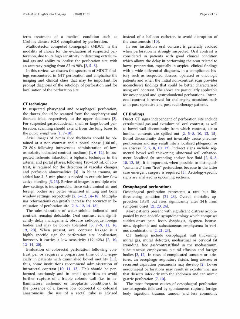
Fig. 1 75-year-old patient with pharyngeal perforation followingrepeated nasogastric tube insertions. Axial contrast-enhanced imageof the neck following oral contrast administration demonstrates acontrast leak through the left aspect of the pharyngealwall (arrowhead)
Pouli et al. Insights into Imaging (2020) 11:31 Page 3 of 19
NSAIDs, acetylsalicylic acid, corticosteroids (Fig. 5), beva-cizumab, stress, tobacco, alcohol abuse, and less com-monly inflammatory bowel disease and Zollinger-Ellisonsyndrome [31, 33–35]. Ulcers along the anterior walland curvatures perforate directly into the peritonealspace (Fig. 6), while ulcers along the posterior wall orduodenum tend to result in contained perforations [3,27, 29, 33]. The gastric antrum and duodenal bulbare the most common perforation sites in ulcerativedisease [2, 3, 31]. Post-bulbar ulcers are rare (3–5%)
and should raise suspicion for conditions likeZollinger-Ellison syndrome or Crohn’s [35].The retroperitoneal duodenum is the most common
site affected in blunt trauma due to its firm attachmentand proximity to the vertebral column [2, 3, 8]. CT ishelpful in distinguishing a duodenal hematoma, which isassociated with wall thickening and surrounding fluid,from a duodenal perforation which may manifest withgas and/or extravasated oral contrast in the right anter-ior pararenal space in addition to the above findings [10,
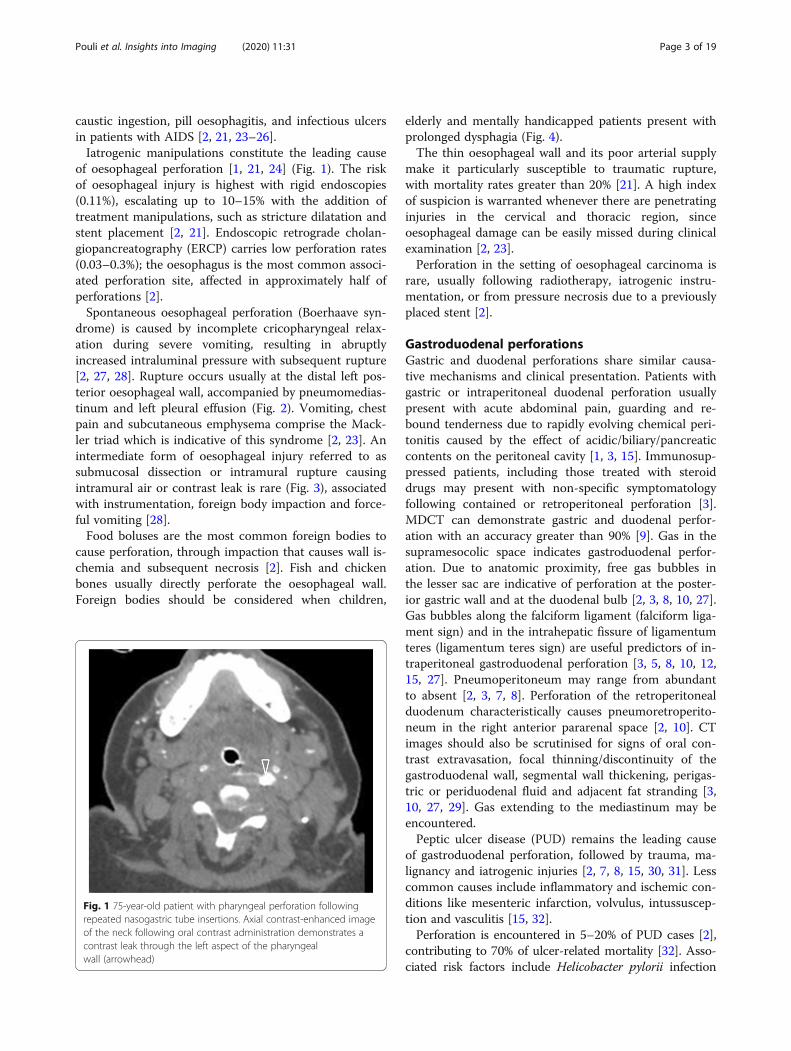
Fig. 2 67-year-old patient with Boerhaave syndrome. a Coronal and (b) axial unenhanced images following oral contrast administration. A largemural defect (black arrow) results in contrast leaking into a paraoesophageal collection (c). Note multiple gas bubbles anteroposterior to theoesophagus, retrocrurally (open arrow), at the right axilla (white arrow) and a right-sided pleural effusion (e). A nasogastric tube (arrowhead)is noted
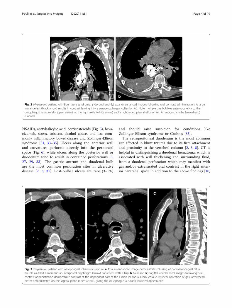
Fig. 3 75-year-old patient with oesophageal intramural rupture. a Axial unenhanced image demonstrates blurring of paraoesophageal fat, adouble air-filled lumen and an interposed diaphragm (arrow) consistent with a flap. b Axial and (c) sagittal unenhanced images following oralcontrast administration demonstrate contrast at the dependent part of the lumen (*) and a submucosal curvilinear collection of gas (arrowhead)better demonstrated on the sagittal plane (open arrow), giving the oesophagus a double-barreled appearance
Pouli et al. Insights into Imaging (2020) 11:31 Page 4 of 19
36]. Gastric injury should be suspected in the presenceof splenic, diaphragmatic or left hemiliver injuries [2,29]. Since the stomach usually collapses following rup-ture, identifying the injury tract on CT may be the onlyclue, suggesting penetrating injury [2, 29]. In the settingof concomitant left diaphragmatic injury, leakage of gas-tric contents into the overlying chest cavity may resultin empyema [2].Iatrogenic perforations occur rarely following ERCP
(0.03–0.3%) (Fig. 7), oesophagogastroduodenoscopy, in-ferior vena cava filter and biliary stent placement [2, 3].ERCP-related duodenal perforations have been classified
in four categories, depending on the mechanism and per-foration site [37]. Lateral or medial wall perforations (typeI) typically require emergent surgery, whereas peri-Vaterian(II) and distal common bile duct injuries (III) are amenableto conservative management. Symptomatic type II patientscan benefit from endoclipping [37, 38]. Cases treated con-servatively may require follow-up CT after 48–72 h [38].Isolated retroperitoneal air from insufflation does not re-quire treatment when asymptomatic (type IV) [37, 38].
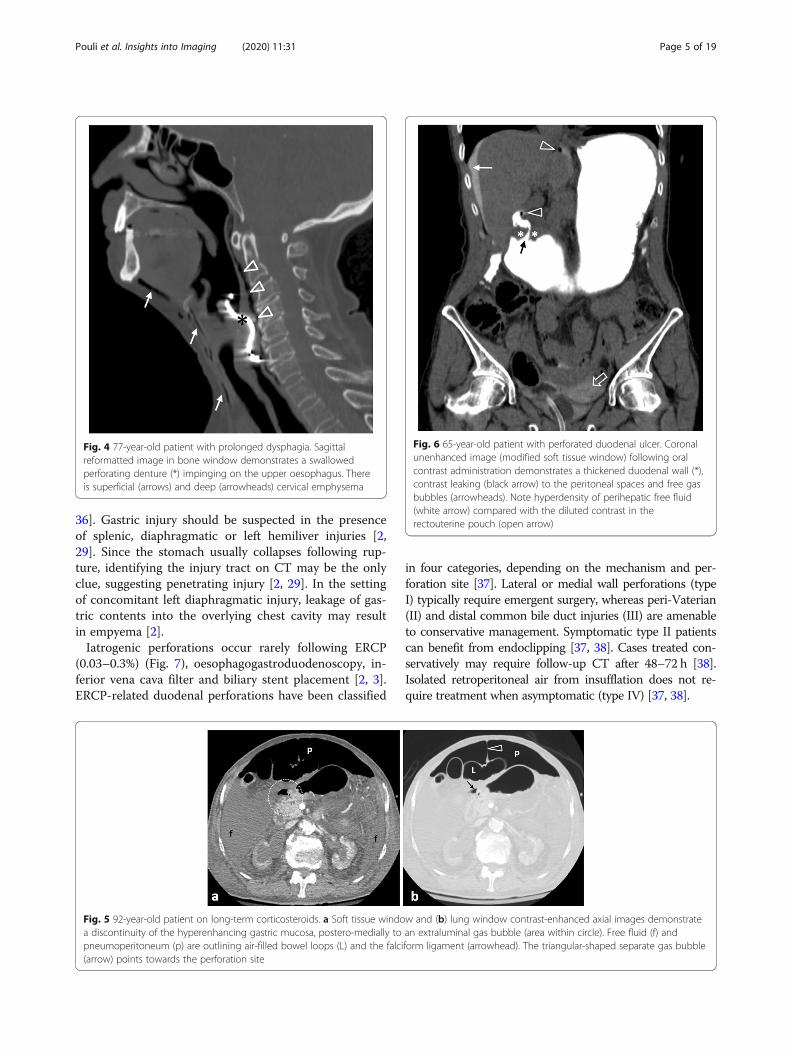
Fig. 4 77-year-old patient with prolonged dysphagia. Sagittalreformatted image in bone window demonstrates a swallowedperforating denture (*) impinging on the upper oesophagus. Thereis superficial (arrows) and deep (arrowheads) cervical emphysema
Fig. 5 92-year-old patient on long-term corticosteroids. a Soft tissue window and (b) lung window contrast-enhanced axial images demonstratea discontinuity of the hyperenhancing gastric mucosa, postero-medially to an extraluminal gas bubble (area within circle). Free fluid (f) andpneumoperitoneum (p) are outlining air-filled bowel loops (L) and the falciform ligament (arrowhead). The triangular-shaped separate gas bubble(arrow) points towards the perforation site
Fig. 6 65-year-old patient with perforated duodenal ulcer. Coronalunenhanced image (modified soft tissue window) following oralcontrast administration demonstrates a thickened duodenal wall (*),contrast leaking (black arrow) to the peritoneal spaces and free gasbubbles (arrowheads). Note hyperdensity of perihepatic free fluid(white arrow) compared with the diluted contrast in therectouterine pouch (open arrow)
Pouli et al. Insights into Imaging (2020) 11:31 Page 5 of 19
Perforation in the setting of gastric banding may presentas an acute or, more commonly, a chronic complicationsecondary to transmural band erosion and manifests withfree or loculated extraluminal gas outlining the band, fatstranding or a subphrenic abscess [17, 29]. Part of theband may be seen in the gastric lumen [17, 29].Gastric perforation associated with malignancy is rare
(0.4–6%), occurring particularly with ulcerated masses,e.g. adenocarcinoma, lymphoma, and large gastrointestinalstromal tumours (GISTs) [2, 29]. Irregular wall thickening,submucosal mass, heaped-up ulcer craters, perivisceralsoft-tissue extension, peritoneal spread, lymphadenopathyand distal metastatic disease are highly suggestive ofunderlying malignancy (Fig. 8) [2, 29, 35].Less than 1% of cases of ingested foreign bodies lead
to perforation [16, 18]. Some patients may not recallingesting a foreign body, present with nonspecific
symptoms and may be diagnosed months or years later[16]. Patients at higher risk are those with reduced palatesensitivity (use of dentures), alcohol abuse, young chil-dren, elderly and mentally handicapped [16, 18]. A for-eign body may be radiopaque depending on itscomposition [18]. Fish bones are the most common cul-prit throughout the GIT except for the oesophagus andusually result in contained perforations which are sealedoff by surrounding omentum and inflammation [16].Consequently, associated pneumoperitoneum is uncom-mon [16, 18]. A foreign body may rarely perforate andmigrate into an adjacent organ, most commonly the lefthemiliver, presenting with fistula and abscess formation(Fig. 9) [ 16,18]. In neglected or chronic cases, where theforeign body gradually erodes through the bowel wall,the resulting inflammatory changes may mimic malig-nancy. Duodenal perforations in particular may mimic
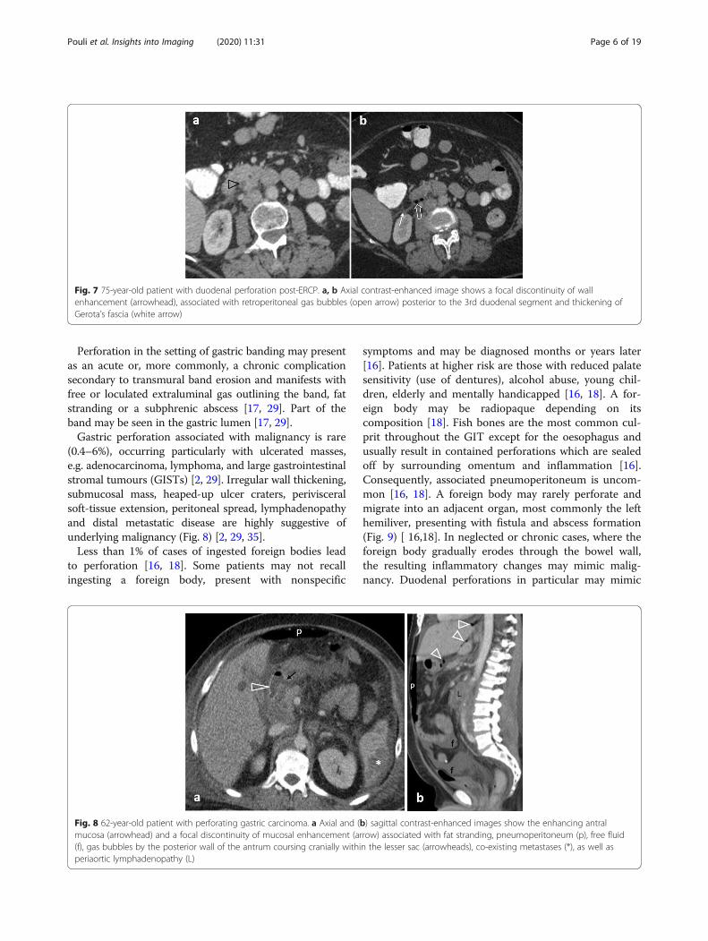
Fig. 7 75-year-old patient with duodenal perforation post-ERCP. a, b Axial contrast-enhanced image shows a focal discontinuity of wallenhancement (arrowhead), associated with retroperitoneal gas bubbles (open arrow) posterior to the 3rd duodenal segment and thickening ofGerota’s fascia (white arrow)
Fig. 8 62-year-old patient with perforating gastric carcinoma. a Axial and (b) sagittal contrast-enhanced images show the enhancing antralmucosa (arrowhead) and a focal discontinuity of mucosal enhancement (arrow) associated with fat stranding, pneumoperitoneum (p), free fluid(f), gas bubbles by the posterior wall of the antrum coursing cranially within the lesser sac (arrowheads), co-existing metastases (*), as well asperiaortic lymphadenopathy (L)
Pouli et al. Insights into Imaging (2020) 11:31 Page 6 of 19
pancreatitis or a pancreatic mass since they may run alonger and relatively asymptomatic course. In thesecases, it is essential to identify a hyperdense structure asthe offending foreign body [16].Presenting symptoms, imaging and clinical consider-
ations in relation to site and causes of upper GIT perfor-ation are summarised (Table 1).
Small bowel perforationsPerforation of the jejunum or ileum carries an incidenceof 1 in 300–350,000 and accounts for 0.4% cases of acuteabdomen [20]. Presentation is nonspecific, consisting ofabrupt onset persistent abdominal pain unresponsive tomedication which evolves into sepsis and peritonitis ifleft untreated [2, 5, 39].Trauma is the leading cause of perforation, followed
by closed-loop obstruction and tumours in developedcountries and by infection in developing countries (ty-phoid fever, tuberculosis, HIV and hookworms amongothers) [5, 20]. Crohn’s disease (CD), ischemia, iatro-genic manipulations, foreign bodies, small bowel diver-ticulitis and medications (e.g. NSAIDs, potassiumchloride) constitute less common causes [2, 5, 20, 27].Pneumoperitoneum may be absent or too subtle to be
detected, noted in approximately half the cases [5, 7, 10,20, 27]. For this reason, images should be scrutinised forindirect findings including wall thickening, mesentericfluid and stranding and ancillary findings indicative ofthe underlying cause such as a mass, an abscess, an in-carcerated hernia or a foreign body [5, 12, 20, 27]. At-tention must be paid to localised interloop collections ofextraluminal fluid or gas, as they can easily be mistakenfor intraluminal contents [20].
Trauma, although uncommon, constitutes the mostfrequent cause of jejunal/ileal perforation [5]. The smallbowel is the third most frequently involved site in ab-dominal blunt trauma, following the liver and spleen [5].Since abdominal traumatic lesions are rarely isolated,the abdomen should be scrutinised for coexisting injur-ies (Fig. 10). CT diagnosis of small bowel blunt injury ischallenging because specific signs for bowel injury arefrequently absent [20]. The combination of bowel wallthickening and mural discontinuity is the most accurateindicator of bowel injury [14]. Indirect signs whichshould raise suspicion for occult bowel injury but notnecessarily perforation include mesenteric fat strandingand a moderate to large volume of unexplained intraper-itoneal fluid in the absence of solid organ injury [2, 14].Investigation with CT in penetrating injury is controver-sial as it may significantly delay surgical management[2]. In penetrating injury, free intraperitoneal gas is notdiagnostic, as air can be introduced into the peritonealcavity by the entry wound [14] (Fig. 11). In this setting,demonstrating a wound track that extends to an injuredintestinal segment is the most sensitive finding [2, 14].Perforation secondary to ischemia can result from ei-
ther strangulated bowel obstruction (Figs. 12 and 13) orfrom a primary vascular event (large vessel occlusion,venous outflow obstruction, or vasculitis) [20]. Ischemiacan additionally result from marked and prolongedhypotension secondary to sepsis, congestive heart failure,acute myocardial infarction and hypovolemic shock. CTchanges suggestive of underlying ischemia mainly de-pend on the aetiology as well as the duration and extentof the ischemic attack and include perfusion abnormal-ities, ranging from bowel wall hyperenhancement to re-duced or absent wall enhancement, segmental bowelwall thickening, localised fluid/fat stranding and embolior thrombi in mesenteric vessels. Pneumatosis intestina-lis and portomesenteric gas in the setting of mesentericischemia indicate transmural infarction [2, 5, 20].Small vessel vasculitis is a rare cause of intestinal per-
foration. Diagnosis is based on indirect signs including anon-vascular territory distribution of multiple and occa-sionally discontinuous ischemic bowel segments [2, 40].Ischemic involvement of the duodenum is nearly alwaysindicative of vasculitis (Fig. 14) [40].Approximately 75–80% of patients with Crohn’s
require surgery within the first 5–20 years from diag-nosis due to the development of stricturing or pene-trating disease [41, 42]. Transmural Crohn’s may leadto contained perforation due to the presence of adhe-sions between adjacent structures or bowel loops [20,27]. Subsequent phlegmon and abscess formation withlocalised peritonitis may develop [14]. Extraluminalcomplications of penetrating CD also include sinustracts and/or fistulas between bowel loops and
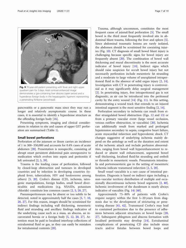
Fig. 9 70-year-old patient presenting with fever and right upperquadrant pain for 3 days. Axial contrast-enhanced imagedemonstrates a gas-containing liver abscess (open arrow) and ahyperdense foreign body in the hepatogastric ligament representinga penetrating fishbone (arrowhead)
Pouli et al. Insights into Imaging (2020) 11:31 Page 7 of 19
between bowel and other visceral organs. Free perfor-ation is a rare life-threatening complication that oc-curs in 1–3% of cases [2, 14, 20] (Fig. 15). Additionalfindings like discontinuous and/or long segmentbowel wall thickening, engorgement of vasa recta ad-jacent to an inflamed bowel loop and mural stratifica-tion characterise active disease, and may suggest theaetiology in otherwise undiagnosed CD [2, 14]. In-flammatory stranding in the small bowel mesenteryadjacent to a thick-walled segment of bowel is notspecific for perforation in patients with CD [27].
Perforation secondary to small bowel tumours occursmore often in primary malignant lymphoma especiallywhen treated with chemotherapy and steroids, in post-transplant lymphoproliferative disorder and following ra-diation therapy [2, 14, 20], although adenocarcinomas,GISTs and metastases can also perforate [20]. Circum-ferential bowel wall thickening with luminal aneurysmaldilatation is highly suggestive of lymphoma [2]. Add-itional findings suggesting GIT lymphoma are multifocalbowel involvement, lymphadenopathy and hepatospleno-megaly [2, 43]. GISTs rarely perforate and in this setting
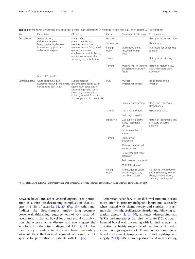
Table 1 Presenting symptoms, imaging and clinical considerations in relation to site and causes of upper GIT perforation
Site Presentation CT findings Causes Cause-specific findings Considerations
Oesophagus Severe distress,sudden-onset pain,fever, dysphagia, dyspnea,hoarseness, dysphonia,tachycardia, crepitus
Mural defect,pneumomediastinum,free mediastinal contrast,free mediastinal fluid, muralgas, subcutaneousemphysema, wall thickening,mediastinal or cervical fatstranding, pleural effusion
Iatrogenic History of instrumentation
Spontaneous Hyperemesis
Foreignbody
Visible food bolus,impacted foreignbody
Investigate for underlyingstricture
Trauma History of penetratinginjury
Tumour Massive wall thickening,oesophago-respiratoryfistula
History of radiotherapy,instrumentation, stentplacement
Fever, SIRS, shock*
Gastroduodenal Acute abdominal pain,guarding, rebound tenderness,non-specific pain (in RP)
Supramesocolicpneumoperitoneum, gas inligamentum teres, gas infalciform ligament, gas inlesser sac, oral contrastleakage, mural defect, gas inanterior pararenal space (in RP)
PUD Mucosalhyperenhancement
Helicobacter pyloriiinfection
Luminal outpouching Drugs, stress, tobacco,alcohol abuse
Trauma Gas in wound track History of trauma
Solid organ injuries
Iatrogenic Gas outlining gastricband, subphrenicabscess
History of instrumentationor history of gastricbanding
Intraluminal band/sutures
Tumour Irregular wallthickening
Mucosal/submucosalenhancement
Perivisceral soft-tissueextension
Peritoneal/nodal spread
Metastatic disease
Foreignbody
Radiopaque structureby a fistula, opacityby a liver abscess
Individuals with reducedpalate sensitivity, alcoholabuse, children, elderly,mentally handicapped
*In late stages. SIRS systemic inflammatory response syndrome, RP retroperitoneal perforation, IP intraperitoneal perforation, RT right
Pouli et al. Insights into Imaging (2020) 11:31 Page 8 of 19
usually present with heterogeneous attenuation and alamellated pattern reflecting areas of haemorrhage ornecrotic degeneration [2]. Since ascites is uncommon inGISTs, such an associated finding should prompt thor-ough inspection for tumour rupture and/or metastaticspread [2].Small bowel diverticula occur in 0.06–2.3% of the
population and rarely perforate [20] (Fig. 16). Meckel’sdiverticulum is located at the antimesenteric side wall ofthe distal ileum, most commonly containing gastric mu-cosa (62%), and can be complicated by bleeding and,rarely, perforation [27, 44]. Reformatted images canbetter illustrate the relationship of diverticula to thelumen of the bowel and suggest them as the perfor-ation site [20].Ingested foreign bodies may rarely cause small bowel
perforation [27]. Common sites include narrowed orangulated portions of the GIT, such as the ileocaecalarea [14, 16, 18, 27]. As with gastroduodenal perfora-tions, pneumoperitoneum is minimal if any, since theforeign body is gradually impacted and is walled-off byomentum and inflammatory changes [2, 16, 20] (Fig. 16).Identifying a partially extraluminal foreign body
confirms the diagnosis, and it is more easily visualised inbone window settings (Fig. 17) [2, 14]. The foreign bodymay be identified distal to the perforation site, since it ispossible to cause a perforation and then move distallywithin the bowel lumen [20].Iatrogenic perforation following open or laparoscopic
surgery most commonly involves the small bowel andcarries high morbidity and mortality, especially if notrecognised intraoperatively [2, 20]. Anastomotic leakageusually occurs within the first postoperative week [10].Endoscopic procedures, misplaced percutaneous drain-age catheters, radiation-induced injury and paracentesiscan also result in small bowel injury and subsequentperforation [20]. Intraperitoneal free gas is difficult tointerpret since it is normally expected post-laparotomyfor up to 2 weeks and approximately for up to 3 daysfollowing laparoscopic procedures [7]. Oral contrastmay be useful in this setting, as contrast leakage withan intact anastomotic site suggests the diagnosis of ac-cidental iatrogenic bowel injury [2, 10, 14]. Persistentor progressively increasing free air and/or ascitesshould raise concern for perforation or anastomoticleakage [8, 10].
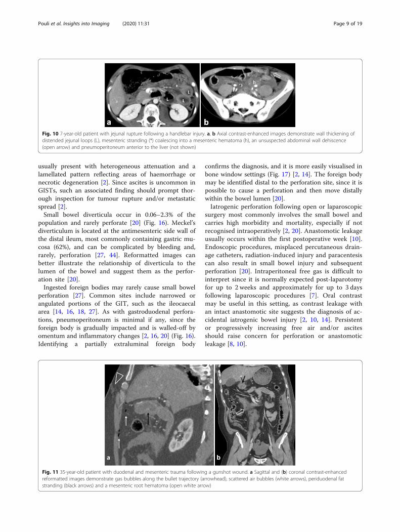
Fig. 10 7-year-old patient with jejunal rupture following a handlebar injury. a, b Axial contrast-enhanced images demonstrate wall thickening ofdistended jejunal loops (L), mesenteric stranding (*) coalescing into a mesenteric hematoma (h), an unsuspected abdominal wall dehiscence(open arrow) and pneumoperitoneum anterior to the liver (not shown)
Fig. 11 35-year-old patient with duodenal and mesenteric trauma following a gunshot wound. a Sagittal and (b) coronal contrast-enhancedreformatted images demonstrate gas bubbles along the bullet trajectory (arrowhead), scattered air bubbles (white arrows), periduodenal fatstranding (black arrows) and a mesenteric root hematoma (open white arrow)
Pouli et al. Insights into Imaging (2020) 11:31 Page 9 of 19
Appendiceal perforationAppendiceal perforation results from obstruction due toan appendicolith in the setting of appendicitis or mayrarely be associated with an underlying tumour ormucocele [8, 27]. Appendicitis is rarely caused byobstructing fruit seeds, vegetables, lymphoid hyperplasia,intestinal worms (Ascaris), malignancy, and foreign bod-ies [45]. Delay in presentation is strongly associated withperforation in the setting of appendicitis [45].The role of CT in detecting perforating appendicitis at
an early stage or appendiceal micro-perforations is con-troversial, especially in young patients [2]. In neglected/complicated cases, elderly patients and possible dualpathology, CT has an established role.The presence of extraluminal gas, which is usually min-
imal (< 2mL) or absent, an appendiceal wall defect, peri-appendiceal abscess, and extraluminal appendicolith arehighly suggestive of perforation [2, 7, 8, 10, 27] (Fig. 18).Cystic dilatation of the appendix with a luminal diam-
eter greater than 1.3 cm, along with the presence ofmural calcifications, suggests a mucocele or a mucinousneoplasm [2, 46]. Secondary perforation, caused by anappendiceal mucinous neoplasm, leads to pseudomyx-oma peritonei [2] (Fig. 19).Presenting symptoms, imaging and clinical consider-
ations in relation to site and causes of small bowel andappendiceal perforation are summarised (Table 2).
Colorectal perforationColorectal perforation carries the highest complicationrate (55%) compared with other GIT perforation sites[47]. This is unsurprising considering the bacterial con-tent of the large bowel that evokes bacterial peritonitis[1, 4]. Perforations contained in retroperitoneal spacesusually present with subtle symptomatology [6, 27].Malignancy is the commonest cause, accounting for
36% of perforations [48]. Perforation may also be iatro-genic (20%), diverticula-related (19%) and less commonlydue to trauma, foreign body ingestion, faecal impaction,
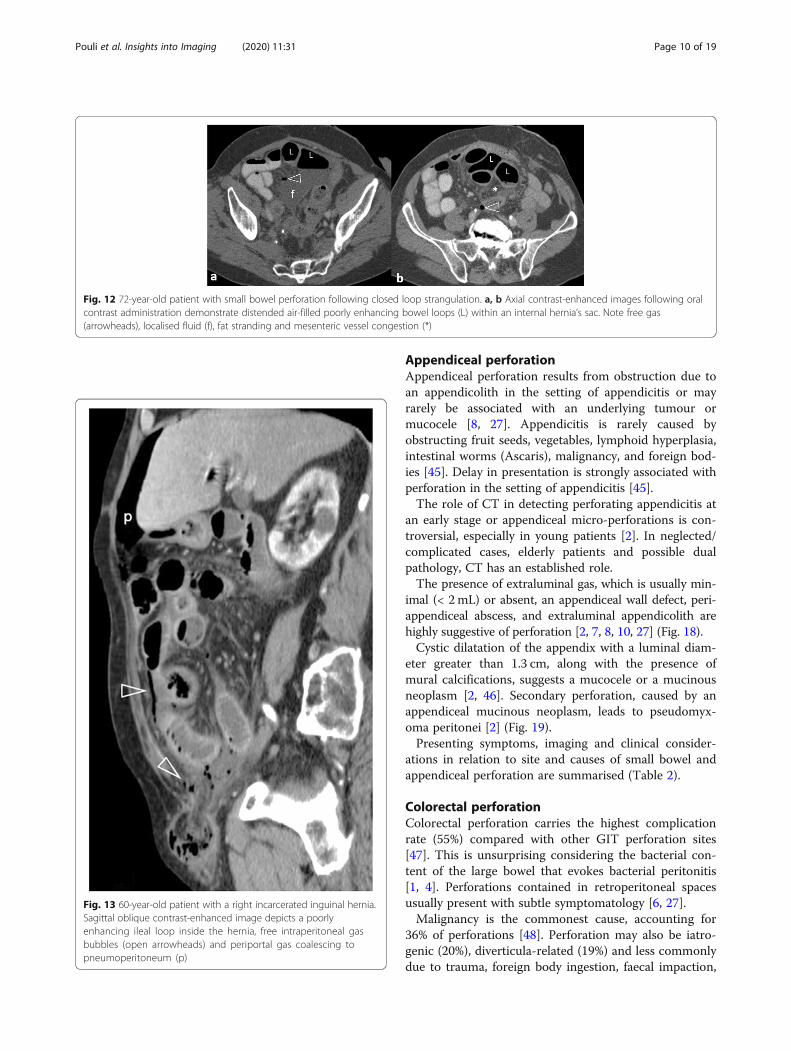
Fig. 12 72-year-old patient with small bowel perforation following closed loop strangulation. a, b Axial contrast-enhanced images following oralcontrast administration demonstrate distended air-filled poorly enhancing bowel loops (L) within an internal hernia’s sac. Note free gas(arrowheads), localised fluid (f), fat stranding and mesenteric vessel congestion (*)
Fig. 13 60-year-old patient with a right incarcerated inguinal hernia.Sagittal oblique contrast-enhanced image depicts a poorlyenhancing ileal loop inside the hernia, free intraperitoneal gasbubbles (open arrowheads) and periportal gas coalescing topneumoperitoneum (p)
Pouli et al. Insights into Imaging (2020) 11:31 Page 10 of 19
ischemia, inflammatory bowel disease, endometriosis,connective tissue disease, radiotherapy, drugs and spon-taneous [4, 6, 7, 10, 11, 49]. The site of perforation isoften linked with its cause [4, 8, 10]; therefore, neoplas-tic, spontaneous, diverticular (in western countries),blunt trauma and ischemic perforations commonlyoccur on the left side of colon, whereas inflammatory
bowel disease, diverticulitis (in eastern countries) andpenetrating trauma perforations tend to be right-sided.The rectosigmoid is most commonly affected in iatro-genic manipulations and foreign bodies [4, 6]. Thececum may perforate when it reaches 12–14 cm in diam-eter as a result of a rapid increase in intraluminal pres-sure in bowel obstruction with a competent ileocaecalvalve, toxic megacolon, stercoral colitis and acute co-lonic pseudo-obstruction [4, 8, 11]. Extraluminal gaslimited in the pelvis or within retroperitoneal compart-ments suggests colorectal perforation with the exceptionof right-sided retropneumoperitoneum which may alsoresult from duodenal perforation.The accuracy of MDCT in predicting the perforation
site is lower in colorectal perforations compared withupper GIT perforations [9]. Direct findings includeextraluminal gas and oral/rectal contrast, as well as walldiscontinuity, faecal material looking like a “dirty” massor foreign bodies protruding through the colonic wall orlying free within the abdominal cavity [4, 8, 11, 27, 50].Associated findings include bowel wall thickening, perico-lonic fat stranding, free fluid, abnormal wall enhancement,abscess, and the presence of a localised inflammatory massadjacent to the colon [4, 8, 11, 27].Free gas may be massive, especially if there is coexistent
obstruction or following colonoscopy [4, 27]. Perforationof intraperitoneal colon, i.e. of cecum, transverse, sigmoidand upper 2/3 of the rectum, leads to pneumoperitoneum[7]. Rupture of the retroperitoneal ascending and descend-ing colon results in pneumoretroperitoneum at the rightand left anterior pararenal spaces respectively [2, 7, 10,11]. Posterior rectal perforations can dissect superiorlyand cause bilateral pneumoretroperitoneum [10, 11]. Inthe setting of diverticulitis or malignancy without mech-anical obstruction, perforation is often associated withsmall volume pneumoperitoneum by the perforation site
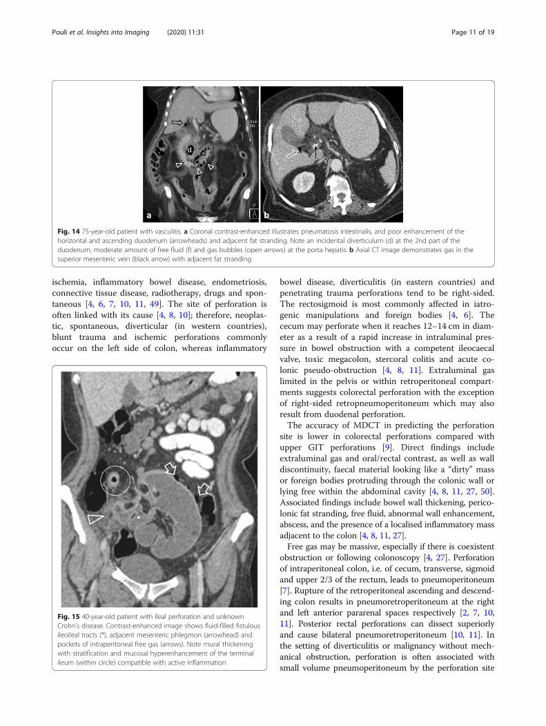
Fig. 14 75-year-old patient with vasculitis. a Coronal contrast-enhanced illustrates pneumatosis intestinalis, and poor enhancement of thehorizontal and ascending duodenum (arrowheads) and adjacent fat stranding. Note an incidental diverticulum (d) at the 2nd part of theduodenum, moderate amount of free fluid (f) and gas bubbles (open arrows) at the porta hepatis. b Axial CT image demonstrates gas in thesuperior mesenteric vein (black arrow) with adjacent fat stranding
Fig. 15 40-year-old patient with ileal perforation and unknownCrohn’s disease. Contrast-enhanced image shows fluid-filled fistulousileoileal tracts (*), adjacent mesenteric phlegmon (arrowhead) andpockets of intraperitoneal free gas (arrows). Note mural thickeningwith stratification and mucosal hyperenhancement of the terminalileum (within circle) compatible with active inflammation
Pouli et al. Insights into Imaging (2020) 11:31 Page 11 of 19
[8, 10]. Free gas present only in the pelvis favours colonicrather than small bowel perforation [2, 6, 11, 27].Perforation complicates 2.6–10% of colon cancers, oc-
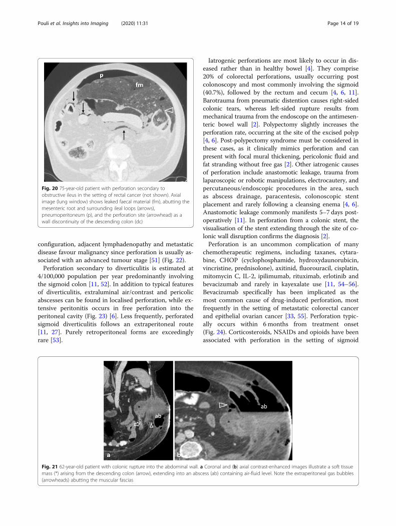
curring most commonly in the sigmoid colon (47.3%)and cecum (24.8%) [2, 6, 11, 49, 51]. Two mechanismsare recognised [2, 4, 6, 11, 49]: the first consists oftumour necrosis and subsequent perforation at thetumour site. The second occurs proximal to the malig-nancy, as a result of bowel distension secondary to ob-struction, frequently occurs at the cecum and usuallyleads to massive pneumoperitoneum (Fig. 20) or pneu-moretroperitoneum. Perforation secondary to tumournecrosis usually causes a small amount of free gas [11].Rarely, the colon perforates into the abdominal wall andpatients may present with extensive cellulitis (Fig. 21).Differentiation between neoplastic and inflammatorycauses is challenging; however, a mural wall thicknessgreater than 1.39 cm, the presence of irregular wall
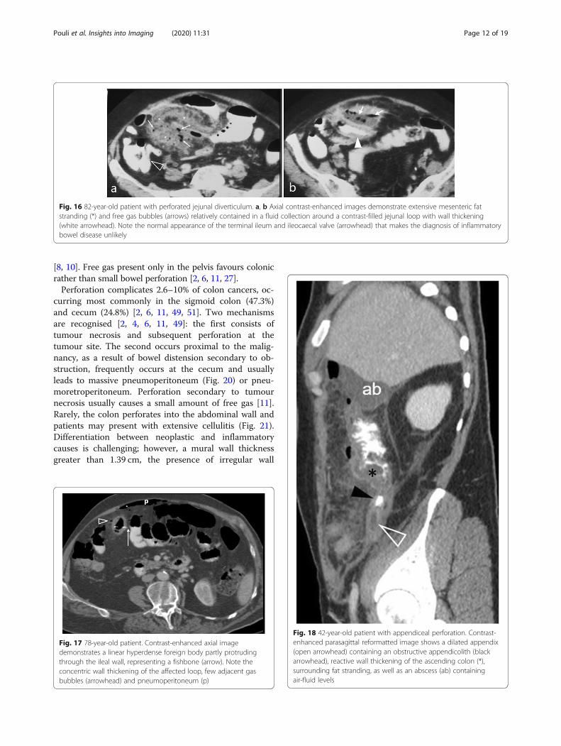
Fig. 16 82-year-old patient with perforated jejunal diverticulum. a, b Axial contrast-enhanced images demonstrate extensive mesenteric fatstranding (*) and free gas bubbles (arrows) relatively contained in a fluid collection around a contrast-filled jejunal loop with wall thickening(white arrowhead). Note the normal appearance of the terminal ileum and ileocaecal valve (arrowhead) that makes the diagnosis of inflammatorybowel disease unlikely
Fig. 17 78-year-old patient. Contrast-enhanced axial imagedemonstrates a linear hyperdense foreign body partly protrudingthrough the ileal wall, representing a fishbone (arrow). Note theconcentric wall thickening of the affected loop, few adjacent gasbubbles (arrowhead) and pneumoperitoneum (p)
Fig. 18 42-year-old patient with appendiceal perforation. Contrast-enhanced parasagittal reformatted image shows a dilated appendix(open arrowhead) containing an obstructive appendicolith (blackarrowhead), reactive wall thickening of the ascending colon (*),surrounding fat stranding, as well as an abscess (ab) containingair-fluid levels
Pouli et al. Insights into Imaging (2020) 11:31 Page 12 of 19
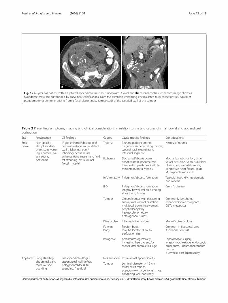
Table 2 Presenting symptoms, imaging and clinical considerations in relation to site and causes of small bowel and appendicealperforation
Site Presentation CT findings Causes Cause specific findings Considerations
Smallbowel
Non-specific,abrupt sudden-onset pain, vomit-ing, anorexia, nau-sea, sepsis,peritonitis
IP gas (minimal/absent), oralcontrast leakage, mural defect,wall thickening, poor/inhomogeneous muralenhancement, mesenteric fluid,fat stranding, extraluminalfaecal material
Trauma Pneumoperitoneum notdiagnostic in penetrating trauma,wound track extending tointestinal segment.
History of trauma
IIschemia Decreased/absent bowelenhancement, pneumatosisintestinalis, gas/thrombi withinmesenteric/portal vessels
Mechanical obstruction, largevessel occlusion, venous outflowobstruction, vasculitis, sepsis,congestive heart failure, acuteMI, hypovolemic shock
Inflammatory Phlegmon/abscess formation Typhoid fever, HIV, tuberculosis,hookworms
IBD Phlegmon/abscess formation,lengthy bowel wall thickenning,sinus tracts, fistulas
Crohn's disease
Tumour Circumferential wall thickeninganeurysmal luminal dilatationmultifocal bowel involvementlymphadenopathyhepatosplenomegalyheterogeneous mass
Commonly lymphomaadenocarcinoma malignantGISTs metastases
Diverticulae Inflamed diverticulum Meckel's diverticulum
Foreignbody
Foreign body,may be located distal toperforation site
Common in ileocaecal areaAvoid oral contrast
Iatrogenic persistent/progressivelyincreasing free gas and/orascites, oral contrast leakage
Laparoscopic surgery,anastomotic leakage, endoscopicprocedures. Pneumoperitoneumnormal< 2 weeks post laparoscopy
Appendix Long standingabdominal pain,fever, muscleguarding
Periappendiceal/IP gas,appendiceal wall defect,phlegmon/abscess, fatstranding, free fluid
Inflammation Extraluminal appendicolith
Tumour Luminal diameter > 1.3 cm,mural calcifications,pseudomyxoma peritonei, mass,enhancing wall nodularity
IP intraperitoneal perforation, MI myocardial infarction, HIV human immunodeficiency virus, IBD inflammatory bowel disease, GIST gastrointestinal stromal tumour
Fig. 19 65-year-old patient with a ruptured appendiceal mucinous neoplasm. a Axial and (b) coronal contrast-enhanced image shows ahypodense mass (m), surrounded by curvilinear calcifications. Note the extensive enhancing encapsulated fluid collections (c), typical ofpseudomyxoma peritonei, arising from a focal discontinuity (arrowhead) of the calcified wall of the tumour
Pouli et al. Insights into Imaging (2020) 11:31 Page 13 of 19
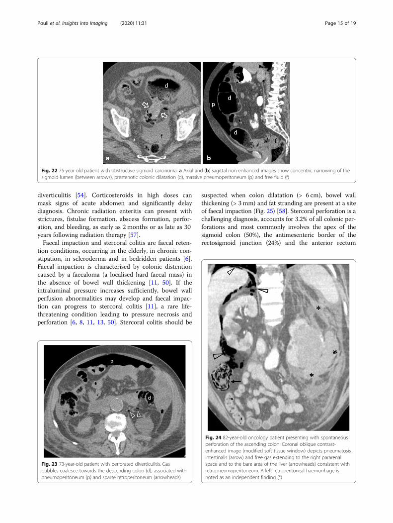
configuration, adjacent lymphadenopathy and metastaticdisease favour malignancy since perforation is usually as-sociated with an advanced tumour stage [51] (Fig. 22).Perforation secondary to diverticulitis is estimated at
4/100,000 population per year predominantly involvingthe sigmoid colon [11, 52]. In addition to typical featuresof diverticulitis, extraluminal air/contrast and pericolicabscesses can be found in localised perforation, while ex-tensive peritonitis occurs in free perforation into theperitoneal cavity (Fig. 23) [6]. Less frequently, perforatedsigmoid diverticulitis follows an extraperitoneal route[11, 27]. Purely retroperitoneal forms are exceedinglyrare [53].
Iatrogenic perforations are most likely to occur in dis-eased rather than in healthy bowel [4]. They comprise20% of colorectal perforations, usually occurring postcolonoscopy and most commonly involving the sigmoid(40.7%), followed by the rectum and cecum [4, 6, 11].Barotrauma from pneumatic distention causes right-sidedcolonic tears, whereas left-sided rupture results frommechanical trauma from the endoscope on the antimesen-teric bowel wall [2]. Polypectomy slightly increases theperforation rate, occurring at the site of the excised polyp[4, 6]. Post-polypectomy syndrome must be considered inthese cases, as it clinically mimics perforation and canpresent with focal mural thickening, pericolonic fluid andfat stranding without free gas [2]. Other iatrogenic causesof perforation include anastomotic leakage, trauma fromlaparoscopic or robotic manipulations, electrocautery, andpercutaneous/endoscopic procedures in the area, suchas abscess drainage, paracentesis, colonoscopic stentplacement and rarely following a cleansing enema [4, 6].Anastomotic leakage commonly manifests 5–7 days post-operatively [11]. In perforation from a colonic stent, thevisualisation of the stent extending through the site of co-lonic wall disruption confirms the diagnosis [2].Perforation is an uncommon complication of many
chemotherapeutic regimens, including taxanes, cytara-bine, CHOP (cyclophosphamide, hydroxydaunorubicin,vincristine, prednisolone), axitinid, fluorouracil, cisplatin,mitomycin C, IL-2, ipilimumab, rituximab, erlotinib andbevacizumab and rarely in kayexalate use [11, 54–56].Bevacizumab specifically has been implicated as themost common cause of drug-induced perforation, mostfrequently in the setting of metastatic colorectal cancerand epithelial ovarian cancer [33, 55]. Perforation typic-ally occurs within 6 months from treatment onset(Fig. 24). Corticosteroids, NSAIDs and opioids have beenassociated with perforation in the setting of sigmoid
Fig. 20 75-year-old patient with perforation secondary toobstructive ileus in the setting of rectal cancer (not shown). Axialimage (lung window) shows leaked faecal material (fm), abutting themesenteric root and surrounding ileal loops (arrows),pneumoperitoneum (p), and the perforation site (arrowhead) as awall discontinuity of the descending colon (dc)
Fig. 21 62-year-old patient with colonic rupture into the abdominal wall. a Coronal and (b) axial contrast-enhanced images illustrate a soft tissuemass (*) arising from the descending colon (arrow), extending into an abscess (ab) containing air-fluid level. Note the extraperitoneal gas bubbles(arrowheads) abutting the muscular fascias
Pouli et al. Insights into Imaging (2020) 11:31 Page 14 of 19
diverticulitis [54]. Corticosteroids in high doses canmask signs of acute abdomen and significantly delaydiagnosis. Chronic radiation enteritis can present withstrictures, fistulae formation, abscess formation, perfor-ation, and bleeding, as early as 2 months or as late as 30years following radiation therapy [57].Faecal impaction and stercoral colitis are faecal reten-
tion conditions, occurring in the elderly, in chronic con-stipation, in scleroderma and in bedridden patients [6].Faecal impaction is characterised by colonic distentioncaused by a faecaloma (a localised hard faecal mass) inthe absence of bowel wall thickening [11, 50]. If theintraluminal pressure increases sufficiently, bowel wallperfusion abnormalities may develop and faecal impac-tion can progress to stercoral colitis [11], a rare life-threatening condition leading to pressure necrosis andperforation [6, 8, 11, 13, 50]. Stercoral colitis should be
suspected when colon dilatation (> 6 cm), bowel wallthickening (> 3mm) and fat stranding are present at a siteof faecal impaction (Fig. 25) [58]. Stercoral perforation is achallenging diagnosis, accounts for 3.2% of all colonic per-forations and most commonly involves the apex of thesigmoid colon (50%), the antimesenteric border of therectosigmoid junction (24%) and the anterior rectum
Fig. 22 75-year-old patient with obstructive sigmoid carcinoma. a Axial and (b) sagittal non-enhanced images show concentric narrowing of thesigmoid lumen (between arrows), prestenotic colonic dilatation (d), massive pneumoperitoneum (p) and free fluid (f)
Fig. 23 73-year-old patient with perforated diverticulitis. Gasbubbles coalesce towards the descending colon (d), associated withpneumoperitoneum (p) and sparse retroperitoneum (arrowheads)
Fig. 24 82-year-old oncology patient presenting with spontaneousperforation of the ascending colon. Coronal oblique contrast-enhanced image (modified soft tissue window) depicts pneumatosisintestinalis (arrow) and free gas extending to the right pararenalspace and to the bare area of the liver (arrowheads) consistent withretropneumoperitoneum. A left retroperitoneal haemorrhage isnoted as an independent finding (*)
Pouli et al. Insights into Imaging (2020) 11:31 Page 15 of 19
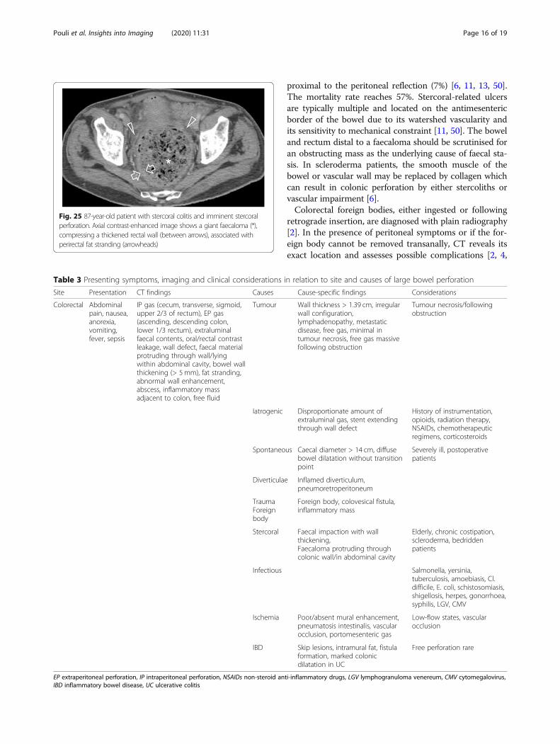
proximal to the peritoneal reflection (7%) [6, 11, 13, 50].The mortality rate reaches 57%. Stercoral-related ulcersare typically multiple and located on the antimesentericborder of the bowel due to its watershed vascularity andits sensitivity to mechanical constraint [11, 50]. The boweland rectum distal to a faecaloma should be scrutinised foran obstructing mass as the underlying cause of faecal sta-sis. In scleroderma patients, the smooth muscle of thebowel or vascular wall may be replaced by collagen whichcan result in colonic perforation by either stercoliths orvascular impairment [6].Colorectal foreign bodies, either ingested or following
retrograde insertion, are diagnosed with plain radiography[2]. In the presence of peritoneal symptoms or if the for-eign body cannot be removed transanally, CT reveals itsexact location and assesses possible complications [2, 4,
Fig. 25 87-year-old patient with stercoral colitis and imminent stercoralperforation. Axial contrast-enhanced image shows a giant faecaloma (*),compressing a thickened rectal wall (between arrows), associated withperirectal fat stranding (arrowheads)
Table 3 Presenting symptoms, imaging and clinical considerations in relation to site and causes of large bowel perforation
Site Presentation CT findings Causes Cause-specific findings Considerations
Colorectal Abdominalpain, nausea,anorexia,vomiting,fever, sepsis
IP gas (cecum, transverse, sigmoid,upper 2/3 of rectum), EP gas(ascending, descending colon,lower 1/3 rectum), extraluminalfaecal contents, oral/rectal contrastleakage, wall defect, faecal materialprotruding through wall/lyingwithin abdominal cavity, bowel wallthickening (> 5 mm), fat stranding,abnormal wall enhancement,abscess, inflammatory massadjacent to colon, free fluid
Tumour Wall thickness > 1.39 cm, irregularwall configuration,lymphadenopathy, metastaticdisease, free gas, minimal intumour necrosis, free gas massivefollowing obstruction
Tumour necrosis/followingobstruction
Iatrogenic Disproportionate amount ofextraluminal gas, stent extendingthrough wall defect
History of instrumentation,opioids, radiation therapy,NSAIDs, chemotherapeuticregimens, corticosteroids
Spontaneous Caecal diameter > 14 cm, diffusebowel dilatation without transitionpoint
Severely ill, postoperativepatients
Diverticulae Inflamed diverticulum,pneumoretroperitoneum
TraumaForeignbody
Foreign body, colovesical fistula,inflammatory mass
Stercoral Faecal impaction with wallthickening,Faecaloma protruding throughcolonic wall/in abdominal cavity
Elderly, chronic costipation,scleroderma, bedriddenpatients
Infectious Salmonella, yersinia,tuberculosis, amoebiasis, Cl.difficile, E. coli, schistosomiasis,shigellosis, herpes, gonorrhoea,syphilis, LGV, CMV
Ischemia Poor/absent mural enhancement,pneumatosis intestinalis, vascularocclusion, portomesenteric gas
Low-flow states, vascularocclusion
IBD Skip lesions, intramural fat, fistulaformation, marked colonicdilatation in UC
Free perforation rare
EP extraperitoneal perforation, IP intraperitoneal perforation, NSAIDs non-steroid anti-inflammatory drugs, LGV lymphogranuloma venereum, CMV cytomegalovirus,IBD inflammatory bowel disease, UC ulcerative colitis
Pouli et al. Insights into Imaging (2020) 11:31 Page 16 of 19
13]. Bowel wall disruption and extraluminal position ofthe object are direct findings of perforation, whereas freegas is uncommon as the perforation site is usually“sealed-off” [2, 6, 11, 16, 18]. Occasionally a colovesicalfistula or inflammatory mass is encountered [16].Perforation secondary to ischemic colitis predomin-
antly results from non-occlusive disease (low-flow state)rather than complete vascular occlusion [11]. Low-flowstates particularly affect watershed areas, such as Grif-fith’s and Sudeck’s critical points, whereas arterial occlu-sion affects whole vascular territories [11, 17]. Thesepatients may initially present with mild abdominal painand tenderness and develop bloody diarrhoea within 24h from symptom onset. Pneumatosis intestinalis in thesetting of ischemia is associated with poor prognosis[17]. Poor or no mural enhancement is suggestive of is-chemia while free air is pathognomonic of transmuralnecrosis [59].Acute colonic pseudo-obstruction (Ogilvie’s syndrome)
usually affects patients with significant comorbidities orpostoperatively [11]. CT findings comprise marked dif-fuse dilatation of colonic and rectal lumen without atransition point to collapsed bowel [11]. Although per-foration is a rare complication (1–3%), carrying a 50–71% mortality rate, the risk increases up to 23% whencaecal diameter is > 14 cm [11].In Ehler-Danlos syndrome, spontaneous perforation is
the most common GIT complication, affecting most com-monly the sigmoid colon [60]. Perforation usually occursearly in the disease (mean age of presentation 24–26years), usually precedes arterial or solid organ rupture andcan be the first symptom in a young patient otherwise un-diagnosed with Ehler-Danlos syndrome [60, 61].Perforation from blunt trauma is uncommon, being re-
ported in approximately 0.5% of all major blunt traumas[4, 13], more commonly in the sigmoid, right colon andcecum [4]. Transverse colon injury may coexist withpancreatic and/or duodenal injury [4]. Oral contrast ad-ministration, when not contraindicated in the emergencysetting, can increase the detection rate of bowel wallthickening and small mesenteric hematomas [13]. Freefluid typically forms polygonal-shaped collections amongloops and within mesenteric folds [4]. Traumatic perfor-ation of a retroperitoneal segment tends to remain local-ised adjacent to the injury site [4].Colonic perforation in inflammatory bowel disease is
rare, with free perforation occurring in about 3% of pa-tients with Crohn’s, less common compared with sealed-off perforations [6]. Free perforation from ulcerative col-itis occurs in about 2% of patients and is associated withtoxic megacolon [6].Presenting symptoms, imaging and clinical consider-
ations in relation to site and causes of colorectal perfor-ation are summarised (Table 3).
ConclusionsWhile the diagnosis of GIT perforation may be straight-forward based on clinical and radiographic findings, thecontribution of CT is twofold: subtle findings not visibleon x-rays may be revealed, therefore supporting thediagnosis and directing appropriate management. Add-itionally, ancillary findings not necessarily related to theperforation may suggest underlying conditions that needfurther investigation following primary repair of rup-tured bowel. Knowledge of the heterogeneous causes ofGIT perforation is important for both the surgeon andthe radiologist in order to ensure optimal management.
AbbreviationsCD: Crohn’s disease; CT: Computed tomography; ERCP: Endoscopicretrograde cholangiopancreatography; GISTs: Gastrointestinal stromaltumours; GIT: Gastrointestinal tract; iv: Intravenous; MDCT: Multidetectorcomputed tomography; NSAIDs: Non-steroid anti-inflammatory drugs;PUD: Peptic ulcer disease
Authors’ contributionsSP was the main author of the article. AK conceived of the article,contributed cases and edited text. IP edited the article for clinical accuracy.MD contributed cases, edited text and formatted figures. MR was thesupervising author, team coordinator, case contributor and text editor. Allauthors read and approved the final manuscript.
FundingThis research did not receive any specific grant from funding agencies in thepublic, commercial, or not-for-profit sectors.
Availability of data and materialsData sharing is not applicable to this article as no datasets were generatedor analysed during the current study.
Ethics approval and consent to participateNot applicable
Consent for publicationNot applicable
Competing interestsThe authors declare that they have no competing interests.
Author details1Department of Radiology, University Hospital of Heraklion, Faculty ofMedicine-University of Crete, Stavrakia, Voutes 21110, Heraklion, Crete,Greece. 2Department of Internal Medicine, University Hospital of Heraklion,Heraklion, Greece.
Received: 26 August 2019 Accepted: 29 November 2019
References1. Langell JT, Mjtulvihill SJ (2008) Gastrointestinal perforation and the acute
abdomen. Med Clin North Am 92(3):599–6252. Del Gaizo AJ, Lall C, Allen BC, Leyendecker JR (2014) From esophagus to
rectum: a comprehensive review of alimentary tract perforations atcomputed tomography. Abdom Imaging 39(4):802–823
3. Picone D, Rusignuolo R, Midiri F et al (2016) Imaging assessment ofgastroduodenal perforations. Semin Ultrasound CT MR 37(1):16–22
4. Saturnino PP, Pinto A, Liguori C, Ponticiello G, Romano L (2016) Role ofmultidetector computed tomography in the diagnosis of colorectalperforations. Semin Ultrasound CT MR 37(1):49–53
5. Lo Re G, Mantia FL, Picone D, Salerno S, Vernuccio F, Midiri M (2016) Smallbowel perforations: what the radiologist needs to know. Semin UltrasoundCT MR 37(1): 23–30
Pouli et al. Insights into Imaging (2020) 11:31 Page 17 of 19
6. Zissin R, Hertz M, Osadchy A, Even-Sapir E, Gayer G (2008) Abdominal CTfindings in nontraumatic colorectal perforation. Eur J Radiol 65(1):125–132
7. Borofsky S, Taffel M, Khati N, Zeman R, Hill M (2015) The emergency roomdiagnosis of gastrointestinal tract perforation: the role of CT. Emerg Radiol22(3):315–327
8. Kim SH, Shin SS, Jeong YY, Heo SH, Kim JW, Kang HK (2009) Gastrointestinaltract perforation: MDCT findings according to the perforation sites. Korean JRadiol 10(1):63–70
9. Kim HC, Yang DM, Kim SW, Park SJ (2014) Gastrointestinal tract perforation:evaluation of MDCT according to perforation site and elapsed time. EurRadiol 24(6):1386–1393
10. Furukawa A, Sakoda M, Yamasaki M et al (2005) Gastrointestinal tractperforation: CT diagnosis of presence, site, and cause. Abdom Imaging30(5):524–534
11. Kothari K, Friedman B, Grimaldi GM, Hines JJ (2017) Nontraumatic largebowel perforation: spectrum of etiologies and CT findings. Abdom Radiol(NY) 42(11):2597–2608
12. Faggian A, Berritto D, Iacobellis F, Reginelli A, Cappabianca S, Grassi R (2016)Imaging patients with alimentary tract perforation: literature review. SeminUltrasound CT MR 37(1):66–69
13. Maddu KK, Mittal P, Arepalli CD, Shuaib W, Tewari A, Khosa F (2014)Colorectal emergencies and related complications: a comprehensiveimaging review-noninfectious and noninflammatory emergencies of colon.AJR Am J Roentgenol 203(6):1217–1229
14. Zissin R, Osadchy A, Gayer G (2009) Abdominal CT findings in small bowelperforation. Br J Radiol 82(974):162–171
15. Paolantonio P, Rengo M, Ferrari R, Laghi A (2016) Multidetector CT inemergency radiology: acute and generalized non-traumatic abdominal pain.Br J Radiol, 89(1061):20150859
16. Paixão TS, Leão RV, de Souza Maciel Rocha Horvat N et al (2017) Abdominalmanifestations of fishbone perforation: a pictorial essay. Abdom Radiol (NY)42(4):1087–1095
17. Ecanow JS, Gore RM (2015) Evaluating patients with left upper quadrantpain. Radiol Clin North Am 53(6):1131–1157
18. Kuzmich S, Burke CJ, Harvey CJ et al (2015) Perforation of gastrointestinaltract by poorly conspicuous ingested foreign bodies: Radiological diagnosis.Br J Radiol 88(1050):20150086
19. O'Malley RG, Al-Hawary MM, Kaza RK, Wasnik AP, Platt JF, Francis IR (2015)MDCT findings in small bowel obstruction: implications of the cause andpresence of complications on treatment decisions. Abdom Imaging 40(7):2248–2262
20. Hines J, Rosenblat J, Duncan DR, Friedman B, Katz DS (2013) Perforation of themesenteric small bowel: etiologies and CT findings. Emerg Radiol 20(2):155–161
21. Eroglu A, Aydin Y, Yilmaz O (2018) Thoracic perforations—surgicaltechniques. Ann Transl Med 6(3):40
22. Markar SR, Mackenzie H, Wiggins T et al (2015) Management and outcomesof esophageal perforation: a national study of 2,564 patients in England. AmJ Gastroenterol 110(11):1559–1566
23. Søreide JA, Viste A (2011) Esophageal perforation: diagnostic work-up andclinical decision-making in the first 24 hours. Scand J Trauma Resusc EmergMed 19:66
24. Sdralis EIK, Petousis S, Rashid F, Lorenzi B, Charalabopoulos A (2017)Epidemiology, diagnosis, and management of esophageal perforations:systematic review. Dis Esophagus 30(8):1–6
25. Gaissert HA, Roper CL, Patterson GA, Grillo HC (2003) Infectious necrotizingesophagitis: outcome after medical and surgical intervention. Ann ThoracSurg 75(2):342–347
26. Pace F, Antinori S, Repici A (2009) What is new in esophageal injury(infection, drug-induced, caustic, stricture, perforation)? Curr OpinGastroenterol 25(4):372–379
27. Singh JP, Steward MJ, Booth TC, Mukhtar H, Murray D (2010) Evolution ofimaging for abdominal perforation. Ann R Coll Surg Engl 92(3):182–188
28. Young CA, Menias CO, Bhalla S, Prasad SR (2008) CT features of esophagealemergencies. Radiographics 28(6):1541–1553
29. Guniganti P, Bradenham CH, Raptis C, Menias CO, Mellnick VM (2015) CT ofgastric emergencies. Radiographics 35(7):1909–1921
30. Marsicano E, Vuong GM, Prather CM (2014) Gastrointestinal causes ofabdominal pain. Obstet Gynecol Clin North Am 41(3):465–489
31. Sung JJ, Kuipers EJ, El-Serag HB (2009) Systematic review: the globalincidence and prevalence of peptic ulcer disease. Aliment Pharmacol Ther29(9):938–946
32. Ahn E, Luk A, Chetty R, Butany J (2009) Vasculitides of the gastrointestinaltract. Semin Diagn Pathol 26(2):77–88
33. Søreide K (2016) Current insight into pathophysiology of gastroduodenalulcers: why do only some ulcers perforate? J Trauma Acute Care Surg 80(6):1045–1048
34. Søreide K, Thorsen K, Harrison EM et al (2015) Perforated peptic ulcer.Lancet 386(10000):1288–1298
35. Baghdanian AH, Baghdanian AA, Puppala S, Tana M, Ohliger MA (2018)Imaging manifestations of peptic ulcer disease on computed tomography.Semin Ultrasound CT MR 39(2):183–192
36. Kunin JR, Korobkin M, Ellis JH, Francis IR, Kane NM, Siegel SE (1993) Duodenalinjuries caused by blunt abdominal trauma: value of CT in differentiatingperforation from hematoma. AJR Am J Roentgenol 160(6):1221–1223
37. Stapfer M, Selby RR, Stain SC et al (2000) Management of duodenalperforation after endoscopic retrograde cholangiopancreatography andsphincterotomy. Ann Surg 232:191–198
38. Tonolini M, Pagani A, Bianco R (2015) Cross-sectional imaging of commonand unusual complications after endoscopic retrogradecholangiopancreatography. Insights Imaging 6:323–338
39. Brown CV (2014) Small bowel and colon perforation. Surg Clin North Am94(2):471–475
40. Rha SE, Ha HK, Lee SH et al (2000) CT and MR imaging findings of bowelischemia from various primary causes. Radiographics 20(1):29–42
41. Peng QH, Wang YF, He MQ, Zhang C, Tang Q (2015) Clinical literaturereview of 1858 Crohn’s disease cases requiring surgery in China. World JGastroenterol 21(15):4735–4743
42. Griffey RT, Fowler KJ, Theilen A, Gutierrez A (2017) Considerations inimaging among emergency department patients with inflammatory boweldisease. Ann Emerg Med 69(5):587–599
43. Gurvits GE, Lan G (2014) Enterolithiasis. World J Gastroenterol 20(47):17819–17829
44. Choi SY, Hong SS, Park HJ, Lee HK, Shin HC, Choi GC (2017) The many facesof Meckel’s diverticulum and its complications. J Med Imaging Radiat Oncol61(2):225–231
45. Shirah BH, Shirah HA, Alhaidari WA (2016) Perforated appendix - delay inpresentation rather than delay in the surgical intervention: retrospectivedatabase analysis of 2573 Saudi Arabian patients in 10 years. Int J Sci Stud4(1):32–36
46. Bennett GL, Tanpitukpongse TP, Macari M, Cho KC, Babb JS (2009) CTdiagnosis of mucocele of the appendix in patients with acute appendicitis.AJR Am J Roentgenol 192(3):W103–W110
47. Seishima R, Okabayashi K, Hasegawa H et al (2015) Computed tomographyattenuation values of ascites are helpful to predict perforation site. World JGastroenterol 21(5):1573–1579
48. Shinkawa H, Yasuhara H, Naka S et al (2003) Factors affecting the earlymortality of patients with nontraumatic colorectal perforation. Surg Today33(1):13–17
49. Otani K, Kawai K, Hata K et al (2019) Colon cancer with perforation. SurgToday 49(1):15–20
50. Bodmer NA, Thakrar KH (2015) Evaluating the patient with left lowerquadrant abdominal pain. Radiol Clin North Am 53(6):1171–1188
51. Gong XH, Zhuang ZG, Zhu J, Feng Q, Xu JR, Qian LJ (2017) Differentiationof cancerous and inflammatory colorectal perforations using multi-detectorcomputed tomography. Abdom Radiol (NY) 42(9):2233–2242
52. Vermeulen J, van der Harst E, Lange JF (2010) Pathophysiology andprevention of diverticulitis and perforation. Neth J Med 68(10):303–309
53. Yaacoub B, Boulay-Coletta I, Jullès MC, Zins M (2011) CT findings ofmisleading features of colonic diverticulitis. Insights Imaging 2(1):69–84
54. Marginean EC (2016) The ever-changing landscape of drug-inducedinjury of the lower gastrointestinal tract. Arch Pathol Lab Med 140(8):748–758
55. Gray EJ, Darvishzadeh A, Sharma A, Ganeshan D, Faria SC, Lall C (2016)Cancer therapy-related complications in the bowel and mesentery: animaging perspective. Abdom Radiol (NY) 41(10):2031–2047
56. Kroschinsky F, Stölzel F, von Bonin S et al (2017) New drugs, new toxicities:severe side effects of modern targeted and immunotherapy of cancer andtheir management. Crit Care 21(1):89
57. Harb AH, Abou Fadel C, Sharara AI (2014) Radiation enteritis. CurrGastroenterol Rep 16(5):383
58. Ünal E, Onur MR, Balcı S, Görmez A, Akpınar E, Böge M (2017) Stercoralcolitis: diagnostic value of CT findings. Diagn Interv Radiol. 23(1):5–9
Pouli et al. Insights into Imaging (2020) 11:31 Page 18 of 19
59. Maddu KK, Mittal P, Shuaib W, Tewari A, Ibraheem O, Khosa F (2014)Colorectal emergencies and related complications: a comprehensiveimaging review--imaging of colitis and complications. AJR Am J Roentgenol203(6):1205–1216
60. El Masri H, Loong TH, Meurette G, Podevin J, Zinzindohoue F, Lehur PA(2018) Bowel perforation in type IV vascular Ehlers–Danlos syndrome. Asystematic review. Tech Coloproctol 22(5):333–341
61. Kulas Søborg ML, Leganger J, Rosenberg J, Burcharth J (2017) Increasedneed for gastrointestinal surgery and increased risk of surgery-relatedcomplications in patients with Ehlers-Danlos syndrome: a systematic review.Dig Surg 34(2):161–170
Publisher’s NoteSpringer Nature remains neutral with regard to jurisdictional claims inpublished maps and institutional affiliations.
Pouli et al. Insights into Imaging (2020) 11:31 Page 19 of 19
Copyright © 2022 FDOKUMEN