The Ubiquitin Ligase XIAP Recruits LUBAC for NOD2 Signaling in Inflammation and Innate Immunity

ORIGINAL ARTICLE
Virtual screening for potential inhibitors of homologymodeled Leptospira interrogans MurD ligase
Amineni Umamaheswari & Dibyabhaba Pradhan &
Marisetty Hemanthkumar
Received: 21 April 2010 /Accepted: 22 April 2010 /Published online: 13 May 2010# Springer-Verlag 2010
Abstract The life-threatening infections caused by Lepto-spira serovars remain a global challenge since long time.Prevention of infection by controlling environmentalfactors being difficult to practice in developing countries,there is a need for designing potent anti-leptospirosis drugs.ATP-dependent MurD involved in biosynthesis of peptido-glycan was identified as common drug target amongpathogenic Leptospira serovars through subtractive ge-nomic approach. Peptidoglycan biosynthesis pathway beingunique to bacteria and absent in host represents promisingtarget for antimicrobial drug discovery. Thus, MurD 3Dmodels were generated using crystal structures of 1EEHand 2JFF as templates in Modeller9v7. Structural refine-ment and energy minimization of the model was carried outin Maestro 9.0 applying OPLS-AA 2001 force field andwas evaluated through Procheck, ProSA, PROQ, andProfile 3D. The active site residues were confirmed fromthe models in complex with substrate and inhibitor. Fourpublished MurD inhibitors (two phosphinics, one sulfon-amide, and one benzene 1,3-dicarbixylic acid derivative)were queried against more than one million entries ofLigand.Info Meta-Database to generate in-house library of1,496 MurD inhibitor analogs. Our approach of virtualscreening of the best-ranked compounds with pharmacoki-netics property prediction has provided 17 novel MurD
inhibitors for developing anti-leptospirosis drug targetingpeptidoglycan biosynthesis pathway.
Keywords Peptidoglycan biosynthesis . Leptospirosis .
Virtual high-throughput screening .MurD inhibitors
Introduction
The widespread emergence of bacterial resistance toexisting antibiotics is a global health threat and hasemphasized the need to develop new antibacterial agentsdirected toward novel targets [1]. Human leptospirosis thatcaused by spirochete pathogen Leptospira interrogans is aworldwide zoonosis of global concern [2, 3]. The diseasedisplays the danger of epidemic through contaminated water,rodents, or pets. Thus, deadly outbreaks may result duringpost-flood conditions. People exposed to infected animalsand contaminated water due to their occupational compul-sions such as recreational activities and farming are also atmajor risk of getting infected by leptospirosis [4, 5]. Due towide range of wild reservoirs for the pathogen Leptospira,prevention of infection by controlling environmental factorsis difficult to practice in developing countries. Either aneffective and safe leptospirosis vaccine or a potent drug fortreatment of severe form of leptospirosis is yet to be invented[6, 7]. Thus, structure-based virtual screening procedurewould be highly useful for discovery of potential inhibitorstargeting novel common drug targets identified frompathogenic L. interrogans serovars through subtractivegenomic approach [data not shown].
Leptospira is a Gram-negative bacterium; hence, pepti-doglycan is an important component to provide structuralintegrity to the cell wall. The peptidoglycan is traditionally atarget of choice with respect to selective toxicity [8]. Properly
A. Umamaheswari (*) :D. PradhanSVIMS Bioinformatics Centre, SVIMS University,Tirupati, Andhra Pradesh 517 507, Indiae-mail: [email protected]
M. HemanthkumarAgricultural Research Station,Perumallapalle,Tirupati, Andhra Pradesh 517507, India
J Chem Biol (2010) 3:175–187DOI 10.1007/s12154-010-0040-8

constructed peptidoglycan provides rigidity, flexibility, andstrength that are necessary for bacterial cells to grow anddivide, while withstanding high internal osmotic pressure [9].Peptidoglycan is composed of a β-1,4-linked glycans ofalternating N-acetyl-glucosamine and N-acetyl-muramic acidsugar [10]. Among the intracellular stages of bacterialpeptidoglycan biosynthesis, the ATP-dependent Mur ligases(MurC, MurD, MurE, and MurF) deserve particular attention.These enzymes successively add L-Ala, D-Glu, meso-A2pmor L-Lys, and D-Ala-D-Ala to the nucleotide precursor, UDP-MurNAc, and they represent promising targets for antibacte-rial drug discovery [8]. The present study focused on MurD(UDP-N-acetylmuramoylalanine-D-glutamate ligase) from 88common drug targets identified among L. interrogansserovars [data not shown]. MurD catalyzes the formation ofthe peptide bond between UDP-MurNAc-L-Ala (UMA) andD-Glu. The reaction starts by phosphorylation of UMA toform an acylphosphate, followed by nucleophilic attack bythe amino group of the incoming D-Glu. A high-energytetrahedral intermediate is formed, which eventually collapsesto yield UDP-MurNAc-L-Ala-D-Glu, ADP, and inorganicphosphate [11]. High specificity, ubiquity among bacteria,and absence in mammals make MurD a promising target forantibacterial therapy [12].
In this paper, L. interrogans MurD 3D structure wasconstructed using homology modeling technique. Structuralrefinement and energy minimization of built 3D model wasdone using Maestro 9.0. The structural quality of thepredicted model was verified using Procheck, ProSA,PROQ, and Profile 3D. Validity of the model was assessed
by docking natural substrates (UMA and D-glutamic acid)and published MurD inhibitors (phosphinic, sulfonamide,and benzene 1,3-dicarbixylic acid derivatives) [13–15]. Thepurpose of the present study was to use virtual high-throughput screening (VHTS) to find novel inhibitors of theMurD followed by scoring and ranking of the compoundsto identify potential hits. The novel inhibitors proposedhere would be highly useful for developing antimicrobialdrug against leptospirosis.
Materials and methods
Hardware and software
The present work was carried out in Sun Microsystems SGIFuel Workstation with 3.0 GHz processor, 4 GB RAM,300 GB hard drive, and an Nvidia FX 1700 graphics cardrunning in Linux operating system. Modeller9v7 [16],Schrodinger 2009 [17], and online bioinformatics resourceswere employed to propose the research findings.
Homology modeling of MurD
MurD was selected as drug target against pathogenicLeptospira through subtractive genomic approach. Thesequence of L. interrogans MurD was obtained from theUniprot. The protein primary sequence was analyzed usingProtParam [18], and secondary structure was predictedusing PSI-PRED [19]. A Pfam [20] search yielded
Fig. 1 a Phosphinate inhibitor 1(IC50=95±15 µM). b Phosphi-nate inhibitor 2 (IC50=78±19 µM). c N-sulfonyl-glutamicacid inhibitor (LK2). d Benzene1,3-dicarboxylic acid inhibitor(IC50 = 270 µM)
176 A. Umamaheswari et al.

conserved domains. SCOP [21] analysis was performed todetect domains based on similarity from experimentalstructures. The involvement of the drug target in pathogensmetabolic pathways were analyzed at the Kyoto Encyclo-pedia of Genes and Genome [22]. Local alignments werepredicted using Basic Local Alignment Search Tool(BLASTP) [23] at the NCBI and homologous entries wereobtained from the Protein Data Bank (PDB). Cocrystallizedstructure of Escherichia coli MurD with substrate UMA(PDB ID:1EEH) [24] was chosen as template. TheBLASTP alignment was further refined using ClustalX[25]. The sequence alignment file was used as input to theModeller9v7 [16] to construct homology models for L.interrogans MurD in complex with UMA. A bundle of 20models from random generation of the starting structurewas calculated, and the best model (structure with lowestDOPE score) was subjected for further analysis. To gainbetter relaxation and more correct arrangement of theatoms, refinement was done on the built Leptospira MurD
model using Maestro 9.0 by applying OPLS-AA 2001 forcefield [17]. Similarly, L. interrogans MurD 3D model incomplex with D-Glu containing sulfonamide inhibitor(LK2) was predicted based on crystal structure of 2JFF toobtain residue details of D-glutamic acid binding site [26].The models and their features were visualized to list UMAand LK2 binding residues with Maestro 9.0 [17]. Themodel generated in complex with UMA was selected forfurther validation and virtual screening.
Evaluation of model quality
The stereochemical parameters of the energy minimizedMurD model were assessed by Procheck, ProSA, ProQ[27–29], and Profile 3D [16]. Procheck and ProSA areoptimized to find native structures, while ProQ is a neuralnetwork-based predictor that is based on a number ofstructural features to predict the quality of a protein model.The target and template Density Optimization Potential
Fig. 2 Secondary structure diagram for the Leptospira interrogans MurD showing location of secondary structural elements with confidence levelof prediction
Virtual screening for potential inhibitors of homology modeled Leptospira interrogans MurD ligase 177

Energy (DOPE) profiles were plotted using gnuplot (http://www.gnuplot.info/), and Modeller SuperPose commandwas used to superimpose the 3D structures of templateand target [16]. The developed 3D model of L. interrogansMurD was submitted to Protein Model Data Base (PMDB)[30], which collects 3D models obtained by structureprediction methods.
Ligand-based VHTS
The Ligand.Info tool can interactively cluster sets ofmolecules on the user side and automatically downloadsimilar molecules from the server. The low-molecular-weightphosphinates (Fig. 1a and b) [13], N-sulfonyl derivative(Fig. 1c) [14], and benzene 1,3-dicarboxylic acid derivative(Fig. 1d) [15] MurD inhibitors were searched for structuralanalogs against Ligand.Info Meta-Database [31]. The top 50structural analogs for each inhibitors were downloaded fromeach sub databases such as Havard’s ChemBank, ChemPDB,KEEG Ligand, Druglikeliness National Cancer Institute(NCI), Anti-HIV NCI, Unannotated NCI, AkoS GmhB,Asinex Ltd, etc. [31, 32]. An in-house library of 1,496 MurDinhibitor analogs was generated.
Docking and scoring
All the docking and scoring calculations were performedusing the Schrodinger software suite 2009 (Maestro 9.0)[17]. The inhibitor analogs were prepared using LigPrep[33]. A grid box was generated on the receptor (modeledL. interrogans MurD) by picking the active site residues inGlide 5.5 [34]. Maestro 9.0 virtual screening protocolconstraints such as Run QikProp, Lipinski filter, andReactive filter were set to filter ligands with suitablepharmacological property and no reactive functionalgroup. The filters demonstrated ability of screenedligand to follow ADME rule. Glide HTVS andStandard Precision method parameters were checkedand set to save 10% of the good scoring ligands [17,34].
Validation of MurD inhibitors
Maestro 9.0 virtual screening protocol was used to dockMurD natural substrates (UMA and D-glutamic acid) andfour published MurD inhibitors (Fig. 1). The Glide scoreand interaction mode of these compounds with L. inter-
Fig. 3 Target (Q8F7V4)–template (1EEH) alignmentusing CLUSTALX
178 A. Umamaheswari et al.

rogans MurD were kept as reference to validate novelMurD inhibitors.
Results and discussion
L. interrogans MurD as drug target
Peptidoglycans are the main constituents of the outer cellwall of Gram-negative bacteria, thus, designing competitive
inhibitors against MurD would disintegrate rigidity, flexi-bility, and strength that are necessary for bacterial cells togrow and divide and making the pathogen prone to osmoticlysis. MurD of L. interrogans is of 463 amino acid lengthwith 51.3 kD molecular weight. The protein is expressed incytoplasm. A potential ATP binding site was noticed from109 to 115 amino acid residues [18]. The high confidencelevel of secondary structure prediction (Fig. 2) would beuseful for evaluating protein 3D model [19, 29]. Theprotein was found having three functional domains. A Pfam
Fig. 4 a Profile 3D plot of template 1EEH and target Leptospirainterrogans MurD. b Ramachandran plot of predicted Leptospirainterrogans MurD 3D model. c ProSA-web Z-scores of all proteinchains in PDB determined by X-ray crystallography (light blue) and
NMR spectroscopy with respect to their length. The Z-score ofLeptospira interrogans MurD was present in that range represented inblack dot. d Energy plot for the predicted Leptospira interrogansMurD
Virtual screening for potential inhibitors of homology modeled Leptospira interrogans MurD ligase 179

search of L. interrogans MurD sequence had detectedpresence of Mur ligase middle domain (Mur ligase M,IPR013221, Pfam Acc No. PF08245) from 105 to 278amino acid position and Mur ligase family Glutamate ligasedomain (Mur ligase C, IPR004101, Pfam Acc No.PF02875) from 298 to 335 amino acid position [35]. Anadditional MurD N-terminal domain was predicted from 1to 93 amino acid regions through SCOP analysis. The N-terminal domain responsible for binding the UDP-substrate,the central domain bearing resemblance to the ATP-bindingdomains of a number of ATP- or GTP-ases, and a C-terminal domain is involved in the binding of the incomingamino acid [36]. Mur ligases have “closed” and “open”conformations, and the closure of the domains is believedto be caused by ligand binding [36]. The MurD of L.interrogans serovars (Copenhageni and Lai) were showing99% sequence identity and was highly conserved amongthe leptospires.
Homology modeling and model evaluation
Cocrystallized MurD with UMA (1EEH) and N-sulfonyl-glutamic acid inhibitor (2JFF) from E. coli were twostructural homologous proteins found by BLASTP analysisand hence, chosen as templates for developing theLeptospira MurD 3D model. The multiple sequencealignment was followed by pairwise alignment amongLeptospira MurD sequence and 1EEH (Fig. 3). A total of20 models of Leptospira MurD were generated in complexwith substrate UMA using Modeller9v7. The substrateUMA was incorporated to the Leptospira MurD 3D modelin order to enhance the model prediction accuracy and todetect active site residues. The structure with lowest DOPEscore from the bundle of 20-modeled MurD structures wasselected for further validation. The DOPE plots of target(Fig. 4a) and template 1EEH had revealed similar 3Dprofiles. Stereochemistry assessment of model had shown99.5% residues in favorable region and 0.5% residues indisallowed region and was found to compare favorably withdata of crystal structure 1EEH (99.7% in favorable regionand 0.3% residues in disallowed region; Fig. 4b). Evalua-
Fig. 6 a Leptospira interrogans 3D model in complex with LK2(blue spheres). b The picture showing residues (blue) within 4 A° ofLK2 (structures generated using PYMOL, Delano WL. DeLanoScientific LLC, USA, 2005)
Fig. 5 a Leptospira interrogans MurD 3D model in complex withUMA (blue spheres). b The picture showing residues (blue) within4 A° of UMA forming hydrogen bonds (red dotted lines) with UMA(structures generated using PYMOL, Delano WL. DeLano ScientificLLC, USA, 2005)
180 A. Umamaheswari et al.

tion of Leptospira MurD 3D model with ProSA-webrevealed a Z-score value of −7.54, which is well withinthe range of native conformations of crystal structures.The ProSA-web analysis had shown that overall theresidue energies of the Leptospira MurD 3D model werelargely negative (importantly all active site residues werelargely negative) except for some peaks in the middleregion. The residue energies including pair energy,combined energy, and surface energy are all negative andhas similar surface energy tendency with template (Fig. 4c
and d). Protein Quality Predictor (ProQ) tool predictionefficiency increases by 15% when the 3D modelsevaluated along with its secondary structure. The second-ary structure (Fig. 2) and 3D model of L. interrogansMurD while submitted to ProQ tool had shown LGscoreof 5.045. The result suggests that the model is ofextremely good quality [29]. RMS-superimposition ofmodeled structure was performed to check the structurecompatibility with template. The tertiary structure ofLeptospira MurD had shown close resemblances to
Table 1 Novel Leptospira interrogans MurD inhibitors
Sl. no
Lead molecules Mol. Wt. Glide score (Kcal/mol)
Predicted oral absorption
1 364.372 -10.275645 Medium
2 374.454 -10.230890 Medium
3 354.377 -9.891519 Medium
4 348.373 -9.891401 Medium
5 389.404 -9.822184 Medium
Virtual screening for potential inhibitors of homology modeled Leptospira interrogans MurD ligase 181

crystallized 1EEH with a Cα RMSD of 0.35 A° and anoverall RMSD of 0.69 A°. The low overall RMSD reflectthe high structural conservation making it a good systemfor homology modeling. Through this assessment andanalysis process, it can be concluded that the L. inter-rogans MurD model generated in the present study isreliable to characterize protein–substrate and protein–ligand interactions and to investigate the relation betweenthe structure and function. With all these evaluations thepredicted L. interrogans MurD model was submitted toPMBD, and it has accepted the model with less than 3%stereochemical check failures. PMDB ID for the devel-oped L. interrogans MurD model in complex with UMAwas PM0075991.
Active site region
L. interrogans MurD has three ligand-binding sites (UMA,ATP, and D-glutamic acid). These binding site residueswere predicted from the model in complex with UMA(Fig. 5a and b) and model in complex with LK2 (Fig. 6aand b). The UMA-binding site residues were Gly16, Gly18,Ile19, Ser20, Gly21, Asp39, Gln40, Asn41, Arg45, Ser69,Pro70, Gly71, Ile72, Lys113, Gly135, Asn136, Gly138,Pro140, Ser157, Tyr159, Gln160, and His181 (Fig. 5b).The residues present in LK2-binding site were Gly16,Asp39, Asn41, Glu44, Ser69, Gly71, Ile72, Tyr159,His181, Arg184, Thr-320, Lys347, Ala435, Ser436,Phe437, Asn442, and Phe443 (Fig. 6b). These binding site
Table 1 (continued)
6 368.404 -9.491632 Medium
7 416.423 -9.469696 Medium
8 363.384 -9.439469 Medium
9 286.370 -9.332832 High
182 A. Umamaheswari et al.

residues were reported as important for UMA (N-terminaldomain), ATP (MurD middle domain), and D-glutamic acid(MurD ligase C-terminal domain) binding in cocrystallizedstructures of E. coli [24]. Thus, all these residues wereconfirmed as L. interrogans MurD active site residues andpicked to generate grid in the centroid of these residues forvirtual screening.
Lead identification
One of the most widely used methods for VHTS is dockingof small molecules into active site of protein target and
subsequent scoring. A wide range of different dockingprograms are available, most of which use semi-rigiddocking, where the ligands are treated as flexible and thereceptors as rigid. The Glide 5.5 software was used for pro-ligand docking. Glide offers the full spectrum of speed andaccuracy from high-throughput virtual screening of millionsof compounds to extremely accurate binding mode pre-dictions, providing consistently high enrichment at everylevel.
Virtual screening from in-house MurD inhibitor analogslibrary was performed using Glide 5.5. For this, 4,366conformers were generated from 1,496 MurD inhibitor
Table 1 (continued)
10 230.263 -9.096446 High
11 354.377 -9.068876 Medium
12 341.381 -9.030411 Medium
13 280.280 -8.828546 Medium
14 425.842 -8.753455 High
Virtual screening for potential inhibitors of homology modeled Leptospira interrogans MurD ligase 183

Fig. 7 Molecular docking inter-actions of best ranked lead andLeptospira interrogans MurD.The protein is shown in ribbons,and ligand in ball and sticks.Hydrogen bonds are shown inred dotted lines
Table 1 (continued)
15 463.575 -8.735190 High
16 246.305 -8.661520 High
17 447.307 -8.646942 Medium
184 A. Umamaheswari et al.
analogs. Conformers (2,136) were passed in Lipinski filterand reactive filter from 4,366 conformers. The dataset wasfurther condensed to 213 based on best-scoring ligandsthrough HTVS, out of which 17 ligands were identified aslead candidates through careful inspection of the dockingposes and possible interactions with the active site for all ofthe active compounds. These 17 leads were novel carboxylicacid derivatives and suggested for first time as MurDinhibitor (Table 1). Three inhibitors (5, 8, and 13) werehaving intact D-Glu fragment [13–15]. All lead moleculeswere satisfying pharmacological properties of 95% drugs anddemonstrated high to medium oral absorption availability.The 10th ranked lead had illustrated 100% oral availability.In decreasing order, lead 1 and lead 17 demonstrated a Glidescore of −10.28 and −8.65 Kcal/mol, respectively. Twobinding modes were observed while analyzing MurDinhibitor docking complexes. Sixteen leads were well alignedto LK2-binding site, and lead 15 was well aligned to UMA-binding site. Thus, analyzing docking interaction of bestranked lead and 15th ranked lead would signify the mode ofMurD competitive inhibition.
The best ranked lead (Glidescore −10.28 Kcal/mol) hadshown an overall 256 good van der Waal contacts and nougly contacts. The docking complex revealed that Lys125,
Leu131, Glu133, Lys132, Gly138, Ile139, Phe144, Lys146,Lys318, Ser319, Thr320, Arg301, Phe302, Asn321,Ser324, Ala327, and Gly328 were involved in good vander Waals interaction; Leu131, Ile139, Phe144, and Ala327were involved in hydrophobic interaction, and threehydrogen bonds were formed with Asn321, Leu131, andSer324 (Fig. 7).
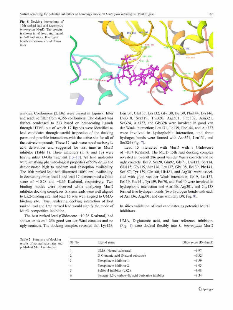
Lead 15 interacted with MurD with a Glidescoreof −8.74 Kcal/mol. The MurD 15th lead docking complexrevealed an overall 286 good van der Waals contacts and nougly contacts. Ile19, Ser20, Glu92, Gly71, Lys113, Ser114,Glu115, Gly135, Asn136, Leu137, Gly138, Ile139, Phe141,Ser157, Tyr 159, Gln160, His181, and Arg301 were associ-ated with good van der Waals interaction; Ile19, Leu137,Ile139, Phe141, Tyr159, Pro70, and Pro140 were involved inhydrophobic interaction and Asn136, Arg301, and Gly138formed five hydrogen bonds (two hydrogen bonds with eachof Asn136, Arg301, and one with Gly138; Fig. 8).
In silico validation of lead candidates as potential MurDinhibitors
UMA, D-glutamic acid, and four reference inhibitors(Fig. 1) were docked flexibly into L. interrogans MurD
Fig. 8 Docking interactions of15th ranked lead and Leptospirainterrogans MurD. The proteinis shown in ribbons, and ligandin ball and sticks. Hydrogenbonds are shown in red dottedlines
Sl. No. Ligand name Glide score (Kcal/mol)
1 UMA (Natural substrate) −6.972 D-Glutamic acid (Natural substrate) −5.323 Phosphinate inhibitor-1 −6.594 Phosphinate inhibitor-2 −6.035 Sulfonyl inhibitor (LK2) −9.086 benzene 1,3-dicarboxylic acid derivative inhibitor −6.54
Table 2 Summary of dockingresults of natural substrates andpublished MurD inhibitors
Virtual screening for potential inhibitors of homology modeled Leptospira interrogans MurD ligase 185

active site for validation of identified lead candidates asinhibitor. UMA was docked to MurD with a Glide scoreof −6.97 Kcal/mol with seven hydrogen bonds (twohydrogen bonds with Asn136 and one hydrogen bond eachwith Ile19, Ser20, Asp39, Lys113, and Gly138). D-Glutamicacid had docked with a Glide score of −5.32 Kcal/mol withthree hydrogen bonds (Arg301, Asp316, and Lys318). Thedocking of two phosphinate inhibitors (Fig. 1a and b) with L.interrogans MurD demonstrated a Glidescore of −6.59and −6.03 Kcal/mol, respectively. The sulfonyl inhibitor(Fig. 1c) demonstrated Glidescore of −9.08 Kcal/mol, andbenzene 1,3-dicarboxylic acid derivatives MurD inhibitor(Fig. 1d) had shown Glidescore of −6.54 kcal/mol. Thephosphinate inhibitors were well aligned to UMA-bindingsite and benzene 1,3-dicarboxylic acid derivative inhibitorwas aligned to LK2-binding site.
The lower Glidescore (−10.28 to −8.65 Kcal/mol) ofidentified lead candidates (Table 1) compared to substratesand reference inhibitors (Table 2) in respective dockingcomplexes revealed that the novel leads would bind morecompetitively into MurD active site than substrate UMA,D-glutamic acid, and existing inhibitors. The bindingmodes observed in these 17 lead candidates were wellaligned with existing MurD inhibitors. Importantly, theinvolvement of identified lead molecule in blockingsubstrate binding residues through hydrogen bonding(Asn136, Gly138, Arg301) van der Waals (Ser20, Asp39,Lys318) and hydrophobic interaction (Ile19, Thr320)justified them as potential inhibitors against L. interrogansMurD. These novel inhibitors may be validated in vitrothrough biochemical assays. MurD being unique to bacte-rial species and the active site being almost conserved inbacteria, the same inhibitor may also be trialed against otherGram-negative bacterial pathogens in order to designcommon effective inhibitor.
Conclusion
Peptidoglycan biosynthesis pathway is unique to Lepto-spira and absent in host human. Properly constructedpeptidoglycan provides rigidity, flexibility, and strengththat are necessary for bacterial cells to grow and divide,while withstanding high internal osmotic pressure. MurDligase is essential for addition of D-Glu to nucleotideprecursor; UDP-MurNAc, thus, is critical for properconstruction of peptidoglycans. Metabolic pathway analysishad revealed that no alternative enzyme could add D-Glu toUDP-MurNAc in Leptospira. Thus, Leptospira MurD is ofsignificant interest for novel inhibitor design to overcomethe challenges of severe leptospirosis. A high-qualityhomology model of L. interrogans MurD was reportedthrough computational validation. Our approach employing
Glide for virtual screening along with QikProp ADMEevaluation provided 17 novel MurD inhibitors. The novel17 carboxylic acid derivative inhibitors identified throughhigh-throughput virtual screening using L. interrogansMurD homology model would be of interest as commoninhibitor against leptospiral serovars. As peptidoglycanbiosynthesis pathway is unique to bacteria, the in silicoidentified MurD inhibitors are likely to be broad spectrumGram-negative inhibitors if synthesized and tested inanimal models.
Acknowledgments The study was supported by grants from DBT,Ministry of Science and Technology, Government of India, NewDelhi. We are grateful to Dr. B. Vengamma, Director, for providingconstant support and encouragement for research at SVIMS Bio-informatics Centre, where this work has been performed. We thankProf. S. Ramakumar, IISc., Bangalore for rendering critical valuablesuggestions on the present work.
References
1. Chopra I, Schofield C, Everett M, O'Neill A, Miller K, Wilcox M,Frère JM, Dawson M, Czaplewski L, Urleb U, Courvalin P (2008)Treatment of health-care-associated infections caused by Gram-negative bacteria: a consensus statement. Lancet Infect Dis 8:133–139
2. World Health Organization (1999) Leptospirosis worldwide. WklyEpidemiol Rec 74:237–242
3. Bharti AR, Nally JE, Ricaldi JN, Matthias MA, Diaz MM, LovettMA, Levett PN, Gilman RH, Willig MR, Gotuzzo E, Vinetz JM(2003) Leptospirosis: a zoonotic disease of global importance.The Lancet Infect Dis 3:757–771
4. Trueba G, Zapata S, Madrid K, Cullen P, Haake D (2004) Cellaggregation: a mechanism of pathogenic Leptospira to survive infresh water. Int Microbiol 7:35–40
5. Levett PN (2001) Leptospirosis. Clin Microbiol Rev 14:296–3266. Guidugli F, Castro AA, Atallah (2000) Antibiotics for preventing
leptospirosis. Cochrane Database Syst Rev 4:CD0013057. Wang Z, Jin L, Wegrzyn A (2007) Leptospirosis vaccines. Microb
Cell Fact 6:398. Barreteau H, Kovac A, Boniface A, Sova M, Gobec S, Blanot D
(2008) Cytoplasmic steps of peptidoglycan biosynthesis. FEMSMicrobiol Rev 32:168–207
9. Vollmer W, Blanot D, Pedro MA (2008) Peptidoglycan structureand architecture. FEMS Microbiol Rev 32:149–167
10. Van Heijeinoot J (2001) Recent advances in the formation ofbacterial peptidoglycan monomer unit. Nat Prod Rep 18:503–519
11. Bouhss A, Dementin S, van Heijenoort J, Parquet C, Blanot D(2002) MurC and MurD synthetases of peptidoglycan biosynthe-sis: borohydride trapping of acyl-phosphate intermediates. Meth-ods Enzymol 354:189–196
12. El Zoeiby A, Sanschagrin F, Levesque RC (2003) Structure andfunction of the Mur enzymes: development of novel inhibitors.Mol Microbiol 47:1–12
13. Strancar K, Blanot D, Gobec S (2006) Design, synthesis andstructure-activity relationships of new phosphinate inhibitors ofMurD. Bioorg Med Chem Lett 16:343–348
14. Humljan J, Kotnik M, Contreras-Martel C, Blanot D, Urleb U,Dessen A, Solmajer T, Gobec S (2008) Novel naphthalene-N-sulfonyl-D-glutamic acid derivatives as inhibitors of MurD, a keypeptidoglycan biosynthesis enzyme. J Med Chem 51:7486–7494
186 A. Umamaheswari et al.

15. Perdih A, Kovac A, Wolber G, Blanot D, Gobec S, Solmajer T(2009) Discovery of novel benzene 1,3-dicarboxylic acid inhib-itors of bacterial MurD and MurE ligases by structure-basedvirtual screening approach. Bioorg Med Chem Lett 19:2668–2673
16. Eswar N, Eramian D, Webb B, Shen MY, Sali A (2008) Proteinstructure modeling with MODELLER. Methods Mol Biol426:145–159
17. Maestro 9.0, versuib 70110, Schrodinger, New York18. Gasteiger E, Hoogland C, Gattiker A, Duvaud S, Wilkins MR,
Appel RD, Bairoch A (2005) Protein Identification andAnalysis Tools on the ExPASy Server. In: John M. Walker(ed) The Proteomics Protocols Handbook. Humana Press, pp571–607
19. Bryson K, McGuffin LJ, Marsden RL, Ward JJ, Sodhi JS, JonesDT (2005) Protein structure prediction servers at UniversityCollege London. Nucleic Acids Res 33:W36–W38
20. Sonnhammer EL, Eddy SR, Birney E, Bateman A, Durbin R(1998) Pfam: multiple sequence alignments and HMM-profiles ofprotein domains. Nucleic Acids Res 26:320–322
21. Murzin AG, Brenner SE, Hubbard T, Chothia C (1995) SCOP: astructural classification of proteins database for the investigationof sequences and structures. J Mol Biol 247:536–540
22. Kanehisa M, Goto S (2000) KEGG: Kyoto Encyclopedia ofGenes and Genomes. Nucleic Acids Res 28:27–30
23. Altschul SF, Madden TL, Schäffer AA, Zhang J, Zhang Z, MillerW, Lipman DJ (1997) Gapped BLAST and PSI BLAST: a newgeneration of protein database search programs. Nucleic AcidsRes 25:3389–3402
24. Bertrand JA, Fanchon E, Martin L, Chantalat L, Auger G, BlanotD, van Heijenoort J, Dideberg O (2000) Open structures of MurD:domain movements and structural similarities with folylpolyglu-tamate synthetase. J Mol Biol 301:1257–1266
25. Thompson JD, Gibson TJ, Plewniak F, Jeanmougin F, HigginsDG (1997) The ClustalX windows interface: flexible strategies formultiple sequence alignment aided by quality analysis tools.Nucleic Acids Res 24:4876–4882
26. Kotnik M, Humljan J, Contreras-Martel C, Oblak M, Kristan K,Hervé M, Blanot D, Urleb U, Gobec S, Dessen A, Solmajer T(2007) Structural and functional characterization of enantiomericglutamic acid derivatives as potential transition state analogueinhibitors of MurD ligase. J Mol Biol 370:107–115
27. Laskowski RA, MacArthur MW, Moss DS, Thornton JM (1993)PROCHECK: a program to check the stereochemical quality ofprotein structures. J Appl Crystallogr 26:283–291
28. Wiederstein M, Sippl MJ (2007) ProSA-web: interactive webservice for the recognition of errors in three-dimensional struc-tures of proteins. Nucleic Acids Res 35:W407–W410
29. Wallner B, Elofsson A (2003) Can correct protein models beidentified? Protein Sci 12:1073–1086
30. Castrignano T, De Meo PD, Cozzetto D, Talamo IG, TramontanoA (2006) The PMDB protein model database. Nucleic Acids Res34:D306–D309
31. von Grotthuss M, Pas J, Rychlewski L (2003) Ligand-Info,searching for similar small compounds using index profiles.Bioinformatics 19:1041–1042
32. Plewczynski D, Hoffmann M, von Grotthuss M, Ginalski K,Rychewski L (2007) In silico prediction of SARS proteaseinhibitors by virtual high throughput screening. Chem Biol DrugDes 69:269–279
33. Brooks WH, Daniel KG, Sung SS, Guida WC (2008) Computa-tional validation of the importance of absolute stereochemistry invirtual screening. J Chem Inf Model 48:639–645
34. Friesner RA, Banks JL, Murphy RB, Halgren TA, Klicic JJ,Mainz DT, Repasky MP, Knoll EH, Shelley M, Perry JK, ShawDE, Francis P, Shenkin PS (2004) Glide: a new approach forrapid, accurate docking and scoring. 1. Method and assessment ofdocking accuracy. J Med Chem 47:1739–1749
35. Faine S, Adler B, Bolin C, Perolat P (1999) Appendix 2 Speciesand serovar list. In: Leptospira and leptospirosis, 2nd edn.Medisci, Melbourne, Australia, pp 138–139
36. Smith CS (2006) Structure, function and dynamics in the Murfamily of bacterial cell wall ligases. J Mol Biol 362:640
Virtual screening for potential inhibitors of homology modeled Leptospira interrogans MurD ligase 187
Copyright © 2022 FDOKUMEN