Requirement for a Drosophila E3Ubiquitin Ligase in Phagocytosis of Apoptotic Cells
Upload
independentCategory
view
2download
0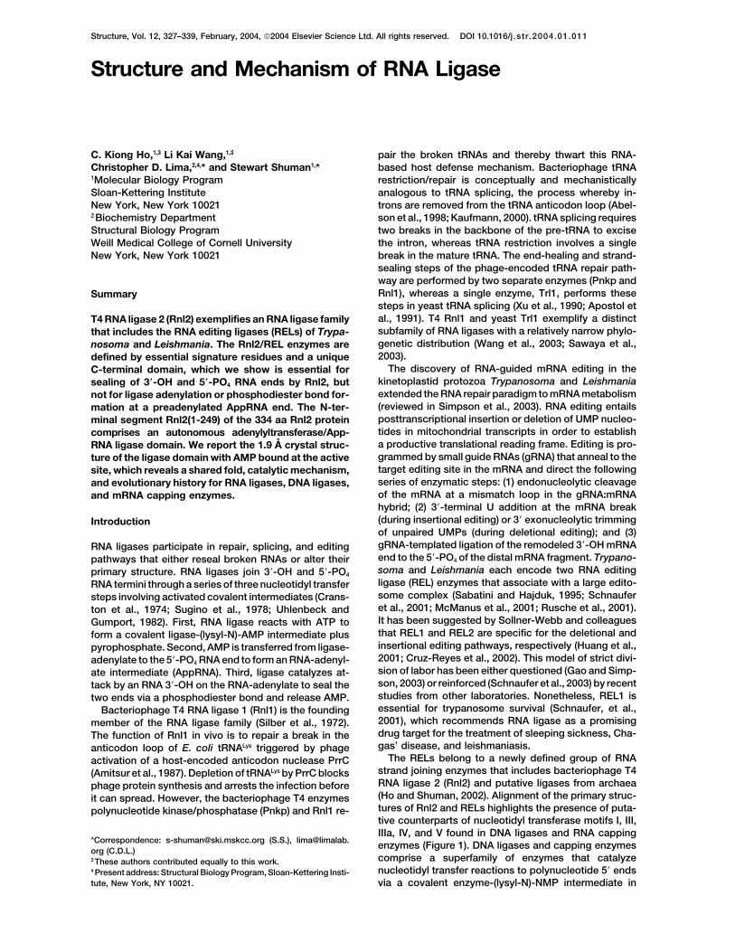
Structure, Vol. 12, 327–339, February, 2004, 2004 Elsevier Science Ltd. All rights reserved. DOI 10.1016/j .str .2004.01.011
Structure and Mechanism of RNA Ligase
pair the broken tRNAs and thereby thwart this RNA-C. Kiong Ho,1,3 Li Kai Wang,1,3
Christopher D. Lima,2,4,* and Stewart Shuman1,* based host defense mechanism. Bacteriophage tRNArestriction/repair is conceptually and mechanistically1Molecular Biology Program
Sloan-Kettering Institute analogous to tRNA splicing, the process whereby in-trons are removed from the tRNA anticodon loop (Abel-New York, New York 10021
2 Biochemistry Department son et al., 1998; Kaufmann, 2000). tRNA splicing requirestwo breaks in the backbone of the pre-tRNA to exciseStructural Biology Program
Weill Medical College of Cornell University the intron, whereas tRNA restriction involves a singlebreak in the mature tRNA. The end-healing and strand-New York, New York 10021sealing steps of the phage-encoded tRNA repair path-way are performed by two separate enzymes (Pnkp andRnl1), whereas a single enzyme, Trl1, performs theseSummarysteps in yeast tRNA splicing (Xu et al., 1990; Apostol etal., 1991). T4 Rnl1 and yeast Trl1 exemplify a distinctT4 RNA ligase 2 (Rnl2) exemplifies an RNA ligase familysubfamily of RNA ligases with a relatively narrow phylo-that includes the RNA editing ligases (RELs) of Trypa-genetic distribution (Wang et al., 2003; Sawaya et al.,nosoma and Leishmania. The Rnl2/REL enzymes are2003).defined by essential signature residues and a unique
The discovery of RNA-guided mRNA editing in theC-terminal domain, which we show is essential forkinetoplastid protozoa Trypanosoma and Leishmaniasealing of 3�-OH and 5�-PO4 RNA ends by Rnl2, butextended the RNA repair paradigm to mRNA metabolismnot for ligase adenylation or phosphodiester bond for-(reviewed in Simpson et al., 2003). RNA editing entailsmation at a preadenylated AppRNA end. The N-ter-posttranscriptional insertion or deletion of UMP nucleo-minal segment Rnl2(1-249) of the 334 aa Rnl2 proteintides in mitochondrial transcripts in order to establishcomprises an autonomous adenylyltransferase/App-a productive translational reading frame. Editing is pro-RNA ligase domain. We report the 1.9 A crystal struc-grammed by small guide RNAs (gRNA) that anneal to theture of the ligase domain with AMP bound at the activetarget editing site in the mRNA and direct the followingsite, which reveals a shared fold, catalytic mechanism,series of enzymatic steps: (1) endonucleolytic cleavageand evolutionary history for RNA ligases, DNA ligases,of the mRNA at a mismatch loop in the gRNA:mRNAand mRNA capping enzymes.hybrid; (2) 3�-terminal U addition at the mRNA break(during insertional editing) or 3� exonucleolytic trimmingIntroductionof unpaired UMPs (during deletional editing); and (3)gRNA-templated ligation of the remodeled 3�-OH mRNARNA ligases participate in repair, splicing, and editingend to the 5�-PO4 of the distal mRNA fragment. Trypano-pathways that either reseal broken RNAs or alter theirsoma and Leishmania each encode two RNA editingprimary structure. RNA ligases join 3�-OH and 5�-PO4
ligase (REL) enzymes that associate with a large edito-RNA termini through a series of three nucleotidyl transfersome complex (Sabatini and Hajduk, 1995; Schnaufersteps involving activated covalent intermediates (Crans-et al., 2001; McManus et al., 2001; Rusche et al., 2001).ton et al., 1974; Sugino et al., 1978; Uhlenbeck andIt has been suggested by Sollner-Webb and colleaguesGumport, 1982). First, RNA ligase reacts with ATP tothat REL1 and REL2 are specific for the deletional andform a covalent ligase-(lysyl-N)-AMP intermediate plusinsertional editing pathways, respectively (Huang et al.,pyrophosphate. Second, AMP is transferred from ligase-2001; Cruz-Reyes et al., 2002). This model of strict divi-adenylate to the 5�-PO4 RNA end to form an RNA-adenyl-sion of labor has been either questioned (Gao and Simp-ate intermediate (AppRNA). Third, ligase catalyzes at-son, 2003) or reinforced (Schnaufer et al., 2003) by recenttack by an RNA 3�-OH on the RNA-adenylate to seal thestudies from other laboratories. Nonetheless, REL1 istwo ends via a phosphodiester bond and release AMP.essential for trypanosome survival (Schnaufer, et al.,Bacteriophage T4 RNA ligase 1 (Rnl1) is the founding2001), which recommends RNA ligase as a promisingmember of the RNA ligase family (Silber et al., 1972).drug target for the treatment of sleeping sickness, Cha-The function of Rnl1 in vivo is to repair a break in thegas’ disease, and leishmaniasis.anticodon loop of E. coli tRNALys triggered by phage
The RELs belong to a newly defined group of RNAactivation of a host-encoded anticodon nuclease PrrCstrand joining enzymes that includes bacteriophage T4(Amitsur et al., 1987). Depletion of tRNALys by PrrC blocksRNA ligase 2 (Rnl2) and putative ligases from archaeaphage protein synthesis and arrests the infection before(Ho and Shuman, 2002). Alignment of the primary struc-it can spread. However, the bacteriophage T4 enzymestures of Rnl2 and RELs highlights the presence of puta-polynucleotide kinase/phosphatase (Pnkp) and Rnl1 re-tive counterparts of nucleotidyl transferase motifs I, III,IIIa, IV, and V found in DNA ligases and RNA capping
*Correspondence: [email protected] (S.S.), lima@limalab. enzymes (Figure 1). DNA ligases and capping enzymesorg (C.D.L.)
comprise a superfamily of enzymes that catalyze3 These authors contributed equally to this work.nucleotidyl transfer reactions to polynucleotide 5� ends4 Present address: Structural Biology Program, Sloan-Kettering Insti-
tute, New York, NY 10021. via a covalent enzyme-(lysyl-N)-NMP intermediate in

Structure328
Figure 1. Rnl2/REL Family of RNA Ligases
The amino acid sequence of T4 Rnl2 fromresidue 2 to 333 is aligned to the sequences ofvibriophage KVP40 Rnl2 and REL1 and REL2from Leishmania tarentolae (Lt) and Trypano-soma brucei (Tb). Gaps in the alignment areindicated by dashes. Positions of amino acidside chain identity or similarity in all six pro-teins are indicated by “�.” Conserved resi-dues identified by mutational analysis as es-sential for Rnl2 activity are indicated by “|.”Nucleotidyl transferase motifs I, III, IIIa, IV,and V are highlighted in shaded boxes. Thesecondary structure of the Rnl2 nucleotidyltransferase domain is shown above the se-quence.
which the nucleotide is attached to the lysine of motif that are signature features of Rnl2/REL-like ligases. Yet,the similarity extends to the C-terminal domain down-I, KxxG (Tomkinson et al., 1991; Shuman and Schwer,
1995; Hakansson et al., 1997; Lee et al., 2000; Odell et stream of motif V (Figure 1). The C-terminal segment ofRnl2 and RELs bears no apparent sequence similarity toal., 2000; Fabrega et al., 2003). RNA ligases employ a
similar mechanism of covalent catalysis in which AMP the carboxyl OB-fold domains of DNA ligases or cappingenzymes. Here we show that the C-terminal domain ofis linked to the motif I lysine (Cranston et al., 1974;
Thogersen et al., 1985; Xu et al., 1990). Mutational analy- Rnl2 is essential for sealing of 3�-OH and 5�-PO4 RNAends (pRNA ligation), but not for ligase adenylation orsis of Rnl2 has pinpointed 12 individual amino acids that
are essential for strand joining (indicated by “|” in Figure for phosphodiester bond formation at a preadenylated5� end (AppRNA ligation). The truncated N-terminal poly-1) (Ho and Shuman, 2002; Yin et al., 2003); these include
at least one conserved residue in each of the nucleotidyl peptide Rnl2(1-249) comprises an autonomous adenyl-yltransferase/AppRNA ligase domain. We report thetransferase motifs. The concordance of mutation data
for Rnl2, capping enzymes, and DNA ligases suggests 1.9 A crystal structure of the adenylyltransferase/App-RNA ligase domain with AMP bound at the active site.that DNA ligases, capping enzymes, and RNA ligases
may have evolved from an ancestral RNA strand-joiningenzyme. This model remains speculative because there Resultsis no atomic structure available for an NTP-dependentRNA ligase. The C-terminal Domain of Rnl2 is Essential
for pRNA LigationT4 Rnl2 provides an excellent model for structuraland mechanistic analysis of the Rnl2/REL class of RNA To understand the structural requirements for catalysis
of RNA strand joining, we analyzed the effects of serialjoining enzymes. There is primary structure similaritybetween phage-encoded Rnl2 and the kinetoplastid deletions of the C-terminal domain of the 334 aa Rnl2
polypeptide. Truncated proteins Rnl2(1-319), Rnl2(1-306),RELs through the entire length of the Rnl2 protein (Figure1). Many of the conserved positions in the N-terminal Rnl2(1-285), and Rnl2(1-249) were produced in E. coli
as N-terminal His10 fusions and purified from solubleportion localize to the nucleotidyl transferase motifs,including essential residues Glu34 and Asn40 of motif I bacterial extracts by nickel-agarose chromatography in
RNA Ligase Structure329
Figure 2. Purification and Adenylyltransfer-ase Activity of Rnl2(1-249)
(A) Aliquots (5 �g) of the nickel-agarose prep-arations of full length Rnl2(1-334) and the indi-cated C-terminal truncated Rnl2 proteinswere analyzed by SDS-PAGE. The Coomas-sie blue-stained gel is shown. The positionsand sizes (in kDa) of marker polypeptides areindicated on the left.(B and C) Adenylyltransferase activity. Reac-tion mixtures (20 �l) containing 50 mM Trisbuffer (either Tris acetate [pH 5.0, 5.5, 6.0,6.5, or 7.0] or Tris-HCl [pH 6.0, 6.5, 7.0, 7.5,8.0, 8.5, 9.0, 9.5]), 5 mM DTT, 1 mM MgCl2,20 �M [�32P] ATP, 0.2 �g (5 pmol) Rnl2, and1 �g (32 pmol) Rnl2(1-249) were incubatedfor 5 min at 22�C. The reactions were quenchedwith SDS, and the products were analyzedby SDS-PAGE. The ligase-[32P]AMP adductwas visualized by autoradiography of thedried gel (C) and quantified by scanning thegel with a phosphorimager. The extents ofRnl2-AMP (�) and Rnl2(1-249)-AMP (�) com-plex formation are plotted as a function of pHin (B).
parallel with full-length wild-type Rnl2 (Figure 2A). SDS- mulation of AppRNA and suppresses formation of li-gated circles (Figure 3B). The explanation for the ATPPAGE analysis of the Rnl2(1-319), Rnl2(1-306), and
Rnl2(1-285) preparations revealed the presence of seri- effect is that Rnl2 is prone to dissociate from the newlyformed RNA-adenylate product of step 2 and that anally truncated Rnl2 polypeptide of the expected size, as
well as a polypeptide of �34 kDa common to the three immediate reaction of Rnl2 with ATP to form ligase-adenylate precludes it from rebinding to the RNA-ade-Rnl2 deletions that corresponds to a His-tagged N-ter-
minal Rnl2 fragment. The abundance of the 34 kDa frag- nylate for subsequent catalysis of strand joining (Ho andShuman, 2002; Yin et al., 2003).ment relative to the expected Rnl2� polypeptide in-
creased progressively as the ligase was truncated by The instructive findings were that the truncatedRnl2(1-249) protein was defective in the composite15, 28, and 49 aa from the C terminus. In contrast, the
recombinant Rnl2(1-249) protein consisted of a single pRNA ligation reaction in the absence of ATP (Figure3A) and in the transfer of AMP to the 5�-PO4 RNA endspecies (�33 kDa) migrating just slightly ahead of the
proteolytic fragment of the other Rnl2� proteins (Figure to form AppRNA in the presence of ATP (Figure 3B).From the protein titration data in Figures 3A and 3B, we2A). We surmise that removal of the C terminus of Rnl2
exposes the enzyme to proteolysis and that the site calculated that the specific activity of Rnl2(1-249) inpRNA ligation and AppRNA formation was �1% of theof protease cleavage is close to amino acid 249. This
landmark (indicated by an arrowhead in Figure 1) corre- specific activity of full-length Rnl2. The deletion mutantRnl2(1-319) was similarly defective in pRNA ligation andsponds to a gap in the alignment between phage Rnl2
and kinetoplastid REL proteins and may therefore corre- AppRNA formation (data not shown). Thus, the C-ter-minal domain of Rnl2 is essential for pRNA ligation, andspond to a domain boundary. This notion was under-
scored by the fact that the recovery of soluble recombi- removal of as few as 15 aa from the C terminus resultsin loss of function.nant Rnl2(1-249) protein was �10-fold greater than that
of full-length wild-type Rnl2 or the other Rnl2� proteins.Rnl2 and Rnl2(1-249) were tested in parallel for strand Rnl2(1-249) Retains Adenylyltransferase Activity
To determine whether the failure of Rnl2(1-249) to cata-joining activity with a 5� 32P-labeled single-stranded 18-mer RNA substrate. Circularization of the 18-mer RNA lyze pRNA ligation was attributable to a defect in the
first step of the strand joining pathway—formation ofby preformed Rnl2-AMP in the enzyme preparation isthe predominant outcome when the ligation reaction of the ligase-AMP intermediate—we assayed the adenylyl-
transferase activity of Rnl2 and Rnl2(1-249) by labelwild-type Rnl2 is performed in the absence of exoge-nous ATP (Figure 3A). The preference for circularization transfer from [�-32P]ATP to the enzyme. In the experi-
ment shown in Figure 2C, both proteins were includedis construed to reflect proximity of the intramolecular3�-OH terminus to the Rnl2 active site. Inclusion of 1 in the same reaction mixture, and the Rnl2-[32P]AMP and
Rnl2(1-249)-[32P]AMP products were resolved by SDS-mM ATP in the strand joining reaction promotes accu-

Structure330
Figure 3. RNA Sealing Activity of Rnl2(1-249)
The RNA adenylation and sealing steps of thereaction pathway are illustrated schemati-cally at the top of the figure.(A and B) pRNA ligation. Reaction mixtures(10 �l) containing 50 mM Tris-acetate (pH6.5), 5 mM DTT, 1 mM MgCl2, 1 pmol of 5�32P-labeled 18-mer RNA oligonucleotide (5�-UUUAAUCAAUUGCGACCC), either 1 mMATP (�ATP, panel B) or no ATP (�ATP, panelA) as specified, and increasing amounts ofenzyme [1, 3, 9, 27, or 81 pmol of Rnl2 orRnl2(1-249), proceeding from left to right ineach titration series] were incubated for 15min at 22�C. Ligase was omitted from controlreactions analyzed in lanes labeled “�.”(C) AppRNA ligation. Reaction mixtures (10�l) containing 50 mM Tris-acetate (pH 6.5), 5mM DTT, 1 mM MgCl2, 200 fmol of 32P-labeledRNA-adenylate strand AppUUUAAUCAAUUGCGACCC (synthesized and gel-purifiedaccording to Yin et al. [2003]), and increasingamounts of enzyme [4, 12, 37, 110, 330, or1000 fmol of Rnl2 or Rnl2(1-249), proceedingfrom left to right in each titration series] wereincubated for 15 min at 22�C. The reactionswere quenched by adding 5 �l of 90% form-amide, 20 mM EDTA. The products were ana-lyzed by electrophoresis through an 18%polyacrylamide gel containing 7 M urea in 45mM Tris-borate, 1 mM EDTA and visualizedby autoradiography. Enzyme was omittedfrom control reaction analyzed in the lane la-beled “�.”
PAGE. Both proteins were active in ligase adenylation, (1-249) to ligate pRNA was likely caused by a defect ina step subsequent to ligase adenylation.but they displayed distinctive pH optima. The adenylyl-
transferase activity of full-length Rnl2 had a bell-shapedpH profile with an optimum at pH 6.5, whereas Rnl2 Phosphodiester Formation at a Preadenylated
RNA 5� End(1-249) was optimal at pH 9.0–9.5 (Figure 2B). The samepH dependence of the Rnl2 and Rnl2(1-249) adenylyl- A preadenylated RNA substrate (AppRNA) was em-
ployed for analysis of step 3 of the ligation pathway intransferase activities was observed when the proteinswere assayed in separate reaction mixtures (data not isolation. Formation of a phosphodiester at the activated
5� end by wild-type Rnl2 was evinced by the appearanceshown). The less extensively truncated protein Rnl2(1-319) displayed an alkaline pH dependence in its ade- of a sealed circular RNA product, the yield of which was
proportional to the amount of input enzyme (Figure 3C).nylyltransferase activity similar to that of Rnl2(1-249)(data not shown). Thus, deletion of the C terminus did Eighty-five percent of the AppRNA substrate was con-
verted to circular RNA at saturating Rnl2 concentrations.not preclude the reaction of Rnl2 with ATP, but eliciteda 3 unit alkaline shift in the pH profile. We calculated A small fraction of the input AppRNA was deadenylated
during the reaction to yield pRNA, which migrated be-that 68% of the input Rnl2 protein and 25% of the inputRnl2(1-249) protein was adenylated with 32P-AMP at their tween RNA-adenylate substrate and the ligated circle
(Figure 3C). Deadenylation is the reverse of step 2 ofrespective pH optima. The remaining fraction of the pro-tein preparation likely consists of preformed ligase-AMP the ligation pathway. The remarkable finding was that
Rnl2(1-249) was 10-fold more active than full-length Rnl2intermediate.The assays of pRNA ligation and RNA adenylation in AppRNA ligation on a per enzyme basis (Figure 3C).
From the slope of the titration curve, we calculated thatshown in Figure 3 were performed at pH 6.5, which isthe optimal pH for full-length Rnl2 (Ho and Shuman, Rnl2(1-249) circularized 8.4 fmol of AppRNA per fmol
of enzyme. Deletion of the C-terminal domain did not2002). In light of the shifted pH profile of the Rnl2(1-249)adenylyltransferase activity, we repeated the ligation ex- appear to affect the balance between the sealing and
deadenylation reactions. It is worth emphasizing that theperiments at alkaline pH. We found that Rnl2(1-249) wasas defective in RNA circularization (�ATP) and RNA ade- 10-fold higher specific activity of Rnl2(1-249) in AppRNA
ligation cannot be attributed to a difference in the occu-nylation (�ATP) at pH 8.5 as it was at pH 6.5 (data notshown). Thus, we conclude that the failure of Rnl2 pancy of the adenylate binding pocket by preformed

RNA Ligase Structure331
ligase-AMP in the Rnl2 and Rnl2(1-249) enzyme prepara- 1fvi; z score 9.4; rmsd of 3.3 A over 141 C� positionsof equivalence to Rnl2), and the GTP-dependent mRNAtions. Only the Rnl2 apoenzyme is capable of sealing
AppRNA, and any Rnl2-AMP in the enzyme preparation capping enzyme of Chlorella virus PBCV-1 (PDB 1fvi;z score 9.4; rmsd of 3.3 A over 141 C� positions ofwould be unreactive in AppRNA ligation. From the exper-
iments presented in Figures 2 and 3, we estimated that equivalence to Rnl2). Structural similarity between Rnl2-like RNA ligases, DNA ligases, and RNA capping en-68% of the Rnl2 preparation was apoenzyme (which
could be adenylated in vitro) and �20% was Rnl2-AMP zymes had been predicted based on the results of muta-tional analysis of Rnl2 (Ho and Shuman, 2002; Yin et al.,(which could circularize the pRNA substrate). In the case
of Rnl2(1-249), we estimated that 25% of the preparation 2003).A structure-based alignment of the amino acid se-was catalytically competent apoenzyme; however, the
fraction of preformed ligase-AMP could not be as- quences of the nucleotidyl transferase domains of T4Rnl2, the Tfi, T7, and Chlorella virus DNA ligases, andsessed, because the truncated enzyme is defective in
catalysis of pRNA ligation. Chlorella virus capping enzyme revealed ten positionsof side chain identity/similarity in these five proteins.Thus, we infer that the functional groups required for
catalysts of RNA phosphodiester bond formation reside The conserved residues are localized to the strandsthat comprise the nucleotide binding site and encom-within the N-terminal adenylyltransferase domain of
Rnl2, and the C-terminal domain is actually an impedi- pass the nucleotidyl transferase motifs. A salient featureof the structural comparison is that five of the side chainsment to either binding or sealing of the AppRNA sub-
strate. Furthermore, we infer that the inability of the that are essential for the activity of Rnl2 (Lys35, Glu99,Phe119, Glu204, and Lys227) are conserved and occupyN-terminal adenylyltransferase/AppRNA ligase domain
to catalyze pRNA ligation stems from a specific require- equivalent positions in the tertiary structures of theNAD� and ATP-dependent DNA ligases and GTP-ment for the C-terminal domain for the second step of
the ligation pathway—transfer of AMP from the ligase dependent capping enzymes.to the 5�-PO4 end to form AppRNA. Because Rnl2(1-249)is capable of deadenylating AppRNA via the reverse of
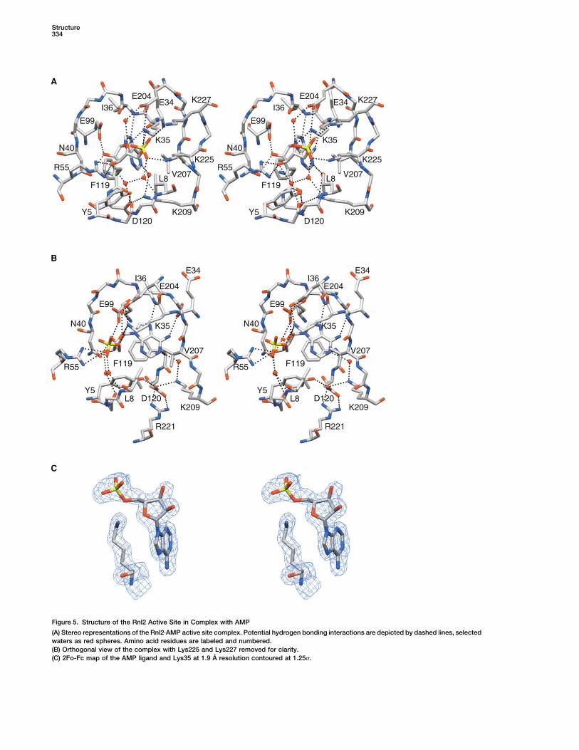
The Adenylate Binding Sitestep 2 (Figure 3C), we suspect that loss of the C-terminalThe refined density map showed that an AMP moietydomain affects the pRNA binding/recognition compo-was bound at the active site (Figure 5C), even thoughnent of the step 2 reaction, rather than the chemistry ofRnl2(1-249) was not intentionally exposed to ATP oradenylate transfer to and from RNAAMP during purification and crystal growth. The AMPphosphate was 2.9 A from the N of Lys35, a positioninconsistent with a covalent connection between theCrystal Structure of the Rnl2 Adenylyltransferase/
AppRNA Ligase Domain motif I lysine nucleophile and AMP. A plausible scenariois that the phosphoamide linkage of the covalent Rnl2(1-We crystallized Rnl2(1-249) and solved the structure by
single isomorphous replacement with anomalous scat- 249)-AMP intermediate was hydrolyzed in situ after puri-fication or in the crystal.tering using diffraction data from native and Hg-deriva-
tized crystals. The ligase crystals were orthorhombic The adenylate binding pocket is composed of a cageof strands and interstrand loops that includes contri-(P21212) with one monomer in the asymmetric unit. The
refined model at 1.9 A resolution had a crystallographic butions from nucleotidyl transferase motifs I, III, IIIa,IV, and V (Figures 1 and 4). The adenine base of theR factor of 18.9% and an Rfree of 22.7% with excellent
stereochemistry (Table 1). The structure comprised a nucleotide is located at the bottom of the pocket (Figure4B), where it is sandwiched between the aromatic ringcontinuous polypeptide from Rnl2 residues 1 to 233.
The N-terminal His-tag and the C-terminal segment from of Phe119 in motif IIIa, the hydrophobic side chain ofVal207 in motif III, and the aliphatic portion of the Lys35aa 234 to 249 were disordered and had no interpretable
electron density. The protein consists of a central nucle- side chain in motif I (Figures 5A and 5B). A similar aro-matic/purine/hydrophobic sandwich is found in T7 li-otide binding pocket lined by a two six-stranded antipar-
allel sheets (composed of strands 3, 4, 5, 6, 7, and 8 gase bound to ATP, Tfi DNA ligase-adenylate, Chlorellavirus DNA ligase-adenylate, Chlorella virus and Candidaand strands 1, 2, 9, 10, 11, and 12, respectively) and the
N-terminal peptide segment from aa 1 to 8 (Figure 4A). albicans capping enzymes bound to GTP, and the Chlo-rella virus and Candida albicans capping enzymesSeven � helices decorate the inferior and lateral surfaces
(Figure 4B). bound covalently to GMP (Subramanya et al., 1996; Ha-kansson et al., 1997; Lee et al., 2000; Odell et al., 2000,The overall fold of Rnl2 resembles that of DNA ligase
and mRNA capping enzyme. The best matches in a 2003; Fabrega et al., 2003). The � stacking of Phe119on adenine is apparently critical for Rnl2 activity, insofardatabase search using DALI (Holm and Sander, 1993)
were to the nucleotidyl transferase domains of the as replacement of Phe119 by alanine blocks adenylyl-transferase activity and overall pRNA ligation and leu-NAD�-dependent DNA ligases from Thermus filiformis
(PBD 1dgs; z score 11.2; rmsd of 3.2 A over 172 C� cine does not fully revive Rnl2 function (Yin et al., 2003).Additional contacts of the adenine base with Rnl2positions of equivalence to Rnl2) and Bacillus stearo-
thermophilus (PDB 1b04; z score 11.1; rmsd of 3.1 A include a hydrogen bond from N7 to the backbone amideof Ile36 and a hydrogen bond from the exocyclic 6-aminoover 165 C� positions of equivalence to Rnl2), the ATP-
dependent DNA ligases of bacteriophage T7 (PDB 1a01; group to the main chain carbonyl of Glu34 (Figure 5B);the latter interaction may explain the specificity of Rnl2z score 10.3; rmsd of 3.3 A over 164 C� positions of
equivalence to Rnl2) and Chlorella virus PBCV-1 (PDB for ATP (Ho and Shuman, 2002; and our unpublished
Structure332
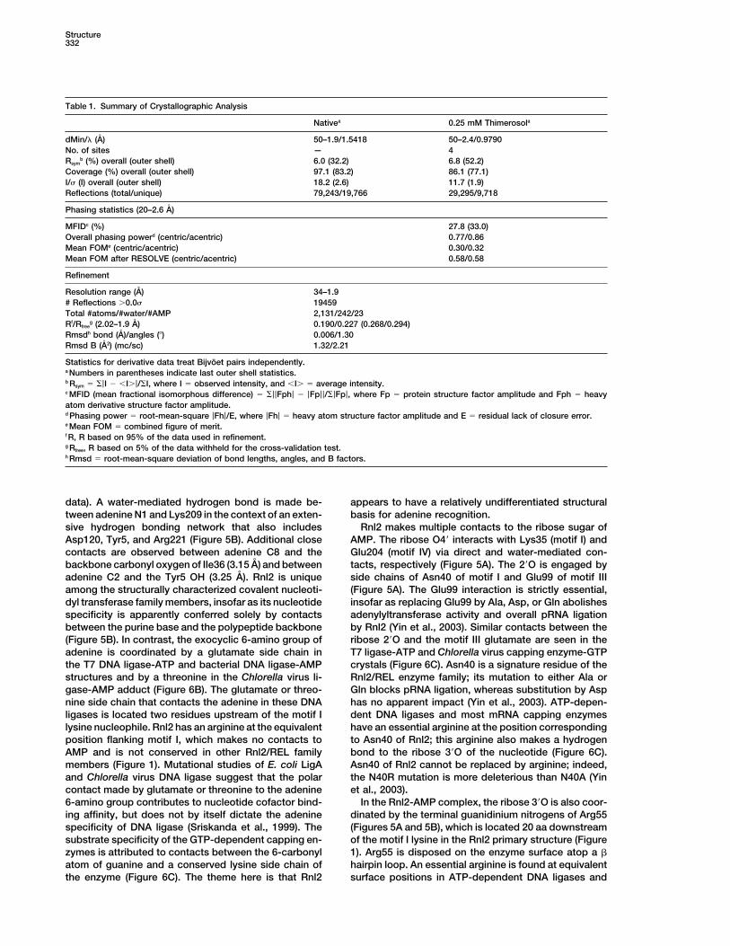
Table 1. Summary of Crystallographic Analysis
Nativea 0.25 mM Thimerosola
dMin/� (A) 50–1.9/1.5418 50–2.4/0.9790No. of sites — 4Rsym
b (%) overall (outer shell) 6.0 (32.2) 6.8 (52.2)Coverage (%) overall (outer shell) 97.1 (83.2) 86.1 (77.1)I/ (I) overall (outer shell) 18.2 (2.6) 11.7 (1.9)Reflections (total/unique) 79,243/19,766 29,295/9,718
Phasing statistics (20–2.6 A)
MFIDc (%) 27.8 (33.0)Overall phasing powerd (centric/acentric) 0.77/0.86Mean FOMe (centric/acentric) 0.30/0.32Mean FOM after RESOLVE (centric/acentric) 0.58/0.58
Refinement
Resolution range (A) 34–1.9# Reflections �0.0 19459Total #atoms/#water/#AMP 2,131/242/23Rf/Rfree
g (2.02–1.9 A) 0.190/0.227 (0.268/0.294)Rmsdh bond (A)/angles (�) 0.006/1.30Rmsd B (A2) (mc/sc) 1.32/2.21
Statistics for derivative data treat Bijvoet pairs independently.a Numbers in parentheses indicate last outer shell statistics.b Rsym � �|I � �I�|/�I, where I � observed intensity, and �I� � average intensity.c MFID (mean fractional isomorphous difference) � �||Fph| � |Fp||/�|Fp|, where Fp � protein structure factor amplitude and Fph � heavyatom derivative structure factor amplitude.d Phasing power � root-mean-square |Fh|/E, where |Fh| � heavy atom structure factor amplitude and E � residual lack of closure error.e Mean FOM � combined figure of merit.f R, R based on 95% of the data used in refinement.g Rfree, R based on 5% of the data withheld for the cross-validation test.h Rmsd � root-mean-square deviation of bond lengths, angles, and B factors.
data). A water-mediated hydrogen bond is made be- appears to have a relatively undifferentiated structuralbasis for adenine recognition.tween adenine N1 and Lys209 in the context of an exten-
sive hydrogen bonding network that also includes Rnl2 makes multiple contacts to the ribose sugar ofAMP. The ribose O4� interacts with Lys35 (motif I) andAsp120, Tyr5, and Arg221 (Figure 5B). Additional close
contacts are observed between adenine C8 and the Glu204 (motif IV) via direct and water-mediated con-tacts, respectively (Figure 5A). The 2�O is engaged bybackbone carbonyl oxygen of Ile36 (3.15 A) and between
adenine C2 and the Tyr5 OH (3.25 A). Rnl2 is unique side chains of Asn40 of motif I and Glu99 of motif III(Figure 5A). The Glu99 interaction is strictly essential,among the structurally characterized covalent nucleoti-
dyl transferase family members, insofar as its nucleotide insofar as replacing Glu99 by Ala, Asp, or Gln abolishesadenylyltransferase activity and overall pRNA ligationspecificity is apparently conferred solely by contacts
between the purine base and the polypeptide backbone by Rnl2 (Yin et al., 2003). Similar contacts between theribose 2�O and the motif III glutamate are seen in the(Figure 5B). In contrast, the exocyclic 6-amino group of
adenine is coordinated by a glutamate side chain in T7 ligase-ATP and Chlorella virus capping enzyme-GTPcrystals (Figure 6C). Asn40 is a signature residue of thethe T7 DNA ligase-ATP and bacterial DNA ligase-AMP
structures and by a threonine in the Chlorella virus li- Rnl2/REL enzyme family; its mutation to either Ala orGln blocks pRNA ligation, whereas substitution by Aspgase-AMP adduct (Figure 6B). The glutamate or threo-
nine side chain that contacts the adenine in these DNA has no apparent impact (Yin et al., 2003). ATP-depen-dent DNA ligases and most mRNA capping enzymesligases is located two residues upstream of the motif I
lysine nucleophile. Rnl2 has an arginine at the equivalent have an essential arginine at the position correspondingto Asn40 of Rnl2; this arginine also makes a hydrogenposition flanking motif I, which makes no contacts to
AMP and is not conserved in other Rnl2/REL family bond to the ribose 3�O of the nucleotide (Figure 6C).Asn40 of Rnl2 cannot be replaced by arginine; indeed,members (Figure 1). Mutational studies of E. coli LigA
and Chlorella virus DNA ligase suggest that the polar the N40R mutation is more deleterious than N40A (Yinet al., 2003).contact made by glutamate or threonine to the adenine
6-amino group contributes to nucleotide cofactor bind- In the Rnl2-AMP complex, the ribose 3�O is also coor-dinated by the terminal guanidinium nitrogens of Arg55ing affinity, but does not by itself dictate the adenine
specificity of DNA ligase (Sriskanda et al., 1999). The (Figures 5A and 5B), which is located 20 aa downstreamof the motif I lysine in the Rnl2 primary structure (Figuresubstrate specificity of the GTP-dependent capping en-
zymes is attributed to contacts between the 6-carbonyl 1). Arg55 is disposed on the enzyme surface atop a hairpin loop. An essential arginine is found at equivalentatom of guanine and a conserved lysine side chain of
the enzyme (Figure 6C). The theme here is that Rnl2 surface positions in ATP-dependent DNA ligases and

RNA Ligase Structure333
be capped with GMP. The equivalent arginine of theC. albicans guanylyltransferase-GMP intermediate co-ordinates a phosphate on the enzyme surface locatedclose to the lysyl-GMP phosphate; this phosphate isproposed to mark the position of the 5� diphosphateRNA terminus prior to the second GMP transfer step ofthe capping pathway (Fabrega et al., 2003). Thus, thecontact of Arg55 with the ribose is a seemingly uniquefeature of the Rnl2 structure. However, it is conceivablethat the contacts of Arg55 are subject to change atdifferent steps of the RNA ligase reaction; e.g., the Argmay well interact with either the � phosphate of ATPduring the ligase adenylation step or the 5�-PO4 of RNAduring the RNA adenylation step. Indeed, the defaultcontact of Arg55 with the AMP ribose may reflect theabsence of a phosphate-like ligand in the crystallizationsolution. The finding that mutation of Arg55 to alanine orglutamine abolishes Rnl2 activity, whereas conservativereplacement with lysine restores ligase function (Yin etal., 2003), is at least consistent with a role for Arg55 incoordinating a phosphate at some point during the RNAligase reaction.
The adenosine nucleoside in the Rnl2 AMP complexis in the syn conformation (Figures 5B and 6A). This issimilar to the syn conformation of adenosine in the crys-tal of T7 ligase bound to ATP and the syn conformationof guanosine in the Chlorella virus capping enzyme-GTPcomplex (Figure 6C) and the closed conformation of theChlorella virus capping enzyme-GMP covalent interme-diate. In contrast, the nucleoside is in the anti confor-mation in the Chlorella virus DNA ligase-AMP covalentintermediate (Figure 6B), the NAD�-dependent Tfi li-gase-AMP covalent intermediate, and the Candida albi-cans capping enzyme-GMP covalent intermediate. Thelatter structures represent open conformational states ofthe ligases and capping enzymes in which the C-terminaldomain (which is not present in the Rnl2 structure) ismoved away from the nucleotidyl transferase domainto allow ingress of the nucleic acid substrate to thecovalently bound nucleotide. It has been proposed thatFigure 4. Structure of RNA Ligase (Rnl2)a change in nucleoside conformation from syn to anti(A and B) Orthogonal views of the Rnl2 structure as depicted byafter formation of the covalent enzyme-NMP intermedi-ribbon diagrams. “N” and “C” demarcate the termini of Rnl2(1-233). �ate and prior to NMP transfer to the 5� end of the poly-helices are colored cyan, strands are colored red, and connecting
polypeptide is colored yellow. AMP is shown in bond representation nucleotide substrate is a conserved feature of thewith standard color-coding for carbon (gray), oxygen (red), nitrogen nucleotidyl transferase superfamily.(blue), and phosphorus (yellow). Images were generated with SETOR The AMP phosphate is coordinated by Lys35 in motif(Evans, 1993) unless noted otherwise.
I (the predicted site of covalent attachment of AMP toRnl2) and by Lys225 and Lys227 in motif V (Figure 5A).
mRNA capping enzymes (Figures 6B and 6C). The crys- In addition, a water-mediated hydrogen bond networktal structure of the Chlorella virus DNA ligase-AMP inter- links the AMP phosphate and the ribose O3� to the Ser6mediate reveals that the Arg coordinates a sulfate ion backbone carbonyl oxygen and the Leu8 backbone am-on the enzyme surface located approximately 5 A from ide (Figure 5A). Lys225 and Lys227 are essential for Rnl2the � phosphate of AMP (Figure 6B). The sulfate con- activity. The structure implicates one or both of the motiftacts are proposed to mimic the interactions of DNA V lysines as direct catalysts of nucleotidyl transfer, e.g.,ligase with the reactive 5� phosphate of the polynucleo- via stabilization of the transition state of the � phospho-tide and with the � phosphate of ATP (Odell et al., 2000). rus of ATP during ligase adenylylation or the AMP phos-Indeed, in the closed conformation of the Chlorella virus phorus during the RNA adenylylation step.guanylyltransferase-GTP complex, the equivalent Arg Lys35 forms an ion pair with Glu204 (motif IV) (Figureside chain directly coordinates the � phosphate of GTP 5A). Glu204 is essential for both the ligase adenylylation(Hakansson et al., 1997). In the covalent guanylyltrans- and AppRNA sealing steps of the Rnl2 pathway (Yin etferase-GMP intermediate, the arginine coordinates a al., 2003), and this residue is conserved as a carboxylatesulfate ion on the protein surface, likely mimicking in DNA ligases, capping enzymes, and RNA ligases. An
ion pair between the motif IV carboxylate and the motifcontacts to the 5� terminus of the RNA substrate to
Structure334
Figure 5. Structure of the Rnl2 Active Site in Complex with AMP
(A) Stereo representations of the Rnl2·AMP active site complex. Potential hydrogen bonding interactions are depicted by dashed lines, selectedwaters as red spheres. Amino acid residues are labeled and numbered.(B) Orthogonal view of the complex with Lys225 and Lys227 removed for clarity.(C) 2Fo-Fc map of the AMP ligand and Lys35 at 1.9 A resolution contoured at 1.25 .

RNA Ligase Structure335
Figure 6. Structural Comparison of RNA, Ligase, DNA Ligase, and Capping Enzyme Active Sites
(A) Stereo representation of the Rnl2·AMP active site complex.(B) Stereo representation of the active site of the Chlorella virus DNA ligase-adenylate intermediate (PDB 1FVI). A sulfate ion is bound on theenzyme surface by Arg42.(C) Stereo representation of the open conformation of the Chlorella virus mRNA capping enzyme-GTP complex (PDB 1CKM). Amino acidresidues are labeled and numbered. Potential hydrogen bonding interactions are depicted by dashed lines. Waters were removed for clarity.All models are in bond representation with standard atom color-coding.
Structure336
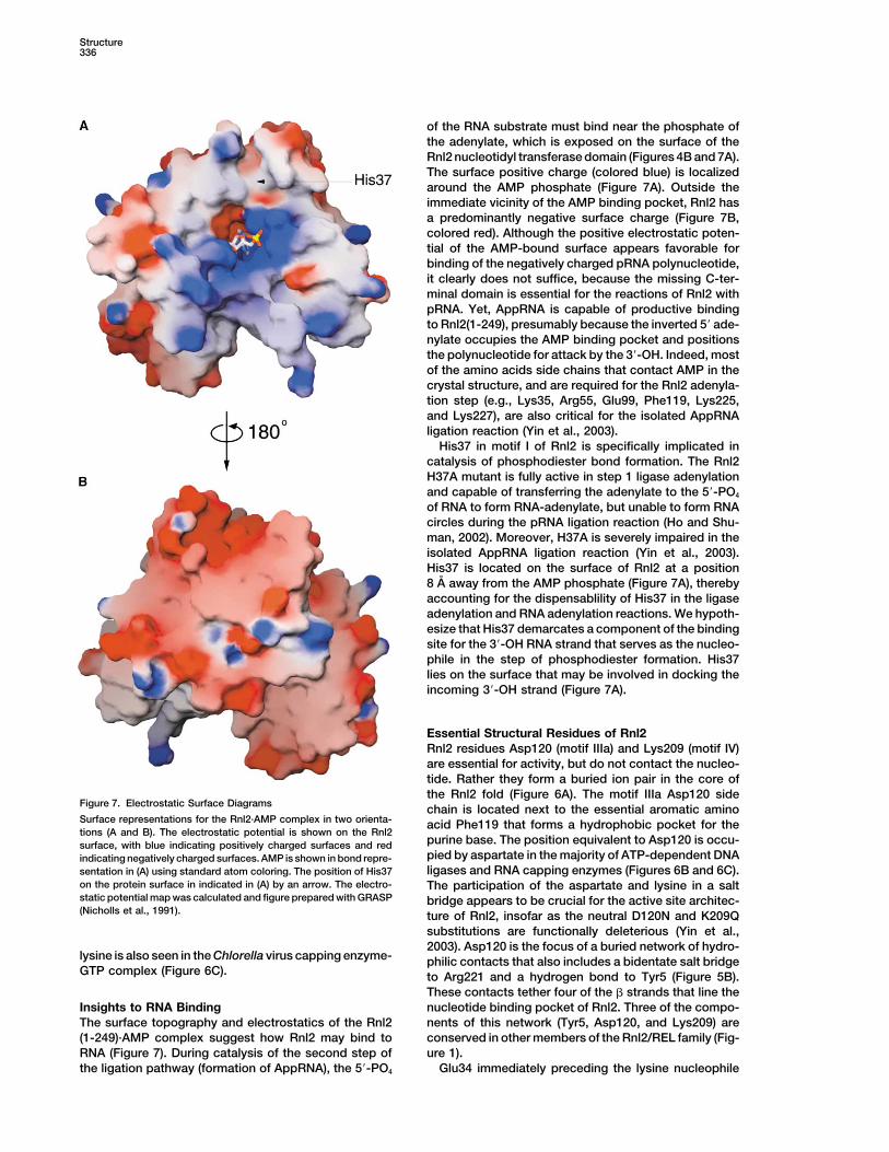
of the RNA substrate must bind near the phosphate ofthe adenylate, which is exposed on the surface of theRnl2 nucleotidyl transferase domain (Figures 4B and 7A).The surface positive charge (colored blue) is localizedaround the AMP phosphate (Figure 7A). Outside theimmediate vicinity of the AMP binding pocket, Rnl2 hasa predominantly negative surface charge (Figure 7B,colored red). Although the positive electrostatic poten-tial of the AMP-bound surface appears favorable forbinding of the negatively charged pRNA polynucleotide,it clearly does not suffice, because the missing C-ter-minal domain is essential for the reactions of Rnl2 withpRNA. Yet, AppRNA is capable of productive bindingto Rnl2(1-249), presumably because the inverted 5� ade-nylate occupies the AMP binding pocket and positionsthe polynucleotide for attack by the 3�-OH. Indeed, mostof the amino acids side chains that contact AMP in thecrystal structure, and are required for the Rnl2 adenyla-tion step (e.g., Lys35, Arg55, Glu99, Phe119, Lys225,and Lys227), are also critical for the isolated AppRNAligation reaction (Yin et al., 2003).
His37 in motif I of Rnl2 is specifically implicated incatalysis of phosphodiester bond formation. The Rnl2H37A mutant is fully active in step 1 ligase adenylationand capable of transferring the adenylate to the 5�-PO4
of RNA to form RNA-adenylate, but unable to form RNAcircles during the pRNA ligation reaction (Ho and Shu-man, 2002). Moreover, H37A is severely impaired in theisolated AppRNA ligation reaction (Yin et al., 2003).His37 is located on the surface of Rnl2 at a position8 A away from the AMP phosphate (Figure 7A), therebyaccounting for the dispensablility of His37 in the ligaseadenylation and RNA adenylation reactions. We hypoth-esize that His37 demarcates a component of the bindingsite for the 3�-OH RNA strand that serves as the nucleo-phile in the step of phosphodiester formation. His37lies on the surface that may be involved in docking theincoming 3�-OH strand (Figure 7A).
Essential Structural Residues of Rnl2Rnl2 residues Asp120 (motif IIIa) and Lys209 (motif IV)are essential for activity, but do not contact the nucleo-tide. Rather they form a buried ion pair in the core ofthe Rnl2 fold (Figure 6A). The motif IIIa Asp120 side
Figure 7. Electrostatic Surface Diagrams chain is located next to the essential aromatic aminoSurface representations for the Rnl2·AMP complex in two orienta- acid Phe119 that forms a hydrophobic pocket for thetions (A and B). The electrostatic potential is shown on the Rnl2
purine base. The position equivalent to Asp120 is occu-surface, with blue indicating positively charged surfaces and redpied by aspartate in the majority of ATP-dependent DNAindicating negatively charged surfaces. AMP is shown in bond repre-ligases and RNA capping enzymes (Figures 6B and 6C).sentation in (A) using standard atom coloring. The position of His37
on the protein surface in indicated in (A) by an arrow. The electro- The participation of the aspartate and lysine in a saltstatic potential map was calculated and figure prepared with GRASP bridge appears to be crucial for the active site architec-(Nicholls et al., 1991). ture of Rnl2, insofar as the neutral D120N and K209Q
substitutions are functionally deleterious (Yin et al.,2003). Asp120 is the focus of a buried network of hydro-
lysine is also seen in the Chlorella virus capping enzyme- philic contacts that also includes a bidentate salt bridgeGTP complex (Figure 6C). to Arg221 and a hydrogen bond to Tyr5 (Figure 5B).
These contacts tether four of the strands that line thenucleotide binding pocket of Rnl2. Three of the compo-Insights to RNA Binding
The surface topography and electrostatics of the Rnl2 nents of this network (Tyr5, Asp120, and Lys209) areconserved in other members of the Rnl2/REL family (Fig-(1-249)·AMP complex suggest how Rnl2 may bind to
RNA (Figure 7). During catalysis of the second step of ure 1).Glu34 immediately preceding the lysine nucleophilethe ligation pathway (formation of AppRNA), the 5�-PO4
RNA Ligase Structure337
is an essential signature residue of the Rnl2/REL family evidence for the common origin of the nucleotidyl trans-ferase domains of Rnl2-like RNA ligases, DNA ligases,(Figure 1), which is also conserved in the Rnl2-like pro-
teins of archaea. Replacement of Glu34 with Ala, Gln, and RNA capping enzymes.RNA ligases, DNA ligases, and capping enzymes differor Asp inactivates Rnl2 (Yin et al., 2003). The Rnl2 crystal
structure reveals that Gln34 makes no contact with AMP in their nucleotide cofactor specificity (for ATP, NAD�,or GTP) and their specificity for nucleotidylation of RNA(Figure 5B), but rather is buried within the protein core
where it tethers the back side of the motif I loop to the versus DNA at 5�-PO4 ends (ligases) versus 5� diphos-phate ends (capping). Several studies suggest that thestart of the �6 helix via a hydrogen bond to Ser170. We
surmise that the essential role of Glu34 is to ensure the polynucleotide substrate specificities of DNA ligasesand capping enzymes are dictated by their C-terminalproper conformation of the loop that contains the lysine
nucleophile (Figure 5B). OB-fold domains (Doherty and Wigley, 1999; Doherty,1999; Timson and Wigley, 1999; Kaczmarek et al., 2001;Lim et al., 2001; Sriskanda and Shuman, 2002). The pres-
Discussion ent demonstration that the C-terminal domain of Rnl2is essential for adenylate transfer to 5�-PO4 RNA is con-
Polynucleotide ligases participate in a general pathway sistent with this notion. However, it is clear that theof nucleic acid repair in which a high-energy nucleotide nucleotidyl transferase domain of Rnl2 suffices forcofactor is used to activate a broken 5�-PO4 end for strand sealing when the RNA adenylation step is by-rejoining to a 3�-OH. The ubiquity and essentiality of passed by the use of an AppRNA substrate. Thus, theDNA ligases attests to the premium placed on the avoid- recognition of the 3�-OH terminus and catalysis of itsance of strand breaks in the DNA genome. Because attack on AppRNA is mediated by amino acids withinRNA is even more susceptible than DNA to chemical or the N-terminal domain. The C-terminal domain of Rnl2enzymatic breakage, it is sensible to think that poly- is homologous to the C termini of kinetoplastid RELs, butnucleotide ligases initially evolved to catalyze RNA re- is apparently unrelated (at least at the level of primarypair/recombination reactions during a primordial RNA- structure) to the OB domains that flank the nucleotidylprotein world. The chemistry of RNA and DNA ligation transferase domains of DNA ligases (ATP and NAD�
entails an identical series of transformations of the phos- dependent) and mRNA capping enzymes, and to thephoanhydride linkage of the nucleotide cofactor, first to domains flanking the nucleotidyl transferase domainsa phosphoamide in the enzyme-adenylate intermediate, of the archaeal Rnl2-like proteins. Preliminary studiesthen back to a phosphoanhydride in the AppRNA or indicate that the biochemical defects elicited by deletionAppDNA intermediates, and finally to a phosphodiester of the C-terminal domain of Rnl2 can be phenocopiedin the sealed RNA or DNA products. The first two steps by single-alanine changes within the C-terminal domain;of polynucleotide ligation are chemically analogous to the residues thereby implicated in step 2 of pRNA liga-the steps of mRNA capping that results in a 5� guanyly- tion appear to be unique to the Rnl2/REL1 family.lated GpppRNA end. We envision that the ancestral enzyme was a “stand-
Prior speculation about the kinds of reactions cata- alone” nucleotidyl transferase domain homologous tolyzed by an ancestral covalent nucleotidyl transferase Rnl2. The ancestor domain would have contained thefrom which capping enzymes and polynucleotide li- defining lysine nucleophile and the five motifs that func-gases evolved emphasized the fact that the mRNA cap tion in nucleotide binding and catalysis of enzyme-AMPstructure and the cap-synthetic enzymes are found ex- adduct formation. We posit that the polynucleotide li-clusively in eukaryotes and eukaryotic viruses. The ge- gase and capping enzyme families evolved by: (1) thenomes of eubacteria and archaea do not encode any acquisition by gene fusion of C-terminal domain mod-RNA guanylyltransferase, at least not one with recogniz- ules that impart biological specificity and (2) differentia-able similarity to known capping enzymes. DNA ligases, tion of the nucleotidyl transferase domains toward ATP,on the other hand, are ubiquitous in eubacteria, archaea, NAD�, and GTP-specific binding pockets. Because Rnl2and eukaryotes. Did capping enzyme evolve from a DNA achieves ATP specificity without side chain contacts toligase very early during the emergence of eukaryotes? adenine, we speculate that its binding pocket may mimicOr is RNA ligase the better candidate for an ancestral that of an undifferentiated ATP-dependent ancestor. Ac-precursor of both DNA ligases and capping enzymes. quisition of GTP specificity by eukaryotic guanylyltrans-Until recently, these issues were difficult to evaluate, ferases can be explained by a few amino acid changesbecause few RNA ligases had been characterized other localized within nucleotidyl transferase domain. For ex-than T4 Rnl1; also, because Rnl1-like enzymes have a ample, the Lys188 side chain of the Chlorella virus cap-narrow phylogenetic distribution and lack two of the five ping enzyme, which makes contacts to the guanine O6nucleotidyl transferase motifs (Wang et al., 2003), they and N7 atoms of GTP (Figure 6C), effectively subvertsmay not be the nearest relatives of the ancestral RNA the adenine-specific backbone contacts seen in thejoining enzyme. The unification of kinetoplastid RELs RNA ligase structure (Figure 6A). In contrast, the reactiv-and T4 Rnl2 as a separate lineage of RNA ligases that ity of bacterial LigA proteins with NAD� depends onincludes putative homologs in archaea and eukaryotic an extra N-terminal domain module (domain Ia) that isDNA viruses revealed a distinct family of RNA strand unique to LigA, and which likely binds the NMN moietyjoining enzymes spanning all three domains of the uni- of NAD� (Sriskanda and Shuman, 2002).versal phylogenetic tree. The solution here of the crystal The T4 Rnl2 structure provides a platform for directingstructure of the catalytic module of Rnl2, together with functional studies of the kinetoplastid RNA editing li-
gases. The nucleotidyl transferase domains of the RELsantecedent structure-function data, provides definitive
Structure338
the supernatant was concentrated by centifugal ultrafiltration to 17are similar in primary structure to Rnl2, especially themg of protein/ml (yield � 150 mg protein).sequences of the strands that compose the AMP binding
pocket, which makes it likely that the active site of theCrystallographic AnalysisRELs will adhere closely to that of Rnl2. The RELs con-Crystals of His-Rnl2(1-249) were grown at 23�C by the hanging droptain a conserved hydrophilic peptide of 27–29 aa in-vapor diffusion method. The protein sample was mixed with an equal
serted between motifs III and IIIa, which is absent in the volume of the reservoir buffer containing 100 mM Tris-HCl (pH 8.5),phage Rnl2 proteins (Figure 1). Reference to the Rnl2 0.2 M sodium acetate, 5 mM DTT, and 28% PEG-3350. Crystalsstructure indicates that this peptide is inserted at the grew over 10 days to a size of 50 �M. Prior to diffraction, the crystals
were cryopreserved in reservoir buffer containing 20% glycerol andapex of a surface exposed loop. We suggest that thisthen flash-frozen in liquid nitrogen. The thimerosol derivative wasextra loop facilitates a macromolecular interaction spe-generated by soaking a crystal for 12 hr at 18�C in mother liquorcific to RNA editing, e.g., binding of RELs to the mRNA/that contained 0.25 mM thimerosol. Diffraction data for a native
gRNA duplex or contacts between RELs and other pro- crystal were collected at a laboratory copper K� source (Rigakutein components of the editosome. There is a pressing RU200) equipped with a confocal Osmic multilayer system and aneed for new therapeutic options for trypanosomiasis Raxis-IV imaging plate detector system. X-ray diffraction data for
the mercury thimerosol derivative were collected at the Nationaland leishmaniasis, and the RELs are promising drugSynchrotron Light Source (Brookhaven, NY) at beamline X4A usingtargets because they are essential for survival of try-an ADSC quantum 4 CCD detector. Data were reduced with DENZO,panosomes and there is no recognizable Rnl2-like ho-SCALEPACK (Otwinowski and Minor, 1997), and CCP4 (CCP4, 1994)
molog in the proteomes of any higher eukaryotic organ- (Table 1). Rnl2(1-249) crystallized in space group P21212 (a�57.72 A,ism. Modeling a structure of RELs based on the crystal b� 89.89 A, c�47.74 A; �,,� � 90�). Four mercury sites were usedstructure of Rnl2 can provide a useful template for in to calculate 2.6 A phases with SOLVE and RESOLVE (Terwilliger
and Berendzen, 1999). The asymmetric unit contained one mono-silico docking of small molecules in the ligase activemer. The electron density was interpreted and traced using O (Jonessite as a shortcut to identifying candidate REL inhibitors.et al., 1991) and refined with CNS (Brunger et al., 1998). Whenrefinement neared completion, water atoms and a model for AMPwere added (Table 1). The model has excellent geometry with noExperimental ProceduresRamachandran outliers.
Deletion Analysis of Rnl2C-terminal deletion mutants were constructed by PCR amplification Acknowledgmentsof the rnl2 gene with mutagenic antisense-strand primers that intro-duced a stop codon in lieu of the codons for Met320, Ala307, Met286, We thank the staff of Howard Hughes Medical Institute beamlineor Ala250 and a BamHI site immediately 3� of the new stop codon. X4A at the National Synchrotron Light Source, a DOE facility. ThisThe PCR products were digested with NdeI and BamHI and inserted work was supported by NIH grants GM63611 (S.S.) and GM61906into pET16b (Novagen). The inserts of the mutant pET-RNL2 plas- (C.D.L.) and by awards from the Rita Allen and the Arnold and Mabelmids were sequenced completely to exclude the acquisition of un- Beckman Foundations (C.D.L.).wanted changes during amplification and cloning. pET-RNL2 plas-mids were transformed into E. coli BL21(DE3). Induction of Rnl2
Received: October 3, 2003production with IPTG and purification of Rnl2 from soluble bacterialRevised: December 4, 2003extracts by Ni-agarose chromatography were performed as de-Accepted: December 10, 2003scribed previously (Ho and Shuman, 2002). The wild-type and mu-Published: February 10, 2004tant Rnl2 preparations were stored at �80�C. Protein concentrations
were determined using the Bio-Rad dye binding assay with bovineReferencesserum albumin as a standard.
Abelson, J., Trotta, C.R., and Li, H. (1998). tRNA splicing. J. Biol.Large Scale Purification of Rnl2(1-249) Chem. 273, 12685–12688.Cultures (4 liters) of E. coli BL21(DE3)/pET-Rnl2(1-249) were grown
Amitsur, M., Levitz, R., and Kaufman, G. (1987). Bacteriophage T4at 37�C in Luria-Bertani medium containing 0.1 mg/ml ampicillin
anticodon nuclease, polynucleotide kinase, and RNA ligase re-until the A600 reached 0.6. The cultures were adjusted to 0.4 mM
process the host lysine tRNA. EMBO J. 6, 2499–2503.isopropyl-D-thiogalactopyranoside (IPTG), and incubation was con-
Apostol, B.L., Westaway, S.K., Abelson, J., and Greer, C.L. (1991).tinued at 17�C for 18 hr. Cells were harvested by centrifugation, andDeletion analysis of a multifunctional yeast tRNA ligase polypeptide:the pellet was stored at �80�C. All subsequent procedures wereidentification of essential and dispensable functional domains. J.performed at 4�C. Thawed bacteria were resuspended in 180 ml ofBiol. Chem. 266, 7445–7455.buffer A (50 mM Tris-HCl [pH 7.5], 1.2 M NaCl, 15 mM imidazole,
10% glycerol). Lysozyme, PMSF, benzamidine, and Triton X-100 Brunger, A.T., Adams, P.D., Clore, G.M., DeLano, W.L., Gros, P.,were added to final concentrations of 1 mg/ml, 0.2 mM, 1 mM, and Grosse-Kunstleve, R.W., Jiang, J.S., Kuszewski, J., Nilges, M.,0.2%, respectively. The lysates were sonicated to reduce viscosity, Pannu, N.S., et al. (1998). Crystallography & NMR system: a newand insoluble material was removed by centrifugation for 40 min at software suite for macromolecular structure determination. Acta17,000 rpm in a Sorvall SS34 rotor. The soluble extract (180 ml; 1.5 g Crystallogr. D 54, 905–921.of protein) was mixed with 15 ml of Ni-nitrilotriacetic acid-agarose CCP4 (Collaborative Computational Project 4) (1994). The CCP4(Qiagen) for 2 hr with constant rotation. The resin was recovered suite: programs for protein crystallography. Acta Crystallogr. D 50,by centrifugation, washed once with 200 ml of buffer A, and resus- 760–763.pended in 50 ml of buffer A. The slurry was poured into a column,
Cranston, J.W., Silber, R., Malathi, V.G., and Hurwitz, J. (1974). Stud-washed sequentially with 100 ml of buffer A, 50 ml of 50 mM imidaz-ies on ribonucleic acid ligase: characterization of an adenosineole in buffer B (50 mM Tris-HCl [pH 7.5], 0.2 M NaCl, 10% glycerol),triphosphate-inorganic pyrophosphate exchange reaction and dem-and 25 ml of 100 mM imidazole in buffer B. Rnl2(1-249) was step-onstration of an enzyme-adenylate complex with T4 bacteriophage-eluted with 200 mM imidazole in buffer B as 2.5 ml fractions wereinduced enzyme. J. Biol. Chem. 249, 7447–7456.collected. The polypeptide compositions of the eluate fractions were
monitored by SDS-PAGE. The peak fractions were pooled and dia- Cruz-Reyes, J., Zhelonkina, A.G., Huang, C.E., and Sollner-Webb, B.(2002). Distinct functions of two RNA ligases in active Trypanosomalyzed against buffer containing 50 mM Tris-HCl (pH 8.0), 0.2 M NaCl,
and 1 mM DTT. The dialysate was clarified by centrifugation, and brucei RNA editing complexes. Mol. Cell. Biol. 22, 4652–4660.
RNA Ligase Structure339
Doherty, A.J. (1999). Conversion of a DNA ligase into an RNA capping chemical analysis of the functional domains of yeast tRNA ligase.J. Biol. Chem. 278, 43928–43938.enzyme. Nucleic Acids Res. 16, 3253–3258.
Schnaufer, A., Panigrahi, A.K., Panicucci, B., Igo, R.P., Salavati, R.,Doherty, A.J., and Wigley, D.B. (1999). Functional domains of anand Stuart, K. (2001). An RNA ligase essential for RNA editing andATP-dependent DNA ligase. J. Mol. Biol. 285, 63–71.survival of the bloodstream form of Trypanosoma brucei. ScienceEvans, S.V. (1993). SETOR: hardware-lighted three-dimensional291, 2159–2162.solid model representations of macromolecules. J. Mol. Graph. 11,Schnaufer, A., Ernst, N.L., Palazzo, S.S., O’Rear, J., Salavati, R., and134–138.Stuart, K. (2003). Separate insertion and deletion subcomplexes ofFabrega, C., Shen, V., Shuman, S., and Lima, C.D. (2003). Structurethe Trypanosoma brucei RNA editing complex. Mol. Cell 12,of an mRNA capping enzyme bound to the phosphorylated carboxyl-307–319.terminal domain of RNA polymerase II. Mol. Cell 11, 1549–1561.Shuman, S., and Schwer, B. (1995). RNA capping enzyme and DNAGao, G., and Simpson, L. (2003). Is the Trypanosoma brucei REL1ligase: a superfamily of covalent nucleotidyl transferases. Mol. Mi-ligase specific for U-deletion RNA editing and is the REL2 RNA ligasecrobiol. 17, 405–410.specific for U-insertion editing? J. Biol. Chem. 278, 27570–27574.Silber, R., Malathi, V.G., and Hurwitz, J. (1972). Purification andHakansson, K., Doherty, A.J., Shuman, S., and Wigley, D.B. (1997).properties of bacteriophage T4-induced RNA ligase. Proc. Natl.X-ray crystallography reveals a large conformational change duringAcad. Sci. USA 69, 3009–3013.guanyl transfer by mRNA capping enzymes. Cell 89, 545–553.Simpson, L., Sbicego, S., and Aphasizhev, R. (2003). Uridine inser-Ho, C.K., and Shuman, S. (2002). Bacteriophage T4 RNA ligase 2tion/deletion RNA editing in trypanosome mitochondria: a complex(gp24.1) exemplifies a family of RNA ligases found in all phylogeneticbusiness. RNA 9, 265–276.domains. Proc. Natl. Acad. Sci. USA 99, 12709–12714.Sriskanda, V., and Shuman, S. (2002). Conserved residues in domainHolm, L., and Sander, C. (1993). Protein structure comparison byIa are required for the reaction of Escherichia coli DNA ligase withalignment of distant matrices. J. Mol. Biol. 233, 123–138.NAD�. J. Biol. Chem. 277, 9685–9700.
Huang, C.E., Cruz-Reyes, J., Zhelonkina, A.G., O’Hearn, S., Wirtz,Sriskanda, V., Schwer, B., Ho, C.K., and Shuman, S. (1999). Muta-E., and Sollner-Webb, B. (2001). Roles for ligases in the RNA editingtional analysis of E. coli DNA ligase identifies amino acids requiredcomplex of Trypanosoma brucei: band IV is needed for U-deletionfor nick-ligation in vitro and for in vivo complementation of theand RNA repair. EMBO J. 20, 4694–4704.growth of yeast cells deleted for CDC9 and LIG4. Nucleic Acids
Jones, T.A., Zou, J.Y., Cowan, S.W., and Kjeldgaard, M. (1991). Res. 27, 3953–3963.Improved methods for building protein models in electron density
Subramanya, H.S., Doherty, A.J., Ashford, S.R., and Wigley, D.B.maps and the location of errors in these models. Acta Crystallogr.
(1996). Crystal structure of an ATP-dependent DNA ligase from bac-A 47, 110–118.
teriophage T7. Cell 85, 607–615.Kaczmarek, F.S., Zaniewski, R.P., Gootz, T.D., Danley, D.E., Man-
Sugino, A., Snopek, T.J., and Cozarelli, N.R. (1978). Bacteriophagesour, M.N., Griffor, M., Kamath, A.V., Cronan, M., Mueller, J., Sun,
T4 RNA ligase: reaction intermediates and interaction of substrates.D., et al. (2001). Cloning and functional characterization of an NAD�-
J. Biol. Chem. 252, 1732–1738.dependent DNA ligase from Staphylococcus aureus. J. Bacteriol.
Terwilliger, T.C., and Berendzen, J. (1999). Automated MAD and183, 3016–3024.MIR structure solution. Acta Crystallogr. D 55, 849–861.
Kaufmann, G. (2000). Anticodon nucleases. Trends Biochem. Sci.Thogersen, H.C., Morris, H.R., Rand, K.N., and Gait, M.J. (1985).25, 70–74.Location of the adenylation site in T4 RNA ligase. Eur. J. Biochem.
Lee, J.Y., Chang, C., Song, H.K., Moon, J., Yang, J., Kim, H.K., Kwon, 147, 325–329.S.T., and Suh, S.W. (2000). Crystal structure of NAD�-dependent
Timson, D.J., and Wigley, D.B. (1999). Functional domains of anDNA ligase: modular architecture and functional implications. EMBONAD�-dependent DNA ligase. J. Mol. Biol. 285, 73–83.J. 19, 1119–1129.Tomkinson, A.E., Totty, N.F., Ginsburg, M., and Lindahl, T. (1991).Lim, J.H., Choi, J., Kim, W., Ahn, B.Y., and Han, Y.S. (2001). Muta-Location of the active site for enzyme-adenylate formation in DNAtional analyses of Aquifex pyrophilus DNA ligase define essentialligases. Proc. Natl. Acad. Sci. USA 88, 400–404.domains for self-adenylation and DNA binding activity. Arch. Bio-Uhlenbeck, O.C., and Gumport, R.I. (1982). RNA ligase. Enzymeschem. Biophys. 388, 253–260.15, 31–58.McManus, M.T., Shimamura, M., Grams, J., and Hajduk, S.L. (2001).Wang, L.K., Ho, C.K., Pei, Y., and Shuman, S. (2003). MutationalIdentificaton of candidate mitochondrial RNA editing ligases fromanalysis of bacteriophage T4 RNA ligase 1: different functionalTrypanosoma brucei. RNA 7, 167–175.groups are required for the nucleotidyl transfer and phosphodiester
Nicholls, A., Sharp, K.A., and Honig, B. (1991). Protein folding andbond formation steps of the ligation reaction. J. Biol. Chem. 278,
association: insights from the interfacial and thermodynamic prop-29454–29462.
erties of hydrocarbons. Proteins. 11, 281–296.Xu, Q., Teplow, D., Lee, T.D., and Abelson, J. (1990). Domain struc-
Odell, M., Sriskanda, V., Shuman, S., and Nikolov, D. (2000). Crystalture in yeast tRNA ligase. Biochemistry 29, 6132–6138.
structure of eukaryotic DNA ligase-adenylate illuminates the mecha-Yin, S., Ho, C.K., and Shuman, S. (2003). Structure-function analysisnism of nick sensing and strand joining. Mol. Cell 6, 1183–1193.of T4 RNA ligase 2. J. Biol. Chem. 278, 17601–17608.
Odell, M., Malinina, L., Sriskanda, V., Teplova, M., and Shuman, S.(2003). Analysis of the DNA joining repertoire of Chlorella virus DNA
Accession Numbersligase and a new crystal structure of the ligase-adenylate intermedi-ate. Nucleic Acids Res. 31, 5090–5100.
The coordinates have been deposited with the PDB (1S68).Otwinowski, Z., and Minor, W. (1997). Processing of X-ray diffractiondata collected in oscillation mode. Methods Enzymol. 276, 307–326.
Rusche, L.N., Huang, C.E., Piller, K.J., Hemann, M., Wirtz, E., andSollner-Webb, B. (2001). The two RNA ligases of the Trypanosomabrucei RNA editing complex: cloning of the essential band IV geneand identifying the band V gene. Mol. Cell. Biol. 21, 979–989.
Sabatini, R., and Hajduk, S.L. (1995). RNA ligase and its involvementin guide RNA/mRNA chimera formation: evidence for a cleavage-ligation mechanism of Trypanosoma brucei mRNA editing. J. Biol.Chem. 270, 7233–7240.
Sawaya, R., Schwer, B., and Shuman, S. (2003). Genetic and bio-
Copyright © 2022 FDOKUMEN