Fundamentals of Torque- tension and Coefficient of Friction ...
Upload
independentCategory
view
2download
0
BioMed CentralComparative Hepatology
ss
Open AcceResearchThe influence of oxygen tension on the structure and function of isolated liver sinusoidal endothelial cellsInigo Martinez*1, Geir I Nedredal2, Cristina I Øie1, Alessandra Warren3, Oddmund Johansen1, David G Le Couteur3 and Baard Smedsrød1Address: 1Department of Cell Biology and Histology, IMB, Department of Medicine, IKM, Department of Orthopaedic Surgery, IKM, University of Tromsø, Norway, 2Surgical Research Lab, IKM, University of Tromsø, Norway and 3Centre for Education and Research on Ageing and the ANZAC Research Institute, Concord RG Hospital and University of Sydney, Australia
Email: Inigo Martinez* - [email protected]; Geir I Nedredal - [email protected]; Cristina I Øie - [email protected]; Alessandra Warren - [email protected]; Oddmund Johansen - [email protected]; David G Le Couteur - [email protected]; Baard Smedsrød - [email protected]
* Corresponding author
AbstractBackground: Liver sinusoidal endothelial cells (LSECs) are specialized scavenger cells, with crucialroles in maintaining hepatic and systemic homeostasis. Under normal physiological conditions, theoxygen tension encountered in the hepatic sinusoids is in general considerably lower than theoxygen tension in the air; therefore, cultivation of freshly isolated LSECs under more physiologicconditions with regard to oxygen would expect to improve cell survival, structure and function. Inthis study LSECs were isolated from rats and cultured under either 5% (normoxic) or 20%(hyperoxic) oxygen tensions, and several morpho-functional features were compared.
Results: Cultivation of LSECs under normoxia, as opposed to hyperoxia improved the survival ofLSECs and scavenger receptor-mediated endocytic activity, reduced the production of the pro-inflammatory mediator, interleukin-6 and increased the production of the anti-inflammatorycytokine, interleukin-10. On the other hand, fenestration, a characteristic feature of LSECsdisappeared gradually at the same rate regardless of the oxygen tension. Expression of the cell-adhesion molecule, ICAM-1 at the cell surface was slightly more elevated in cells maintained athyperoxia. Under normoxia, endogenous generation of hydrogen peroxide was drastically reducedwhereas the production of nitric oxide was unaltered. Culture decline in high oxygen-treatedcultures was abrogated by administration of catalase, indicating that the toxic effects observed inhigh oxygen environments is largely caused by endogenous production of hydrogen peroxide.
Conclusion: Viability, structure and many of the essential functional characteristics of isolatedLSECs are clearly better preserved when the cultures are maintained under more physiologicoxygen levels. Endogenous production of hydrogen peroxide is to a large extent responsible forthe toxic effects observed in high oxygen environments.
Published: 5 May 2008
Comparative Hepatology 2008, 7:4 doi:10.1186/1476-5926-7-4
Received: 24 August 2007Accepted: 5 May 2008
This article is available from: http://www.comparative-hepatology.com/content/7/1/4
© 2008 Martinez et al; licensee BioMed Central Ltd. This is an Open Access article distributed under the terms of the Creative Commons Attribution License (http://creativecommons.org/licenses/by/2.0), which permits unrestricted use, distribution, and reproduction in any medium, provided the original work is properly cited.
Page 1 of 11(page number not for citation purposes)

Comparative Hepatology 2008, 7:4 http://www.comparative-hepatology.com/content/7/1/4
BackgroundThe liver sinusoids are lined by endothelial cells that havea unique structure and function essential for hepatic andsystemic homeostasis. Much of our understanding of thebiology of the LSECs has been generated in experimentscurried out on cultured LSECs, mostly derived fromrodents. However, in vitro preservation of functionallyintact LSECs during isolation and culture has been a chal-lenge because isolated LSECs have poor viability and rap-idly loose many of their functional and morphologicalcharacteristics [1,2]. Some improvements have beenachieved with autologous serum [3], hepatocyte-condi-tioned medium [2], VEGF or sophisticated syntheticserum-free medium [4].
Traditionally, most of the cell cultivation of today is per-formed in static culture systems maintained under atmos-pheric or hyperoxic oxygen levels (20%). As yet, the effectsof different oxygen tensions on isolated LSECs have notbeen investigated. Oxygen is an important modulator ofcellular function in both normal and disease states. Thus,hypoxic conditions (5–15 mmHg O2) are characterized bya shift to more anaerobic metabolic processes in the cells,or to the expression of signalling molecules that promoteoxygen delivery, such as pro-angiogenic switches [5]. Incontrast, hyperoxic conditions (≥ 160 mmHg O2) oftenresults in the formation of reactive oxygen species that aredirectly implicated in the induction of cell injury via lipidperoxidation and expression of pro-inflammatorycytokines [6]. In the liver, baseline metabolism and func-tions occur typically in normoxic environments rangingfrom 30–90 mmHg O2. Thus although oxygen gradientsoccurs between the periportal and perivenous parts of theliver lobule, average oxygen tension is always significantlylower than atmospheric oxygen tension (160 mmHg O2).Variation in oxygen levels could represent a critical ele-ment in LSEC viability because it drastically interfereswith cellular energy metabolism and the generation ofoxidative stress. LSECs are particularly sensitive to hyper-oxia and oxidative stress induced either by hydrogen per-oxide or tert-butylhydroperoxide [7,8]. Accordinglystrategies to reduce oxidative stress such as lowering theoxygen tension might be useful in preserving funtionalLSECs.
In this study we compared essential morphological andfunctional features of LSECs during in vitro culture usingeither atmospheric oxygen tension or more reduced oxy-gen conditions. The results indicate that most LSEC func-tions are better preserved when the cells are incubatedunder low oxygen tension.
ResultsIn vivo and in vitro oxygen tensionBaseline oxygen levels were measured in blood samplesfrom the portal vein, hepatic artery or the hepatic vein ofanesthetized animals kept mechanically ventilated to sta-bilize body constants. Similarly, baseline measurementsin culture supernatants were obtained after 24 h in CO2incubators adjusted to either 20% O2 or 5% O2. Results inTable 1 show absolute values of oxygen measurementsgiven in kilo Pascals (kPa). Of note, oxygen levels encoun-tered in cultures maintained at 5% O2 are slightly higherthan the values found in venous blood entering and leav-ing the liver.
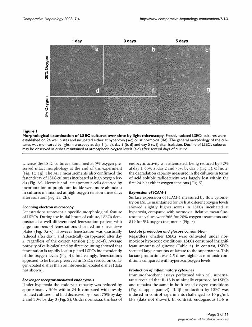
Cell viability assays and morphological analysisAll in vitro experiments in this study were carried out withan especially tailored serum-free medium which has beenshown to preserve LSECs morphology and viability betterthan regular RPMI, DMEM or their serum-containing var-iants. Quickly after isolation the cells were placed inatmospheric or low oxygen environments and the mor-phologic development of the culture was monitored overtime by conventional light microscopy. LSEC prolifera-tion analysed by BrDU incorporation was undetectable atany culture condition (data not shown). No significantdifferences in the morphology were observed during thefirst 48 h of culture (
. 1a, 1d). However, the number of viable cells per well, asmeasured by the MTT assay significantly decreased athyperoxic conditions already at 24 h (Fig. 2). From the 3rd
day in culture, LSECs maintained at atmospheric oxygentension started to collapse gradually, as observed by theformation of small areas with rounded dying cells anddetached cells scattered all over the cultures (Fig. 1b, 1f).The areas with dead cells and cell-remnants were moreprominent at the 5th day of cultures kept at high oxygenlevels, representing about 85% of the total seeded area,
Table 1: In vivo and in vitro oxygen measurements.
In vivo In vitro
Portal vein Hepatic vein Hepatic artery 5%CO2/95% air 5%CO2/5%O2
Oxygen tension (kPa) 7.22 (± 0.9) 6.70 (± 0.3) 20.9 (± 4.6) 18.46 (± 1.2) 7.39 (± 0.9)
Blood samples were collected from the indicated vascular beds and from 24 h-conditioned culture supernatants. Glass capillary tubes were used to avoid equilibration of the samples with atmospheric oxygen. The total oxygen content in the samples is given in kilo Pascals (kPa). The results are representative data obtained from three independent measurements.
Page 2 of 11(page number not for citation purposes)
Comparative Hepatology 2008, 7:4 http://www.comparative-hepatology.com/content/7/1/4
whereas the LSEC cultures maintained at 5% oxygen pre-served intact morphology at the end of the experiment(Fig. 1c, 1g). The MTT measurements also confirmed thefaster decay of LSEC cultures incubated at high oxygen lev-els (Fig. 2c). Necrotic and late apoptotic cells detected byincorporation of propidium iodide were more abundantin cultures maintained at high oxygen tension three daysafter isolation (Fig. 2a, 2b).
Scanning electron microscopyFenestrations represent a specific morphological featureof LSECs. During the initial hours of culture, LSECs dem-onstrated a well differentiated fenestration pattern withlarge numbers of fenestrations clustered into liver sieveplates (Fig. 3a–c). However fenestration was drasticallyreduced after day 1 and practically disappeared after day2, regardless of the oxygen tension (Fig. 3d–f). Averageporosity of cells calculated by direct counting showed thatfenestration is rapidly lost in plated LSECs independentlyof the oxygen levels (Fig. 4). Interestingly, fenestrationsappeared to be better preserved in LSECs seeded on colla-gen-coated dishes than on fibronectin-coated dishes (datanot shown).
Scavenger receptor-mediated endocytosisUnder hyperoxia the endocytic capacity was reduced byapproximately 50% within 24 h compared with freshlyisolated cultures, and had decreased by about 75% by day2 and 90% by day 3 (Fig. 5). Under normoxia, the loss of
endocytic activity was attenuated, being reduced by 32%at day 1, 65% at day 2 and 75% by day 3 (Fig. 5). Of note,the degradation capacity measured in the cultures in termsof acid soluble radioactivity was largely lost within thefirst 24 h at either oxygen tensions (Fig. 5).
Expression of ICAM-1Surface expression of ICAM-1 measured by flow cytome-try on LSECs maintained for 24 h at diferent oxygen levelsshowed slightly higher scores in LSECs incubated athyperoxia, compared with normoxia. Relative mean fluo-rescence values were 966 for 20% oxygen treatments and819 for 5% oxygen treatments (Fig. 7)
Lactate production and glucose consumptionRegardless whether LSECs were cultivated under nor-moxic or hyperoxic conditions, LSECs consumed insignif-icant amounts of glucose (Table 2). In contrast, LSECssecreted large amounts of lactate to the supernatant. Thislactate production was 2.5 times higher at normoxic con-ditions compared with hyperoxic oxygen levels.
Production of inflammatory cytokinesImmunoabsorbent assays performed with cell superna-tants revealed that IL-1β is minimally expressed by LSECsand remains the same in both tested oxygen conditions(Fig. 6, upper pannel). IL-1β production by LSEC wasinduced in control experiments challenged to 10 µg/mLLPS (data not shown). In contrast, endogenous IL-6 is
Morphological examination of LSEC cultures over time by light microscopyFigure 1Morphological examination of LSEC cultures over time by light microscopy. Freshly isolated LSECs cultures were established on 24 well plates and incubated either at hyperoxia (a-c) or at normoxia (d-f). The general morphology of the cul-tures was monitored by light microscopy at day 1 (a, d), day 3 (b, d) and day 5 (c, f) after isolation. Decline of LSECs cultures may be observed in dishes maintained at atmospheric oxygen levels (a-c) after several days of culture.
1 day 3 days 5 days
20%
Ox
yg
en
5%
Ox
yg
en
a
f g
b c
d
Page 3 of 11(page number not for citation purposes)

Comparative Hepatology 2008, 7:4 http://www.comparative-hepatology.com/content/7/1/4
actively released by LSECs cultured under hyperoxic oxy-gen levels, whereas the levels of this cytokine are reducedby 40% under normoxic conditions during the first 24 hand up to 48 h of culture (Fig. 6, middle panel). Produc-tion of the anti-inflammatory cytokine IL-10 correlatedinversely with IL-6. The levels of IL-10 measured in super-natants of LSECs cultured under normoxia were twice thelevels found in hyperoxia (Fig. 6, bottom panel). This sce-nario persisted also the following 24 h of culture.
Production of reactive oxygen and nitrogen speciesThere was little expression of NO during the first 48 h ofculture at both oxygen tensions (Fig. 8a). Elevation of NOlevels was observed in LPS-treated cultures that were usedas positive controls (data not shown). LSECs cultured athyperoxia generated nearly three – fold larger amounts ofH2O2 when compared to LSECs maintained at normoxia(Fig. 8b). The levels of H2O2 approached the assay detec-tion limit at 48 h of culture in LSECs kept at lower oxygen.Exogenous administration of 1000 U/mL of catalase from
the beginning of cell culture, efficiently blocked the pro-duction of endogenous H2O2 during the first 48 hours ofculture (Fig. 9a) and was able to revert the cell survivalrates observed in cultures kept at hyperoxic conditions(Fig. 9b).
DiscussionIn this study, we demonstrate that atmospheric oxygenlevels represent a deleterious environment for LSECs, andthat different oxygen environments induce significantfunctional and structural changes in these cells. LSECs areknown to be particularly vulnerable to variations of oxy-gen levels, as demonstrated by ischemia-reperfusion chal-lenges of livers, or in anoxia-reoxygenation experiments invitro [9]. During the re-oxygenation periods LSECs, KCsand hepatocytes produce large amounts of oxygen radi-cals, and LSECs gradually go into apoptosis induced byoxidative stress [8]. These findings suggest that LSECs arepoorly equipped to adapt to hyperoxic conditions. It istherefore a curious fact that all published studies usingcultured LSECs have been conducted with cells main-tained under atmospheric or hyperoxic oxygen tension.We here demonstrate that LSECs cultured under moder-ately low oxygen tension exhibit improved survival andmaintain their in vivo characteristics better as compared toLSECs cultured under atmospheric oxygen tension.
At physiologic conditions the ratio of portal vs. arterialblood flow entering the liver is around 4:1. In the rat sys-tem, most arterial blood reaches the sinusoids indirectly,via initial anastomosis between the terminal hepatic arte-riole and the portal venule [10]. In average, the oxygencontent of hepatic blood is rather low (~55 mmHg). How-ever, oxygen gradients normally exist between the peripor-tal and the perivenous areas of the liver lobule, rangingfrom 60–70 mmHg O2 in the periportal area to 25–35mmHg O2 in the perivenous areas [11]. Some laboratorieshave developed complex bioreactor systems that allow theformation of steady state oxygen gradients in culture [12].Zonal heterogeneity with regard to the oxygen tension hasbeen studied in hepatocyte cultures but not in culturedLSECs. Practically all published in vitro studies on LSECsare conducted in static culture systems where the levels ofoxygen have not been an issue.
Many research groups use long-term cultures of LSECs toexplore these cells [13,14]. In spite of this fact, surpris-ingly few studies have focused on identifying changesinduced on LSEC functions, morphology or viability dur-ing normal in vitro culture. Nevertheless, some laborato-ries have reported that signature LSEC functions such asendocytic capacity or fenestration are drastically reducedafter 1 or 2 days in culture [4,1]. Some research groupshave attempted to develop protocols to extend the in vitrolifetime of functionally intact LSECs. Most strategies have
Comparative viability of LSECs cultures maintained under high and low oxygen levelsFigure 2Comparative viability of LSECs cultures maintained under high and low oxygen levels. Cell death was moni-tored by incorporation of propidium iodide in late apoptotic or necrotic cells in cultures maintained in either high (a) or low (b) oxygen tension. Separately, viability was determined at the indicated time points by MTT colorimetric assay (c). Freshly isolated LSECs cultures were established on 24 well-plates and incubated either at hyperoxia (open bars) or at normoxia (filled bars). The obtained results demonstrate a faster decay of loss of cells in cultures maintained at hyper-oxic conditions. Statistical analyses by t-student test: *P < 0.05, **P < 0.001.
1 day 3 days 5 days
Ab
so
rban
ce
(57
0 n
m)
0
0.2
04
0.6
0.8
1.0
1.2
1.4 * ** **
a b
c
Page 4 of 11(page number not for citation purposes)

Comparative Hepatology 2008, 7:4 http://www.comparative-hepatology.com/content/7/1/4
used specially designed culture media, including tumour-conditioned medium [15], hepatocyte-conditionedmedium [2], autologous rat serum [3], phorbol ester sup-plemented medium, vascular endothelial growth factor-containing medium [16] or orthovanadate [1]. In the
present study we used a serum-free medium developedpreviously in our laboratory [4]. With this medium, wewere able to maintain fuctionally intact rat LSEC culturesduring 48 h. However, from the third day of incubation,the cultures gradually lost their integrity. This loss wasnevertheless prevented by incubation of cultures underlow oxygen tension, enabling maintenance of intact mor-phology after six days of culture. This demonstrates thatatmospheric oxygen levels itself is a disfavourable envi-ronment for LSECs.
Several reports have shown that a major biological func-tion of LSECs is to rid the blood of an array of naturallyoccurring soluble macromolecular and colloidal wastesubstances, via clathrin-mediated endocytosis (for reviewsee [17]). Based on the knowledge that receptor-mediatedendocytosis represents a characteristic function of LSECs,we compared the ability of the cells to internalize anddegrade formaldehyde treated serum albumin (FSA), thatis specifically taken up by the scavenger receptor of LSEC,in high and low oxygen environments. Although theendocytic capacity decreased gradually over time in bothconditions, the total uptake measured at 24, 48 and 72 hwas significantly higher under physiological oxygen con-ditions than under hyperoxic conditions. Yet, culturing ofLSECs under low oxygen levels per se was not enough tomaintain the endocytic capacity at the same level as meas-ured in freshly prepared cultures. The reason for this is at
Porosity measurementsFigure 4Porosity measurements. Porosity analysis of LSECs seeded on coverslips at 6, 24 and 48 h. Fenestrae and gaps greater that 300 nm were excluded from the analysis. Poros-ity measurements are expressed as percentage of the total area covered by cells in each coverslip. Black columns: 20% oxygen. White columns: 5% oxygen.
Avera
ge
po
rosity
-fe
n>
300
nm
0
2
4
6
8
10
12
14
16
6 hours 24 hours 48 hours
Fenestration pattern in cultured LSEC analysed by scanning electron microscopy (SEM)Figure 3Fenestration pattern in cultured LSEC analysed by scanning electron microscopy (SEM). The evolution of fenestrae in isolated LSECs seeded on fibronectin-coated coverslips was monitored at different time points by SEM. LSECs cul-tures were maintained at high (a-c) or low (d-f) oxygen levels. Highly fenestrated cells can be observed during early time points of culture. Fenestration is gradually lost over time in both normoxic or hypoxic conditions.
6 hours 24 hours 48 hours
20%
Ox
yg
en
5%
Ox
yg
en
a b c
d e f
Page 5 of 11(page number not for citation purposes)

Comparative Hepatology 2008, 7:4 http://www.comparative-hepatology.com/content/7/1/4
least three-fold, based on the fact that LSEC monocultureslack: i) essential factors produced locally or brought to thecells via the portal circulation, ii) interaction with otherliver cells, and/or iii) interaction with native extracellularmatrix. Focusing in the present work on the impact of oxy-gen tension, we show here that a low oxygen level in vitro,approaching that of the local sinusoidal environment invivo, significantly prolong the naturally high scavengeractivity of LSECs when compare to traditional culturesestablished at atmospheric oxygen levels.
Another important physiological function carried out byLSECs in vivo is the filtration of numerous plasma compo-nents towards the liver parenchyma through a well organ-ized net of transcytoplasmic holes called fenestrae. Thisfenestration represents a hallmark of intact mammalianLSEC. It is noteworthy that several reports conclude thatthe cells undergo a rapid defenestration after isolation andculture [18]. Assessing fenestration using scanning elec-tron microscopy, we found that this feature is graduallylost over time independent of the culture conditions used.Of note, LSECs lost fenestration more rapidly whenseeded on fibronectin-coated dishes than on collagen-coated ones. This shows that the key factors which enablethe maintenance of the LSEC fenestrae in the intact liverare lost upon cultivation, regardless the oxygen levels.
Adaptation to hypoxia is known to induce changes in theenergy metabolic routes of cells, most commonly shiftingfrom the oxidative phosphorylation pathways to the glyc-olytic routes. Morphometric studies of LSECs in the intactliver have shown that the cells contain unusually fewmitochondria [19,20]. This observation, along with the
Production of inflammatory cytokines by LSECsFigure 6Production of inflammatory cytokines by LSECs. Secretion of IL-1β (a), IL-6 (b), or IL-10 (c) was measured in LSEC culture supernatants by ELISA at different time-points. Conditioned media were collected from LSECs cultured at hyperoxia (open bars) or normoxia (filled bars) at 24 hours intervals. Final concentrations were estimated from individual standard curves. Values are means of triplicate measure-ments. The results are representative data obtained from three independent experiments. Statistical analyses by t-stu-dent test: *P < 0.001.
0
200
400
600
800
1000
1200
IL-1β
0
500
1000
1500
2000
2500 IL-6
0
20
40
60
80
100
120
140
160
0-24 hours 24-48 hours
IL-10
**
**
**
**
pg
/ m
l /
10
5c
ell
sp
g/
ml
/ 1
05c
ell
sp
g/
ml
/ 1
05c
ell
s
Time-course analysis of LSECs endocytic capacityFigure 5Time-course analysis of LSECs endocytic capacity. Endocytosis of 125I-FSA, a ligand for the LSEC scavenger receptor was monitored at different time points after incuba-tion of cells at normoxic or hyperoxic conditions. Each col-umn represents separate values of cell-associated (lower part) and degraded (upper part) 125I-FSA. Total endocytosis is the result of adding cell associated and degraded ligand (full column size), calculated as percentage of total 125I-FSA added to cultures. Values are means of triplicate measurements. The results are representative data obtained from three independent experiments. Statistical analyses by Student's t-test: *P < 0.05, **P < 0.001.
0
10
20
30
40
50*
**
**
21% O2
5% O2
2 hours 24 hours 48 hours 72 hours
Radio
activity
(% o
f to
tal)
Page 6 of 11(page number not for citation purposes)

Comparative Hepatology 2008, 7:4 http://www.comparative-hepatology.com/content/7/1/4
fact that rat LSECs produce large amounts of lactate andacetate, even when cultured at high oxygen levels, stronglysuggest that these cells are geared to a largely anaerobictype of metabolism. Conceivingly, LSECs perform lessoxidative phosphorylation compared to most other cellstypes, and it has been suggested that in LSECs, glutamineand fatty acid oxidation are the main sources of energy[21]. An alternative path is the anaerobic conversion ofpyruvate, originating from the catabolism of glucogenicaminoacids from the growth medium, into lactate [22].Examining glucose consumption and lactate productionunder different oxygen tensions to explore possible varia-tions in the energy sources of the cells, we found that glu-cose was not consumed by LSECs under hyperoxic ornormoxic conditions. This suggests that the energy meta-bolic routes are independent of the oxygen levels. In con-trast, the amount of lactate generated by LSECs under
normoxic conditions was enhanced almost three times,suggesting that metabolic energy reactions are drivenmore efficiently under low oxygen.
As a rule, procedures used to isolate and cultivate cellsinduce cell activation to some extent. Desirable in vitromodels should be based on non-activated or low-acti-vated cells. In the present study we measured the produc-tion of inflammatory cytokines, reactive oxygen speciesand the expression of adhesion molecules after cell culti-vation as indicators of cell activation. Our results confirmthat LSECs produce high levels of IL-6 when culturedunder "standard" high (20%) oxygen pressure. Notably,the expression of this cytokine was reduced by 50% whenthe cells were maintained at low oxygen levels. In contrast,the production of IL-10, an anti-inflammatory mediator,was enhanced when the cells were incubated at 5% oxy-
ICAM expression on cultured LSECsFigure 7ICAM expression on cultured LSECs. Quantitative measurements of ICAM-1 expressed on the surface of LSEC were done by flow cytometry after incubation of LSECs on normoxic and hyperoxic environments during 24 h. Relative mean fluo-rescence values for 20% O2 levels were 966, whereas 5% O2 scored 819. The results are representative data obtained from two independent experiments.
Co
un
ts
Relative fluorescence
20
40
60
80
100
120
101 102 103
Unlabeled
5% Oxygen
20% Oxygen
ICAM-1
Table 2: Lactate and glucose measurements in supernatants obtained from dishes with or without cells.
Culture Supernatants Time (h) Lactate (mmol/L/106 cells) Glucose (mmol/L/106 cells)
Cell-free (20% O2) 0–24 0.10 (± 0.20) 11.81 (± 0.24)LSECs (20% O2) 0–24 1.19 (± 0.42) 10.96 (± 0.40)LSECs (5% O2) 0–24 2.77 (± 0.59) 10.83 (± 0.20)LSECs (20% O2) 24–48 1.31 (± 0.38) 10.70 (± 0.20)LSECs (5% O2) 24–48 3.07 (± 1.12) 10.36 (± 0.20)
Conditioned media collected from cell free incubations, or LSEC cultures were analysed for glucose and lactate. Values are means of triplicate measurements. The results are representative data obtained from three independent experiments.
Page 7 of 11(page number not for citation purposes)

Comparative Hepatology 2008, 7:4 http://www.comparative-hepatology.com/content/7/1/4
gen. Flow cytometric analysis of ICAM-1 expression at thecell surface show strong signal on LSECs cultured for 24 hat 20% oxygen. These values are however slightly reducedupon incubation of cells at low oxygen tensions. The over-all results indicate that LSECs have a less activated pheno-type when they are incubated at low oxygen levels.
The transfer of LSECs from an in vivo low oxygen tensionto an in vitro high oxygen tension may exert effects on thecells similar to those observed in LSECs during hypoxia-reoxygenation of the intact liver. Indeed, we observedlarge production of hydrogen peroxide by LSECs when thecells were kept at atmospheric oxygen conditions. Of note,this production was much lower at low oxygen tension.Hydrogen peroxide induces toxic effects on LSECs, mostlybecause these cells are not well equipped to metabolizethis reactive substance [8,9]. Addition of catalase to themedium, an enzyme that mediates directly the catabolism
of H2O2, was able to abrogate to a large extent the forma-tion of H2O2, and had beneficial effects on the morphol-ogy and survival of LSEC cultures. We thus entertain theidea that endogenous H2O2 may be largely responsible forthe rapid culture decline observed during high oxygenincubation.
Effect of catalase on H2O2 formation and on cell survival in LSEC culturesFigure 9Effect of catalase on H2O2 formation and on cell sur-vival in LSEC cultures. Formation of H2O2 was measured on LSEC cultures (fluorescence values) during 6 h following the first 24 h of incubation at 5% O2, or at 20% O2 in the presence or absence of catalase (a). Additionally, survival rates at the third day of culture (absorbance values) were measured on dishes treated the same way (b). All results are representative data obtained from two independent experi-ments.
0
0,5
1
1,5
2
2,5
3
0
0,2
0,4
0,6
0,8
1
1,2
100
U/mL
1000
U/mL
20% Oxygen
a
b
5%
Oxygen
Catalase
Flu
ore
sce
nce
(fo
ld 5
% O
2)
Ab
sorb
ance
(fo
ld 5
% O
2)
Production of nitric oxide (NO) and hydrogen peroxide (H2O2) by LSECFigure 8Production of nitric oxide (NO) and hydrogen perox-ide (H2O2) by LSEC. Secretion of nitric oxide (NO) was measured in LSEC culture supernatants by Griess reaction at 24 and 48 h (a). Culture media was collected from hyperoxic conditions (open bars) or normoxic conditions (filled bars) at 24 hours intervals. Final concentrations were estimated from individual standard curves. Generation of endogenous H2O2 was monitored in separate experiments at the indicated time-points in LSEC cultures by H2O2-mediated oxidation of DCFH-DA into DFC during 6 h (b). Values are total fluores-cence emitted at 545 nm.
24 hours 48 hours
Flu
ore
scen
ce(4
88/5
45n
m)
Nit
rite
( µµ µµM
/mill
. cel
ls)
0
400
800
1200
1600
*
*
H2O2
NO
0
20
40
60
80
100
a
b
Page 8 of 11(page number not for citation purposes)

Comparative Hepatology 2008, 7:4 http://www.comparative-hepatology.com/content/7/1/4
ConclusionIn this study we report that atmospheric oxygen tensionhas harmful effects on isolated rat LSECs during long termcultivation. These effects may be largely abrogated byincubation of the cells at physiological O2 conditions. Ourfindings are compatible with a better preservation ofessential morphologic and functional LSEC featuresunder more physiological oxygen tensions. Based onthese data, we recommend low oxygen environments forcultivation of LSECs, especially when long-term culturesare used.
MethodsIsolation and culture of LSECPreparation of highly purified rat liver sinusoidalendothelial cells was performed as previously described[23]. Briefly, rat livers were perfused with collagenase(Worthington Biochemical Corporation, Lakewood, NJ,USA.) and the resulting suspension of single cells was sub-jected to low speed centrifugation to eliminate most of thehepatocytes, followed by discontinuous density centrifu-gation in Percoll (Amersham Biotech, Uppsala, Sweden)gradients. The resulting non-parenchymal cells were sus-pended in pre-warmed serum-free culture medium.Kupffer cells were eliminated by selective attachment toglutaraldehyde-treated albumin (Ostapharma, Ziegel-brucke, Switzerland), and the enriched LSEC suspensionwas seeded on dishes coated with either rat tail collagentype I (Nutacon, Leimunden, Netherlands) or humanfibronectin (isolated at the laboratory from pooledhuman blood samples). For all experiments the cells wereincubated in DM110/SS serum-free medium [4]. Incuba-tions were undertaken under hyperoxia (20%) or nor-moxia 95%N2/5%O2 to reflect physiologic conditions inthe liver sinusoids. The monolayer cultures were moni-tored by conventional light microscopy.
Endocytosis measurementsCultures of LSEC (0.5 × 10⁶) were established in 2 cm2
wells and maintained in serum-free medium. After exper-imental treatments, cells were cultured for 90 min at 37°Cin 250 µl of RPMI, containing 1% Human Serum Albu-min (HAS) and trace amounts (40.000 cpm, 50 ng/ml) ofradioiodinated formaldehyde-treated bovine serum albu-min (125I-FSA). Endocytosis experiments were terminatedby transferring incubation medium and two washing vol-umes to tubes containing 800 µl of 20% TCA, therebyinducing precipitation of non-degraded proteins. Aftercentrifugation, precipitated and soluble radioactivity wasmeasured using a Geiger counter. Acid soluble radioactiv-ity was considered to indicate degraded FSA, and resultswere evaluated from total radioactivity added to cultures.Cell associated radioactivity was calculated by solubilisa-tion of cell monolayers with 1% SDS, and measured with
a γ-counter (Cobra II, Packard). Values were normalizedfor the total number of cells counted per well.
Scanning electron microscopyScanning EM was performed as previously described [24].LSECs that had been cultured in hypoxic and normoxicconditions for 6, 24 and 48 h were fixed for 1 hour with2.5% glutaraldehyde in 0.1 mol/l sodium cacodylatebuffer (1% sucrose). Coverslips were treated with tannicacid (1% in 0.15 mol/l cac. buffer), osmicated (1% OsO4/0.1 mol/l cacodylate buffer), dehydrated in a series of eth-anol gradients and finally incubated in hexamethyldisila-zane for 2 min. Gold coated coverslips were viewed usinga Jeol scanning microscope. Five representative cells fromeach time point were photographed (magnification3,000–5,000 ×) and fenestral diameter and porosity (per-centage of surface area occupied by fenestrations) ana-lysed using ImageJ software. The results are expressed asmean ± S.D.
ELISA measurements of cytokine productionThe production of the inflammatory mediators IL-1β, IL-6 and IL-10 in LSEC culture supernatants was determinedwith specific rat IL-1β, IL-6 and IL-10 ELISA kit (R&D Sys-tems, Minneapolis, USA) according to manufacturers'instructions. Briefly, after the experimental incubations ofcultures, the supernatants were collected and underwenthigh speed centrifugation, then kept at -20°C. 96 well-plates were coated with "capture" antibodies and 100 µlof 1:2 diluted supernatants were added to each well andincubated for 2 h at room temperature. The "detection"antibody was then applied for 2 h and the wells were ulti-mately subjected to peroxidase reaction. Absorbance wasmeasured at 450 nm and the values were converted intoµg/ml according to the standard curve. Values were nor-malized after the total number of cells counted per well.
ICAM expression by flow cytometryFor measurements of ICAM-1 expression at the cell sur-face, LSEC were incubated at different oxygen tension dur-ing 24 h, detached from wells by 20 min incubation inEDTA buffer, and immediately fixed in 4% paraformalde-hyde. Fixative was removed by cell sedimentation and thecells were then resuspended in 500 µl of PBS containing1% BSA. Specific monoclonal antibody against rat ICAM(Biodesign International, Saco, ME, USA) was added totubes containing fixed LSEC and incubated during 30 minat room temperature. Unlabeled antibody was eliminatedby a series of cell washings, followed by incubation with asecondary antibody against mouse IgG FITC-congugated(Dako Denmark A/S). A group of cells incubated onlywith secondary antibodies was used as negative controls.Fluorescent cells were then analyzed on a BD FACScanflow cytometer
Page 9 of 11(page number not for citation purposes)

Comparative Hepatology 2008, 7:4 http://www.comparative-hepatology.com/content/7/1/4
Measurement of endogenous H2O2 productionTo measure endogenous production of H2O2, cells wereseeded in 24 well plates and incubated for the indicatedtime points at different oxygen levels. After incubation,cells were incubated with 20 mM/L 2',7'-dichlorofluores-cein-diacetate (DCFH-DA) for 30 min at 37°C. This is anon-polar compound that readily diffuses into cells. Thecultures were washed twice to eliminate excess amount ofreagent and the cultures were further incubated for 6 h at37°C. Once the acetate groups are cleaved by intracellularesterase, H2O2 produced by the cells oxidizes DCFH to thefluorescent compound 2',7'-dichlorofluorescein (DCF).In this way, fluorescence intensity is proportional to theamount of H2O2 generated. DFC fluorescence wasrecorded using a computerized plate-scanning microfluo-rimeter (CytoFluor-2350 system, Millipore Co. Bedford,MA) at both 485/22-nm excitation with 530/25-nm emis-sion filter at high sensitivity settings. Non-DCFH-DA-incubated cells were used to subtract basal auto fluores-cence.
Nitric oxide analysisThe stable end product of nitric oxide, nitrite (NO2
-), wasmeasured in culture supernatants by standard colorimet-ric assay [25]. Briefly, 50 µl aliquots of medium were col-lected from individual wells and treated with an equalvolume of Griess reagent (1% sulphanilamide and 0.1%napthylenediamide dihydrochloride in 2.5% H3PO4) atroom temperature for 10 min. The optical density of thesamples was recorded using TiterTek Multiskan at 540nm. A standard curve using NaNO2 in clean culturemedium was used for calculating NO2
- concentration. Allvalues are means ± S.D. of triplicate measurements forthree separate experiments.
Lactate, glucose and oxygen measurementsLactate and glucose concentrations were measured withYSI-analyzer (Cobas, Switzerland). Cells were seeded in24 well-plates and incubated at different oxygen levels.Cell supernatants were collected every 24 h and cleanedfrom particles and debris by high speed centrifugation.For each measurement, 150 µl of culture supernatant wasutilized. The different oxygen tensions in blood in anes-thetised pigs were measured in the portal vein, hepaticvein and aorta. The partial pressures of oxygen and pH inthe samples (culture supernatants and blood) were ana-lyzed with a blood-gas analyzer (Rapidlab 865, ChironDiagnostics, UK). The sampling was performed with aglass capillary tube.
Cell viability assay: MTT assayLSEC were seeded on 24 well-plates and cultivated for theindicated time-points. Cells were subsequently exposed to0.25 mg/mL of MTT (3-[4,5-dimethylthiazol-2-yl]-2,5-dephenyl tetrazolium bromide; Sigma-Aldrich) reagent
and incubated for 2 h at 37°C. After 2 h, 200 µL of dissolv-ing solution (96,7% Isopropanol/3,3% HCl) was addedto each well, followed by incubation for 1 h at 37°C in arocker plate. The absorbance at 570 nm of each samplewell was measured by using an automated plate readerand compared between the groups.
Propidium iodide staining for cell deathNecrotic or late apoptotic cells were identified on LSECcultures by propidium iodide incorporation. AdherentLSEC cultures were established on 2 wells thermanoxslides (NUNC International, Tokio, Japan), treated withfibronectin. Cultures were maintained at high and lowoxygen environments during three days, and the culturemedia was renewed every 24 h. Propidium iodide stainingwas performed with Apoptosis/necrosis detection kit fromCalbiochem following manufacturer instructions. Thespecimen were embedded in Dako Fluoromount (Dako,Glostrup, Denmark), and examined in a fluorescencemicroscope (Zeiss Axiophot, Germany) equipped with aNikon DS-5MC digital camera.
StatisticsResults are presented as mean ± S.E.M. and the two exper-imental conditions compared using the Student's t-testwith P < 0.05 considered significant. In all experiments,the obtained raw data were normalized against to theamount of cells counted in each treatment. For the quan-titative measurements done in SEM, comparisonsbetween groups were undertaken using Kruskal-Wallis testwith a post hoc Dunn analysis. P value of < 0.05 was con-sidered statistically significant.
Competing interestsThe authors declare that they have no competing interests.
Authors' contributionsIM and BS conceived of the study. IM carried out most ofthe experimental work and the writing. AW and DGL par-ticipated in the determination of fenestration parameters,statistical analysis and assisted in the writing. CIØ carriedout the isolation and characterization of the cells. GINparticipated in the oxygen measurements, as well as in thedetermination of glucose and lactate in culture superna-tants. OJ and BS participated in the design of the study,coordination and helped to draft the manuscript.
AcknowledgementsThe authors highly appreciate the technical assistance for the flow cytomet-ric analysis by Bjørn Thorvald Moe, at the University of Tromsø. This work was supported by grants from the Norwegian Research Council and the Medical Faculty at the University of Tromsø.
References1. Ohi N, Nishikawa Y, Tokairin T, Yamamoto Y, Doi Y, Omori Y,
Enomoto K: Maintenance of Bad phosphorylation prevents
Page 10 of 11(page number not for citation purposes)

Comparative Hepatology 2008, 7:4 http://www.comparative-hepatology.com/content/7/1/4
Publish with BioMed Central and every scientist can read your work free of charge
"BioMed Central will be the most significant development for disseminating the results of biomedical research in our lifetime."
Sir Paul Nurse, Cancer Research UK
Your research papers will be:
available free of charge to the entire biomedical community
peer reviewed and published immediately upon acceptance
cited in PubMed and archived on PubMed Central
yours — you keep the copyright
Submit your manuscript here:http://www.biomedcentral.com/info/publishing_adv.asp
BioMedcentral
apoptosis of rat hepatic sinusoidal endothelial cells in vitroand in vivo. Am J Pathol 2006, 168(4):1097-1106.
2. Krause P, Markus PM, Schwartz P, Unthan-Fechner K, Pestel S, Fan-drey J, Probst I: Hepatocyte-supported serum-free culture ofrat liver sinusoidal endothelial cells. J Hepatol 2000,32(5):718-726.
3. de Leeuw AM, Barelds RJ, de Zanger R, Knook DL: Primary cul-tures of endothelial cells of the rat liver: a model forultrastructural and functional studies. Cell Tissue Res 1982,223(1):201-215.
4. Elvevold K, Nedredal GI, Revhaug A, Bertheussen K, Smedsrod B:Long-term preservation of high endocytic activity in primarycultures of pig liver sinusoidal endothelial cells. Eur J Cell Biol2005, 84(9):749-764.
5. Semenza GL: Regulation of mammalian O2 homeostasis byhypoxia-inducible factor 1. Annu Rev Cell Dev Biol 1999,15:551-578.
6. D'Angio CT, Finkelstein JN: Oxygen regulation of gene expres-sion: a study in opposites. Mol Genet Metab 2000, 71(1–2):371-380.
7. Cogger VC, Mross PE, Hosie MJ, Ansselin AD, McLean AJ, Le CouteurDG: The effect of acute oxidative stress on the ultrastructureof the perfused rat liver. Pharmacol Toxicol 2001, 89(6):306-311.
8. Motoyama S, Minamiya Y, Saito S, Saito R, Matsuzaki I, Abo S, InabaH, Enomoto K, Kitamura M: Hydrogen peroxide derived fromhepatocytes induces sinusoidal endothelial cell apoptosis inperfused hypoxic rat liver. Gastroenterology 1998,114(1):153-163.
9. Samarasinghe DA, Tapner M, Farrell GC: Role of oxidative stressin hypoxia-reoxygenation injury to cultured rat hepatic sinu-soidal endothelial cells. Hepatology 2000, 31(1):160-165.
10. Yamamoto K, Sherman I, Phillips MJ, Fisher MM: Three-dimen-sional observations of the hepatic arterial terminations inrat, hamster and human liver by scanning electron micros-copy of microvascular casts. Hepatology 1985, 5(3):452-456.
11. Allen JW, Bhatia SN: Formation of steady-state oxygen gradi-ents in vitro: application to liver zonation. Biotechnol Bioeng2003, 82(3):253-262.
12. Allen JW, Khetani SR, Bhatia SN: In vitro zonation and toxicity ina hepatocyte bioreactor. Toxicol Sci 2005, 84(1):110-119.
13. Rauen U, Elling B, de Groot H: Injury to cultured liver endothe-lial cells after cold preservation: mediation by reactive oxy-gen species that are released independently of the knowntrigger hypoxia/reoxygenation. Free Radic Biol Med 1997,23(3):392-400.
14. Gerlach JC, Zeilinger K, Spatkowski G, Hentschel F, Schnoy N, Kol-beck S, Schindler RK, Neuhaus P: Large-scale isolation of sinusoi-dal endothelial cells from pig and human liver. J Surg Res 2001,100(1):39-45.
15. Irving MG, Roll FJ, Huang S, Bissell DM: Characterization and cul-ture of sinusoidal endothelium from normal rat liver: lipo-protein uptake and collagen phenotype. Gastroenterology 1984,87(6):1233-1247.
16. Esser S, Wolburg K, Wolburg H, Breier G, Kurzchalia T, Risau W:Vascular endothelial growth factor induces endothelial fen-estrations in vitro. J Cell Biol 1998, 140(4):947-959.
17. Smedsrod B, Pertoft H, Gustafson S, Laurent TC: Scavenger func-tions of the liver endothelial cell. Biochem J 1990,266(2):313-327.
18. DeLeve LD, Wang X, Hu L, McCuskey MK, McCuskey RS: Rat liversinusoidal endothelial cell phenotype is maintained by para-crine and autocrine regulation. Am J Physiol Gastrointest Liver Phys-iol 2004, 287(4):G757-763.
19. Wisse E: An ultrastructural characterization of the endothe-lial cell in the rat liver sinusoid under normal and variousexperimental conditions, as a contribution to the distinctionbetween endothelial and Kupffer cells. J Ultrastruct Res 1972,38(5):528-562.
20. Fujii Y, Ohno N, Li Z, Terada N, Baba T, Ohno S: Morphologicaland histochemical analyses of living mouse livers by new 'cry-obiopsy' technique. J Electron Microsc (Tokyo) 2006, 55(2):113-122.
21. Spolarics Z, Lang CH, Bagby GJ, Spitzer JJ: Glutamine and fattyacid oxidation are the main sources of energy for Kupfferand endothelial cells. Am J Physiol 1991, 261(2 Pt 1):G185-190.
22. Nedredal GI, Elvevold K, Ytrebo LM, Fuskevag OM, Pettersen I, Ber-theussen K, Langbakk B, Smedsrod B, Revhaug A: Significant con-
tribution of liver non-parenchymal cells to metabolism ofammonia and lactate, and cocultivation augments the func-tions of a bioartificial liver. Am J Physiol Gastrointest Liver Physiol2007.
23. Smedsrod B, Pertoft H, Eggertsen G, Sundstrom C: Functional andmorphological characterization of cultures of Kupffer cellsand liver endothelial cells prepared by means of density sep-aration in Percoll, and selective substrate adherence. Cell Tis-sue Res 1985, 241(3):639-649.
24. Cogger VC, Muller M, Fraser R, McLean AJ, Khan J, Le Couteur DG:The effects of oxidative stress on the liver sieve. J Hepatol2004, 41(3):370-376.
25. AH D, CF N, DJ S: Release of reactive nitrogen intermediatesand reactive oxygen intermediates from mouse peritonealmacrophages. Comparison of activating cytokines and evi-dence for independent production. J Immunol 1988,141(7):2407-2412.
Page 11 of 11(page number not for citation purposes)
Copyright © 2022 FDOKUMEN