Transendocardial, Autologous Bone Marrow Cell Transplantation for Severe, Chronic Ischemic Heart...
10
Transendocardial, Autologous Bone Marrow Cell Transplantation for Severe, Chronic Ischemic Heart Failure Emerson C. Perin, MD, PhD*; Hans F.R. Dohmann, MD*; Radovan Borojevic, PhD; Suzana A. Silva, MD; Andre L.S. Sousa, MD; Claudio T. Mesquita, MD, PhD; Maria I.D. Rossi, PhD; Antonio C. Carvalho, MD, PhD; Helio S. Dutra, PhD; Hans J.F. Dohmann, MD, PhD; Guilherme V. Silva, MD; Luciano Belém, MD; Ricardo Vivacqua, MD; Fernando O.D. Rangel, MD; Roberto Esporcatte, MD; Yong J. Geng, MD, PhD; William K. Vaughn, PhD; Joao A.R. Assad, MD; Evandro T. Mesquita, MD, PhD; James T. Willerson, MD Background—This study evaluated the hypothesis that transendocardial injections of autologous mononuclear bone marrow cells in patients with end-stage ischemic heart disease could safely promote neovascularization and improve perfusion and myocardial contractility. Methods and Results—Twenty-one patients were enrolled in this prospective, nonrandomized, open-label study (first 14 patients, treatment; last 7 patients, control). Baseline evaluations included complete clinical and laboratory evaluations, exercise stress (ramp treadmill), 2D Doppler echocardiogram, single-photon emission computed tomography perfusion scan, and 24-hour Holter monitoring. Bone marrow mononuclear cells were harvested, isolated, washed, and resuspended in saline for injection by NOGA catheter (15 injections of 0.2 cc). Electromechanical mapping was used to identify viable myocardium (unipolar voltage 6.9 mV) for treatment. Treated and control patients underwent 2-month noninvasive follow-up, and treated patients alone underwent a 4-month invasive follow-up according to standard protocols and with the same procedures used as at baseline. Patient population demographics and exercise test variables did not differ significantly between the treatment and control groups; only serum creatinine and brain natriuretic peptide levels varied in laboratory evaluations at follow-up, being relatively higher in control patients. At 2 months, there was a significant reduction in total reversible defect and improvement in global left ventricular function within the treatment group and between the treatment and control groups (P0.02) on quantitative single-photon emission computed tomography analysis. At 4 months, there was improvement in ejection fraction from a baseline of 20% to 29% (P0.003) and a reduction in end-systolic volume (P0.03) in the treated patients. Electromechanical mapping revealed significant mechanical improvement of the injected segments (P0.0005) at 4 months after treatment. Conclusions—Thus, the present study demonstrates the relative safety of intramyocardial injections of bone marrow– derived stem cells in humans with severe heart failure and the potential for improving myocardial blood flow with associated enhancement of regional and global left ventricular function. (Circulation. 2003;107:2294-2302.) Key Words: cells heart failure ischemia revascularization gene therapy A fter myocardial infarction, chronically ischemic (hibernat- ing) myocardium may persist in association with variable degrees of scar tissue. In most circumstances, native angiogen- esis is insufficient to prevent the resultant remodeling when significant injury occurs. As a consequence, infarct-related heart failure remains a major cause of morbidity and mortality. The understanding that vasculogenesis can occur in the adult has led to intense investigation into stem cell therapy. Several recent experimental studies have confirmed the po- tential of pluripotential cells in differentiating into cardio- myocytes and endothelial cells. 1,2 Further evidence from animal models has confirmed that pluripotential cells from Received March 7, 2003; revision received March 25, 2003; accepted March 26, 2003. From the Texas Heart Institute at St Luke’s Episcopal Hospital, Houston, Tex (E.C.P., G.V.S., Y.J.G., W.K.V., J.T.W.); Hospital Procardiaco, Rio de Janeiro, Brazil (H.F.R.D., S.A.S., A.L.S.S., C.T.M., H.J.F.D., L.B., R.V., F.O.D.R., R.E., J.A.R.A., E.T.M.); Federal University, Rio de Janeiro, Brazil (R.B., M.I.D.R., A.C.C., H.S.D.); and Brazilian Millennium Institute for Tissue Bioengineering (H.F.R.D., R.B., A.C.C.). *Drs Perin and Dohmann are co-principal investigators. Guest editor for this article was Valentin Fuster, MD, PhD, Mount Sinai School of Medicine, NY. This article originally appeared Online on April 21, 2003 (Circulation. 2003;107:r75–r83). Correspondence to Emerson C. Perin, MD, 6624 Fannin, Suite 2220, Houston, TX 77030 (e-mail [email protected]), and Hans F.R. Dohmann, MD, Rua General Polidoro, 192, CEP 22080-000 –Botafogo, Rio de Janeiro, Brazil (e-mail [email protected]). © 2003 American Heart Association, Inc. Circulation is available at http://www.circulationaha.org DOI: 10.1161/01.CIR.0000070596.30552.8B 2294 Clinical Investigation and Reports by guest on August 18, 2015 http://circ.ahajournals.org/ Downloaded from
Transcript of Transendocardial, Autologous Bone Marrow Cell Transplantation for Severe, Chronic Ischemic Heart...

Transendocardial, Autologous Bone Marrow CellTransplantation for Severe, Chronic Ischemic Heart Failure
Emerson C. Perin, MD, PhD*; Hans F.R. Dohmann, MD*; Radovan Borojevic, PhD;Suzana A. Silva, MD; Andre L.S. Sousa, MD; Claudio T. Mesquita, MD, PhD; Maria I.D. Rossi, PhD;
Antonio C. Carvalho, MD, PhD; Helio S. Dutra, PhD; Hans J.F. Dohmann, MD, PhD;Guilherme V. Silva, MD; Luciano Belém, MD; Ricardo Vivacqua, MD; Fernando O.D. Rangel, MD;Roberto Esporcatte, MD; Yong J. Geng, MD, PhD; William K. Vaughn, PhD; Joao A.R. Assad, MD;
Evandro T. Mesquita, MD, PhD; James T. Willerson, MD
Background—This study evaluated the hypothesis that transendocardial injections of autologous mononuclear bonemarrow cells in patients with end-stage ischemic heart disease could safely promote neovascularization and improveperfusion and myocardial contractility.
Methods and Results—Twenty-one patients were enrolled in this prospective, nonrandomized, open-label study (first 14patients, treatment; last 7 patients, control). Baseline evaluations included complete clinical and laboratory evaluations,exercise stress (ramp treadmill), 2D Doppler echocardiogram, single-photon emission computed tomography perfusionscan, and 24-hour Holter monitoring. Bone marrow mononuclear cells were harvested, isolated, washed, andresuspended in saline for injection by NOGA catheter (15 injections of 0.2 cc). Electromechanical mapping was usedto identify viable myocardium (unipolar voltage �6.9 mV) for treatment. Treated and control patients underwent2-month noninvasive follow-up, and treated patients alone underwent a 4-month invasive follow-up according tostandard protocols and with the same procedures used as at baseline. Patient population demographics and exercise testvariables did not differ significantly between the treatment and control groups; only serum creatinine and brainnatriuretic peptide levels varied in laboratory evaluations at follow-up, being relatively higher in control patients. At 2months, there was a significant reduction in total reversible defect and improvement in global left ventricular functionwithin the treatment group and between the treatment and control groups (P�0.02) on quantitative single-photonemission computed tomography analysis. At 4 months, there was improvement in ejection fraction from a baseline of20% to 29% (P�0.003) and a reduction in end-systolic volume (P�0.03) in the treated patients. Electromechanicalmapping revealed significant mechanical improvement of the injected segments (P�0.0005) at 4 months after treatment.
Conclusions—Thus, the present study demonstrates the relative safety of intramyocardial injections of bone marrow–derived stem cells in humans with severe heart failure and the potential for improving myocardial blood flow withassociated enhancement of regional and global left ventricular function. (Circulation. 2003;107:2294-2302.)
Key Words: cells � heart failure � ischemia � revascularization � gene therapy
After myocardial infarction, chronically ischemic (hibernat-ing) myocardium may persist in association with variable
degrees of scar tissue. In most circumstances, native angiogen-esis is insufficient to prevent the resultant remodeling whensignificant injury occurs. As a consequence, infarct-related heartfailure remains a major cause of morbidity and mortality.
The understanding that vasculogenesis can occur in theadult has led to intense investigation into stem cell therapy.Several recent experimental studies have confirmed the po-tential of pluripotential cells in differentiating into cardio-myocytes and endothelial cells.1,2 Further evidence fromanimal models has confirmed that pluripotential cells from
Received March 7, 2003; revision received March 25, 2003; accepted March 26, 2003.From the Texas Heart Institute at St Luke’s Episcopal Hospital, Houston, Tex (E.C.P., G.V.S., Y.J.G., W.K.V., J.T.W.); Hospital Procardiaco, Rio de
Janeiro, Brazil (H.F.R.D., S.A.S., A.L.S.S., C.T.M., H.J.F.D., L.B., R.V., F.O.D.R., R.E., J.A.R.A., E.T.M.); Federal University, Rio de Janeiro, Brazil(R.B., M.I.D.R., A.C.C., H.S.D.); and Brazilian Millennium Institute for Tissue Bioengineering (H.F.R.D., R.B., A.C.C.).
*Drs Perin and Dohmann are co-principal investigators.Guest editor for this article was Valentin Fuster, MD, PhD, Mount Sinai School of Medicine, NY.This article originally appeared Online on April 21, 2003 (Circulation. 2003;107:r75–r83).Correspondence to Emerson C. Perin, MD, 6624 Fannin, Suite 2220, Houston, TX 77030 (e-mail [email protected]), and Hans F.R. Dohmann, MD,
Rua General Polidoro, 192, CEP 22080-000–Botafogo, Rio de Janeiro, Brazil (e-mail [email protected]).© 2003 American Heart Association, Inc.
Circulation is available at http://www.circulationaha.org DOI: 10.1161/01.CIR.0000070596.30552.8B
2294
Clinical Investigation and Reports
by guest on August 18, 2015http://circ.ahajournals.org/Downloaded from

bone marrow improve myocardial function and perfusion inthe setting of ischemic heart disease.3,4 In addition, recentpublications5,6 have described beneficial effects of intracoro-nary infusion of autologous, mononuclear bone marrow in theimmediate postinfarction period in humans. A recent reportby Tse et al7 described improvement in myocardial perfusionand segmental contractility (as assessed by cardiac magneticresonance imaging) in ischemic myocardial segments treatedwith catheter-based delivery.
The present study addresses primarily the safety of endo-cardial bone marrow mononuclear cell (BMMNC) injectionsand secondarily the hypothesis that endocardial injections ofautologous BMMNCs (ABMMNCs) in patients with end-stage ischemic heart disease may promote neovascularizationand may overcome the failure of the natural myocardialhealing process.
MethodsPatient PopulationThis is a prospective, nonrandomized, open-label study of 21 patientswith severe ischemic heart failure and no other option for standardrevascularization therapies. Patients were enrolled sequentially, withthe first 14 patients assigned to the treatment group and the last 7patients to the control group. In accordance with the ethics commit-tee’s recommendations, an initial group of 4 patients was enrolled asa safety study. After 4 months’ follow-up of the initially injectedpatients (once safety was determined), the remaining study patientswere enrolled. All patients were placed on maximally toleratedmedical therapy at time of enrollment. The following inclusioncriteria were required for patient enrollment: (1) chronic coronaryartery disease with reversible perfusion defect detectable by single-photon emission computed tomography (SPECT); (2) left ventricular(LV) ejection fraction (EF) �40%; (3) ineligibility for percutaneousor surgical revascularization, as assessed by coronary arteriography;and (4) signed, informed consent. Ineligibility for surgical orpercutaneous revascularization procedures was determined by 2expert committees: a surgical committee comprising 2 cardiovascu-lar surgeons and a noninvasive cardiologist, and an interventionalcommittee comprising 2 interventional cardiologists and 1 noninva-sive cardiologist. Patients were not enrolled in the study if any 1 ofthe following exclusion criteria was met: (1) difficulty in obtainingvascular access for percutaneous procedures; (2) previous or currenthistory of neoplasia or other comorbidity that could impact thepatient’s short-term survival; (3) significant ventricular dysrhyth-mias (sustained ventricular tachycardia); (4) LV aneurysm; (5)unexplained abnormal baseline laboratory abnormalities; (6) bonetissue with abnormal radiological aspect; (7) primary hematologicdisease; (8) acute myocardial infarction within 3 months of enroll-ment in the study; (9) presence of intraventricular thrombus by 2DDoppler echocardiogram; (10) hemodynamic instability at the timeof the procedure; (11) atrial fibrillation; or (12) any condition that, inthe judgment of the investigator, would place the patient at unduerisk.
The ethics committee of Pro-Cardiaco Hospital (Rio de Janeiro)and the Brazilian National Research Ethics Council approved thestudy protocol.
Baseline EvaluationBaseline evaluation in the treatment group included a completeclinical evaluation (history and physical), laboratory evaluation(complete blood count, blood chemistry, C-reactive protein [CRP],brain natriuretic peptide [BNP], creatine kinase [CK]-MB andtroponin serum levels), exercise stress test with ramp treadmillprotocol,8 2D Doppler echocardiogram, dipyridamole SPECT perfu-sion scan, and 24-hour Holter monitoring.
The control group underwent the above-mentioned baseline eval-uation except for 24-hour Holter monitoring, CK-MB, and troponinserum levels.
Periprocedural EvaluationPatients in the treatment group had serum CRP, complete bloodcount, CK, troponin, and BNP (only 9 patients) levels measured andan ECG performed just before the procedure. Immediately after theprocedure, another ECG and 2D Doppler echocardiogram wereperformed, and 24-hour Holter monitoring was begun. Serum CRP,CK, and troponin levels were also assessed at 24 hours. Patients weremonitored in the cardiac intensive care unit for 48 hours after theinjection procedure.
Bone Marrow Aspiration and Isolation ofMononuclear CellsApproximately 4 hours before the cell injection procedure, bonemarrow (50 mL) was aspirated under local anesthesia from theposterior iliac crest. BMMNCs were isolated by density gradient onFicoll-Paque Plus (Amersham Biosciences). Mononuclear cells wereexhaustively washed with heparinized saline containing 5% humanserum albumin and filtered through 100-�m nylon mesh to removecell aggregates. The cells were finally resuspended in saline with 5%human serum albumin for injection. A small fraction of the cellsuspension was used for cell counting and viability testing withtrypan blue exclusion. Cell viability was shown to be �90%(96.2�4.9%), assuring the quality of the cell suspension. Post-hoccharacterization of leukocyte differentiation markers by flow cytom-etry and functional assays was done on another fraction of cells. Theclonogenic capacity of hematopoietic progenitors was evaluated bycolony-forming assays (granulocyte-macrophage colony-formingunit) as previously described.9
A high correlation between granulocyte-macrophage colony-forming units and CD45loCD34� cells was seen (Spearman r�0.77,P�0.0012). Fibroblast colony-forming assay was done as previouslydescribed10 to determine the presence of putative progenitor mesen-chymal lineages. Bacterial and fungal cultures of the clinically usedcell preparations were performed and proved negative.
Antibodies and Staining Procedure forFluorescence-Activated Cell Sorter AnalysisThe following antibodies were either biotinylated or conjugated withfluorescein isothiocyanate (Pharmingen), phycoerythrin (PE), orPerCP: anti-CD45 as a pan-leukocyte marker (clone HI30), anti-CD34 as a hematopoietic progenitor marker (clone HPCA-II),anti-CD3 as a pan–T-cell marker (clone SK7), anti-CD4 as a T-cellsubpopulation marker (clone SK3), and anti-CD8 as a T-cell sub-population marker (clone SK1) from Becton Dickinson; anti-CD14as a monocyte marker (clone TUK4), anti-CD19 as a pan–B-cellmarker (clone SJ25-C1), and anti-CD56 as an NK-cell marker (cloneNKI nbl-1), from Caltag Laboratories (Burlingame, Calif); andanti-HLA–DR (MHC-II, clone B8.12.2) from Beckman-Coulter. Thebiotinylated antibodies were revealed with Streptavidin PECy7(Caltag Laboratories). Three-color immunofluorescence analysiswas used for the identification of leukocyte populations in totalnucleated bone marrow cell suspensions. After staining, erythrocyteswere lysed with the Becton Dickinson lysis buffer solution accordingto the manufacturer’s instructions, and CD45 antibody was used toassess the percentages of leukocytes in each sample. Data acquisitionand analyses were performed on a fluorescence-activated cell sorterCalibur with CellQuest 3.1 software (Becton Dickinson).
Transendocardial Delivery of ABMMNCsIn the cell-injection treatment group, patients were taken to thecardiac catheterization laboratory �1 hour before the anticipatedarrival of the bone marrow cells from the laboratory. Left heartcatheterization with biplane LV angiography was performed. Sub-sequently, electromechanical mapping (EMM) of the left ventriclewas performed as previously described.11 The general region fortreatment was selected by matching the area identified as ischemic
Perin et al Transendocardial Bone Marrow Cell Transplantation 2295
by guest on August 18, 2015http://circ.ahajournals.org/Downloaded from

by previous SPECT perfusion imaging. The electromechanical mapwas then used to target the specific treatment area by identifyingviable myocardium (unipolar voltage �6.9 mV)12 within that region.Areas associated with decreased mechanical activity (local linearshortening �12%, indicating hibernating myocardium) werepreferred.
The NOGA injection catheter (Figure 1) was prepared by adjust-ing the needle extension at 0° and 90° flex and by placing 0.1 cc ofABMMNCs to fill the needle dead space. The injection catheter tipwas placed across the aortic valve and into the target area, and eachinjection site was carefully evaluated before the cells were injected.Before every injection of cells into the LV wall, the followingcriteria had to be met: (1) perpendicular position of the catheter to theLV wall; (2) excellent loop stability (�4 mm); (3) underlyingvoltage �6.9 mV; and (4) presence of a premature ventricularcontraction on extension of the needle into the myocardium. Fifteeninjections of 0.2 cc (mean of 25.5�6.3�106 cells/patient) weredelivered (Figure 2).
Two-Month Noninvasive Follow-Up EvaluationAll patients, both treated and control, underwent noninvasivefollow-up evaluations at 2 months, which consisted of a clinicalevaluation, ramp treadmill protocol, 2D Doppler echocardiogram,and dipyridamole SPECT perfusion scan. Patients in the treatmentgroup had repeat 24-hour Holter monitoring. The ramp treadmillprotocol was selected because it is better than standard incrementalprotocols in estimating functional capacity in these severely illpatients.8
The predicted V̇O2max was used to tailor the patient workload.Treadmill speed was initially 0.5 mph, and inclination was 0% to10% with a planned duration of 10 minutes of exercise.13,14 Theechocardiographic data were analyzed by 2 independent, blinded,experienced observers. Images were stored digitally and analyzedoffline. If a discrepancy between the readings of �5% was noted, athird blinded observer was called and a consensus achieved. Theend-systolic volume (ESV), end-diastolic volume (EDV), and EFwere measured according to standard protocols.
Dipyridamole stress and resting SPECT imaging were performedwith the same stress procedure at baseline and at follow-up. Studieswere read by a blinded, experienced observer. Approximately 740MBq of technetium-99m sestamibi was injected at rest and afterstress, with dipyridamole infusion at a rate of 142 �g/kg of bodyweight per minute infused for 4 minutes. One hour later, SPECTimaging was initiated, using a 15% window centered over the140-keV photopeak. Acquisitions were performed with a 1-detectorgamma camera (Ecam, Siemens), acquiring 32 projections over 180°(right anterior oblique 45° to left posterior oblique 45°) (low-energy,high-resolution collimation; 64�64 matrixes; and 35 seconds perprojection). Short-axis and vertical and horizontal long-axis tomo-
grams of the left ventricle were extracted from the reconstructedtransaxial tomograms by performing coordinate transformation withappropriate interpolation. No attenuation or scatter correction wasapplied. Quantitative SPECT analysis was performed on an ICONworkstation computer (Siemens). The analysis was performed withthe use of a completely automated software package, with theexception of a quality-control check to verify the maximum countcircumferential profiles. The methods for quantitative analysis havebeen previously described.15,16 In brief, processing parameters,including the apical and most basal tomographic short-axis slices, thecentral axis of the LV chamber, and a limiting radius for myocardialcount search, were automatically derived. Short-axis tomograms
Figure 1. The NOGA Myostar injection catheter,with the needle in the extended position (insert).
Figure 2. Injection catheter advanced into the left ventriclethrough the aortic valve. The catheter tip is placed against theendocardial surface (insert) with the needle extended into themyocardium delivering ABMMNCs.
2296 Circulation May 13, 2003
by guest on August 18, 2015http://circ.ahajournals.org/Downloaded from

were then sampled by using a maximum-count circumferentialprofile sampling technique with a cylindrical approach for samplingthe body of the left ventricle and a spherical approach for samplingthe LV apex. Comparisons were made to sex-matched normallimits.16 Polar map displays and quantitative values were thengenerated to indicate stress myocardial perfusion defect extent andseverity.16,17
Four-Month Invasive Follow-Up EvaluationPatients in the control group did not undergo NOGA mapping orrepeat LV angiograms at late follow-up (because of ethics committeerecommendations).
Patients in the treatment group had 4-month invasive follow-upevaluations consisting of LV angiograms and EMM. LV angiogra-phy was performed through the femoral approach with the use of a5F pigtail catheter. All angiograms were obtained in 2 planes—a 30°right anterior oblique view and a 60° left anterior oblique view—during a period of stable sinus rhythm. Ventricular volume was notmeasured during or after a premature beat. A 40-mm sphere wasused as calibration device. LV EDV, ESV, and EF were calculatedby 2 blinded, experienced observers who used the area-lengthmethod.18
EMM was performed according to established criteria11 with a fillthreshold of 15 mm. After the acquisition of points, postprocessinganalysis was performed with a series of filters (moderate setting) toeliminate inner points, points that do not fit the standard stabilitycriteria (location stability �4 mm, loop stability �6 mm, and cyclelength variation �10%), points acquired during ST-segment eleva-tion, and points not related to the left ventricle (eg, those in theatrium). A blinded, expert observer used a 12-segment bull’s-eye tocompare electromechanical values (unipolar voltage and local linearshortening) of injected segments at baseline and follow-up.
Statistical AnalysesUnivariate differences in demographic characteristics (Table 1)between the control and treated groups were assessed with �2/Fisher’s exact test and t tests for discrete and continuous variables,respectively. Multivariable logistic regression was also used todetermine the independent relationship between each demographicvariable and treatment group. No statistically significant differencesbetween the 2 groups were found. Because each patient in bothgroups was used as his or her own control, changes between baselineand 8 weeks in the control and treated groups were assessed withpaired t tests. Logistic regression analysis was utilized to comparemedications (Table 2) at baseline, 8 weeks, and 16 weeks within the
control and treatment groups and between the control and treatmentgroups.
Comparisons of the changes from baseline to 8 weeks in thecontrol and treatment groups were made with repeated-measuresANOVA. The ANOVA model included the control versus treatmentand baseline versus 8 weeks as factors and also included theinteraction between the 2 factors. A probability value �0.05 wasconsidered statistically significant.
ResultsPatient population demographics did not differ significantlybetween the treatment and control groups (Table 1). Therewere no significant differences in �-blocker, ACE inhibitor,or nitrate use between the 2 groups (Table 2).
Procedural DataThe total procedural time for mapping and injection was 81�19minutes. Electromechanical maps comprised an average of92�16 points. Patients received an average of 15�2 cellinjections in a mean of 2�0.7 segments (6 inferior, 14 lateral, 2anterior, and 5 septal). Each injection of 2 million cells wasdelivered in a volume of 0.2 cc. The cell population compriseda mean of 2.44�1.33% CD45loCD34� cells (Table 3).
Safety DataOne patient in the control group died 2 weeks after enroll-ment in the study and was not included in the analysis. Apatient in the treatment group died at 14 weeks, presumablyof sudden cardiac death. This patient had onset of severeangina and was found to be in asystole by emergency medicalpersonnel. The patient had persistent improvement in cardiac
TABLE 1. Demographics of the Treatment and Control Groups
Treatment(n�14)
Control(n�7) P
Age 56.9�9.8 64.3�7.2 0.1
Male gender, % 86 90 0.53
Hypertension, % 64 71 0.74
Diabetes, % 29 57 0.35
Hypercholesterolemia, % 79 57 0.35
Smoking, % 7 0 0.47
Previous myocardial infarction, % 100 100 1.0
Previous percutaneous coronary intervention, % 7 43 0.09
Previous coronary artery bypass grafting, % 64 86 0.61
Previous stroke, % 29 0 0.26
Peripheral vascular disease, % 57 71 0.66
Chronic renal failure, % 14 14 1.0
Multivessel disease, % 100 100 1.0
Values are mean�SD or percentage of patients.
TABLE 2. Percentage of Patients Receiving Selected CardiacMedications at Baseline and 8- and 16-Week Follow-Up
Baseline 8 Weeks 16 Weeks P
ACE�ARB
Control 86 86 86 1.0
Treatment 86 100 93 0.32
P 0.63*
Nitrates
Control 86 86 86 0.99
Treatment 93 93 93 0.91
P 0.95*
�-Blockers
Control 43 57 57 0.59
Treatment 71 71 64 0.73
P 0.94*
Diuretics
Control 71 71 71 1.0
Treatment 86 79 71 0.56
P 0.65*
Ca channel blockers
Control 14 14 29 0.49
Treatment 21 29 21 0.89
P 0.62*
*P for comparison of all 3 time periods between treatment and controlgroups.
Perin et al Transendocardial Bone Marrow Cell Transplantation 2297
by guest on August 18, 2015http://circ.ahajournals.org/Downloaded from

function, as assessed by echocardiography. Baseline EF was30% by echocardiography and increased to 57% at 2-monthfollow-up, demonstrating a similar response as the rest of thetreatment group with regard to increased contractile function.In both cases, the families refused postmortem exams.
There were no major periprocedural complications. Onepatient had a transient episode of pulmonary edema that waseasily reversed with loop diuretics after the procedure. Nosustained arrhythmias were associated with the injectionprocedures, nor did any significant arrhythmias occur whilethe patients were hospitalized. There were no sustainedventricular arrhythmias found on 24-hour Holter monitoringat baseline or when repeated after the injection procedure andno significant differences in the number or percentage ofpremature ventricular contractions. No postprocedural peri-cardial effusions were seen on 2D Doppler echocardiograms.All patients were discharged on the third hospital day as perprotocol.
Two-Month Noninvasive Follow-Up EvaluationsOf all baseline and follow-up laboratory values (Table 4),only serum creatinine and BNP levels varied between thecontrol and treatment groups at follow-up. Follow-up serumcreatinine levels were significantly elevated in the controlgroup as compared with the treatment group (P�0.03). Thelevels of CRP at baseline and follow-up were not signifi-cantly different between the two groups (Table 4). There wasa trend toward increased difference of BNP levels atfollow-up between the two groups, with higher levels in thecontrol group (P�0.06).
Patients in the treatment group experienced less heartfailure and fewer anginal symptoms at the 2-month follow-upwhen compared with the control group, by both New YorkHeart Association (NYHA) and Canadian CardiovascularSociety Angina Score (CCSAS) distribution (Table 5). Base-line exercise test variables (METs and V̇O2max) were similarfor the 2 groups. There was a significant increase, however, inMETs and V̇O2max at follow-up in the treatment group(P�0.0085 and 0.01, respectively). There was a trend toward
improvement when these variables were compared with thecontrol group (P�0.08 for both variables).
Baseline comparison of ESV, EDV, and LVEF betweenthe treatment and control groups revealed significant differ-
TABLE 3. Characteristics of Bone Marrow Mononuclear Cells Injected Into the Myocardium*
Cell Population and PhenotypePercent of
Injected CellsNo. of Cells Injected,
(�103)/mm2
Hematopoietic progenitor cells (CD45loCD34�) 2.4�1.3* 57.4�61.4*
Early hematopoietic progenitor cells (CD45loCD34�HLA-DR�) 0.1�0.1 2.1�1.8
CD4� T cells (CD45�CD3�CD4�) 28.4�10.8 537.0�265.7
CD8� T cells (CD45�CD3�CD8�) 14.9�5.9 311.0�221.6
B cells (CD45�CD19�) 1.9�1.0 232.5�174.8
Monocytes (CD45�CD14�) 10.0�4.0 202.8�161.0
NK cells (CD45�CD56�) 1.2�0.5 21.2�13.5
Functional assayNo. Colonies/106
BMMNCNo. of Cells Injected,
(�103)/mm2
Fibroblast colony-forming assay 7.8�9.7 0.2�0.2
Granulocyte-macrophage colony-forming unit assay 719.6�385.3 16.4�18.5
Values are average�SD.*Results for 14 patients in the treatment group, except: CD34�CD45loHLA-DR�, 13 patients; CD45�CD19�, 13
patients; CD45�CD14�, 11 patients; and CD45�CD56�, 9 patients.
TABLE 4. Laboratory Values for the Treatment and theControl Groups
Treatment(n�14)
Control(n�7) P
White blood cells, nL
Before treatment 8.3�2.8 8.6�1.5 0.39
After treatment 8.3�2.1 9.2�1.4 0.19
P 0.85 0.14
Creatinine, mg/dL
Before treatment 1.17�0.32 1.35�1.02 0.60
After treatment* 1.10�0.26 1.63�0.08 0.030
P 0.23 0.09
CRP, mg/dL
Before treatment 1.00�0.70 0.76�0.50 0.43
After treatment 1.03�1.0 0.61�0.57 0.33
P 0.94 0.59
BNP, pg/mL
Before treatment 328.1�410.7 404.4�421.6 0.73
After treatment 281.8�286.6 565.1�366.3 0.06
P 0.91 0.19
CK-MB, ng/mL
Before treatment 2.67�0.42 NA NA
24 Hours 3.08�1.43 NA NA
P 0.35 NA NA
Troponin, ng/mL
Before treatment 0.14�0.09 NA NA
24 Hours 1.13�0.84 NA NA
P 0.0007 NA NA
CK-MB indicates myocardial muscle creatine kinase isoenzyme; NA, notapplicable.
*After treatment�2 months.
2298 Circulation May 13, 2003
by guest on August 18, 2015http://circ.ahajournals.org/Downloaded from

ences: The control group had smaller LV volumes (P�0.001)and a trend (P�0.054) toward higher baseline EF. Cardiacfunction (measured by EF on echocardiograms) had anabsolute increase of 6% over the 2-month follow-up period inthe cell-treated group. In contrast, the mean EF decreased,although not significantly, in the control group. In addition,when the 2 groups were compared, the treatment groupshowed a significant improvement in EF after 2 months
(P�0.03). Cardiac geometry, as assessed by ESV, alsoimproved. A significant fall in ESV (P�0.03) and a trendtoward reduction in EDV (P�0.07) were noted in thetreatment group. Volumes remained unchanged within thecontrol group. When the two groups were compared atfollow-up, a significant reduction in ESV was seen in thetreated patients (P�0.04).
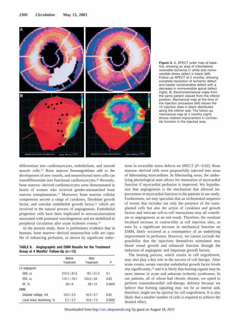
Nuclear perfusion imaging studies were similar at baselinefor the amount of total reversible defect and percent of restdefect with 50% activity (scar). Within the control group,there was no significant change in these two variables atfollow-up. Within the treatment group, there was no signifi-cant change in rest defect, with 50% activity at 2-monthfollow-up, but there was a significant 73% reduction in totalreversible defect (P�0.022; from 15.15�14.99% to4.53�10.61%). A typical example of resolution of inferolat-eral ischemia (baseline to follow-up) in a cell-treated patientis shown in Figure 3A.
Four-Month Invasive Follow-Up EvaluationsResults from LV angiography at baseline and 4-monthfollow-up are shown in Table 6. There was a sustainedimprovement in LVEF from baseline, an increase from 20%to 29% at 4 months (31% relative increase) (P�0.0003) in thetreated patients. There was also a continued reduction in ESV(P�0.03) at 4 months. EDV remained unchanged (P�0.1).Control group patients did not have repeat LV angiograms.
On EMM, segmental analysis revealed a significant me-chanical improvement of the injected segments (P�0.0005)(Table 6). Significant improvement in mechanical function atthe injection site is illustrated by EMM in Figure 3B.Unipolar voltage values did not change from baseline tofollow-up.
DiscussionThe present study describes for the first time ABMMNCtransplantation with the use of transendocardial injections inpatients with severe LV dysfunction, end-stage ischemicheart disease, and no other option for treatment. The results ofour study suggest that injection of ABMMNCs is safe andimproves perfusion and myocardial contractility when viableareas of myocardium are targeted.
Wound healing is a multifaceted process that involvescomplex interactions between inflammatory cells, cytokines,and a number of extracellular matrix proteins, and thedevelopment of new capillaries. Because the normal repara-tive mechanisms seem to be overwhelmed when clinicallysignificant myocardial injury occurs, a logical next stepwould be to amplify one part of this response artificially byapplying stem cells locally in the setting of ischemia orinfarction when a large amount of heart muscle has beeninjured.
In experimental animals, bone marrow–derived cells havebeen shown to regenerate areas of infarcted myocardium andcoronary capillaries,1 thus limiting functional impairmentafter myocardial infarction. Transendocardial injection ofABMMNCs has been shown to increase myocardial contrac-tility and perfusion in swine.4 Various cell lineages have beenused to generate evidence that bone marrow stem cells
TABLE 5. Comparison of Baseline and 2-Month Follow-UpValues for the Treatment and Control Groups
Treatment(n�14)
Control(n�7) P *
NYHA class
Before treatment 2.21�0.89 2.71�0.75
After treatment 1.14�0.36 2.71�0.76 0.0001
P 0.0003 1.0
CCSAS class
Before treatment 2.64�0.84 2.57�0.97
After treatment 1.28�0.61 2.14�0.89
P 0.0001 0.06 0.001
Ramp treadmillMETs
Before treatment 5.09�2.5 5.07�1.96
After treatment 6.68�2.35 5.16�2.45 0.078
P 0.0085 0.84
V̇O2max
Before treatment 17.96�8.78 17.75�6.85
After treatment 23.38�8.31 18.08�8.58 0.08
P 0.01 0.84
Echocardiogram
ESV, cc
Before treatment 146.78�53.46 89.42�26.23
After treatment 123.21�47.88 98.85�20.52 0.041
P 0.026 0.36
EDV, cc
Before treatment 211.35�76.89 135.71�26.08
After treatment 189.14�67.54 145�27.62 0.09
P 0.065 0.50
EF, %
Before treatment 30�5.56 36�11.73
After treatment 35.5�7.85 31.85�7.55 0.029
P 0.027 0.31
SPECT
Total reversible defect, %
Before treatment 15.15�14.99 10.71�16.60
After treatment 4.53�10.61 32.28�37.25 0.022
P 0.016 0.23
% Rest defect (50%)
Before treatment 40.77�11.13 35.85�10.09
After treatment 38.84�8.79 36.42�12.08 0.65
P 0.44 0.77
*P values reflect comparison of the differences between treatment andcontrol groups over time (see Methods).
Perin et al Transendocardial Bone Marrow Cell Transplantation 2299
by guest on August 18, 2015http://circ.ahajournals.org/Downloaded from
differentiate into cardiomyocytes, endothelium, and smoothmuscle cells.19 Bone marrow hemangioblasts add to thedevelopment of new vessels, and mesenchymal stem cells cantransdifferentiate into functional cardiomyocytes.20 Recently,bone marrow–derived cardiomyocytes were demonstrated inhearts of women who received gender-mismatched bonemarrow transplantation.21 Moreover, bone marrow cellularcomponents secrete a range of cytokines, fibroblast growthfactor, and vascular endothelial growth factor,22 which areinvolved in the natural process of angiogenesis. Endothelialprogenitor cells have been implicated in neovascularizationassociated with postnatal vasculogenesis and are mobilized toperipheral circulation after acute ischemic events.23
In the present study, there is preliminary evidence that inhumans, bone marrow–derived mononuclear cells are capa-ble of enhancing perfusion, as shown by significant reduc-
tions in reversible stress defects on SPECT (P�0.02). Bonemarrow–derived cells were purposefully injected into areasof hibernating myocardium. In hibernating areas, the under-lying physiological state allows for restoration of myocardialfunction if myocardial perfusion is improved. We hypothe-size that angiogenesis is the mechanism that allowed im-provement in myocardial function in the patients in our study.Furthermore, we may speculate that an orchestrated sequenceof events that includes not only the presence of the trans-planted cells but also the action of cytokines and growthfactors and intricate cell-to-cell interactions may all contrib-ute to angiogenesis as an end result. Therefore, the resultantlocalized increase in contractility at cell injection sites, asseen by a significant increase in mechanical function onEMM, likely occurred as a consequence of an underlyingimprovement in perfusion. However, we cannot exclude thepossibility that the injections themselves stimulated newblood vessel growth and enhanced function through theinduction of angiogenic and important growth factors.
The homing process, which results in cell engraftment,may also play a key role in the success of cell therapy. Afteracute events, serum vascular endothelial growth factor levelsrise significantly,23 and it is likely that homing signals may bemore intense in acute and subacute ischemic syndromes. Inour patients, all of whom had chronic disease, we opted toperform transendocardial cell-therapy delivery because webelieve that homing signaling may not be as intense and,therefore, might not be optimal for cell engraftment. It is alsolikely that a smaller number of cells is required to achieve thedesired effect.
Figure 3. A, SPECT polar map at base-line, showing an area of inferolateral,reversible ischemia in white and nonre-versible stress defect in black (left).Follow-up SPECT at 2 months, showingcomplete resolution of ischemic defectand basilar nonreversible defect with adecrease in nonreversible apical defect(right). B, Electromechanical maps fromthe same patient viewed from the inferiorposition. Mechanical map at the time ofthe injection procedure (left) shows the15 injection sites in black distributedalong the inferior wall. The follow-upmechanical map at 4 months (right)shows marked improvement in contrac-tile function in the injected area.
TABLE 6. Angiographic and EMM Results for the TreatmentGroup at 4 Months’ Follow-Up (n�13)
BeforeTreatment
AfterTreatment P
LV angiogram
EDV, cc 213.5�81.6 181�51.3 0.1
ESV, cc 174.1�78.7 133.5�54 0.03
EF, % 20�9 29�13 0.0003
EMM
Unipolar voltage, mV 10.5�3.5 10.3�2.7 0.65
Local linear shortening, % 5.7�3.7 10.8�7.5 0.0005
2300 Circulation May 13, 2003
by guest on August 18, 2015http://circ.ahajournals.org/Downloaded from

EMM technology has been widely confirmed to be accu-rate for delineating and identifying scarred and viable myo-cardium and for differentiating degrees of infarct transmural-ity.11,12,24 EMM thus offers a theoretical benefit over surgicalor intracoronary approaches because viability of the site canbe determined before each injection. Injections would then beperformed only to targeted, viable areas of hibernatingmyocardium. Many treated sites targeted in this study were inareas of totally occluded epicardial vascular beds, makingintracoronary delivery impossible. Furthermore, potential is-chemia provoked by coronary manipulation is avoided. Thisapproved procedure seemed safer for these chronically ill,high-risk patients because it avoided associated surgicalmorbidity and mortality.
Tse et al7 recently demonstrated improvement in myocar-dial perfusion and segmental contractility after ABMMNCtransendocardial injections. Those results are somewhat sim-ilar to results of the present study, although Tse and col-leagues did not see improvement in global EF. The maindifference between the studies is the significant baseline LVdysfunction present in our group (mean EF, 20%) as com-pared with a normal mean EF (56.9%) in the Tse study.7 Thepreliminary data of Tse and colleagues also suggest therelative safety of the procedure.
The use of transendocardial delivery proved to be safe inour study, as cellular therapy was successfully delivered inevery case without any major periprocedural events (eg,death, myocardial infarction, ventricular arrhythmias, cardiacperforation, pericardial effusion, or development of intramyo-cardial tumor). Troponin levels increased by a small butsignificant amount, consistent with delivery via intramuscularinjection (Table 4), but the absolute rise was relatively smallbiologically. The stability between levels of CRP in thetreatment and control groups suggests that we did not initiatea significant inflammatory reaction with cell injection.
The major limitations of this study are the small number ofpatients enrolled and the study design, which limits conclu-sions about efficacy. Because of ethics committee concerns,the control group was not enrolled concurrently with treatedpatients, did not receive a placebo injection, and did notundergo invasive follow-up. However, treatment and controlgroups had similar follow-up up to 2 months. The benefitsseen in this study with cell therapy could be attributable to theplacebo effect seen in phase 1 trials. Potential biases includeselection bias (eg, tertiary hospital population) and investiga-tor bias when assessing symptoms at follow-up (CCSAS andNYHA class) although echocardiographic, angiographic, andSPECT studies were read blindly. In addition, smaller LVvolumes and a trend toward higher EFs were present in thecontrol group. However, both groups were matched in termsof demographics, medication use, baseline laboratory values,functional status classification, treadmill workload, andV̇O2max. More importantly, similar baseline reversible andfixed ischemic defects were present in both groups, as one ofthe most important end points assessed in this study was theamount of reversible perfusion defect at follow-up. The endpoint of contractility is more difficult to evaluate in light ofthe differences between the groups at baseline; however,changes in opposite directions occurred at follow-up. In
addition, the slightly better LVEFs and smaller hearts shouldlogically have biased results against the cell-treated group.
Although the mechanisms by which cell therapy confersclinical benefit are not well understood, correlation betweencell phenotype subpopulation analysis and long-term clinicaloutcomes is beyond the scope of the present study. Futureanalyses will be performed in this regard when longer-termfollow-up is available.
The treatment of patients with heart failure has becomeincreasingly important given the growing number of casesand their economic impact on the healthcare system.25,26
More aggressive and widespread therapy in patients withchronic, ischemic heart failure will ultimately lead to apopulation harboring more advanced disease with a potentialyearly mortality rate as high as 50%.27 For these patients,therapeutic options remain limited. The very high-risk natureof the patient population represented in our study cohort isunderscored by the fact that there was a death in both thecontrol and the treatment groups. However, the significantimprovement in LVEF noted in the treatment group onangiographic follow-up at 4 months (from 20% to 29%) mayimply an improved clinical state and, it is hoped, providesome reduction in risks for the future.28
ConclusionIn this initial prospective, nonrandomized, open-label studyin no-other-option coronary artery disease patients with LVdysfunction, we noted improvement in symptoms, cardiacfunction, and perfusion with transendocardial ABMMNCtherapy, without any clinical evidence of significant harmfrom the procedure itself. We believe there may be clinicalpotential for this relatively novel therapy. Further investiga-tion in a larger, randomized trial is warranted.
AcknowledgmentsA NOGA mapping system and catheters were provided by CordisCorporation (Miami Lakes, Fla).
We thank Rita Weiler, Ana Cristina Reis, RN, and Patricia Souza,RN, for their enthusiastic support and coordination of the studypatients; Cristine Rutherford for the psychological assessment andsupport of the patients; David R. Buskohl, Mark Martin, andJacqueline Grant for their outstanding technical support; and Mari-anne Mallia, ELS, for editorial assistance in the preparation of themanuscript.
References1. Orlic D, Kajstura J, Chimenti S, et al. Bone marrow cells regenerate
infarcted myocardium. Nature. 2001;410:701–705.2. Kocher AA, Schuster MD, Szabolcs MJ, et al. Neovascularization of
ischemic myocardium by human bone-marrow– derived angioblastsprevents cardiomyocyte apoptosis, reduces remodeling and improvescardiac function. Nat Med. 2001;7:430–436.
3. Kawamoto A, Gwon HC, Iwaguro H, et al. Therapeutic potential of exvivo expanded endothelial progenitor cells for myocardial ischemia. Cir-culation. 2001;103:634–637.
4. Fuchs S, Baffour R, Zhou YF, et al. Transendocardial delivery ofautologous bone marrow enhances collateral perfusion and regionalfunction in pigs with chronic experimental myocardial ischemia. J AmColl Cardiol. 2001;37:1726–1732.
5. Strauer BE, Brehm M, Zeus T, et al. Repair of infarcted myocardium byautologous intracoronary mononuclear bone marrow cell transplantationin humans. Circulation. 2002;106:1913–1918.
Perin et al Transendocardial Bone Marrow Cell Transplantation 2301
by guest on August 18, 2015http://circ.ahajournals.org/Downloaded from

6. Assmus B, Schächinger V, Teupe C, et al. Transplantation of progenitorcells and regeneration enhancement in acute myocardial infarction(TOPCARE-AMI). Circulation. 2002;106:3009–3017.
7. Tse HF, Kwong YL, Chan JKF, et al. Angiogenesis in ischaemic myo-cardium by intramyocardial autologous bone marrow mononuclear cellimplantation. Lancet. 2003;361:47–49.
8. Gibbons RJ, Balady GJ, Bricker TJ, et al. ACC/AHA 2002 guidelineupdate for exercise testing: summary article. A report of the AmericanCollege of Cardiology/American Heart Association Task Force onPractice Guidelines (Committee to Update the 1997 Exercise TestingGuidelines). J Am Coll Cardiol. 2002;40:1531–1540.
9. Coutinho LH, Gilleece MH, de Wynter EA, et al. Clonal and long-termcultures using human bone marrow. In: Testa NG, Molineux G, eds.Haemopoiesis: A Practical Approach. New York, NY: Oxford UniversityPress, 1993:84–85.
10. Castro-Malaspina H, Gay RE, Resnick G, et al. Characterization ofhuman bone marrow fibroblast colony-forming cells (CFU-F) and theirprogeny. Blood. 1980;56:289–301.
11. Perin EC, Silva GV, Sarmento-Leite R, et al. Assessing myocardialviability and infarct transmurality with left ventricular electromechanicalmapping in patients with stable coronary artery disease: validation bydelayed-enhancement magnetic resonance imaging. Circulation. 2002;106:957–961.
12. Perin EC, Silva GV, Leite RS. Left ventricular electromechanicalmapping as a diagnostic method. In: Abela GS, ed. Myocardial Revas-cularization: Novel Percutaneous Approaches. New York, NY:Wiley-Liss; 2001:183–195.
13. Kaminsky LA, Whaley MH. Evaluation of a new standardized rampprotocol: the BSU/Bruce Ramp protocol. J Cardiopulm Rehabil. 1998;18:438–444.
14. American College of Sport Medicine. Guidelines for Exercise Testing andExercise Prescription. 6th ed. Philadelphia, Pa: Lippincott Williams &Wilkins; 2000.
15. Garcia EV, Cooke CD, Van Train KF, et al. Technical aspects of myo-cardial SPECT imaging with technetium-99m sestamibi. Am J Cardiol.1990;66:23E–31E.
16. Van Train K, Areeda J, Garcia EV, et al. Quantitative same-day rest stresstechnetium-99 m sestamibi SPECT: definition and validation of stressnormal limits and criteria for abnormality. J Nucl Med. 1993;34:1494–1502.
17. Van Train K, Garcia EV, Maddahi J, et al. Multicenter trial validation forquantitative analysis of same-day rest-stress technetium-99m sestamibimyocardial tomograms. J Nucl Med. 1994;35:609–618.
18. Dodge HT, Sandler H, Ballew DW, et al. The use of biplane angiographyfor the measurement of left ventricular volume in man. Eur Heart J.1960;60:762–776.
19. Toma C, Pittenger MF, Cahill KS, et al. Human mesenchymal stem cellsdifferentiate to a cardiomyocyte phenotype in the adult murine heart.Circulation. 2002;105:93–98.
20. Asahara T, Murohara T, Sullivan A, et al. Isolation of putative progenitorendothelial cells for angiogenesis. Science. 1997;275:964–967.
21. Badorff C, Brandes RP, Popp R, et al. Transdifferentiation of blood-derived human adult endothelial progenitor cells into functionally activecardiomyocytes. Circulation. 2003;107:1024–1032.
22. Bikfalvi A, Han ZC. Angiogenic factors are hematopoietic factors andvice versa. Leukemia. 1994;8:523–529.
23. Shintani S, Murohara T, Ikeda H, et al. Mobilization of endothelialprogenitor cells in patients with acute myocardial infarction. Circulation.2001;103:2776–2779.
24. Wolf T, Gepstein L, Dror V, et al. Detailed electromechanical mappingaccurately predicts the transmural extent of myocardial infarction. J AmColl Cardiol. 2001;37:1590–1597.
25. American Heart Association. 2000 Heart and Stroke Statistical Update.Dallas, Tex: American Heart Association; 2001.
26. O’Connell JB, Birstow MR. Economic impact of heart failure in theUnited States: time for a different approach. J Heart Lung Transplant.1994;13:S107–S112.
27. Califf RM, Adams KF, McKenna WJ, et al. A randomized controlled trialof epoprostenol therapy for severe congestive heart failure: the FlolanInternational Randomized Trial (FIRST). Am Heart J. 1997;134:44–54.
28. Marantz PR, Tobin JN, Wassertheil-Smoller S, et al. Prognosis in ische-mic heart disease. Can you tell as much at the bedside as in the nuclearlaboratory? Arch Intern Med. 1992;152:2433–2437.
2302 Circulation May 13, 2003
by guest on August 18, 2015http://circ.ahajournals.org/Downloaded from

and James T. WillersonRoberto Esporcatte, Yong J. Geng, William K. Vaughn, Joao A.R. Assad, Evandro T. Mesquita
Dohmann, Guilherme V. Silva, Luciano Belém, Ricardo Vivacqua, Fernando O.D. Rangel,Sousa, Claudio T. Mesquita, Maria I.D. Rossi, Antonio C. Carvalho, Helio S. Dutra, Hans J.F.
Emerson C. Perin, Hans F.R. Dohmann, Radovan Borojevic, Suzana A. Silva, Andre L.S.Ischemic Heart Failure
Transendocardial, Autologous Bone Marrow Cell Transplantation for Severe, Chronic
Print ISSN: 0009-7322. Online ISSN: 1524-4539 Copyright © 2003 American Heart Association, Inc. All rights reserved.
is published by the American Heart Association, 7272 Greenville Avenue, Dallas, TX 75231Circulation doi: 10.1161/01.CIR.0000070596.30552.8B
2003;107:2294-2302; originally published online April 21, 2003;Circulation.
http://circ.ahajournals.org/content/107/18/2294World Wide Web at:
The online version of this article, along with updated information and services, is located on the
http://circ.ahajournals.org//subscriptions/
is online at: Circulation Information about subscribing to Subscriptions:
http://www.lww.com/reprints Information about reprints can be found online at: Reprints:
document. Permissions and Rights Question and Answer this process is available in the
click Request Permissions in the middle column of the Web page under Services. Further information aboutOffice. Once the online version of the published article for which permission is being requested is located,
can be obtained via RightsLink, a service of the Copyright Clearance Center, not the EditorialCirculationin Requests for permissions to reproduce figures, tables, or portions of articles originally publishedPermissions:
by guest on August 18, 2015http://circ.ahajournals.org/Downloaded from