Intracoronary infusion of autologous mononuclear cells from bone marrow or granulocyte...
20
Intracoronary infusion of autologous mononuclear cells from bone marrow or G-CSF mobilised apheresis product may not improve remodelling, contractile function, perfusion or infarct size in a swine model of large myocardial infarction Ranil de Silva, MRCP PhD, Amish N. Raval, MD, Mohiuddin Hadi, MD † , Karena M. Gildea, BS, Aylin C. Bonifacino, MS * , Zu-Xi Yu, MD PhD ¶ , Yu Ying Yau, RN ϕ , Susan F. Leitman, MD ϕ , Stephen L. Bacharach, PhD † , Robert E. Donahue, VMD * , Elizabeth J. Read, MD ϕ , and Robert J. Lederman, MD *Cardiovascular Branch, Haematology Branch, National Institutes of Health, Bethesda, Maryland, USA ¶Pathology Core, National Institutes of Health, Bethesda, Maryland, USA ϕDivision of Intramural Research, National Heart Lung and Blood Institute; Department of Transfusion Medicine, National Institutes of Health, Bethesda, Maryland, USA †Department of Nuclear Medicine, Clinical Center. National Institutes of Health, Bethesda, Maryland, USA Abstract Background—In a blinded, placebo controlled study, we investigated whether intracoronary infusion of autologous mononuclear cells from G-CSF mobilised apheresis product or bone marrow (BM) improved sensitive outcome measures in a swine model of large MI. Methods and Results—Four days after LAD occlusion and reperfusion, cells from BM or apheresis product of saline (Placebo) or G-CSF injected animals were infused into the LAD. Large infarcts were created: baseline ejection fraction (EF) by MRI of 35.3 ± 8.5%, no difference between the Placebo, G-CSF and BM groups (p=0.16 by ANOVA). At 6 weeks EF fell to a similar degree in the Placebo, G-CSF and BM groups (−7.9±6.0%, −8.5±8.8% and −10.9±7.6%, p=0.78 by ANOVA). Left ventricular volumes and infarct size by MRI deteriorated similarly in all 3 groups. Quantitative PET demonstrated significant decline in FDG uptake rate in the LAD territory at follow-up, with no histological, angiographic or PET perfusion evidence of functional neovascularisation. Immunofluorescence failed to demonstrate transdifferentiation of infused cells. Conclusion—Intracoronary infusion of mononuclear cells from either bone marrow or G-CSF mobilised apheresis product may not improve or limit deterioration in systolic function, adverse ventricular remodelling, infarct size or perfusion in a swine model of large MI. Keywords Angiogenesis; imaging; myocardial infarction; myogenesis; stem cell Correspondence to: Ranil de Silva MRCP PhD, NHLI, Imperial College London, Royal Brompton and Harefield NHS Trust, Sydney St, London SW3 6NP, UK Email: [email protected], Telephone: +44 20 73518626 Fax: +44 20 73518629. Conflicts of Interest Disclosures None. NIH Public Access Author Manuscript Eur Heart J. Author manuscript; available in PMC 2009 January 1. Published in final edited form as: Eur Heart J. 2008 July ; 29(14): 1772–1782. doi:10.1093/eurheartj/ehn216. NIH-PA Author Manuscript NIH-PA Author Manuscript NIH-PA Author Manuscript
-
Upload
independent -
Category
Documents
-
view
0 -
download
0
Transcript of Intracoronary infusion of autologous mononuclear cells from bone marrow or granulocyte...

Intracoronary infusion of autologous mononuclear cells frombone marrow or G-CSF mobilised apheresis product may notimprove remodelling, contractile function, perfusion or infarctsize in a swine model of large myocardial infarction
Ranil de Silva, MRCP PhD, Amish N. Raval, MD, Mohiuddin Hadi, MD†, Karena M. Gildea,BS, Aylin C. Bonifacino, MS*, Zu-Xi Yu, MD PhD¶, Yu Ying Yau, RNϕ, Susan F. Leitman,MDϕ, Stephen L. Bacharach, PhD†, Robert E. Donahue, VMD*, Elizabeth J. Read, MDϕ, andRobert J. Lederman, MD*Cardiovascular Branch, Haematology Branch, National Institutes of Health, Bethesda, Maryland, USA
¶Pathology Core, National Institutes of Health, Bethesda, Maryland, USA
ϕDivision of Intramural Research, National Heart Lung and Blood Institute; Department of TransfusionMedicine, National Institutes of Health, Bethesda, Maryland, USA
†Department of Nuclear Medicine, Clinical Center. National Institutes of Health, Bethesda, Maryland, USA
AbstractBackground—In a blinded, placebo controlled study, we investigated whether intracoronaryinfusion of autologous mononuclear cells from G-CSF mobilised apheresis product or bone marrow(BM) improved sensitive outcome measures in a swine model of large MI.
Methods and Results—Four days after LAD occlusion and reperfusion, cells from BM orapheresis product of saline (Placebo) or G-CSF injected animals were infused into the LAD. Largeinfarcts were created: baseline ejection fraction (EF) by MRI of 35.3 ± 8.5%, no difference betweenthe Placebo, G-CSF and BM groups (p=0.16 by ANOVA). At 6 weeks EF fell to a similar degree inthe Placebo, G-CSF and BM groups (−7.9±6.0%, −8.5±8.8% and −10.9±7.6%, p=0.78 by ANOVA).Left ventricular volumes and infarct size by MRI deteriorated similarly in all 3 groups. QuantitativePET demonstrated significant decline in FDG uptake rate in the LAD territory at follow-up, with nohistological, angiographic or PET perfusion evidence of functional neovascularisation.Immunofluorescence failed to demonstrate transdifferentiation of infused cells.
Conclusion—Intracoronary infusion of mononuclear cells from either bone marrow or G-CSFmobilised apheresis product may not improve or limit deterioration in systolic function, adverseventricular remodelling, infarct size or perfusion in a swine model of large MI.
KeywordsAngiogenesis; imaging; myocardial infarction; myogenesis; stem cell
Correspondence to: Ranil de Silva MRCP PhD, NHLI, Imperial College London, Royal Brompton and Harefield NHS Trust, Sydney St,London SW3 6NP, UK Email: [email protected], Telephone: +44 20 73518626 Fax: +44 20 73518629.Conflicts of Interest DisclosuresNone.
NIH Public AccessAuthor ManuscriptEur Heart J. Author manuscript; available in PMC 2009 January 1.
Published in final edited form as:Eur Heart J. 2008 July ; 29(14): 1772–1782. doi:10.1093/eurheartj/ehn216.
NIH
-PA Author Manuscript
NIH
-PA Author Manuscript
NIH
-PA Author Manuscript

IntroductionPreclinical investigations of bone marrow derived progenitor cell therapy in large animalmodels of acute myocardial infarction, with prognostically significant reductions of leftventricular function, have not been reported. This would appear valuable given the increasingclinical interest in cardiac regenerative strategies for such patients1. We therefore performeda blinded placebo controlled study of intracoronary infusion of autologous mononuclear cellsderived from either cytokine mobilised apheresis product or bone marrow in a swine model oflarge myocardial infarction (MI).
MethodsAnimal Procedures
Animal procedures were approved by the NHLBI Animal Care and Use Committee. Theexperimental protocol is summarised in Figure 1. Twenty-four Yucatan miniswine (wt 37–52kg) underwent 90 minute balloon occlusion of the left anterior descending (LAD) artery asdescribed previously2.
In a blinded fashion, animals were alternately assigned to receive no injections or dailysubcutaneous injections of either human recombinant granulocyte colony stimulating factor(G-CSF, n=8, 10 μg/kg, Neupogen, Amgen) or placebo (n=7, equal volume of normal saline)for five days. The first injection was administered after LAD occlusion and daily thereafter.All injected animals underwent apheresis. Animals receiving no injections underwent bonemarrow aspiration (BM, n=7) without apheresis. Animals in the placebo group formed thecellular control against which the G-CSF and BM groups were compared.
Apheresis and Bone Marrow AspirationApheresis was performed using citrate anticoagulant in a 1:13 ratio to the whole blood flowrate. Aseptic bone marrow aspiration from both iliac crests was performed under generalanaesthesia.
Cell PreparationImmediately after apheresis or bone marrow aspiration, the cellular product was prepared forinfusion. The product was incubated with ammonium chloride (ACK, BioWhittaker), washed,labelled with CM-DiI (Molecular Probes) according to manufacturer’s instructions, andresuspended in plasmalyte A/2% human serum albumin which was heparinised (30u/mL).
Colony Forming AssaysHaematopoietic colony forming assays were performed as an index of progenitor cell activity(see Supplementary Methods). The combined number of myeloid (CFU G/M) and erythroid(BFU E) colonies counted 2 weeks after plating determined the total number of colony formingunits (CFU). The number of colony forming cells (CFC) was calculated assuming one CFUper CFC. All procedures were performed by investigators blinded to treatment allocation andimaging results.
Intracoronary Cell InfusionIntracoronary cell infusion was performed into the LAD as reported previously3, byinvestigators blinded to the identity of the cell product, followed by coronary angiography toconfirm vessel patency.
de Silva et al. Page 2
Eur Heart J. Author manuscript; available in PMC 2009 January 1.
NIH
-PA Author Manuscript
NIH
-PA Author Manuscript
NIH
-PA Author Manuscript

PETDynamic PET was performed using a GE Advance scanner (GE Medical Systems). Myocardialblood flow was measured at rest and during adenosine infusion (140 μg/kg/min) using a bolusadministration of 50 mCi of oxygen-15 labeled water (H2
15O) 4. Myocardial FDG uptake wasmeasured after intravenous fluorine-18 fluorodeoxyglucose (FDG, 15 mCi) under anhyperinsulinaemic euglycaemic glucose clamp 5.
MRIMRI was performed at 1.5 T (Sonata, Siemens Medical Systems) using an 8 channel phasedarray surface coil (Nova Medical Inc). Breath-held, ECG-gated cine steady state free precession(SSFP) MRI was acquired (TR/TE, 3.6/1.8 ms; flip angle, 65°; field of view (FOV), 300 × 244mm; matrix, 256 × 127 pixels, slice thickness, 8 mm, bandwidth 1085 Hz/pixel). Delayedenhancement (DE-MRI) images were acquired after intravenous Gd-DTPA (0.2 mmol/kg)using a phase sensitive inversion recovery sequence (TR/TE, 11/4.45 ms; flip angle, 30°;inversion-time, 300 ms; FOV, 350 × 241 mm; matrix, 256 × 141 pixels; slice-thickness, 8 mm,bandwidth 140 Hz/pixel).
Follow-upAt six weeks, animals returned for cardiac catheterisation, MRI and PET after which they wereeuthanised. Tissues were prepared for histology (Data Supplement).
Data AnalysisAll analyses were performed by investigators blinded to treatment allocation after 6 animalsin each group had completed the protocol.
MRI—All images were analysed on a Leonardo workstation (Siemens). Ejection fraction (EF),end-systolic volume (ESV), end-diastolic volume (EDV), and stroke volume (SV) werecalculated. Regional wall thickening was determined using a 16-sector model. Sectors werecategorised as either hypokinetic (wall thickening > 0 mm and <1.5 mm) or akinetic (wallthickening ≤ 0 mm). Infarct volumes were quantified from DE-MRI images using a previouslyvalidated tool which automatically windows, levels, and contours images of infarctedregions6.
PET—PET and MRI images were registered (Data Supplement). Manually segmented infarct,peri-infarct and remote myocardial regions on MR images were transformed on to PET imagesfor kinetic analysis. Myocardial blood flow values in the infarct, peri-infarct and remote regionswere calculated from the dynamic H2
15O data4. The ratio of myocardial blood flow duringadenosine infusion to resting myocardial blood flow was defined as the adenosine vasodilatorresponse. Rate constants for net myocardial FDG uptake (Ki) were calculated using Patlakanalysis. Parametric Ki images were generated on a pixel by pixel basis in each animal7. Partialvolume correction of the FDG data was based on the H2
15O data for corresponding regions.
StatisticsThe primary endpoint was change in EF from baseline to 6 weeks. Secondary endpointsincluded EF, EDV, ESV, SV, and infarct volume at 6 weeks. Data were assessed for normalityusing the Kolmogorov-Smirnov method. Continuous parameters are shown as mean ± SD.Data were analysed using one way ANOVA with specific intergroup comparisons made usingunpaired Student t-tests with Bonferroni correction for multiple comparisons. p<0.05 wasconsidered significant.
de Silva et al. Page 3
Eur Heart J. Author manuscript; available in PMC 2009 January 1.
NIH
-PA Author Manuscript
NIH
-PA Author Manuscript
NIH
-PA Author Manuscript

ResultsSix of 24 animals failed to complete or were excluded from the protocol due to death beforeintracoronary cell infusion (n=4), severe biventricular failure due to extensive left and rightventricular infarction 7 days after cell delivery (n=1, G-CSF), or baseline MRI scanner failure(n=1, BM). Data are presented from 18 animals. G-CSF increased total leukocyte count andCFC concentration in peripheral blood (See Data Supplement). The composition of infusedcells is summarised in Table 1. Trypan blue viability for all cellular products was > 90%. Theweight-adjusted dose of colony forming cells administered by intracoronary infusion wassimilar in the G-CSF and BM groups were similar (8.04 ± 4.13 × 10³ v 7.27 ± 3.38 × 10³ colonyforming cells/kg) (Table 1).
Apheresis was tolerated in all animals despite recent large myocardial infarctions.Intracoronary cell infusion was performed successfully and without complication in 18/19animals. In one G-CSF treated animal, receiving the highest infused dose of total nucleatedcells (1.57 ×10⁹ cells/kg), no-reflow occurred.
At 6 weeks, there was no haemodynamic improvement in any group. Cardiac output wasmaintained by increased heart rate, not improved stroke volume (Data Supplement). Coronaryangiography revealed no collateralisation of the LAD territory.
Large infarcts were created as evidenced by baseline EF of 34.3±9.7%, 31.2±7.8%, and 40.4±6.0% in the Placebo (n=6), G-CSF (n=6) and BM (n=6) groups respectively (p=0.16 byANOVA) (Table 1). EF, ESV and EDV deteriorated in all groups at 6 weeks. There was nosignificant difference in any parameter between treatment groups (Figure 2). At 6 weeks, thenumber of sectors with a wall motion abnormality increased to a similar degree in all 3 groups(Figure 3), with an increasing proportion of akinetic segments (Table 3). After completingblinded analyses, assuming the null hypothesis could be rejected, we estimated that n=134 pigsper group would be required to achieve 80% power to detect a significant difference (p<0.05)in change in EF from baseline to 6 weeks follow-up. The study was therefore terminated ongrounds of futility.
Transmural delayed enhancement was evident in the antero-septal wall in all animals atbaseline (Figure 4). At 6 weeks, transmural delayed enhancement persisted consistent withnonviable myocardium. These regions underwent significant thinning and expansion, andaccordingly, the mass of tissue showing delayed enhancement at follow-up was reduced(Figure 4).
Quantitative myocardial FDG uptake (Ki) images before intracoronary cell infusiondemonstrated preserved or enhanced FDG uptake in sectors showing transmural delayedenhancement (Figure 5). At follow-up, these regions thinned, and exhibited markedly reducedFDG uptake despite partial volume correction, suggesting non-viable infarcted myocardium(Figure 5 and 6). Ki calculated on remote, infarct and peri-infarct regions reflected a similartrend.
Adenosine vasodilator response, an index of coronary microcirculatory function, was measuredin the infarcted and peri-infarct myocardium at baseline and at six weeks follow-up and wasnot different in any of the 3 groups in the peri-infarct regions (Figure 7).
Haematoxylin and Eosin and Masson-Trichrome stained sections from the border and infarctzones of the hearts in all 3 groups demonstrated equivalent degrees of myocyte hypertrophy,myocyte degeneration, oedema, fibrosis, inflammatory cell infiltrate, hemorrhage, and vasculardensity. These findings are inconsistent with myocyte regeneration or neovascularisation inany group.
de Silva et al. Page 4
Eur Heart J. Author manuscript; available in PMC 2009 January 1.
NIH
-PA Author Manuscript
NIH
-PA Author Manuscript
NIH
-PA Author Manuscript
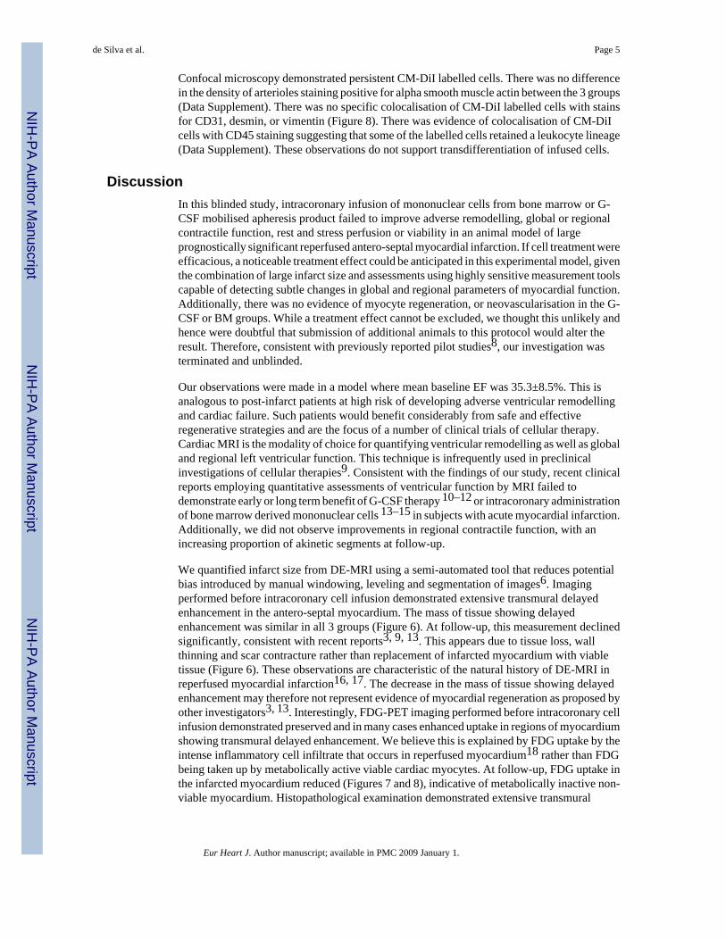
Confocal microscopy demonstrated persistent CM-DiI labelled cells. There was no differencein the density of arterioles staining positive for alpha smooth muscle actin between the 3 groups(Data Supplement). There was no specific colocalisation of CM-DiI labelled cells with stainsfor CD31, desmin, or vimentin (Figure 8). There was evidence of colocalisation of CM-DiIcells with CD45 staining suggesting that some of the labelled cells retained a leukocyte lineage(Data Supplement). These observations do not support transdifferentiation of infused cells.
DiscussionIn this blinded study, intracoronary infusion of mononuclear cells from bone marrow or G-CSF mobilised apheresis product failed to improve adverse remodelling, global or regionalcontractile function, rest and stress perfusion or viability in an animal model of largeprognostically significant reperfused antero-septal myocardial infarction. If cell treatment wereefficacious, a noticeable treatment effect could be anticipated in this experimental model, giventhe combination of large infarct size and assessments using highly sensitive measurement toolscapable of detecting subtle changes in global and regional parameters of myocardial function.Additionally, there was no evidence of myocyte regeneration, or neovascularisation in the G-CSF or BM groups. While a treatment effect cannot be excluded, we thought this unlikely andhence were doubtful that submission of additional animals to this protocol would alter theresult. Therefore, consistent with previously reported pilot studies8, our investigation wasterminated and unblinded.
Our observations were made in a model where mean baseline EF was 35.3±8.5%. This isanalogous to post-infarct patients at high risk of developing adverse ventricular remodellingand cardiac failure. Such patients would benefit considerably from safe and effectiveregenerative strategies and are the focus of a number of clinical trials of cellular therapy.Cardiac MRI is the modality of choice for quantifying ventricular remodelling as well as globaland regional left ventricular function. This technique is infrequently used in preclinicalinvestigations of cellular therapies9. Consistent with the findings of our study, recent clinicalreports employing quantitative assessments of ventricular function by MRI failed todemonstrate early or long term benefit of G-CSF therapy 10–12 or intracoronary administrationof bone marrow derived mononuclear cells 13–15 in subjects with acute myocardial infarction.Additionally, we did not observe improvements in regional contractile function, with anincreasing proportion of akinetic segments at follow-up.
We quantified infarct size from DE-MRI using a semi-automated tool that reduces potentialbias introduced by manual windowing, leveling and segmentation of images6. Imagingperformed before intracoronary cell infusion demonstrated extensive transmural delayedenhancement in the antero-septal myocardium. The mass of tissue showing delayedenhancement was similar in all 3 groups (Figure 6). At follow-up, this measurement declinedsignificantly, consistent with recent reports3, 9, 13. This appears due to tissue loss, wallthinning and scar contracture rather than replacement of infarcted myocardium with viabletissue (Figure 6). These observations are characteristic of the natural history of DE-MRI inreperfused myocardial infarction16, 17. The decrease in the mass of tissue showing delayedenhancement may therefore not represent evidence of myocardial regeneration as proposed byother investigators3, 13. Interestingly, FDG-PET imaging performed before intracoronary cellinfusion demonstrated preserved and in many cases enhanced uptake in regions of myocardiumshowing transmural delayed enhancement. We believe this is explained by FDG uptake by theintense inflammatory cell infiltrate that occurs in reperfused myocardium18 rather than FDGbeing taken up by metabolically active viable cardiac myocytes. At follow-up, FDG uptake inthe infarcted myocardium reduced (Figures 7 and 8), indicative of metabolically inactive non-viable myocardium. Histopathological examination demonstrated extensive transmural
de Silva et al. Page 5
Eur Heart J. Author manuscript; available in PMC 2009 January 1.
NIH
-PA Author Manuscript
NIH
-PA Author Manuscript
NIH
-PA Author Manuscript

infarction with no evidence of transdifferentiation (Figure 8), cardiac regeneration orneovascularisation (Data Supplement).
Certain haematopoietic progenitor cell populations may be angiogenic19. Histologicalassessment of the density of vascular structures in peri-infarct and infarcted regions was similarin all three groups (Data Supplement). Furthermore, the number of arterioles staining positivefor anti-smooth muscle actin was similar in all 3 groups (Data Supplement). Adenosinevasodilator response measured quantitatively in the peri-infarct regions with H2
15O-PETshowed no improvement and was similar in all 3 groups (Figure 7). Selective coronaryangiography failed to demonstrate collateral growth. These data do not support functionalneovascularisation in this model.
Clinical studies have reported few immediate procedural complications from infusion ofmononuclear cells into infarct related coronary arteries. The total nucleated cell dose in thesestudies has progressively increased from 9×10⁶ to 24.6± 8.4×10⁸ 3, 13, 15, 20, 21. In oneanimal, no-reflow was observed at the highest infused total nucleated cell dose. Pending resultsfrom further dose escalation studies, we would caution against exceeding this intracoronarycell dose in clinical studies.
Study LimitationsWe recognise the limitations of simulating human clinical disease using animal models. Unlikemany clinical observations where EF improves after reperfusion in myocardial infarction22,we observed a consistent decrease in EF in all groups. This has been observed in clinical studies23 and may be due to the extensive myocardial infarctions created in this study24. The smallnumber of animals reflects the pilot nature of this protocol and is consistent with sample sizesin previously published preclinical investigations using large animals25, 26. We estimated that~400 animals should be studied to detect a significant difference (beneficial or otherwise)between the treatment groups. While the sample size in this study is small, the measurementtechniques were highly sensitive and reproducible, and performed in a model of large infarctionin which any benefits should have been easily discerned. Our results in part may be explainedby the use of an unselected cell product. Currently, clinically relevant progenitor cellpopulations cannot be selected in swine due to lack of appropriate antibodies. Furthermore,intramyocardial rather than intracoronary cell delivery may be more efficacious in this modelsystem. The follow-up timepoint in the current study was chosen as previous preclinicalinvestigations have demonstrated functional improvements by this time which plateauthereafter 9, 25–28. A cellular control (mononuclear cells from non-mobilised apheresisproduct) was chosen to maintain study blind and help determine if the test cell populationswould exert any effect over and above simple interstitial reinforcement of the infarctregion29. We cannot exclude the possibility that all 3 cell populations administered in thecurrent study conferred improvement relative to an acellular control, though this seems unlikelyas previous studies have not shown benefit over intracoronary administration of medium onejection fraction in reperfused myocardial infarction9.
ConclusionsIntracoronary infusion of mononuclear cells from either bone marrow or G-CSF mobilisedapheresis product may not improve or limit deterioration of left ventricular systolic function,rest or stress perfusion and infarct size in a swine model of large prognostically significantreperfused antero-septal myocardial infarction. These cell populations may not necessarilyconfer benefit to analogous patient populations. Studies in animal models of large reperfusedmyocardial infarction which employ local delivery systems and endpoint assessmentsequivalent to those used in contemporary clinical studies may represent an essential step in
de Silva et al. Page 6
Eur Heart J. Author manuscript; available in PMC 2009 January 1.
NIH
-PA Author Manuscript
NIH
-PA Author Manuscript
NIH
-PA Author Manuscript

selecting cellular products that ultimately demonstrate sustained therapeutic benefit in clinicaltrials30.
Supplementary MaterialRefer to Web version on PubMed Central for supplementary material.
AcknowledgementsWe thank Kathryn Hope, Katherine Lucas and Joni Taylor for their expert animal care; William Schenke and VictorWright for technical assistance; Cengizhan Ozturk and Ludovic Le Meunier for assistance with MRI and PET imaging;Peter Kellman for the phase sensitive inversion recovery sequence; and Li-Yueh Hsu for infarct volume analysissoftware. We are grateful to Dawson Smith of Gambro BCT for providing the Cobe Spectra system. Amgen providedNeupogen (recombinant human G-CSF) under a materials collaborative research and development agreement withNIH. Finally we pay tribute to and posthumously thank Charles Carter for his invaluable contribution without whichthis study would not have been possible.
Funding Sources
This work was supported by NIH Z01-HL005062-04 (RJL).
References1. Penn MS. Stem-cell therapy after acute myocardial infarction: the focus should be on those at risk.
Lancet 2006;367(9505):87–88. [PubMed: 16413856]2. de Silva R, Gutierrez LF, Raval AN, McVeigh ER, Ozturk C, Lederman RJ. X-ray fused with magnetic
resonance imaging (XFM) to target endomyocardial injections: validation in a swine model ofmyocardial infarction. Circulation 2006;114(22):2342–2350. [PubMed: 17101858]
3. Wollert KC, Meyer GP, Lotz J, Ringes-Lichtenberg S, Lippolt P, Breidenbach C, Fichtner S, Korte T,Hornig B, Messinger D, Arseniev L, Hertenstein B, Ganser A, Drexler H. Intracoronary autologousbone-marrow cell transfer after myocardial infarction: the BOOST randomised controlled clinical trial.Lancet 2004;364(9429):141–148. [PubMed: 15246726]
4. Bonow RO, Dilsizian V, Cuocolo A, Bacharach SL. Identification of viable myocardium in patientswith chronic coronary artery disease and left ventricular dysfunction. Comparison of thalliumscintigraphy with reinjection and PET imaging with 18F-fluorodeoxyglucose. Circulation 1991;83(1):26–37. [PubMed: 1984883]
5. Chareonthaitawee P, Schaefers K, Baker CS, Turkheimer F, Stegger L, Banner NR, Yacoub M, BonserRS, Iozzo P, Camici PG, Rimoldi O. Assessment of infarct size by positron emission tomography and[18F]2-fluoro-2-deoxy-D-glucose: a new absolute threshold technique. Eur J Nucl Med Mol Imaging2002;29(2):203–215. [PubMed: 11930884]
6. Hsu LY, Natanzon A, Kellman P, Hirsch GA, Aletras AH, Arai AE. Quantitative myocardial infarctionon delayed enhancement MRI. Part I: Animal validation of an automated feature analysis and combinedthresholding infarct sizing algorithm. J Magn Reson Imaging 2006;23(3):298–308. [PubMed:16450367]
7. Choi Y, Hawkins RA, Huang SC, Gambhir SS, Brunken RC, Phelps ME, Schelbert HR. Parametricimages of myocardial metabolic rate of glucose generated from dynamic cardiac PET and 2-[18F]fluoro-2-deoxy-d-glucose studies. J Nucl Med 1991;32(4):733–738. [PubMed: 2013815]
8. Penicka M, Horak J, Kobylka P, Pytlik R, Kozak T, Belohlavek O, Lang O, Skalicka H, Simek S,Palecek T, Linhart A, Aschermann M, Widimsky P. Intracoronary injection of autologous bonemarrow-derived mononuclear cells in patients with large anterior acute myocardial infarction: aprematurely terminated randomized study. J Am Coll Cardiol 2007;49(24):2373–2374. [PubMed:17572255]
9. Moelker AD, Baks T, van den Bos EJ, van Geuns RJ, de Feyter PJ, Duncker DJ, van der Giessen WJ.Reduction in infarct size, but no functional improvement after bone marrow cell administration in aporcine model of reperfused myocardial infarction. Eur Heart J 2006;27(24):3057–3064. [PubMed:17135284]
de Silva et al. Page 7
Eur Heart J. Author manuscript; available in PMC 2009 January 1.
NIH
-PA Author Manuscript
NIH
-PA Author Manuscript
NIH
-PA Author Manuscript
10. Ripa RS, Haack-Sorensen M, Wang Y, Jorgensen E, Mortensen S, Bindslev L, Friis T, Kastrup J.Bone marrow derived mesenchymal cell mobilization by granulocyte-colony stimulating factor afteracute myocardial infarction: results from the Stem Cells in Myocardial Infarction (STEMMI) trial.Circulation 2007;116(11 Suppl):I24–30. [PubMed: 17846310]
11. Ripa RS, Jorgensen E, Wang Y, Thune JJ, Nilsson JC, Sondergaard L, Johnsen HE, Kober L, GrandeP, Kastrup J. Stem cell mobilization induced by subcutaneous granulocyte-colony stimulating factorto improve cardiac regeneration after acute ST-elevation myocardial infarction: result of the double-blind, randomized, placebo-controlled stem cells in myocardial infarction (STEMMI) trial.Circulation 2006;113(16):1983–1992. [PubMed: 16531621]
12. Zohlnhofer D, Ott I, Mehilli J, Schomig K, Michalk F, Ibrahim T, Meisetschlager G, von Wedel J,Bollwein H, Seyfarth M, Dirschinger J, Schmitt C, Schwaiger M, Kastrati A, Schomig A. Stem cellmobilization by granulocyte colony-stimulating factor in patients with acute myocardial infarction:a randomized controlled trial. Jama 2006;295(9):1003–1010. [PubMed: 16507801]
13. Janssens S, Dubois C, Bogaert J, Theunissen K, Deroose C, Desmet W, Kalantzi M, Herbots L,Sinnaeve P, Dens J, Maertens J, Rademakers F, Dymarkowski S, Gheysens O, Van Cleemput J,Bormans G, Nuyts J, Belmans A, Mortelmans L, Boogaerts M, Van de Werf F. Autologous bonemarrow-derived stem-cell transfer in patients with ST-segment elevation myocardial infarction:double-blind, randomised controlled trial. Lancet 2006;367(9505):113–121. [PubMed: 16413875]
14. Meyer GP, Wollert KC, Lotz J, Steffens J, Lippolt P, Fichtner S, Hecker H, Schaefer A, Arseniev L,Hertenstein B, Ganser A, Drexler H. Intracoronary bone marrow cell transfer after myocardialinfarction: eighteen months’ follow-up data from the randomized, controlled BOOST (BOne marrOwtransfer to enhance ST-elevation infarct regeneration) trial. Circulation 2006;113(10):1287–1294.[PubMed: 16520413]
15. Lunde K, Solheim S, Aakhus S, Arnesen H, Abdelnoor M, Egeland T, Endresen K, Ilebekk A,Mangschau A, Fjeld JG, Smith HJ, Taraldsrud E, Grogaard HK, Bjornerheim R, Brekke M, MullerC, Hopp E, Ragnarsson A, Brinchmann JE, Forfang K. Intracoronary injection of mononuclear bonemarrow cells in acute myocardial infarction. N Engl J Med 2006;355(12):1199–1209. [PubMed:16990383]
16. Fieno DS, Hillenbrand HB, Rehwald WG, Harris KR, Decker RS, Parker MA, Klocke FJ, Kim RJ,Judd RM. Infarct resorption, compensatory hypertrophy, and differing patterns of ventricularremodeling following myocardial infarctions of varying size. J Am Coll Cardiol 2004;43(11):2124–2131. [PubMed: 15172424]
17. Baks T, van Geuns RJ, Biagini E, Wielopolski P, Mollet NR, Cademartiri F, van der Giessen WJ,Krestin GP, Serruys PW, Duncker DJ, de Feyter PJ. Effects of primary angioplasty for acutemyocardial infarction on early and late infarct size and left ventricular wall characteristics. J Am CollCardiol 2006;47(1):40–44. [PubMed: 16386662]
18. Go LO, Murry CE, Richard VJ, Weischedel GR, Jennings RB, Reimer KA. Myocardial neutrophilaccumulation during reperfusion after reversible or irreversible ischemic injury. Am J Physiol1988;255(5 Pt 2):H1188–1198. [PubMed: 2461099]
19. Rafii S, Lyden D. Therapeutic stem and progenitor cell transplantation for organ vascularization andregeneration. Nat Med 2003;9(6):702–712. [PubMed: 12778169]
20. Assmus B, Honold J, Schachinger V, Britten MB, Fischer-Rasokat U, Lehmann R, Teupe C, PistoriusK, Martin H, Abolmaali ND, Tonn T, Dimmeler S, Zeiher AM. Transcoronary transplantation ofprogenitor cells after myocardial infarction. N Engl J Med 2006;355(12):1222–1232. [PubMed:16990385]
21. Assmus B, Schachinger V, Teupe C, Britten M, Lehmann R, Dobert N, Grunwald F, Aicher A, UrbichC, Martin H, Hoelzer D, Dimmeler S, Zeiher AM. Transplantation of Progenitor Cells andRegeneration Enhancement in Acute Myocardial Infarction (TOPCARE-AMI). Circulation2002;106(24):3009–3017. [PubMed: 12473544]
22. Baks T, van Geuns RJ, Biagini E, Wielopolski P, Mollet NR, Cademartiri F, Boersma E, van derGiessen WJ, Krestin GP, Duncker DJ, Serruys PW, de Feyter PJ. Recovery of left ventricular functionafter primary angioplasty for acute myocardial infarction. Eur Heart J 2005;26(11):1070–1077.[PubMed: 15716283]
23. Tarantini G, Razzolini R, Cacciavillani L, Bilato C, Sarais C, Corbetti F, Marra MP, Napodano M,Ramondo A, Iliceto S. Influence of transmurality, infarct size, and severe microvascular obstruction
de Silva et al. Page 8
Eur Heart J. Author manuscript; available in PMC 2009 January 1.
NIH
-PA Author Manuscript
NIH
-PA Author Manuscript
NIH
-PA Author Manuscript

on left ventricular remodeling and function after primary coronary angioplasty. Am J Cardiol 2006;98(8):1033–1040. [PubMed: 17027566]
24. Chareonthaitawee P, Christian TF, Hirose K, Gibbons RJ, Rumberger JA. Relation of initial infarctsize to extent of left ventricular remodeling in the year after acute myocardial infarction. J Am CollCardiol 1995;25(3):567–573. [PubMed: 7860898]
25. Fuchs S, Baffour R, Zhou YF, Shou M, Pierre A, Tio FO, Weissman NJ, Leon MB, Epstein SE,Kornowski R. Transendocardial delivery of autologous bone marrow enhances collateral perfusionand regional function in pigs with chronic experimental myocardial ischemia. J Am Coll Cardiol2001;37(6):1726–1732. [PubMed: 11345391]
26. Silva GV, Litovsky S, Assad JA, Sousa AL, Martin BJ, Vela D, Coulter SC, Lin J, Ober J, VaughnWK, Branco RV, Oliveira EM, He R, Geng YJ, Willerson JT, Perin EC. Mesenchymal stem cellsdifferentiate into an endothelial phenotype, enhance vascular density, and improve heart function ina canine chronic ischemia model. Circulation 2005;111(2):150–156. [PubMed: 15642764]
27. Kocher AA, Schuster MD, Szabolcs MJ, Takuma S, Burkhoff D, Wang J, Homma S, Edwards NM,Itescu S. Neovascularization of ischemic myocardium by human bone-marrow-derived angioblastsprevents cardiomyocyte apoptosis, reduces remodeling and improves cardiac function. Nat Med2001;7(4):430–436. [PubMed: 11283669]
28. Orlic D, Kajstura J, Chimenti S, Jakoniuk I, Anderson SM, Li B, Pickel J, McKay R, Nadal-GinardB, Bodine DM, Leri A, Anversa P. Bone marrow cells regenerate infarcted myocardium. Nature2001;410(6829):701–705. [PubMed: 11287958]
29. Wall ST, Walker JC, Healy KE, Ratcliffe MB, Guccione JM. Theoretical Impact of the Injection ofMaterial Into the Myocardium: A Finite Element Model Simulation. Circulation 2006;114(24):2627–2635. [PubMed: 17130342]
30. Unger EF. All is not well in the world of translational research. J Am Coll Cardiol 2007;50(8):738–740. [PubMed: 17707177]
de Silva et al. Page 9
Eur Heart J. Author manuscript; available in PMC 2009 January 1.
NIH
-PA Author Manuscript
NIH
-PA Author Manuscript
NIH
-PA Author Manuscript

Figure 1.Schematic describing the study design. Abbreviations: AP – apheresis product mononuclearcells; BM – bone marrow mononuclear cells; CFU – colony forming unit; IC –intracoronary;LAD – left anterior descending coronary artery; MRI – magnetic resonance imaging; PB –peripheral blood mononuclear cells; PET – positron emission tomography; R + L Ht Cath –right and left heart cardiac catheterisation.
de Silva et al. Page 10
Eur Heart J. Author manuscript; available in PMC 2009 January 1.
NIH
-PA Author Manuscript
NIH
-PA Author Manuscript
NIH
-PA Author Manuscript

Figure 2.Global systolic function and remodelling assessed by SSFP-MRI (Placebo n=6; G-CSF n=5;BM n=5). Increased end-systolic volume and end-diastolic volume without significant changein stroke volume occurred in all groups (Panel A). There was no significant difference betweenthe 3 groups for the change stroke volume (p=0.86), end-systolic volume (p=0.34) or end-diastolic volume (p=0.38) by ANOVA. EF deteriorated in all 3 groups (Panel B) withoutsignificant differences between groups by ANOVA (p=0.78).
de Silva et al. Page 11
Eur Heart J. Author manuscript; available in PMC 2009 January 1.
NIH
-PA Author Manuscript
NIH
-PA Author Manuscript
NIH
-PA Author Manuscript

Figure 3.Regional wall thickening analysis (Placebo n=6; G-CSF n=5; BM n=5). The number ofsegments with a regional wall motion abnormality expressed as an absolute number (Panel A)or as a proportion of the left ventricle (Panel B) increased in all treatment groups at 6 weekscompared with baseline. This is illustrated on the end-diastolic SSFP MRI images for theplacebo (Panels C and D), G-CSF (Panels E and F) and BM (Panels G and H) groups.
de Silva et al. Page 12
Eur Heart J. Author manuscript; available in PMC 2009 January 1.
NIH
-PA Author Manuscript
NIH
-PA Author Manuscript
NIH
-PA Author Manuscript
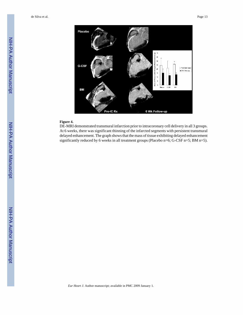
Figure 4.DE-MRI demonstrated transmural infarction prior to intracoronary cell delivery in all 3 groups.At 6 weeks, there was significant thinning of the infarcted segments with persistent transmuraldelayed enhancement. The graph shows that the mass of tissue exhibiting delayed enhancementsignificantly reduced by 6 weeks in all treatment groups (Placebo n=6; G-CSF n=5; BM n=5).
de Silva et al. Page 13
Eur Heart J. Author manuscript; available in PMC 2009 January 1.
NIH
-PA Author Manuscript
NIH
-PA Author Manuscript
NIH
-PA Author Manuscript

Figure 5.Overlay of registered short axis DE-MRI and FDG uptake (Ki) images. FDG uptake inmyocardium with transmural delayed enhancement on MRI before intracoronary cell infusionis preserved or increased. By 6 weeks, there was concordance between the FDG uptake imagesand the DE-MRI images with all treatment groups showing reduced FDG uptake in regionsshowing thinning and persistent transmural delayed enhancement.
de Silva et al. Page 14
Eur Heart J. Author manuscript; available in PMC 2009 January 1.
NIH
-PA Author Manuscript
NIH
-PA Author Manuscript
NIH
-PA Author Manuscript
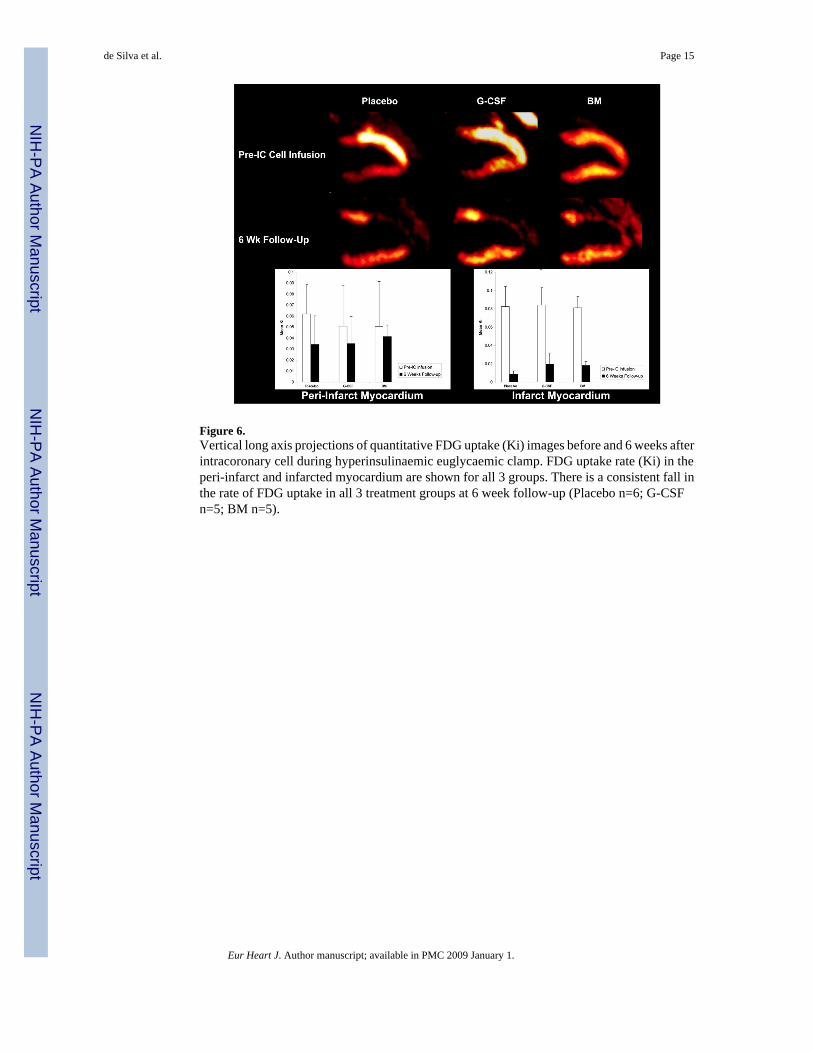
Figure 6.Vertical long axis projections of quantitative FDG uptake (Ki) images before and 6 weeks afterintracoronary cell during hyperinsulinaemic euglycaemic clamp. FDG uptake rate (Ki) in theperi-infarct and infarcted myocardium are shown for all 3 groups. There is a consistent fall inthe rate of FDG uptake in all 3 treatment groups at 6 week follow-up (Placebo n=6; G-CSFn=5; BM n=5).
de Silva et al. Page 15
Eur Heart J. Author manuscript; available in PMC 2009 January 1.
NIH
-PA Author Manuscript
NIH
-PA Author Manuscript
NIH
-PA Author Manuscript

Figure 7.The upper panels show measurements of adenosine vasodilator response in the peri-infarct andinfarcted myocardium pre-intracoronary cell infusion and at 6 weeks measured by H2
15O-PET.The lower panels show histologically measured vessel scores. No increase in vessel density orimprovement in vasodilator response was observed in any treatment group.
de Silva et al. Page 16
Eur Heart J. Author manuscript; available in PMC 2009 January 1.
NIH
-PA Author Manuscript
NIH
-PA Author Manuscript
NIH
-PA Author Manuscript

Figure 8.Representative high power laser confocal micrographs showing no colocalisation of CM-DiIpositive cells (red) with cells staining positive for CD31 (panel A), desmin (panel B) andvimentin (panel C). Similar findings were observed in all treatment groups.
de Silva et al. Page 17
Eur Heart J. Author manuscript; available in PMC 2009 January 1.
NIH
-PA Author Manuscript
NIH
-PA Author Manuscript
NIH
-PA Author Manuscript

NIH
-PA Author Manuscript
NIH
-PA Author Manuscript
NIH
-PA Author Manuscript
de Silva et al. Page 18
Table 1Composition of infused cells
Placebo G-CSF BM
Total Nucleated Cells (×108) 47.4±17.6 186±231 10.1±6.29Mononuclear Cells (×106/kg) 110±42.9 98.1±50.3 5.33±2.48
Colony Forming Cells (×102/kg) 3.62±1.42 80.4±41.3 72.7±33.8Granulocytes (×108) 1.63±3.04 147±225 7.58±5.33
Platelets (×109) 16.2±11.6 27.1±35.1 13.3±8.8
Eur Heart J. Author manuscript; available in PMC 2009 January 1.

NIH
-PA Author Manuscript
NIH
-PA Author Manuscript
NIH
-PA Author Manuscript
de Silva et al. Page 19Ta
ble
2G
loba
l Mea
sure
s of S
ysto
lic F
unct
ion
and
Rem
odel
ing
from
MR
I Dat
a
Plac
ebo
G-C
SFB
M
Pre-
IC R
x6
Wk
FUPr
e-IC
Rx
6 W
k FU
Pre-
IC R
x6
Wk
FUn=
6n=
6n=
6n=
5n=
6n=
5E
F (%
)34
.3±9
.726
.5±6
.8*
31.2
±7.8
24.3
±5.1
*40
.4±6
.029
.6±8
.1*
ESV
(mL
)56
.5±1
8.4
69.2
±14.
1*49
.5±1
2.2
72.3
±12.
9*35
.7±1
1.3
52.9
±22.
8*E
DV
(mL
)84
.2±1
6.0
93.9
±14.
8†72
.1±1
5.7
95.8
±19.
2†60
.1±1
6.7
73.4
±23.
7†SV
(mL
)27
.7±4
.124
.7±6
.322
.6±7
.223
.7±8
.524
.2±6
.920
.5±3
.9L
VM
(g)
80.1
±11.
280
.7±6
.268
.6±1
3.8
79.5
±12.
183
.0±8
.781
.7±1
2.0
EF –
eje
ctio
n fr
actio
n; E
DV
– e
nd-d
iast
olic
vol
ume;
ESV
– e
nd-s
ysto
lic v
olum
e; S
V –
stro
ke v
olum
e; L
VM
– le
ft ve
ntric
ular
mas
s; P
re-I
C R
x –
Pre-
Intra
coro
nary
Cel
l Inf
usio
n; 6
Wk
FU –
6 W
eek
Follo
w-u
p.
* p<0.
01 v
pre
-IC
Rx;
† p<0.
05 v
pre
-IC
Rx.
Eur Heart J. Author manuscript; available in PMC 2009 January 1.

NIH
-PA Author Manuscript
NIH
-PA Author Manuscript
NIH
-PA Author Manuscript
de Silva et al. Page 20Ta
ble
3R
egio
nal W
all M
otio
n A
naly
sis o
f MR
I Dat
a
Pre-
IC C
ell I
nfus
ion
6 W
k Fo
llow
-Up
# WM
A%
Hyp
o%
Aki
n# W
MA
%H
ypo
%A
kin
Plac
ebo
40±1
674
±626
±654
±15*
63±1
337
±13
G-C
SF45
±13
60±1
240
±12
57±4
.8*
50±1
2*50
±12*
BM
32±5
64±7
36±7
50±1
2†56
±13*
44±1
3*
# WM
A –
num
ber o
f sec
tors
with
a w
all m
otio
n ab
norm
ality
; %H
ypo
- % o
f hyp
okin
etic
sect
ors;
%A
kin
- % o
f aki
netic
sect
ors.
* p<0.
05 v
Pre
-IC
Cel
l Inf
usio
n;
† p<0.
02 v
Pre
-IC
Cel
l Inf
usio
n
Eur Heart J. Author manuscript; available in PMC 2009 January 1.