ICAM-1-mediated Endothelial Nitric Oxide Synthase Activation via Calcium and AMP-activated Protein...
11
Molecular Biology of the Cell Vol. 20, 995–1005, February 1, 2009 ICAM-1–mediated Endothelial Nitric Oxide Synthase Activation via Calcium and AMP-activated Protein Kinase Is Required for Transendothelial Lymphocyte Migration Roberta Martinelli, Matthew Gegg, Rebecca Longbottom, Peter Adamson, Patric Turowski,* and John Greenwood* Division of Cell Biology, Institute of Ophthalmology, University College London, London EC1V 9EL, United Kingdom Submitted June 23, 2008; Revised October 20, 2008; Accepted December 3, 2008 Monitoring Editor: Martin A. Schwartz As a gatekeeper of leukocyte trafficking the vasculature fulfills an essential immune function. We have recently shown that paracellular transendothelial lymphocyte migration is controlled by intercellular adhesion molecule 1 (ICAM-1)- mediated vascular endothelial cadherin (VEC) phosphorylation [Turowski et al., J. Cell Sci. 121, 29 –37 (2008)]. Here we show that endothelial nitric oxide synthase (eNOS) is a critical regulator of this pathway. ICAM-1 stimulated eNOS by a mechanism that was clearly distinct from that utilized by insulin. In particular, phosphorylation of eNOS on S1177 in response to ICAM-1 activation was regulated by src family protein kinase, rho GTPase, Ca 2 , CaMKK, and AMPK, but not Akt/PI3K. Functional neutralization of any component of this pathway or its downstream effector guanylyl cyclase significantly reduced lymphocyte diapedesis across the endothelial monolayer. In turn, activation of NO signaling promoted lymphocyte transmigration. The eNOS signaling pathway was required for T-cell transmigration across primary rat and human microvascular endothelial cells and also when shear flow was applied, suggesting that this pathway is ubiquitously used. These data reveal a novel and essential role of eNOS in basic immune function and provide a key link in the molecular network governing endothelial cell compliance to diapedesis. INTRODUCTION The events controlling the capture and subsequent migra- tion of circulating lymphocytes across the vascular wall have been studied extensively, and many of its generic principles are known. However, the signaling mechanisms that under- pin this process remain poorly defined. Endothelial cells (ECs) actively participate in directing and regulating the process of lymphocyte migration across the vascular wall via adhesion molecules such as vascular cell adhesion cell molecule 1 (VCAM-1; Engelhardt, 2006), platelet and endo- thelial cell adhesion molecule-1 (PECAM-1; Liao et al., 1997), and intercellullar adhesion molecule 1 (ICAM-1; Turowski et al., 2005). In the CNS, where ECs form a tight specialized barrier, lymphocyte recruitment utilizes the same adhesion molecules, with the pairing of lymphocyte function-associ- ated antigen 1 (LFA-1)/ICAM-1 being preeminent in con- trolling diapedesis (Greenwood et al., 1995; Turowski et al., 2005). Previous studies have also shown that ICAM-1 is not only involved in the adhesion process, but its engagement acti- vates diverse signaling pathways in ECs, some of which are required for subsequent migration. For example, ICAM-1 has been reported to interact with members of the ezrin/ radixin/moesin (ERM) family, to activate rho GTPases, src, protein kinase C (PKC), and mitogen-activated protein (MAP) kinases and a series of proteins involved in the formation of focal adhesions such as FAK, paxillin, and p130 cas (Turowski et al., 2005). A rise of intracellular calcium appears to be central to ICAM–mediated events (Etienne- Manneville et al., 2000) and precedes the activation of the other master regulators such as rho GTPases and PKC. Im- portantly, EC signaling via Ca 2 and rho is essential for subsequent lymphocyte migration (Adamson et al., 1999; Etienne-Manneville et al., 2000) at least in part by regulating adherens junction modulation during paracellular diapede- sis (Allingham et al., 2007; Turowski et al., 2008). Indeed, ICAM-1 engagement induces phosphorylation of vascular endothelial cadherin (VEC) and concomitant paracellular permeability. This suggests that lymphocyte adhesion to ICAM-1 modulates the paracellular space much like vaso- active compounds such as vascular endothelial growth fac- tor (VEGF) or histamine (Dejana et al., 2008). A central mediator of VEGF or thrombin-induced endothelial perme- ability is nitric oxide (van Hinsbergh and van Nieuw Amer- ongen, 2002). Two nitric oxide synthase (NOS) systems are of physiological significance in ECs. Inducible NOS (iNOS) is regulated at the transcriptional level and is mainly utilized during long-term adaptation of the vasculature to extracel- This article was published online ahead of print in MBC in Press (http://www.molbiolcell.org/cgi/doi/10.1091/mbc.E08 – 06 – 0636) on December 10, 2008. * These authors contributed equally to this work. Address correspondence to: John Greenwood (j.greenwood@ ucl.ac.uk) or Patric Turowski ([email protected]). Abbreviations used: EC, endothelial cell; ROS, reactive oxygen spe- cies; VEC, vascular endothelial cadherin; NO, nitric oxide; eNOS, endothelial nitric oxide synthase; XO, xanthine oxidase; NOX, NAPDH oxidase; AMPK, AMP-activated protein kinase; CaMKK, calcium/calmodulin kinase kinase; ICAM-1, intercellular adhesion molecule-1; VCAM-1, vascular cell adhesion molecule-1; L-NAME, N G -nitro-l-arginine methyl ester; ODQ, 1H-[1,2,4]oxadiazole[4,3- a]quinoxalin-1-one. © 2009 by The American Society for Cell Biology 995 http://www.molbiolcell.org/content/suppl/2008/12/10/E08-06-0636.DC1.html Supplemental Material can be found at:
-
Upload
independent -
Category
Documents
-
view
0 -
download
0
Transcript of ICAM-1-mediated Endothelial Nitric Oxide Synthase Activation via Calcium and AMP-activated Protein...

Molecular Biology of the CellVol. 20, 995–1005, February 1, 2009
ICAM-1–mediated Endothelial Nitric Oxide SynthaseActivation via Calcium and AMP-activated Protein KinaseIs Required for Transendothelial Lymphocyte MigrationRoberta Martinelli, Matthew Gegg, Rebecca Longbottom, Peter Adamson,Patric Turowski,* and John Greenwood*
Division of Cell Biology, Institute of Ophthalmology, University College London, London EC1V 9EL,United Kingdom
Submitted June 23, 2008; Revised October 20, 2008; Accepted December 3, 2008Monitoring Editor: Martin A. Schwartz
As a gatekeeper of leukocyte trafficking the vasculature fulfills an essential immune function. We have recently shownthat paracellular transendothelial lymphocyte migration is controlled by intercellular adhesion molecule 1 (ICAM-1)-mediated vascular endothelial cadherin (VEC) phosphorylation [Turowski et al., J. Cell Sci. 121, 29–37 (2008)]. Here weshow that endothelial nitric oxide synthase (eNOS) is a critical regulator of this pathway. ICAM-1 stimulated eNOS bya mechanism that was clearly distinct from that utilized by insulin. In particular, phosphorylation of eNOS on S1177 inresponse to ICAM-1 activation was regulated by src family protein kinase, rho GTPase, Ca2�, CaMKK, and AMPK, butnot Akt/PI3K. Functional neutralization of any component of this pathway or its downstream effector guanylyl cyclasesignificantly reduced lymphocyte diapedesis across the endothelial monolayer. In turn, activation of NO signalingpromoted lymphocyte transmigration. The eNOS signaling pathway was required for T-cell transmigration acrossprimary rat and human microvascular endothelial cells and also when shear flow was applied, suggesting that thispathway is ubiquitously used. These data reveal a novel and essential role of eNOS in basic immune function and providea key link in the molecular network governing endothelial cell compliance to diapedesis.
INTRODUCTION
The events controlling the capture and subsequent migra-tion of circulating lymphocytes across the vascular wall havebeen studied extensively, and many of its generic principlesare known. However, the signaling mechanisms that under-pin this process remain poorly defined. Endothelial cells(ECs) actively participate in directing and regulating theprocess of lymphocyte migration across the vascular wallvia adhesion molecules such as vascular cell adhesion cellmolecule 1 (VCAM-1; Engelhardt, 2006), platelet and endo-thelial cell adhesion molecule-1 (PECAM-1; Liao et al., 1997),and intercellullar adhesion molecule 1 (ICAM-1; Turowski etal., 2005). In the CNS, where ECs form a tight specializedbarrier, lymphocyte recruitment utilizes the same adhesionmolecules, with the pairing of lymphocyte function-associ-ated antigen 1 (LFA-1)/ICAM-1 being preeminent in con-
trolling diapedesis (Greenwood et al., 1995; Turowski et al.,2005).
Previous studies have also shown that ICAM-1 is not onlyinvolved in the adhesion process, but its engagement acti-vates diverse signaling pathways in ECs, some of which arerequired for subsequent migration. For example, ICAM-1has been reported to interact with members of the ezrin/radixin/moesin (ERM) family, to activate rho GTPases, src,protein kinase C (PKC), and mitogen-activated protein(MAP) kinases and a series of proteins involved in theformation of focal adhesions such as FAK, paxillin, andp130cas (Turowski et al., 2005). A rise of intracellular calciumappears to be central to ICAM–mediated events (Etienne-Manneville et al., 2000) and precedes the activation of theother master regulators such as rho GTPases and PKC. Im-portantly, EC signaling via Ca2� and rho is essential forsubsequent lymphocyte migration (Adamson et al., 1999;Etienne-Manneville et al., 2000) at least in part by regulatingadherens junction modulation during paracellular diapede-sis (Allingham et al., 2007; Turowski et al., 2008). Indeed,ICAM-1 engagement induces phosphorylation of vascularendothelial cadherin (VEC) and concomitant paracellularpermeability. This suggests that lymphocyte adhesion toICAM-1 modulates the paracellular space much like vaso-active compounds such as vascular endothelial growth fac-tor (VEGF) or histamine (Dejana et al., 2008). A centralmediator of VEGF or thrombin-induced endothelial perme-ability is nitric oxide (van Hinsbergh and van Nieuw Amer-ongen, 2002). Two nitric oxide synthase (NOS) systems areof physiological significance in ECs. Inducible NOS (iNOS)is regulated at the transcriptional level and is mainly utilizedduring long-term adaptation of the vasculature to extracel-
This article was published online ahead of print in MBC in Press(http://www.molbiolcell.org/cgi/doi/10.1091/mbc.E08–06–0636)on December 10, 2008.
* These authors contributed equally to this work.
Address correspondence to: John Greenwood ([email protected]) or Patric Turowski ([email protected]).
Abbreviations used: EC, endothelial cell; ROS, reactive oxygen spe-cies; VEC, vascular endothelial cadherin; NO, nitric oxide; eNOS,endothelial nitric oxide synthase; XO, xanthine oxidase; NOX,NAPDH oxidase; AMPK, AMP-activated protein kinase; CaMKK,calcium/calmodulin kinase kinase; ICAM-1, intercellular adhesionmolecule-1; VCAM-1, vascular cell adhesion molecule-1; L-NAME,NG-nitro-l-arginine methyl ester; ODQ, 1H-[1,2,4]oxadiazole[4,3-a]quinoxalin-1-one.
© 2009 by The American Society for Cell Biology 995 http://www.molbiolcell.org/content/suppl/2008/12/10/E08-06-0636.DC1.htmlSupplemental Material can be found at:

lular cues. Endothelial NOS (eNOS) is constitutively ex-pressed in the vasculature and in skeletal muscle, and itsactivity is highly regulated on many levels. For instance,caveolin-1, G-protein–coupled receptors (in particular bra-dykinin receptor 2 and the angiotensin II R1), and the eNOS-binding protein NOSIP bind to and inhibit eNOS activity(Fulton et al., 2001).
Calmodulin and hsp90 have also been reported to stimu-late eNOS activity (Pober and Sessa, 2007). Most impor-tantly, eNOS activity has been shown to be modulated byphosphorylation on serine and threonine residues in re-sponse to a variety of agonists such as VEGF, insulin, andshear stress (Fulton et al., 2001; Marletta, 2001). In particular,six residues, namely S116, T497, S617, S635, S1177, and Y83,have been identified as being phosphorylated during acti-vation (Gallis et al., 1999; Harris et al., 2001; Fulton et al.,2005). Phosphoinositide 3-kinase (PI3K) and Akt/PKB havebeen shown to be important regulators of eNOS both in vitroand in vivo (Dimmeler et al., 1999; Fulton et al., 1999; Six etal., 2002). However, other pathways for eNOS activation areknown. For example, thrombin-mediated eNOS activationdepends on CaM kinase kinase (CaMKK) and AMP-acti-vated protein kinase (AMPK; Stahmann et al., 2006), and NOhas been implicated in mediating some of the functions ofAMPK (Morrow et al., 2003; Nagata et al., 2003).
The purpose of this study was to test whether eNOS waspart of ICAM-1–mediated signaling in microvascular ECs.We also searched for upstream activators of eNOS. Finally,we analyzed whether ICAM-1–mediated paracellular regu-lation, including the compliance to lymphocyte diapedesis,was regulated by eNOS.
MATERIALS AND METHODS
MaterialsMouse mAbs to rat ICAM-1 (1A29 and 3H8) were generated from hybrid-omas provided by Dr. M. Miyasaka (Osaka University, Osaka, Japan) and Dr.W. Hickey (Dartmouth Medical School, Hanover, NH), respectively. Alterna-tively, low endotoxin IgG from Serotec (Oxford, United Kingdom) were used.Mouse mAb to human ICAM-1 (clone 15.2), rat CD11a (clone WT.1), and ratCD18 (clone WT.3) were also purchased from Serotec. Goat anti-mouse Abs(GAM) were from Sigma-Aldrich (Poole, United Kingdom). Polyclonal Abspecific for eNOS, Akt, AMPK, and their phosphorylated forms (eNOSSer1177, AKT Ser 473, and AMPK Thr172) were from Cell Signaling Technol-ogy (Beverly, MA). Anti-CaMKK was purchased from BD Biosciences (Ox-ford, United Kingdom). Biotinylated anti-rabbit Ig and streptavidin-fluores-cein were purchased from GE Healthcare (Amersham, United Kingdom).L-NAME (NG-nitro-l-arginine methyl ester), ODQ (1H-[1,2,4]oxadiazolo-[4,3-a]quinoxalin-1-one), AICAR (5-aminoimidazole-4-carboxamide ribonucleo-side), and BAPTA-AM were from Sigma-Aldrich. SU6656, wortmannin,LY294002, compound C, Akt1/2i, BAY-412272, and calyculin A were fromMerck Biosciences (Nottingham, United Kingdom). STO-609 was from Tocris(Bristol, United Kingdom), and cell-permeable C3 transferase was from Cy-toskeleton (Denver, CO). Phospho-specific peptide to eNOS IRTQ(pS)FSLQand its control IRTQSFSLQ were from Genepep (Prades le Lez, France).
ECs and Endothelioma LinesThe immortalized Lewis rat brain microvascular EC line GPNT and themouse endothelioma cells bEnd5 and ICAM-1-deficient (bEndI1.1) weregrown as previously described (Lyck et al., 2003; Romero et al., 2003). Primarycultures of cerebral ECs were prepared from 5–7-wk-old Lewis rats, 5–7-wk-old eNOS�/� mice (eNOS, F1 hybrid between SV129 and C57BLK/6, kindlyprovided by Dr. Adrian Hobbs, UCL, London) or wild-type mice, as previ-ously described (Abbott et al., 1992). Primary mouse and rat EC cells wereseeded on collagen IV/fibronectin-coated plates and maintained in EGM-2MV (Clonetics, Cambrex BioScience, Wokingham, United Kingdom). Theprimary dermal EC line, HDMEC-c (Promocell, Heidelberg, Germany), wasalso grown in EGM-2 MV medium.
Rho Activation AssayCells were grown to confluence in 10-cm dishes. After appropriate stimula-tion, the cells were washed twice with ice-cold PBS and then lysed in 50 mMTris buffer (pH 7.2), containing 500 mM NaCl, 10 mM MgCl2, 1% Triton X-100,
1 mM DTT, and mixed protease inhibitors (Roche Diagnostics, Burgess Hill,United Kingdom). Each lysate was incubated with ca. 10 �g rhotekin rho-binding domain (Ren et al., 1999) absorbed to glutathione beads at 4°C for 1 h.The beads were washed three times with lysis buffer and then processed forimmunoblotting using monoclonal anti-rhoA antibody (Santa Cruz Biotech-nology, Santa Cruz, CA).
ImmunofluorescenceCells were grown to confluence in 35-mm dishes, stimulated, and fixed in3.7% formaldehyde. After extraction in �20°C acetone, cells were processedfor indirect immunocytochemistry. ICAM-1 distribution after incubation withmAb 1A29 was detected with anti-rat Cy3-conjugated antibody (1:50). Detec-tion of phospho-eNOS was performed with phospho-specific antibodies (1:50), followed by biotinylated anti-rabbit Ig (1:100) and streptavidin-FITC(1:200). Where indicated, diluted primary antibody was preincubated with 10�M eNOS-specific peptides IRTQ(pS)FSLQ or its nonphosphorylated control.Confocal microscopy was performed as previously described (Turowski et al.,2004). For the analysis of ICAM-1 clustering confocal sections spanning themembrane area of cells and containing more than 90% of the ICAM-1 stainingwere stacked and quantified. The diameter of all fluorescent clusters (abovethe resolution limit of 0.2 �m) within an area of 25 �m2 was measured usingZeiss LSM imaging software (Welwyn Garden City, United Kingdom).
Shear StressImmortalized Lewis rat brain microvascular ECs (GPNT) were seeded inregular 12-well plates. Once confluent, cells were exposed to 10 dyn/cm ofsteady shear stress for 24 h on an orbital rotator as already described (Leyet al., 1989).
T-cellsFor migration assays, myelin basic protein (MBP)-specific T lymphocyte lineswere used (kind gift of Dr. E. Beraud, Marseille, France). Human whole bloodwas processed using Ficoll Paque to collect the peripheral blood mononuclearcells, following which T-cells were separated by positive selection usingCD4� MACS beads (Miltenyi, Bisley, United Kingdom) according to themanufacturer’s instructions. T-cells were cultured overnight at 1.5 � 106/mlin RPMI-1640 supplemented with 10% FCS, 100 U/ml penicillin, 100 �g/mlstreptomycin,1 mM sodium pyruvate, 1 mM nonessential amino acids, 2 mMl-Glu, and 50 �m �-mercaptoethanol in the presence of recombinant IL-2 (50U/ml).
Lymphocyte Migration In VitroLymphocyte migration assays on immortalized Lewis rat brain ECs (GPNT)were conducted as previously described (Adamson et al., 1999). Rat T-cellswere allowed to migrate for 30 min to 1 h at which point rates were deter-mined by time-lapse video microscopy (see also Supplemental Videos). Mi-gration of human T-cells across human dermal ECs was performed overcourse of 4 h. Migration data from static cultures was collected from triplicateexperiments each representing a minimum of six wells. In shear stress exper-iments, rat T-cells were added to 12-well plates containing GPNT monolayersand incubated for 1.5 h at 37°C on an orbital rotator before recording by videomicroscopy. Added numbers from a minimum of four fields per assay wereused.
Measurement of NO ProductionCells were grown in 96-well plates and, at confluence, cultured in serum-freemedium overnight. Medium from each well was removed and replaced withprewarmed phenol-red free HBSS containing the fluorescent dye 5 4,5-dia-minofluorescein diacetate (DAF-2DA, Molecular Probes, Eugene, OR; 10 �M,100 �l/well) for 120 min at 37°C for the detection of NO. After incubation,cells were washed twice with prewarmed HBSS before stimulation. Thefluorescence of each well was measured in a fluorescence plate reader (exci-tation 490 nm, emission 520 nm). Results were obtained by subtracting thereading of controls from the correspondent reading for every time point afterstimulation.
siRNA-mediated Knockdown of AMPK and CaMKK�Specific siRNA duplexes targeting � subunits of AMPK, CaMKK�, and non-silencing controls were purchased from Dharmacon (Chicago, IL). GPNT cells(3 � 105) were seeded into six-well plates and transfected 24 h later withOligofectamine Reagent following the manufacturer’s instructions (Invitro-gen, Paisley, United Kingdom). Briefly, 200 pmol siRNA was complexed withtransfection reagent and added to the cells in serum-free medium for 4 h.Subsequently, serum was added, and the incubation proceeded for 24 hbefore repeating the transfection with siRNA. Cells were cultured for afurther 48 h before experiments were performed.
Immunoprecipitation and Western BlottingCells were seeded at 1 � 105/ml and, at confluence, cultured in serum-freemedium overnight. Cells were pretreated and stimulated as detailed in the
R. Martinelli et al.
Molecular Biology of the Cell996

figure legends and then lysed in boiling 50 mM Tris/Cl, pH 6.8, 2% SDS, 10%glycerol, 100 mM DTT, 100 nM calyculin A (200 �l/60-mm dish). Immuno-precipitation of VEC was performed as previously described (Turowski et al.,2008). Samples were subjected to SDS-PAGE and transferred to nitrocelluloseby semidry electrotransfer. The membranes were blocked in TBS containing5% milk, 0.1% Tween-20, and 0.1% Triton X-100 for 2 h before incubationovernight at 4°C with the appropriate antibody diluted at 1:1000 in TBScontaining 0.1% Tween-20 and 0.1% Triton X-100. Membranes werewashed three times with PBS/0.1% Tween-20 before 1-h incubation withan anti-mouse or anti-rabbit HRP-conjugated IgG (GE Healthcare) at adilution of 1:10,000 and 1:2000, respectively. After three washes in PBS/0.1% Tween-20, membranes were developed using the ECL reagents(Roche) according to the manufacturer’s instructions and exposed to x-rayfilm. Protein bands were evaluated by densitometric quantification, nor-malized against the amount of total protein, and averaged over at leastthree independent experiments.
Statistical AnalysesData are presented as mean � SEM. Variances of mean values were statisti-cally analyzed by the Student’s t test. *p � 0.05; **0.001 � p � 0.01; ***p �0.001. Time-course data were analyzed by linear regression, and the signifi-cance of slopes was determined by analyses of covariance (ANCOVA) usingthe Prism software package.
RESULTS
LFA1–ICAM-1 clustering plays a fundamental role duringleukocyte transmigration (Barreiro et al., 2002, Carman et al.,2003). Subsequent endothelial ICAM-1 signaling involvesCa2�, rho GTPases, protein kinases, and actin rearrange-ments and has generally been studied by antibody-mediatedcross-linking of serum-starved cells (Turowski et al., 2005).Specifically, experimental protocols usually involve incuba-
tion of cells with a primary anti-ICAM-1 antibody for 5–30min (believed to be a neutral period), followed by clusteringusing secondary antibody that triggers the cellular signalingresponse (Figure 1A). This was illustrated by a loss of sig-naling to actin when ICAM-1 clustering was prevented(Supplemental Figure S1A). Additionally, we have foundthat a variety of monoclonal primary anti-ICAM-1 antibod-ies on their own, i.e., not followed by cross-linking with asecondary Ab, induced ICAM-1 signaling in ECs, exempli-fied by receptor surface clustering (Figure 1B), rho GTPaseactivation (Figure 1C), Ca2� transients (Supplemental FigureS1B), and VEC phosphorylation (see Figure 3C). This indi-cated that ICAM-1–mediated signaling was activated usingprimary antibody alone and suggested that certain immedi-ate early signals such as oscillating Ca2� (SupplementalFigure S1B) or NO production (see below) may have previ-ously been overlooked. In the following, all EC activationwas performed by either anti-ICAM-1 antibody alone orT-cells.
ICAM-1 Induces eNOS PhosphorylationeNOS activation was determined by measuring phosphory-lation of S1177 (Harris et al., 2001). Primary rat cerebralmicrovascular ECs were stimulated with 1A29 mAb forvarious times and the level of activated eNOS determined inWestern blots using anti-phospho S1177 eNOS antibodies(Figure 2A). ICAM-1 ligation induced strong phosphoryla-tion of eNOS within 15 min. Positive control cells, stimu-lated with insulin (Montagnani et al., 2001; Fleming et al.,
Figure 1. ICAM-1–mediated endothelial sig-naling. (A) Schematic representation of strate-gies used to activate ICAM-1 on ECs. (B) Anti-body-induced ICAM-1 surface clustering.Confluent, serum-starved GPNT cells were ei-ther left untreated (NT) or incubated with 10�g/ml ICAM-1 antibody 1A29 for 60 min(ICAM-1 ligation). Alternatively, at 30 min cellswere briefly washed, and receptor cross-link-ing was induced by the addition of 10 �g/mlgoat anti-mouse IgG for 30 min (XL). Cellswere fixed and stained for ICAM-1 and ana-lyzed by confocal microscopy. Cluster size wasdetermined as described in Materials and Meth-ods in at least 20 random areas, and their sizedistribution is represented in box plots. Aver-age cluster density in these areas is representedon the right. Data are representative of threeindependent experiments. Bar, 1 �m. (C) Con-fluent, serum-starved GPNT cells were eitherleft untreated or incubated with insulin (1 �M,15 min), lysophosphatidic acid (LPA, 10 �M, 15min), or ICAM-1 antibody 1A29 (10 �g/ml) forthe times indicated (left). Alternatively, at 30min, cells were briefly washed and receptorcross-linking induced by the addition of 10�g/ml goat anti-mouse IgG for the indicatedtimes (right). Cells were then lysed, and theamount of activated (top) and total rho GTPase(bottom) was determined by pulldown assays.Similar results were obtained in at least threeindependent experiments.
eNOS and Lymphocyte Migration
Vol. 20, February 1, 2009 997
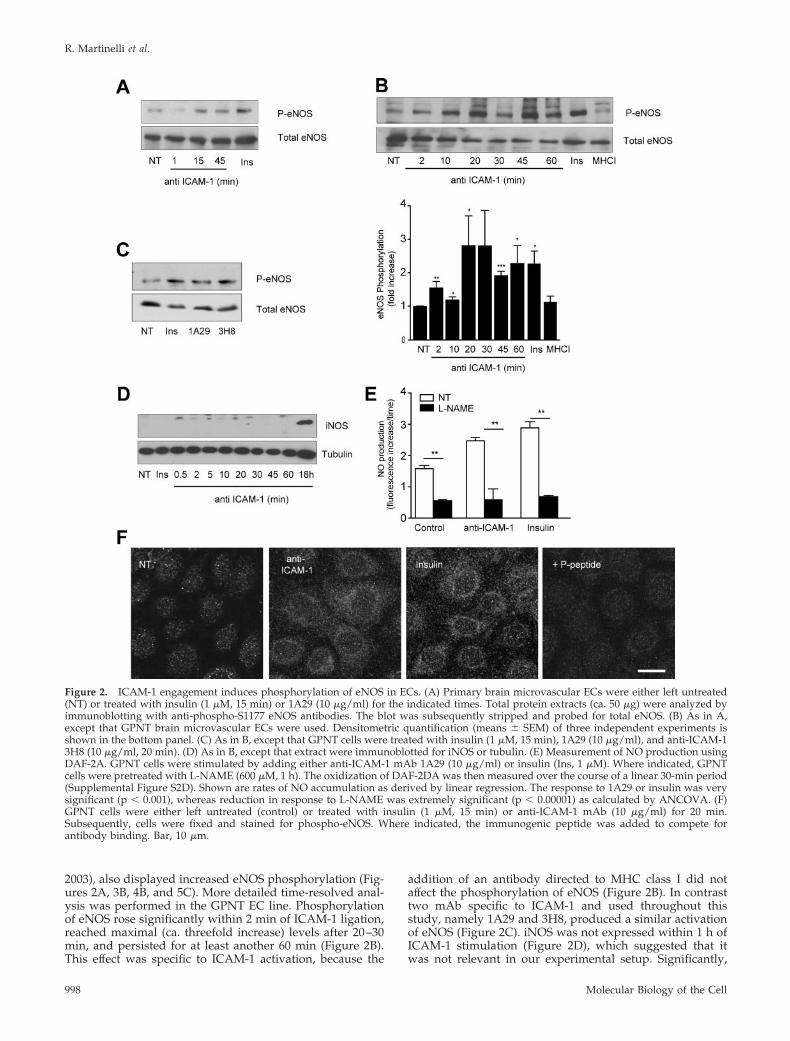
2003), also displayed increased eNOS phosphorylation (Fig-ures 2A, 3B, 4B, and 5C). More detailed time-resolved anal-ysis was performed in the GPNT EC line. Phosphorylationof eNOS rose significantly within 2 min of ICAM-1 ligation,reached maximal (ca. threefold increase) levels after 20–30min, and persisted for at least another 60 min (Figure 2B).This effect was specific to ICAM-1 activation, because the
addition of an antibody directed to MHC class I did notaffect the phosphorylation of eNOS (Figure 2B). In contrasttwo mAb specific to ICAM-1 and used throughout thisstudy, namely 1A29 and 3H8, produced a similar activationof eNOS (Figure 2C). iNOS was not expressed within 1 h ofICAM-1 stimulation (Figure 2D), which suggested that itwas not relevant in our experimental setup. Significantly,
Figure 2. ICAM-1 engagement induces phosphorylation of eNOS in ECs. (A) Primary brain microvascular ECs were either left untreated(NT) or treated with insulin (1 �M, 15 min) or 1A29 (10 �g/ml) for the indicated times. Total protein extracts (ca. 50 �g) were analyzed byimmunoblotting with anti-phospho-S1177 eNOS antibodies. The blot was subsequently stripped and probed for total eNOS. (B) As in A,except that GPNT brain microvascular ECs were used. Densitometric quantification (means � SEM) of three independent experiments isshown in the bottom panel. (C) As in B, except that GPNT cells were treated with insulin (1 �M, 15 min), 1A29 (10 �g/ml), and anti-ICAM-13H8 (10 �g/ml, 20 min). (D) As in B, except that extract were immunoblotted for iNOS or tubulin. (E) Measurement of NO production usingDAF-2A. GPNT cells were stimulated by adding either anti-ICAM-1 mAb 1A29 (10 �g/ml) or insulin (Ins, 1 �M). Where indicated, GPNTcells were pretreated with L-NAME (600 �M, 1 h). The oxidization of DAF-2DA was then measured over the course of a linear 30-min period(Supplemental Figure S2D). Shown are rates of NO accumulation as derived by linear regression. The response to 1A29 or insulin was verysignificant (p � 0.001), whereas reduction in response to L-NAME was extremely significant (p � 0.00001) as calculated by ANCOVA. (F)GPNT cells were either left untreated (control) or treated with insulin (1 �M, 15 min) or anti-ICAM-1 mAb (10 �g/ml) for 20 min.Subsequently, cells were fixed and stained for phospho-eNOS. Where indicated, the immunogenic peptide was added to compete forantibody binding. Bar, 10 �m.
R. Martinelli et al.
Molecular Biology of the Cell998

increased phosphorylation of eNOS correlated with an in-crease of NO. As reported by others (Matheny et al., 2000),the adhesion of lymphocytes to GPNT induced reactiveoxygen species (ROS) in the ECs (Supplemental Figure S2).Unlike in other EC systems, ROS accumulation in GPNTcells was largely dependent on ICAM-1 and specificallyabolished by the NOS inhibitor L-NAME. This suggestedthat these ROS were in fact reactive nitrogen species, andthis was tested directly in GPNT loaded with DAF-2DA, adye that fluoresces upon binding to oxidized species of NO.Stimulation with 1A29 or insulin induced a time-dependent,linear increase in DAF-2DA fluorescence (Supplemental Fig-ure S2D) at a significantly higher rate than nonstimulatedcontrol cells (Figure 2E). Changes in DAF-2DA fluorescencewere sensitive to L-NAME pretreatment suggesting theywere due to eNOS activity (Figure 2E). Detailed subcellularanalysis of the distribution of activated eNOS was per-formed using anti-phospho eNOS antibodies in indirect im-munofluorescence (Figure 2F). Insulin and ICAM-1 activa-tion induced a similar increase in phospho-eNOS staining inGPNT cells. In both cases specific staining was punctate,suggesting vesicular eNOS association, and was enriched inpericellular areas of the cell. Collectively, these data sug-gested that endothelial ICAM-1 stimulation induced eNOSand the production of oxygen species, among which NOand/or oxidized derivatives constituted the vast majority.
ICAM-1 and Insulin Activate eNOS by Different SignalTransduction PathwaysEndothelial signaling after ICAM-1 stimulation is controlledby src protein kinases (Etienne-Manneville et al., 2000; Wanget al., 2003) and rho GTPase (Etienne et al., 1998; Adamson etal., 1999). Pharmacological inhibition of these major signaltransducers abolished ICAM-1–induced activation of eNOS(Figure 3A). Ca2� is a critical mediator of ICAM-1 signaling(Etienne-Manneville et al., 2000; Supplemental Figure S1B)and has also been implicated in the activation of eNOS
(Busse and Mulsch, 1990). We therefore tested whether Ca2�
was also involved in ICAM-1– or insulin-mediated eNOSactivation. Intracellular Ca2� was chelated using BAPTA-AM, and subsequently eNOS phosphorylation was assessed(Figure 3B). Depletion of Ca2� did not affect insulin-inducedeNOS phosphorylation, in line with previous observations(Montagnani et al., 2001). By contrast, ICAM-1–inducedeNOS activation was reduced to basal levels in BAPTA-AM–pretreated cells. These data implicated Ca2�, rho GTPases,and src protein kinases upstream of eNOS activation in re-sponse to ICAM-1 activation. Endothelial ICAM-1 signalingregulates a variety of processes ranging from focal adhesion totranscription. However, among these, only modulation of in-tercellular junctions through VEC phosphorylation has so farbeen shown to be relevant to leukocyte migration (Turowskiet al., 2008). We tested whether ICAM-1 engagement mediatedphosphorylation of VEC via a pathway involving eNOS.ICAM-1 activation led to an �2.5-fold increase in VEC ty-rosine phosphorylation (Figure 3C). In cell pretreated withL-NAME, the ICAM-1–induced phosphorylation was totallyabsent. Significantly, insulin did not induce any phosphor-ylation of VEC (data not shown), further underlining thatinsulin and ICAM-1 engaged separate signaling pathways inmicrovascular ECs.
ICAM-1 Induces eNOS Activation via AMPKAkt/PKB has been shown to directly activate eNOS in ECsin response to VEGF or insulin (Dimmeler et al., 1999; Hill etal., 2001). Phospho-Akt could be detected in insulin-stimu-lated GPNT cells but not in response to the ligation ofICAM-1 (Figure 4A). This inability of ICAM-1 to activateAkt/PKB was expected because, at least in B-cells, this is aprerogative of ICAM-2 (Perez et al., 2002). Inhibition of PI3Kby wortmannin obliterated insulin-induced eNOS activationbut left the response to ICAM-1 intact (Figure 4B), furthersupporting that insulin and ICAM-1 signaling engage sepa-rate pathways in brain microvascular ECs. In search of a
Figure 3. ICAM-1-induced VEC phosphory-lation is mediated by eNOS. (A) GPNT cellswere either left untreated (NT) or pretreatedwith SU6656 (10 �M, 1 h) or cell-permeable C3transferase (3.75 �g/ml, 2 h) before ICAM-1stimulation (45 min) and immunoblot analysisof phosphorylated and total eNOS. (B) Whereindicated, GPNT cells were pretreated withBAPTA-AM (20 �M, 30 min) before analysis(performed as in A). Densitometric quantifica-tion (means � SEM) of three independent ex-periments is shown below. (C) GPNT cellswere pretreated with 1 mM L-NAME or leftuntreated before stimulation with anti-ICAM-1mAb (10 �g/ml) for 15 min. VEC was thenimmunoprecipitated and its tyrosine phos-phorylation analyzed with the phosphospecificantibody 4G10. A representative figure anddensitometric quantification (means � SEM) ofthree independent experiments is shown.
eNOS and Lymphocyte Migration
Vol. 20, February 1, 2009 999

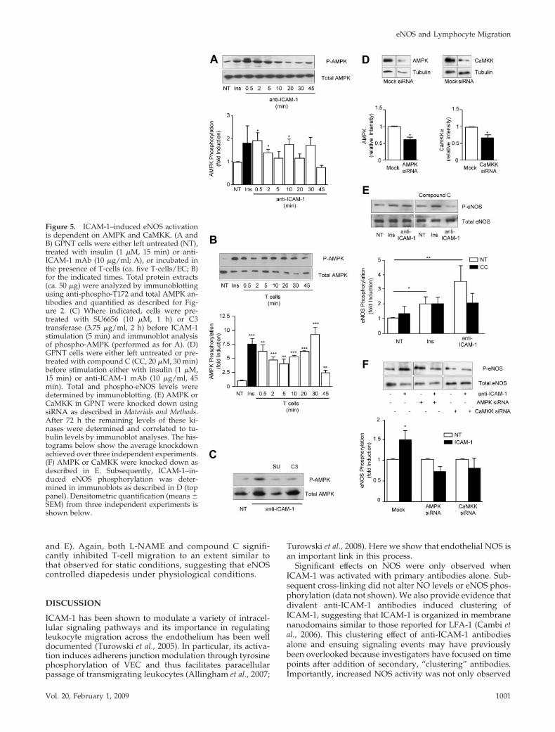
mediator of ICAM-1–induced eNOS activation we turned toAMPK, which can also phosphorylate eNOS S1177 (Morrowet al., 2003; Nagata et al., 2003). To investigate whetherAMPK was involved in ICAM-1–dependent signaling in ratmicrovascular ECs, kinase phosphorylation was monitoredusing specific antibodies. Rapid and transient phosphoryla-tion of AMPK at Thr172 was detected in response to ICAM-1stimulation, with a distinct peak at 0.5 min and a return tocontrol levels within 20 min (Figure 5A). Similar phosphor-ylation of endothelial AMPK was obtained when T-cellswere added (Figure 5B). Phosphorylation was sensitive topretreatment with inhibitors targeting src protein kinases orrho GTPase, placing AMPK downstream of this well-estab-lished axis of ICAM-1 signaling (Figure 5C). Next, we inves-tigated whether AMPK was required for ICAM-1–inducedeNOS activation. For this, GPNT were pretreated using com-pound C, a selective inhibitor of AMPK (Zhou et al., 2001).Significant reduction in the phosphorylation of eNOS wasdetected, indicating that AMPK was instrumental to ICAM-1–induced eNOS stimulation (Figure 5D). In contrast, insu-lin-induced eNOS phosphorylation was insensitive to com-pound C, again underlining the molecular differences insignaling. The involvement of AMPK in ICAM-1–mediatedeNOS phosphorylation was further corroborated by siRNA-mediated knockdown. Transfection of GPNT cells withsiRNA complexes specifically targeting AMPK led to nearly50% reduction in protein levels (Figure 5E). Cells with re-duced levels of AMPK displayed somewhat higher phos-phorylation of eNOS, but this was completely abolished in
response to ICAM-1 (Figure 5F). Because calcium plays animportant role in ICAM-1–mediated signaling (Supplemen-tal Figure S1B) and AMPK can be activated by the calcium-dependent CaMKK (Hurley et al., 2005), we investigatedwhether this kinase was relevant in our system. siRNA-mediated knockdown targeting CaMKK led to a 40% reduc-tion in protein levels (Figure 5E). Under these conditionseNOS phosphorylation was also significantly reduced inresponse to ICAM-1 stimulation (Figure 5F). These resultsrevealed that VEC phosphorylation is linked to ICAM-1activation via Ca2�, CaMKK, AMPK, and eNOS.
Lymphocyte Diapedesis Requires eNOS SignalingFinally, we investigated whether eNOS signaling was alsorequired for leukocyte transmigration. For this, we used awell-documented in vitro system of an antigen activatedT-cell line cocultured with primary or established brain mi-crovascular ECs (Adamson et al., 1999). T-cell transmigrationis monitored by time-lapse video microscopy and impor-tantly, occurs in the absence of cytokine stimulation or che-mokine gradients (see also Supplemental Videos). Transmi-gration is strongly dependent on ICAM-1 (Greenwood et al.,1995). Specifically, we observed more than 60% reduction ofT-cell migration across GPNT monolayers when LFA-1 wasblocked beforehand (Figure 6A). L-NAME pretreatment ofGPNT cells induced a similar block in migration, as did theinhibition of the NO effector soluble guanylyl cyclase (sGC)using ODQ (Figure 6B). In sharp contrast, inhibition of otherimportant intracellular oxidase systems such as xanthineoxidase (XO) and NADPH oxidase (NOX) had no effect onmigration rates (Supplemental Figure S3). L-NAME did notaffect lymphocyte migration across primary brain ECs de-rived from eNOS�/� mice, indicating that this NOS inhibi-tor has no other relevant intracellular target (SupplementalFigure S4A). Exogenous addition of the NO donor DEA-NOto cocultures led to a modest increase in T-cell migration butpotently reversed a block exerted by BAPTA-AM (Figure6B). Chelating endothelial Ca2� is one of the most effectiveways of inhibiting leukocyte migration (Etienne-Mannevilleet al., 2000), and this blockade was completely reversedwhen ECs were pretreated with DEA-NO or when the sGCagonist BAY-412272 was added during migration. However,inhibition of migration through ODQ could not be reversedby addition of exogenous NO (Figure 6B), indicating thatNO was required downstream of Ca2� and upstream of sGCsignaling. Significantly, in all cases, inhibition of migrationwas due to reduced diapedesis because lymphocyte adhe-sion to ECs was unchanged (Supplemental Figure S5). ThuseNOS and NO played an integral part of ICAM-1–depen-dent lymphocyte migration across GPNT cells. To furtherconfirm the importance of eNOS signaling we tested theupstream regulators. Inhibition of endothelial PI3K or Aktdid not affect lymphocyte transmigration, confirming thatthis pathway is not relevant for ICAM-1 signaling (Figure6C). In clear contrast the pharmacological inhibition of eitherAMPK or CaMKK led to a reduction in migrated lympho-cytes similar to that observed when ICAM-1 engagement orNOS was blocked (Figure 6D). This effect was specificbecause siRNA-mediated knockdown of these kinasesalso significantly reduced transmigration (Figure 6D). Theeffect of L-NAME was not only restricted to the GPNT cellline. Migration across primary rat cerebral or human der-mal microvascular ECs was also inhibited in response toL-NAME or compound C treatment (Figure 7,A–C). Shearflow is an important parameter in all processes of thevascular endothelium. T-cell migration across GPNT wasalso assessed after exposure to shear stress (Figure 7, D
Figure 4. ICAM-1–induced eNOS activation is independent ofPKB/Akt. (A) GPNT cells were either left untreated (NT), treatedwith insulin (1 �M, 15 min) or anti-ICAM-1 mAb (10 �g/ml) for theindicated times. Total protein extracts (ca. 50 �g) were analyzed byimmunoblotting using anti-phospho-T493 and total Akt antibodies.(B) GPNT cells were treated with insulin (1 �M, 15 min) or anti-ICAM-1 mAb (10 �g/ml, 45 min). Where indicated, cells werepretreated with wortmannin (WN, 20 �M, 30 min). eNOS activationwas analyzed and quantified as in Figure 2.
R. Martinelli et al.
Molecular Biology of the Cell1000
and E). Again, both L-NAME and compound C signifi-cantly inhibited T-cell migration to an extent similar tothat observed for static conditions, suggesting that eNOScontrolled diapedesis under physiological conditions.
DISCUSSION
ICAM-1 has been shown to modulate a variety of intracel-lular signaling pathways and its importance in regulatingleukocyte migration across the endothelium has been welldocumented (Turowski et al., 2005). In particular, its activa-tion induces adherens junction modulation through tyrosinephosphorylation of VEC and thus facilitates paracellularpassage of transmigrating leukocytes (Allingham et al., 2007;
Turowski et al., 2008). Here we show that endothelial NOS isan important link in this process.
Significant effects on NOS were only observed whenICAM-1 was activated with primary antibodies alone. Sub-sequent cross-linking did not alter NO levels or eNOS phos-phorylation (data not shown). We also provide evidence thatdivalent anti-ICAM-1 antibodies induced clustering ofICAM-1, suggesting that ICAM-1 is organized in membranenanodomains similar to those reported for LFA-1 (Cambi etal., 2006). This clustering effect of anti-ICAM-1 antibodiesalone and ensuing signaling events may have previouslybeen overlooked because investigators have focused on timepoints after addition of secondary, “clustering” antibodies.Importantly, increased NOS activity was not only observed
Figure 5. ICAM-1–induced eNOS activationis dependent on AMPK and CaMKK. (A andB) GPNT cells were either left untreated (NT),treated with insulin (1 �M, 15 min) or anti-ICAM-1 mAb (10 �g/ml; A), or incubated inthe presence of T-cells (ca. five T-cells/EC; B)for the indicated times. Total protein extracts(ca. 50 �g) were analyzed by immunoblottingusing anti-phospho-T172 and total AMPK an-tibodies and quantified as described for Fig-ure 2. (C) Where indicated, cells were pre-treated with SU6656 (10 �M, 1 h) or C3transferase (3.75 �g/ml, 2 h) before ICAM-1stimulation (5 min) and immunoblot analysisof phospho-AMPK (performed as for A). (D)GPNT cells were either left untreated or pre-treated with compound C (CC, 20 �M, 30 min)before stimulation either with insulin (1 �M,15 min) or anti-ICAM-1 mAb (10 �g/ml, 45min). Total and phospho-eNOS levels weredetermined by immunoblotting. (E) AMPK orCaMKK in GPNT were knocked down usingsiRNA as described in Materials and Methods.After 72 h the remaining levels of these ki-nases were determined and correlated to tu-bulin levels by immunoblot analyses. The his-tograms below show the average knockdownachieved over three independent experiments.(F) AMPK or CaMKK were knocked down asdescribed in E. Subsequently, ICAM-1–in-duced eNOS phosphorylation was deter-mined in immunoblots as described in D (toppanel). Densitometric quantification (means �SEM) from three independent experiments isshown below.
eNOS and Lymphocyte Migration
Vol. 20, February 1, 2009 1001

in response to antibody-mediated clustering but also duringlymphocytes adhesion (Supplemental Figure S2).
At times that appeared relevant to transendothelial lym-phocyte migration we observed a time-dependent phos-phorylation of eNOS on S1177, an activation site in thereductase domain of the enzyme (Fulton et al., 2001). Acorroborative rise in intracellular concentration of reactivenitrogen species was also detected, both in response toICAM-1 ligation and insulin. Reactive nitrogen species weremeasured in cells loaded with DAF-2DA, a dye that fluo-resces upon binding to a variety of oxidized species of NO.They are likely to represent NO because inhibition of theintracellular NO receptor sGC displayed effects similar tothat of NOS inhibition. Significant eNOS phosphorylationwas subcellularly restricted to vesicular structures, suggest-ing that ICAM-1–induced nitrogen oxide species affectedsignaling locally.
The phosphorylation of at least five S/T residues plays arole in the activation of eNOS and PKB/Akt has been iden-tified as a critical activator in many situations (Fulton et al.,1999; Montagnani et al., 2001). When eNOS was activated inresponse to ICAM-1, PKB/Akt clearly did not play a role.First, ICAM-1 failed to induce PKB/Akt phosphorylation inagreement with a previous report (Perez et al., 2002). Second,neither PI3K nor Akt inhibitors affected ICAM-1–inducedphosphorylation or ICAM-1–dependent lymphocyte migra-tion. There is precedence of Akt-independent activation ofeNOS, exemplified by stimulation of human umbilical ve-nous endothelial cells (HUVECs) with fenofibrate (Mu-rakami et al., 2006) or when bovine aortic ECs are exposed toshear stress (Boo et al., 2002) or thrombin (Motley et al.,2007). Although we were unable to demonstrate an involve-ment of PI3K or PKB/Akt, a role for AMPK was clearlyshown. This protein kinase is an energy-sensing enzyme that
has been reported to mediate a number of metabolic signalssuch as inhibition of cholesterol, fatty acid and protein syn-thesis, and enhancement of glucose uptake and blood flow
Figure 7. eNOS signaling is required for T-cell migration across avariety of ECs. (A) T-cell migration assay similar to Figure 6, exceptthat primary rat brain ECs were used. (B and C) T-cell migration assaysimilar to Figure 6, except that human primary dermal cells were used.(D and E) The effect of L-NAME and compound C on migration acrossGPNT cells was assessed when shear stress was operational.
Figure 6. Signaling pathways required for transen-dothelial lymphocyte migration. (A) Contribution ofICAM-1 to T-cell migration. T-cells were either leftuntreated or preincubated with antibodies againstCD11a (10 �g/ml) and CD18 (10 �g/ml) for 1 hbefore assessing migration across GPNT cells mono-layers. (B) GPNT cells were left untreated (Control)or treated with various pharmacological inhibitors,alone or in combination, before migration. ECs werepretreated with L-NAME (600 �M) or ODQ (100�M) for 1 h or BAPTA-AM (20 �M) for 30 minbefore migration. Alternatively cells were stimu-lated with DEA-NO (5 �M) for 30 min before lym-phocyte migration assays. The sGC agonist BAY41–2272 (50 �M) were added for 30 min before andkept throughout the lymphocyte migration assays.(C) Migration assay similar to B. GPNT cells werepretreated with the Akt inhibitor Akti1/2 (2.5 �M,30 min), the PI3-kinase inhibitor LY294002 (20 �M,1 h), the AMPK inhibitor compound C (20 �M, 30min), or the CaMKK inhibitor STO-609 (10 �M, 1 h)before migration. (D) GPNT cells were transfectedwith siRNA targeting CaMKK� or AMPK 72 h be-fore the migration assay. All results are the mean �SEM of six replicates from at least three independentexperiments.
R. Martinelli et al.
Molecular Biology of the Cell1002
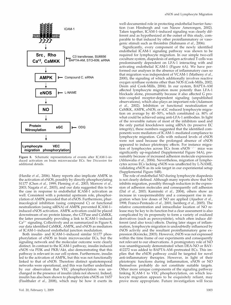
(Hardie et al., 2006). Many reports also implicate AMPK inthe activation of eNOS, possibly by directly phosphorylatingS1177 (Chen et al., 1999; Fleming et al., 2003; Morrow et al.,2003; Nagata et al., 2003), and our data suggested this to bethe case in response to endothelial ICAM-1 activation aswell. Consistent with a potential upstream role, phosphor-ylation of AMPK preceded that of eNOS. Furthermore, phar-macological inhibition (using compound C) or functionalneutralization (using siRNA) of AMPK prevented ICAM-1–induced eNOS activation. AMPK activation could be placeddownstream of src protein kinase, rho GTPase and CaMKK,the latter presumably providing a link to ICAM-1–inducedCa2� signaling. Collectively and as summarized in Figure 8,our data identified CaMKK, AMPK, and eNOS as mediatorsof ICAM-1–induced endothelial junction modulation.
Both insulin- and ICAM-1–induced similar S1177 phos-phorylation of eNOS in brain microvascular ECs, but thesignaling network and the molecular outcome were clearlydistinct. In contrast to the ICAM-1 pathway, insulin inducedeNOS via PI3K and PKB/Akt, also demonstrating that thispathway is relevant in our cell system. Furthermore, insulinled to the activation of AMPK, but this was not functionallylinked to that of eNOS. Therefore distinct spatiotemporalnetworks were operational, and this was further underlinedby our observation that VEC phosphorylation was un-changed in the presence of insulin (data not shown). Indeed,insulin has also been shown to phosphorylate eNOS on Y657(Fisslthaler et al., 2008), which may be how it exerts its
well-documented role in protecting endothelial barrier func-tion (van Hinsbergh and van Nieuw Amerongen, 2002).Taken together, ICAM-1–induced signaling was clearly dif-ferent and as hypothesized at the outset of this study, com-parable to that induced by other proinflammatory or vaso-genic stimuli such as thrombin (Stahmann et al., 2006).
Significantly, every component of the newly identifiedendothelial ICAM-1 signaling pathway was shown to berequired for lymphocyte migration. In our simple two-cellcoculture system, diapedesis of antigen activated T-cells waspredominantly dependent on LFA-1 interacting with andactivating endothelial ICAM-1 (Figure 6A). We have per-formed our analyses in the absence of inflammatory cues sothat migration was independent of VCAM-1 (Matheny et al.,2000), the signaling of which additionally involves reactiveoxygen synthase systems other than NOS (Cook-Mills, 2002;Deem and Cook-Mills, 2004). In our system, BAPTA-AMaffected lymphocyte migration more potently than LFA-1blockade alone, presumably because it also affected G pro-tein–coupled receptor-dependent signaling (unpublishedobservations), which also plays an important role (Adamsonet al., 2002). Inhibition or functional neutralization ofCaMKK, AMPK, eNOS, or sGC reduced lymphocyte migra-tion on average by 40–50%, which constituted ca. 80% ofwhat could be achieved using anti-LFA-1 antibodies. In lightof the reversible nature of most of the inhibitors used andthe only partial knockdown using siRNA (to preserve ECintegrity), these numbers suggested that the identified com-ponents were mediators of ICAM-1–mediated compliance tolymphocyte migration. Cells with reduced levels of eNOSwere not used because the prolonged absence of eNOSappeared to induce pleiotropic effects. For instance migra-tion of lymphocytes across ECs from eNOS�/� mice wassignificantly up-regulated (Supplemental Figure S4A), pre-sumably because of increased adhesion molecule expression(Ahluwalia et al., 2004). Nevertheless, migration of lympho-cytes across ECs lacking eNOS was unaffected by L-NAME,validating eNOS as its sole target in our experimental setup(Supplemental Figure S4B).
The role of endothelial NO during lymphocyte diapedesisis not clearly defined. Although many reports show that NOinhibits migration, possibly through reduction in the expres-sion of adhesion molecules and consequently cell adhesion(Dal et al., 2003; Kaminski et al., 2004), others show anincrease in vasopermeability and a consequent rise in mi-gration when low doses of NO are applied (Ajuebor et al.,1998; Franco-Penteado et al., 2001; Isenberg et al., 2005). Therelative concentration and intracellular location of NO re-lease may be key to its function but a clear assessment is alsocomplicated by its propensity to form a variety of oxidizedderivatives (such as peroxynitrite), which often induce dif-ferent (and also toxic) effects. During later stages of inflam-mation, lymphocyte migration is undoubtedly influenced byiNOS activity and the resultant proinflammatory gene ex-pression (Kroncke, 2003). However, iNOS was not expressedwithin the time frame of our experiments and consequentlynot relevant to our observations. A promigratory role of NOwas unambiguously demonstrated when DEA-NO or BAY-412272 was added to BAPTA-AM–pretreated ECs. This im-plies that the eNOS pathway could be targeted for novelanti-inflammatory therapies. However, in light of theirpleiotropic functions during inflammation, eNOS or NOthemselves probably do not constitute suitable targets.Other more unique components of the signaling pathwaylinking ICAM-1 to VEC phosphorylation, on which leu-kocyte migration appears to be exquisitely reliant, mayprove more appropriate. Future investigation will focus
Figure 8. Schematic representations of events after ICAM-1–in-duced activation on brain microvascular ECs. See Discussion foradditional details.
eNOS and Lymphocyte Migration
Vol. 20, February 1, 2009 1003

on the spatiotemporal requirements of ICAM-1–inducedeNOS and VEC phosphorylation as well as the kinasesand phosphatase involved. eNOS activation may lead tothe rapid inactivation of a VEC tyrosine phosphatase bydirect nitrosylation. However, the sensitivity of our sys-tem to the sGC inhibitor ODQ suggests a more complexnetwork of regulation.
ACKNOWLEDGMENTS
We thank Jeremy Pearson (King’s College London, London, United Kingdom),Britta Engelhardt (University of Bern, Bern, Switzerland), and Evelyn Beraud forproviding us with ICAM-1 CHO cells, mouse endothelioma cell lines, andantigen-activated T-cells, respectively. Adrian Hobbs kindly provided eNOSknockout mice. We thank Veronica Hollis, Miriam Palacios-Callender, and Sal-vador Moncada for help with direct NO measurements and helpful commentsthroughout this work and Reiner Haseloff for helpful comments throughout thiswork. This work was supported by a Program Grant from the Wellcome Trust(062403).
REFERENCES
Abbott, N. J., Hughes, C. C., Revest, P. A., and Greenwood, J. (1992). Devel-opment and characterisation of a rat brain capillary endothelial culture:towards an in vitro blood-brain barrier. J. Cell Sci. 103(Pt 1), 23–37.
Adamson, P., Etienne, S., Couraud, P. O., Calder, V., and Greenwood, J.(1999). Lymphocyte migration through brain endothelial cell monolayersinvolves signaling through endothelial ICAM-1 via a rho-dependent path-way. J. Immunol. 162, 2964–2973.
Adamson, P., Wilbourn, B., Etienne-Manneville, S., Calder, V., Beraud, E.,Milligan, G., Couraud, P. O., and Greenwood, J. (2002). Lymphocyte traffick-ing through the blood-brain barrier is dependent on endothelial cell hetero-trimeric G-protein signaling. FASEB J. 16, 1185–1194.
Ahluwalia, A., Foster, P., Scotland, R. S., McLean, P. G., Mathur, A., Perretti,M., Moncada, S., and Hobbs, A. J. (2004). Antiinflammatory activity of solubleguanylate cyclase: cGMP-dependent down-regulation of P-selectin expressionand leukocyte recruitment. Proc. Natl. Acad. Sci USA 101, 1386–1391.
Ajuebor, M. N., Virag, L., Flower, R. J., Perretti, M., and Szabo, C. (1998). Roleof inducible nitric oxide synthase in the regulation of neutrophil migration inzymosan-induced inflammation. Immunology 95, 625–630.
Allingham, M. J., van Buul, J. D., and Burridge, K. (2007). ICAM-1-mediated,Src- and Pyk2-dependent vascular endothelial cadherin tyrosine phosphory-lation is required for leukocyte transendothelial migration. J. Immunol. 179,4053–4064.
Barreiro, O., Yanez-Mo, M., Serrador, J. M., Montoya, M. C., Vicente-Manzanares,M., Tejedor, R., Furthmayr, H., and Sanchez-Madrid, F. (2002). Dynamic inter-action of VCAM-1 and ICAM-1 with moesin and ezrin in a novel endothelialdocking structure for adherent leukocytes. J. Cell Biol. 157, 1233–1245.
Boo, Y. C., Sorescu, G., Boyd, N., Shiojima, I., Walsh, K., Du, J., and Jo, H.(2002). Shear stress stimulates phosphorylation of endothelial nitric-oxidesynthase at Ser1179 by Akt-independent mechanisms: role of protein kinaseA. J. Biol. Chem. 277, 3388–3396.
Busse, R., and Mulsch, A. (1990). Calcium-dependent nitric oxide synthesis inendothelial cytosol is mediated by calmodulin. FEBS Lett. 265, 133–136.
Cambi, A., Joosten, B., Koopman, M., de, L. F., Beeren, I., Torensma, R.,Fransen, J. A., Garcia-Parajo, M., van Leeuwen, F. N., and Figdor, C. G. (2006).Organization of the integrin LFA-1 in nanoclusters regulates its activity. Mol.Biol. Cell 17, 4270–4281.
Carman, C. V., Jun, C. D., Salas, A., and Springer, T. A. (2003). Endothelialcells proactively form microvilli-like membrane projections upon intercellularadhesion molecule 1 engagement of leukocyte LFA-1. J. Immunol. 171, 6135–6144.
Chen, Z. P., Mitchelhill, K. I., Michell, B. J., Stapleton, D., Rodriguez-Crespo,I., Witters, L. A., Power, D. A., Ortiz de Montellano, P. R., and Kemp, B. E.(1999). AMP-activated protein kinase phosphorylation of endothelial NOsynthase. FEBS Lett. 443, 285–289.
Cook-Mills, J. M. (2002). VCAM-1 signals during lymphocyte migration: roleof reactive oxygen species. Mol. Immunol. 39, 499–508.
Dal, S. D., Paron, J. A., de Oliveira, S. H., Ferreira, S. H., Silva, J. S., and Cunha,F. Q. (2003). Neutrophil migration in inflammation: nitric oxide inhibitsrolling, adhesion and induces apoptosis. Nitric Oxide 9, 153–164.
Deem, T. L., and Cook-Mills, J. M. (2004). Vascular cell adhesion molecule 1(VCAM-1) activation of endothelial cell matrix metalloproteinases: role ofreactive oxygen species. Blood 104, 2385–2393.
Dejana, E., Orsenigo, F., and Lampugnani, M. G. (2008). The role of adherensjunctions and VE-cadherin in the control of vascular permeability. J. Cell Sci.121, 2115–2122.
Dimmeler, S., Fleming, I., Fisslthaler, B., Hermann, C., Busse, R., and Zeiher,A. M. (1999). Activation of nitric oxide synthase in endothelial cells byAkt-dependent phosphorylation. Nature 399, 601–605.
Engelhardt, B. (2006). Molecular mechanisms involved in T cell migrationacross the blood-brain barrier. J. Neural Transm. 113, 477–485.
Etienne, S., Adamson, P., Greenwood, J., Strosberg, A. D., Cazaubon, S., andCouraud, P. O. (1998). ICAM-1 signaling pathways associated with Rhoactivation in microvascular brain endothelial cells. J. Immunol. 161, 5755–5761.
Etienne-Manneville, S., Manneville, J. B., Adamson, P., Wilbourn, B., Greenwood,J., and Couraud, P. O. (2000). ICAM-1-coupled cytoskeletal rearrangements andtransendothelial lymphocyte migration involve intracellular calcium signaling inbrain endothelial cell lines. J. Immunol. 165, 3375–3383.
Fisslthaler, B., Loot, A. E., Mohamed, A., Busse, R., and Fleming, I. (2008).Inhibition of endothelial nitric oxide synthase activity by proline-rich tyrosinekinase 2 in response to fluid shear stress and insulin. Circ. Res. 102, 1520–1528.
Fleming, I., Schulz, C., Fichtlscherer, B., Kemp, B. E., Fisslthaler, B., and Busse,R. (2003). AMP-activated protein kinase (AMPK) regulates the insulin-in-duced activation of the nitric oxide synthase in human platelets. Thromb.Haemost. 90, 863–871.
Franco-Penteado, C. F., DeSouza, I., Teixeira, S. A., Ribeiro-DaSilva, G., De,N. G., and Antunes, E. (2001). Role of nitric oxide on the increased vascularpermeability and neutrophil accumulation induced by staphylococcal entero-toxin B into the mouse paw. Biochem. Pharmacol. 61, 1305–1311.
Fulton, D., Gratton, J. P., McCabe, T. J., Fontana, J., Fujio, Y., Walsh, K.,Franke, T. F., Papapetropoulos, A., and Sessa, W. C. (1999). Regulation ofendothelium-derived nitric oxide production by the protein kinase Akt. Na-ture 399, 597–601.
Fulton, D., Gratton, J. P., and Sessa, W. C. (2001). Post-translational control ofendothelial nitric oxide synthase: why isn’t calcium/calmodulin enough?J. Pharmacol. Exp. Ther. 299, 818–824.
Fulton, D., Church, J. E., Ruan, L., Li, C., Sood, S. G., Kemp, B. E., Jennings,I. G., and Venema, R. C. (2005). Src kinase activates endothelial nitric-oxidesynthase by phosphorylating Tyr-83. J. Biol. Chem. 280, 35943–35952.
Gallis, B., et al. (1999). Identification of flow-dependent endothelial nitric-oxide synthase phosphorylation sites by mass spectrometry and regulation ofphosphorylation and nitric oxide production by the phosphatidylinositol3-kinase inhibitor LY294002. J. Biol. Chem. 274, 30101–30108.
Greenwood, J., Wang, Y., and Calder, V. L. (1995). Lymphocyte adhesion andtransendothelial migration in the central nervous system: the role of LFA-1,ICAM-1, VLA-4 and VCAM-1. Immunology 86, 408–415.
Hardie, D. G., Hawley, S. A., and Scott, J. W. (2006). AMP-activated proteinkinase–development of the energy sensor concept. J. Physiol. 574, 7–15.
Harris, M. B., Ju, H., Venema, V. J., Liang, H., Zou, R., Michell, B. J., Chen,Z. P., Kemp, B. E., and Venema, R. C. (2001). Reciprocal phosphorylation andregulation of endothelial nitric-oxide synthase in response to bradykininstimulation. J. Biol. Chem. 276, 16587–16591.
Hill, M. M., Andjelkovic, M., Brazil, D. P., Ferrari, S., Fabbro, D., and Hemmings,B. A. (2001). Insulin-stimulated protein kinase B phosphorylation on Ser-473 isindependent of its activity and occurs through a staurosporine-insensitive kinase.J. Biol. Chem. 276, 25643–25646.
Hurley, R. L., Anderson, K. A., Franzone, J. M., Kemp, B. E., Means, A. R., andWitters, L. A. (2005). The Ca2�/calmodulin-dependent protein kinase kinasesare AMP-activated protein kinase kinases. J. Biol. Chem. 280, 29060–29066.
Isenberg, J. S., Tabatabai, N., and Spinelli, H. M. (2005). Nitric oxide modu-lation of low-density mononuclear cell transendothelial migration. Microsur-gery 25, 452–456.
Kaminski, A., Pohl, C. B., Sponholz, C., Ma, N., Stamm, C., Vollmar, B., andSteinhoff, G. (2004). Up-regulation of endothelial nitric oxide synthase inhibitspulmonary leukocyte migration following lung ischemia-reperfusion in mice.Am. J. Pathol. 164, 2241–2249.
Kroncke, K. D. (2003). Nitrosative stress and transcription. Biol. Chem. 384,1365–1377.
Ley, K., Lundgren, E., Berger, E., and Arfors, K. E. (1989). Shear-dependentinhibition of granulocyte adhesion to cultured endothelium by dextran sul-fate. Blood 73, 1324–1330.
R. Martinelli et al.
Molecular Biology of the Cell1004

Liao, F., Ali, J., Greene, T., and Muller, W. A. (1997). Soluble domain 1 ofplatelet-endothelial cell adhesion molecule (PECAM) is sufficient to blocktransendothelial migration in vitro and in vivo. J. Exp. Med. 185, 1349–1357.
Lyck, R., Reiss, Y., Gerwin, N., Greenwood, J., Adamson, P., and Engelhardt,B. (2003). T-cell interaction with ICAM-1/ICAM-2 double-deficient brainendothelium in vitro: the cytoplasmic tail of endothelial ICAM-1 is necessaryfor transendothelial migration of T cells. Blood 102, 3675–3683.
Marletta, M. A. (2001). Another activation switch for endothelial nitric oxidesynthase: why does it have to be so complicated? Trends Biochem. Sci. 26,519–521.
Matheny, H. E., Deem, T. L., and Cook-Mills, J. M. (2000). Lymphocytemigration through monolayers of endothelial cell lines involves VCAM-1signaling via endothelial cell NADPH oxidase. J. Immunol. 164, 6550–6559.
Montagnani, M., Chen, H., Barr, V. A., and Quon, M. J. (2001). Insulin-stimulated activation of eNOS is independent of Ca2� but requires phosphor-ylation by Akt at Ser(1179). J. Biol. Chem. 276, 30392–30398.
Morrow, V. A., Foufelle, F., Connell, J. M., Petrie, J. R., Gould, G. W., and Salt,I. P. (2003). Direct activation of AMP-activated protein kinase stimulatesnitric-oxide synthesis in human aortic endothelial cells. J. Biol. Chem. 278,31629–31639.
Motley, E. D., Eguchi, K., Patterson, M. M., Palmer, P. D., Suzuki, H., andEguchi, S. (2007). Mechanism of endothelial nitric oxide synthase phosphor-ylation and activation by thrombin. Hypertension 49, 577–583.
Murakami, H., et al. (2006). Fenofibrate activates AMPK and increases eNOSphosphorylation in HUVEC. Biochem. Biophys. Res. Commun. 341, 973–978.
Nagata, D., Mogi, M., and Walsh, K. (2003). AMP-activated protein kinase(AMPK) signaling in endothelial cells is essential for angiogenesis in responseto hypoxic stress. J. Biol. Chem. 278, 31000–31006.
Perez, O. D., Kinoshita, S., Hitoshi, Y., Payan, D. G., Kitamura, T., Nolan,G. P., and Lorens, J. B. (2002). Activation of the PKB/AKT pathway byICAM-2. Immunity 16, 51–65.
Pober, J. S., and Sessa, W. C. (2007). Evolving functions of endothelial cells ininflammation. Nat. Rev. Immunol. 7, 803–815.
Ren, X. D., Kiosses, W. B., and Schwartz, M. A. (1999). Regulation of the smallGTP-binding protein Rho by cell adhesion and the cytoskeleton. EMBO J. 18,578–585.
Romero, I. A., Radewicz, K., Jubin, E., Michel, C. C., Greenwood, J., Couraud,P. O., and Adamson, P. (2003). Changes in cytoskeletal and tight junctionalproteins correlate with decreased permeability induced by dexamethasone incultured rat brain endothelial cells. Neurosci. Lett. 344, 112–116.
Six, I., Kureishi, Y., Luo, Z., and Walsh, K. (2002). Akt signaling mediatesVEGF/VPF vascular permeability in vivo. FEBS Lett. 532, 67–69.
Stahmann, N., Woods, A., Carling, D., and Heller, R. (2006). Thrombin acti-vates AMP-activated protein kinase in endothelial cells via a pathway involv-ing Ca2�/calmodulin-dependent protein kinase kinase beta. Mol. Cell. Biol.26, 5933–5945.
Turowski, P., Adamson, P., and Greenwood, J. (2005). Pharmacological tar-geting of ICAM-1 signaling in brain endothelial cells: potential for treatingneuroinflammation. Cell Mol. Neurobiol. 25, 153–170.
Turowski, P., Adamson, P., Sathia, J., Zhang, J. J., Moss, S. E., Aylward, G. W.,Hayes, M. J., Kanuga, N., and Greenwood, J. (2004). Basement membrane-dependent modification of phenotype and gene expression in human retinalpigment epithelial ARPE-19 cells. Invest. Ophthalmol. Vis. Sci. 45, 2786–2794.
Turowski, P. et al. (2008). Phosphorylation of vascular endothelial cadherincontrols lymphocyte emigration. J. Cell Sci. 121, 29–37.
van Hinsbergh, V., and van Nieuw Amerongen, G. P. (2002). Intracellularsignalling involved in modulating human endothelial barrier function. J.Anat. 200, 549–560.
Wang, Q., Pfeiffer, G. R., and Gaarde, W. A. (2003). Activation of SRC tyrosinekinases in response to ICAM-1 ligation in pulmonary microvascular endothe-lial cells. J. Biol. Chem. 278, 47731–47743.
Zhou, G., et al. (2001). Role of AMP-activated protein kinase in mechanism ofmetformin action. J. Clin. Invest. 108, 1167–1174.
eNOS and Lymphocyte Migration
Vol. 20, February 1, 2009 1005