Eotaxin and FGF enhance signaling through an Extracellular signal-related kinase (ERK)-dependent...
9
RESEARCH Open Access Eotaxin and FGF enhance signaling through an Extracellular signal-related kinase (ERK)-dependent pathway in the pathogenesis of Eosinophilic Esophagitis Jennifer J Huang 1 , Jae Won Joh 1 , Judy Fuentebella 1 , Anup Patel 1 , Tammie Nguyen 1 , Scott Seki 1 , Lisa Hoyte 1 , Neha Reshamwala 1 , Christine Nguyen 2 , Anthony Quiros 2 , Dorsey Bass 1 , Eric Sibley 1 , William Berquist 1 , Kenneth Cox 1 , John Kerner 1 , Kari C Nadeau 1* Abstract Background: Eosinophilic esophagitis (EoE) is characterized by the inflammation of the esophagus and the infiltration of eosinophils into the esophagus, leading to symptoms such as dysphagia and stricture formation. Systemic immune indicators like eotaxin and fibroblast growth factor were evaluated for possible synergistic pathological effects. Moreover, blood cells, local tissue, and plasma from EoE and control subjects were studied to determine if the localized disease was associated with a systemic effect that correlated with presence of EoE disease. Method: Real-time polymerase chain reaction from peripheral blood mononuclear cells (PBMC), immunohistochemistry from local esophageal biopsies, fluid assays on plasma, and fluorescence-activated cell sorting on peripheral blood cells from subjects were used to study the systemic immune indicators in newly diagnosed EoE (n = 35), treated EoE (n = 9), Gastroesophageal reflux disease (GERD) (n = 8), ulcerative colitis (n = 5), Crohn’s disease (n = 5), and healthy controls (n = 8). Result: Of the transcripts tested for possible immune indicators, we found extracellular signal-regulated kinase (ERK), Bcl-2, bFGF (basic fibroblast growth factor), and eotaxin levels were highly upregulated in PBMC and associated with disease presence of EoE. Increased FGF detected by immunohistochemistry in esophageal tissues and in PBMC was correlated with low levels of pro-apoptotic factors (Fas, Caspase 8) in PBMC from EoE subjects. Plasma-derived bFGF was shown to be the most elevated and most specific in EoE subjects in comparison to healthy controls and disease control subjects. Conclusion: We describe for the first time a possible mechanism by which increased FGF is associated with inhibiting apoptosis in local esophageal tissues of EoE subjects as compared to controls. Eotaxin and FGF signaling pathways share activation through the ERK pathway; together, they could act to increase eosinophil activation and prolong the half-life of eosinophils in local tissues of the esophagus in EoE subjects. Introduction Eosinophilic esophagitis (EoE) is an inflammatory disor- der of the esophagus that can be characterized by feed- ing difficulties, heartburn, regurgitation, vomiting, abdominal pain, dysphagia, and food impaction [1-3]. EoE seems to predominantly affect children, and in particular, males [4]. Epidemiologic data from 2008 sug- gest that in children the annual incidence of EoE is approximately 1.3-1.6 per 10,000 and the prevalence is approximately 4.3-9.1 per 10,000 [5,6]. Approximately 50% of subjects with EoE have co-existing atopic condi- tions, such as food allergy, asthma, or eczema [7-12]. Food allergy is particularly prominent, but aeroallergens also may play a role [13]. * Correspondence: [email protected] 1 Stanford School of Medicine, Stanford, CA 94305, USA Full list of author information is available at the end of the article Huang et al. Allergy, Asthma & Clinical Immunology 2010, 6:25 http://www.aacijournal.com/content/6/1/25 ALLERGY, ASTHMA & CLINICAL IMMUNOLOGY © 2010 Huang et al; licensee BioMed Central Ltd. This is an Open Access article distributed under the terms of the Creative Commons Attribution License (http://creativecommons.org/licenses/by/2.0), which permits unrestricted use, distribution, and reproduction in any medium, provided the original work is properly cited.
Transcript of Eotaxin and FGF enhance signaling through an Extracellular signal-related kinase (ERK)-dependent...

RESEARCH Open Access
Eotaxin and FGF enhance signaling throughan Extracellular signal-related kinase(ERK)-dependent pathway in the pathogenesisof Eosinophilic EsophagitisJennifer J Huang1, Jae Won Joh1, Judy Fuentebella1, Anup Patel1, Tammie Nguyen1, Scott Seki1, Lisa Hoyte1,Neha Reshamwala1, Christine Nguyen2, Anthony Quiros2, Dorsey Bass1, Eric Sibley1, William Berquist1,Kenneth Cox1, John Kerner1, Kari C Nadeau1*
Abstract
Background: Eosinophilic esophagitis (EoE) is characterized by the inflammation of the esophagus and theinfiltration of eosinophils into the esophagus, leading to symptoms such as dysphagia and stricture formation.Systemic immune indicators like eotaxin and fibroblast growth factor were evaluated for possible synergisticpathological effects. Moreover, blood cells, local tissue, and plasma from EoE and control subjects were studied todetermine if the localized disease was associated with a systemic effect that correlated with presence of EoE disease.
Method: Real-time polymerase chain reaction from peripheral blood mononuclear cells (PBMC),immunohistochemistry from local esophageal biopsies, fluid assays on plasma, and fluorescence-activated cellsorting on peripheral blood cells from subjects were used to study the systemic immune indicators in newlydiagnosed EoE (n = 35), treated EoE (n = 9), Gastroesophageal reflux disease (GERD) (n = 8), ulcerative colitis(n = 5), Crohn’s disease (n = 5), and healthy controls (n = 8).
Result: Of the transcripts tested for possible immune indicators, we found extracellular signal-regulated kinase(ERK), Bcl-2, bFGF (basic fibroblast growth factor), and eotaxin levels were highly upregulated in PBMC andassociated with disease presence of EoE. Increased FGF detected by immunohistochemistry in esophageal tissuesand in PBMC was correlated with low levels of pro-apoptotic factors (Fas, Caspase 8) in PBMC from EoE subjects.Plasma-derived bFGF was shown to be the most elevated and most specific in EoE subjects in comparison tohealthy controls and disease control subjects.
Conclusion: We describe for the first time a possible mechanism by which increased FGF is associated withinhibiting apoptosis in local esophageal tissues of EoE subjects as compared to controls. Eotaxin and FGF signalingpathways share activation through the ERK pathway; together, they could act to increase eosinophil activation andprolong the half-life of eosinophils in local tissues of the esophagus in EoE subjects.
IntroductionEosinophilic esophagitis (EoE) is an inflammatory disor-der of the esophagus that can be characterized by feed-ing difficulties, heartburn, regurgitation, vomiting,abdominal pain, dysphagia, and food impaction [1-3].EoE seems to predominantly affect children, and in
particular, males [4]. Epidemiologic data from 2008 sug-gest that in children the annual incidence of EoE isapproximately 1.3-1.6 per 10,000 and the prevalence isapproximately 4.3-9.1 per 10,000 [5,6]. Approximately50% of subjects with EoE have co-existing atopic condi-tions, such as food allergy, asthma, or eczema [7-12].Food allergy is particularly prominent, but aeroallergensalso may play a role [13].* Correspondence: [email protected]
1Stanford School of Medicine, Stanford, CA 94305, USAFull list of author information is available at the end of the article
Huang et al. Allergy, Asthma & Clinical Immunology 2010, 6:25http://www.aacijournal.com/content/6/1/25 ALLERGY, ASTHMA & CLINICAL
IMMUNOLOGY
© 2010 Huang et al; licensee BioMed Central Ltd. This is an Open Access article distributed under the terms of the Creative CommonsAttribution License (http://creativecommons.org/licenses/by/2.0), which permits unrestricted use, distribution, and reproduction inany medium, provided the original work is properly cited.

The clinical symptoms of EoE are often similar tothose of gastroesophageal reflux disease (GERD) andinitial illness may be thought to be GERD. However, thesymptoms do not resolve with gastric acid suppression,and EoE presents with esophageal abnormalities duringendoscopy, showing eosinophil infiltration in theesophagus. EoE is currently defined as the presence ofmore than 15 intraepithelial eosinophils per high-powered field (eos/HPF) in both the proximal and distalesophagus [14]. Endoscopy of the subjects often showssigns of longitudinal linear furrows, trachealization,white plaques, and strictures but usually with a negativepH probe result. Subjects with more severe disease sta-tus present with severe stricturing, furrowing, and/ortrachealization, which may lead to mechanical dilationof the esophagus or food impaction that requiressurgical removal [13].EoE diagnosis can only be made through endoscopy
and biopsy of esophageal tissue. Prognostic tests that areeasily obtained and efficiently run through minimallyinvasive techniques to predict disease progression arelacking. Current guidelines suggest continual biopsiesfor monitoring of disease progress and treatment effi-ciency [15]. Since endoscopy with biopsy entails risks topatients, we aimed to study immune indicators found inplasma as secreted proteins that correlate with local pre-sence in esophageal tissues in EoE subjects as comparedto controls. Moreover, we hypothesized that through thediscovery of specific EoE immune indicators, we couldfurther interrogate pathological mechanisms of EoE dis-ease. Specifically, we focused on examining immuneindicators associated with EoE, for example, eotaxin andFGF. FGF is a protein involved in cell development, celldifferentiation and tissue repair [16]. Additionally, bFGFhas been shown to enhance the half-life of cells [17] andboth bFGF and eotaxin coordinate their activity throughthe activation of ERK [18,19]. Thus, in particular, wehypothesized that FGF could enhance activation andhalf-life of eosinophils in local esophageal tissues ascompared to peripheral blood.Altogether, the data presented here provide a mechan-
istic explanation for how basic fibroblast growth factorcould worsen the pathogenesis of EoE by enhancingactivation of eosinophils through synergy with eotaxinvia the ERK signaling pathway. The data may be used inthe possible diagnosis and prognosis of EoE. In addition,our results could provide an explanation for increasedeosinophil numbers, and prolongation of eosinophilhalf-life in EoE disease pathology.
MethodsHuman Clinical Data collectionThe study was approved by the Stanford AdministrativePanel on Human Subjects in Medical Research. All
subjects signed informed consent forms before partici-pating in the study. The study was performed accordingto Declaration of Helsinki guidelines. Proximal and dis-tal esophageal tissue and peripheral blood were obtainedfrom subjects during a diagnostic endoscopy and subse-quent biopsy. Middle esophageal biopsy tissue was alsoobtained from some patients. Biopsy samples were pre-served as formalin-fixed and paraffin-embedded sections.Histological diagnosis of EoE, GERD, or other disorders(i.e, ulcerative colitis and Crohn’s disease) was made bya licensed and certified clinical pathologist in gastroen-terology at Stanford University School of Medicine Clin-ical laboratory using guidelines by calculating the meannumber of eosinophils per high power field (HPF) inmore than 3 areas per biopsy sample [7]. Subjects whohad no pathological basis for symptoms and who had anegative pH probe were considered healthy controls(HCs). Subjects who had a negative pH probe result andwhose histological sections contained greater than15 eos/HPF were diagnosed with EoE as per guidelines[14,20]. All EoE subjects had failed a trial of protonpump inhibitor therapy (specifically, for an average of6-8 weeks of therapy) before being diagnosed with EoE.Subjects with a positive pH probe test and a negativebiopsy result were diagnosed with GERD. GERD sub-jects had less than 6 eosinophils/HPF in the esophaguson biopsy. Subjects labeled as “treated EoE” were con-firmed with biopsies of esophagus showing improvement(less than 5-10 eosinophils/HPF) and demonstratedresolution of clinical symptoms. Subjects with eosino-philic gastritis, eosinophilic colitis, acute or chronicinfections (viral, bacterial or fungal), autoimmune dis-ease, or neoplasm were excluded from the EoE, HC, andGERD groups. Allergic subjects were defined as havinga total serum IgE of >25 IU/ml and positive skin pricktesting as compared with positive histamine control tofoods. Patients were not allergy patch-tested routinely.None of the subjects had active infections and no sub-jects had positive findings for parasitic infections. Over-all, samples were obtained on newly diagnosed EoE(n = 35), treated EoE (n = 9), GERD (n = 8), ulcerativecolitis (UC, n = 5), and Crohn’s disease (CD, n = 5),healthy controls (n = 8) respectively. Demographics foreach subject are found in Additional File 1, Table S1.
Plasma and PBMC separationEach blood sample was centrifuged at 1800 RPM for10 minutes, and the supernatant plasma layer wascollected and centrifuged for another 10 minutes at13,000 RPM. Plasma was stored in 250 μl aliquots at-80°C. Phosphate-buffered saline (PBS) was added to theremaining blood cell layer until the total volume wasdouble the original collected blood volume. The homo-geneous PBS-blood mixture was layered over the Ficoll
Huang et al. Allergy, Asthma & Clinical Immunology 2010, 6:25http://www.aacijournal.com/content/6/1/25
Page 2 of 9

reagent (MP Biomedicals, Solon, OH) at a 2:1 volumeratio and subsequently centrifuged at 2200 RPM for20 minutes. The peripheral blood mononuclear cell(PBMC) layer was removed and washed twice with PBSat 1800 RPM for 5 minutes. PBMCs were then re-suspended at 1 million cells per milliliter in a solutionof 90% FBS and 10% DMSO and stored for later use forQT-PCR.
Fluid AssaysPlasma samples underwent testing with Luminex 35-plextechnology (Invitrogen, Carlsbad, CA). 35 cytokines andchemokines were assayed: fibroblast growth factor basic(bFGF or FGF-2); eotaxin (1, 2, and 3); IL-1a; IL-1b; IL-1receptor antagonist (IL-1RA); IL-2; IL-4; IL-5; IL-6; IL-7;IL-8; IL-10; IL-12-p40; IL-12-p70; IL-13; IL-15; IL-17; IL-17F; epithelial cell-derived neutrophil-activating protein-78 (ENA78); granulocyte colony-stimulating factor(G-CSF); granulocyte-macrophage colony-stimulating fac-tor (GM-CSF); growth-related oncogene-alpha (GRO-a);interferon-gamma (IFN-g); interferon-inducible protein10 (IP10); leptin; monocyte chemotactic protein-3 (MCP-3); monokine induced by gamma interferon (MIG);macrophage inflammatory protein 1 alpha (MIP-1a);macrophage inflammatory protein 1 beta (MIP-1b); nervegrowth factor (NGF); platelet-derived growth factor-BB(PDGF-BB); regulated upon activation, normal T cellexpressed and secreted (RANTES); transforming growthfactor beta (TGF-b); tumor necrosis factor alpha (TNF-a);tumor necrosis factor beta (TNF-b), and vascular endothe-lial growth factor (VEGF). Samples were tested and nor-malized with standard curves to ensure consistency andcalibrations occurred before each run, per manufacturer’sinstructions (Luminex Technologies, Invitrogen, Carlsbad,CA). Furthermore, each sample was run in duplicate forquality control. In addition, some plasma samples (n = 10)from subjects were run through Cytometric Bead Array(CBA) Multiplex technology (BD Biosciences, San Jose,CA; Th1/Th2 cytokine Assay, used per manufacturer’sinstructions) to test the reproducibility of the Luminex35-plex technology. For those cytokines tested (i.e.,eotaxin-3, basic fibroblast growth factor (bFGF), G-CSF,IFN-, IL-17, IL-1a, IL4, IL-5, macrophage inflammatoryprotein 1a, and nerve growth factor), similar results wereobtained with CBA and Luminex technologies.
ImmunohistochemistryParaffin-embedded tissue samples were soaked in xyleneand then solutions of 100%, 95%, and 70% ethanolsequentially to remove the paraffin wax. Antigenunmasking was performed by heating the slides in adecloaking chamber to 120°C in Diva Decloaking buffer(Biocare Medical, Concord, CA), and then cooling toroom temperature. H2O2 block (Lab Vision, Fremont,
CA) and protein block (Dako, Glostrup, Denmark) werethen applied to the tissue to prevent non-specific bind-ing and block endogenous peroxidases. Unconjugatedmouse anti-human FGF-9 primary antibody (Clone D-8)(Santa Cruz Biotechnology) was diluted 1:50 and appliedfor 2 hours at 25°C. After washing, a secondary MACH2 Mouse HRP (Biocare Medical, Concord, CA) antibodywas applied for 30 minutes. After further washing, theslides were stained with DAB (Vector Labs, Burlingame,CA) and subsequently counterstained with hematoxylinbefore being mounted. Cells were counted per high-powered field (HPF, 400×) at three different sites in thetissue and the mean HPF was calculated. Polyclonalunconjugated rabbit anti-human FGF-2 primary anti-body (Abcam, Cambridge, MA) was diluted 1:250 withantibody diluent (Dako, Glostrup, Denmark) and wasdouble-stained with mouse anti-human EG2 primaryantibody (gift of Dr. Reinhard B. Raggam). A MACH 2Double Stain HRP-AP secondary antibody (BiocareMedical, Concord, CA) was used.
QT-PCR RNA analysisPBMCs from each subject were used for QT-PCR RNAanalysis. For cDNA synthesis, 500 ng total RNA wastranscribed with cDNA transcription reagents (AppliedBiosystems, Foster City, CA) using random hexamers,according to the manufacturer’s instructions. Geneexpression was measured in real-time with the Gen-eAmp 7900 Sequence Detection System (Applied Biosys-tems, Foster City, CA) using primers and other reagentspurchased from Applied Biosystems. Relative quantifica-tion was measured using the Comparative CT(Threshold Cycle) method. The expression level of agene in a given sample was represented as 2-ΔΔ Ct whereΔΔCT = [ΔCT(experimental)] - [ΔCT(medium)] and ΔCT =[CT(experimental)] - [CT(housekeeping)]. All PCR assays wereperformed in triplicate. 100 ng of total isolated RNAwas submitted to reverse transcription (Invitrogen,Carlsbad, CA). 50 ng of resulting cDNA was submittedto TaqMan™PCR on an ABI Systems qPCR machine(Applied Biosystems, Foster City, CA) at the StanfordDepartment of Pediatrics QT-PCR Facility using gene-specific, fluorochrome-labeled probe/primer sets pur-chased from Applied Biosystems, Inc. EoE, GERD, HC,UC, and CD samples were tested for fibroblast growthfactor (FGF), fibroblast growth factor receptor (FGF-R),eotaxin, C-C chemokine receptor type 3 (CCR3), extra-cellular signal-regulated kinase (ERK), c-Jun N terminalkinase (JNK), IL-13, IL-5, Fas, B cell lymphoma protein2 (Bcl-2), and normalized to the b-2-microglobulin gene.
StatisticsStatistical analysis was performed with the GraphPadPrism software (GraphPad Software, La Jolla, CA).
Huang et al. Allergy, Asthma & Clinical Immunology 2010, 6:25http://www.aacijournal.com/content/6/1/25
Page 3 of 9

Statistical comparisons of data among groups were per-formed using the one-way analysis of variance(ANOVA) non-parametric Kruskal-Wallis test and theDunn’s Multiple Comparison post-test. Differences wereconsidered significant at a p-value of less than or equalto 0.05. Correlation analysis between fold expressionswas performed using the Spearman correlation.
ResultsDemographicsData were analyzed from newly diagnosed EoE (n = 35),treated EoE (n = 9), GERD (n = 8), ulcerative colitis(UC, n = 5), and Crohn’s disease (CD, n = 5), healthycontrols (HC, n = 8) respectively. Demographics foreach subject are found in Additional File 1, Table S1. Consistent with past findings, EoE seemed to parti-
cularly affect males (25/35 subjects). Subjects (71%) hadelevated IgE levels and positive prick skin testing to atleast one food allergen (performed at the StanfordAllergy and Immunology Clinics). None of the UC orCD subjects were on medications at the time of biopsysince these were representative newly diagnosed subjectbiopsies for the UC and CD subjects.Prior to the subjects’ endoscopy for possible diagnosis
of EoE, subjects had been treated with anti-reflux ther-apy for 6-8 weeks without a positive response. Sevensubjects were treated with only swallowed budesonide, 6were treated with only swallowed fluticasone, 3 weretreated with only an elimination diet, 6 were treatedwith only an elemental diet, and 11 were treated withboth swallowed budesonide and an elimination diet(Additional File 1, Table S2).
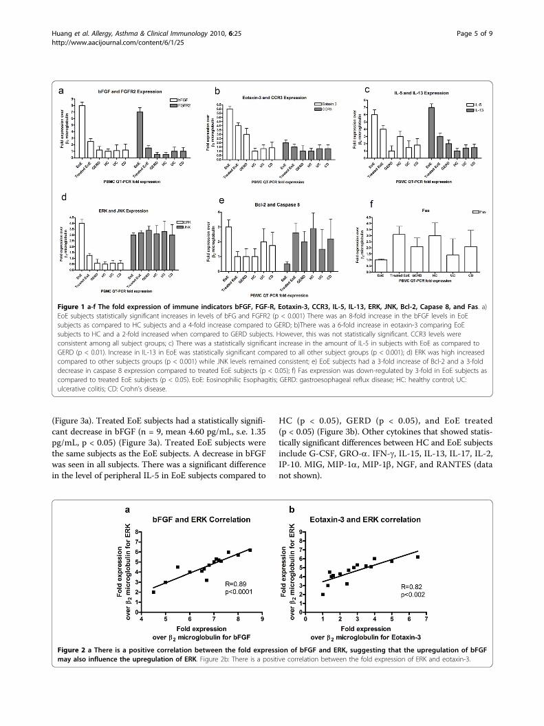
QT-PCR Assays demonstrate differential expression ofimmune indicators in EoERNA purified from subject PBMCs was used for QT-PCR assays. Transcripts for bFGF were found to beincreased in newly diagnosed EoE subjects and expres-sion was decreased after treatment. QT-PCR datashowed that untreated EoE subjects had an increasedexpression of bFGF that was up to eight-fold higher(n = 35) compared to the HCs (n = 8, 1×, p < 0.05), andcompared to when on treatment (n = 9 EoE subjects,1.5×, p < 0.001). Although FGF levels in GERD subjectswere slightly elevated (n = 8), it was not statisticallysignificant compared to HC (p > 0.05) (Figure 1a). EoEsubjects had statistically significant increased FGFR2expression compared to HC (14×, p < 0.05) (Figure 1a).EoE subjects had a slight but not statistically significantincrease in eotaxin-3 expression when compared toGERD (2×, p > 0.05) and to HC (6×, p > 0.05) (Figure1b). Eotaxin-1 and 2 transcripts were also assessed andfound similar to eotaxin-3 expression patterns (data notshown). CCR3, the eotaxin-3 receptor, also had an
increased expression factor of 2× compared to GERDand HC (Figure 1b). IL-5 expression was a six-foldincrease in EoE subjects versus GERD and a three-foldincrease versus HC (Figure 1c). However, the differencewas not statistically significant. IL-13 had a statisticallysignificant seven-fold increase in EoE subjects versusHC (p < 0.05) and a 3.5 fold increase over GERD sub-jects (p < 0.001) (Figure 1c). IL-13 increases were asso-ciated with EoE subjects with concomitant food allergies(data not shown).In summary, specific immune indicators with increased
expression in newly diagnosed EoE compared to treatedEoE, HC, GERD, UC, and CD were: bFGF, FGF-Receptor2, IL-13, IL-5, eotaxin, and CCR3 (in order of highest tolowest extent of increased transcript expression). Inter-estingly, upon examination of transcript expression ofsignaling pathway proteins and apoptosis-related proteinssuch as ERK, JNK, Bcl-2, caspase 8, and Fas, for the newlydiagnosed EoE subjects, it was found that the signalingpathway transcripts of ERK but not JNK were found tobe increased relative to HC, UC, CD and treated EoE(Figure 1d). ERK was expressed at a minimum of four-fold increase at statistically significant differences com-pared to all other subjects (p < 0.05) (Figure 1d). Theextent of the increases in ERK was correlated with bFGFincreases in expression (R = 0.89, p < 0.05) (Figure 2a)and with eotaxin increases in expression (R= 0.82,p < 0.05) (Figure 2b). This suggests that FGF signalingcould occur through ERK but not JNK, and that this sig-naling could enhance the ERK-dependent signaling path-ways associated with eotaxin. EoE subjects had a 3-foldincrease in Bcl-2 expression compared to treated EoEpatients while caspase 8 showed a 3-fold decrease(p < 0.05) (Figure 1e). Likewise, there was a 3-folddecrease in Fas expression levels in EoE subjects com-pared to the treated EoE subjects (p < 0.05) (Figure 1f).
Fluid Assays on plasma show highly elevated bFGFspecifically in EoE subjectsSince we found bFGF and other immune indicators to bespecifically increased in PBMCs of EoE subjects, we thenperformed fluid assays to determine whether they werepresent systemically in the plasma. Our fluid assay data(performed via Luminex 35-plex technology per manufac-turer’s instructions, Invitrogen and via Cytometric BeadArray Technology per manufacturer’s instructions, BDBiosciences) were important in the narrowing of potentialimmune indicators specific to eosinophilic esophagitis. Inplasma from HC subjects (n = 10), bFGF levels were low(mean 0.13 pg/mL, s.e. 0.09 pg/mL) while bFGF was sig-nificantly upregulated in EoE subjects (n = 10, mean 81.98pg/mL, s.e. 17.23 pg/mL for EoE, p < 0.05). Differences inbFGF levels between EoE and GERD were also significant(n = 10, mean 1.74 pg/mL, s.e. 0.64 for GERD, p < 0.05)
Huang et al. Allergy, Asthma & Clinical Immunology 2010, 6:25http://www.aacijournal.com/content/6/1/25
Page 4 of 9
(Figure 3a). Treated EoE subjects had a statistically signifi-cant decrease in bFGF (n = 9, mean 4.60 pg/mL, s.e. 1.35pg/mL, p < 0.05) (Figure 3a). Treated EoE subjects werethe same subjects as the EoE subjects. A decrease in bFGFwas seen in all subjects. There was a significant differencein the level of peripheral IL-5 in EoE subjects compared to
HC (p < 0.05), GERD (p < 0.05), and EoE treated(p < 0.05) (Figure 3b). Other cytokines that showed statis-tically significant differences between HC and EoE subjectsinclude G-CSF, GRO-a. IFN-g, IL-15, IL-13, IL-17, IL-2,IP-10. MIG, MIP-1a, MIP-1b, NGF, and RANTES (datanot shown).
Figure 1 a-f The fold expression of immune indicators bFGF, FGF-R, Eotaxin-3, CCR3, IL-5, IL-13, ERK, JNK, Bcl-2, Capase 8, and Fas. a)EoE subjects statistically significant increases in levels of bFG and FGFR2 (p < 0.001) There was an 8-fold increase in the bFGF levels in EoEsubjects as compared to HC subjects and a 4-fold increase compared to GERD; b)There was a 6-fold increase in eotaxin-3 comparing EoEsubjects to HC and a 2-fold increased when compared to GERD subjects. However, this was not statistically significant. CCR3 levels wereconsistent among all subject groups; c) There was a statistically significant increase in the amount of IL-5 in subjects with EoE as compared toGERD (p < 0.01). Increase in IL-13 in EoE was statistically significant compared to all other subject groups (p < 0.001); d) ERK was high increasedcompared to other subjects groups (p < 0.001) while JNK levels remained consistent; e) EoE subjects had a 3-fold increase of Bcl-2 and a 3-folddecrease in caspase 8 expression compared to treated EoE subjects (p < 0.05); f) Fas expression was down-regulated by 3-fold in EoE subjects ascompared to treated EoE subjects (p < 0.05). EoE: Eosinophilic Esophagitis; GERD: gastroesophageal reflux disease; HC: healthy control; UC:ulcerative colitis; CD: Crohn’s disease.
Figure 2 a There is a positive correlation between the fold expression of bFGF and ERK, suggesting that the upregulation of bFGFmay also influence the upregulation of ERK. Figure 2b: There is a positive correlation between the fold expression of ERK and eotaxin-3.
Huang et al. Allergy, Asthma & Clinical Immunology 2010, 6:25http://www.aacijournal.com/content/6/1/25
Page 5 of 9

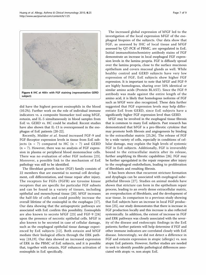
ImmunohistochemistryTo test whether there were increases in expression ofFGF in the local tissue of the esophagus in EoE com-pared to controls, immunohistochemistry (IHC) usingthe FGF antibody was conducted on slides from HCsubjects (n = 7), GERD subjects (n = 6), and EoE sub-jects (n = 7). Cells were counted at 400× (Figure 4). EoEsubjects were verified to have more than 15 eos/HPF asshown by EG2 antibody staining. HCs showed no cellssurrounded by FGF, and there was only a minimal pre-sence in GERD subjects. EoE subjects, however, consis-tently showed FGF in significantly higher countscompared to either GERD (p < 0.05) or HC (p < 0.05)(Figures 4, 5 and 6), based on an average of three areasper slide. The antibody was not guaranteed to beisoform-specific.
DiscussionOur data indicate that fibroblast growth factor is amonga set of factors that are differentially regulated in the
periphery in EoE, suggesting that eosinophilic esophagi-tis is not only a local condition but also a systemic dis-order that may be detected through analysis of plasmasamples. Although symptoms of disease of may be loca-lized at just the esophagus, our results show that EoEsubjects have a set of unique peripheral immune indica-tors that could be used as diagnostic indicators. In addi-tion, through analysis of PBMCs (which consist mainlyof lymphocytes, monocytes, macrophages, and dendriticcells rather than granulocytes), we were able to detectan activation state present in other immune cells in EoEthat could lead to subsequent activation of eosinophils.This would prove especially useful since current diagno-sis involves an esophageal tissue biopsy, which carriespotentially unnecessary risks.The plasma fluid assay data provided here show that
bFGF was upregulated in EoE subjects. Along withbFGF upregulation, an increase in eotaxin was also seenin EoE subjects. Interestingly, IL-5 was increased in theplasma of EoE subjects compared to that of GERD orHC. The subject with the highest IL-5 expression level
Figure 3 a) EoE subjects had an elevated level of bFGF in comparison (p < 0.05) to those of HC, GERD, and treated EoE subjects(same subjects as EoE subject but now on therapy); b)IL-5 was also elevated in EoE subjects compared to treated EoE subjects (p <0.05) and gastroesophageal reflux disease and healthy control subjects (p < 0.05). (*p < 0.05).
Figure 4 The number of cells that are FGF positive cells aresignificantly greater in EoE subjects than in HC (p < 0.05) orGERD (p < 0.05). (* p < 0.05).
Figure 5 IHC at 400× with FGF staining (representative EoEsubject).
Huang et al. Allergy, Asthma & Clinical Immunology 2010, 6:25http://www.aacijournal.com/content/6/1/25
Page 6 of 9
did have the highest percent eosinophils in the blood(10.2%). Further work on the role of individual immuneindicators vs. a composite biomarker tool using bFGF,eotaxin, and IL-5 simultaneously in blood samples fromEoE vs. GERD vs. HC could be studied. Recent studieshave also shown that IL-13 is overexpressed in the eso-phagus of EoE patients [20-22].Recently, Mulder et al. found increased FGF-9 and
FGF-Receptor expression levels in tissue from EoE sub-jects (n = 7) compared to HC (n = 7) and GERD(n = 7). However, there was no analysis of FGF expres-sion in plasma or peripheral blood mononuclear cells.There was no evaluation of other FGF isoforms [23].Moreover, a possible link to the mechanism of EoEpathology was still to be identified.The fibroblast growth factor (FGF) family consists of
22 members that are essential to normal cell develop-ment, cell differentiation, and tissue repair after injury.The receptors for FGFs (FGFR) are tyrosine kinasereceptors that are specific for particular FGF subsetsand can be found in a variety of tissues, includingepithelial and mesenchymal tissue [20]. bFGF increasesthe half-life of cells and could possibly increase theoverall lifetime of the eosinophil in the esophagus [17].Our data showing that the antiapoptotic pathways areassociated with EoE confirm this possibility. Eosinophilsare also known to secrete bFGF [23] and FGF-9 [24]upon the presence of necrotic epithelial cells. bFGF isalso known to be secreted in case of cellular damage,such as the esophageal epithelial tissue damage experi-enced by EoE subjects [12]. Both eotaxin and bFGFmediate their biological effects through the ERK path-way [18,19]. Our data showed an increased expressionof ERK in the PBMC of EoE subjects, and it is possiblethat, together with eotaxin, FGF enhances activation ofeosinophils in EoE specifically.
The increased global expression of bFGF led to theinvestigation of the local expression bFGF of the eso-phageal biopsies of the subjects. Our data show thatFGF, as assessed by IHC of local tissue and bFGFassessed by QT-PCR of PBMC, are upregulated in EoE.Initial immunohistochemistry antibody stains of FGFdemonstrate an increase in local esophageal FGF expres-sion levels in the lamina propria. FGF is diffusely spreadover the lamina propria, close to the surface mucinousepithelium and covers mucosal glands as well. Whilehealthy control and GERD subjects have very lowexpression of FGF, EoE subjects show higher FGFexpression. It is important to note that bFGF and FGF-9are highly homologous, sharing over 54% identical orsimilar amino acids (Protein BLAST). Since the FGF-9antibody was made against the entire length of theamino acid, it is likely that homologous isoforms of FGFsuch as bFGF were also recognized. These data furthersuggested that FGF expression levels may help differ-entiate EoE from GERD, since EoE subjects have asignificantly higher FGF expression level than GERD.bFGF may be involved in the esophageal tissue fibrosis
that is common to many EoE subjects. Past research hasdemonstrated that bFGF is a pro-fibrotic cytokine thatmay promote both fibrosis and angiogenesis by bindingto the extracellular matrix [25,26]. The release of FGFby a wide variety of cells, especially in conditions of cel-lular damage, may explain the high levels of systemicFGF in EoE subjects. Additionally, FGF is irreversiblybound to the extracellular matrix after its release,further amplifying its fibrotic capabilities [26]. FGF maybe further upregulated in the repair response after injuryto the esophageal endothelium, leading to proliferationof fibroblasts and resulting fibrosis.It has been shown that recurrent stricture formation
and dysphagia can be associated with esophageal sube-pithelial fibrosis [27]. Studies on animal models haveshown that stricture can form in the epithelium repairprocess, leading to an overly dense extracellular matrix,an overproduction of fibroblasts, and the development ofscar tissue. In comparison to a previous study showingthat EoE subjects have an increase in local FGF produc-tion [25], our study demonstrates that there is increase inFGF production locally and this increase is also reflectedsystemically. In addition, the extent of increase in FGFand ERK pathways was closely associated with the sever-ity of the disease and endoscopic findings in the EoEpatients; further patients will help determine if FGF andother immune indicators are correlated closely with EoEdisease. Interestingly, we did not detect a difference inFGF and eotaxin expression in atopic (allergic) vs. nonatopic EoE patients. However, further studies are neededto seek to identify possible pathological differences asso-ciated with atopic vs. non atopic EoE.
Figure 6 IHC at 400× with FGF staining (representative GERDsubject).
Huang et al. Allergy, Asthma & Clinical Immunology 2010, 6:25http://www.aacijournal.com/content/6/1/25
Page 7 of 9

bFGF expression is essential to the transcription ofERK [18] and ERK feeds into the eotaxin-3 pathway,potentially further activating the eosinophil and improv-ing its sensitivity and migration towards eotaxin in theesophageal tissue. Activation of ERK may induce bothdegranulation and chemotaxis of eosinophils [19]. It doesappear that eosinophils also express bFGF [28]. The eosi-nophils may be operating in a positive feedback loop inwhich the expression bFGF is encouraging the increasedactivation of the eosinophil, which then leads to morebFGF expression and better chemotaxis. Additionally,like bFGF, FGF-9 can activate both ERK1 and ERK2 [29].Further analysis of more subjects is necessary to iden-
tify additional immune indicators that constitute aunique EoE plasma protein composite or individualmarker profile. Lower esophageal eosinophilia is com-mon in GERD, and further determination of numbers ofeosinophils in the tissues of GERD subjects is needed;we believe our current results reflect a relative differencein GERD and EoE subjects. We have focused on EoE todetermine the involvement of specific blood immuneindicators in the disease; we will continue to study othereosinophilic gastroenterological disorders such as eosi-nophilic gastroenteritis and eosinophilic colitis to deter-mine the role of FGF in those disease entities.We have demonstrated that fibroblast growth factors
may play an important role in the pathophysiology ofEoE and may be part of a set of immune indicators thatcould, without biopsy, differentiate EoE subjects fromsubjects with other clinically similar symptoms such asGERD. In this effort to determine possible pathologicalmechanisms, we found that FGF increase was associatedwith activation of ERK and of anti-apoptotic, pathwayswhich could enhance eotaxin signaling and increaseeosinophil lifespan, respectively.
ConclusionsSubjects with eosinophilic esophagitis had differentimmune indicator profiles, specifically with increases inbasic fibroblast growth factor in blood plasma, periph-eral blood mononuclear cells, and local esophagealtissue compared to subjects with gastroesophageal refluxdisease, ulcerative colitis, Crohn’s disease, and healthycontrols. The upregulation of fibroblast growth factorwas found to be associated with ERK expression, whichis in turn essential to the expression eotaxin-3, an eosi-nophil chemoattractant. Additionally, increases in basicfibroblast growth factor were found to be associatedwith activation of anti-apoptotic pathways. Therefore,FGF with eotaxin and antiapoptic factors could enhancemigration and prolong the life-span of eosinophil,respectively. This may also explain the prolonged pre-sence of higher than normal numbers of eosinophils inthe esophagus.
Additional material
Additional file 1: Supplementary tables. Table S1a: Demographics ofEoE patients. D (distal) and P (proximal) reflect location of the eosinophilcount. Eosinophil counts are given as per high powered field (> greaterthan; = equal to). If negative for any allergies, total IgE less than 10 kU/mL. Table S1b: Demographics of GERD patients Table S1c: Demographicsof healthy controls. Table S1 d. Demographics of Crohn’s disease andulcerative colitis patients. Table S2: Treatment emographics of EoEsubjects.
AcknowledgementsJennifer Huang, Jae Joh, and Scott Seki would like to thank the StanfordVice Provost for Undergraduate Education (VPUE) grants for undergraduatestudents for supporting the work done. Judy Fuentebella received afellowship grant from the Siegelman Fellowship Award at the StanfordSchool of Medicine and Anup Patel received a fellowship grant from theTissue and Transplant Engineering Award at Stanford School of Medicine.Kari Nadeau received a Institute for Immunity, Transplantation, and InfectiousDiseases seed grant and a Stanford Digestive Disease Center award. We aregrateful to the nurses in the Ambulatory Procedure Unit for their hard workand help in collecting the subject samples. Also, we would like to thank theStanford FACS facility, the QT-PCR facility, and the Institute for Immunity,Transplantation, and Infectious Diseases and the Human Immune MonitoringCenter for their services.
Author details1Stanford School of Medicine, Stanford, CA 94305, USA. 2California PacificMedical Center, San Francisco, CA 94118, USA.
Authors’ contributionsJH and JJ performed the plasma and PBMC separation, theimmunohistochemistry, the QT-PCR tissue preparation, and the data analysis.JF, AP, TN, and SS aided in the immunohistochemistry. VS, CN, AQ, DB, WB,KC, JK, JP, and LN and KN aiding in obtaining biopsy and blood samples. NRand LH were involved in enrolling and consenting patients. JH drafted themanuscript. All authors read and approved the final manuscript.
Competing interestsThe authors declare that they have no competing interests.
Received: 6 March 2010 Accepted: 5 September 2010Published: 5 September 2010
References1. Norvell JM, Venarske D, Hummell DS: Eosinophilic esophagitis: an
allergist’s approach. Ann Allergy Asthma Immunol 2007, 98(3):207-14.2. Aceves SS, Newbury RO, Dohil R, Schwimmer J, Bastian JF: Distinguishing
eosinophilic esophagitis in pediatric patients: clinical, endoscopic, andhistologic features of an emerging disorder. J Clin Gastroenterol 2007,41(3):252-6.
3. Katzka DA: Eosinophilic esophagitis: it’s all in the family. GastrointestEndosc 2007, 65(2):335-6.
4. Ferguson DD, Foxx-Orenstein AE: Eosinophilic esophagitis: an update. DisEsophagus 2007, 20(1):2-8.
5. Noel RJ, Putnam PE, Rothenberg ME: Eosinophilic esophagitis. N Engl JMed 2004, 351(9):940-1.
6. Buckmeier B, Rothenberg M, Collins M: The incidence and prevalence ofeosinophilic esophagitis. J Allergy Clin Immunol 2008, 121(2):S71.
7. Furuta GT, Straumann A: Review article: the pathogenesis andmanagement of eosinophilic oesophagitis. Aliment Pharmacol Ther 2006,24(2):173-82.
8. Liacouras CA, Spergel JM, Ruchelli E, Verma R, Mascarenhas M, Semeao E,Flick J, Kelly J, Brown-Whitehorn T, Mamula JE, Markowitz : Eosinophilicesophagitis: a 10-year experience in 381 children. Clin GastroenterolHepatol 2005, 3(12):1198-206.
Huang et al. Allergy, Asthma & Clinical Immunology 2010, 6:25http://www.aacijournal.com/content/6/1/25
Page 8 of 9
9. Kelly KJ, Lazenby AJ, Rowe PC, Yardley JH, Perman JA, Sampson HA:Eosinophilic esophagitis attributed to gastroesophageal reflux:improvement with an amino acid-based formula. Gastroenterology 1995,109(5):1503-12.
10. Fogg MI, Ruchelli E, Spergel JM: Pollen and eosinophilic esophagitis.J Allergy Clin Immunol 2003, 112(4):796-7.
11. Mishra A, Hogan S, Brandt EB, Rothenberg ME: An etiological role foraeroallergens and eosinophils in experimental esophagitis. J Clin Invest2001, 107(1):83-90.
12. Spergel JM, Beausoleil JL, Mascarenhas M, Liacouras CA: The use of skinprick tests and patch tests to identify causative foods in eosinophilicesophagitis. J Allergy Clin Immunol 2002, 109(2):363-8.
13. Chehade M, Sampson HA: Epidemiology and etiology of eosinophilicesophagitis. Gastrointest Endosc Clin N Am 2008, 18(1):33-44, viii.
14. Furuta GT, Liacouras CA, Collins MH, Gupta SK, Justinich C, Putnam PE,Bonis P, Hassall E, Straumann A, Rothenberg ME: Eosinophilic esophagitisin children and adults: a systematic review and consensusrecommendations for diagnosis and treatment. Gastroenterology 2007,133(4):1342-63.
15. Straumann A: Eosinophilic esophagitis: a rapidly emerging disease.Esophagus 2008, 5(4):177-183.
16. Ornitz DM, Itoh N: Fibroblast growth factors. Genome Biol 2001, 2(3):REVIEWS3005.
17. Karsan A, Yee E, Poirier GG, Zhou R, Craig JM, Harlan : Fibroblast growthfactor-2 inhibits endothelial cell apoptosis by Bcl-2-dependent andindependent mechanisms. Am J Pathol 1997, 151(6):1775-84.
18. Pintucci G, Moscatelli D, Saponara F, Biernacki PR, Baumann FG, Bizekis C,Galloway AC, Basilico C, Mignatti : Lack of ERK activation and cellmigration in FGF-2-deficient endothelial cells. FASEB J 2002, 16(6):598-600.
19. Kampen GT, Stafford S, Adachi T, Jinquan T, Quan S, Grant JA, Skov PS,Poulsen LK, Alam R: Eotaxin induces degranulation and chemotaxis ofeosinophils through the activation of ERK2 and p38 mitogen-activatedprotein kinases. Blood 2000, 95(6):1911-7.
20. Rothenberg ME: Biology and treatment of eosinophilic esophagitis.Gastroenterology 2009, 137(4):1238-49.
21. Mishra A, Rothenberg ME: Intratracheal IL-13 induces eosinophilicesophagitis by an IL-5, eotaxin-1, and STAT6-dependent mechanism.Gastroenterology 2003, 125(5):1419-27.
22. Blanchard C, Mishra A, Saito-Akei H, Monk P, Anderson I, Rothenberg ME:Inhibition of human interleukin-13-induced respiratory and oesophagealinflammation by anti-human-interleukin-13 antibody (CAT-354). Clin ExpAllergy 2005, 35(8):1096-103.
23. Stenfeldt AL, Wenneras C: Danger signals derived from stressed andnecrotic epithelial cells activate human eosinophils. Immunology 2004,112(4):605-14.
24. Mulder DJ, Pacheco I, Hurlbut DJ, Mak N, Furuta GT, MacLeod RJ,Justinich CJ: FGF9-induced proliferative response to eosinophilicinflammation in oesophagitis. Gut 2009, 58(2):166-73.
25. Dinbergs ID, Brown L, Edelman ER: Cellular response to transforminggrowth factor-beta1 and basic fibroblast growth factor depends onrelease kinetics and extracellular matrix interactions. J Biol Chem 1996,271(47):29822-9.
26. Lawrence A, Khanna D, Misra R, Aggarwal A: Increased expression of basicfibroblast growth factor in skin of patients with systemic sclerosis.Dermatol Online J 2006, 12(1):2.
27. Chehade M, Sampson HA, Morotti RA, Magid MS: Esophageal subepithelialfibrosis in children with eosinophilic esophagitis. J Pediatr GastroenterolNutr 2007, 45(3):319-28.
28. Hoshino M, Takahashi M, Aoike N: Expression of vascular endothelialgrowth factor, basic fibroblast growth factor, and angiogeninimmunoreactivity in asthmatic airways and its relationship toangiogenesis. J Allergy Clin Immunol 2001, 107(2):295-301.
29. Antoine M, et al: Expression and function of fibroblast growth factor(FGF) 9 in hepatic stellate cells and its role in toxic liver injury. BiochemBiophys Res Commun 2007, 361(2):335-41.
doi:10.1186/1710-1492-6-25Cite this article as: Huang et al.: Eotaxin and FGF enhance signalingthrough an Extracellular signal-related kinase (ERK)-dependent pathwayin the pathogenesis of Eosinophilic Esophagitis. Allergy, Asthma & ClinicalImmunology 2010 6:25.
Submit your next manuscript to BioMed Centraland take full advantage of:
• Convenient online submission
• Thorough peer review
• No space constraints or color figure charges
• Immediate publication on acceptance
• Inclusion in PubMed, CAS, Scopus and Google Scholar
• Research which is freely available for redistribution
Submit your manuscript at www.biomedcentral.com/submit
Huang et al. Allergy, Asthma & Clinical Immunology 2010, 6:25http://www.aacijournal.com/content/6/1/25
Page 9 of 9