Eosinophilic Granulomatosis with Polyangiitis and Diffuse Gastrointestinal Involvement

Fish & Shellfish Immunology (2000) 10, 143–154doi:10.1006/fsim.1999·0233Available online at http://www.idealibrary.com on
Spontaneous cytotoxic activity of eosinophilic granulecells separated from the normal peritoneal cavity of
Dicentrarchus labrax
MATTEO CAMMARATA1, MIRELLA VAZZANA
1, MELCHIORRE CERVELLO2,
VINCENZO ARIZZA1
AND NICOLOv PARRINELLO1*
1Laboratory of Marine Immunobiology, Department of Animal Biology,University of Palermo, Via Archirafi 18, 90123, Palermo, Italy,
2Institute of Developmental Biology, National Research Council,Via Ugo La Malfa, 153, 90146, Palermo, Italy
(Received 1 March 1999, accepted 22 June 1999)
In this study the spontaneous in vitro cytotoxic activity to tumour cell lines,(K562), by unstimulated sea bass (Dicentrarchus labrax) leukocytes wasexamined by trypan blue exclusion test and lactate dehydrogenase releaseassay. A high anti-tumour cell line activity of resident peritoneal leukocyteswas found at an e#ector to target ratio (E:T) of 25:1 after incubation for 2 h at18� C. Rabbit and sheep erythrocytes were not lysed. A low activity wasdisplayed by head kidney and spleen cell populations whereas blood leuko-cytes revealed no significant activity. The e#ect of E:T ratio on cytotoxicity aswell as microscopy observations suggested that the cytotoxic reactionrequired e#ector-target cell contact. Eosinophilic granule cells, isolated on aPercoll density gradient from a peritoneal wash, appeared to be responsiblefor the in vitro cytotoxic activity. � 2000 Academic Press
Key words: Dicentrarchus labrax, Teleostei, cytotoxicity, peritoneal cavity,eosinophilic granule cell.
I. Introduction
Natural cellular immunity represents a first barrier against pathogens. It is arapid and e#ective response that predominantly involves phagocytes andcytotoxic cells. Spontaneous cytotoxic activity of leukocytes against certainmammalian and fish cell lines (Hinuma et al., 1980; Graves et al., 1984;Suzumura et al., 1994), virus-infected cells (Yoshinaga et al., 1994) and aprotozoan (Graves et al., 1985) has been demonstrated in some species of fish.In elasmobranchs, spontaneous cytotoxicity requiring no in vitro activationis a property of macrophage-like cells (McKinney et al., 1986). In teleoststhe cytolytic activity of leukocytes seems to be mediated by various e#ectorcells globally referred to as non-specific cytotoxic cells (NCC) (Evans et al.,1988). Stuge et al. (1995) reported that the cytotoxic e#ector cells in catfish
* Corresponding author. Department of Animal Biology, Via Archirafi, 18 90123 Palermo, Italy.E-mail:[email protected]
1431050–4648/00/020143+12 $35.00/0 � 2000 Academic Press
144 M. CAMMARATA ET AL.
peripheral blood are di#erent from those found in the pronephros. Further-more, the cold target inhibition test showed that carp cytotoxic cells (NK-like)do not consist of a single population for target recognition (Suzumura et al.,1994). In this species, NCCs consist of small agranular lymphocytes ormonocytes in the blood (Meseguer et al., 1994) or neutrophilic granulocytes inthe head kidney (Kurata et al., 1995). In Oncorynchus mykiss the e#ector cellsseem to be small agranular lymphocytes in the head kidney (Greenlee et al.,1991) and in the intestinal intraepithelial tissue (McMillan & Secombes, 1997).
In this study spontaneous cytotoxic activity to tumour cell lines anderythrocytes by Sea bass, Dicentrarchus labrax, leukocytes is reported. Thekilling activity and organ distribution of the e#ector cells were examined,with particular reference to resident peritoneal leukocytes. The peritonealcavity, both in mammals (Appelberg et al., 1991) and fishes (Afonso et al., 1997),provides a model of distribution and mobilisation of cells that are quickly andextensively attracted by intraperitoneal injection of stimulants (Afonso et al.,1998). In the case of fish, it has been reported that the sterile unstimulatedperitoneal cavity of rainbow trout contains a significant population ofresident macrophages but very few neutrophils (Afonso et al., 1997).Bodammer (1986) showed that the principal cells recovered from the peri-toneal exudate of striped bass (Morone saxatilis) challenged with bacteria aremacrophages, eosinophilic granule cells and neutrophils. We found that thecell population in the unstimulated peritoneal cavity of D. labrax, obtainedby simple washing with physiological medium, contains macrophages, neutro-phils, lymphocytes and eosinophilic granule cells. These cell types wereisolated on Percoll density gradients and tested for in vitro cytotoxic activity.
II. Materials and Methods
ANIMALS
Sea bass (Dicentrarchus labrax) (200–250 g) were obtained from acommercial fish farm (Petrosino, TP).
COLLECTION OF EFFECTOR CELLS
The fish were anaesthetised with 0·05% MS222 (3-aminobenzoic acid ethylester) (Sigma) in seawater and sampled at the farm. The tissues and peritonealcells were kept on ice and taken quickly to the laboratory. Blood was collectedfrom the heart into a sterile plastic syringe containing 0·2 ml of heparin, andcentrifuged at 800�g for 10 min at 4� C. The head kidney and spleen weresurgically removed and placed in cold medium (Leibovitz L15 medium contain-ing 2% fetal calf serum, 100 units penicillin ml�1, 100 units streptomycinml�1, and 10 units heparin ml�1). All culture medium components were fromGibco.
Peritoneal cavity cells (PCCs) were obtained as follows: after disinfection ofthe ventral surface of the fish with 70% ethyl alcohol, the peritoneal cavitywas injected with 15 ml of isotonic (370 mOsm kg�1) Leibovitz medium. Aftermassaging the ventral surface of the fish for 10 min, the medium containing

CYTOTOXICITY OF SEA BASS LEUKOCYTES 145
the PCCs was harvested with a syringe. The PCCs were obtained by centrifu-gation at 400�g for 10 min at 4� C. The fish serum and medium osmolarity wasmeasured by an osmometer (Röebling).
TARGET CELLS
The human erythromyeloid leukaemia-derived cell line (K562), humanhystocytic lymphoma-derived cell line (U937), and Burkitt lymphoma derivedcell line (Raji) were maintained in RPMI 1640 medium (Gibco) supplementedwith 10% heat-inactivated fetal calf serum (Flow), gentamycin, streptomycinand HEPES bu#er (Boerhinger), at 37� C with 5% CO2. The culture was fedbiweekly in order to maintain constant exponential cell growth.
The Zooprophylaxis Institute (Palermo, Italy) provided sheep erythrocytes.Rabbit erythrocytes (RE) were from Sclavo (Siena, Italy).
DENSITY GRADIENT SEPARATION OF CELLS
Pronephros and spleen cell suspensions or blood were centrifuged on a34/46% Percoll/Hanks balanced salt solution (HBSS: NaCl 190 mM, KCl5·36 mM, glucose 5·54 mM, KH2PO4 0·44 mM, Na2HPO4 0·56 mM; pH 7·6,370 mOsm kg�1) density gradient. A band of enriched leukocytes was visibleat the interface. Cellular debris was separated at the top of the gradient andthe erythrocyte pellet was packed at the bottom.
To separate the leukocyte populations present in the peritoneal wash, acontinuous density gradient was formed with 55% Percoll in HBSS bycentrifugation at 25 000�g for 20 min at 4� C. The PCC suspension wasimmediately layered on the preformed Percoll gradient and centrifuged at2500�g for 25 min at 7� C. After centrifugation two bands of cells wereformed. The cells from each band were counted in a haemocytometer and theleukocytes adjusted to 2·5 �105 ml�1 in HBSS.
TRYPAN BLUE EXCLUSION TEST
Dead cells were determined by addition of 0·01% trypan blue to the medium.This test was also used to evaluate the cytotoxic activity against tumour celllines, the dye was added into the reaction mixture after 2 h incubation.
To show target cell death following an in vitro cytotoxic reaction, thetrypan blue was added to the medium 20 min after the leukocytes were mixedwith target cells. Samples of the reaction mixture were smeared on slides andexamined under the microscope.
METHODS FOR STAINING LEUKOCYTE SMEARS
Samples (50 �l) of the leukocyte suspensions (5�106 cells ml�1) werelayered on a clean slide, allowed to settle for 30 min and then fixed withundiluted methanol for 7 min at 4–8� C. Fixed smears were treated withMay-Grünwald for 3 min and after washing, with Giemsa for 10 min. The slides
146 M. CAMMARATA ET AL.
were rinsed with deionised water and, after ethanol dehydration and xylenetreatment, were mounted in Permount (Sigma).
CYTOTOXIC ASSAY
The cytotoxic assay against erythrocytes has been previously described(Cammarata et al., 1997). Briefly, 200 �l of leukocyte suspension (E, 1·5�105
cells) in phosphate bu#ered saline (PBS: NaCl 103·6 mM, KH2PO4 1·46 mM,Na2HPO4 0·8 mM, KCl 2·6 mM, CaCl22H2O 0·9 mM, MgCl2 6H2O 0·49 mM, pH 7·4)were mixed with an equal volume of freshly prepared rabbit erythrocytes (T,8�106 cells) in the same medium. Spontaneous haemoglobin release neverexceeded 5% of the total release obtained after keeping the erythrocytes indistilled water. The mixture was incubated at 18� C for 2 h, centrifuged at200�g for 5 min at 7� C and the amount of released haemoglobin in thesupernatant was estimated by reading the absorbance at 541 nm. The degree ofhaemolysis was determined according to the equation: Percent haemolysis=measured release – spontaneous release/complete release – spontaneousrelease �100.
Cytotoxicity assay against tumour cell lines was performed using a cyto-toxic detection Kit (Boehringer) based on determination of lactate dehydro-genase (LDH) activity released from lysed target cells (Korzeniewski &Callewaert, 1983). The target cells were washed and suspended in PBSsupplemented with 1% bovine serum albumin (PBS-BSA, 370 mOsm kg�1) at aconcentration of 1�105 cells ml�1. All tests were performed in triplicate with1�104 target cells well�1 and with appropriate e#ector-to-target ratios inV-shaped microplates (Nunc) in a total volume of 200 �l. Plates were centri-fuged for 1 min at 100�g and incubated for 2 h at 18� C. The plates were thencentrifuged for 5 min at 400�g, and the release of LDH from lysed cells in100 �l of supernatant from each well was determined by reading the absorb-ance at 490 nm in a microplate reader (Uniskan I, Labsystems). Spontaneousand maximum release were measured in 100 �l of supernatant from wellscontaining target cells only or target cells with 1% Triton X-100 (Fluka).Spontaneous baseline LDH release from target (1�104 cells well�1) ande#ector cells were used as controls. The values of the controls were subtractedfrom the degree of target cell lysis determined according to the equation:Percent lysis=(measured release – spontaneous target release) – spontaneouse#ector release/complete release – spontaneous target release �100.
Appropriate controls showed that leukocyte mortality in PBS, under theexperimental conditions employed, was lower than 5%. The living cells wereobserved through Nomarski di#erential interference contrast optics (DIC).Unless otherwise specified the cytotoxic reactions to both erythrocytes andtumour cells were carried out at 18� C for 2 h, at 25:1 E/T ratio, in 370 mOsm/kg�1 medium.
STATISTICAL METHODS
The data were examined by Student’s t-test to determine the statisticalprobability, P<0·01 being the lowest significance level.

CYTOTOXICITY OF SEA BASS LEUKOCYTES 147
III. Results
50:10
E:T ratio
Per
cen
t ce
ll m
edia
ted
lysi
s
40
30
20
10
1.6:1 3.2:1 6.3:1 25:112.5:1
**
B2
Fig. 1. E#ect of E:T ratio on spontaneous cytotoxic activity of Dicentrarchus labrax byunseparated leukocytes or separated eosinophilic granule cells (EGCs) from perito-neal cavity co-incubated with K562 target cells. Lactate dehydrogenase release intothe supernatant was used to calculate the percentage of target cell lysis. Thepercent of target cell lysis was also determined by trypan blue exclusion test(TBET). Unseparated leukocytes assayed by TBET (�); Unseparated leukocytesassayed by LDH release method (�); Percoll density enriched eosinophilic granulecells population (B2) (�). *: The same number of cells contained in each reactionmixture were assayed for the spontaneous LDH release in the absence of targetcells: () unseparated leukocytes; () EGCs. � TBET of unseparated leukocytes inthe absence of target cells. Percent cell lysis of target cells, assayed by LDH releasemethod or TBET, were less than 5%.
EFFECT OF OSMOLARITY AND E:T RATIO ON PCC CYTOTOXIC ACTIVITY
Leukocyte mortality of less than 5% and highest values of cytotoxicity(36�8·2%) were obtained when the osmolarity was 370�30 mOsm kg�1, thesame as that measured in the serum of D. labrax. A significant decrease(P<0·001) of the PCC cytotoxic activity against the K562 cell line (10�6%)was found when the medium of the reaction mixture was at the physiologicalosmolarity of the target (280 mOsm kg�1). In that medium leukocytemortality was greater than 20%.
The time course and e#ector:target ratio experiments were carried outat 370 mOsm kg�1 using both LDH release and trypan blue assays. Thetime course (0·25–12 h) experiments showed that the highest activity wasreached after 2 h incubation at 18� C. Higher temperature values (20–37� C)significantly increased the leukocyte mortality.
To examine the cytotoxic mechanism, cytotoxicity was assayed at di#erentE:T ratios (Fig. 1). The curve, obtained by plotting the numbers of unfraction-ated leukocytes against the lysis of K562 tumour cells, was sigmoidal in shape.A significant rise in lysis was found when 2·5�106 leukocytes ml�1 were
148 M. CAMMARATA ET AL.
incubated with 1·0�105 target cells ml�1 (E:T ratio 25:1). At the highest E:Tvalues an increased background of spontaneous target lysis was evident.
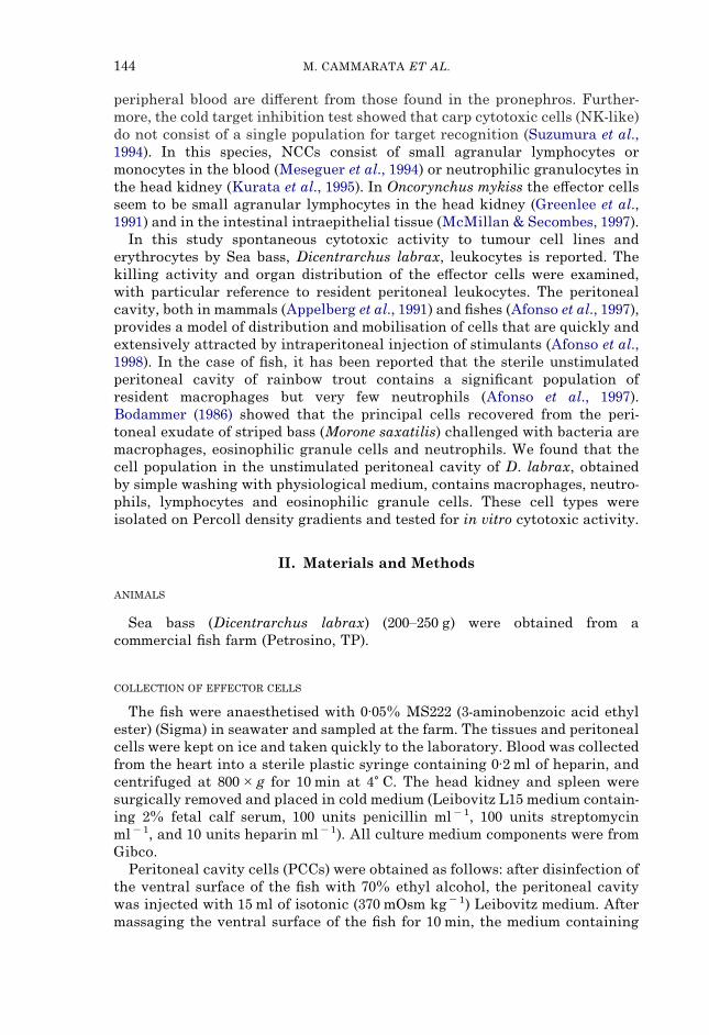
CYTOTOXIC ACTIVITY OF LEUKOCYTES FROM VARIOUS TISSUES EXAMINED BY LDH METHOD
Fig. 2 shows the anti-K562 cytotoxic activity of the leukocytes separatedfrom the various tissues. PCCs exhibited the highest activity. A lower level(P<0·001) of activity was shown by head kidney and spleen leukocytes, bloodrevealed no significant activity.
CYTOTOXIC ACTIVITY AGAINST VARIOUS TARGETS
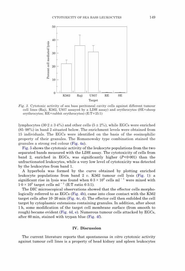
To examine the specificity range of the e#ector peritoneal cavity cells, LDHassays against Raji and U937 tumour cell lines as well as against sheep andrabbit erythrocytes were performed. As shown in Fig. 3, Raji and U937 cellsappeared to be more sensitive than K562 cell lines. Both the erythrocyte typeswere unsensitive even when assayed at an E:T ratio of 100:1.
0
Per
cen
t ce
ll m
edia
ted
lysi
s 40
30
20
10
Pronephros
50
Spleen P.C.C. Blood
Fig. 2. Cytotoxic activity (LDH assay) of sea bass leukocytes from pronephros, spleen,peritoneal cavity (PCC) and blood against K562 tumour cell line (E:T=25:1).
PERCOLL ENRICHMENT OF EOSINOPHILIC GRANULE CELLS FROM PERITONEAL CAVITY
Microscopical observation revealed that cells morphologically identified asmacrophages (26·4�7·8%), neutrophils (22·8�6·7%), lymphocytes (32·3�3·4%) and eosinophilic granule cells (EGCs) (18·5�6·4%) were contained inthe unseparated leukocyte suspension from the peritoneal wash.
To identify and characterise the e#ector cells from the peritoneal cavity, thecytotoxic assays were carried out with leukocytes separated from eachspecimen on a continuous 55% Percoll/HBSS density gradient. After centri-fugation, two distinct bands were visible within the Percoll gradient. Theupper band 1 contained macrophages (34�5%), neutrophils (30·8�5·7%),
CYTOTOXICITY OF SEA BASS LEUKOCYTES 149
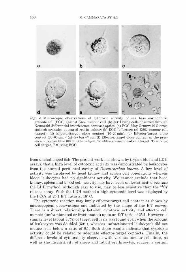
lymphocytes (30·2�3·4%) and other cells (5�2%), while EGCs were enriched(85–98%) in band 2 situated below. The enrichment levels were obtained from15 individuals. The EGCs were identified on the basis of the eosinophilicproperty of their granules. The Romanowsky type combination stained thegranules a strong red colour (Fig. 4a).
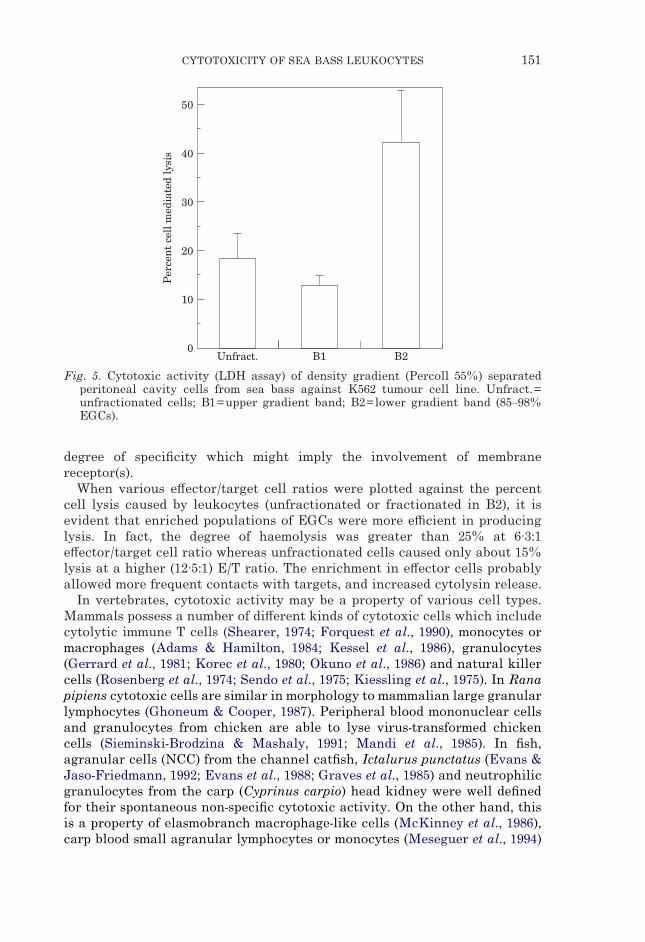
Fig. 5 shows the cytotoxic activity of the leukocyte populations from the twoseparated bands measured with the LDH assay. The cytotoxicity of cells fromband 2, enriched in EGCs, was significantly higher (P<0·001) than theunfractionated leukocytes, while a very low level of cytotoxicity was detectedby the leukocytes from band 1.
A hyperbola was formed by the curve obtained by plotting enrichedleukocyte populations from band 2 v. K562 tumour cell lysis (Fig. 1): asignificant rise in lysis was found when 6·3�105 cells ml�1 were mixed with1·0�105 target cells ml�1 (E:T ratio 6·3:1).
The DIC microscopical observations showed that the e#ector cells morpho-logically referred to as EGCs (Fig. 4b), came into close contact with the K562target cells after 10–20 min (Fig. 4c, d). The e#ector cell then enfolded the celltarget by cytoplasmic extensions containing granules. In addition, after about1 h, some modification of the target cell membrane surface (from smooth torough) became evident (Fig. 4d, e). Numerous tumour cells attacked by EGCs,after 60 min, stained with trypan blue (Fig. 4f).
0
Per
cen
t ce
ll m
edia
ted
lysi
s 40
30
20
10
K562
50
Raji U937 RE SE
Target
Fig. 3. Cytotoxic activity of sea bass peritoneal cavity cells against di#erent tumourcell lines (Raji, K562, U937 assayed by a LDH assay) and erythrocytes (SE=sheeperythrocytes; RE=rabbit erythrocytes) (E:T=25:1)
IV. Discussion
The current literature reports that spontaneous in vitro cytotoxic activityagainst tumour cell lines is a property of head kidney and spleen leukocytes
150 M. CAMMARATA ET AL.
Fig. 4. Microscopic observations of cytotoxic activity of sea bass eosinophilicgranule cell (EGC) against K562 tumour cell. (b)–(e): Living cells observed throughNomarski di#erential interference contrast optics. (a) EGC May Grunwald Giemsastained; granules appeared red in colour; (b) EGC (e#ector); (c) K562 tumour cell(target); (d) E#ector/target close contact (10–20 min); (e) E#ector/target closecontact (30–60 min), (a)–(e) bar=7 �m; (f) E#ector/target close contact in the pres-ence of trypan blue (60 min) bar=6 �m. Td=blue stained dead cell target, Ta=livingcell target, E=living EGC.
from unchallenged fish. The present work has shown, by trypan blue and LDHassays, that a high level of cytotoxic activity was demonstrated by leukocytesfrom the normal peritoneal cavity of Dicentrarchus labrax. A low level ofactivity was displayed by head kidney and spleen cell populations whereasblood leukocytes had no significant activity. We cannot exclude that headkidney, spleen and blood cell activity may have been underestimated becausethe LDH method, although easy to use, may be less sensitive than the 51Crrelease assay. With the LDH method a high cytotoxic level was displayed bythe PCCs at 25:1 E:T ratio at 18� C.
The cytotoxic reaction may imply e#ector-target cell contact as shown bymicroscopical observations and indicated by the shape of the E:T curves.There is a direct relationship between cytotoxic activity and e#ector cellnumber (unfractionated or fractionated) up to an E:T ratio of 25:1. However, asimilar level (about 35%) of target cell lysis was found even when the amountof leukocytes was doubled (50:1), whereas unfractionated leukocytes did notinduce lysis below a ratio of 6:1. Both these results indicate that cytotoxicactivity could be related to adequate e#ector-target contacts. Finally, thedi#erent levels of cytotoxicity observed with various tumour cell lines, aswell as the insensitivity of sheep and rabbit erythrocytes, suggest a certain
CYTOTOXICITY OF SEA BASS LEUKOCYTES 151
0
Per
cen
t ce
ll m
edia
ted
lysi
s 40
30
20
10
Unfract.
50
B2B1
Fig. 5. Cytotoxic activity (LDH assay) of density gradient (Percoll 55%) separatedperitoneal cavity cells from sea bass against K562 tumour cell line. Unfract.=unfractionated cells; B1=upper gradient band; B2=lower gradient band (85–98%EGCs).
degree of specificity which might imply the involvement of membranereceptor(s).
When various e#ector/target cell ratios were plotted against the percentcell lysis caused by leukocytes (unfractionated or fractionated in B2), it isevident that enriched populations of EGCs were more e$cient in producinglysis. In fact, the degree of haemolysis was greater than 25% at 6·3:1e#ector/target cell ratio whereas unfractionated cells caused only about 15%lysis at a higher (12·5:1) E/T ratio. The enrichment in e#ector cells probablyallowed more frequent contacts with targets, and increased cytolysin release.
In vertebrates, cytotoxic activity may be a property of various cell types.Mammals possess a number of di#erent kinds of cytotoxic cells which includecytolytic immune T cells (Shearer, 1974; Forquest et al., 1990), monocytes ormacrophages (Adams & Hamilton, 1984; Kessel et al., 1986), granulocytes(Gerrard et al., 1981; Korec et al., 1980; Okuno et al., 1986) and natural killercells (Rosenberg et al., 1974; Sendo et al., 1975; Kiessling et al., 1975). In Ranapipiens cytotoxic cells are similar in morphology to mammalian large granularlymphocytes (Ghoneum & Cooper, 1987). Peripheral blood mononuclear cellsand granulocytes from chicken are able to lyse virus-transformed chickencells (Sieminski-Brodzina & Mashaly, 1991; Mandi et al., 1985). In fish,agranular cells (NCC) from the channel catfish, Ictalurus punctatus (Evans &Jaso-Friedmann, 1992; Evans et al., 1988; Graves et al., 1985) and neutrophilicgranulocytes from the carp (Cyprinus carpio) head kidney were well definedfor their spontaneous non-specific cytotoxic activity. On the other hand, thisis a property of elasmobranch macrophage-like cells (McKinney et al., 1986),carp blood small agranular lymphocytes or monocytes (Meseguer et al., 1994)
152 M. CAMMARATA ET AL.
and trout (Oncorhynchus mykiss) intestinal intraepithelial lymphocytes(McMillan & Secombes, 1997).
In the present work the e#ector cytotoxic cells of the sea bass peritonealcavity were characterised as eosinophilic granular cells (EGCs). These cells,recovered from the unstimulated peritoneal cavity, could be identified on thebasis of their morphology and cytochemical properties (i.e. myeloperoxidase,data not shown) as macrophages, eosinophilic granule cells and neutrophils.Bodammer (1986) reported that similar cells were present in the peritonealcavity of striped bass injected with bacteria and that all three of the cell typeswere phagocytic. For the first time, we isolated the ‘eosinophilic granule cells’from the peritoneal cavity and showed, by using the LDH release method andNomarski contrast microscopical observations, that they are responsible forthe spontaneous cytotoxic activity in the sea bass.
We thank Prof. L. D’Ancona (Histology Institute, Palermo) for critical comments;Prof. S. Plescia (Dept. of Chemistry and Pharmaceutic Technologies, Palermo) andDr S. Caracappa (Zooprophylaxis Institute, Palermo) for kindly providing the K562,Raji and U937 cells, rabbit and sheep erythrocytes. This work was supported by grantsfrom the CNR and MURST.
References
Adams, D. O. & Hamilton, T. A. (1984). The cell biology of macrophage activation.Annual Review of Immunology 2, 283–318.
Afonso, A., Ellis, A. E. & Silva, M. T. (1997). Leucocyte population of the unstimulatedperitoneal cavity of rainbow trout (Oncorhynchus mykiss). Fish & ShellfishImmunology 7, 335–348.
Afonso, A., Silva, J., Lousada, S., Ellis, A. E. & Silva, M. T. (1998). Uptake ofneutrophils and neutrophilic components by macrophages in the inflamedperitoneal cavity of rainbow trout (Oncorhynchus mykiss). Fish & ShellfishImmunology 8, 319–338.
Appelberg, R., Pedrosa, J. M. & Silva, M. T. (1991). Host and bacterial factors controlthe Mycobacterium avium induced chronic peritoneal granulocytosis in mice.Clinical and Experimental Immunology 83, 302–307.
Bodammer, J. E. (1986). Ultrastructural observations on peritoneal exudate cells fromstriped bass. Veterinary Immunology and Immunopathology 12, 127–140.
Cammarata, M., Arizza, V., Parrinello, N., Candore, G. & Caruso, C. (1997).Phenoloxidase-dependent cytotoxic mechanism in ascidian (Styela plicata)hemocytes active against erythrocytes and K562 tumor cells. European Journalof Cell Biology 74, 302–307.
Evans, D. L. & Jaso-Friedmann, L. (1992). Nonspecific cytotoxic cells as e#ectors ofimmunity in fish. Annual Review of Fish Diseases 2, 109–121.
Evans, D. L., Jaso-Friedmann, L., Smith, E. E., John, A., St., Koren, H. S. & Harris,D. T. (1988). Identification of a putative antigen receptor on fish nonspecificcytotoxic cells with monoclonal antibodies. Journal of Immunology 141, 324–332.
Forquest, F., Calin, V., Trescol-Biemount, M. C., Kanellopoulos, J., Mottez, E.,Kourilsky, P., Rabourdin-Combe, C. & Gerlier, D. (1990). Generation of hen egglysozyme-specific and major histocompatibility complex class I-restricted cyto-toxic T lymphocytes: recognition of cytosolic and secreted antigen expressed bytransfected cells. European Journal of Immunology 20, 2325–2332.
Gerrard, T. L., Cohen, D. J. & Kaplan, A. M. (1981). Human neutrophil-mediatedcytotoxicity to tumor cells. Journal of the National Cancer Institute 66, 483–488.
Ghoneum, M. H. & Cooper, E. L. (1987). Inhibition of frog SK e#ector-target cellbinding. Developmental and Comparative Immunology 11, 167–178.
CYTOTOXICITY OF SEA BASS LEUKOCYTES 153
Graves, S. S., Evans, D. L., Cobb, D. & Dawe, D. L. (1984). Nonspecific cytotoxic cellsin fish (Ictalurus punctatus) I. Optimum requirements for target cell lysis.Developmental and Comparative Immunology 8, 293–302.
Graves, S. S., Evans, D. L. & Dawe, D. L. (1985). Antiprotozoan activity of nonspecificcytotoxic cells (NCC) from the channel catfish (Ictalurus punctatus). Journal ofImmunology 134, 78–85.
Greenlee, A. R., Brown, R. A. & Ristow, S. S. (1991). Nonspecific cytotoxic cells ofrainbow trout (Oncorhynchus mykiss) kill YAC-1 targets by both necrotic andapoptic mechanisms. Developmental and Comparative Immunology 15, 153–164.
Hinuma, S., Abo, T., Kumagai, K. & Hata, M. (1980). The potent activity of fresh waterfish kidney cells in cell-killing I. Characterization and species-distribution ofcytotoxicity. Developmental and Comparative Immunology 4, 653–666.
Kessel, K. P. M., van Strijp, J. A. G. & van Verhoef, J. (1986). Cytotoxicity by humanadherent cells: oxygen-dependent and independent cytotoxic reactions bydi#erent cell populations. Immunology 58, 291–296.
Kiessling, R., Klein, E. & Wigzell, H. (1975). ‘Natural’ killer cells in the mouse 1.Cytotoxic cells with specificity for mouse moloney leukemia cells. Specificityand distribution according to genotype. European Journal of Immunology 5,112–117.
Korec, S., Herberman, R. B., Dean, J. H. & Cannon, G. B. (1980). Cytostasis of tumorcell lines by human granulocytes. Cellular Immunology 53, 104–115.
Korzeniewski, C. & Callewaert, D. M. (1983). An enzyme-release assay for naturalcytotoxicity. Journal of Immunological Methods 64, 313–320.
Kurata, O., Okamoto, N. & Ikeda, Y. (1995). Neutrophilic granulocytes in carp,Cyprinus carpio, possess a spontaneous cytotoxic activity. Developmental andComparative Immunology 4, 315–325.
Mandi, Y., Seprenyi, G., Pusztai, R. & Beladi, I. (1985). Natural cytotoxicity inchickens. Immunobiology 170, 284–292.
McKinney, E. C., Haynes, L. & Droese, A. L. (1986). Macrophage-like e#ector ofspontaneous cytotoxicity from the shark. Developmental and ComparativeImmunology 10, 497–508.
McMillan, N. D. & Secombes, C. J. (1997). Isolation of rainbow trout (Oncorhynchusmykiss) intestinal intraepithelial lymphocytes (IEL) and measurement of theircytotoxic activity. Fish & Shellfish Immunology 7, 527–541.
Meseguer, J., Esteban, M. A., Lopez-Ruiz, A. & Bielek, E. (1994). Ultrastructure ofnonspecific cytotoxic cells in teleosts. I. E#ector-target cell binding in a marineand a freshwater species (Seabream: Sparus aurata L., and Carp: Cyprinus carpioL.). The Anatomical Record 239, 468–474.
Okuno, T., Takagaki, Y., Pluznik, D. H. & Djeu, J. Y. (1986). Natural cytotoxic (NC) cellactivity in basophilic cells: release of NC-specific cytotoxic factor by IgEreceptor triggering. Journal of Immunology 136, 4652–4658.
Rosenberg, E. B., McCoy, J. L., Green, S. S., Donnelly, F. C., Siwarski, D. C., Levine,P. H. & Herberman, R. B. (1974). Destruction of human lymphoid tissue-culturecell lines by human peripheral lymphocytes in 51Cr-release cellular cytotoxicityassays. Journal of the National Cancer Institute 52, 345–352.
Sendo, F., Aoki, T., Boyse, E. A. & Buafo, C. K. (1975). Natural occurrence oflymphocytes showing cytotoxic activity to BALB/c radiation-induced leukemiaRL 1 cells. Journal of the National Cancer Institute 55, 603–609.
Shearer, G. M. (1974). Cell-mediated cytotoxicity to trinitrophenyl-modified syngeneiclymphocytes. European Journal of Immunology 4, 527–533.
Sieminski-Brodzina, L. M. & Mashaly, M. M. (1991). Characterization by scanning andtransmission electron microscopy of avian peripheral blood mononuclear cellsexhibiting natural killer-like (NK) activity. Developmental and ComparativeImmunology 15, 181–188.
Stuge, T. B., Miller, N. W. & Clem, L. W. (1995). Channel catfish cytotoxic e#ectorcells from peripheral blood and pronephroi are di#erent. Fish & ShellfishImmunology 5, 469–471.

154 M. CAMMARATA ET AL.
Suzumura, E., Kurata, O., Okamoto, N. & Ikeda, Y. (1994). Characteristics of naturalkiller-like cells in carp. Fish Pathology 29, 199–203.
Yoshinaga, K., Okamoto, N., Kurata, O. & Ikeda, Y. (1994). Individual variations ofnatural killer activity of rainbow trout leucocytes against IPN virus-infectedand uninfected RTG-2 cells. Fish Pathology 29, 1–4.
Copyright © 2022 FDOKUMEN