Sea bass (Dicentrarchus labrax L.) vitellogenin. I—Induction, purification and partial...
12
Pergamon Comp. Biochem. Physiol. Vol. 107B, No. 2, pp. 205-216, 1994 Copyright © 1994 Elsevier Science Ltd Printed in Great Britain. All rights reserved 0305-0491/94 $6.00 + 0.00 Sea bass (Dicentrarchus labrax L.) vitellogenin. I Induction, purification and partial characterization E. Mafian6s,* S. Zanuy,* F. Le Menn,1" M. Carfillo* and J. Nfifiez *Instituto de Acuicultura de Torre de la Sal, Ribera de Cabanes, 12595 Torre de la Sal, Castell6n, Spain; i'Laboratoire de Biologie Marine, Universit6 de Bordeaux I, Av. des Facult6s, 33405 Talence Cedex, France; and :~ORSTOM, Centre de Recherches Oc~anologiques (CRO), BP VI8 Abidjan, Ivory Coast In sea bass (males and non-vitellogenic females) estradiol (~) treatment caused the appearance in the bloodstream of a new protein that was identified as the vitellogenin (VTG) molecule for this teleost species. The molecular weight of the sea bass VTG (445 kDa) and its amino acid composition was similar to that of other teleosts. VTG was purified by double-step chromatography and then used to obtain specific antibodies. The localization of VTG in the liver and in the ovary was carried out by immunocytochemistry. The effects of the E2 treatment on the synthetic activity of the liver were studied throughout the modifications observed at the histological and biochemical levels. Key words: Dicentrarchus labrax; Vitellogenin. Comp. Biochem. Physiol. 107B, 205-216, 1994. Introduction Vitellogenin (VTG), a lipophosphoglyco- protein, is known to be the yolk precursor protein in oviparous vertebrates. VTG is synthesized in the liver under the stimulation of estrogens, released into the bloodstream and selectively incorporated by the growing oocytes via receptor-mediated endocytosis. In the oocytes, the proteolytic cleavage of VTG gives rise to the yolk proteins, lipovitellin and phosvitin (Wallace, 1985; Wallace and Selman, 1990). The synthesis of VTG has been artificially induced using gonadotropic hormones (Burzawa-Gerard and Dumas-Vidai, 1991), androgens (Hori et al., 1979; Le Menn, 1979) and estrogens (Sundararaj and Nath, 1981). Van Bohemen et al. (1982) found that estradiol was the most potent estrogen for the induction of VTG synthesis in Oncorhynchus mykiss. Since then, estradiol has been normally used for the induction of VTG synthesis in many fish species (Mommsen and Walsh, 1988). Treatment of Correspondence to: S. Zanuy, Instituto de Acuicultura de Torre de la Sal, Ribera de Cabanes, 12595 Torre de la Sal, Castell6n, Spain, Fax: (964) 31-03-50. Received 1 July 1993; accepted 6 August 1993. animals with estradiol is associated with the stimulation of the synthetic activity of the liver, as observed at biochemical (Emmersen and Emmersen, 1976; Ng et al., 1984; Haux and Norberg, 1985; Korsgaard et al., 1986; Korsgaard, 1990) and histological levels (Seiman and Wallace, 1983; Tam et al., 1983; Ng et al., 1984). In hepatocytes, estradiol causes the expression of the VTG genes, the accumu- lation into the cytoplasm of the VTG mRNAs and the synthesis of the primary VTG peptide (Wahli et al., 1981). Carbohydrates, lipids, phosphorus and different ions such as calcium and iron, are successively added to the core of this primary peptide, resulting in the final form of the circulating VTG (Tata and Smith, 1979). Although the structure of the VTG is widely conserved, differences are observed between species, probably due to differences in the translocation processes (Ding et al., 1989). VTG has been purified and characterized in several species of fish including teleosts (Hara and Hirai, 1978; Hara et al., 1980, 1983; De Vlaming et al., 1980; So et al., 1985; Hamazaki et al., 1987b; Fremont and Riazi, 1988; Bradley and Grizzle, 1989; Takemura et al., 1991; 205
-
Upload
independent -
Category
Documents
-
view
3 -
download
0
Transcript of Sea bass (Dicentrarchus labrax L.) vitellogenin. I—Induction, purification and partial...

Pergamon Comp. Biochem. Physiol. Vol. 107B, No. 2, pp. 205-216, 1994
Copyright © 1994 Elsevier Science Ltd Printed in Great Britain. All rights reserved
0305-0491/94 $6.00 + 0.00
Sea bass (Dicentrarchus labrax L.) vitellogenin. I Induction, purification and partial characterization
E. Mafian6s,* S. Zanuy,* F. Le Menn,1" M. Carfillo* and J. Nfifiez *Instituto de Acuicultura de Torre de la Sal, Ribera de Cabanes, 12595 Torre de la Sal, Castell6n, Spain; i'Laboratoire de Biologie Marine, Universit6 de Bordeaux I, Av. des Facult6s, 33405 Talence Cedex, France; and :~ORSTOM, Centre de Recherches Oc~anologiques (CRO), BP VI8 Abidjan, Ivory Coast
In sea bass (males and non-vitellogenic females) estradiol ( ~ ) treatment caused the appearance in the bloodstream of a new protein that was identified as the vitellogenin (VTG) molecule for this teleost species. The molecular weight of the sea bass VTG (445 kDa) and its amino acid composition was similar to that of other teleosts. VTG was purified by double-step chromatography and then used to obtain specific antibodies. The localization of VTG in the liver and in the ovary was carried out by immunocytochemistry. The effects of the E2 treatment on the synthetic activity of the liver were studied throughout the modifications observed at the histological and biochemical levels.
Key words: Dicentrarchus labrax; Vitellogenin.
Comp. Biochem. Physiol. 107B, 205-216, 1994.
Introduction Vitellogenin (VTG), a lipophosphoglyco- protein, is known to be the yolk precursor protein in oviparous vertebrates. VTG is synthesized in the liver under the stimulation of estrogens, released into the bloodstream and selectively incorporated by the growing oocytes via receptor-mediated endocytosis. In the oocytes, the proteolytic cleavage of VTG gives rise to the yolk proteins, lipovitellin and phosvitin (Wallace, 1985; Wallace and Selman, 1990).
The synthesis of VTG has been artificially induced using gonadotropic hormones (Burzawa-Gerard and Dumas-Vidai, 1991), androgens (Hori et al., 1979; Le Menn, 1979) and estrogens (Sundararaj and Nath, 1981). Van Bohemen et al. (1982) found that estradiol was the most potent estrogen for the induction of VTG synthesis in Oncorhynchus mykiss. Since then, estradiol has been normally used for the induction of VTG synthesis in many fish species (Mommsen and Walsh, 1988). Treatment of
Correspondence to: S. Zanuy, Instituto de Acuicultura de Torre de la Sal, Ribera de Cabanes, 12595 Torre de la Sal, Castell6n, Spain, Fax: (964) 31-03-50.
Received 1 July 1993; accepted 6 August 1993.
animals with estradiol is associated with the stimulation of the synthetic activity of the liver, as observed at biochemical (Emmersen and Emmersen, 1976; Ng et al., 1984; Haux and Norberg, 1985; Korsgaard et al., 1986; Korsgaard, 1990) and histological levels (Seiman and Wallace, 1983; Tam et al., 1983; Ng et al., 1984). In hepatocytes, estradiol causes the expression of the VTG genes, the accumu- lation into the cytoplasm of the VTG mRNAs and the synthesis of the primary VTG peptide (Wahli et al., 1981). Carbohydrates, lipids, phosphorus and different ions such as calcium and iron, are successively added to the core of this primary peptide, resulting in the final form of the circulating VTG (Tata and Smith, 1979). Although the structure of the VTG is widely conserved, differences are observed between species, probably due to differences in the translocation processes (Ding et al., 1989).
VTG has been purified and characterized in several species of fish including teleosts (Hara and Hirai, 1978; Hara et al., 1980, 1983; De Vlaming et al., 1980; So et al., 1985; Hamazaki et al., 1987b; Fremont and Riazi, 1988; Bradley and Grizzle, 1989; Takemura et al., 1991;
205
206 E. Mafianrs et al.
Murakami et al., 1991; Sullivan et al., 1991; Kishida et al., 1992; Goodwin et aL, 1992), elasmobranchs (Craik, 1978; Perez and Callard, 1992) and cyclostomes (Yu et aL, 1991). In recent years, sea bass has gained a growing importance in European aquaculture and, as a consequence, several studies related to the en- vironmental and hormonal control of its reproductive cycle have been performed (Carrillo et al., 1989; Pra te t al., 1990). The vitellogenic process of sea bass has been studied at histological, ultrastructural and elec- trophoretical levels (Mayer et al., 1988; Zanuy et al., 1989; Mayer et al., 1990; Mosconi et al., 1992; Carnevali et al., 1992) but to date, no attempt has been made to identify, characterize and purify its VTG in order to accomplish a more dynamic study of the process of vitellogen- esis in this species.
The aim of this study was, (l) the purification and characterization of the VTG molecule in this species and (2) to observe the changes produced in liver and plasma by the E2 treatment. With this study we want to have an appropriate back- ground for further studies on the vitellogenic processes in this economically important species.
Materials and Methods Hormone treatment
Adult male and female sea bass (between 1.5-2.5kg of body weight), reared in our laboratory (east coast of Spain, 40°N 0°E) in natural conditions of temperature and photo- period, were distributed, at the beginning of June, into four homogenous groups: three experimental groups treated with different doses of estradiol (0.5, 2 and 5 mg E2/kg body wt) and one control group injected with the vehicle (ethanol diluted 1:9 in NaCI 0.9%). Animals received six intraperitoneal injections, given every 2 days.
At the end of the experiment, blood was collected in heparinized tubes containing PMSF (1 mM), centrifuged (1500g for 20 min at 4°C) and plasma stored at -20°C. Plasma was collected and stored in the presence of PMSF in order to prevent the proteolytical breakdown of the VTG. Liver and gonads were taken up and either frozen (-20°C) or fixed for biochemical or histological studies. RNA and DNA contents of the liver were determined according to Buckley (1979). The levels of total proteins in liver and plasma were measured by the method of Lowry et al. (1951). VTG plasma levels were measured by ELISA (Mafian6s et al., 1994).
Viteilogenin purification
Plasma of E2-treated animals, stored at -20°C with PMSF (1 mM) was centrifuged
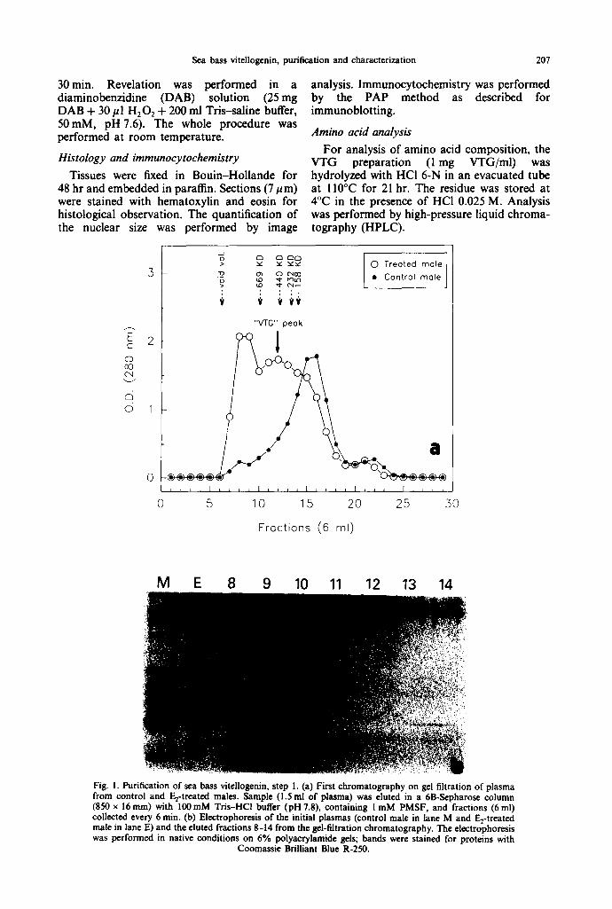
(i 500 g for l 0 min at 4°C) and l ml applied on to a 6B-Sepharose column (850 x 16mm). The sample was eluted with Tris-HCl buffer 100 mM (pH 7.8), containing PMSF I mM, at a flow rate of 60 mi/hr and fractions were collected every 6 min. Fractions containing VTG were pooled and concentrated to a final vol of 5 ml using a Sartorius ultrafiltration system. Immediately after, this solution was applied on to a DEAE- Sepharose column (350 x 16ram) and eluted with a linear gradient (150-300mM) of Tris-HCl buffer (pH 7.8), PMSF I mM, at a flow rate of 36 ml/hr. Fractions of 3 ml were collected and their absorbance read at 280 nm. The whole procedure was performed at 4°C. Molecular weight determinations were carried out both by gel filtration on 6B-Sepharose using thyroglobulin (669kDa), ferritin (440kDa), catalase (232 kDa) and aldolase (158 kDa) as standard proteins, and by SDS electrophoresis using phosphorylase-b (94 kDa), albumin (67 kDa), ovalbumin (43 kDa) and carbonic an- hydrase (30 kDa) as standard proteins.
Immuniza t ion procedure
The VTG preparation (1 mg VTG/ml) was mixed with complete Freund's adjuvant (1:1) and injected subcutaneously in rabbits. Injec- tions of l ml (for each rabbit) of the mixed solution were given every week for 4 weeks; after 15 days of "resting period" the injections were repeated every 2 weeks until no further increase in the antibody titer could be detected (four injections after the "resting period"). Then, blood was collected, allowed to clot at 4°C for 48 hr and serum stored at - 20°C.
Electrophoresis and immunoblot t ing
Native and SDS-PAGE (0.1% SDS) were performed in discontinuous gels with 4.5% stacking gel and resolving gel with a linear gradient of acrylamide 4-16% (Laemmli, 1970). Gels were stained for proteins with Coomassie Brilliant Blue R-250.
For immunoblotting, proteins were trans- ferred on to a nitrocellulose paper (NTC). Transfer was performed for 2 hr (50 mA) at 4°C in Tris-HCl buffer 25 mM (containing 14.4 g/1 of glycine), pH 8.3. After this, the paper was satu- rated for 2hr in Tris-saline buffer, 10mM, pH 7.6 (TBS), containing 5% swine serum and washed three times (10 min) with TBS. After the saturation step, the paper was incubated with the antibody solution (diluted 1:3000 in TBS) overnight. The antigen-antibody complexes were detected by the peroxidase-anti-peroxidase (PAP) method: secondary antibody incubation (goat-anti-rabbit diluted l: 1000) for 45 min, fol- lowed by PAP incubation (diluted I: 500) for
Sea bass vitellogcnin, purification and characterization 207
30min. Revelation was performed in a diaminobenzidine (DAB) solution (25 mg DAB + 30/~1 H20: + 200 ml Tris-saline buffer, 50mM, pH 7.6). The whole procedure was performed at room temperature.
Histology and immunocytochemistry Tissues were fixed in Bouin-Hollande for
48 hr and embedded in paraffin. Sections (7 #m) were stained with hematoxylin and eosin for histological observation. The quantification of the nuclear size was performed by image
analysis. Immunocytochemistry was performed by the PAP method as described for immunoblotting.
Amino acid analysis For analysis of amino acid composition, the
VTG preparation (1 mg VTG/ml) was hydrolyzed with HCI 6-N in an evacuated tube at 110°C for 21 hr. The residue was stored at 4°C in the presence of HCI 0.025 M. Analysis was performed by high-pressure liquid chroma- tography (HPLC).
E c 2 o 0o
c~ © 1
0
0
> v 5£ Y ' ¢ "
.~_ o~ o c-400 o LO ,d- ~ ' )U ' )
, : : - ,
$ t $$v
0 Treated male
• Control male
"VTG" peak
. / a
5 10 15 2.0 25 30
Fractions (6 ml)
M E 8 9 10 11 12 13 14
• - , % :=? . ~¢~ ...
. . ~ . , ~ : .
Fig. I. Purification of sea bass vitellogenin, step I. (a) First chromatography on gel filtration of plasma from control and E2-treated males. Sample (1.5 ml of plasma) was eluted in a 6B-Scpharos¢ column (850 x 16ram) with 100raM Tris-HCl buffer (pH 7.8), containing I mM PMSF, and fractions (6ml) collected every 6 min. (b) Electrophoresis of the initial plasmas (control male in lane M and E2-treated male in lane E) and the eluted fractions 8-14 from the gel-filtration chromatography. The electrophoresis was performed in native conditions on 6% polyacrylamide gels; bands were stained for proteins with
Coomassie Brilliant Blue R-250.
208 E. Mafian6s e t al.
Results Purification and partial characterization of the viteliogenin molecule
Sea bass VTG was purified from plasma of E2-treated animals using a double chromatog- raphy method, a gel filtration on 6B-Sepharose followed by ion-exchange on DEAE-Sepharose. Figure l(a) shows the elution profile on gel filtration of plasmas from control and Fro- treated males. The E2 treatment produced an increase of the peak eluted just after the void volume and the appearance of a peak centered around fraction 12. Electrophoresis of the eluted fractions, together with the initial plasmas, is shown in Fig. l(b). Plasma from the E2-treated males presented one protein that was absent in the control males. This E2-induced protein was suspected to be the VTG molecule. This protein was eluted in the first fractions of the gel filtration (fractions 9-14 of Fig. la) but it was not completely resolved from other plasma proteins. Fractions containing this protein were pooled and applied to the second chromatography step.
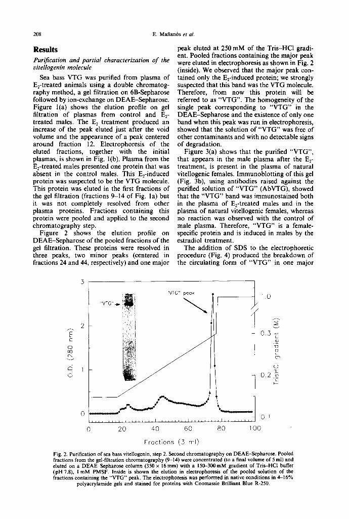
Figure 2 shows the elution profile on DEAE-Sepharose of the pooled fractions of the gel filtration. These proteins were resolved in three peaks, two minor peaks (centered in fractions 24 and 44, respectively) and one major
peak eluted at 250 mM of the Tris-HCl gradi- ent. Pooled fractions containing the major peak were eluted in electrophoresis as shown in Fig. 2 (inside). We observed that the major peak con- tained only the Erinduced protein; we strongly suspected that this band was the VTG molecule. Therefore, from now this protein will be referred to as "VTG". The homogeneity of the single peak corresponding to "VTG" in the DEAE-Sepharose and the existence of only one band when this peak was run in electrophoresis, showed that the solution of "VTG" was free of other contaminants and with no detectable signs of degradation.
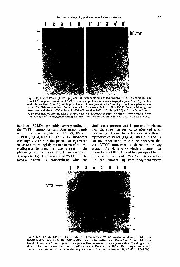
Figure 3(a) shows that the purified "VTG", that appears in the male plasma after the E2- treatment, is present in the plasma of natural vitellogenic females. Immunoblotting of this gel (Fig. 3b), using antibodies raised against the purified solution of "VTG" (AbVTG), showed that the "VTG" band was immunostained both in the plasma of E2-treated males and in the plasma of natural vitellogenic females, whereas no reaction was observed with the control of male plasma. Therefore, "VTG" is a female- specific protein and is induced in males by the estradiol treatment.
The addition of SDS to the electrophoretic procedure (Fig. 4) produced the breakdown of the circulating form of "VTG" in one major
F C
o oo (N
~4
(5
"VTG" peak
i : "VTG" .~. iF!
i (:i / 2 i"(: I
i~. ::..: :':i
,
0 _ t O
0 20 40 60 80 100
Fractions (3 rnl)
1.0 /
/
0..3
0.2
C (D
O
CT~
:E
C
Fig. 2. Purification of sea bass vitellogenin, step 2. Second chromatography on DEAE-Sepharose. Pooled fractions from the gel-filtration chromatography (9-14) were concentrated (to a final volume of 5 ml) and eluted on a DEAE-Sepharose column (350 x 16ram) with a 150-300mM gradient of Tris-HCl buffer (pH 7.8), i mM PMSF. Inside is shown the elution in electrophorcsis of the pooled solution of the fractions containing the "VTG" peak. The electrophoresis was performed in native conditions in 4--16%
polyacrylamide gels and stained for proteins with Coomassie Brilliant Blue R-250.
Sea bass vitellogenin, purification and characterization
3 4 5 I' 2' 3' 4' 5'
..:i. . . : •
~ . : . . . . ~ • . •
~.,::L . • • .~• ~• ~i?:!' i ~ - + ~ : . ? : . i : . "
t+~? , :
;~;+i+.,",+ i + . ~ ~l,~,l:.e ... :.,.
• . . L
: i (::.:~
. . . . . Y::C:!+i'i:i??~ • .: . . . . • +~.~+;c,~:~
, - . . . . ?~ ..r , : ~+~ .+ >.::+-+.i.:-+/:'~ ~ • +,..~, .+:+r, ~ . ~
• +: • . : . , . , : . - - ;.+:+~.;+¢+~+.~ ~ . p ~
• : ++'-'.++,~G~;~~
+
I"VTG"
209
: : ; . ; ' , i . . V ~ i ~ ) ? , ~ . ~ - , l ~ % . , ~ ! r - ~ , f L . ' L : : + ~ r : : ~ i ~ 5 ~ _ ~ 4 £ ~ ; ~ e ~ f f , f f ~ , ' - ~ . : - - , - ~ . : ; : " ~ : . • ~ • " . . . . " • • •
Fig. 3. (a) Native PAGE (4--16% gel) and (b) immunoblotting of the purified "VTG" preparation (lane I and I'), the pooled solution of "VTG" after the gel filtration chromatography (lane 2 and 2'), control male plasma (lane 3 and 3'), vitellogenic female plasma (lane 4 and 4') and E2-treated male plasma (lane 5 and 5'). Gels were stained for proteins with Coomassie Brilliant Blue R-250. Immunoblotting was performed with the AbVTG (diluted 1/3000 in Tris-saline buffer, I 0 mM, pH 7.6) and complexes detected by the PAP method after transfer of the proteins to a nitrocellulose paper. On the left, arrowheads indicate the position of the molecular weight markers (from top to bottom, 669, 440, 232, 140 and 67 kDa).
band of 180kDa, probably corresponding to the "VTG" monomer, and four minor bands with molecular weights of 113, 97, 86 and 73 kDa (Fig. 4, lane 1). The "VTG" monomer was highly visible in the plasma of E2-treated males and more slightly in the plasma of natural vitellogenic females, but was absent in the plasma of control males (Fig. 4, lanes 4, 2 and 3, respectively). The presence of "VTG" in the female plasma is concomitant with the
vitellogenic process and is present in plasma over the spawning period, as observed when comparing plasma from females at different reproductive stages (Fig. 4, lanes 5, 6 and 7). On the other hand, it can be observed that the "VTG" monomer is absent in an egg extract (Fig. 4, lane 8) which contained one major band of 88 kDa, and two groups of bands of around 70 and 25kDa. Nevertheless, Fig. 5(b) showed, by immunocytochemistry,
"VTG"
1 2 3 4 5 6 7 8
Fig. 4. SDS-PAGE (0.1% SDS) in 4-16% gel, of the purified "VTG+' preparation (lane I), vitellogenic female plasma (lane 2), control male plasma (lane 3), E2-treated male plasma (lane 4), previtellogenic female plasma (lane 5), vitellogenic female plasma (lane 6), ovulated female plasma (lane 7) and egg extract (lane 8). Gels were stained for proteins with Coomassie Brilliant Blue R-250. On the right, arrowheads
indicate the position of the molecular weight markers (from top to bottom, 94, 67, 43 and 30 kDa).

210 E. Mafian6s et al.
Fig. 5. Ovary from a vitellogenic female, stained with hematoxylin and eosin in (a) and immunostained using the AbVTG in (b). Strong immunoreaction was detected in the growing oocytes (arrow) while no reaction was shown in the previtellogenic and immature ones (arrowhead). AbVTG was diluted 1:3000 in Tris-saline buffer, 10 mM, pH 7.6 and the reaction detected by the PAP method. Scale bar = 600 tim.
that AbVTG recognized antigenic "VTG" sites in the growing oocytes, indicating that these egg yolk proteins were derived, at least in part, from the "VTG" molecule. Immature and previtel- logenic oocytes remained unstained.
The molecular weight of the circulating form Asp 6.3 6.5 8.0 Tbr 4.9 5.5 4.7 o f " V T G " was estimated to be around 445 kDa Ser 8.4 6.9 10.3 by gel filtration. The amino acid composition of Glu I 1.1 11.9 10.6 sea bass "VTG" is shown in Table 1. Alanine Pro 6.0 5.5 4.2 and glutamic acid are the most concentrated Gly 4.8 4.6 4.4
Ala 13.2 12.8 10.4 amino acids while methionine and histidine are Val 7.2 6.9 6.8 the least concentrated. Met 0.9 2.0 2.4
lie 6.3 6.6 5.7 Effects of the estradiol treatment Leu 10.2 10.8 9.7
The induc t ion o f V T G synthesis by E2 Tyr 3.1 2.6 3.6 Phe 3.6 2.9 3.4 treatment produced an increase in the activity of His 1.5 2.3 2.4 protein synthesis in the liver, as reflected by Lys 6.9 7.0 7.6 changes in its composition and structure. Arg 5.4 4.9 5.0 Table 2 summarizes some biochemical changes 99.8 99.7 99.2 which occurred in the liver after the E2 treatment. The content of total proteins in the liver was slightly increased in the treated groups
Table 1. Amino acid composition of sea bass VTG compared with that of VTG from other teleost species
Amino acid residue (mol.%) Amino Rainbow acid Sea bass Goldfish* Medakat trout~
8.4 5,0 7.5
11.5 5.2 4.2
11.7 7.1 2.6 5.5 9.5 3.0 4.0 2.1 7.1 4.5
98.84
*Data from De Vlaming et al. (1980). tData from Hamazaki et al. (1987b). ~:Data from Hara and Hirai (1978).
Sea bass vitellogenin, purification and characterization
Table 2. Biochemical modifications induced by estradiol treatment in the fiver of sea bass
211
Total proteins RNA DNA Group (mg/g fiver) (mg/g liver) (mg/g fiver) R N A / D N A Prot/DNA Control 96.81 + 9.13 (4)* 17.31 :t: 1.08 (7)* 3.08 + 0.16 (6)* 5.99 + 0.26 (6)* 30.37 + 3.62 (4)" 0.5mg E2/kg i11.49+4.06 (6)" 23.09+0.20 (6) b 2.99+0.14 (7)* 7.98+0.49 (6)* 37.05 + 1.34 (6)* 2mg ~/kg 112.87+4.71 (5)* 22.96+0.77 (5) b 2.60+0.17 (5)* 9.05+0.73 (5) b 44.05-I-2.53 (5) b 5rag Fa/kg 118.56-I-8.48 (4)* 24.66+2.16 (4) b 2.78-I-0.25 (4)* 9.21 :I: 1.28 (4) b 42.95-I-0.87 (4) b
Differences for every parameter between the groups are expressed with different letters (P < 0.05).
with respect to the controls (not significantly different); when expressed in relation to DNA, this increase was significantly different (P < 0.05) for the groups injected with 2 and 5mg Ez/kg. The content of liver RNA was significantly (P < 0.05) increased in all treated groups with respect to the controls; neverthe- less, when it was expressed in relation to DNA only the groups injected with the higher doses (2 and 5mg E2/kg) maintained this difference. DNA content of the liver was not affected by the Ez treatment.
Table 3 summarizes some changes in the structure of the liver associated with the F_q treatment. The hepatosomatic index (HSI) and the nuclear size of the hepatocytes were increased in all treated groups with respect to the controls but this increment was significantly different (P < 0.05) only for the groups injected with 2 and 5 mg F~/kg. The hypertrophy of the hepatocytes (assessed by the number of nuclei/area unit) was only observed in the two groups that received the highest doses of ~ .
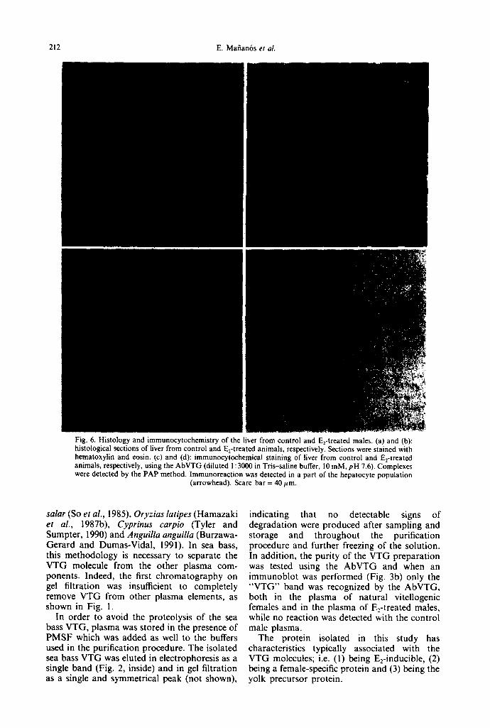
Figure 6 shows histological sections of liver from control and E2-treated animals. The hepatocytes of the control group (Fig. 6a) were smaller and more vacuolized than those of the treated ones (Fig. 6b). On the other hand, liver slices were immunostained with the AbVTG for the localization of VTG into the hepatocytes. No reaction was observed in the liver of the control group (Fig. 6c) whereas in the liver of the E2-treated animals a positive immunostain- ing was detected in the cytoplasm of some hepatocytes (Fig. 6d).
In plasma, the E 2 treatment caused a signifi- cant increase in the content of both VTG and total proteins (Fig. 7). Before the Ez injections, the plasma VTG levels were undetected in males and very low (around 100/lg/ml) in females.
These levels remained unchanged in the control group throughout the experiment. Two days after the first injection a slight, but not signifi- cant (P < 0.05), increase in the VTG levels was observed in the three treated groups, when compared to the controls. Six days after the first injection, the VTG levels were significantly increased in all treated groups with respect to the controls and reached a maximum of around 50mg/ml after the fourth injection. Total proteins were significantly increased in all the treated groups with respect to the controls at day 8 after the beginning of the experiment.
Discussion
In this study, E2 was used for the induction of VTG synthesis in male and female sea bass, because it is known to be the most potent estrogen for the induction of VTG synthesis in fish (Sundararaj and Nath, 1981; van Bohemen et al., 1982).
Several methods have been developed for the purification of amphibian VTG, ultracentri- fugation (Redshaw and Follett, 1971), precipitation with dimethyiformamide (Ansari et al., 1971), selective precipitation with Mg 2+- EDTA (Wiley et al., 1979) and chromatography (Wallace, 1970; Wiley et al., 1979). In fish, the purification of VTG has normally been per- formed by chromatography, because the other methods have failed to completely isolate VTG from other plasma components (Wiley et al., 1979; de Vlaming et ai., 1980). In this study, sea bass VTG was purified from plasma of E2- treated animals using a double chromatography method. Similar procedures have been successfully used for the purification of VTG in Anguilla japonica (Hara et al., 1980), Salmo
Table 3. Structural modifications induced by estradiol treatment in the liver of sea bass
Nuclei/area* Group HS! (* = 13.2/~m 2) (/am 2) Control 0.87+0.09 (7)" 117:t: 10 (7)' 14.95 + 0.20 (7)" 0.Stag E2/kg 1.25-t-0.07 (7)" 86+ 5 (7) b 15.05+0.16 (7)" 2mg Ee/kg 2.38+0.12 (7) b 58+3 (7) c 18.53:1:0.62 (7) b 5mg ~/kg 2.18:1:0.17 (6) b 57___3 (7) c 20.20+0.56 (7) b
Differences for every parameter between the groups are expressed with different letters (P < 0.05).
212 E. Mafian6s et al.
i:i
" : ' : 7
• - . r : "
I I I
Fig. 6. Histology and immunocytochemistry of the liver from control and E2-treated males. (a) and (b): histological sections of liver from control and E2-treated animals, respectively. Sections were stained with hematoxylin and eosin. (c) and (d): immunocytochemical staining of liver from control and E2-treated animals, respectively, using the AbVTG (diluted l: 3000 in Tris-saline buffer, l0 raM, pH 7.6). Complexes were detected by the PAP method, lmmunoreaction was detected in a part of the hepatocyte population
(arrowhead). Scare bar = 40 lain.
salar (So et al., 1985), Oryzias latipes (Hamazaki et al., 1987b), Cyprinus carpio (Tyler and Sumpter, 1990) and Anguilla anguilla (Burzawa- Gerard and Dumas-Vidal, 1991). In sea bass, this methodology is necessary to separate the VTG molecule from the other plasma com- ponents. Indeed, the first chromatography on gel filtration was insufficient to completely remove VTG from other plasma elements, as shown in Fig. I.
In order to avoid the proteolysis of the sea bass VTG, plasma was stored in the presence of PMSF which was added as well to the buffers used in the purification procedure. The isolated sea bass VTG was eluted in electrophoresis as a single band (Fig. 2, inside) and in gel filtration as a single and symmetrical peak (not shown),
indicating that no detectable signs of degradation were produced after sampling and storage and throughout the purification procedure and further freezing of the solution. In addition, the purity of the VTG preparation was tested using the AbVTG and when an immunoblot was performed (Fig. 3b) only the "V TG " band was recognized by the AbVTG, both in the plasma of natural vitellogenic females and in the plasma of E2-treated males, while no reaction was detected with the control male plasma.
The protein isolated in this study has characteristics typically associated with the VTG molecules; i.e. (I) being E2-inducible, (2) being a female-specific protein and (3) being the yolk precursor protein.

Sea bass vitellogenin, purification and characterization 2 ! 3
8O • c o n t r o l
0 a d T a a -- 0.5 mg E2/ml
E 6 0 a 2 mg e/ml -d---- - - - .~m-----~ ~ . . . . a
E
.c_ 40 C ¢ ) C ~ o
20
~ b ~ b b & I , 1 A I , I ~ I , I ~ I L
100 8 a
E ~ a a
E 80 a
.c_
Cl
~ 4o *S
t t t t t t b 1 , I L I , I , I , 1 , I ,
0 2 4 6 8 10 12
Days
Fig. 7. Plasma levels of (a) vitellogenin and (b) total proteins, during the estradiol treatment. The VTG levels were measured by ELISA and the levels of total proteins by Lowry et al. (1951). At the bottom, arrows indicate the time of ¢stradiol injections. Different letters represent significant differences (P < 0.05) between the groups for the same sampling (when there is only one letter for the four groups in a sampling point, it means that there are no differences between them); (*) over the lines represents differences
(P < 0.05) from the previous sampling for the same group; N = 7 for each sampling.
Firstly, the E 2 inductivity of sea bass VTG was observed by gel filtration and electrophor- esis by comparing plasma from treated and untreated males (Fig. 1). In sea bass, no other protein was observed to be induced by the E2 treatment whereas, in the teleost Oryz ias latipes, Hamazaki et al. (1987a) found that another protein different to VTG was induced by treat- ment with estradiol. This protein, a glycoprotein synthesized by the liver, was found to be a component of the egg envelope (Hamazaki et al., 1987c). Other E2-inducible proteins different to VTG were also found in amphibians (Mitchell et al., 1985; Holland and Wangh, 1987), but the nature and function of these proteins remain unclear. On the other hand, it is well known that VTG is synthesized in the liver under the influence of estradiol and in accordance with that, sea bass VTG was local- ized in the liver tissue of the E2-treated animals using the AbVTG (Fig. 6). Thus, considering that previously discussed, this new protein purified from the plasma of F-a-treated sea bass, can be considered as the probable VTG.
Secondly, this protein is female-specific as shown by its presence in the plasma of natural vitellogenic females and its absence in the plasma of non-treated males and previtellogenic females (SDS-PAGE, Fig. 4). On the other hand, the AbVTG did not react with any of the bands of the male plasma, whereas a strong reaction was found with the band identified as VTG in the vitellogenic female plasma (Fig. 3b). This methodology has been widely used in other teleost species (Hara et al., 1983; Bradley and Grizzle, 1989) and it has proved to be a good tool for the characterization of VTG as a fe- male-specific protein. Notwithstanding, Ding et al. (1989) and Pelissero et al. (1991) detected the presence of VTG in the male plasma of Ore- ochromis aureus and Acipenser baeri, respect- ively. Although the significance of the presence of VTG in males of tilapia remains unclear, in the Siberian sturgeon it has been demonstrated to be induced by the presence of phytoestrogens in the diet (Pelissero et al., 1991).
Thirdly, the protein isolated in this study was found to be related to the yolk proteins, as
214 E. Mafian6s et al.
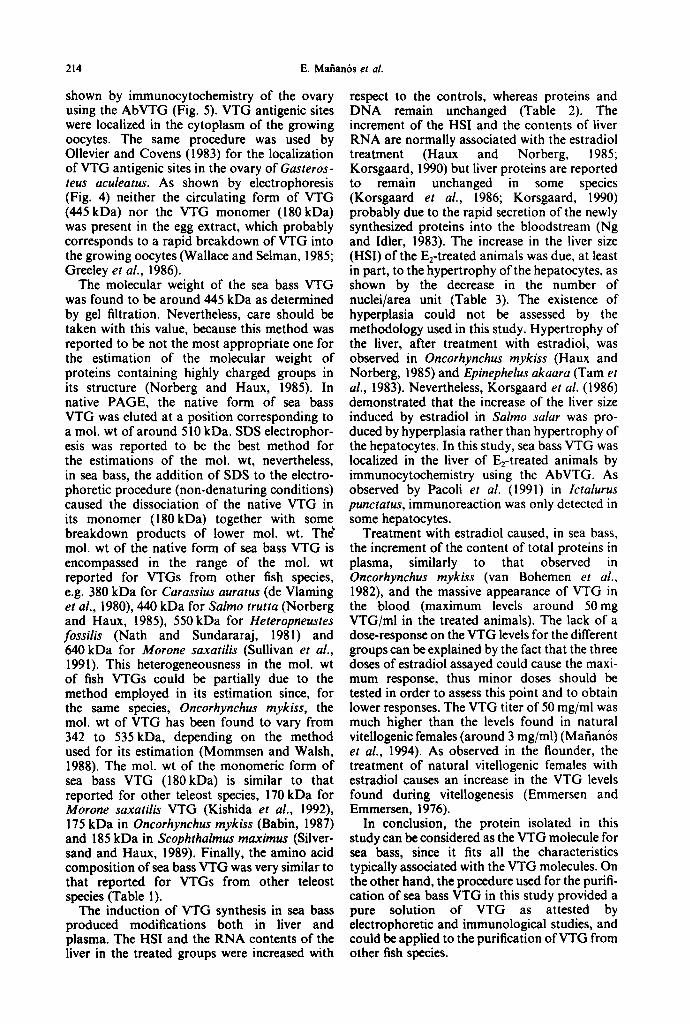
shown by immunocytochemistry of the ovary using the AbVTG (Fig. 5). VTG antigenic sites were localized in the cytoplasm of the growing oocytes. The same procedure was used by Ollevier and Covens (1983) for the localization of VTG antigenic sites in the ovary of Gasteros- teus aculeatus. As shown by electrophoresis (Fig. 4) neither the circulating form of VTG (445kDa) nor the VTG monomer (180kDa) was present in the egg extract, which probably corresponds to a rapid breakdown of VTG into the growing oocytes (Wallace and Selman, 1985; Greeley et al., 1986).
The molecular weight of the sea bass VTG was found to be around 445 kDa as determined by gel filtration. Nevertheless, care should be taken with this value, because this method was reported to be not the most appropriate one for the estimation of the molecular weight of proteins containing highly charged groups in its structure (Norberg and Haux, 1985). In native PAGE, the native form of sea bass VTG was eluted at a position corresponding to a mol. wt of around 510 kDa. SDS electrophor- esis was reported to be the best method for the estimations of the tool. wt, nevertheless, in sea bass, the addition of SDS to the electro- phoretic procedure (non-denaturing conditions) caused the dissociation of the native VTG in its monomer (180kDa) together with some breakdown products of lower mol. wt. Thd mol. wt of the native form of sea bass VTG is encompassed in the range of the mol. wt reported for VTGs from other fish species, e.g. 380 kDa for Carassius auratus (de Vlaming et ai., 1980), 440 kDa for Salmo trutta (Norberg and Haux, 1985), 550kDa for Heteropneustes fossilis (Nath and Sundararaj, 1981) and 640 kDa for Morone saxatilis (Sullivan et al., 1991). This heterogeneousness in the mol. wt of fish VTGs could be partially due to the method employed in its estimation since, for the same species, Oncorhynchus mykiss, the mol. wt of VTG has been found to vary from 342 to 535 kDa, depending on the method used for its estimation (Mommsen and Walsh, 1988). The mol. wt of the monomeric form of sea bass VTG (180kDa) is similar to that reported for other teleost species, 170 kDa for Morone saxatilis VTG (Kishida et ai., 1992), 175 kDa in Oncorhynchus mykiss (Babin, 1987) and 185 kDa in Scophthalmus rnaximus (Silver- sand and Haux, 1989). Finally, the amino acid composition of sea bass VTG was very similar to that reported for VTGs from other teleost species (Table 1).
The induction of VTG synthesis in sea bass produced modifications both in liver and plasma. The HSI and the RNA contents of the liver in the treated groups were increased with
respect to the controls, whereas proteins and DNA remain unchanged (Table 2). The increment of the HSI and the contents of liver RNA are normally associated with the estradiol treatment (Haux and Norberg, 1985; Korsgaard, 1990) but liver proteins are reported to remain unchanged in some species (Korsgaard et al., 1986; Korsgaard, 1990) probably due to the rapid secretion of the newly synthesized proteins into the bloodstream (Ng and Idler, 1983). The increase in the liver size (HSI) of the E2-treated animals was due, at least in part, to the hypertrophy of the hepatocytes, as shown by the decrease in the number of nuclei/area unit (Table 3). The existence of hyperplasia could not be assessed by the methodology used in this study. Hypertrophy of the liver, after treatment with estradiol, was observed in Oncorhynchus mykiss (Haux and Norberg, 1985) and Epinephelus akaara (Tam et al., 1983). Nevertheless, Korsgaard et al. (1986) demonstrated that the increase of the liver size induced by estradiol in Salmo salar was pro- duced by hyperplasia rather than hypertrophy of the hepatocytes. In this study, sea bass VTG was localized in the liver of E2-treated animals by immunocytochemistry using the AbVTG. As observed by Pacoli et al. (1991) in Ictalurus punctatus, immunoreaction was only detected in some hepatocytes.
Treatment with estradiol caused, in sea bass, the increment of the content of total proteins in plasma, similarly to that observed in Oncorhynchus mykiss (van Bohemen et al., 1982), and the massive appearance of VTG in the blood (maximum levels around 50mg VTG/ml in the treated animals). The lack of a dose-response on the VTG levels for the different groups can be explained by the fact that the three doses of estradiol assayed could cause the maxi- mum response, thus minor doses should be tested in order to assess this point and to obtain lower responses. The VTG titer of 50 mg/ml was much higher than the levels found in natural vitellogenic females (around 3 mg/ml) (Mafian6s et al., 1994). As observed in the flounder, the treatment of natural viteliogenic females with estradiol causes an increase in the VTG levels found during vitellogenesis (Emmersen and Emmersen, 1976).
In conclusion, the protein isolated in this study can be considered as the VTG molecule for sea bass, since it fits all the characteristics typically associated with the VTG molecules. On the other hand, the procedure used for the purifi- cation of sea bass VTG in this study provided a pure solution of VTG as attested by electrophoretic and immunological studies, and could be applied to the purification of VTG from other fish species.

Sea bass vitellogenin, purification and characterization 215
Acknowledgements--This work was supported by a research grant from the CICYT (No. MAR88-0231) and from a Spanish-French joint research program (No. 127/3). We are also grateful to Dr F. Vandesande of the Zoological Institute, Catholic University of Leuven (Belgium), for his help in the immunological processes and to Dr O. Kah, of the CNRS (France), for his technical assistance and the review of the manusc/ipt.
References Ansari A. Q., Dolphin P. J., Lazier C. B., Munday K. A.
and Akhtar M. (1971) Chemical composition of an oestrogen-induced calcium-binding glycolipophospho- protein in Xenopus laevis. Biochem. J. 122, 107-113.
Babin P. J. (1987) Apolipoproteins and the association of egg yolk proteins with plasma high-density lipoproteins after ovulation and follicular atresia in the rainbow trout (Salmo gairtb~eri). J. biol. Chem. 262, 4290-4296.
Bohemen Ch. G. van, Lambert J. G. D., Goos H. J. Th. and van Oordt P. G. W. J. (1982) Estrone and estradiol participation during exogenous vitellogenesis in the female rainbow trout, Salmo gairdneri. Gen comp. Endocr. 46, 81-92.
Bradley J. T. and Grizzle J. M. (1989) Vitellogenin induction by estradiol in channel catfish, Ictalurus punctatus. Gen. comp. Endocr. 73, 28-39.
Buckley L. J. (1979) Relationship between RNA-DNA ratio, prey density and growth rate in Atlantic cod (Gadus morhua) larvae. J. Fish. Res. Bd Can. 36, 1497-1502.
Burzawa-Gerard E. and Dumas-Vidal A. (1991) Effects of 17fl-estradiol and carp gonadotropin on vitellogenesis in normal and hypophysectomized European silver female eel (Anguilla anguilla L.) employing a homologous radio- immunoassay for vitellogenin. Gen. comp. Endocr. 84, 264-276.
Carnevali O., Mosconi G., Roncarati A., Belvedere P. and Polzonetti-Magni A. M. (1992) Changes in the electro- phoretic patterns of the yolk proteins during vitellogenesis in sea bream (Sparus aurato, L.) and sea bass (Dicentrarchus labrax, L.). Riv. ltal. Aquacolt. 27, 61-70.
Carrillo M., Bromage N., Zanuy S., Serrano R. and Prat F. (1989) The effect of modifications in photoperiod on spawning time, ovarian development and egg quality in the sea bass (Dicemrarch~ labrax L.). Aquaculture 81, 351-365.
Craik J. C. A. (1978) The effects of oestrogen treatment on certain plasma constituents associated with vitellogenesis in the elasmobranch Scyliorhinus canicula L. Gen. comp. Endocr. 35, 455-464.
De Vlaming V. L., Wiley H. S., Delahunty G. and Wallace R. A. (1980) Goldfish (Carassius auratus) vitellogenin: induction, isolation, properties and relationship to yolk proteins. Comp. Biochem. Physiol. 67B, 613~23.
Devaucbelle N. and Coves D. (1988a) The characteristics of sea bass (Dicentrarchus labrax) eggs: description, biochemical composition and hatching performances. Aquat. Living Resour. 1, 223-230.
Devauchelle N. and Coves D. (1988b) Sea bass (Dicentrarchus labrax) reproduction in captivity: gameto- genesis and spawning. Aquat. Living Resour. !, 215-222.
Ding J. L., Hee P. L. and Lam T. J. (1989) Two forms of vitellogenin in the plasma and gonads of male Ore- ochromis aureus. Comp. Biochem. Physiol. 93B, 363-370.
Emmersen B. K. and Emmersen J. (1976) Protein, RNA and DNA metabolism in relation to ovarian vitellogenic growth in the flounder Platichthys flesus (L.). Comp. Biochem. Physiol. 55, 315-321.
Fremont L. and Riazi A. (1988) Biochemical analysis of vitellogenin from rainbow trout (Salmo gairdneri): fatty acid composition of phospholipids. Reprod. Nutr. Devel. 28, 939-952.
Goodwin A. E., Grizzle J. M., Bradley J. T. and Estridge B. H. (1992) Monoclonal antibody-based immunoassay of vitellogenin in the blood of male channel catfish (Ictalurus punctatus). Comp. Biochem. PhysioL 101B, 441~46.
Greeley M. S., Calder D. R. and Wallace R. A. (1986) Changes in teleost yolk proteins during oocyte maturation: correlation of yolk proteolysis with oocyte hydration. Comp. Biochem. Physiol. 84B, I-9.
Hamazaki T. S., luchi I. and Yamagami K. (1987a) Production of a "spawning female-specific substance" in hepatic cells and its accumulation in the ascites of the estrogen-treated adult fish, Oryzios latipes. J. exp. Zool. 242, 325-332.
Hamazaki T. S., Iuchi I. and Yamagami K. (1987b) Purifi- cation and identification of vitellogenin and its immuno- histochemical detection in growing oocytes of the teleost, Oryzias latipes. J. exp. Zool. 242, 333-341.
Hamazaki T. S., Iuchi I. and Yamagami K. (1987c) Isolation and partial characterization of a "spawning female-specific substance" in the teleost, Oryzias latipes. J. exp. Zool. 242, 343-349.
Hara A. and Hirai H. (1978) Comparative studies on immunochemical properties of female-specific serum protein and egg yolk proteins in rainbow trout (Salmo gairdneri). Comp. Biochem. Physiol. 59B, 339-343.
Hara A., Yamauchi K. and Hirai H. (1980) Studies on female-specific serum protein (viteUogenin) and egg yolk protein in Japanese eel (Anguilla japonica). Comp. Biochem. Physiol. 65B, 315-320.
Hara A., Takano K. and Hirai H. (1983) Immunochemical identification of female-specific serum protein, vitel- Iogenin, in the medaka, Oryzias latipes (teleosts). Comp. Biochem. Physiol. 76A, 135-141.
Haux C. and Norberg B. (1985) The influence of estradiol- 17,6 on the liver content of protein, lipids, glycogen and nucleic acids in juvenile rainbow trout, Salmo gairdneri. Comp. Biochem. Physiol. 81B, 275--279.
Holland L. J. and Wangh L. J. (1987) Estrogen induction of a 45 kDa secreted protein coordinately with vitel- Iogenin in Xenopus liver. Molec. Cell Endocr. 49, 63-73.
Hori S. H., Kodama T. and Tanahashi K. (1979) Induction of vitellogenin synthesis in goldfish by massive doses of androgens. Gen. comp. Endocr. 37, 306-320.
Kishida M., Anderson T. R. and Specker J. (1992) Induction by fl-estradiol of vitellogenin in striped bass (Morone saxatilis): characterization and quantification in plasma and mucus. Gen. comp. Endocr. 88, 29-39.
Korsgaard B., Mommsen T. P. and Saunders R. L. 0986) The effect of temperature on the vitellogenic response in Atlantic salmon post-smolts (Salmo salar). Gen. comp. Endocr. 62, 193-201.
Korsgaard B. 0990) Estrogen treatment and its influence on protein synthesis and amino acid metabolism in Zoarces viviparus (L) males. Fish Physiol. Biochem. 8, 121-127.
Laemmli U. K. (1970) Cleavage of structural proteins during the assembly of the head of bacteriophage T4. Nature, Lond. 227, 68ff4585.
Le Menn F. (1979) Induction de vitellogtnine par l'estradiol et par des androgtnes chez un ttltostten marin: Gobius niger L. C. r. Acad. Sci. Paris. D. 289, 413~,16.
Lowry O. H., Rosebrough N. J., Farr A. L. and Randall R. J. (1951) Protein measurements with the Folin reagent. J. biol. Chem. 193, 265-275.
Mafian6s E., Nfifiez J., Zanuy S., Carrilo M. and I.,¢ Menn F. (1994) Sea bass (Dicentrarchus labrax L.) vitellogenin: II. Validation of an enzyme-linked immunosorbent assay (ELISA). Comp. Biochem. Physiol. 107B, 217-230.
Mayer I., Shackley S. E. and Ryland J. S. (1988) Aspects of the reproductive biology of the bass, Dicentrarchus labrax L. I. A historical and histochemical study of oocyte development. J. Fish Biol. 33, 609~622.
CIII~ I0712--D

216 E. Mafian6s et al.
Mayer I., Shackley S. E. and Witthames P. R. (1990) Aspects of the reproductive biology of the bass, Dicentrarchus labrax L. I1. Fecundity and pattern of oocyte development. J. Fish Biol. 36, 141-148.
Mitchell R. O., Dean W. L., Hess P. P. and Feldhoff R. C. (1985) Purification and characterization of a non- vitellogenin, estrogen-induced plasma protein from the American bullfrog Rana catesbeiana. Biochemistry 24, 3672-3677.
Mommsen T. P. and Walsh P. J. (1988) Viteliogenesis and oocyte assembly. In Fish Physiology (Edited by Hoar W. S. and Randall D. J.), Vol. XIA, pp. 347-406. Academic Press, New York.
Mosconi G., Carnevali O., Roncarati A., Sabieti M. G., Can'era E. and Polzonetti-Macni A. M. (1992) Morpho- logical changes of yolk globules during oocytes maturation of sea bass (Dicentrarchus labraxL.) and sea bream (Sparus aurata, L.). Riv. hal. Aquac. 27, 71-79.
Murakami M., luchi I. and Yamagami K. (1991) Partial characterization and subunit analysis of major phospho- proteins of egg yolk in the fish, Oryzias latipes. Comp. Biochem. Physiol. 100B, 587-593.
Nath P. and Sundararaj B. I. (1981) Induction of vitelloge- nesis in the hypophysectomized catfish, Heteropneustes fossilis (Bloch): effects of piscine and mammalian hormones. Gen. comp. Endocr. 43, 191-200.
Ng T. B. and Idler D. R. (1983) Yolk formation and differentiation in teleost fishes. In Fish Physiology (Edited by Hoar W. S., Randall D. J. and Donaldson E. M.), Vol. IXA, pp. 373--404. Academic Press, New York.
Ng T. B., Woo N. Y. S., Tam P. P. L. and Au C. Y. W. (1984) Changes in metabolism and hepatic ultrastructure induced by estradiol and testosterone in immature female Epinephelus akaara (Teleostei, Scrranidae). Cell Tissue Res. 236, 651-659.
Norberg B. and Haux C. (1985) Induction, isolation and a characterization of the lipid content of plasma vitellogenin from two salmo species: rainbow trout (Salmo gairdheri) and sea trout (Salmo trutta). Comp. Biochem. Physiol. 81B, 869--876.
Ollevier F. and Covens M. (1983) Vitellogenesis in Gasteros- teus aculeatus. Ann. Soc. r. zool. Belg. 113, (suppl. I) 327-334.
Pacoli C. Q., Grizzle J. M. and Bradley J. T. (1991) Immunohistochemical localization of vitellogenin and associated morphologic changes in hepatocytes of chan- nel catfish, lctalurus panctatus. Can. J. Zool. 69, 7--14.
Pelissero C., Bennetau B., Babin P., Le Menn F. and Dunogues J. (1991) Estrogenic activity of certain phy- toestrogens on vitellogenin synthesis in the Siberian stur- geon (Acipenser baeri). J. Steroid Biochem. molec. Biol. 38, 293-299.
Perez L. E. and Callard 1. P. (1992) Identification of vitellogenin in the little skate (Raja erinacea). Comp. Biochem. Physiol. 103B, 699-705.
Prat F., Zanuy S., Carrillo M., de Mones A. and Fostier A. (1990) Seasonal changes in the plasma of gonodal steroids of sea bass Dicentrarchus labrax U and their relationship to growth, oocyte development, spawning and fecundity. Gen. comp. Endocr. 78, 361-373.
Redshaw M. R. and Follett B. K. (1971) The crystalline yolk-platelet proteins and their soluble plasma precursor in an amphibian, Xenopus laevis. Biochem. J. 124, 759-766.
S¢lman K. and Wallace R. A. (1983) Oogenesis in Fundulus heteroclitus. I11. Vitellogenesis. J. exp. Zool. 226, 441-457.
Silversand C. and Haux C. (1989) Isolation of turbot (Scophthalmus maximus) by high-performance anion- exchange chromatography. J. Chromat. 478, 387-397.
So Y. P., Idler D. R. and Hwang S. J. (1985) Plasma vitellogenin in landlocked Atlantic Salmon (Salmo salar Ouananiche): isolation, homologous radioimmunoassay and immunological cross-reactivity with vitellogenin from other teleosts. Comp. Biochem. Physiol. 81B, 63-71.
Sullivan C. V., Tao Y., Hodson R. G., Hara A., Bennett R. O. and Woods II L. C. (1991) Vitellogenin. In Repro- ductive Physiology offish (Edited by Scott A. P., Sumpter J. P., Kime D. E. and Rolfe M. S.), pp. 315-317. Department of Animal and Plant Sciences, the University, Sheffield.
Sundararaj B. I. and Nath P. (1981) Steroid-induced synthesis of vitellogenin in the catfish, Heteropneustes fossilis (Bloch). Gen. comp. Endocr. 43, 201-210.
Takemura A., Hara A. and Takano K. (1991) lmmuno- chemical identification and partial characterization of female-specific serum proteins in white-edged rockfish, Sebastes taczanowskff. Envir. Biol. Fish. 30, 49-56.
Tam P. P. L., Ng T. B. and Woo Y. S. (1983) Effects of oestradiol-17/] and testosterone on the histology of pitu- itary, liver, ovary and skin of previtellogenic Epinephelus akaara (Teleostei, Serranidae). Cell Tissue Res. 231, 579-592.
Tara J. R. and Smith D. F. (1979) Vitellogenesis: a versatile model for hormonal regulation of gene expression. Rec. Prog. Horm. Res. 35, 47-95.
Tyler C. R. and Sumpter J. P. (1990) The purification and partial characterization of carp, Cyprinus carpio, vitellogenin. Fish Physiol. Biochem. 8, 111-120.
Wahli, W., Dawid I. B., Ryfl'el G. U. and Weber R. (1981) Vitellogenesis and the vitellogenin gene family. Science 212, 298-304.
Wallace R. A. (1970) Studies on amphibian yolk. IX. Xenopus vitellogenin. Biochim. biophys. Acta 215, 176-183.
Wallace R. A. (1985) Vitellogenesis and oocyte growth in non-mammalian vertebrates. In Developmental Biology (Edited by Browder L.), Vol. I, pp. 127--177. Plenum Press, New York.
Wallace R. A. and Selman K. (1985) Major protein changes during vitellogenesis and maturation of Fundulus oocytes. Devl Biol. 110, 492-498.
Wallace R. A. and Sclman K. (1990) Ultrastructural aspects of oogenesis and oocyte growth in fish and amphibians. J. Elect. Microsc. Tech. 16, 175-201.
Wiley H. S., Opresko L. and Wallace R. A. (1979) New methods for the purification of vertebrate vitellogenin. Analyt. Biochem. 97, 145-152.
Wiley H. S. and Wallace R. A. (1981) The structure of vitellogenin. Multiple vitellogenins in Xenopus laevis give rise to multiple forms of the yolk proteins. J. biol. Chem. 256, 8626-8634.
Yu J. Y., Dickhoff W. W., Swanson P. and Gorbman A. (1981) Vitellogenesis and its hormonal regulation in the Pacific hagfish, Eptatretus stouti L. Gen. comp. Endocr. 43, 492-502.
Zanuy S., Le Menn F., Carrillo M. and Nufiez-Rodriguez J. (1989) Ultrastructural aspects of sea bass (Dicentrarchus labrax L.) follicles during vitellogenesis. Sat. Syrup. Applic. Comp. Endocr. Fish Cult. (Compiled by Carrillo, M., Zanuy S. and Planas J.), pp. 77 (Abstract). Publication of the University of Barcelona, Spain.