Mass Mortalities in Mari-Cultured European Sea Bass (Dicentrarchus labrax) at Northern Egypt.
15
ISSN: 0975-8585 July - August 2014 RJPBCS 5(4) Page No. 95 Research Journal of Pharmaceutical, Biological and Chemical Sciences Mass Mortalities in Mari-Cultured European Sea Bass (Dicentrarchus labrax) at Northern Egypt. Moustafa M a , Eissa AE a , Laila AM b , Gaafar AY b , Abumourad IMK b *, and Elgendy MY b . a Department of Fish Diseases and Management, Faculty of Veterinary Medicine, Cairo University, Giza, 11221 Egypt. b Department of Hydrobiology, National Research Centre, El Buhouth St., Dokki, 12311 Cairo, Egypt. ABSTRACT Septicemic bacterial diseases represent the most prominent sector of diseases with direct colossal impacts on Egyptian mariculture. Significant mortalities have been recorded among sea bass, Dicentrarchus labrax L. cultured in at floating net-cages and earthen ponds located within the North of Egypt during the year of 2012. Moribund and ∕ or freshly dead fish samples showing a picture of clinical septicemia were collected for bacteriological and histopathological studies. A total number of 100 fish samples were inspected through the course of these episodes. Vibrio alginolyticus (V. Alginolyticus) was the most prevalent bacterial pathogen 32.25 %, followed by Pseudomonas fluorescens (P. fluorescens) 24.19% , Tenacibacullum maritimum (T. maritimum) 17.74 % and Streptococcus agalactiae (S. agalactiae) 14.51 %. Vibrio vulnificus (V. vulnificus) infections recorded the lowest rate 11.29 %. Bacterial infections were linked with numerous pathological changes. Adverse water quality measures recorded in the investigated fish farms were strongly incriminated in triggering the infections with an ultimate end of mass mortalities. Keywords: Sea bass, Mass mortalities, Bacterial pathogens, PCR, Water quality *Corresponding author
Transcript of Mass Mortalities in Mari-Cultured European Sea Bass (Dicentrarchus labrax) at Northern Egypt.

ISSN: 0975-8585
July - August 2014 RJPBCS 5(4) Page No. 95
Research Journal of Pharmaceutical, Biological and Chemical
Sciences
Mass Mortalities in Mari-Cultured European Sea Bass (Dicentrarchus labrax) at Northern Egypt.
Moustafa Ma, Eissa AE a, Laila AMb, Gaafar AYb, Abumourad IMKb*, and Elgendy MYb.
aDepartment of Fish Diseases and Management, Faculty of Veterinary Medicine, Cairo University, Giza, 11221
Egypt. bDepartment of Hydrobiology, National Research Centre, El Buhouth St., Dokki, 12311 Cairo, Egypt.
ABSTRACT
Septicemic bacterial diseases represent the most prominent sector of diseases with direct colossal
impacts on Egyptian mariculture. Significant mortalities have been recorded among sea bass, Dicentrarchus labrax L. cultured in at floating net-cages and earthen ponds located within the North of Egypt during the year of 2012. Moribund and ∕ or freshly dead fish samples showing a picture of clinical septicemia were collected for bacteriological and histopathological studies. A total number of 100 fish samples were inspected through the course of these episodes. Vibrio alginolyticus (V. Alginolyticus) was the most prevalent bacterial pathogen 32.25 %, followed by Pseudomonas fluorescens (P. fluorescens) 24.19% , Tenacibacullum maritimum (T. maritimum) 17.74 % and Streptococcus agalactiae (S. agalactiae) 14.51 %. Vibrio vulnificus (V. vulnificus) infections recorded the lowest rate 11.29 %. Bacterial infections were linked with numerous pathological changes. Adverse water quality measures recorded in the investigated fish farms were strongly incriminated in triggering the infections with an ultimate end of mass mortalities. Keywords: Sea bass, Mass mortalities, Bacterial pathogens, PCR, Water quality
*Corresponding author

ISSN: 0975-8585
July - August 2014 RJPBCS 5(4) Page No. 96
INTRODUCTION
Nationally, aquaculture has been identified as the best answer to reduce the expanding gap between supply and demand for fish production [1]. Aspects for growing the Egyptian mari-culture are highly diversifying and promising. The country has vast aquatic marine resources that potentially support the establishment of wide varieties of aquaculture facilities [2, 3]. Bacterial diseases of cultured fishes are among the influential constraints hindering the establishment and expansion of competent aquaculture programs. It constitutes the greatest proportion of disease problems in various types of fish production to an extent that exceeds all other diseases combined. Virtually, for each type of aquaculture and for every fish species, specific bacterial pathogens are responsible for serious mortalities [4,5]. Outbreaks are exacerbated with the development of intensive marine fish farming which leads to the emergence of lofty of pathologies [6,7]. The potential of a bacterial disease to cause mass mortality in fish is a multi-factorial syndrome [8]. Variable factors related to the host, environment and the pathogen may work in concert to define the nature of the triggered course of infection. Environmental factors such as oxygen level, temperature, salinity and pH might be involved in triggering disease outbreaks especially in intensive aquaculture systems [9,10]. Sea bass, Dicentrarchus labrax L. is one of the leading Mediterranean mari-cultured fish species [11] . Production systems are diverse but mainly run under the category of intensive land based and sea cages mari-culture systems [12]. Despite their economic importance, there are no considerable publications on the dynamics of bacterial pathogens and their relationship to unfavorable water quality. In this study, we discuss the bacterial etiologies incriminated in sea bass mass mortalities reported in two Egyptian fish farms throughout the year of 2012.
MATERIALS AND METHODS
Fish samples and rearing conditions
A total of 100 fish were investigated through the course of two epidemics that hit the sea bass reared in floating net-cages and earthen ponds located in two different coastal provinces at the North of Egypt. The winter episode attacked the floating net-cages located at the city of King Marriott west of Alexandria province. The summer outbreaks hit the earthen ponds within the vicinity of Muthallath El-Deeba region at Damietta. A total of 50 moribund and ∕ or freshly dead fish presenting the typical picture of clinical septicemia were collected and examined through every outbreak for the causative agent / agents behind these episodic mass mortalities. The body weights of fish samples obtained from Alexandria fish farm ranging between 100-220 g while those collected from Damietta province were 250-400 g respectively.
Bacteriological examination Sampling and sample processing
Samples for bacteriological examination were obtained from gills, liver, kidney, spleen and brain under aseptic condition. Loopfuls from the above mentioned tissues were cultured

ISSN: 0975-8585
July - August 2014 RJPBCS 5(4) Page No. 97
onto ordinary and selective media. Marine agar, tryptic soy agar supplemented with 1.5 % (w/v) NaCl, thiosulphate citrate bile salt sucrose agar (TCBS), Pseudomonas agar base medium supplemented with CFC (cetrimide, fusidin and cephaloridine supplement and 1.5% (w/v) NaCl, aeromonas agar base supplemented with ampicillin and 1.5 (w/v) % NaCl, and sheep blood agar supplemented with 1.5 % (w/v) NaCl. The inoculated plates were incubated at 25 °C for 18-48 hours.
Identification of isolates
Identification of bacterial isolates was performed by studying the phenotypic and biochemical characteristics using both conventional and commercial miniaturized API 20 E and API strept systems following the criteria proposed by those described in[13].
Molecular identification DNA extraction
DNA from bacterial colonies was extracted using Bacterial Genomic DNA extraction Kit (Fermentas, Vilnius, Lithuania) following the manufacturer’s instructions. Concentration and purity of DNA samples were measured using a spectrophotometer (Konica Minolta, Tokyo, Japan).
Oligonucleotide primers and PCR
The sequence of each set of primers specific for every bacterial isolate was inquired from previous studies references (Table 1). PCR was performed in 25 μl volumes consisting of 5 μM master mix, 100ng of genomic DNA template and 1 pMol of each specific primer for different bacterial isolates.
The thermal cycling profiles of PCR reactions for the different bacterial isolates (V.
alginolyticus, P. fluorescens, T. maritimum and S. agalactiae) were as follows: Initial denaturation for 5 minutes at 94 °C, consisted total of 35 cycles at 94 °C for 1 minute, 50 °C for 1 minute, 72 °C for 1 minute and a final extension at 72 °C for 10 minutes. On the contrary, the thermal profiles of PCR reactions for V. vulnificus isolates were, initial denaturation for 5 minutes at 94 °C. The reaction mixtures were cycled 30 cycles at 94 °C for 30 second, 54 °C for 30 second, 72 °C for 30 second and a final extension at 72 °C for 10 minutes (Table 1). The amplification products were analyzed by running through 1.0 % agrose gel electrophoresis, stained with ethidium bromide and visualized under UV trans-illumination system. A DNA ladder with 100-bp increment (Fermentas, Vilnius, Lithuania) was used as a molecular weight marker.

ISSN: 0975-8585
July - August 2014 RJPBCS 5(4) Page No. 98
Table 1: Target genes, oligonucleotide primer sequence and thermal profiles for (PCR) reactions.
Histopathological examination
Tissue specimens from gills, liver, spleen, kidney and brain of infected fish were preserved in 10% buffered formalin, embedded in paraffin, sectioned at 5 μm, and stained with Hematoxylin and Eosin (H&E) according to the method described by [14]. The histopathological alterations of H&E stained tissue sections were microscopically assessed under low / high microscopic powers.
Water quality examination
Water samples were obtained from different locations within each aquaculture facility in sterile plastic bottles and stored according to standard methods described by [15,16]. Temperature, dissolved Oxygen (DO), pH and salinity were measured on spot while un-ionized ammonia (NH3), nitrites, nitrates and heavy metals (iron, copper, zinc, cobalt, cadmium and lead) were measured in laboratory according to methods adopted from [16].
RESULTS
The bacteriological examination revealed a total of sixty two bacterial isolates retrieved
from forty nine infected fish. The majority of these isolates were Gram-negative (85.48 %) that were categorized into Vibrio alginolyticus (32.25 %), Pseudomonas fluorescens (24.19 %), Tenicibaculum maritimum (17.74%) and Vibrio vulnificus (11.29 %). Interestingly, Streptococcus agalactiae was the only Gram-positive bacterial pathogen incriminated in such outbreaks 14.51 %. The full phenotypic and biochemical characteristics of recovered bacterial isolates are illustrated in table (2, 3) and the frequency of isolation of the different bacterial species is summarized in table (4).
Bacterial species Target gene Sequence Thermal profile of cycling program PCR products molecular weight
Reference
No. of cycles Den. Ann Ext.
V.Alginolyticus Collagenase Forward: 5΄CGAGTACAGTCACTTGAAAGCC3΄ Revers: 3΄CACAACAGAACTCGCGTTACC5΄
35 94°C\1 min
50°C\1 m
72°C\ 10 min
737 Di Pinto et al ., (2005)
V.Vulnificus 16S rDNA Forward: 5΄TCTAGCGGAGACGCTGGA3΄ Revers: 3΄GCTCACTTTCGCAAGTTGGCC5΄
30 94°C\30 S 54°C\30 S
72°C\10 min
273 kim&Jeong (2001
P. fluorescens 16S rDNA Forward: 5΄TGCATTCAAAACTGACTG3΄ Revers: 3΄AATCACACCGTGGTAACCG5΄
35 94°C\1 m 50°C\1 m
72°C\10 min
850 Scarpellini et al., (2004)
T. maritimum 16S rRNA Forward: 5΄TGTAGCTTGCTACAGATGA3΄ Revers: 3΄AAATACCTACTCGTAGGTACG5΄
35 94°C\1 min
50°C\1 m
72°C\10 min
400 Cepeda et al., (2003)
S. agalactiae 16S-23Sintergenic
region
Forward: 5΄ CCACGATCTAGAAATAGATTG3΄ Revers: 3΄TGCCAAGGCATCCACC5΄
35 94°C\1 min
50°C\1 m
72°C\10 min
150 Berridge et al., (2001)

ISSN: 0975-8585
July - August 2014 RJPBCS 5(4) Page No. 99
Table 2: Phenotypic and biochemical characteristics of gram- negative bacterial isolates retrieved from naturally infected sea bass.
Item V. alginolyticus V. vulnificus P. fluorescens T. maritimum
Colony characters Onto marine agar and TSA
smooth, convex, white to creamy,
round, raised and shiny
smooth, convex, white to creamy,
round, raised and shiny
circular, smooth, convex and
yellow-green and glistening
Pale yellow flat with uneven edges, Spreading and strongly adherent to
agar surface. Absorb Congo red with red-
colour development of colonies
Colony characters Onto TCBS yellow-colored green-colored -Ve -Ve
Colony characters Onto Pseudomonas agar base medium
-Ve -Ve yellow-green
colored colonies -Ve
Microscopical characters Gram -Ve, straight to slightly curved
rods.
Gram -Ve, straight to slightly
curved rods.
Gram –Ve, short bacilli.
Gram –Ve, long, slender, pleomorphic bacilli
O/129 sensitivity (150 mg) +Ve +Ve -Ve -Ve
Motility Motile Motile Motile Motile
Biochemical characters
B –Galactosidase production (OPNG)
- - - -
Arginine dihydrolase production (ADH)
- - + -
Lysine decarboxylase production(LDC)
+ Variable - -
Ornithine decarboxylase production(ODC)
- Variable - -
Citrate utilization (CIT) + - Variable -
H2S production(H2S) - - - -
Urease production(URE) Variable - - -
Tryptophane deaminase production (TDA)
Variable - - -
Indole production(IND) + Variable - -
Acetoin production(VP) - - + -
Gelatinase production(CEL) Variable Variable - +
Acid from glucose(GLU) + Variable - -
Acid from manitol(MAN) + Variable - -
Acid from inositol(INO) - - - -
Acid from Sorbitol(SOR) Variable - - -
Acid from rhamnose(RHA) - - - -
Acid from sucrose(SAC) + - - -
Acid from melibiose(MEL) - - Variable -
Acid from amygdalin (AMY) - + - -
Acid from arabinose (ARA) - - - -
Cytochrome oxidase prod(OX) + + + +

ISSN: 0975-8585
July - August 2014 RJPBCS 5(4) Page No. 100
Table 3: Morpho-chemical characteristics of S. agalactiae isolates retrieved from naturally infected sea bass.
Colony characters Onto marine and TSAS* agar Small pinpoint whitish round and slightly raised
Onto Blood agar β Haemolysis
Microscopical Examination Gram- stain and cell form Gram positive cocci arranged in short chains
Biochemical characteristics obtained from API 20 Strept
Voges-Proskauer ( VP ) +
Hippurate ( hip ) +
Aesculin ( ESC ) -
pyrrolidonyl arylamidase (PYRA ) -
α galactosidase ( α-GAl ) -
glucuronidase ( GUR ) Variable
galactosidase ( GAL ) -
Alkaline phosphatase ( PAL) +
leucine arylamidase ( LAP) +
Arginine dihydrolase ( ADH) +
Ribose ( RIB) +
Arabinose (ARA) -
Mannitol ( MAN) -
Sorbitol ( SOR) -
Lactose ( LAC) Variable
Trehalose ( TRE) Variable
Inulin ( INU) -
Raffinose ( RAF) -
Amygdalin ( AMD) Variable
Glycogen ( GLYG) -
*TSAS: Tryptic Soy Agar Supplemented with NaCl
Table 4: Incidences of bacterial infections in sea bass during winter and summer outbreaks.
Bacterial strains Winter outbreak Summer outbreak Total
No. of isolates
Seasonal incidence
No. of isolates
Seasonal incidence
No. of isolates
Total incidence
V. alginolyticus 8 12.9 12 19.35 20 32.25
V. vulnificus 0 0 7 11.29 7 11.29
P. fluorescens 13 20.96 2 3.22 15 24.19
T. maritimum 3 4.83 8 12.9 11 17.74
S. agalactiae 0 0 9 14.51 9 14.51
The total number of isolates
24 38.7 38 61.29 62 100
The percentage was calculated according to the total number of retrieved isolates (62).
ISSN: 0975-8585
July - August 2014 RJPBCS 5(4) Page No. 101
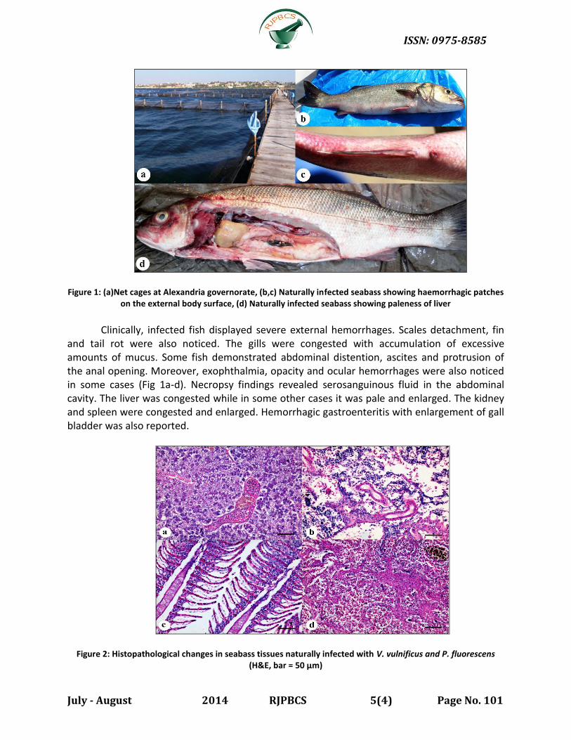
Figure 1: (a)Net cages at Alexandria governorate, (b,c) Naturally infected seabass showing haemorrhagic patches on the external body surface, (d) Naturally infected seabass showing paleness of liver
Clinically, infected fish displayed severe external hemorrhages. Scales detachment, fin
and tail rot were also noticed. The gills were congested with accumulation of excessive amounts of mucus. Some fish demonstrated abdominal distention, ascites and protrusion of the anal opening. Moreover, exophthalmia, opacity and ocular hemorrhages were also noticed in some cases (Fig 1a-d). Necropsy findings revealed serosanguinous fluid in the abdominal cavity. The liver was congested while in some other cases it was pale and enlarged. The kidney and spleen were congested and enlarged. Hemorrhagic gastroenteritis with enlargement of gall bladder was also reported.
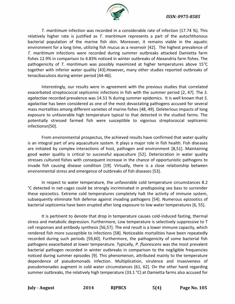
Figure 2: Histopathological changes in seabass tissues naturally infected with V. vulnificus and P. fluorescens (H&E, bar = 50 µm)

ISSN: 0975-8585
July - August 2014 RJPBCS 5(4) Page No. 102
a: Hepatopancreas of seabass naturally infected with V. vulnificus showing congestion of main blood vessels and sinusoidal spaces with diffuse vacuolar degeneration and hepatocytic necrosis revealing densely basophic stained hepatocytes with activation of melanomacrophage centers. b: Posterior kidney of seabass naturally infected with P. fluorescens showing diffuse necrosis of the interstitial tissue with discrete tubular necrosis leaving cyst like spaces with moderate mononuclear cell infiltration in-between necrotic tissue. c: Gills of seabass naturally infected with P. fluorescens showing separation in-between the epithelial cell lining of the secondary gill lamellae and the underlying capillary bed with hyperplasia of the epithelial lining at the base of the secondary gill lamellae.d: Spleen of seabass naturally infected with T. maritimum showing multifocal necrosis with discrete depletion of white pulp with activation of melanomacrophage centers.
PCR yielded specific amplicons identical to the size of target gene sequence for each bacterial strain. In particular, V. alginolyticus as well as V.vulnificus primers produced specific amplicons 737-bp and 273 bp respectively. Furthermore, P. fluorescens primers amplified a fragment of 850 bp. Also T. maritimum primers yielded specific 400 bp amplicon. On the hand, S. agalactiae primers produced specific 150-bp fragments ( Fig. 3).
Figure 3: Identification of bacterial strains by PCR
Improper values of water quality parameters were recorded in both investigated fish
farms (Table 5). Average water temperatures were 8.2 °C and 33.1 °C in Alexandria and Damietta farms respectively. Dissolved oxygen levels were lower than the optimal values 4.3 and 3.8ppm. The pH values of water in Alexandria and Damietta farms were 7.1 and 8.4 respectively. Salinity was within the expected range 31 ‰ and 37 ‰ respectively.
The results also demonstrated that nitrogenous waste products may be significantly accused for predisposing fish to surrender these outbreaks. The values in Alexandria farm recorded for NH3, NO2 and NO3 were far from the optimum recommended levels 0.8 mg/l, 0.75 mg/l and 1.3 mg/l respectively. The same measures in Damietta farm were also more noxious 1.3 mg/l, 0.99 mg/l and 1.69 mg/l respectively. Levels of some detected metals were higher than the recommended marine high reliability trigger value. The measures recorded for Cobalt,

ISSN: 0975-8585
July - August 2014 RJPBCS 5(4) Page No. 103
zinc, lead, iron and copper recorded in Alexandria farm were, 0.068 mg/l, 0.058 mg/l, 0.099 mg/l, 0.821 mg/l and 0.062 mg/l, respectively, while the same measures noticed in Damietta farm were, 0.118 mg/l, 0.0881 mg/l, 0.0362 mg/l, 1.07 mg/l and 0.066 mg/l respectively. It is worthy to mention that cadmium was not detected in both farms.
Table (5): Water quality measures in examined fish farms during outbreaks.
Sampling Site 1: Floating Net cage El-Hwarria, Marriott- Alexandria, Sampling Site 2: Fish ponds Muthallath El-Deeba – Damietta (ND): not detected.. *Average of readings was calculated in all measured parameters.
Histologically, cardiovascular degenerative, proliferative and necrotic changes were evident. Some bacterial agents were detected in histological sections (Fig. 2). Extensive hemorrhages were noticed in hemopoeitic tissues with congestion of hepatic, renal, splenic and cerebral blood vessels. Moreover, diffuse vacuolar degeneration and discrete necrotic changes were commonly evident in hepatocytes, pancreatic acinar cells, glomerulo-tubular and interstitial renal tissue. The necrotic areas were replaced by empty spaces and tissue debris. Furthermore, multifocal depletion of heamopoeitic elements was eminent concurrent with hyper-activation of melanomacrophage centers and multifocal infiltration of mononuclear cells. Ultimately, the gills showed severe congestion of lamellar blood vessels with frequent telangiectasis, diffuse and multifocal hyperplasia of epithelial lining of the secondary lamellae as well as the proliferation of malpighian cells concurrently with some infiltration by eosinophilic granular cells.
DISCUSSION
Sea bass is one of the predominant mari-cultured fish species at the majority of Mediterranean countries [17]. The high commercial value of Sea bass has encouraged aquaculturists to invest in its mass production [18]. However the rapid development of intensive culture systems has been faced in many incidences with infectious disease epidemics with consequent serious economic losses [6]. Virtually, diseases outbreaks are recognized as a significant constraint to aquaculture production and its economic development in many countries [4].
In agreement with [8,19], the improper water quality measures recorded in the
investigated fish farms are strongly incriminated in provoking the high mortalities noticed in theses farms. This is justified by the fact that pathogenesis of bacterial fish disease is a multi-factorial process .Such process necessitates that interaction of variable factors related to
Sampling sites
Season Temp
°C Salinity
% D.O. mg/l
pH NH3 mg/l
NO2 mg/l
NO3 mg/l
Heavy metals (mg/l)
Cd Co Zn Pb Fe Cu
Site 1 Winter 8.2°C 31 4.3 7.1 0.9 0.85 1.1 ND 0.058 0.054 0.091 0.721 0.052
Site 2 Summer 33.1°C 37 3.8 8.4 1.2 0.96 1.48 ND 0.115 0.0781 0.0359 1.03 0.046

ISSN: 0975-8585
July - August 2014 RJPBCS 5(4) Page No. 104
invading pathogens, hostile environment and fish host, which work together in synergism to shape the nature of the triggered course of infection [20].
In the current research, the bacteriological and molecular studies have confirmed that 49 % of the investigated sea bass were found to be infected. V. alginolyticus was the most prevalent bacterial pathogen (32.25 %) retrieved from moribund / freshly dead fishes followed by P. fluorescens (24.19%), T. maritimum (17.74 %) and S. agalactiae (14.51 %) with the lowest infection rate (11.29) for V. vulnificus . The high magnitude of vibrios infection (43.54 %.) during the epidemics confirms the potential of such pathogen in implementing serious impacts on cultured marine fish [21,6]. This may partly attributed to the ubiquitous existence of vibrios in seawater, sediment as well as alimentary tract of marine fishes [22-24]. Thus, the emergence of deteriorated environmental conditions would predictably drive them to vigorously attack fish with consequent eruptions of mass mortalities [19, 25].
V. alginolyticus have been involved in many events of mass mortalities noticed in several aquaculture facilities along the Mediterranean sea-coast [26-28,6]. The frequent mass mortalities associated with V. alginolyticus infections have alarmed fish farmers to the immense threat of such pathogen onboth cultured marine fish [29] as well as wild fish populations [2].
In respect to V. vulnificus, the results emphasized the great impacts of this pathogen on
the health status of sea bass, especially during the summer period. For several decades, V. vulnificus has been considered as a primary fish pathogen particularly in immune-compromised fish [30,31]. Numerous epizootics relevant to V. vulnificus have been recorded in a variety of cultured marine fishes during the past two decades [28,32]. Some studies ignored the relationship between vibrio infections and season of occurrence as vibrios attack cultured fish year-round without reliance to a particular season [33]. Studies in wild marine habitat also proved the association between unfavorable extreme water temperature and the flourishing of vibrio infections in fish [2].
The relatively low prevalence of vibriosis reported during winter episode in comparison
to summer epidemics can be justified by the fact that vibrios may survive in a dormant state in cold-water circumstances. The unfavorable conditions of low temperature and nutrient deprivation are the main triggering causes of induction for dormant stages of such pathogen [34, 35]. When appropriate environmental conditions are established, these microorganisms are nevertheless able to recover from this dormant state and infect fish [36].
Results also indicated that the recorded P. fluorescens infections approached 24.19 %.
This pathogen has long been considered to be among the most prevalent bacterial pathogens attacking variety of marine fish; in particular, stressed and immunocompromised species [19,37,38]. Some other studies showed extremely higher frequencies approaching 88% in gilthead sea bream larvae [39] and 72 % in captive Atlantic cod, Gadus morhua [40]. The maximal activity of its proteases is recorded during lower water temperatures (winter season) [9].In agreement with our results, several reports has concluded that lower water temperatures have triggered robust attacks during winter period [41].
ISSN: 0975-8585
July - August 2014 RJPBCS 5(4) Page No. 105
T. maritimum infection was recorded in a considerable rate of infection (17.74 %). This relatively higher rate is justified as T. maritimum represents a part of the autochthonous bacterial population of the marine fish skin. Moreover, it remains viable in the aquatic environment for a long time, utilizing fish mucus as a reservoir [42]. The highest prevalence of T. maritimum infections were recorded during summer outbreaks attacked Damietta farm fishes 12.9% in comparison to 4.83% noticed in winter outbreaks of Alexandria farm fishes. The pathogenicity of T. maritimum was possibly maximized at higher temperatures above 15°C together with inferior water quality [43].However, many other studies reported outbreaks of tenacibaculosis during winter period [44-46].
Interestingly, our results were in agreement with the previous studies that correlated
exacerbated streptococcal septicemic infections in fish with the summer period [2, 47]. The S. agalactiae recorded prevalence was 14.51 % during summer epidemics. It is well known that S. agalactiae has been considered as one of the most devastating pathogens accused for several mass mortalities among different varieties of marine fishes [48, 49]. Deleterious impacts of long exposure to unfavorable high temperature typical to that detected in the studied farms. The potentially stressed farmed fish were susceptible to vigorous streptococcal septicemic infectionsn[50].
From environmental prospectus, the achieved results have confirmed that water quality is an integral part of any aquaculture system. It plays a major role in fish health. Fish diseases are initiated by complex interactions of host, pathogen and environment [8,51]. Maintaining good water quality is critical to successful aquaculture [52]. Deterioration in water quality stresses cultured fishes with consequent increase in the chance of opportunistic pathogens to invade fish causing disease condition [19]. Virtually, there is a close relationship between environmental stress and emergence of outbreaks of fish diseases [53].
In respect to water temperature, the unfavorable cold temperature circumstances 8.2
°C detected in net-cages could be strongly incriminated in predisposing sea bass to surrender these epizootics. Extreme cold temperatures completely halt the activity of immune system, subsequently eliminate fish defense against invading pathogens [54]. Numerous epizootics of bacterial septicemia have been erupted after long exposure to low water temperatures [6, 55].
It is pertinent to denote that drop in temperature causes cold-induced fasting, thermal
stress and metabolic depression. Furthermore, Low temperature is selectively suppressive to T cell responses and antibody synthesis [56,57]. The end result is a lower immune capacity, which rendered fish more susceptible to infections [58]. Noticeable mortalities have been repeatedly recorded during such periods [59,60]. Furthermore, the pathogenicity of some bacterial fish pathogens exacerbated at lower temperature. Typically, P. fluorescens was the most prevalent bacterial pathogen recorded in winter outbreaks in comparison to the negligible frequencies noticed during summer episodes [9]. This phenomenon, attributed mainly to the temperature dependence of pseudomonads infection. Multiplication, virulence and invasiveness of pseudomonades augment in cold water circumstances [61, 62]. On the other hand regarding summer outbreaks, the relatively high temperature (33.1 °C) at Damietta farms also accused for

ISSN: 0975-8585
July - August 2014 RJPBCS 5(4) Page No. 106
these septicemic infections. Numerous septicemic bacterial infections affecting marine fish have been correlated with exposure to long periods of extreme temperature [2].
In respect to dissolved oxygen levels (DO), unfavorable levels lower than the optimal recommended values were recorded (4.3 mg/l) and (3.8 mg/l) in Alexandria and Damietta farms respectively. These relatively low DO levels synergized with other viable environmental aquatic components in both aquaculture facilities to produce the eminent cases of mortalities [63]. Impaired immune mechanisms triggered by theses hostile conditions are accused for the establishment of these outbreaks [2, 64]. Furthermore, the virulence of the causative pathogens is exacerbated by exposure to reduced dissolved oxygen levels [53].
Regarding the pH, the recorded values (7.1 and 8.4) seem to be conductive to the higher
prevalence of vibrios infections recorded in the study. The pH affects some virulence characteristics of many fish pathogens. Vibrios withstand higher pH values. Moreover, the adhesion of vibrios to fish mucus is greatly influenced by pH values [65, 66].
Results demonstrated that ammonia and nitrite are involved in modulating fishes to
surrender these episodes recorded in land based or cage culture systems. Unionized ammonia (NH3) should not exceed 0.1 mg/L for saltwater fish species [67]. These levels were far from those recorded in our investigations (0.9 and 1.2 mg/l) during winter and summer outbreaks respectively pointing to ammonia in disturbing the general health status of sea bream and sea bass in several ways. High ammonia levels enhance microbial infections through suppressing the immune capacity of fish. Phagocytic and clearance efficiency are diminished. As an ultimate fate for the colossal immumo-suppression of fishes, bacterial invasion will be the most probable event [2, 39, 68, 69]. On the other hand, prolonged exposure to low levels irritates gills and skin creating proper environment for parasitic infestations and opens new portal of entry for opportunistic bacterial infections [70].
For the heavy metals, achieved results also confirmed the synergistic effect of some
heavy metals, particularly, copper and iron in disturbing the health status of cultured sea bass which speed up the course of outbreaks. The mean of some detected metal values were higher than levels recommended by [67]. The recorded levels of Co, Zn, Pb, Fe and Cu in Alexandria Fish farm were 0.058 mg/l, 0.054 mg/l, 0.091 mg/l, 0.721mg/l and 0.052 mg/l compared to 0.115 mg/l, .0781mg/l, 0.0359 mg/l, 1.03 mg/l, and 0.046 mg/l respectively in Damietta farm Achieved results indicated the possible involvement of these metals in triggering such epizootics. According to ANZECC (2000) the marine high reliability trigger value recommended for ; copper, iron, lead, zinc, cobalt and cadmium are; 1.3 μg/L, 300 μg/L, 4.4 μg/L, 15 μg/L, 1 μg/L and 5.5 μg/L respectively. It is well established fact that polluted water weakens the fish host defenses, allowing increased opportunities for epizootic diseases to affect fish populations. Juveniles collected from polluted environments are more vulnerable to variety of microbial infections than those from non-contaminated habitats a result of immune-suppression. The role of heavy metals in rendering fish more susceptible to bacterial fish diseases is documented. Fish reared in copper and iron contaminated habitats are vulnerable to a variety of bacterial pathogens such as vibriosis and tenicibaculosis [42].

ISSN: 0975-8585
July - August 2014 RJPBCS 5(4) Page No. 107
Ultimately, the histopathological lesions detected in investigated fishes included circulatory, degenerative, proliferative and necrotic changes. Destruction of the critical components of the circulatory and immune system by toxic bacterial extracellular products (proteases and hemolysins) is thought to be the cornerstone behind these pathological alterations [71, 72].
CONCLUSION
V. alginolyticus, V. vulnificus, P. fluorescens T. maritimum, and S. agalactiae, are considered to be the most threatening bacterial pathogens involved in the septicemic bacterial infections of sea bass and therefore should be included in the future strategies of prophylaxis and treatment of farmed sea bass. The results also clearly point out that maintaining a favorable fish culture, environment and good husbandry practice are crucial for growing healthy fish.
ACKNOWLEDGMENTS
We greatly appreciate the assistance of Dr. Ahmad Noor El-Den, Researcher at the
National Research Centre, Egypt, for helping us to obtain samples. We also would like to thank all members of the Hydrobiology Department for their invaluable support during all stages of this research project.
REFERENCES
[1] El-Gendy MY. Epizootiological studies on some bacterial infections in marine fishes. In:
Fish Diseases and Management. Cairo, Faculty of Veterinary Medicine, 2007. [2] Moustafa M, Laila AM, Mahmoud MA, Soliman WS, & MY E.-G. J American Sci
2010;6:603 -612. [3] Sadek S. Fish Physiol Biochem 2000;22:171-178. [4] Meyer FP. J Anim Sci 1991;69:4201-4208. [5] Lem A, & Karunasagar I. The FAO Aquaculture Newsletter 2007;13-16. [6] Zorrilla I, Chabrillon M, Arijo S, Diaz-Rosales P, Martinez-Manzanares E, Balebona MC, &
Morinigo MA. Aquaculture 2003;218, 11-20. [7] Hedrick RP. J Aquatic Animal Health 1998;10:107-111. [8] Snieszko S. J Fish Biol 1974;6:197-208. [9] Eissa A, Tharwat N, & Zaki M. Chemosphere 2013;90: 1061-1068. [10] Eissa AE, Zaki MM, & Aziz AA. Flavobacterium columnare/Myxobolus tilapiae Concurrent
Infection in the Earthen Pond Reared Nile Tilapia (Oreochromis niloticus) during the Early Summer. Interdisciplinary Bio Central, 2010.
[11] Papoutsoglou SE, Tziha G, Vrettos X, & Athanasiou A. Aquacult Eng 1998;18:135-144. [12] Blancheton JP. Aquacult Eng 2000;22:17-31. [13] Buller NB. Bacteria from fish and other aquatic animals: a practical identification
manual, Cabi, 2004. [14] Bancroft JD, Gamble M. Theory and practice of histological techniques, Churchill
Livingstone, Edinburgh, 1996.

ISSN: 0975-8585
July - August 2014 RJPBCS 5(4) Page No. 108
[15] Boyd CE. Water Quality in Ponds for Aquaculture, Alabama Agricultural Experiment Station, Auburn University, AL, USA, 1990.
[16] Apha APHA. Standard Methods for the Examination of Water and Wastewater, Washington, D. C, 2000.
[17] Balebona MC, Andreu MJ, Bordas MA, Zorrilla I, Morinigo MA, & Borrego JJ. Appl Environ Microbiol 1998;64, 4269-4275.
[18] Arechavala-Lopez P, Uglem I, Fernandez-Jover D, Bayle-Sempere JT, & Sanchez-Jerez P. Fisheries Res 2012;121:126-135.
[19] Roberts RJ. (2012) Fish pathology, Wiley-Blackwell, W.B. Saunders, Philadelphia, PA.Sahoo P, Tanuja S, Dash SJ. Stress factors in fish culture ponds. e-planet, 2009.44:44.
[20] Zaki MM, Eissa AE, & Saeid S. World J Fish Marine Sci 2011;3: 21-36. [21] Sedano J, Zorrilla I, Morinigo M, Balebona M, Vidaurreta A, Bordas M, & Borrego J.
Aquacult Res 1996;27:323-333. [22] Vandenberghe J, Thompson FL, Gomez-Gil B, & Swings J. Aquaculture 2003;219:9-20. [23] Pedersen K, Dalsgaard I, & Larsen JL. Appl Environ Microbiol 1997;63:3711-3715. [24] Chen MX, Li HY, Li G, & Zheng TL. Braz J Microbiol 2011;42:884-896. [25] Snoussi M, Hajlaoui H, Noumi E, Zanetti S, & Bakhrou FA. Ann Microbiol 2008;58, 141-
146. [26] Bakhrouf A, Ben Ouada H, & Oueslati R. Marine Life 1995;5:47-53. [27] Kahla-Nakbi AB, Chaieb K, Besbes A, Zmantar T, & Bakhrouf A. Vet Microbiol
2006;117:321-327. [28] Yiagnisis M, Athanassopoulou F. Bacteria Isolated from Diseased Wild and Farmed
Marine Fish in Greece. In: (ed. by D.F. Aral). InTech, 2011. [29] Zhuzhuan H, Hejian G, & Huangzh J. Identificafio and Pathogenicity of Pathogen of
Epinephalus fario and E. awoara Ulceration Disease Acta Scientiarum Naturalium Universitatis Sunyatseni 2000;1:278-282.
[30] Hoi L, Larsen JL, Dalsgaard I, & Dalsgaard A. Appl Environ Microbiol 1998;64: 7-13. [31] Song Y, Cheng W, Shen C, Ou Y, & Song H. Occurrence of Vibrio vulnificus in cultured
shrimp and eel in Taiwan. In: NSC Symp. Ser, pp. 1990;172-179. [32] Biosca E, Amaro C, Esteve C, Alcaide E, & Garay E. J Fish Dis 1991;14:103-109. [33] Golomazou E, Athanassopoulou F, Vagianou S, Sabatakou O, Tsantilas H, Rigos G, &
Kokkokiris L. Turkish J Vet Ani Sci 2006;3:0389-396. [34] Oliver JD. J Microbiol 2005;43:93-100. [35] Colwell RR, & Huq A. Ann New York Acad Sci 1994;740, 44-54. [36] Whitesides MD, & Oliver JD. Appl Environ Microbiol 1997;63:1002-1005. [37] Plumb JA. Vet Hum Toxicol 1991; 33 (1):34-39. [38] Nedoluha PC, & Westhoff D. Food Microbiol 1997;14:255-264. [39] Ibrahem M, Hatem M. Mass mortality caused by Pseudomonas fluorescens and
Cryptocaryon irritans in gilthead sea bream (Sparus aurata L.) in Egyptian mariculture. In: Proceedings of the 2nd Global Fisheries and Aquaculture Research Conference, Cairo International Convention Center, 24-26 October 2009:pp. 275-289.
[40] Khan RA, Campbell J, & Lear H. J Wildl Dis 1981;17:521-527. [41] Hoshino T, Ishizaki K, Sakamoto T, Kumeta H, Yumoto I, Matsuyama H, & Ohgiya S. Lett
App Microbiol 1997;25:70-72.

ISSN: 0975-8585
July - August 2014 RJPBCS 5(4) Page No. 109
[42] Avendano-Herrera R, Magarinos B, Morinigo M, Romalde J, & Toranzo A. Bull European Assoc Fish Pathol 2005;25:68-72.
[43] Avendano-Herrera R, Núnez S, Magarinos B, & Toranzo A. Bull European Assoc Fish Pathol 2004;24:280-286.
[44] Wakabayashi H, Hikida M, Masumura K. Int J Systematic Bacteriol 1986;36:396-398. [45] Bernardet JF, Kerouault B, Michel C. Fish Pathol 1994;29: 105-111. [46] Soltani M, Munday BL, Burke CM. Aquaculture 1996;140:259-264. [47] Bragg R, Broere J. Bull European Assoc Fish Pathol 1986:6. [48] Eldar A, Bejerano Y, Livoff A, Horovitcz A, Bercovier H. Vet Microbiol 1995;43:33-40. [49] Pasnik DJ, Evans JJ, Panangala VS, Klesius PH, Shelby RA, Shoemaker CA. J Fish Dis
2005;28: 205-212. [50] Yuasa K, Kitancharoen N, Kataoka Y, Al-Murbaty FA. J Aquatic Animal Health 1999;11:87-
93. [51] Chen S. Revue Scientifique Et Technique (International Office of Epizootics),
1991;10:609. [52] Arrignon J. Management of freshwater fisheries, Science Publishers, Inc, 1999. [53] Mellergaard S, Nielsen E. Dis Aquatic Org 1995;22:101-114. [54] Sahoo P, Tanuja S, Dash SJ. e-planet 2009;44:44. [55] Doménech A, et al. Aquaculture 1997;156:317-326. [56] Clem LW, Miller NW, Bly JE. Phylogenesis Immune Functions 1991:191-213. [57] Ainsworth AJ, Dexiang C, Waterstrat P, Greenway T. Comp Biochem Physiol Part A:
Physiol 1991;100:907-912. [58] Ibarz A, et al. Rev Fish Biol Fish 2010;20: 539-556. [59] Sarusic G. Bull European Assoc Fish Pathol 1999;19. [60] Gallardo M, et alAquaculture 2003;223:15-27. [61] Berthe FCJ, Michel C, Bernardet JF. Dis Aquatic Org 1995;21: 151-155. [62] Doménech A, et al. J Fish Dis 1996;19:33-38. [63] Haley R, Davis SP, Hyde JM. Progressive Fish-Culturist, 1967;29:193. [64] Snieszko SF. Adv Vet Sci Comp Med 1973;17: 291-314. [65] Gordon AS, Gerchakov SM, Udey LR. Can J Microbiol 1981;27:698-703. [66] Balebona M, et al. Aquaculture 1995;132:113-120. [67] Anzecc. Australian and New Zealand Guidelines for Fresh and Marine Water Quality,
Australian and New Zealand Environmental and Conservation Council, 2000. [68] Cheng SY, Lee WC, Shieh LW, Chen JC. Arch Environ Contam Toxicol 2004;47:352-362. [69] Cheng W, Hsiao IS, Chen JC. Fish Shellfish Immunol 2004;17:193-202. [70] Noga EJ. Fish disease: diagnosis and treatment, John Wiley & Sons, 2010. [71] Kimura H, Kusuda R. J Fish Dis 1979;2:501-510. [72] Woo PT. Fish Diseases and Disorders: Volume 3: Viral, Bacterial and Fungal Infections,
Cabi, 2011.