Eosinophilic esophagitis. The North against the South? A bio ...
9
Revista de Gastroenterología de México. 2017;82(4):328---336 www.elsevier.es/rgmx REVISTA DE DE MEXICO GASTROENTEROLOGIA ´ ´ REVIEW ARTICLE Eosinophilic esophagitis. The North against the South? A bio-economic-social mechanistic approach and clinical implications D. García-Compeán * , J.A. González-González, E.I. González-Moreno, H.J. Maldonado-Garza Servicio de Gastroenterología, Departamento de Medicina Interna, Hospital Universitario y Facultad de Medicina, Universidad Autónoma de Nuevo León, Monterrey, Mexico Received 28 October 2016; accepted 9 February 2017 Available online 12 August 2017 KEYWORDS Eosinophilic esophagitis; Prevalence; Esophageal eosinophilia; Helicobacter pylori Abstract Eosinophilic esophagitis is a chronic antigen-mediated disease characterized by esophageal symptoms, esophageal eosinophilia, and the absence of response to proton pump inhibitors. It is the most frequent cause of dysphagia and food impaction in adults. Its inci- dence and prevalence is very high in the developed countries (USA, Europe, Australia), where its connotation is that of an emerging epidemic. While studies have been published with large case series in the developed countries, those published in Latin America are small or consist of isolated case reports. The differences in the prevalence of the disease between the developed and developing regions are unknown. Genetic or racial causes have been cited. Nevertheless, the epidemic nature of the disease suggests that environmental causes are the most powerful. Based on the published hypotheses, as well as on epidemiologic studies, the present review discusses some of the possible causes of the disparity in the prevalence of eosinophilic esophagi- tis between the two types of countries. The ‘hygiene hypothesis’ is reviewed, together with the possible relation of Helicobacter pylori, intestinal parasites, and modifications of the esophageal microbiota in patients with eosinophilic esophagitis. In reference to studies con- ducted in the United States, the clinical behavior and progression of eosinophilic esophagitis in Hispanics is reviewed and a possible predominant phenotype in Mexican and other Latin Amer- ican patients is discussed. Finally, based on the above, an algorithm for studying the disease in the Latin American countries is proposed. © 2017 Asociaci´ on Mexicana de Gastroenterolog´ ıa. Published by Masson Doyma M´ exico S.A. This is an open access article under the CC BY-NC-ND license (http://creativecommons.org/licenses/ by-nc-nd/4.0/). Please cite this article as: García-Compeán D, González-González JA, González-Moreno EI, Maldonado-Garza HJ. La esofagitis eosi- nofílica. ¿El Norte contra el Sur? Enfoque mecanicista bio-económico-social e implicaciones clínicas. Revista de Gastroenterología de México. 2017;82:328---336. * Corresponding author. Servicio de Gastroenterología, Departamento de Medicina Interna, Hospital Universitario y Facultad de Medicina, Universidad Autónoma de Nuevo León, Avenida Madero y Gonzalitos S/N, Col. Mitras Centro, C.P. 64460 Monterrey, Mexico. Tel.: +52-81-83487315; Fax: +52 81 89891381. E-mail address: [email protected] (D. García-Compeán). 2255-534X/© 2017 Asociaci´ on Mexicana de Gastroenterolog´ ıa. Published by Masson Doyma M´ exico S.A. This is an open access article under the CC BY-NC-ND license (http://creativecommons.org/licenses/by-nc-nd/4.0/).
-
Upload
khangminh22 -
Category
Documents
-
view
3 -
download
0
Transcript of Eosinophilic esophagitis. The North against the South? A bio ...
Revista de Gastroenterología de México. 2017;82(4):328---336
www.elsevier.es/rgmx
REVISTA DE
DE MEXICO
GASTROENTEROLOGIA´
´
REVIEW ARTICLE
Eosinophilic esophagitis. The North against the South?
A bio-economic-social mechanistic approach and
clinical implications�
D. García-Compeán ∗, J.A. González-González, E.I. González-Moreno,H.J. Maldonado-Garza
Servicio de Gastroenterología, Departamento de Medicina Interna, Hospital Universitario y Facultad de Medicina, Universidad
Autónoma de Nuevo León, Monterrey, Mexico
Received 28 October 2016; accepted 9 February 2017
Available online 12 August 2017
KEYWORDSEosinophilicesophagitis;Prevalence;Esophagealeosinophilia;Helicobacter pylori
Abstract Eosinophilic esophagitis is a chronic antigen-mediated disease characterized by
esophageal symptoms, esophageal eosinophilia, and the absence of response to proton pump
inhibitors. It is the most frequent cause of dysphagia and food impaction in adults. Its inci-
dence and prevalence is very high in the developed countries (USA, Europe, Australia), where
its connotation is that of an emerging epidemic. While studies have been published with large
case series in the developed countries, those published in Latin America are small or consist of
isolated case reports. The differences in the prevalence of the disease between the developed
and developing regions are unknown. Genetic or racial causes have been cited. Nevertheless,
the epidemic nature of the disease suggests that environmental causes are the most powerful.
Based on the published hypotheses, as well as on epidemiologic studies, the present review
discusses some of the possible causes of the disparity in the prevalence of eosinophilic esophagi-
tis between the two types of countries. The ‘hygiene hypothesis’ is reviewed, together with
the possible relation of Helicobacter pylori, intestinal parasites, and modifications of the
esophageal microbiota in patients with eosinophilic esophagitis. In reference to studies con-
ducted in the United States, the clinical behavior and progression of eosinophilic esophagitis in
Hispanics is reviewed and a possible predominant phenotype in Mexican and other Latin Amer-
ican patients is discussed. Finally, based on the above, an algorithm for studying the disease in
the Latin American countries is proposed.
© 2017 Asociacion Mexicana de Gastroenterologıa. Published by Masson Doyma Mexico S.A. This
is an open access article under the CC BY-NC-ND license (http://creativecommons.org/licenses/
by-nc-nd/4.0/).
� Please cite this article as: García-Compeán D, González-González JA, González-Moreno EI, Maldonado-Garza HJ. La esofagitis eosi-
nofílica. ¿El Norte contra el Sur? Enfoque mecanicista bio-económico-social e implicaciones clínicas. Revista de Gastroenterología de México.
2017;82:328---336.∗ Corresponding author. Servicio de Gastroenterología, Departamento de Medicina Interna, Hospital Universitario y Facultad de Medicina,
Universidad Autónoma de Nuevo León, Avenida Madero y Gonzalitos S/N, Col. Mitras Centro, C.P. 64460 Monterrey, Mexico.
Tel.: +52-81-83487315; Fax: +52 81 89891381.
E-mail address: [email protected] (D. García-Compeán).
2255-534X/© 2017 Asociacion Mexicana de Gastroenterologıa. Published by Masson Doyma Mexico S.A. This is an open access article under
the CC BY-NC-ND license (http://creativecommons.org/licenses/by-nc-nd/4.0/).
Eosinophilic esophagitis. A bio-economic-social mechanistic approach 329
PALABRAS CLAVEEsofagitiseosinofílica;Prevalencia;Eosinofilia esofágica;Helicobacter pylori
La esofagitis eosinofílica. ¿El Norte contra el Sur? Enfoque mecanicista
bio-económico-social e implicaciones clínicas
Resumen La esofagitis eosinofílica (EEo) es una enfermedad crónica mediada por alérgenos,
caracterizada por síntomas esofágicos, eosinofilia esofágica y ausencia de respuesta favorable
a inhibidores de la bomba de protones (IBP). En los adultos es la causa más frecuente de
disfagia e impactación alimentaria. Su incidencia y prevalencia son muy altas en los países
desarrollados (EUA, Europa y Australia), en donde ha alcanzado una connotación de epidemia.
Mientras que en los países desarrollados se publican estudios con grandes series de pacientes,
en nuestro subcontinente se reporta en series con pocos casos o en casos aislados. Las causas
de las diferencias de la prevalencia de la enfermedad entre ambas regiones se desconocen. Se
han invocado causas genéticas o raciales. No obstante, el carácter epidémico de la enfermedad
sugiere que las causas ambientales son más poderosas.
Con base en hipótesis publicadas, así como en los estudios epidemiológicos, en la presente
revisión se discutirán algunas de las posibles causas de la disparidad de prevalencia de la
EEo entre ambos tipos de países. Se revisará la «hipótesis de la higiene», así como la posible
relación del Helicobacter pylori, los parásitos intestinales y las modificaciones de la microbiota
esofágica con la EEo. Con base en estudios realizados en EUA, se revisará el comportamiento
clínico y evolutivo de la EEo en individuos hispanos y se discutirá acerca de un posible fenotipo
predominante en los pacientes de América Latina y México. Finalmente, sobre la base anterior,
se propondrá un algoritmo de estudio de la enfermedad en nuestros países.
© 2017 Asociacion Mexicana de Gastroenterologıa. Publicado por Masson Doyma Mexico S.A.
Este es un artıculo Open Access bajo la licencia CC BY-NC-ND (http://creativecommons.org/
licenses/by-nc-nd/4.0/).
Introduction
Eosinophilic esophagitis (EoE) is a chronic disease of theesophagus caused by environmental and food allergens.It is characterized by symptoms of esophageal dysfunc-tion, eosinophilic infiltrate (≥ 15 eosinophils/high powerfield) in the esophageal mucosa, and absence of clinicaland histologic response to treatment with proton pumpinhibitors (PPIs) for 8 weeks.1,2 According to the diagnosticguidelines established by the 2007, 2011, and 2013 interna-tional consensuses on digestive eosinophilia,1---3 EoE shouldbe differentiated from the esophageal eosinophilia causedby gastroesophageal reflux disease and PPI-responsiveesophageal eosinophilia (PPI-REE).3 The latter is clinically,endoscopically, and histologically indistinguishable fromEoE,2,4 and whether it is a phenotype of EoE that respondsto PPIs, or an independent entity, is not known.5
Initially described in children, it was not until 1978 thatEoE was reported in adults.6 In children it manifests as aninflammatory phenotype clinically characterized by vomi-ting, heartburn, chest pain, and difficulty to gain bodyweight. At endoscopy, there is predominant inflammatoryexudate in the esophageal mucosa.7,8 In adults it is currentlythe most frequent cause of dysphagia and food impaction.It behaves as a progressively chronic disease, highly recur-rent, and with a tendency toward esophageal remodeling(submucosal fibrosis) and stricture.9,10 It affects males morefrequently than females, with a 4:1 ratio, and individuals inthe fourth and fifth decades of life.11
The present article describes the general clinical andepidemiologic characteristics of EoE in developed and devel-oping countries (North and South), particularly the Latin
American countries, emphasizing the differences in preva-lence between both regions of the world. At the same time,based on the scant epidemiologic evidence that has beenpublished up to now, we discuss the possible causes involvedin such differences. We then attempt to outline the clini-cal and progressive characteristics of the disease in LatinAmerican populations, gleaned from the available clinicalinformation, and finally, we propose measures to diagnoseand treat the disease in Mexico and other Latin Americancountries.
Eosinophilic esophagitis. Prevalence andincidence in developed and developingcountries
Based on studies conducted on general populations, sinceits initial description in the 1980s, EoE incidence and preva-lence has significantly increased, to the degree that it isconsidered epidemic, in the United States (US), Canada,Western Europe, and Australia (this last country locatedin the South but with economic and social developmentcomparable to that of Europe and the US).12---15 Preva-lence in children and adults in the general population inthose geographic regions has been determined at 10 to 90/100,000 inhabitants with an estimated incidence of 2 to 13cases/100,000 inhabitants. These figures are the highest,compared with those reported in the few studies publishedin Latin America,16---18 in China,19,20 in Japan,21,22 and in SaudiArabia.23 No cases have been reported in India or in NorthAfrica or Sub-Saharan Africa.

330 D. García-Compeán et al.
Disorders of the
mucosal barriers
Environmental
and dietary
factors
Genetic susceptibility
IL-5Maturation of
the bone
marrow
eosinophils
Increase of
permeability to
inflammatory
cells (eosinophils,
basophils, adn
mastocytes)
Epithelial cell
eotaxine-3
production
IL-13
IL-4
Natural killer
T cell Th2 reaction
Eosinophil
and mastocyte
recruitment
Mucosal
inflammation
Fibrosis
TGF-β
Figure 1 Physiopathogenesis of eosinophilic esophagitis.
In Mexico and in other Latin American countries, inci-dence of the disease in the general population is not knownbecause of the lack of studies. There have only been isolatedcase reports or small case series from some of the SouthAmerican countries (Chile, Uruguay, Argentina, Peru).24---26
In two prospective Latin American studies, one from Mex-ico and the other from Brazil, in subjects that underwentesophagogastroduodenoscopy due to esophageal symptoms,prevalence was reported at 1 and 1.7%,16,18 which was lowerthan that reported in similar studies in the US and Spain (5to 8%).27,28 In another Mexican study conducted by our groupon patients with clinical signs of refractory gastroesophagealreflux disease, prevalence was also low, at 4%.17
Possible causes of the eosinophilic esophagitisepidemic in developed countries and of thelow prevalence in developing countries
EoE is an allergic disease with pathophysiologic mechanismssimilar to those of other atopic diseases, such as bronchialasthma, allergic rhinitis, and atopic dermatitis. It is welldocumented that EoE is triggered by allergens containedin foods and the environment, given that the restrictionof certain foods improves symptoms and the esophagealeosinophilic infiltrate. In addition, it has an elevated sea-sonal incidence, with greater frequency between summerand fall, when there is a greater concentration of pollenin the environment.29 These allergens cause an inflamma-tory reaction mediated by helper T lymphocytes, known asthe Th2 reaction, which induces the production of IL-4, IL-5, and IL-13 interleukins and the secretion of eotaxine-3 bythe epithelial cells of the esophageal mucosa. That agentis a powerful recruiter of eosinophils and mast cells, whosepresence causes inflammation of the mucosa and disruptionof the epithelial barrier (fig. 1).
Similar to what has been observed with EoE, other aller-gic diseases, such as asthma, allergic rhinitis, and atopicdermatitis, have had a tendency to increase in the last 30years in Europe and the USA, with asthma becoming an
Table 1 Factors that could be involved in eosinophilic
esophagitis (EoE) as a cause of epidemic in developed
countries and of low prevalence in developing countries.
Hygiene hypothesis: eradication of infections (inducer)
Early exposure to antibiotics (inducer)
Cesarean birth (inducer)
Food processing: preservatives, chemical additives, use of
hormones, pesticides (inducer)
Genetic susceptibility (protector or inducer)
Changes in the esophageal microbiota (protector or inducer)
Helicobacter pylori (protector)
Other bacteria (protector)
Intestinal parasites (protector)
epidemic phenomenon30 and atopic dermatitis doubling ortripling its prevalence.31
Even though the knowledge of the genetic bases of EoEhas been defined in recent years,32 the epidemic behaviorof the disease suggests that environmental causes are morepowerful than genetic ones. It is not clear why food andenvironmental allergens that had been tolerated for manyyears in the affected countries became EoE triggers. Thefactors involved in the epidemic could also explain the lowprevalence in developing countries, including Mexico. Thosefactors are most likely multiple and complex, but discussionof them presently amounts to speculation only, given thatthe majority have not been intentionally studied in relationto EoE, and the few existing studies are in the preliminaryphase (Table 1). Nevertheless, recent epidemiologic obser-vations have produced interesting data, which is discussedin the present article.
The hygiene hypothesis
The ‘‘hygiene hypothesis’’ proposed by Okada et al.in 201033 could explain why individuals from developedcountries have a more intense allergic reaction to foods

Eosinophilic esophagitis. A bio-economic-social mechanistic approach 331
and environmental allergens than they had in the past andwhy their reaction is stronger than that currently experi-enced by individuals from developing countries. Accordingto this hypothesis, the reduction in the incidence of infec-tions in individuals from the developed countries due tothe pharmacologic eradication of bacteria and parasitesis the origin of the increased incidence of allergic andautoimmune diseases, such as inflammatory bowel dis-ease. In support of this hypothesis, over recent decadesan increase in allergic diseases mediated by pathophysio-logic mechanisms similar to EoE, such as asthma, allergicrhinitis, and atopic dermatitis,30 has been reported. Theincidence of atopic dermatitis has doubled or tripled indeveloped countries over the past 3 decades.31 The mech-anisms through which the ‘‘hygiene hypothesis’’ attemptsto explain these phenomena are not well defined, but itproposes that the main one could be related to a redi-rection of the Th1 and Th2 inflammatory reactions. Thehelper T lymphocytes in the Th1 reaction produce inflam-matory cytokines, such as interleukin IL-2, interferon-�,and the tumor necrosis factor alpha (TNF-�), that act oncell-mediated immunity. In contrast, the helper T lym-phocytes of the Th2 reaction produce the IL-4, IL-5, andIL-13 interleukins that contribute to the production oftype E immunoglobulins (IgE) and eosinophil-mediated aller-gic responses. Some authors suggest that in developedcountries, the reduced microbial burden in early childhood(that normally favors a strong Th1 inflammatory reaction)redirects the inflammatory response toward the Th2 pheno-type, thus predisposing the individual to allergic disorders.
Helicobacter pylori
In the context of the ‘‘hygiene hypothesis’’, an inverse rela-tion of Helicobacter pylori (H. pylori) and EoE has recentlybeen reported. In an extensive study on biopsies from indi-viduals in the US general population, Dellon et al. found thatindividuals with esophageal eosinophilia had a significantlyreduced odds ratio of H. pylori, compared with individualswith normal biopsy. They also found a dose-response rela-tion between the increase in the eosinophilic infiltrate andreduced prevalence of H. pylori.34 Another more recentstudy conducted in Germany confirmed those findings. Inthat case-control study with 58 patients, 5.2% of the patientswith EoE had serologic evidence of H. pylori infection, com-pared with seroprevalence in 37.9% of the patients withoutEoE.35
In Mexico, H. pylori prevalence data obtained in anational survey on an open population was 66%36 andit reached 90% in some Central and South Americancountries.37,38 In contrast, prevalence in the developedcountries was significantly lower, at 6 to 20%.39 The dif-ferences in prevalence between the two regions has beenattributed to the exposure and contagion rates of H.
pylori in childhood, which are high in the Latin Americancountries.40,41
It is interesting that since its characterization in 1980and its consequent eradication, the prevalence of H. pylori
has been significantly reduced in the US. In contrast, ithas remained high or has been reduced very little in theless developed countries.42 In the US, H. pylori prevalence
varies among the different racial groups that make up itspopulation. It is lower in Caucasians (8-26%) and higheramong African Americans and Latinos (52-54% and 48-64%,respectively).43---45 This intergroup difference is linked moreto socioeconomic differences than to racial ones, as shownin the study by Malaty and Graham, conducted on a residentHispanic population in the US, in which there was an inverserelation between H. pylori prevalence and socioeconomiclevel. The Hispanics from the high, middle, and low socio-economic classes had prevalences of H. pylori of 6, 43, and86%, respectively.46
In addition to the ethnic differences, it has been specu-lated that differences in climate and exposure to allergenscould explain the unequal prevalence of EoE betweendeveloped and developing countries. The results of studiesconducted in the US could be interpreted as contradictingthose arguments. In those studies, in which the prevalenceof EoE in different population groups was evaluated, EoEwas reported to be significantly less frequent in Hispanicsthan in Caucasians, despite their being exposed to the sameclimate and the same environmental and food allergens. Ina study conducted on an open population of more than 6million individuals, of which 3,360 (0.05%) had EoE, 3,160(94.04%) were Caucasian, 190 (5.65%) were African Amer-ican, and only 10 (0.29%) were Hispanic.47 These resultssuggest that African American and Hispanic individuals mayhave ‘‘protective’’ factors against EoE and could also bearguments in favor of the inverse relation between H. pylori
and EoE and of the hygiene hypothesis. The concomitanteffect of genetic and racial susceptibility cannot be ruledout.
Parasites
Based on the ‘‘hygiene hypothesis’’, intestinal parasitosiscould be a protective factor against EoE, given that para-sitosis prevalence is still high in the developing countries.However, this aspect has not been widely analyzed. In somestudies in tropical and subtropical countries, there is evi-dence that Schistosoma infections have a strong protectivefactor against atopia,48 that Necator americanus possiblyhas a protective effect against asthma, and that the eradica-tion of helminths increases atopic cutaneous sensitivity.49,50
However, in other studies, the eradication of intestinal para-sites has not been observed to provide a protective factoragainst allergic diseases, particularly asthma.51 In addition,the proposed mechanism through which bacterial infectionswould reduce the risks for allergic diseases (redirectionof the Th1 and Th2 inflammatory reactions) would not bethe same for parasites, precisely because they induce Th2reactions. However, other alternate mechanisms could beinvolved, such as antigen competence and immunoregula-tion. The first proposes that two immune responses causedby different antigens tend to inhibit each another.33 The sec-ond postulates that in an immunologic response, regulatingT cells that suppress the immunologic responses differentfrom the responses against the antigens in question couldbe activated.52 The theory about parasites is interesting,but further studies that evaluate the possible relation toEoE are required.

332 D. García-Compeán et al.
Microbiota
The human intestine is colonized with different strainsof bacteria after birth. The gut microbiota is importantfor numerous physiologic functions, for maintaining theintegrity of the mucosal barrier, and for regulating immuno-logic functions.53,54 In recent years it has been pointed outthat alterations in the composition of the gut microbiotacould be involved in the causes of some chronic diseasesand neoplasias.55,56 In that context, the gut microbiotahas been extensively studied. In contrast, the esophagealmicrobiota had not been characterized until recently. Atpresent, there are few studies evaluating the alterationsof the esophageal microbiota in EoE. In one study con-ducted on 33 children with EoE, 18 with active diseaseand 15 with inactive disease were compared with 35 con-trols. The modifications of the esophageal microbiome wereevaluated after dietary manipulation and significant differ-ences in microbiome composition were found in the childrenwith active EoE, compared with the controls (Proterobacte-
ria in EoE and Streptococcus and Atopobium genera in thecontrols), who experienced no modifications from dietarychanges.57 In another study conducted on 70 children,there were significant differences in the composition of theesophageal microbiota in patients with EoE, compared withpatients with gastroesophageal reflux and normal individ-uals. Hemophillus was significantly predominant in patientswith EoE.58 Finally, another study found significant modifi-cations in the esophageal microbiota with chronic PPI use inadults with gastroesophageal reflux disease, Barrett’s esoph-agus, and esophageal and gastric adenocarcinoma.59,60 Eventhough chronic PPI use has not been demonstrated to be arisk factor for EoE, it has been cited as a possible causethrough alternative mechanisms to those related to acidsuppression.61
Although the findings of those studies are interesting,modifications in the esophageal microbiota cannot yet beascertained to be the cause of EoE, but rather an effectof the disease.62 Further studies with respect to this areneeded.
Possible predominance of an attenuatedclinical phenotype of eosinophilic esophagitisin developing countries
In developing countries, EoE is a chronic disease with anelevated clinical and pathologic recurrence rate when treat-ment is suspended.63,64 Recent studies have shown that ithas a tendency to progress from the inflammatory pheno-type (that occurs in children) to the fibrostenotic phenotype(that occurs in adults), resulting in esophageal stricture andfood impaction.65,66 These complications are more frequentas the progression time of the disease increases, and areseen in 70% of the cases that have had symptomatic activityfor more than two decades.67,68
In a 2011 study published by Richter et al., conductedat a single hospital center in the US on 64 patientswith esophageal eosinophilia detected through pathologyreports, and with a 10-year follow-up (81% Caucasians, 12%African Americans, and 6% Hispanics), the following inter-esting data were found: the African Americans and Hispanics
had a clinically atypical form of the disease characterizedby advanced age, a greater frequency of gastroesophagealreflux disease symptoms, less dysphagia for solid foods, andfewer alterations related to fibrosis (circumferential rings)at endoscopy, compared with the Caucasians. The authorsconcluded that those intergroup differences in the clinicalbehavior of esophageal eosinophilia could be due to dif-ferences in the phenotype of the disease itself or to thepresence of different clinical entities.69 The results of thatstudy were confirmed in 2016 in an extensive multicen-ter study conducted at 5 large US hospital centers on 793patients with EoE (476 adults and 317 children), of which83% were Caucasian, 10% were African American, and 7%were Hispanics and Asians. As in the study by Richter et al.,the non-Caucasian groups had an atypical ‘‘benign’’ formof the disease, given that they presented with a signifi-cantly lower frequency of dysphagia (56 and 53% vs 74%)and esophageal food impaction (13 and 13% vs 35%) than theCaucasians. They had a lower frequency of concentric ringsand linear grooves at endoscopy. The authors concludedthat EoE diagnosis should be considered in African Ameri-cans and Hispanics, even if they did not present with typicalsymptoms.70
The findings of those two studies could have broaderimplications. It must be remembered that PPI-REE is acondition that responds favorably to PPIs and is clinicallyand histologically indistinguishable from EoE.71,72 In thelast consensus of the international working group on EoEin 2016, it was suggested that this clinical entity is apart of the EoE spectrum, possibly with different clini-cal and progressive characteristics.73 Its frequency in thepatients with esophageal eosinophilia in general is from 50to 75%.74,75 It has been suggested that PPIs could have arestoring effect on lesions in the esophageal mucosa pro-duced by acid, increasing their permeability and makingthe penetration of allergens possible.76,77 PPIs have alsobeen observed to have anti-inflammatory properties thatare independent of acid suppression, inhibiting eotaxine-3secretion.78,79 Until recently, the long-term natural historyof this entity was unknown. A retrospective study on 94patients demonstrated that patients with PPI-REE, com-pared with EoE, had significantly less risk for distal strictureand narrowings of esophageal caliber after 20 years ofsymptoms (30.2 vs 72.3%).80 At present it is not knownif there are racial differences in the distribution of thisentity.
The results of the studies discussed above suggest thatnon-Caucasian individuals (Hispanics and African Ameri-cans) could possess ‘‘protective’’ or attenuating factors forEoE, particularly against the fibrostenotic phenotype of thedisease. It is possible that in developing countries an atten-uated inflammatory phenotype prevails that has ‘‘atypical’’clinical characteristics and is sensitive to PPIs.71 In the twostudies published in Mexico in 2011 and 2016, a predomi-nance of typical clinical characteristics was described, butthey were small case series.17,81 The quantity and charac-teristics of the underdiagnosed patients in Mexico is notknown and the long-term clinical outcome has not been eval-uated. In our experience with 8 patients seen at our hospitalsince 2007, none of them has required esophageal dilationsand only one presented with food impaction at the time ofdiagnosis (personal data not published).
Eosinophilic esophagitis. A bio-economic-social mechanistic approach 333
Conclusions and recommendations
In conclusion, the incidence of EoE in developed countrieshas been increasing rapidly in recent years, with epidemic
characteristics. In contrast, prevalence is very low in devel-oping countries, including Mexico. The causes of this areunknown. The epidemic behavior of EoE, as in other aller-gic disease, suggests that environmental causes are more
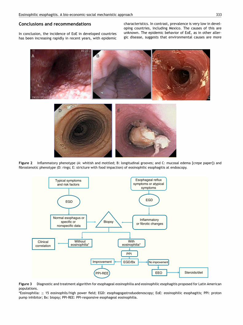
Figure 2 Inflammatory phenotype (A: whitish and mottled; B: longitudinal grooves; and C: mucosal edema [crepe paper]) and
fibrostenotic phenotype (D: rings; E: stricture with food impaction) of eosinophilic esophagitis at endoscopy.
Typical symptoms
and risk factors
Esophageal reflux
symptoms or atypical
symptoms
Inflammatory
or fibrotic changes
Clinical
correlation
Withouteosinophilia*
Witheosinophilia*
PPI
Improvement EGD/Bx No improvement
EEO Steroids/dietPPI-REE
Normal esophagus or
specific or
nonspecific data
EGDEGD
Biopsy
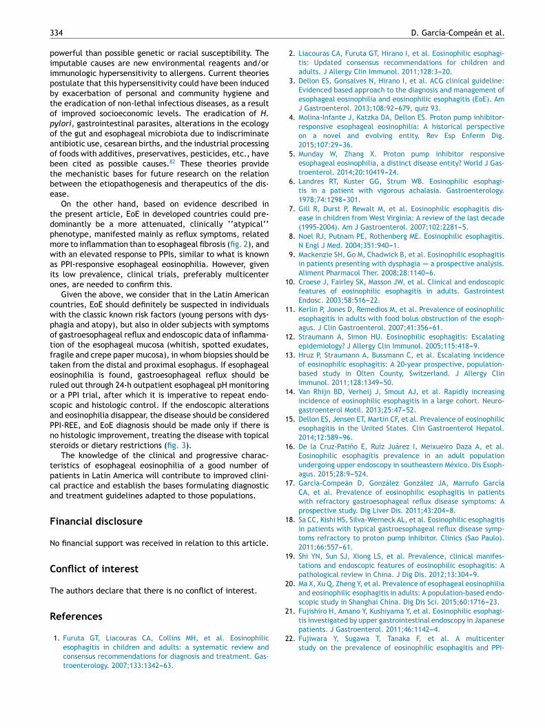
Figure 3 Diagnostic and treatment algorithm for esophageal eosinophilia and eosinophilic esophagitis proposed for Latin American
populations.
*Eosinophilia: ≥ 15 eosinophils/high power field; EGD: esophagogastroduodenoscopy; EoE: eosinophilic esophagitis; PPI: proton
pump inhibitor; Bx: biopsy; PPI-REE: PPI-responsive esophageal eosinophilia.
334 D. García-Compeán et al.
powerful than possible genetic or racial susceptibility. Theimputable causes are new environmental reagents and/orimmunologic hypersensitivity to allergens. Current theoriespostulate that this hypersensitivity could have been inducedby exacerbation of personal and community hygiene andthe eradication of non-lethal infectious diseases, as a resultof improved socioeconomic levels. The eradication of H.
pylori, gastrointestinal parasites, alterations in the ecologyof the gut and esophageal microbiota due to indiscriminateantibiotic use, cesarean births, and the industrial processingof foods with additives, preservatives, pesticides, etc., havebeen cited as possible causes.82 These theories providethe mechanistic bases for future research on the relationbetween the etiopathogenesis and therapeutics of the dis-ease.
On the other hand, based on evidence described inthe present article, EoE in developed countries could pre-dominantly be a more attenuated, clinically ‘‘atypical’’phenotype, manifested mainly as reflux symptoms, relatedmore to inflammation than to esophageal fibrosis (fig. 2), andwith an elevated response to PPIs, similar to what is knownas PPI-responsive esophageal eosinophilia. However, givenits low prevalence, clinical trials, preferably multicenterones, are needed to confirm this.
Given the above, we consider that in the Latin Americancountries, EoE should definitely be suspected in individualswith the classic known risk factors (young persons with dys-phagia and atopy), but also in older subjects with symptomsof gastroesophageal reflux and endoscopic data of inflamma-tion of the esophageal mucosa (whitish, spotted exudates,fragile and crepe paper mucosa), in whom biopsies should betaken from the distal and proximal esophagus. If esophagealeosinophilia is found, gastroesophageal reflux should beruled out through 24-h outpatient esophageal pH monitoringor a PPI trial, after which it is imperative to repeat endo-scopic and histologic control. If the endoscopic alterationsand eosinophilia disappear, the disease should be consideredPPI-REE, and EoE diagnosis should be made only if there isno histologic improvement, treating the disease with topicalsteroids or dietary restrictions (fig. 3).
The knowledge of the clinical and progressive charac-teristics of esophageal eosinophilia of a good number ofpatients in Latin America will contribute to improved clini-cal practice and establish the bases formulating diagnosticand treatment guidelines adapted to those populations.
Financial disclosure
No financial support was received in relation to this article.
Conflict of interest
The authors declare that there is no conflict of interest.
References
1. Furuta GT, Liacouras CA, Collins MH, et al. Eosinophilic
esophagitis in children and adults: a systematic review and
consensus recommendations for diagnosis and treatment. Gas-
troenterology. 2007;133:1342---63.
2. Liacouras CA, Furuta GT, Hirano I, et al. Eosinophilic esophagi-
tis: Updated consensus recommendations for children and
adults. J Allergy Clin Immunol. 2011;128:3---20.
3. Dellon ES, Gonsalves N, Hirano I, et al. ACG clinical guideline:
Evidenced based approach to the diagnosis and management of
esophageal eosinophilia and eosinophilic esophagitis (EoE). Am
J Gastroenterol. 2013;108:92---679, quiz 93.
4. Molina-Infante J, Katzka DA, Dellon ES. Proton pump inhibitor-
responsive esophageal eosinophilia: A historical perspective
on a novel and evolving entity. Rev Esp Enferm Dig.
2015;107:29---36.
5. Munday W, Zhang X. Proton pump inhibitor responsive
esophageal eosinophilia, a distinct disease entity? World J Gas-
troenterol. 2014;20:10419---24.
6. Landres RT, Kuster GG, Strum WB. Eosinophilic esophagi-
tis in a patient with vigorous achalasia. Gastroenterology.
1978;74:1298---301.
7. Gill R, Durst P, Rewalt M, et al. Eosinophilic esophagitis dis-
ease in children from West Virginia: A review of the last decade
(1995-2004). Am J Gastroenterol. 2007;102:2281---5.
8. Noel RJ, Putnam PE, Rothenberg ME. Eosinophilic esophagitis.
N Engl J Med. 2004;351:940---1.
9. Mackenzie SH, Go M, Chadwick B, et al. Eosinophilic esophagitis
in patients presenting with dysphagia ---- a prospective analysis.
Aliment Pharmacol Ther. 2008;28:1140---6.
10. Croese J, Fairley SK, Masson JW, et al. Clinical and endoscopic
features of eosinophilic esophagitis in adults. Gastrointest
Endosc. 2003;58:516---22.
11. Kerlin P, Jones D, Remedios M, et al. Prevalence of eosinophilic
esophagitis in adults with food bolus obstruction of the esoph-
agus. J Clin Gastroenterol. 2007;41:356---61.
12. Straumann A, Simon HU. Eosinophilic esophagitis: Escalating
epidemiology? J Allergy Clin Immunol. 2005;115:418---9.
13. Hruz P, Straumann A, Bussmann C, et al. Escalating incidence
of eosinophilic esophagitis: A 20-year prospective, population-
based study in Olten County, Switzerland. J Allergy Clin
Immunol. 2011;128:1349---50.
14. Van Rhijn BD, Verheij J, Smout AJ, et al. Rapidly increasing
incidence of eosinophilic esophagitis in a large cohort. Neuro-
gastroenterol Motil. 2013;25:47---52.
15. Dellon ES, Jensen ET, Martin CF, et al. Prevalence of eosinophilic
esophagitis in the United States. Clin Gastroenterol Hepatol.
2014;12:589---96.
16. De la Cruz-Patino E, Ruíz Juárez I, Meixueiro Daza A, et al.
Eosinophilic esophagitis prevalence in an adult population
undergoing upper endoscopy in southeastern México. Dis Esoph-
agus. 2015;28:9---524.
17. García-Compeán D, González González JA, Marrufo García
CA, et al. Prevalence of eosinophilic esophagitis in patients
with refractory gastroesophageal reflux disease symptoms: A
prospective study. Dig Liver Dis. 2011;43:204---8.
18. Sa CC, Kishi HS, Silva-Werneck AL, et al. Eosinophilic esophagitis
in patients with typical gastroesophageal reflux disease symp-
toms refractory to proton pump inhibitor. Clinics (Sao Paulo).
2011;66:557---61.
19. Shi YN, Sun SJ, Xiong LS, et al. Prevalence, clinical manifes-
tations and endoscopic features of eosinophilic esophagitis: A
pathological review in China. J Dig Dis. 2012;13:304---9.
20. Ma X, Xu Q, Zheng Y, et al. Prevalence of esophageal eosinophilia
and eosinophilic esophagitis in adults: A population-based endo-
scopic study in Shanghai China. Dig Dis Sci. 2015;60:1716---23.
21. Fujishiro H, Amano Y, Kushiyama Y, et al. Eosinophilic esophagi-
tis investigated by upper gastrointestinal endoscopy in Japanese
patients. J Gastroenterol. 2011;46:1142---4.
22. Fujiwara Y, Sugawa T, Tanaka F, et al. A multicenter
study on the prevalence of eosinophilic esophagitis and PPI-

Eosinophilic esophagitis. A bio-economic-social mechanistic approach 335
responsive esophageal eosinophilic infiltration. Intern Med.
2012;51:3235---9.
23. Al-Hussaini A, Semaan T, El Hag I. Eosinophilic esophagitis in a
developing country: Is it different from developed countries?
Gastroenterol Res Pract. 2013;2013:526037.
24. González FC, Torres J, Molina UR, et al. [Eosinophilic esophagi-
tis: Report of three cases]. Rev Med Chil. 2009;137:666---71.
25. Pavez-Ovalle C, Silva C. J, Díaz F. R. Esofagitis eosinofílica. A
propósito de un caso clínico. Gastr Latinoam. 2006;17:73---8.
26. Planzer M. Esofagitis eosinofílica. Gastr Latinoam.
2007;18:136---40.
27. Veerappan GR, Perry JL, Duncan TJ, et al. Prevalence of
eosinophilic esophagitis in an adult population undergoing upper
endoscopy: A prospective study. Clin Gastroenterol Hepatol.
2009;7:420---6.
28. Molina-Infante J, Ferrando-Lamana L, Ripoll C, et al. Esophageal
eosinophilic infiltration responds to proton pump inhibition in
most adults. Clin Gastroenterol Hepatol. 2011;9:110---7.
29. Hurrell JM, Genta RM, Dellon ES. Prevalence of esophageal
eosinophilia varies by climate zone in the United States. Am
J Gastroenterol. 2012;107:698---706.
30. Masoli M, Fabian D, Holt S, et al. The global burden of
asthma: Executive summary of the GINA Dissemination Com-
mittee report. Allergy. 2004;59:469---78.
31. Bieber T. Atopic dermatitis. N Engl J Med. 2008;358:1483---94.
32. Patel SM, Falchuk KR. Three brothers with dysphagia caused by
eosinophilic esophagitis. Gastrointest Endosc. 2005;61:165---7.
33. Okada H, Kuhn C, Feillet H, et al. The ‘hygiene hypothesis’
for autoimmune and allergic diseases: An update. Clin Exp
Immunol. 2010;160:1---9.
34. Dellon ES, Peery AF, Shaheen NJ, et al. Inverse associa-
tion of esophageal eosinophilia with Helicobacter pylori based
on analysis of a US pathology database. Gastroenterology.
2011;141:1586---92.
35. Von Arnim U, Wex T, Link A, et al. Helicobacter pylori infec-
tion is associated with a reduced risk of developing eosinophilic
esophagitis. Aliment Pharmacol Ther. 2016;43:825---30.
36. Torres J, Leal-Herrera Y, Perez-Perez G, et al. A community-
based seroepidemiologic study of Helicobacter pylori infection
in Mexico. J Infect Dis. 1998;178:1089---94.
37. Porras C, Nodora J, Sexton R, et al. Epidemiology of Helicobac-
ter pylori infection in six Latin American countries (SWOG Trial
S0701). Cancer Causes Control. 2013;24:209---15.
38. Coelho LG, Coelho MC. Clinical management of Helicobacter
pylori: The Latin American perspective. Dig Dis. 2014;32:302---9.
39. Eusebi LH, Zagari RM, Bazzoli F. Epidemiology of Helicobacter
pylori infection. Helicobacter. 2014;19 Suppl. 1:1---5.
40. Graham DY, Adam E, Reddy GT, et al. Seroepidemiology of Heli-
cobacter pylori infection in India. Comparison of developing and
developed countries. Dig Dis Sci. 1991;36:1084---8.
41. Bardhan PK. Epidemiological features of Helicobacter
pylori infection in developing countries. Clin Infect Dis.
1997;25:973---8.
42. Blaser MJ. Helicobacter pylori and esophageal disease: Wake-up
call? Gastroenterology. 2010;139:1819---22.
43. Nguyen T, Ramsey D, Graham D, et al. The prevalence of Heli-
cobacter pylori remains high in African American and Hispanic
veterans. Helicobacter. 2015;20:305---15.
44. Grad YH, Lipsitch M, Aiello AE. Secular trends in Helicobacter
pylori seroprevalence in adults in the United States: Evi-
dence for sustained race/ethnic disparities. Am J Epidemiol.
2012;175:54---9.
45. Everhart JE, Kruszon-Moran D, Perez-Perez GI, et al. Sero-
prevalence and ethnic differences in Helicobacter pylori
infection among adults in the United States. J Infect Dis.
2000;181:1359---63.
46. Malaty HM, Graham DY. Importance of childhood socioecono-
mic status on the current prevalence of Helicobacter pylori
infection. Gut. 1994;35:742---5.
47. Maradey-Romero C, Prakash R, Lewis S, et al. The 2011-2014
prevalence of eosinophilic esophagitis in the elderly amongst 10
million patients in the United States. Aliment Pharmacol Ther.
2015;41:1016---22.
48. Flohr C, Tuyen LN, Lewis S, et al. Poor sanitation and helminth
infection protect against skin sensitization in Vietnamese
children: A cross-sectional study. J Allergy Clin Immunol.
2006;118:1305---11.
49. Lynch NR, Hagel I, Perez M, et al. Effect of anthelmintic treat-
ment on the allergic reactivity of children in a tropical slum. J
Allergy Clin Immunol. 1993;92:404---11.
50. Van den Biggelaar AH, Rodrigues LC, van Ree R, et al.
Long-term treatment of intestinal helminths increases mite
skin-test reactivity in Gabonese schoolchildren. J Infect Dis.
2004;189:892---900.
51. Cooper PJ, Chico ME, Vaca MG, et al. Effect of albendazole
treatments on the prevalence of atopy in children living in
communities endemic for geohelminth parasites: A cluster-
randomised trial. Lancet. 2006;367:1598---603.
52. Belkaid Y, Piccirillo CA, Mendez S, et al. CD4+CD25+ regula-
tory T cells control Leishmania major persistence and immunity.
Nature. 2002;420:502---7.
53. Lee D, Albenberg L, Compher C, et al. Diet in the pathogenesis
and treatment of inflammatory bowel diseases. Gastroenter-
ology. 2015;148:1087---106.
54. Tremaroli V, Bäckhed F. Functional interactions between
the gut microbiota and host metabolism. Nature. 2012;489:
242---9.
55. Tilg H, Moschen AR. Food, immunity, and the microbiome. Gas-
troenterology. 2015;148:1107---19.
56. Wu GD, Lewis JD. Analysis of the human gut microbiome
and association with disease. Clin Gastroenterol Hepatol.
2013;11:774---7.
57. Benitez AJ, Hoffmann C, Muir AB, et al. Inflammation-associated
microbiota in pediatric eosinophilic esophagitis. Microbiome.
2015;3:23.
58. Harris JK, Fang R, Wagner BD, et al. Esophageal microbiome in
eosinophilic esophagitis. PLoS One. 2015;10:e0128346.
59. Blackett KL, Siddhi SS, Cleary S, et al. Esophageal bacterial
biofilm changes in gastro-esophageal reflux disease, Barrett’s
and esophageal carcinoma: Association or causality? Aliment
Pharmacol Ther. 2013;37:1084---92.
60. Yu G, Gail MH, Shi J, et al. Association between upper diges-
tive tract microbiota and cancer-predisposing states in the
esophagus and stomach. Cancer Epidemiol Biomarkers Prev.
2014;23:735---41.
61. Mullin JM, Valenzano MC, Whitby M, et al. Esomeprazole
induces upper gastrointestinal tract transmucosal permeability
increase. Aliment Pharmacol Ther. 2008;28:1317---25.
62. Dellon ES. The esophageal microbiome in eosinophilic esophagi-
tis. Gastroenterology. 2016;151:364---5.
63. Menard-Katcher P, Marks KL, Liacouras CA, et al. The natural his-
tory of eosinophilic esophagitis in the transition from childhood
to adulthood. Aliment Pharmacol Ther. 2013;37:114---21.
64. Liacouras CA, Wenner WJ, Brown K, et al. Primary eosinophilic
esophagitis in children: Successful treatment with oral cortico-
steroids. J Pediatr Gastroenterol Nutr. 1998;26:380---5.
65. Prieto R, Richter JE. Eosinophilic esophagitis in adults: An
update on medical management. Curr Gastroenterol Rep.
2013;15:324.
66. Hirano I. Therapeutic end points in eosinophilic esophagitis: Is
elimination of esophageal eosinophils enough? Clin Gastroen-
terol Hepatol. 2012;10:750---2.
67. Schoepfer AM, Safroneeva E, Bussmann C, et al. Delay in
diagnosis of eosinophilic esophagitis increases risk for stric-

336 D. García-Compeán et al.
ture formation in a time-dependent manner. Gastroenterology.
2013;145:1230---6.
68. Straumann A. The natural history and complications of
eosinophilic esophagitis. Gastrointest Endosc Clin N Am.
2008;18:99---118.
69. Bohm M, Malik Z, Sebastiano C, et al. Mucosal eosinophilia:
Prevalence and racial/ethnic differences in symptoms and
endoscopic findings in adults over 10 years in an urban hospital.
J Clin Gastroenterol. 2012;46:567---74.
70. Moawad FJ, Dellon ES, Achem SR, et al. Effects of race and
sex on features of eosinophilic esophagitis. Clin Gastroenterol
Hepatol. 2016;14:23---30.
71. García-Compeán D, González-González JA, González-Moreno
EI, et al. Proton pump inhibitor-responsive esophageal
eosinophilia: A new entity in search of recognition? Rev Gas-
troenterol Mex. 2016;81:113---5.
72. Dellon ES, Speck O, Woodward K, et al. Clinical and endoscopic
characteristics do not reliably differentiate PPI-responsive
esophageal eosinophilia and eosinophilic esophagitis in patients
undergoing upper endoscopy: A prospective cohort study. Am J
Gastroenterol. 2013;108:1854---60.
73. Molina-Infante J, Bredenoord AJ, Cheng E, et al. Proton pump
inhibitor-responsive esophageal eosinophilia: An entity chal-
lenging current diagnostic criteria for eosinophilic esophagitis.
Gut. 2016;65:524---31.
74. Vazquez-Elizondo G, Ngamruengphong S, Khrisna M, et al. The
outcome of patients with esophageal eosinophilic infiltration
after an eight-week trial of a proton pump inhibitor. Aliment
Pharmacol Ther. 2013;38:1312---9.
75. Molina-Infante J, Rivas MD, Hernandez-Alonso M, et al.
Proton pump inhibitor-responsive esophageal eosinophilia
correlates with downregulation of eotaxin-3 and Th2
cytokines overexpression. Aliment Pharmacol Ther. 2014;40:
955---65.
76. Tobey NA, Hosseini SS, Argote CM, et al. Dilated intercellu-
lar spaces and shunt permeability in nonerosive acid-damaged
esophageal epithelium. Am J Gastroenterol. 2004;99:13---22.
77. Van Rhijn BD, Weijenborg PW, Verheij J, et al. Proton
pump inhibitors partially restore mucosal integrity in patients
with proton pump inhibitor-responsive esophageal eosinophilia
but not eosinophilic esophagitis. Clin Gastroenterol Hepatol.
2014;12:1815---23.
78. Kedika RR, Souza RF, Spechler SJ. Potential anti-inflammatory
effects of proton pump inhibitors: a review and discussion of
the clinical implications. Dig Dis Sci. 2009;54:2312---7.
79. Zhang X, Cheng E, Huo X, et al. Omeprazole blocks STAT6 binding
to the eotaxin-3 promoter in eosinophilic esophagitis cells. PLoS
One. 2012;7:e50037.
80. Podboy A, Katzka DA, Enders F, et al. Esophageal narrowing
on barium oesophagram is more common in adult patients
with eosinophilic esophagitis than PPI-responsive esophageal
eosinophilia. Aliment Pharmacol Ther. 2016;43:1168---77.
81. Soto-Solis R, Santana-de Anda K, González-Uribe N, et al. How
to improve the diagnosis of eosinophilic esophagitis: Experi-
ence from a case series in Mexico. Rev Gastroenterol Mex.
2017;82:12---5.
82. Jensen ET, Dellon ES. Environmental and infectious factors
in eosinophilic esophagitis. Best Pract Res Clin Gastroenterol.
2015;29:29---721.