Neonatal Androgenization Exacerbates Alcohol-Induced Liver Injury in Adult Rats, an Effect Abrogated...
10
Neonatal Androgenization Exacerbates Alcohol-Induced Liver Injury in Adult Rats, an Effect Abrogated by Estrogen Whitney M. Ellefson 1 , Ashley M. Lakner 1 , Alicia Hamilton 2 , Iain H. McKillop 1,3,5 , Herbert L. Bonkovsky 1,4,5 , Nury M. Steuerwald 2,5 , Yvette M. Huet 1 , Laura W. Schrum 1,4,5 * 1 Department of Biology, University of North Carolina at Charlotte, Charlotte, North Carolina, United States of America, 2 Molecular Biology Core Facilities, Carolinas Medical Center, Charlotte, North Carolina, United States of America, 3 Department of General Surgery, Carolinas Medical Center, Charlotte, North Carolina, United States of America, 4 Department of Internal Medicine, Carolinas Medical Center, Charlotte, North Carolina, United States of America, 5 Liver-Biliary-Pancreatic Center, Carolinas Medical Center, Charlotte, North Carolina, United States of America Abstract Alcoholic liver disease (ALD) affects millions of people worldwide and is a major cause of morbidity and mortality. However, fewer than 10% of heavy drinkers progress to later stages of injury, suggesting other factors in ALD development, including environmental exposures and genetics. Females display greater susceptibility to the early damaging effects of ethanol. Estrogen (E2) and ethanol metabolizing enzymes (cytochrome P450, CYP450) are implicated in sex differences of ALD. Sex steroid hormones are developmentally regulated by the hypothalamic-pituitary-gonadal (HPG) axis, which controls sex- specific cycling of gonadal steroid production and expression of hepatic enzymes. The aim of this study was to determine if early postnatal inhibition of adult cyclic E2 alters ethanol metabolizing enzyme expression contributing to the development of ALD in adulthood. An androgenized rat model was used to inhibit cyclic E2 production. Control females (Ctrl), androgenized females (Andro) and Andro females with E2 implants were administered either an ethanol or isocalorically- matched control Lieber-DeCarli diet for four weeks and liver injury and CYP450 expression assessed. Androgenization exacerbated the deleterious effects of ethanol demonstrated by increased steatosis, lipid peroxidation, profibrotic gene expression and decreased antioxidant defenses compared to Ctrl. Additionally, CYP2E1 expression was down-regulated in Andro animals on both diets. No change was observed in CYP1A2 protein expression. Further, continuous exogenous administration of E2 to Andro in adulthood attenuated these effects, suggesting that E2 has protective effects in the androgenized animal. Therefore, early postnatal inhibition of cyclic E2 modulates development and progression of ALD in adulthood. Citation: Ellefson WM, Lakner AM, Hamilton A, McKillop IH, Bonkovsky HL, et al. (2011) Neonatal Androgenization Exacerbates Alcohol-Induced Liver Injury in Adult Rats, an Effect Abrogated by Estrogen. PLoS ONE 6(12): e29463. doi:10.1371/journal.pone.0029463 Editor: Jean-Marc A. Lobaccaro, Clermont Universite ´, France Received September 23, 2011; Accepted November 29, 2011; Published December 20, 2011 Copyright: ß 2011 Ellefson et al. This is an open-access article distributed under the terms of the Creative Commons Attribution License, which permits unrestricted use, distribution, and reproduction in any medium, provided the original author and source are credited. Funding: This work was supported by Grants from National Institutes of Health (DK38825, HLB; AA014891, LWS) and by institutional funds from Carolinas Medical Center. The funders had no role in study design, data collection and analysis, decision to publish, or preparation of the manuscript. Competing Interests: The authors have declared that no competing interests exist. * E-mail: [email protected] Introduction Alcoholic liver disease (ALD) affects millions of people worldwide and is a major cause of morbidity and mortality [1]. ALD encompasses varying degrees of hepatic injury progressing from steatosis (fatty liver) to more advanced damage, including hepatic inflammation and cell death, fibrosis/cirrhosis and hepatocellular carcinoma [2]. However, less than 10% of heavy drinkers progress to later stages of injury, suggesting other contributing factors in development of severe liver injury due to excessive alcohol consumption. Of these factors, health status (obesity), environmental exposures (smoking, diet, endocrine disruptors) and genetics (sex differences) influence development and progression of ALD [3]. Sex differences are observed in rodent models of ALD, with females displaying greater susceptibility to the detriments of alcohol than males [4,5,6]. Since decreasing estrogen (E2) levels [via antiestrogens or ovariectomy (OVX)] [7,8] protects females from ethanol-induced liver injury, E2 is implicated in sex differences observed in ALD. However, in these reports, E2 manipulation was initiated post-pubertally (antiestrogens) or at 4 weeks of age (OVX). No studies to date have determined the contribution of cyclic E2 in the development of ALD. Addition- ally, OVX studies cannot exclude the possibility that other ovarian factors besides E2 (e.g. progesterone, inhibin) may play a role in development and progression of ALD. Gonadal sex steroid hormone levels are regulated by the hypothalamic pituitary gonadal (HPG) axis, which is disrupted by neonatal androgeniza- tion. Further, gonadal hormone production, regulated by the HPG axis, programs sex-specific expression of hepatic enzymes during pubertal development and can profoundly affect adult liver physiology [9]. Perinatal hormonal imprinting of hepatic enzymes including members of the cytochrome P450 family and those involved in steroid metabolism has been shown [9,10]. Alterations in expression of ethanol metabolizing enzymes have been implicated in predisposition to ALD [2]. Following acute and chronic ethanol consumption, hepatocytes (liver parenchyma) are the principal site of ethanol metabolism. Classically, ethanol PLoS ONE | www.plosone.org 1 December 2011 | Volume 6 | Issue 12 | e29463
Transcript of Neonatal Androgenization Exacerbates Alcohol-Induced Liver Injury in Adult Rats, an Effect Abrogated...

Neonatal Androgenization Exacerbates Alcohol-InducedLiver Injury in Adult Rats, an Effect Abrogated byEstrogenWhitney M. Ellefson1, Ashley M. Lakner1, Alicia Hamilton2, Iain H. McKillop1,3,5, Herbert L.
Bonkovsky1,4,5, Nury M. Steuerwald2,5, Yvette M. Huet1, Laura W. Schrum1,4,5*
1 Department of Biology, University of North Carolina at Charlotte, Charlotte, North Carolina, United States of America, 2 Molecular Biology Core Facilities, Carolinas
Medical Center, Charlotte, North Carolina, United States of America, 3 Department of General Surgery, Carolinas Medical Center, Charlotte, North Carolina, United States of
America, 4 Department of Internal Medicine, Carolinas Medical Center, Charlotte, North Carolina, United States of America, 5 Liver-Biliary-Pancreatic Center, Carolinas
Medical Center, Charlotte, North Carolina, United States of America
Abstract
Alcoholic liver disease (ALD) affects millions of people worldwide and is a major cause of morbidity and mortality. However,fewer than 10% of heavy drinkers progress to later stages of injury, suggesting other factors in ALD development, includingenvironmental exposures and genetics. Females display greater susceptibility to the early damaging effects of ethanol.Estrogen (E2) and ethanol metabolizing enzymes (cytochrome P450, CYP450) are implicated in sex differences of ALD. Sexsteroid hormones are developmentally regulated by the hypothalamic-pituitary-gonadal (HPG) axis, which controls sex-specific cycling of gonadal steroid production and expression of hepatic enzymes. The aim of this study was to determine ifearly postnatal inhibition of adult cyclic E2 alters ethanol metabolizing enzyme expression contributing to the developmentof ALD in adulthood. An androgenized rat model was used to inhibit cyclic E2 production. Control females (Ctrl),androgenized females (Andro) and Andro females with E2 implants were administered either an ethanol or isocalorically-matched control Lieber-DeCarli diet for four weeks and liver injury and CYP450 expression assessed. Androgenizationexacerbated the deleterious effects of ethanol demonstrated by increased steatosis, lipid peroxidation, profibrotic geneexpression and decreased antioxidant defenses compared to Ctrl. Additionally, CYP2E1 expression was down-regulated inAndro animals on both diets. No change was observed in CYP1A2 protein expression. Further, continuous exogenousadministration of E2 to Andro in adulthood attenuated these effects, suggesting that E2 has protective effects in theandrogenized animal. Therefore, early postnatal inhibition of cyclic E2 modulates development and progression of ALD inadulthood.
Citation: Ellefson WM, Lakner AM, Hamilton A, McKillop IH, Bonkovsky HL, et al. (2011) Neonatal Androgenization Exacerbates Alcohol-Induced Liver Injury inAdult Rats, an Effect Abrogated by Estrogen. PLoS ONE 6(12): e29463. doi:10.1371/journal.pone.0029463
Editor: Jean-Marc A. Lobaccaro, Clermont Universite, France
Received September 23, 2011; Accepted November 29, 2011; Published December 20, 2011
Copyright: � 2011 Ellefson et al. This is an open-access article distributed under the terms of the Creative Commons Attribution License, which permitsunrestricted use, distribution, and reproduction in any medium, provided the original author and source are credited.
Funding: This work was supported by Grants from National Institutes of Health (DK38825, HLB; AA014891, LWS) and by institutional funds from CarolinasMedical Center. The funders had no role in study design, data collection and analysis, decision to publish, or preparation of the manuscript.
Competing Interests: The authors have declared that no competing interests exist.
* E-mail: [email protected]
Introduction
Alcoholic liver disease (ALD) affects millions of people
worldwide and is a major cause of morbidity and mortality [1].
ALD encompasses varying degrees of hepatic injury progressing
from steatosis (fatty liver) to more advanced damage, including
hepatic inflammation and cell death, fibrosis/cirrhosis and
hepatocellular carcinoma [2]. However, less than 10% of heavy
drinkers progress to later stages of injury, suggesting other
contributing factors in development of severe liver injury due to
excessive alcohol consumption. Of these factors, health status
(obesity), environmental exposures (smoking, diet, endocrine
disruptors) and genetics (sex differences) influence development
and progression of ALD [3].
Sex differences are observed in rodent models of ALD, with
females displaying greater susceptibility to the detriments of
alcohol than males [4,5,6]. Since decreasing estrogen (E2) levels
[via antiestrogens or ovariectomy (OVX)] [7,8] protects females
from ethanol-induced liver injury, E2 is implicated in sex
differences observed in ALD. However, in these reports, E2
manipulation was initiated post-pubertally (antiestrogens) or at 4
weeks of age (OVX). No studies to date have determined the
contribution of cyclic E2 in the development of ALD. Addition-
ally, OVX studies cannot exclude the possibility that other ovarian
factors besides E2 (e.g. progesterone, inhibin) may play a role in
development and progression of ALD. Gonadal sex steroid
hormone levels are regulated by the hypothalamic pituitary
gonadal (HPG) axis, which is disrupted by neonatal androgeniza-
tion. Further, gonadal hormone production, regulated by the HPG
axis, programs sex-specific expression of hepatic enzymes during
pubertal development and can profoundly affect adult liver
physiology [9]. Perinatal hormonal imprinting of hepatic enzymes
including members of the cytochrome P450 family and those
involved in steroid metabolism has been shown [9,10].
Alterations in expression of ethanol metabolizing enzymes have
been implicated in predisposition to ALD [2]. Following acute and
chronic ethanol consumption, hepatocytes (liver parenchyma) are
the principal site of ethanol metabolism. Classically, ethanol
PLoS ONE | www.plosone.org 1 December 2011 | Volume 6 | Issue 12 | e29463

metabolism occurs via alcohol dehydrogenase (ADH), the
microsomal ethanol oxidizing system, comprised predominantly
of inducible cytochrome P450 2E1 (CYP2E1) and catalase [11].
Metabolism by ADH and CYP2E1 generates reactive oxygen
species, promoting lipid peroxidation, protein adduct formation
and collagen synthesis, the major mechanisms of damage in ALD
progression [1]. The liver counteracts the deleterious effects of
oxidative stress via increased antioxidant defense mechanisms
(including superoxide dismutase, catalase and glutathione perox-
idase), providing protection against ALD development [12].
During chronic ethanol exposure, the balance between pro-
oxidants and anti-oxidants may favor pro-oxidants, thus rendering
the cell susceptible to oxidative stress [1]. Despite increased
oxidative stress by ethanol-induced CYP2E1, previous studies
report CYP2E1 expression may not be the predominant
mechanism of alcohol-induced damage, suggesting possible
involvement of other CYP450 family members in ethanol
metabolism and ALD progression [13]. CYP1A2 is also capable
of metabolizing ethanol, and interestingly, is induced in a sex-
specific manner with potential regulation by E2 [14,15,16,17].
Therefore, sex-specific ethanol metabolizing enzymes may con-
tribute to sexual dimorphism in ALD.
In the present study, neonatal androgenization was used as a
model to examine the contribution of adult cyclic E2 in the
development and progression of ALD. Neonatal androgenized
animals undergo persistent oestrus with non-ovulating polyfolli-
cular ovaries and, thus, leads to constant levels of E2 secretion in
the adult (no E2 cyclicity) [18]. The androgenization model was
selected over OVX in order to minimize effects of prepubertal
fluctuations of E2 while still providing a constant basal amount of
E2, similar to levels observed on the morning of proestrus. Since
androgenization programs ‘‘male-like’’ specific gonadotropin
patterns and blocks E2 cyclicity, we anticipated alcohol-induced
liver damage to be less severe than in normal females and to be
more closely aligned with that of normal males. Additionally,
because E2 is implicated in sex differences observed in ALD,
androgenized animals administered constant exogenous E2 (via
E2-packed silastic implants) were expected to display increased
liver injury. Contrary to what we expected, our study demon-
strated that neonatal androgenization exacerbated alcohol-
induced liver injury mediated, in part, by increased oxidative
stress and profibrotic gene expression and that constant E2
administration during adulthood was able to abrogate this effect.
Additionally, CYP2E1 protein expression was decreased by
androgenization suggesting that alcohol metabolism through this
enzyme does not contribute to the damaging effects. Therefore,
these studies provide further insight into the contribution of cyclic
E2 and adult exposure to continuous E2 in alcohol-induced liver
injury, and an improved understanding of sexual dimorphism in
ALD.
Materials and Methods
MaterialsLieber-DeCarli liquid diets were purchased from Dyets, Inc
(Bethlehem, PA). Silastic tubing (I.D.6O.D.: 0.040060.0850) was
purchased from Dow Corning (Midland, MI). Testosterone and
estrogen (estradiol-17b) were purchased from Sigma Chemical Co.
(St. Louis, MO). A BCA kit to measure protein concentrations was
purchased from Pierce Biotechnology, Inc (Santa Cruz, CA) and
540 nm absorbance was measured on a BioTek Synergy HT
Multi-Detection Microplate Reader (BioTek Instruments, Wi-
nooski, VT) using Gen5 Analysis Software. For Western blot
analyses, antibodies against cytochrome P450 2E1 (CYP2E1),
cytochrome P450 1A2 (CYP1A2) and glyceraldehyde-3-phosphate
dehydrogenase (GAPDH) were purchased from Millipore (Bill-
erica, MA). Antibody against 4-Hydroxynonenal (4-HNE) was
purchased from Alpha Diagnostic Intl (San Antonio, TX).
Animals and Experimental ProceduresFemale Wistar rats (Charles River Laboratories, Wilmington,
MA) were used in this study. For inhibition of cyclic E2, an
androgenized rat model was used. Day 5 female pups (birth = day 1)
were administered a single subcutaneous injection of 1.0 mg of
testosterone in 0.02 ml corn oil (androgenized) or a single
injection of corn oil (control) (Figure 1), permanently inhibiting
cyclic E2 production, but maintaining basal levels of E2.
Considering that liver pathology will be assessed due to chronic
effects of androgenization and alcohol, control animals were not
staged according to cycle at time of harvest. At seven weeks
(approximately 170–220g), androgenized female rats were
exposed to constant elevated estrogen (E2) levels via silastic
Figure 1. Summary of experimental design. Day 5 old Wistar female pups were administered a single subcutaneous injection of testosterone(1.0 mg) in 0.02 ml corn oil (androgenized) or corn oil (control). After 6 weeks, androgenized and control females were subcutaneously implantedwith empty silastic tubing or tubing packed with 17b-estradiol. One day after receiving implants rats were acclimated to the ethanol (E) or control (C)Lieber-DeCarli (LDC) diet. Animals were administered the full E-LDC (36% calories from ethanol) or C-LDC (isocalorically-matched) for 4 weeks andthen sacrificed. Five animals were used for each group.doi:10.1371/journal.pone.0029463.g001
Androgenization and ALD
PLoS ONE | www.plosone.org 2 December 2011 | Volume 6 | Issue 12 | e29463
implants (packed with 17b-estradiol powder) or empty silastic
implants placed subcutaneously on back of the neck. One day
after receiving implants, rats were assigned to experimental
groups: control or ethanol-containing Lieber-DeCarli (LDC)
liquid diet (Figure 1). In the ethanol-LDC group (E-LDC), the
amount of ethanol was sequentially increased during the first week
of feeding until 36% of total dietary calories from ethanol were
achieved [19]. The control-LDC (C-LDC) group received an
isocaloric liquid diet in which carbohydrates were substituted for
ethanol. One week post ethanol acclimatization, rats were
administered full ethanol or control diet for four weeks. Water
was provided ad libitum. All experimental procedures were
approved by the Institutional Animal Care and Use Committee
of University of North Carolina at Charlotte (IACUC ID: 09-
009.0) and performed in accordance with the National Institutes
of Health guidelines for Care and Use of Laboratory Animals.
After four weeks of ethanol feeding, animals were sacrificed and
tissues were harvested. Blood was collected and plasma obtained
by centrifugation (1,5006g, 15 minutes, 4uC) and stored at
280uC. Liver tissue was either snap-frozen in liquid nitrogen and
stored at 280uC for protein and RNA analyses or fixed in neutral-
buffered formalin for histology.
Plasma EstradiolPlasma estradiol levels were measured using a high-sensitivity
radioimmunoassay (Beckman Coulter, Brea, CA) with a sensitivity
of 2 pg/ml. Estradiol assays were run in duplicate for each
individual plasma sample, taken at sacrifice. The assay has an
intra-assay coefficient of variation of 8.9%.
Liver HistologyLiver tissue was harvested and fixed in 10% neutral buffered
formalin for histological analysis. Sections (4 mm) from formalin-
fixed paraffin-embedded tissues were cut and deparaffinized with
three changes of xylene (10 minutes), cleared with two changes of
100% ethanol and rehydrated with two changes of 95 and 70%
ethanol and water. For Hematoxylin and Eosin (H&E) staining,
slides were stained in Mayer’s Hematoxylin for 15 minutes and
rinsed for 15 minutes in water, followed by an 80% ethanol rinse
and incubated in Eosin Y for 15 seconds. For 4-HNE staining,
slides were incubated with 4-HNE antibody at 1:500 dilution for 1
hour at room temperature and secondary antibody for 30 minutes,
then developed with DAB substrate (Vector Laboratories,
Burlingame, CA). Tissues were dehydrated and cleared through
95% and 100% ethanol and xylene. Coverslips were then applied
with Permount.
Triglyceride MeasurementLiver tissue triglyceride concentrations were measured using a
Triglyceride Quantification Kit (Biovision, Mountain View, CA).
Fifty mg of liver was homogenized in a 5% NP-40 buffer, and
assay was performed according to manufacturer’s directions.
Absorbance was measured on a BioTek Synergy HT Multi-
Detection Microplate Reader using Gen5 Analysis Software.
Thiobarbituric Acid Reactive Substances AssayThiobarbituric Acid Reactive Substances (TBARS) in tissue
homogenates were quantified using a TBARS Assay Kit through
determination of malondialdehyde (MDA) formation (Cayman
Chemical Company, Ann Arbor, MI). Tissue was homogenized in
RIPA buffer [1% (v/v NP-40, 0.5% (w/v) sodium deoxycholate,
0.1% (w/v) SDS, 0.5 mM phenylmethylsulfonyl fluoride (PMSF),
0.05 mM Na3VO4, 2 mg/ml aprotinin in phosphate-buffered
saline (PBS)], sonicated, centrifuged at 16,0006g for 10 minutes
at 4uC and supernatants stored at 280uC. Assay was performed
according to manufacturer’s directions. Protein concentrations
were measured using a BCA kit and TBARS were normalized to
total protein.
Superoxide Dismutase Activity AssaySuperoxide dismutase (SOD) activity was measured in tissue
homogenates using an SOD Assay Kit (Cayman Chemical
Company). Tissue was homogenized in HEPES buffer (1 mM
EGTA, 210 mM mannitol and 70 mM sucrose, pH 7.2),
centrifuged at 1,5006g for 5 minutes at 4uC and supernatant
stored at 280uC. Assay was performed according to manufactur-
er’s directions. Protein concentrations were measured using a BCA
kit and SOD activity normalized to protein.
Catalase Activity AssayCatalase activity was measured in tissue homogenates using a
Catalase Assay Kit (Cayman Chemical Company). Tissue was
homogenized in potassium phosphate buffer (50 mM potassium
phosphate, 1 mM EDTA, pH 7.0), then centrifuged at 1,5006g
for 5 minutes at 4uC and supernatant stored at 280uC. Assays
were performed according to manufacturer’s directions on a 96-
well plate. Protein concentrations were measured using a BCA kit
and catalase activity normalized to total protein.
Western Blotting AnalysesTotal tissue lysates were prepared in lysis buffer (50 mM Tris-
HCL, 0.1 mM EDTA, 0.2% (w/v) sodium dodecyl sulfate, and
protease inhibitor cocktail), sonicated, and centrifuged
(16,0006g, 10 minutes, 4uC) and supernatant stored at 280uCas previously described [19]. Protein concentration was measured
using a BCA method and protein samples boiled for 5 minutes
with an equal amount of NuPAGEH denaturing buffer (Invitro-
gen, Carslbad, CA). Protein samples were resolved on a 10% (w/
v) polyacrylamide gel prior to transfer onto nitrocellulose
membrane. Ponceau S (Sigma Aldrich, St. Louis, MO) staining
was used to determine equal protein loading and to assess transfer
quality. Membranes were blocked in 5% non-fat dry milk in TBS-
T (25 mM Tris-HCl, pH 8.0, 144 mM NaCl, 0.1% Tween 20),
washed with TBS-T and incubated overnight at 4uC with 1:1,000
dilution of primary antibody and 1:5,000 dilution of HRP-
conjugated secondary antibody. Membranes were washed and
bound enzymes detected with enhanced chemiluminescence
(ECL) solution (Pierce Biotechnology, Inc). Densitometric quan-
tification of band intensity was performed using a densitometric
analysis program (Quantity One, Bio-Rad Laboratories, Inc;
Hercules, CA). Glyceraldehyde-3-phosphate dehydrogenase
(GAPDH) was used as a control.
RNA Isolation and Real-Time Polymerase Chain Reaction(qRT-PCR) Analyses
Total RNA was isolated from liver tissue using TRIzol reagent
(Gibco-BRL, Gaithersburg, MD). For collagen 3a1 (col3a1),
interleukin-6 (IL-6) and cyp2e1 mRNA quantitation, first-strand
complementary DNA was synthesized using an iScriptTM cDNA
synthesis kit (Bio-Rad, Inc), and 50 ng of final product was used as
template for PCR. qRT-PCR was performed using TaqManHProbe-Based Detection (Life Technologies, Carlsbad, CA) with an
ABI Prism 7500 Fast Real-Time PCR System using TaqmanHGene Expression Master Mix (Life Technologies) as previously
reported [20]. For cyp1a2 mRNA expression, total RNA was
reverse-transcribed using Superscript II reverse transcriptase
Androgenization and ALD
PLoS ONE | www.plosone.org 3 December 2011 | Volume 6 | Issue 12 | e29463

(Promega, Madison, WI), and qRT-PCR was conducted using IQ
SYBR Green Supermix (Bio-Rad, Inc) as described previously
[21]. Fold change values were calculated by comparative cycle
threshold (Ct) value analysis after normalizing to GAPDH mRNA.
Statistical AnalysesAll data are presented as mean 6 SEM. In the animal studies, for
each treatment group, five animals were used. Data were analyzed
using two-way ANOVAs testing both sex hormone status and diet
followed by Kruskal-Wallis post-hoc test for pairwise comparisons.
A priori a, P,0.05 was considered significant. Standard square root
transformation was performed prior to statistical analysis for
Cyp2E1 protein expression for E-LDC group.
Results
Androgenization Exacerbates Ethanol-Induced HepaticSteatosis
Representative H&E stained tissue sections from each experi-
mental group are shown in Figure 2A. No significant histological
differences were observed between the three groups maintained on
C-LDC diets (Fig. 2A, panels A–C). However, increased micro-
and macrovesicular steatosis was observed in control female (Ctrl)
livers of rats maintained on E-LDC (panel D) as compared to pair-
matched animals maintained on C-LDC diet (panel A). Andro rats
on the E-LDC diet (panel E) had increased steatosis compared to
the Ctrl on E-LDC (panel D). Andro animals exposed to constant
E2 exhibited improved liver histology with a marked reduction in
steatosis (panel F). To further validate steatosis observed by H&E
staining, liver tissue triglycerides were quantified. These data
demonstrate androgenization increased tissue triglyceride concen-
trations compared to Ctrl on E-LDC diet and exogenous E2
abrogated this effect (Fig. 2B). No significant differences in
triglycerides were observed in animals on C-LDC. Elevated levels
of E2 were successfully released from implants as indicated by
plasma estradiol concentrations (Table 1). Additionally, as
previously demonstrated [22,23,24,25] basal levels of estradiol
were also observed in the androgenized animals (Table 1).
Androgenization Exacerbates Ethanol-Induced OxidativeStress
Oxidative stress was assessed by 4-HNE staining (Fig. 3A) and
TBARS (Fig. 3B) assay. Androgenized animals on E-LDC showed
increased 4-HNE positive cells (Fig. 3A, panel E) as indicated by
Figure 2. Effects of ethanol and androgenization on liver damage. A. Representative images of H&E stained formalin-fixed paraffin-embedded liver tissue from rats maintained on control Lieber-DeCarli (C-LDC, top panels) or ethanol-LDC (E-LDC, bottom panels) diet. Panels A and D,control females (Ctrl); B and E, androgenized females (Andro); panels C and F, Andro + estrogen (Andro + E2). PT, portal triad; CV, central vein. B.Hepatic tissue triglyceride concentrations. Liver tissue (50 mg) was homogenized in 5% NP-40 buffer and triglycerides were measured for all groups.Data are presented as mean values 6 SEM. *P,0.05 compared to all groups.doi:10.1371/journal.pone.0029463.g002
Androgenization and ALD
PLoS ONE | www.plosone.org 4 December 2011 | Volume 6 | Issue 12 | e29463
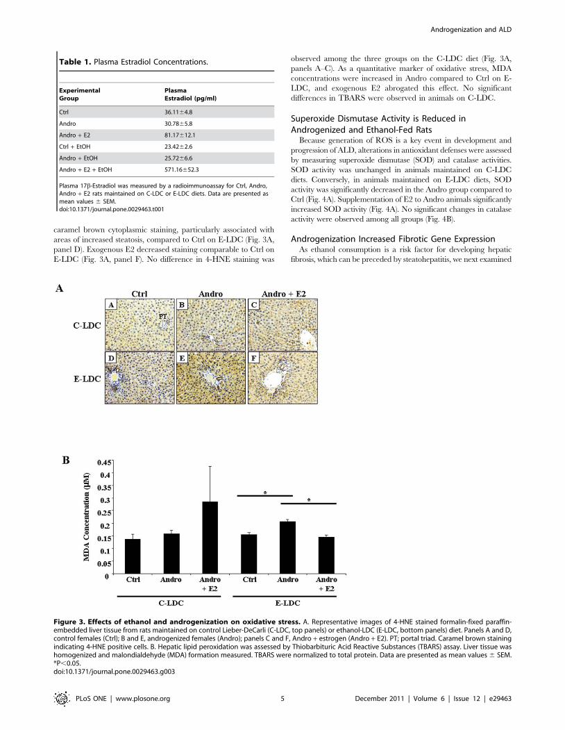
caramel brown cytoplasmic staining, particularly associated with
areas of increased steatosis, compared to Ctrl on E-LDC (Fig. 3A,
panel D). Exogenous E2 decreased staining comparable to Ctrl on
E-LDC (Fig. 3A, panel F). No difference in 4-HNE staining was
observed among the three groups on the C-LDC diet (Fig. 3A,
panels A–C). As a quantitative marker of oxidative stress, MDA
concentrations were increased in Andro compared to Ctrl on E-
LDC, and exogenous E2 abrogated this effect. No significant
differences in TBARS were observed in animals on C-LDC.
Superoxide Dismutase Activity is Reduced inAndrogenized and Ethanol-Fed Rats
Because generation of ROS is a key event in development and
progression of ALD, alterations in antioxidant defenses were assessed
by measuring superoxide dismutase (SOD) and catalase activities.
SOD activity was unchanged in animals maintained on C-LDC
diets. Conversely, in animals maintained on E-LDC diets, SOD
activity was significantly decreased in the Andro group compared to
Ctrl (Fig. 4A). Supplementation of E2 to Andro animals significantly
increased SOD activity (Fig. 4A). No significant changes in catalase
activity were observed among all groups (Fig. 4B).
Androgenization Increased Fibrotic Gene ExpressionAs ethanol consumption is a risk factor for developing hepatic
fibrosis, which can be preceded by steatohepatitis, we next examined
Table 1. Plasma Estradiol Concentrations.
ExperimentalGroup
PlasmaEstradiol (pg/ml)
Ctrl 36.1164.8
Andro 30.7865.8
Andro + E2 81.17612.1
Ctrl + EtOH 23.4262.6
Andro + EtOH 25.7266.6
Andro + E2 + EtOH 571.16652.3
Plasma 17b-Estradiol was measured by a radioimmunoassay for Ctrl, Andro,Andro + E2 rats maintained on C-LDC or E-LDC diets. Data are presented asmean values 6 SEM.doi:10.1371/journal.pone.0029463.t001
Figure 3. Effects of ethanol and androgenization on oxidative stress. A. Representative images of 4-HNE stained formalin-fixed paraffin-embedded liver tissue from rats maintained on control Lieber-DeCarli (C-LDC, top panels) or ethanol-LDC (E-LDC, bottom panels) diet. Panels A and D,control females (Ctrl); B and E, androgenized females (Andro); panels C and F, Andro + estrogen (Andro + E2). PT; portal triad. Caramel brown stainingindicating 4-HNE positive cells. B. Hepatic lipid peroxidation was assessed by Thiobarbituric Acid Reactive Substances (TBARS) assay. Liver tissue washomogenized and malondialdehyde (MDA) formation measured. TBARS were normalized to total protein. Data are presented as mean values 6 SEM.*P,0.05.doi:10.1371/journal.pone.0029463.g003
Androgenization and ALD
PLoS ONE | www.plosone.org 5 December 2011 | Volume 6 | Issue 12 | e29463

profibrotic gene expression by qRT-PCR. Differences in IL-6 and
col3a1 gene expression were not observed in the three experimental
groups maintained on C-LDC (Fig. 5). Up-regulation of IL-6 and
col3a1 mRNA was observed in Andro animals on E-LDC compared
to Ctrl on E-LDC (Fig. 5). Continuous exogenous E2 exposure in
adult rats abrogated the androgenization effect of increased
profibrotic gene expression (Fig. 5).
Cytochrome P450 Expression is Modulated byAndrogenization and Ethanol
Chronic ethanol consumption induces hepatic microsomal
CYP2E1 leading to increased generation of ROS [26]. Hormonal
imprinting of liver enzymes is known to occur [27], which suggests
that changes in sex specific expression of CYP450 family members
may contribute to sex differences in ALD. Therefore, cytochrome
P450 2E1 (CYP2E1) and cytochrome P450 1A2 (CYP1A2, a
predominantly female expressed enzyme) protein expression were
measured to determine effects of androgenization on ethanol
metabolizing enzymes. cyp2E1 expression was unchanged at the
mRNA level across all experimental groups (Fig. 6A), but because
CYP2E1 is predominantly regulated post-transcriptionally, protein
expression was assessed. Androgenization significantly decreased
CYP2E1 protein expression in animals maintained on the C-LDC
diet, and a similar effect was observed in animals maintained on E-
LDC (Fig. 6B). E2 implants did not significantly alter CYP2E1
expression in Andro groups maintained on either C-LDC or E-
LDC diets (Fig. 6B). Significant up-regulation of cyp1a2 mRNA
with the addition of ethanol was observed in the Ctrl, but
androgenization inhibited this effect (Fig. 6C). Additionally, E2
supplementation to Andro group increased cyp1a2 expression in
the ethanol treated animals compared to animals on C-LDC
(Fig. 6C). In contrast to CYP2E1, no significant differences in
CYP1A2 protein expression were detected between any of the
experimental groups (Fig. 6D).
Discussion
Our study demonstrated that inhibition of adult cyclic estrogen
(E2) via neonatal androgenization exacerbated the deleterious
Figure 4. Effects of ethanol and androgenization on antioxidant defense activity. A. Hepatic superoxide dismutase (SOD) activitydetermined by assay kit for control (Ctrl), androgenized (Andro) or androgenized + E2 (Andro + E2) animals maintained on control Lieber-DeCarli (C-LDC) or ethanol-LDC (E-LDC) diet. B. Hepatic catalase activity determined by assay kit for Ctrl, Andro, Andro + E2 rats maintained on C-LDC or E-LDCdiets. Activity levels were normalized to total protein. Data are presented as mean values 6 SEM. *P,0.05.doi:10.1371/journal.pone.0029463.g004
Androgenization and ALD
PLoS ONE | www.plosone.org 6 December 2011 | Volume 6 | Issue 12 | e29463

effects of ethanol through increased hepatic steatosis (H&E and
liver triglycerides). Additionally, androgenized animals on the
ethanol Lieber-DeCarli diet (E-LDC) showed significant increases
in lipid peroxidation (4-HNE and TBARS) compared to Ctrl
animals on E-LDC, and decreased superoxide dismutase (SOD)
activity in these animals further contributed to increased oxidative
stress. Further, disruption of normal cyclic E2 up-regulated
mRNA expression of profibrotic markers, IL-6 and col3a1.
Additionally, all indicators of liver injury were reversed by
exogenous administration of E2, supporting a protective effect of
continuous E2 exposure and/or supraphysiological dose of E2,
observed only in animals on the ethanol diet, in the adult
androgenized rat. Although androgenization exacerbated ethanol-
induced liver injury (e.g. steatosis, lipid peroxidation, oxidative
stress and profibrotic gene expression), only increased steatosis was
observed in Ctrl animals on the E-LDC diet compared to control
LDC (C-LDC), while all other liver injury markers were
unchanged. This was unexpected since the four week LDC diet
leads to mild liver injury (i.e. steatosis); however, a more severe
form of liver injury is achievable by the intragastric feeding model
[28] or a longer duration of ethanol feeding (i.e. eight weeks) which
would result in increased liver damage as observed by increased
inflammation, hepatic stellate cell activation and oxidative stress
[19]. It will be of interest to determine if increased triglyceride
accumulation by our model of androgenization with ethanol
administration occurs in other tissues such as adipose to document
if this is a general phenomenon or liver specific.
Since normal cyclic patterning of hormone release can program
hepatic enzyme expression, CYP2E1 mRNA and protein were
examined. Androgenization decreased CYP2E1 protein expres-
sion compared to Ctrl females on C-LDC and E-LDC diets,
suggesting a relationship between cyclic E2 and expression of
CYP2E1. Previous studies reported the deleterious effects of
ethanol metabolism were independent of CYP2E1 expression, as
CYP2E1 knockout mice were equally susceptible to ethanol-
induced liver injury [13]. Our data also supports that CYP2E1
does not contribute to liver pathology since high E2 concentrations
did not alter CYP2E1 expression between Andro and Andro + E2
groups, although pathology was changed. However, this does not
preclude other sex-specific CYP450 enzymes in the progression of
ALD, including CYP1A2, a predominantly female contributor to
the microsomal ethanol oxidizing system [14,16]. In our study,
cyp1A2 mRNA expression was up-regulated in Ctrl animals on E-
LDC compared to C-LDC and androgenization abrogated this
effect, while no significant difference in protein expression was
detected among all groups. This discrepancy observed between
protein and message may be due to the four week ethanol
exposure. Longer duration of ethanol consumption may lead to
protein expression levels similar to what was observed at the
mRNA level. Additionally, since induction of the microsomal
ethanol oxidizing system is commonly associated with chronic
ethanol consumption, the four week duration of ethanol feeding as
well as the ethanol administration model system used (e.g. Lieber-
DeCarli diet) may account for lack of ethanol-induced expression
of CYP2E1 in Ctrl animals on E-LDC compared to C-LDC.
Ethanol-increased expression of CYP2E1 is largely regulated by a
post-transcriptional mechanism due to protein stabilization [12].
Therefore, in addition to Western blot analyses, future studies
should examine CYP2E1 and CYP1A2 enzyme activity. Collec-
tively, these results support previous studies indicating CYP450
enzymes are not involved in ALD development or in the
underlying sex differences [13]. Collectively, these findings suggest
that cyclic E2 is not solely responsible for the observed sexual
differences in ALD, but instead, variations in gonadal hormone
regulation by the HPG axis may contribute to disease suscepti-
bility.
Even though E2 has been implicated in development and
progression of ALD, previous studies have reported protective
effects of E2 associated with increased antioxidant activities and
subsequent reduction in lipid peroxidation in non-alcoholic liver
diseases including hepatitis C virus, hepatocellular carcinoma and
sepsis [29,30,31]. Similarly, improvement of hepatic steatosis by
E2 has been observed in an animal model of obesity and alcohol
[32]. Specifically, high circulating levels of E2 showed suppressive
effects in a fibrotic animal model [33]. As E2 is a strong
endogenous antioxidant [34], the mechanism of action has been
attributed to reduced levels of ROS generation, lipid peroxidation,
and activation of activator protein (AP)-1 and NFkB [34,35,36].
Interestingly, in our study continuous exogenous administration of
E2 to adult androgenized female rats on the alcohol diet, which
Figure 5. Effects of ethanol and androgenization on profibrotic gene expression. Quantitative Real-Time PCR analysis of Interleukin-6 (IL-6)(black bar) and collagen Type 3 (COL3a1) (striped bar) gene expression in control (Ctrl), androgenized (Andro) and Andro + estrogen (Andro + E2)animals maintained on control Lieber-DeCarli (C-LDC) or ethanol-LDC (E-LDC) diet. Gene expression was normalized to glyceraldehyde-3-phosphatedehydrogenase (GAPDH). Data are presented as mean values 6 SEM. *P,0.05 vs. Ctrl + E-LDC; #P,0.05 vs. Andro + E2 + E-LDC.doi:10.1371/journal.pone.0029463.g005
Androgenization and ALD
PLoS ONE | www.plosone.org 7 December 2011 | Volume 6 | Issue 12 | e29463

increased E2 concentration by 7-fold compared to animals on
control diet (571.16652.3 vs 81.17612.1), displayed protective
effects as marked by decreased steatosis, lipid peroxidation and
profibrotic gene expression. Noteworthy, is the high concentration
of plasma E2 in Andro + E2 animals on the alcohol diet compared
to Andro + E2 on the control diet. Chronic alcohol consumption
alters hormone levels, and our data suggests that alcohol may
impede metabolism of E2 in the liver leading to increased plasma
levels. Further, SOD activity was increased in androgenized
animals implanted with E2, showing protective effects of this
hormone. Even though elevated SOD activity leads to increased
generation of H2O2, all groups displayed similar catalase activity;
therefore, differences in H2O2 cellular concentrations should not
contribute to overall oxidative stress. Although exogenous E2
decreased catalase activity in Andro animals on C-LDC compared
to all other groups, this change did not reach significance.
Additionally, exogenous E2 had no effect on CYP2E1 expression.
Therefore, these data suggest that timing and pattern of E2 release
Figure 6. Effects of ethanol and androgenization on CYP450 expression. A. Quantitative Real-Time PCR analysis of CYP2E1 mRNAexpression in control (Ctrl), androgenized (Andro) and Andro + estrogen (Andro + E2) rats maintained on control Lieber-DeCarli (C-LDC) or ethanol-LDC (E-LDC) diet. B. Representative CYP2E1 protein expression was determined by Western blot analysis (upper panel) and quantified by opticalintegrated volume (lower panel). Data are presented as mean values 6 SEM. Standard square root transformation was performed prior to statisticalanalysis. *P,0.05. C. Quantitative Real-Time PCR analysis of CYP1A2 mRNA expression compared between groups described in A. Data are presentedas mean values 6 SEM. *P,0.05. D. Representative CYP1A2 protein expression was determined by Western blot analysis (upper panel) and quantifiedby optical integrated volume (lower panel). RNA and protein expression were normalized to GAPDH.doi:10.1371/journal.pone.0029463.g006
Androgenization and ALD
PLoS ONE | www.plosone.org 8 December 2011 | Volume 6 | Issue 12 | e29463
regulated by the HPG axis plays a critical role in development and
progression of ALD in adulthood.
Androgenization is a well-established method of hypothalamic-
pituitary masculinization and inhibition of cyclic E2 [constant
baseline levels of E2 are observed (Table 1)] [23,24,25]; therefore,
we chose this model to elucidate the role of cyclic E2 in ALD
progression. Since previous studies implicating E2 in ALD
development regulated E2 expression through OVX and anties-
trogen administration (selective E2 receptor modulators), and did
not observe complete reversal of the damaging effects of ethanol,
the role of E2 remains to be fully elucidated [7,8]. In OVX
ovarian tissue is completely removed, and the contribution of
other ovarian factors, such as progesterone and inhibin, cannot be
discounted. Androgenization inhibits adult cyclic E2 production in
the presence of intact ovaries. However, androgenization not only
inhibits cyclic E2, but also disrupts the HPG axis, therefore,
implicating a role for other sex hormones (e.g. LH and FSH) in
ALD. Studies have speculated differences in androgenization and
OVX models with respect to LH concentrations. In androgenized
rats E2 feedback on the hypothalamus may prevent pituitary LH
storage, while in OVX animals increased amounts of LH and FSH
may occur due to lack of hormonal feedback resulting in release of
these gonadotropins [18]. Chronic ethanol consumption alters
serum LH and FSH, confirming an ethanol effect on pituitary
hormone release [37,38,39]. Therefore, it will be crucial to
determine the contribution of additional sex steroid hormones
regulated by the HPG axis on the development of ALD. Further, it
is possible that postnatal androgenization may also lead to other
endocrine changes in addition to E2 cyclicity in adults or higher
than normal prepubertal estrogen production leading to such
problems as metabolic dysfunction and insulin resistance that
would impact liver physiology.
Many factors, including environmental endocrine disruptors
and toxins, can disrupt the HPG axis. Postnatal exposure to
bisphenol A (BPA; an environmental estrogenic compound) has
been shown to induce anovulation and infertility in female rats
[40,41]. In adulthood, ethanol can directly modulate sex steroid
hormones through aromatization of testosterone to E2, resulting in
feminization of male alcoholics [42,43]. The HPG axis is not only
altered by environmental factors and toxins, but in clinical
disorders such as polycystic ovary syndrome (PCOS), which affects
6-8% of premenopausal women [44]. PCOS leads to hypersecre-
tion of androgens and altered patterning of sex steroid hormone
release [45]. Interestingly, the incidence of non-alcoholic fatty liver
disease in women with PCOS is increased, further supporting the
role of the HPG axis in susceptibility to fatty liver disease [46]. All
aforementioned challenges to the HPG axis postnatally could alter
adult liver physiology of both males and females, potentially
impacting development and progression of ALD.
In summary, cyclic E2 and possibly other sex steroid hormones
regulated by the HPG axis may contribute to the development and
progression of ALD, conferring sex differences. Moreover, timing
and pattern of E2 delivery can contribute to severity of ALD.
Understanding the interplay of sex steroid hormone exposure and
the HPG axis in liver physiology is critical to predicting
susceptibility to ALD and possibly other hepatotoxins.
Acknowledgments
The authors thank Judy Vachris, Rana Elbaz and Robin Major-Harvey for
their assistance with the animal models and tissue collection and Tracy
Walling for her assistance with histology. Additionally, we thank Ashleigh
Everhardt for her help with plasma estradiol measurements. We are also
grateful to Dr. Jim Norton for his statistical guidance.
Author Contributions
Conceived and designed the experiments: LWS YMH NMS. Performed
the experiments: WME AH. Analyzed the data: WME AML LWS YMH
NMS IHM HLB. Contributed reagents/materials/analysis tools: IHM.
Wrote the paper: WME AML LWS YMH.
References
1. Beier JI, McClain CJ (2010) Mechanisms and cell signaling in alcoholic liver
disease. Biol Chem 391: 1249–1264.
2. Gramenzi A, Caputo F, Biselli M, Kuria F, Loggi E, et al. (2006) Review article:
alcoholic liver disease--pathophysiological aspects and risk factors. Aliment
Pharmacol Ther 24: 1151–1161.
3. Day CP (2006) Genes or environment to determine alcoholic liver disease and
non-alcoholic fatty liver disease. Liver Int 26: 1021–1028.
4. Nanji AA, Jokelainen K, Fotouhinia M, Rahemtulla A, Thomas P, et al. (2001)
Increased severity of alcoholic liver injury in female rats: role of oxidative stress,
endotoxin, and chemokines. Am J Physiol Gastrointest Liver Physiol 281:
G1348–1356.
5. Tadic SD, Elm MS, Li HS, Van Londen GJ, Subbotin VM, et al. (2002) Sex
differences in hepatic gene expression in a rat model of ethanol-induced liver
injury. J Appl Physiol 93: 1057–1068.
6. Kono H, Wheeler MD, Rusyn I, Lin M, Seabra V, et al. (2000) Gender
differences in early alcohol-induced liver injury: role of CD14, NF-kappaB, and
TNF-alpha. Am J Physiol Gastrointest Liver Physiol 278: G652–661.
7. Jarvelainen HA, Lukkari TA, Heinaro S, Sippel H, Lindros KO (2001) The
antiestrogen toremifene protects against alcoholic liver injury in female rats.
J Hepatol 35: 46–52.
8. Yin M, Ikejima K, Wheeler MD, Bradford BU, Seabra V, et al. (2000) Estrogen
is involved in early alcohol-induced liver injury in a rat enteral feeding model.
Hepatology 31: 117–123.
9. Gustafsson JA, Eneroth P, Hokfelt T, Mode A, Norstedt G, et al. (1981) Role of
the hypothalamo-pituitary-liver axis in sex differences in susceptibility of the liver
to toxic agents. Environ Health Perspect 38: 129–141.
10. Csaba G, Szeberenyi SZ, Dobozy O (1987) Hormonal imprinting of the
microsomal enzyme system in adults. Microsomal activity change in response to
estrogen (DES, AE) treatment during liver regeneration. Horm Metab Res 19:
493–496.
11. Lieber CS (2004) The discovery of the microsomal ethanol oxidizing system and
its physiologic and pathologic role. Drug Metab Rev 36: 511–529.
12. Cederbaum AI, Lu Y, Wu D (2009) Role of oxidative stress in alcohol-induced
liver injury. Arch Toxicol 83: 519–548.
13. Kono H, Bradford BU, Yin M, Sulik KK, Koop DR, et al. (1999) CYP2E1 isnot involved in early alcohol-induced liver injury. Am J Physiol 277:
G1259–1267.
14. Asai H, Imaoka S, Kuroki T, Monna T, Funae Y (1996) Microsomal ethanol
oxidizing system activity by human hepatic cytochrome P450s. J Pharmacol ExpTher 277: 1004–1009.
15. Degawa M, Tanimura S, Agatsuma T, Hashimoto Y (1989) Hepatocarcinogenic
heterocyclic aromatic amines that induce cytochrome P-448 isozymes, mainly
cytochrome P-448H (P-450IA2), responsible for mutagenic activation of thecarcinogens in rat liver. Carcinogenesis 10: 1119–1122.
16. Iba MM, Fung J, Thomas PE, Park Y (1999) Constitutive and induced
expression by pyridine and beta-naphthoflavone of rat CYP1A is sexuallydimorphic. Arch Toxicol 73: 208–216.
17. Choi SY, Fischer L, Yang K, Chung H, Jeong H (2011) Isoform-specificregulation of cytochrome P450 expression and activity by estradiol in female
rats. Biochem Pharmacol 81: 777–782.
18. Christakos S, Sinha D, Dao TL (1976) Neonatal modification of endocrinefunctions and mammary carcinogenesis in the rat. Br J Cancer 34: 58–63.
19. Karaa A, Thompson KJ, McKillop IH, Clemens MG, Schrum LW (2008) S-adenosyl-L-methionine attenuates oxidative stress and hepatic stellate cell
activation in an ethanol-LPS-induced fibrotic rat model. Shock 30: 197–205.
20. Steuerwald NM, Parsons JC, Bennett K, Bates TC, Bonkovsky HL (2010)Parallel microRNA and mRNA expression profiling of (genotype 1b) human
hepatoma cells expressing hepatitis C virus. Liver Int 30: 1490–1504.
21. Lakner AM, Walling TL, McKillop IH, Schrum LW (2011) Altered aquaporin
expression and role in apoptosis during hepatic stellate cell activation. Liver Int31: 42–51.
22. Davidge ST, Zhang Y, Stewart KG (2001) A comparison of ovariectomy models
for estrogen studies. Am J Physiol Regul Integr Comp Physiol 280: R904–907.
23. Edwards DA (1971) Neonatal administration of androstenedione, testosterone or
testosterone propionate: effects on ovulation, sexual receptivity and aggressivebehavior in female mice. Physiol Behav 6: 223–228.
24. Mizukami S, Yamanouchi K, Arai Y, Yanai R, Nagasawa H (1982) Failure of
ovulation after neonatal administration of 5 alpha-dihydrotestosterone to femalerats. Endokrinologie 79: 1–6.
Androgenization and ALD
PLoS ONE | www.plosone.org 9 December 2011 | Volume 6 | Issue 12 | e29463
25. Pinilla L, Trimino E, Garnelo P, Bellido C, Aguilar R, et al. (1993) Changes in
pituitary secretion during the early postnatal period and anovulatory syndromeinduced by neonatal oestrogen or androgen in rats. J Reprod Fertil 97: 13–20.
26. McKillop IH, Schrum LW (2009) Role of alcohol in liver carcinogenesis. Semin
Liver Dis 29: 222–232.27. Gustafsson JA, Stenberg A (1974) Irreversible androgenic programming at birth
of microsomal and soluble rat liver enzymes active on androstene-3,17-dioneand 5alpha-androstane-3alpha,17beta-diol. J Biol Chem 249: 711–718.
28. Arteel GE (2010) Animal models of alcoholic liver disease. Dig Dis 28: 729–736.
29. Hayashida K, Shoji I, Deng L, Jiang DP, Ide YH, et al. (2010) 17beta-estradiolinhibits the production of infectious particles of hepatitis C virus. Microbiol
Immunol 54: 684–690.30. Lacort M, Leal AM, Liza M, Martin C, Martinez R, et al. (1995) Protective
effect of estrogens and catecholestrogens against peroxidative membranedamage in vitro. Lipids 30: 141–146.
31. Sener G, Arbak S, Kurtaran P, Gedik N, Yegen BC (2005) Estrogen protects the
liver and intestines against sepsis-induced injury in rats. J Surg Res 128: 70–78.32. Hong J, Holcomb VB, Kushiro K, Nunez NP (2011) Estrogen inhibits the effects
of obesity and alcohol on mammary tumors and fatty liver. Int J Oncol.33. Yasuda M, Shimizu I, Shiba M, Ito S (1999) Suppressive effects of estradiol on
dimethylnitrosamine-induced fibrosis of the liver in rats. Hepatology 29:
719–727.34. Shimizu I (2003) Impact of oestrogens on the progression of liver disease. Liver
Int 23: 63–69.35. Inoue H, Shimizu I, Lu G, Itonaga M, Cui X, et al. (2003) Idoxifene and
estradiol enhance antiapoptotic activity through estrogen receptor-beta incultured rat hepatocytes. Dig Dis Sci 48: 570–580.
36. Omoya T, Shimizu I, Zhou Y, Okamura Y, Inoue H, et al. (2001) Effects of
idoxifene and estradiol on NF-kappaB activation in cultured rat hepatocytesundergoing oxidative stress. Liver 21: 183–191.
37. Bell H, Raknerud N, Falch JA, Haug E (1995) Inappropriately low levels of
gonadotrophins in amenorrhoeic women with alcoholic and non-alcoholiccirrhosis. Eur J Endocrinol 132: 444–449.
38. Kostic N, Bozanic M, Lalevic S, Adamov A (1989) [Hormone levels in patients
with alcoholic liver cirrhosis]. Srp Arh Celok Lek 117: 767–776.39. Van Thiel DH, Kumar S, Gavaler JS, Tarter RE (1990) Effect of liver
transplantation on the hypothalamic-pituitary-gonadal axis of chronic alcoholicmen with advanced liver disease. Alcohol Clin Exp Res 14: 478–481.
40. Ramos JG, Varayoud J, Kass L, Rodriguez H, Costabel L, et al. (2003)
Bisphenol a induces both transient and permanent histofunctional alterations ofthe hypothalamic-pituitary-gonadal axis in prenatally exposed male rats.
Endocrinology 144: 3206–3215.41. Fernandez M, Bourguignon N, Lux-Lantos V, Libertun C (2010) Neonatal
exposure to bisphenol a and reproductive and endocrine alterations resemblingthe polycystic ovarian syndrome in adult rats. Environ Health Perspect 118:
1217–1222.
42. Eagon PK, Zdunek JR, van Thiel DH, Singletary BK, Egler KM, et al. (1981)Alcohol-induced changes in hepatic estrogen-binding proteins: a mechanism
explaining feminization in alcoholics. Arch Biochem Biophys 211: 48–54.43. Gordon GG, Southren AL, Vittek J, Lieber CS (1979) The effect of alcohol
ingestion on hepatic aromatase activity and plasma steroid hormones in the rat.
Metabolism 28: 20–24.44. Shayya R, Chang RJ (2010) Reproductive endocrinology of adolescent
polycystic ovary syndrome. BJOG 117: 150–155.45. Doi SA, Towers PA, Scott CJ, Al-Shoumer KA (2005) PCOS: an ovarian
disorder that leads to dysregulation in the hypothalamic-pituitary-adrenal axis?Eur J Obstet Gynecol Reprod Biol 118: 4–16.
46. Vassilatou E, Lafoyianni S, Vryonidou A, Ioannidis D, Kosma L, et al. (2010)
Increased androgen bioavailability is associated with non-alcoholic fatty liverdisease in women with polycystic ovary syndrome. Hum Reprod 25: 212–220.
Androgenization and ALD
PLoS ONE | www.plosone.org 10 December 2011 | Volume 6 | Issue 12 | e29463