The pharmacology of the antioxidant lipoic acid
17
Gen. Pharmac. Vol. 29, No. 3, pp. 315-331, 1997 Copyright © 1997 Elsevier Science Inc. Printed in the USA. ELSEVIER ISSN 0306-3623/97 $17.00 + .00 PII S0306-3623(96)00474-0 All rights reserved REVIEW The Pharmacology of the Antioxidant Lipoic Acid Gerreke Ph. Biewenga, Guido R. M. M. Haenen* and Aalt Bast LEIDEN/AMSTERDAM CENTER FOR DRUG RESEARCH,VRIJE UNIVERSITEIT, DEPARTMENT OF PHARMACOCHEMISTRY, DIVISIONOF MOLECULAR PHARMACOLOGY, DE BOELELAAN 1083, 1081 HV AMSTERDAM, THE NETHERLANDS [FAx: 31-20-4447610; TEL: 31-20-4447582] ABSTRACT. 1. Lipoic acid is an example of an existing drug whose therapeutic effect has been re- lated to its antioxidant activity. 2. Antioxidant activity is a relative concept: it depends on the kind of oxidative stress and the kind of oxidizable substrate (e.g., DNA, lipid, protein). 3. In vitro, the final antioxidant activity of lipoic acid is determined by its concentration and by its antioxidant properties. Four antioxidant properties of lipoic acid have been studied: its metal chelating capacity, its ability to scavenge reactive oxygen species (ROS), its ability to regenerate endogenous anti- oxidants and its ability to repair oxidative damage. 4. Dihydrolipoic acid (DHLA), formed by reduction of lipoic acid, has more antioxidant proper- ties than does lipoic acid. Both DHLA and lipoic acid have metal-chelating capacity and scavenge ROS, whereas only DHLA is able to regenerate endogenous antioxidants and to repair oxidative damage. 5. As a metal chelator, lipoic acid was shown to provide antioxidant activity by chelating Fe z+ and Cu>; DHLA can do so by chelating Cd 2+. 6. As scavengers of ROS, lipoic acid and DHLA display antioxidant activity in most experiments, whereas, in particular cases, pro-oxidant activity has been observed. However, lipoic acid can act as an antioxidant against the pro-oxidant activity produced by DHLA. 7. DHLA has the capacity to regenerate the endogenous antioxidants vitamin E, vitamin C and glutathione. 8. DHLA can provide peptide methionine sulfoxide reductase with reducing equivalents. This en- hances the repair of oxidatively damaged proteins such as oL-1 antiprotease. 9. Through the lipoamide dehydrogenase-dependent reduction of lipoic acid, the cell can draw on its NADH pool for antioxidant activity additionally to its NADPH pool, which is usually consumed during oxidative stress. 10. Within drug-related antioxidant pharmacology, lipoic acid is a model compound that enhances understanding of the mode of action of antioxidants in drug therapy. GEN PHARMAC29;3:315--331, 1997. © 1997 Elsevier Science Inc. KEY WORDS. Antioxidant pharmacology, dihydrolipoic acid, glutathione, lipoic acid, metabolism, metal chelation, peptide methionine sulfoxide reductase, reactive oxygen species, scavenging, vitamin C, vitamin E ANTIOXIDANT PHARMACOLOGY Within pharmacology, a new area of research concentrates on the pharmacology of antioxidants. Whereas at the beginning of the 20th century, P. Ehrlich noticed that most drugs act through bind- ing to DNA or proteins (receptors, enzymes, carrier molecules, ion channels), more recently, other mechanisms by which a drug can af- fect the function of a living system are being recognized. By acting as an antioxidant, a drug may also affect the functioning of an or- ganism. B. Halliwell, a leading scientist in the field of antioxidant research, formulated the definition of an antioxidant as: any sub- stance that, when present at low concentrations compared with those of an oxidizable substrate, significantly delays or prevents oxidation of that subs trate (Halliwell, 1995). With this definition, antioxidants are of interest to anyone who wants to preserve goods: radiation chemists, food scientists and museum curators. The human body also needs to *To whom correspondence should be addressed. Accepted 9 September 1996. be preserved well. Antioxidants such as vitamin C, vitamin E and glutathione protect tissue from oxidative damage. Of course, the statement that living organisms contain natural preservatives is far too popularized. In living systems, a wide range of oxidative pro- cesses take place, delicately tuned by specific antioxidants. In some situations, the balance between the oxidative and the antioxidative processes tips in favor of the oxidative processes. This results in oxi- dative stress and may finally lead to pathology. Antioxidant phar- macology investigates possibilities of therapeutic intervention in oxidative processes (Bast, 1994). Basically, this is done through two routes (Fig. 1). The first route concentrates on existing or potential drugs. Compounds are screened for antioxidant activity, and the contribution to a pharmacological effect is evaluated. The second route concentrates on the pathology. Pathological processes are in- vestigated, and the involvement of oxidative stress is considered. An example of an existing drug of which the therapeutic effect is related to its antioxidant activity is lipoic acid (Bast et al., 1988). Ideally, both research routes converge and result in an effective and
Transcript of The pharmacology of the antioxidant lipoic acid

Gen. Pharmac. Vol. 29, No. 3, pp. 315-331, 1997 Copyright © 1997 Elsevier Science Inc. Printed in the USA.
ELSEVIER
ISSN 0306-3623/97 $17.00 + .00 PII S0306-3623(96)00474-0
All rights reserved
REVIEW
The Pharmacology of the Antioxidant Lipoic Acid
Gerreke Ph. Biewenga, Guido R. M. M. Haenen* and Aalt Bast LEIDEN/AMSTERDAM CENTER FOR DRUG RESEARCH, VRIJE UNIVERSITEIT, DEPARTMENT OF PHARMACOCHEMISTRY, DIVISION OF MOLECULAR PHARMACOLOGY, DE BOELELAAN
1083, 1081 HV AMSTERDAM, THE NETHERLANDS [FAx: 31-20-4447610; TEL: 31-20-4447582]
ABSTRACT. 1. Lipoic acid is an example of an existing drug whose therapeutic effect has been re- lated to its antioxidant activity.
2. Antioxidant activity is a relative concept: it depends on the kind of oxidative stress and the kind of oxidizable substrate (e.g., DNA, lipid, protein).
3. In vitro, the final antioxidant activity of lipoic acid is determined by its concentration and by its antioxidant properties. Four antioxidant properties of lipoic acid have been studied: its metal chelating capacity, its ability to scavenge reactive oxygen species (ROS), its ability to regenerate endogenous anti- oxidants and its ability to repair oxidative damage.
4. Dihydrolipoic acid (DHLA), formed by reduction of lipoic acid, has more antioxidant proper- ties than does lipoic acid. Both DHLA and lipoic acid have metal-chelating capacity and scavenge ROS, whereas only DHLA is able to regenerate endogenous antioxidants and to repair oxidative damage.
5. A s a metal chelator, lipoic acid was shown to provide antioxidant activity by chelating Fe z+ and Cu>; DHLA can do so by chelating Cd 2+.
6. A s scavengers of ROS, lipoic acid and DHLA display antioxidant activity in most experiments, whereas, in particular cases, pro-oxidant activity has been observed. However, lipoic acid can act as an antioxidant against the pro-oxidant activity produced by DHLA.
7. D H L A has the capacity to regenerate the endogenous antioxidants vitamin E, vitamin C and glutathione.
8. D H L A can provide peptide methionine sulfoxide reductase with reducing equivalents. This en- hances the repair of oxidatively damaged proteins such as oL-1 antiprotease.
9. Through the lipoamide dehydrogenase-dependent reduction of lipoic acid, the cell can draw on its N A D H pool for antioxidant activity additionally to its NADPH pool, which is usually consumed during oxidative stress.
10. Within drug-related antioxidant pharmacology, lipoic acid is a model compound that enhances understanding of the mode of action of antioxidants in drug therapy. GEN PHARMAC 29;3:315--331, 1997. © 1997 Elsevier Science Inc.
KEY WORDS. Antioxidant pharmacology, dihydrolipoic acid, glutathione, lipoic acid, metabolism, metal chelation, peptide methionine sulfoxide reductase, reactive oxygen species, scavenging, vitamin C, vitamin E
A N T I O X I D A N T PHARMACOLOGY
Within pharmacology, a new area of research concentrates on the pharmacology of antioxidants. Whereas at the beginning of the 20th century, P. Ehrlich noticed that most drugs act through bind- ing to DNA or proteins (receptors, enzymes, carrier molecules, ion channels), more recently, other mechanisms by which a drug can af- fect the function of a living system are being recognized. By acting as an antioxidant, a drug may also affect the functioning of an or- ganism. B. Halliwell, a leading scientist in the field of antioxidant research, formulated the definition of an antioxidant as: any sub- stance that, when present at low concentrations compared with those of an oxidizable substrate, significantly delays or prevents oxidation of that subs trate (Halliwell, 1995). With this definition, antioxidants are of interest to anyone who wants to preserve goods: radiation chemists, food scientists and museum curators. The human body also needs to
*To whom correspondence should be addressed. Accepted 9 September 1996.
be preserved well. Antioxidants such as vitamin C, vitamin E and glutathione protect tissue from oxidative damage. Of course, the statement that living organisms contain natural preservatives is far too popularized. In living systems, a wide range of oxidative pro- cesses take place, delicately tuned by specific antioxidants. In some situations, the balance between the oxidative and the antioxidative processes tips in favor of the oxidative processes. This results in oxi- dative stress and may finally lead to pathology. Antioxidant phar- macology investigates possibilities of therapeutic intervention in oxidative processes (Bast, 1994). Basically, this is done through two routes (Fig. 1 ). The first route concentrates on existing or potential drugs. Compounds are screened for antioxidant activity, and the contribution to a pharmacological effect is evaluated. The second route concentrates on the pathology. Pathological processes are in- vestigated, and the involvement of oxidative stress is considered. An example of an existing drug of which the therapeutic effect is related to its antioxidant activity is lipoic acid (Bast et al., 1988). Ideally, both research routes converge and result in an effective and
316 G.P . Biewenga et al.
antioxidant profile of a drug therapeutic intervention } . ~ . ~
oxidative processes in a disease
drug related antioxidant pharmacology disease related andoxidant pharmacology
FIGURE 1. Antioxidant pharmacology investigates possibilities for therapeutic intervention in oxidative processes.
therapeutic intervention. In this review, we discuss the first route: research related to the antioxidant activity of the drug lipoic acid.
LIPOIC ACID
Lipoic acid (chemical name: 1,2-dithiolane-3-pentanoic acid, Fig. 2) is present in all kinds of prokaryotic and eukaryotic cells. In hu- man beings, it is part of several 2-oxo acid dehydrogenases that take part in energy formation. Linked to lysine residues of the 2-oxo acid dehydrogenase multienzyme complexes (Fujiwara et al., 1995; Mor- ris et al., 1995), lipoic acid acts as a cofactor. It binds acyl groups and transfers them from one part of the enzyme complex to another. In this process, lipoic acid is reduced to dihydrolipoic acid (DHLA), which is subsequently reoxidized by lipoamide dehydrogenase (Lip- DH) under the formation of NADH. Overall, lipoic acid and DHLA act as a redox couple, carrying electrons from the substrate of the dehydrogenase to NAD +.
In 1966, German physicians started to administer lipoic acid to patients with liver cirrhosis, mushroom poisoning, heavy metal in- toxication and diabetic polyneuropathy. Originally, the rationale for this treatment was the observation that patients with liver cir- rhosis, diabetes mellitus and polyneuropathy had lower levels of li- poic acid (Kleemann et al., 1989). It was assumed that supplementa- tion with lipoic acid helped to overcome the shortage, thereby restoring the 2-oxo acid oxidation. Indeed, destruction of the cofac- tot function of lipoic acid may be involved in pathological pro- cesses. In arsenite intoxication, As 3+ can form a complex with lipoic acid in the 2-oxo acid dehydrogenases, rendering it inactive. In cer- tain types of liver disease, autoantibodies that recognize lipoylated subunits in the multienzymes have been demonstrated in patients' sera (Gut et al., 1995; Tuaillon et al., 1992). Also, oxidative stress near the dehydrogenase complex can lead to oxidative destruction (Gutierrez Correa and Stoppani, 1996), thereby adversely affecting the functioning of lipoic acid as a cofactor.
Lipoic acid is bound to proteins and, consequently, free lipoic acid has not been detected in human beings (Hermann et al., 1996). However, after therapeutic application, free lipoic acid can be found in the circulation (Teichert and PreiB, 1995). It is likely that the therapeutic effects originate from free, unbound lipoic acid. In heavy metal intoxications, free lipoic acid and its metabolites are
HOOC~ ~COOH dihydrolipo'm acid (DHLA) .~ ISH ~ lipoic acid
HOOC S- -S~
isomers ofbeta-lipoic acid '~0
~ COOH
FIGURE2. The chemical structures of dihydrolipoic acid (DHLA), lipoic acid and the isomers of [3-1ipoic acid. The struc- tures contain a chiral center (*). The R-configuration is naturally occurring.
able to trap the metals in the circulation, thereby preventing the damage caused by the metal and (sometimes) improving its secre- tion. In diabetic polyneuropathy, free lipoic acid may enter nerve tissue and prevent glucose-related oxidative damage.
Especially in diabetic polyneuropathy, approaching antioxidant pharmacology via the pathology- and drug-related routes may lead to promising possibilities for therapeutic intervention. It has been shown that oxidative stress is part of the pathological process of dia- betes. It has been suggested that oxidative stress has a role in the causation of noninsulin-dependent diabetes (Salonen et al., 1995). Wolff et al. (1991) demonstrated that high glucose levels produce oxidative stress. In streptozotocin diabetic rats, antioxidants and li- poic acid delayed the onset of polyneuropathy (Cameron et al., 1993; Nagamatsu et al., 1995; Vertommen et al., 1994). In clinical trials, lipoic acid did not improve polyneuropathy over a relatively short period of 3 weeks. The benefit of lipoic acid was found in re- ducing neuropathic complaints such as pain and paresthesia (Ziegler et al., 1995). Although the exact role of oxidative stress in human diabetics is still under investigation (route 2), the obtained results warrant a further investigation of the antioxidant role of lipoic acid in oxidative stress (route 1).
OXIDATIVE STRESS
The functioning of living systems depends on energy. Aerobic or- ganisms derive their energy from the oxidation of fuel molecules such as glucose and fatty acids. In oxidation, electrons are removed from these molecules and subsequently transferred in a chain of re- actions to other molecules until they finally reach their ultimate electron acceptor: 02. Often, oxidative stress originates from im- proper control of this reduction of O2.
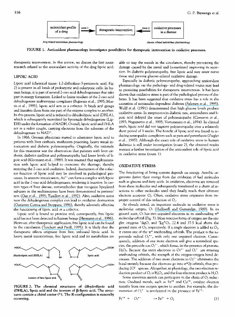
As already noted, an important molecule in oxidative stress is molecular oxygen, 02 (Halliwell and Gutteridge, 1989). In its ground state, Oz has two unpaired electrons in its antibonding ~r* molecular orbital (Fig. 3). More reactive forms of oxygen are the sin- glet oxygens: ~AgO2 and 1~g+O2, 22.4 and 37.5 kcal above the ground state of 02, respectively. If a single electron is added to 02, it enters one of the "rr* antibonding orbitals. The product is the su- peroxide radical O2"-, with only one unpaired electron. Conse- quently, addition of one more electron will give a nonradical spe- cies, the peroxide ion O2 2-, which forms, in the presence of protons, H202. Because the extra electrons in O2" and 022- are entering antibonding orbitals, the strength of the oxygen-oxygen bond de- creases. The addition of two more electrons to 022- eliminates the bond entirely because the electrons go into ~r*2p orbitals, thus pro- ducing 202- species. Altogether, in physiology, the two-electron re- duction product of 02 is H202, and the four-electron product is H20.
Some transition metals can participate in the chain of 02 reduc- tion. Oxidized metals, such as Fe 3+ and C u 2+, catalyze electron transfer from one oxygen species to another. For example, the dis- mutation of 02" is accelerated in the presence of Fe3+:
Fe 3+ + 02"- --+ Fe 3+ + O2 (1)

Pharmacology of Lipoic Acid 317
o 2p
.-d re* 2p ---~-- - - - ~
o 2p @ E cr, 2s '8
o*ls
ground state 02
(3yg- 02 )
+- - - +- + 4 - + + -4¢- -4¢- + -4¢- + -4¢-
-4¢ -4t- + e 44 -44-
-44- -4¢- + -44- 4+ 4¢- 4¢-
singlet 0 2 superoxide peroxide singlet 0 2
(lAg 02) (O2") ( 022-) (IEg+O2)
FIGURE 3. Molecular-orbital energy pattern for the diatomic oxygen molecule.
Fe 2+ + O2"- + 2H + ~ F e 3+ + H202 (2)
202" + 2H + --* 02 + H202 (3)
Reduced metal ions, such Fe z+ and Cu +, can donate one electron and thus start a whole sequence of reactions. An example is the Fen- ton reaction in which the very reactive hydroxyl radical ( 'OH) is formed:
Fe 2+ + H202 ---qntermediate complex--* Fe 3+ + "OH + O H - (4)
Traces of Fe 3+ might be able to react further with H202, although this is very slow at physiological pH:
Fe 3+ + H202 --+intermediate complex---, Fe 2+ + O2"- + 2H +
Even more reactions are possible:
"OH + H202 ~ H20 + H + + O2"-
O2"- + Fe 3+ --'* Fe 2+ + 02
"OH + Fe 2+ ---* Fe 3+ + O H -
The overall sum of these reactions is the iron-catalyzed decomposi- tion of hydrogen peroxide:
2H202--'+ 02 + 2H20 (9)
In aerobic organisms, almost all steps in the reduction of O2 are catalyzed by metals. Mostly, the reactivity of the metals is controlled by incorporating the metal in an enzyme. Examples of such enzymes are superoxide dismutase (SOD), catalase and cytochromes. Dissoci- ated from their protein environments, metals take part in other, often unwanted, secondary reactions (e.g., in the Haber-Weiss reac- tion). The protein environment of SOD prevents the metal-cata- lyzed reaction between 02" and H202, but unbound iron or copper induces hydroxyl radical formation:
Fe > + 02" ---' Fe z+ + 02
Fe 2+ + H202 '+ "OH + O H - + Fe 3+
02"- + H202 ---' "OH + 02 + O H -
In the absence of iron, the reaction rate of the Haber-Weiss reac- tion is virtually zero. This illustrates the extreme importance of iron and copper in the formation of "OH.
The species H202 and "OH, as well as other oxygen-derived spe- cies such as 1AGO2 and HOCI, can damage biomolecules such as
DNA, proteins and lipids. They are generally referred to as reactive oxygen species (ROS). Many ROS were ultimately derived from 02"-. The proposal that O2"- is the major factor in oxygen toxicity is known as the superoxide theory of oxygen toxicity. Superoxide anions can be formed in vivo through several routes. First, some bio- logically important molecules and certain xenobiotics can oxidize in the presence of 02 to yield O2" : they include glyceraldehyde, re- duced forms of riboflavin, hemoglobin, adrenaline, tetrahydropteri- dines, thiols, paraquat and doxorubicin. Second, the electron trans- port chains of mitochondria and the endoplasmatic reticulum can "leak" electrons to 02. In mitochondria, cytochrome oxidase, NADH-coenzyme Q reductase and reduced forms of coenzyme Q it- self are sites of electron leakage. In the endoplasmatic reticulum, im- proper reactions of the cytochrome P-450 enzymes can "leak" elec-
(5) trons to 02. A third source of O2"- are the phagocytic cells. These cells contain a membrane-bound enzyme, NADPH oxidase, which catalyzes the formation of 02"- according to the following stoichi-
(6) ometry:
(7) NADPH + 202--~ 202"- + H + + NADP + (13)
(8) The toxic effect of ROS usefully contributes to microbicidal activity of the phagocytic cells. Although all ROS originate from 02 reduc- tion, not all ROS are derived from 02"-. For example, NO" is formed by NO synthase in an oxidative process resembling a P-450 reaction (Boucher et al., 1992). In this process, reduced oxygen is incorporated into an N%OH-L-arginine intermediate (Nathan, 1992).
When compounds with one or more unpaired electrons are small and freely diffusible, they are referred to as "free radicals." In this view, ROS--just like O2, O2 °-, "OH and metals ions with unpaired electrons in the d orbitals--are considered free radicals. Apart from some exceptions, most reactions in chemistry can be described as polar or radical reactions. In polar reactions, bonds are made when an electron-rich reagent donates a pair of electrons to the electron- poor reagent. On the other hand, in radical reactions, each reactant donates only one electron to the new bond. Biologists have been as-
(10) suming for years that free radicals do not exist in biological systems. (11) However, the observation that radical reactions also take place in
biology has aroused a new trend: "free radical research." Although (12) this terminology is logical on a historical basis, chemically it is awk-
ward. It suggests that it is useful to study "free radicals" and, for ex- ample, "free anions" separately. Nowadays, scientists are aware of this strange terminology and have started to acknowledge the total chain of oxidative processes, regardless of their radical or polar char- acter.

318 G.P. Biewenga et al.
ANTIOXIDANTS
To control oxidative processes, biological systems have been equipped with several antioxidant mechanisms. Antioxidant en- zymes such as SOD, catalase and peroxidases are concerned with the removal of O2"- and H202. Metal ions can be sequestrated to inhibit uncontrolled dismutation of 02"-, the Fenton reaction and the Haber-Weiss reaction. ROS can be scavenged in an aqueous envi- ronment by small molecules such as vitamin C, glutathione and uric acid. In a lipid environment, vitamin E scavenges ROS-derived rad- icals and protects the membranes. When, in spite of all this, the an- tioxidant system has failed, damage to endogenous molecules may be repaired. Organisms contain for this purpose DNA repair systems, protein turnover mechanisms and peptide methionine sulfoxide re- ductase (PMSR).
As stated before, drugs also have antioxidant activity. Generally, the antioxidant properties of a drug are determined by their chemi- cal and physical properties. The combination of these properties and the concentration determine its final antioxidant activity. To be ef- fective, the drug itself and its relevant metabolites should become available to the tissue that is prone to oxidative stress. Within anti- oxidant pharmacology, the absorption, distribution, metabolism and excretion of a drug are studied. The antioxidant properties of the drug and its metabolites are examined. For the evaluation of the an- tioxidant properties of lipoic acid, four aspects have been distin- guished:
1. The metal-chelating capacity 2. The ROS-scavenging capacity 3. The capacity to regenerate endogenous antioxidants 4. The role in repair systems
The four antioxidant properties can be studied as pure chemical re- actions. The corresponding antioxidant activity is determined in re- lation to prevention of oxidative damage. The antioxidant activity is assessed in vitro or in vivo. For lipoic acid, the experiments that have been performed to test its bioavailability, metabolism and anti- oxidant activity are reviewed in the next section.
THE BIOAVAILABILITY OF LIPOIC ACID
Our daily diet contains lipoic acid. Especially, food derived from tis- sue with a high metabolic activity has a high lipoic acid content (Herbert and Guest, 1975; Mattulat and Baltes, 1992). Meat from a metabolic organ such as pig heart has a lipoic acid content of 1.1- 1.6 mg/kg, whereas calf muscle contains only 0.07q). 15 mg/kg (Mat- tulat and Bakes, 1992). This indicates that most lipoic acid in the diet originates from the multienzyme complexes. Proteolytic en- zymes do not effectively cleave the peptide bound between lipoic acid and lysine. Therefore, it has been suggested that, after diges- tion, lipoic acid is absorbed as lipoyllysine (Martulat, 1992). In addi- tion, lipoic acid can be obtained by de novo biosynthesis from fatty acids and cysteine (Carreau, 1979; Spoto et al., 1982). Indications were found that lipoic acid is synthesized in a protein-bound form (Spoto et al., 1982). From food or biosynthesis, only minor amounts of free lipoic acid will enter the circulation. After oral application, free lipoic acid is obtained in relatively high amounts. The thera- peutic dose exceeds the dietary intake by far. Doses have been ad- ministered up to 10 mg/kg IV to rat (Stroman et al., 1989) and to human beings up to 1,200 mg IV (Ziegler et al., 1995), which is equal to the lipoic acid content of circa 1,000 kg of pig heart.
After administration of lipoic acid, its concentration can be de- termined in the blood plasma (Teichert and PreiB, 1995) and in the tissue (Maitra et al., 1996). Originally, all analytical methods for the determination of lipoic acid contained a hydrolysis step to liberate
,-g
2.5 ¸
2 . 0
1 .5
1 .0
0.5 '
frO:
~ 3.ketolipoic a~id
bisnorlipoic acid
, li, poic acid
~ 1 0 0 200 300 food consumption
time (min)
FIGURE 4. The time course of the plasma concentration of li- poic acid (open circles), 3-ketolipoic acid (closed circles) and bis- norlipoic acid (triangles) after oral administration of 1 g R-lipoic acid to a healthy volunteer who had fasted overnight. A meal high in fat content was consumed 85 min after oral intake of R-lipoic acid. The compounds were analyzed by high-performance liquid chromatography coupled to electrochemical detection.
lipoic acid bound to the lysine residue. Because free lipoic acid is thought to be the most important therapeutic form, nowadays free lipoic acid concentrations are evaluated (Biewenga et al., 1996c; Handelman et al., 1994; Hermann et al., 1996). Figure 4 shows that concentrations up to 1,154 ng/ml of free lipoic acid appear in the blood plasma after oral application of 1 g of R-lipoic acid to a healthy male volunteer. Hermann et al. (1996) extensively studied the pharmacokinetics and bioavailability of racemic lipoic acid in different formulations. The half life in plasma is approximately 30 rain (as is also seen in Fig. 4). The mean total plasma clearance was in the same range as the plasma flow of the liver (about 11-17 ml/ (rain kg). The liver presumably eliminates lipoic acid. The absolute bioavailability ( F J was calculated to be between 20% and 38%, de- pending on the isomer and formulation.
A relation between bioavailability and a pharmacological effect caused by antioxidant activity has not yet been established. A rela- tion between lethality and bioavailability seems to exist. Bioavail- ability explains the difference in LDs0 after oral and other routes of application (Table 1). From extrapolation of these data, human be- ings would tolerate several grams of lipoic acid. Indeed, no serious side effects have been reported even after administration of about 1 g of lipoic acid. Injection (i.v.) of 1,200 mg of lipoic acid induced adverse reactions such as nausea or vomiting in 28 of 86 diabetic pa- tients (Ziegler et al., 1995). After i.m. injection of 40 mg, a burning pain on the place of injection was reported (von Schreiber, 1961 ).
THE METABOLISM OF LIPOIC ACID R e d u c t i o n
Lipoic acid can be reduced to the dithiol DHLA (Fig. 2). Because this reduced form greatly contributes to the antioxidant activity of
TABLE 1. The dose at which 50% of the animals die after applica- tion of lipoic acid
Application LDs0 rat LDs0 mouse
p.o. 1,130 mg/kg 502 mg/kg i.p. 200 mg/kg 160 mg/kg s.c. 230 mg/kg 200 mg/kg i.v. 180 mg/kg 210 mg/kg
Adapted from Lewis and Sweet, 1985.

Pharmacology of Lipoic Acid 319
1.5"
2 4 6 8 1'0 [lipoic acid] in mM
FIGURE 5. The initial rate of DHLA formation from racemic li- poic (squares) acid and R-lipoic acid (circles) by LipDH. For com- parison, the observed rate of racemic lipoic acid was multiplied by two. Within the first 60 sec, DHLA formation was monitored at 500 nm using the thiol reagent dithionitrobenzoic acid in the pres- ence of 1.0 mM NADH. The reaction was started by addition of LipDH (5 ~g/ml).
lipoic acid in vivo, mechanisms of reduction have been studied. It is known that disulfides can be reduced by other thiols. Under normal conditions, the thiol glutathione (reduced, GSH) is present abun- dantly. In principle, GSH is able to reduce the intramolecular disul- fide of lipoic acid. However, this reaction proceeds so slowly that substantial reduction of lipoic acid by GSH was never observed (Biewenga et al., 1996b). More important is the enzymatic reduc- tion of lipoic acid. In mitochondria, LipDH can also perform the re- verse reaction of the 2-oxo acid dehydrogenases. It catalyzes the re- duction of free lipoic acid at the expense of NADH. In the cytosol, GSH reductase catalyzes the reduction at the expense of NADPH. Remarkably, both enzymes have opposite stereospecificity. Mamma- lian GSH reductase catalyzes the reduction of S-lipoic acid about twice as fast as the R-lipoic acid reduction (Pick et al., 1995). Mam- malian LipDH reduces the other enantiomer, R-lipoic acid, approxi- mately 28 times as fast as the S-enantiomer (Biewenga et al., 1996a; L6ffelhardt et al., 1995).
Therapeutically, lipoic acid is administered as a racemic mixture. Originally, it was thought that S-lipoic acid, being a poor substrate, could occupy the active site of LipDH and inhibit the reduction of R-lipoic acid. Indeed, i t was shown that S-lipoic acid can act as an inhibitor of LipDH (Biewenga et al., 1996a; L6ffelhardt et al., 1995). However, at saturating NADH concentrations (above 0.1 mM), no decreased DHLA formation was observed for the racemic mixture (Fig. 5). Apparently, R-DHLA formation is not affected by S-lipoic acid. Therefore, S-lipoic acid should be regarded as an iso- meric ballast in the LipDH-dependent reduction of racemic lipoic acid. However, the stereoselectivity of the reduction of racemic lipoic acid is not as strict as predicted. DHLA is a strong reductant. It is able to reduce disulfide groups, as well as the intramolecular disulfide in lipoic acid. After administration of racemic lipoic acid, R-DHLA and S-lipoic acid will coexist in the same cell compart- ment. The nonenzymatic reduction of one isomer by the reduced form of the other occurs.
NADH , , ~ / R - l i p o i c acid
v
NAD + l "~ R-DHLA
For the reduction
S-DHLA
~ S-lipoic acid
(14)
catalyzed by GSH reductase, S-DHLA may non-
~ ° " % / . . . . . ....... 0.%. / . . . .
s - - s F ~ o---- ~ ~ l e d lipoic acid
S--S ~/NAD - - - lipoic acid
NADH + H +
CH~ . . . . . . .
S - - S fCoA_S H
0 . . . . ° : " . . . . . . . . . . . .
S--S C"3 __ poic acid
FIGURE 6. The chemical structure of the principal metabolites of lipoic acid that have been identified.
enzymatically reduce R-lipoic acid. This implies that the poorly re- duced form should not be considered isomeric ballast. Overall, on a theoretical basis, it is expected that the reduction of a racemic mix- ture would be superior to that of both isomers administered apart. In vivo, detection of DHLA is hampered by its high reactivity. In human plasma, for example, DHLA added to plasma instantly reacts with plasma components (Biewenga et al., 1996c; Teichert and Preil~, 1995). However, formation of DHLA was proved by using hu- man erythrocytes (Constantinescu et a l , 1995) and rat eye lenses. By application of lipoic acid to newborn rats, the racemic form pro- duced more DHLA in the lenses than did R-lipoic acid or S-lipoic acid alone. Coincidentally, racemic lipoic acid showed more protec- tion against buthionine sulfoximine (BSO)-induced cataract than did the pure isomers (Maitra et al., 1996).
13-ox/dat/on
Another metabolic event of lipoic acid is the [3-oxidation of its pen- tanoic acid side chain (Fig. 6). In the bacterium Pseudomonas putida LP, bisnorlipoic acid, tetranorlipoic acid and 13-hydroxybisnorlipoic acid were identified as principal metabolites of [3-oxidation (Furr et al., 1978). In urine of rats, the same products plus a keto compound were detected (Spence and McCormick, 1976). In addition, CO2 is a product of 13-oxidation, produced by degradation of acetyl CoA (Fig. 6) in the citric acid cycle. Indeed, after the administration of [1,6-14C]lipoic acid to rats, 14CO2 was expired (Harrison and McCor- mick, 1974). Further support for [3-oxidation has been provided by Harrison and McCormick (1974). Incubation of the [14C]lipoic acid with mitochondrial preparations resulted in the production of 14CO2, which could be decreased by unlabeled fatty acids.
In human beings, the metabolism was studied only recently. It was found that ]3-oxidation also occurs in human beings. In human plasma, bisnorlipoic acid was detected (Fig. 4). The maximum con- centration of bisnorlipoic acid (704 ng/ml) was observed 189 min after oral administration of 1 g of R-lipoic acid to a male volunteer

320 G.P. Biewenga et al.
(Biewenga et al., 1996c). In urine, the main metabolite was $4,S 6. dimethylbisnorlipoic acid (Locher et al., 1995), indicating that [3-oxidation products are further metabolized before they are ex- creted in urine.
In conclusion, DHLA, bisnorlipoic acid, 13-hydroxybisnorlipoic acid and tetranorlipoic acid may contribute to the antioxidant effect of lipoic acid in vivo. As will be described, the antioxidant properties of DHLA have been studied extensively, the antioxidant properties of bisnor- and tetranorlipoic acid have been studied only occasionally and the other metabolites have not been studied.
METAL CHELATION T h e c h e m i s t r y
Metal chelation is a property of a compound that can result either in antioxidant or in pro-oxidant activity. Antioxidant activity is ob- tained when a complex is formed in which the metal is shielded and all coordination sites for 02 are occupied. In addition, antioxidant activity is obtained when electron density is withdrawn from the metal to the chelator, so electrons cannot be transferred to 02. Pro- oxidant activity is obtained when coordination sites for 02 are pres- ent and the metal is reduced. The ligand transfers electrons to the metal, and the electrons are subsequently transferred to 02. The most important transition metal in oxidative stress is iron. Studying iron complexation (Fe 2+ or Fe 3+) by antioxidants in aqueous solu- tions at pH 7.4 is hampered by the formation of insoluble iron hy- droxides. Therefore, iron complexation in an aqueous environment is studied mostly by displacing a known iron chelator.
In polar but nonaqueous solvents, it was shown that lipoic acid forms complexes with Mn 2+, Cu 2+, Zn 2+, Cd > and Pb i+ (Sigel, 1982; Sigel and Prijs, 1978). In addition, lipoic acid does not chelate Fe 3+ (Cornaro et al., 1985). DHLA chelates Co > , Ni 2+, Cu 2+, Zn 2+, Pb 2+ (Sigel, 1982), Hg 2+ (Brown and Edwards, 1970) and Fe 3+ (Comaro et al., 1985; Kawabata et al., 1995), resulting in com- plexes poorly soluble in water. Evidence has been produced that the DHLA complex with Fe 3+ is more stable than that with Fe 2+ (Bo- nomi et al., 1985). The metabolites tetranor- and bisnorlipoic acid can form complexes with Cu 2+, Zn 2+, Cd 2+ and Pb 2+ (Sigel, 1982).
M e t a l che la t i on in vi t ro and in vivo
Lipoic acid may provide antioxidant activity by chelation of iron. This conclusion is based on results in an OH scavenging assay in which deoxyribose was used as a detector molecule (Scott et al., 1994). Deoxyribose binds to iron, inducing site-specific degradation of deoxyribose. However, after addition of lipoic acid, it was con- cluded that lipoic acid displaces the deoxyribose from the deoxyri- bose-iron complex. It had been shown previously that lipoic acid does not chelate Fe 3+ (Comaro et al., 1985). Therefore, it can be deduced that lipoic acid chelates Fe 2+, thus diminishing the amount of "OH detectable by deoxyribose. In a lipid peroxidation test, indi- cations for iron chelation also have been obtained. In this test, oxi- dative stress is induced by incubation of Fe c2+ or 3+) and vitamin C. The vitamin C chelates the iron and reduces it to Fe > . Subse- quently, Fe 2+ can transfer one electron to oxygen or to other ROS and induce oxidative stress. At equimolar amounts of iron and vita- min C, lipoic acid is able to compete with vitamin C for chelation, and consequently protection against peroxidation of lipids was ob- served (Scott et al., 1994). However, when vitamin C is in a 50-fold excess of iron, lipoic acid is unable to compete with vitamin C for chelation, and consequently no prevention of lipid peroxidation is
2 . 0 -
1.5- i
1.0-
~ 0 . 5 "
0 . 0 10 20 30 40 50
time (min)
FIGURE 7. The time course of lipid peroxidation. By measuring thiobarbituric acid (TBA) reactive material, lipid peroxidation was assayed. After reaction with TBA, products are formed that absorb at 535 nm (at 600 nm is corrected for protein precipita- tion). Rat hepatic microsomes were incubated with 0.2 mM vita- min C and (1) no addition, (2) 0.5 mM DHLA, (3) 1 mM GSH, (4) 0.5 mM DHLA plus 1 mM GSH and (5) 0.5 mM DHLA plus 0.5 mM oxidized glutathione (GSSG). Addition of 0.5 mM GSSG or 0.5 mM lipoic acid alone had no effect, and the result was iden- tical with that of time course (1). The effect of adding 0.5 mM li- poic acid plus 1 mM GSH did not differ from that obtained with 1 mM GSH alone (3). The last results are not indicated for the sake of clarity. All reactions were started with 10 I*M Fe z+.
observed (Fig. 7) (Bast and Haenen, 1988). In the same manner, complexation of Cu 2+ by lipoic acid can explain protection in Cu 2+- induced lipid peroxidation (Ou et al., 1995).
The complexation of metals by DHLA also may result in antioxi- dant activity. This was suggested for lipid peroxidation induced by Cd 2+ (Mtiller and Menzel, 1990). However, DHLA is also an effec- tive reducing agent for some transition metals, resulting in pro- oxidant activity. For example, DHLA can easily reduce Fe > to Fe 2+. This reduction increases the amount of Fe 2+ and consequently promotes (Fe2+/vitamin C)-induced lipid peroxidation (Bast and Haenen, 1988; Scott et al., 1994). This pro-oxidant activity is seen in Fig. 7 as a higher final level of TBARS compared with the con- trol. Of course, one should be aware that pro-oxidant activity may be derived from a combination of Fe 3+ reduction and vitamin C re- generation (described later). In general, DHLA has to compete with other chelating molecules for metals. In vitro, it was shown that ex- cess DHLA could effectively compete with ferritine, thereby liberat- ing iron from ferritine (Bonomi and Pagani, 1986), but it is not known whether this occurs in vivo, too. The ability of a compound to accelerate DNA degradation by Fe3+/bleomycin is often used as a measure of its iron-reducing pro-oxidant activity. However, DHLA did not promote (Fe3+/bleomycin)-induced DNA degrada- tion (Scott et al., 1994), indicating that it did not displace bleomy- cin from the complex.
In vivo indications for antioxidant activity through metal chela- tion were found in rats. Lipoic acid prevented (Cd2+)-induced lipid peroxidation in brain, heart and testes (Sumathi et al., 1994).
ROS SCAVENGING CAPACITY The c h e m i s t r y
One method for modulating oxidative stress is to use ROS scaven- gers. Antioxidant activity is obtained when the reaction between the ROS and the scavenger is faster than the reaction between the ROS and target molecule (Fig. 8). In an organism, the many bio- molecules present in tissue and in the intra- and extracellular fluids can be targets of ROS-mediated damage. As in all chemical reac- tions, the reaction rate of a scavenging reaction is determined by the concentration of the scavenger and by the rate constant (k3 in Fig.

Pharmacology of Lipoic Acid 321
generator oll kl lid ( R O S ) ROS !
FIGURE 8. The principle of ROS scavenging. In vivo, endoge- nous biomolecules take the place of the detector molecule.
8). In test-tube experiments designed to test the ROS scavenging capacity of a compound, information about this rate constant is ob- tained. Two procedures have been applied to examine ROS scav- enging capacity: a competition experiment and monitoring of the time course of concentration of a reactant. To illustrate the different tests, the principles of the hypochlorous acid (HOCI) scavenging tests are discussed.
In analogy to the principle of ROS scavenging, in vivo experi- ments can be designed as a competition assay, as shown in Fig. 8. In this assay, the scavenger competes with the detector molecule for the ROS. In the absence of the scavenger, the detector molecule produces a signal that is measured. Scavenging activity of a test compound is revealed by a suppression of the signal produced by the detector molecule. In many scavenging tests, the ROS under inves- tigation is an unstable species and must be generated in the test tube. HOCI can be generated by the myeloperoxidase/H202/C1 sys- tem. However, HOCI is relatively stable and commercially avail- able. Therefore, the test can be simplified by using HOCI instead of the HOC1 generator (Winterbourn, 1985). The experiment starts with the addition of HOCI to a mixture of the scavenger and the detector molecule (Fig. 8). It should be noted that the concentra- tions of the scavenger and detector molecule are chosen in such a way that the amount of detector molecule is easily determined by the analytical equipment. The concentrations do not necessarily have to be in the physiological range. Because no rate constant is known for the reaction between HOCI and a suitable detector mole- cule, the rate constant for the scavenging reaction (k3, Fig. 8) can- not be calculated. Therefore, the HOC1 scavenging capacity of com- pounds is given as a rank order.
Recently, Folkes et al. (1995) tried to obtain rate constants by the second method {i.e., monitoring the time course of concentration of a reactant). They further simplified the HOCI scavenging test by omitting the detector molecule. Consequently, they dealt solely with the HOC1 scavenging reaction. The rate of this reaction was determined by following one of the reactants: HOC1, the scavenger or the oxidized scavenger. It appeared, however, that efficient scav- engers (e.g., GSH) react too fast with HOC1 to determine their rate constants. Nevertheless, the principle of this method was success- fully applied with other ROS (Tables 2 and 3).
DHLA, lipoic acid, bisnorlipoic acid and tetranorlipoic acid have been tested in several HOCI scavenging assays. All compounds ap- peared to be good scavengers in the elastase assay (Biewenga et al., 1994; Haenen and Bast, 1991; Scott et al., 1994). In this assay, the detector molecule is ctl-antiprotease (cq-AP). After reaction with HOCI, the remaining cq-AP is determined as the remaining elastase inhibitory capacity of the mixture (Fig. 9). In the TNB assay, the thiol 5-thio-2-nitrobenzoic acid (TNB) is the detector molecule (Ching et al., 1994). Only lipoic acid was tested in this assay, and it protected TNB more efficiently than did other scavengers such as S-methylglutathione (GSMe). DHLA was not tested because it in- terferes by reacting with oxidized TNB. In the protein carbonyl assay, oxidation of bovine serum albumin (BSA) by HOC1 produces protein carbonyl groups that are detected by 2,4-dinitrophenylhy-
drazine (Yan et al., 1996). DHLA appeared to be the most efficient scavenger of the compounds tested. Remarkably, lipoic acid was less efficient in the prevention of protein carbonyl formation than GSMe. The difference in rank order in HOC1 scavenging activity found in the TNB and carbonyl assays may be explained by interfer- ing reactions. Thiols and amines can interfere in the formation of protein carbonyl groups or by reaction of these compounds with the carbonyls (O'Neill et al., 1994). In general, interference of test com- pounds on signal formation is one of the pitfalls of the competition assay.
Altogether, HOCI scavenging tests have been designed to obtain information about one reaction; that is, the reaction between HOCI and scavenger. Three comments can be made in this regard. First, it should be stressed that ROS scavenging is a matter of reaction rate. This is illustrated by 13-1ipoic acid. Although 13-1ipoic acid is able to react with HOCI, in the elastase assay, the reaction rate is not high enough to compete with ~x~-AP (Biewenga et al., 1994). Second, in vivo, the HOC1 scavenging reaction never takes place as a single reaction but is always accompanied by subsequent reactions. The difference between the TNB and protein carbonyl assays may be explained by these reactions. This observation leads to the third remark. It nicely shows that antioxidant activity is a relative con- cept. TNB is protected by lipoic acid, whereas BSA is not. Thus, strictly speaking, the target molecule has to be considered when an antioxidant activity is ascribed to a compound.
Lipoic acid and its metabolites can also scavenge many other ROS. In Tables 2 and 3, an overview of the ROS scavenging capaci- ties of lipoic acid and DHLA are given. For a complete discussion of the assays and their results, the reader is referred to other papers (Halliwell, 1995; Packer et al., 1995; Scott et al., 1994).
R O S scaveng ing in vitro
Oxidative stress is caused not by a single reaction but by a whole array of radical and nonradical reactions. Finally, oxidative stress can result in the destruction of proteins, DNA or lipids. Several tests have been developed to study the different oxidative processes. The most extensively studied oxidation is the lipid peroxidation process (Halliwell and Gutteridge, 1989). This process can be subdivided in four events (Fig. 10): the initiation reaction, the lipid hydroperox- ide-independent and lipid hydroperoxide-dependent reactions and the termination reactions. The initiation reaction consists of an H' abstraction from a polyunsaturated fatty acid in the lipid membrane:
x / - ~ v + R" " x , F ~ ' - ' ~ y + RH (15)
Subsequently, in a chain of reactions, more and more lipid-derived radicals are formed. Finally, this chain of reactions can stop when two radicals form a stable nonradical product.
In the lipid peroxidation process, two mechanisms of ROS scav- enging lead to an antioxidant activity. First, a compound can scav- enge the ROS that initiates the oxidative process. The chemical re- actions behind this mechanism of ROS scavenging were described in the preceding paragraph. Second, a compound can act as a chain- breaking antioxidant. Vitamin E, for example, is an endogenous chain-breaking antioxidant. It reacts more rapidly with the interme- diate radicals (the peroxy and alkoxy radicals in Fig. 10) than that these radicals can abstract H" from the surrounding lipids.
The effect of lipoic acid and DHLA in microsomal lipid peroxida- tion is shown in Fig. 7. The experiment was started by the genera- tion of ROS in a Fe2+-vitamin C mixture. In this test, antioxiclant activity results in a lag time between the generation of the ROS and the beginning of the peroxidation. However, both lipoic acid and

322
T A B L E 2. R O S scavenging of lipoic acid
G. P. Biewenga et al.
Detector Detected Oxidant E f f e c t Generator molecule reactant Scavenging capacity Reference
O2- - Xanthine oxidase D M P O spin trap - - - - (Suzuki et al., 1991) - Hypoxanthine Cytochrome C - - - - (Scott et a/., 1994)
oxidase H202 - - - Peroxidase - - - - (Scott et al., 1994) °OH + H202/Fe 2+ D M P O spin trap - - D H L A = LA (Suzuki et al., 1991)
+ H202/Fe 2+ Luminol - - - - (Suzuki et al., 1991) + HzO2/Fe3+/ascorbate Deoxyribose - - k = 4.71 × 10l°/M/sec (Scott et al., 1994) + Illumination NP-III DMPO - - k = 1.92 × 101°/M/sec (Matsugo et al., 1995) + Illumination NP-III BSA - - - - (Matsugo et al., 1995) + Illumination NP-III Salicylic acid - - - - (Matsugo et al., 1995) + Illumination NP-III Apo-B-protein - - - - (Matsugo e ta / . , 1995)
HOC1 + - - a~-AP - - DHLA, GSH, LA > (Haenen and Bast, 1991) GSMe > GSSG, cystine (Biewenga et al., 1994)
+ - - TNB - - LA > GSMe > G S S G (Ching et al., 1994) + - - BSA - - DHLA > G S H > GSMe > (Yan e ta / . , 1996)
LA > cystine, GSSG C C 1 3 0 2 . + Radiolysis G e l 4 - - C C l 3 0 2 . k = 1.8 × 10⁸/M/sec (Scott et a/., 1994) 1AgO 2 "i- Rubene autoperoxi- - - Rubrene k = 1 × 10S/M/sec (Stevens et al., 1974)
dation (benzene) + ThermolysisNDPO2 - - 1AGO2 k = 1.38 × 10⁸/M/sec (Kaiser et al., 1989)
O N O O - + - - Tyrosine - - DHLA, G S H > (Whi teman et al., 1996) LA > GSSG
+ - - a l - A P - - DHLA, LA, Met > (Whi teman et al., 1996) G S H > GSSG
ABAP, - Thermolysis A B A P B-phycoerythrin - - - - (Kagan et al., 1992)
Abbreviations: a~-AP, a-l-antiprotease; ABAP, 2,2'-azobis(2-amidinopropane)dihydrochloride; BSA, bovine serum albumin; DHLA, dihydrolipoic acid; DMPO, 5,5 -dimethyl- 1-pyrroline-N-oxide; GSMe, S-methylated glutathione; LA, lipoic acid; NDPOz, endoperoxide of 3,3'-( 1,4-naphthylide )dipropionate; NP-III, N,N'-bis(2-hydroperoxy-2-methoxyethyl)-l,4,5,8-naphthalene-tetracarboxylic diimide); TNB, 5-thio-2-nitrobenzoic acid.
T A B L E 3. R O S scavenging of D H L A
Detector Detected Oxidant E f f e c t Generator molecule reactant Scavenging capacity Reference
0 2 - + Xanthine oxidase D M P O spin trap - - k = 3.3 × 10S/M/sec (Suzuki et al., 1991) + Xanthine oxidase Epinephrine - - k = 7.3 x 10⁵/M/sec (Suzuki et al., 1993) - Hypoxanthine oxidase N B T - - (Scott et al., 1994) + CytP450/adriamycin D M P O spin trap - - k = 4.8 × 10⁵/M/sec (Dikalov et al., 1996) + CytP450/adriamycin - - DHLA k = 4.8 × 10⁵/M/sec (Dikalov et al., 1996)
H2Oz - - - - - H202 - - (Haenen et al., 1990) - - - - - D H L A - - (Scott et al., 1994)
• O H + H202/Fe 2+ D M P O spin trap - - D H L A = LA (Suzuki et al., 1991) - H202/Fe3+/vitamin C Deoxyribose - - - - (Scott et al., 1994)
HOC1 + - - aI-AP - - DHLA, GSH, LA > (Haenen and Bast, 1991) GSMe > GSSG, cystine (Biewenga et al., 1994)
+ - - BSA - - DHLA > G S H > GSMe > (Yan et al., 1996) LA > cystine, G S S G
C C l 3 O z ° q- Radiolysis C C h - - C C l 3 0 2 . k = 2.7 × 10⁷/M/sec (Scott et al., 1994) 1AGO2 - Thermolysis NDPO2 - - 1AGO2 - - (Kaiser et al., 1989)
+ Photolysis duroquinone - - 1AGO2 k = 5.7 × 10S/M/sec (Bisby et al., 1996) N O . + Macrophages - - NO" (as NO2-) - - (Burkat et al., 1993) O N O O - + - - Tyrosine - - DHLA, G S H > (Whi teman et al., 1996)
LA > GSSG + - - a~-AP - - DHLA, LA, Met > (Whi teman et al., 1996)
G S H > GSSG A B A P . + Thermolysis A B A P B-phycoerythrin - - - - (Kagan et al., 1992)
Abbreviations: al-AP, a-l-antiprotease; ABAP, 2,2'-azobis(2-amidinopropane)dihydrochloride; BSA, bovine serum albumin; DHLA, dihydrolipoic acid; DMPO, 5,5-dimethyl-l-pyrroline-N-oxide; GSMe, S-methylated glutathione; LA, lipoic acid; NBT, nitro-blue tetrazolium NDPOz, endoperoxide of 3,3 - (1,4-naphthylide)dipropionate; TNB, 5-thio-2-nitrobenzoic acid.

Pharmacology of Lipoic Acid 323
100
80
:i 60
~ . ~" 20"
0 20 40 60 80 100
concentration(btM)
FIGURE 9. The HOCI scavenging capacity of DHLA (trian- gles), lipoic acid (solid circles) and 13-1ipoic acid (open circles). The test compounds were mixed with 20 ~g of a l -AP before 50 I~M of HOCI was added. The remaining amount of detector mole- cule, a l -AP, was expressed as its physiological functioning. The percentage of elastase inhibition was determined by using N-t- BOC-L-alanine p-nitrophenolester as a substrate. The rate of for- mation of the cleavage product was observed at 410 nm.
DHLA did not produce a lag time, indicating that they do not act as an antioxidant in this test. From this result, it is concluded that both compounds are not able to scavenge the initiating ROS pro- duced by Fe>/vitamin C. In addition, the results indicate that lipoic acid and DHLA do not act as a chain-breaking antioxidant.
When the lipid peroxidation is initiated by thermolysis of 2,2'- azobis(2,4-dimethyl-valeronitrile (AMVN), a different result is ob- tained (Table 4). In this test, lipoic acid was a moderate antioxidant
LH
,SL-
LOOH
L o °
HO e
LO0 °
LOOH
LOOH-independent lipid peroxidation
LOOH-dependent lipid peroxidation
FIGURE 10. Sequence of reactions in lipid peroxidation. LH is a polyunsaturated fatty acid, and LOOH the corresponding lipid hydroperoxide.
and DHLA a good antioxidant (Kagan et al., 1992). This can be ex- plained by prevention of the initiation of the lipid peroxidation pro- cess. DHLA probably scavenges the AMVN radical, which initiates the oxidative process because it was observed that it scavenges the structurally related ABAP radical (Table 3).
The different effects in the Fe2+-vitamin C and AMVN-induced lipid peroxidation again illustrate the relativity of the term "antioxi- dant" Besides the importance of the kind of target molecule, the kind of oxidative process plays a role. In most in vitro ROS scavenger tests, the oxidative process is somehow related to an etiology. Cen- tral in those tests is the oxidizable substrate (e.g., the proteins, lipids, DNA, cell or organ).
For lipoic acid and DHLA, whether ROS scavenging tests can re- sult in antioxidant activity has been examined (Table 4). De- pending on the kind of oxidative stress and the kind of tissue that should be protected, it has been concluded that both lipoic acid and DHLA can act as antioxidant by scavenging ROS in vitro.
R O S scavenging i n v i v o
Several tests have been performed to determine the antioxidant ac- tivity of lipoic acid and DHLA in vivo (Table 5). Most in vivo tests consist of an animal model for a certain disease of which it is as- sumed that oxidative stress accompanies its etiology. For example, streptozotocin is used in animal models for diabetes. It induces high blood glucose levels, which subsequently induce oxidative stress. So the nature of oxidative stress is more or less related to the disease process. However, oxidative stress can also be induced chemi- cally. For example, 1-methyl-4-phenyl-l,2,3,6-tetrahydropyridine (MPTP; Table 5) and alloxan (Fig. 11) have been used. In these cases, it should be noted that the nature of oxidative stress is unre- lated to the disease. If, in chemically induced oxidative stress, anti- oxidant activity is observed, it only means that the compound effec- tively reaches the tissue to protect it from the chemically induced oxidative stress.
In vivo antioxidant activity can be determined by the measure- ment of: (1) the antioxidant levels, (2) the degree of damage (e.g., to lipids, proteins) and (3) a physiological function. The first point will be discussed in the paragraph about regeneration. As seen in Table 5, the amount of damage was measured by the determination of lipid peroxidation products, enzyme activity or cell viability. The third point, the prevention of the loss of a physiological function is, of course, the final goal of an antioxidant therapy. Only limited in- formation about the molecular mechanism is obtained. For lipoic acid, protection of different pharmacological functions has been studied. For example, the glucose level indicates the functioning of [3-cells, the dopamine level indicates the function of dopamine neu- rons and locomotor activity is related to the function of forebrain neuronal networks (see also Table 5).
In all tests, it is impossible to determine the mechanism of anti- oxidant activity--that is, by metal chelation, ROS scavenging, en- dogenous antioxidant regeneration or repair of oxidative damage. If specific products are generated in the reaction between ROS and antioxidant, the presence of these products in vivo provides evidence for ROS scavenging in vivo. For example, reaction products that are formed specifically in the ROS scavenging reaction have been ana- lyzed in vivo for salicylic acid (Grootveld and Halliwell, 1986). For lipoic acid, 13-1ipoic acid is a ROS scavenging product (Biewenga et al., 1994; Stary et al., 1975). Therefore, measurement of [3-1ipoic acid may elucidate whether ROS scavenging took place in antioxi- dant activity. 13-Lipoic acid formation has not yet been examined in in vivo antioxidant tests.

324
TABLE 4. In v/tro antioxidant activity of lipoic acid and DHLA
G. P. Biewenga et al.
Generation of Effect of oxidative stress by Product to be protected Measurement lipoic acid DHLA Reference
IAgO2 generation by pBR322-DNA Single-strand breaks + n.d. (Devasagayam et al., 1993) thermolysis of NDIK) 2
Myoglobin/I-I202 Arachidonic acid Lipid peroxidation - + (Scott et al., 1994)
Thermolysis of AMVN Liposomes, Lipid peroxidation - + (Kagan et cd., 1992) tissue homogenates, n.d. + microsomes (_+)* +
Fe2+/vitamin C Microsomes Lipid peroxidation, - - (Bast and Haenen, 1988) Ca2+-ATPase activity n.d. + (Pruijn et al., 1991)
ADP/Fe3+/NADPH Microsomes Lipid peroxidation - + (Scholich et al., 1989)
Fe3+/vitamin C Liposomes Lipid peroxidation + - (Scott et al., 1994)
Addition of 03 Blood plasma Protein oxidation, n.d. - (van der Vliet et al., 1995) lipid peroxidation, n.d. -- vitamin C level n.d. q-
Cigarette smoke Plasma proteins Creatine kinase activity n.d. ~ (Scott et al., 1994)
Cu2+/HzOffepinephrine LipDH LipDH activity - + (Gutierrez Correa and Stoppani, 1996)
H202 addition Rat heart mitochondria Lipid peroxidation n.d. + (Scheer and Zimmer, 1993)
c a 2+ Hepatocytes Cell viability, + n.d. (Moiler, 1989) sorbitol dehydrogenase
activity, + n.d. lipid peroxidation + n.d.
Activated macrophages Islet cells Cell lysis n.d. + (Burkat et al., 1993)
Hypoxanthine oxidase Islet cells Metabolic activity n.d. + (Burkat et al., 1993)
Ischemia Perfused Langendorff Heart function n.d. - (Haramaki et al., 1993) hearts
Perfused Langendorff Heart function, + n.d. hearts** cell viability, + n.d.
protein oxidation + n.d.
Aortic flow + n.d.
Dissociation of NF-KB, + + NFqcB-mediated gene
expression, + + ATPase activity + (R-isomer) n.d.
Cell viability, + n.d. antioxidant (GSH) status + n.d.
Ischemia
Hypoxia/reoxygenation
TNF-a and PKC stimulation of T cells
Isolated rat heart
Transcription factor NF-KB present in living T cells
Irradiation Murine tumor cells
(Serbinova et al., 1992)
(Zimmer et al., 1995)
(Suzuki and Packer, 1994)
(Busse et al., 1992)
Abbreviations: +, antioxidant activity; - , no antioxidant activity; I, pro-oxidant activity; AMVN, 2,2'-azobis(2,4-dimethyl-valeronitrile); LipDH, lipoamide dehydrogenase; n.d., not determined; NDPO2, endoperoxide of 3,3'-(1,4-naphthylide)dipropionate; PKC, protein kinase C; TNF, tumor necrosis factor a; (-+)*, ca 15% inhibition of the lipid peroxidation, not concentration dependent; **, hearts from animals fed with lipoic acid.
R E G E N E R A T I O N OF E N D O G E N O U S A N T I O X I D A N T S The chemistry
Scavenging of ROS, especially radicals, is efficient only when it forms a relatively stable oxidation product that can be safely regen- erated or degraded. Glutathione and vitamin C are endogenous molecules that take part in the regeneration of oxidized antioxi- dants. Through a cooperative set of reactions, the different antioxi- dants interact with each other. For example, after scavenging, the vitamin C radical is formed, leading to the following interplay of an- tioxidants (the name of the enzyme that might catalyze the reaction is given parenthetically):
2 vit C ' ---+ vit C + DHaJk (nonenzymatically) (16)
DHAA + 2 GSH ---, GSSG + vit C (GSH-dehydro- (17) ascorbate reductase)
NADH + 2 vit C" --* NAD + + 2 vit C (NADH-semidehy- (18) dro-ascorbate reductase)
The vitamin E radical also can be regenerated. The following stoichiometric reactions have been described:
2v i tE" + 2 G S H ~ 2 v i t E + GSSG (free radical (19) reductase)
2 vit E. + vitC ---, 2 vit E + DHAA (nonenzymatically) (20)
Additionally, the GSSG formed in these reactions can be reduced back to GSH:
Pharmacology of Lipoic Acid 325
Generation of Species oxidative stress by
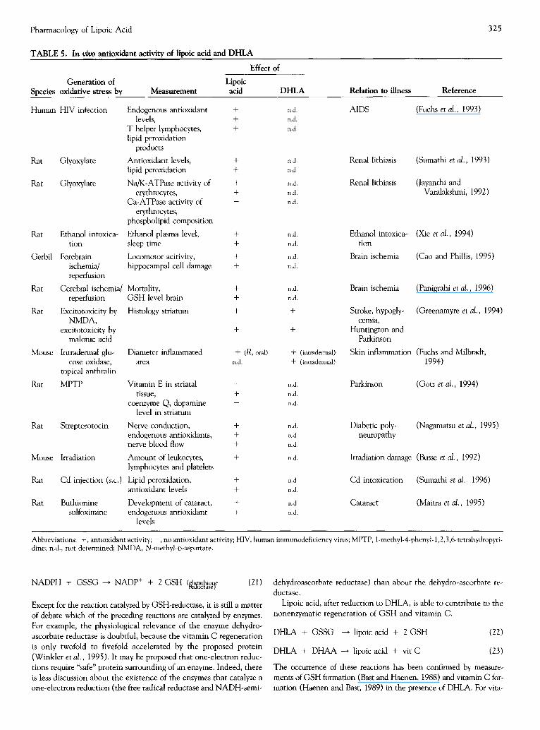
TABLE 5. In v/vo antioxidant activity of lipoic acid and D H L A
Effect of
Lipoic Measurement acid
Human HIV infection
Rat Glyoxylate
Rat Glyoxylate
Rat Ethanol intoxica- tion
Gerbil Forebrain ischemia/ repeffusion
Rat Cerebral ischemia/ reperfusion
Rat Excitotoxicity by NMDA,
excitotoxicity by malonic acid
Mouse Intradermal glu- cose oxidase,
topical anthralin
Rat MPTP
Rat Streptozotocin
Mouse Irradiation
Rat Cd injection (s.c.)
Rat Buthionine sulfoximine
Endogenous antioxidant + levels, +
T helper lymphocytes, + lipid peroxidation
products
Antioxidant levels, + lipid peroxidation +
Na/K-ATPase activity of + erythrocytes, +
Ca-ATPase activity of erythrocytes,
phospholipid composition
Ethanol plasma level, + sleep time +
Locomotor acitivity, + hippocampal cell damage +
D H L A
n.d. n.d. n.d.
n.d. n.d.
n.d. n.d. n.d.
n.d. n.d.
n.d. n.d.
Mortality, + n.d. GSH level brain + n.d.
Histology striatum + +
+ +
Diameter inflammated + (R, oral) area n.d.
+ (intradermal) + (intradern, al)
Vitamin E in striatal - n.d. tissue, + n.d.
coenzyme Q, dopamine - n.d. level in striatum
Nerve conduction, + n.d. endogenous antioxidants, + n.d. nerve blood flow + n.d.
Amount of leukocytes, + n.d. lymphocytes and platelets
Lipid peroxidation, + n.d. antioxidant levels + n.d.
Development of cataract, + n.d. endogenous antioxidant + n.d.
levels
Relation to illness Reference
AIDS (Fuchs et al., 1993)
Renal lithiasis
Renal lithiasis
(Sumathi et al., 1993)
(Jayanthi and Varalakshmi, 1992)
Ethanol intoxica- tion
Brain ischemia
(Xie et al., 1994)
(Cao and Phillis, 1995)
Brain ischemia (Panigrahi et al., 1996)
Stroke, hypogly- (Greenamyre et al., 1994) cemia,
Huntington and Parkinson
Skin inflammation (Fuchs and Milbradt, 1994)
Parkinson (Gotz et al., 1994)
Diabetic poly- (Nagamatsu et al., 1995) neuropathy
Irradiation damage (Busse et d . , 1992)
Cd intoxication (Sumathi et al., 1996)
Cataract (Maitre et al., 1995)
Abbreviations: +,antioxidant activity; - , no antioxidant activity;HIV, humanimmunodeficiencyvirus;MPTP, 1-methyl-4-phenyl-l,2,3,6-tetrahydropyn- dine; n.d., not determined; NMDA, N-methyl-D-aspartate.
NADPH + GSSG ---* NADP + + 2 GSH (glu, tathior~e (21) reauctasel
Except for the reaction catalyzed by GSH-reductase, it is still a matter of debate which of the preceding reactions are catalyzed by enzymes. For example, the physiological relevance of the enzyme dehydro- ascorbate reductase is doubtful, because the vitamin C regeneration is only twofold to fivefold accelerated by the proposed protein (Winkler et al. , 1995). It may be proposed that one-electron reduc- tions require "safe" protein surrounding of an enzyme. Indeed, there is less discussion about the existence of the enzymes that catalyze a one-electron reduction (the free radical reductase and NADH-semi-
dehydroascorbate reductase) than about the dehydro-ascorbate re- ductase.
Lipoic acid, after reduction to DHLA, is able to contribute to the nonenzymatic regeneration of GSH and vitamin C.
DHLA + GSSG ---* lipoic acid + 2 GSH (22)
DHLA + DHAA ---* lipoic acid + vit C (23)
The occurrence of these reactions has been confirmed by measure- ments of GSH formation (Bast and Haenen, 1988) and vitamin C for- mation (Haenen and Bast, 1989) in the presence of DHLA. For vita-

326 G.P. Biewenga et al.
o
30"
20"
0 no alloxan
33??:".::":: :: ::.3 -:- .:- 3 .:--7-
:%" 7 7.7 7 7 "-- . . . . . . . . . . . . . .
::::.5:3 -5 -7.:7-:7-.
:2:2.3-.3?.7.:.7.23:.:.: :: :73-373?)?3?:-: , ; , ; , ;<<,, , , , :
a l loxan
avatment
l ipoic acid + al loxan
FIGURE 11. Plasma glucose levels 72 hr after treatment with al- loxan (100 mg/kg, IV). Lipoic acid (100 mg/kg, IP) was given 30 min prior to the alloxan injection. The male SE outbred mice (ca. 30 g) fasted 4 hr before measurement of the glucose level in plasma derived from the tail veins. Data are shown as the mean of at least four experiments_+ SD.
N A D H - x ~ f lipoic acid
NAD + ~" "~ DHLA
vitamin C ~ D H A A
(25)
The regeneration of GSH also was studied by measuring GSH lev- els. Busse et al. (1992) observed an increase in the amount of GSH in murine neuroblastoma and melanoma cells after incubation with lipoic acid. Han et al. (1995) found similar results in human T-lymphocyte Jurkat cells. Remarkably, the elevation of GSH levels was larger than the amount of GSSG normally present in these cells. This may also indicate that liberation of GSH takes place from mixed disulfides [reaction (26)], although it may not be ruled out that lipoic acid stimulates the biosynthesis of glutathione by an un- known mechanism.
T - G l U ) c y s - S - - S - - C y s - p r o t e i n + DHLA
Gly (G-S-S-pmtein) ~. GSH + l i lmicacid + protein-SH
(26)
min C regeneration, the rote constant has been estimated (Table 6). DHLA shows greater reactivity toward dehydroascorbic acid (DHAA) than does GSH, indicating that, at certain concentrations, DHLA is preferentially used for vitamin C regeneration over GSH.
Interestingly, DHLA regenerates not only endogenous antioxi- dants. It was shown that DHLA could regenerate ebselen selenol (Haenen et al., 1990) and that the oxidation product of lipoic acid, [3-1ipoic acid, is reduced by DHLA (Fig. 12):
DHLA + 13-1ipoic acid ---, 2 lipoic acid (24)
In vitro r e g e n e r a t i o n
Two approaches are possible for studying the interplay of antioxi- dants in vitro. After addition of the proposed regenerating antioxi- dant, either the amount of the antioxidants or a synergistic protec- tion against oxidative damage can be quantified.
The first approach was applied by Xu and Wells (1996); they studied the regeneration of vitamin C. They determined the amount of vitamin C formed by incubating rat liver mitochondria or mi- toplasts with lipoic acid. In the presence of NADH, the addition of lipoic acid resulted in increased vitamin C levels. The necessity for NADH indicates that DHLA is the regenerating species:
TABLE 6. Nonenzymatic regeneration of vitamin C
Reductor Method* k (M/rain) SD
GSH 1 32.8 1.0 DHLA 2 1603 300
1 875 95
*(1) The rate constant was determined by following the rate of vitamin C formation at 265 nm. A concentration of (0.2-0.1 mM) dehydroascorbate was incubated with (0.35~0.50 raM) DHLA.
(2) The rate constant was determined from the steady-state concentra- tion of vitamin C observed at 265 nm. A concentration of 0.25 ~g/ml was incubated with ascorbate oxidase with 0.05 mM vitamin C and 1.0-1.5 mM DHLA.
Both reactions were performed at 37°C, 50 mM KHzPOJKOH, pH 7.4.
The second approach, studying the synergism in the prevention of oxidative damage, is best applied when the regenerating antioxi- dant alone does not contribute to overall antioxidant activity (Fig. 13). Such circumstances were found by Bast and Haenen (1988) and Kagan et al. (1992). Kagan et al. found that DHLA did not pro- tect dioleoylphosphatidylcholine liposomes from ultraviolet-induced peroxidation. And, in another in vitro test, DHLA showed no pro- tection from lipoxygenase-induced peroxidation in microsomes. After induction of the lipid peroxidation process, the electron spin resonance (ESR) signal of the chromanoxyl radical present in the lipid bilayer was an indication that the peroxidation was proceed- ing. After addition of vitamin C to the lipid suspension, the chro- manoxyl radical disappeared and the ascorbyl radical appeared. Gradually, the ascorbyl radical decreased in time, and progressively, the chromanoxyl radical appeared. The interaction of DHLA with dehydroascorbate was seen as a longer lifetime of the ESR signal of the ascorbyl radical before the lipid peroxidation continued.
scavenging interplay disproportio- nation R°~vitE ~vitC- ~ vitC*
RH vit E° vit C DHAA (27)
lipoic acid DHLA
Bast and Haenen (1988) studied the interplay between DHLA and GSH. They observed synergism in the protection against Fe2+-vitamin C-induced lipid peroxidation. As seen in Fig. 7, antioxidant activity is found for DHLA only when it is coincubated with GSSG.
DHLA does not regenerate the chromanoxyl radical present in vitamin E directly. This can be concluded from the study reported by Kagan et al. (1992). DHLA alone did not decrease the signal of the chromanoxyl radical in the liposomes. However, vitamin E can be regenerated by DHLA in a cascade of regenerating reactions.

Pharmacology of Lipoic Acid 327
8
1 . 0 -
0.5-
=
2~:~0 280 300 3~0 340 360 380
wavelength in nm
FIGURE 12. A concentration of 5 mM DHLA (spectrum t=0) was incubated with 5.16 mM I~-lipoic acid at 37°C and pH 7.4. Every 10 min, a spectrum was recorded. The increase at 333 nm shows the formation of lipoic acid. The second-order rate constant was determined: k=0.51 × 10 -2 M/min.
Scholich et al. (1989) reported a nonenzymatic regeneration of vita- min E by DHLA, independently of GSH. However, Bast and Haenen (1988) found that protection against lipid peroxidation by DHLA depended on GSH (Fig. 7). They proposed that the regener- ation of GSH [reaction (22)] is followed by the enzymatic regenera- tion of vitamin E [reaction (19)].
In vivo r e g e n e r a t i o n
The two approaches used for in vitro tests have been applied to the study of the regeneration of antioxidants; that is, the measurement of antioxidant levels or the determination of synergistic protection. Vitamin C regeneration has been studied in healthy newborn rats. It was shown that the administration of lipoic acid did not statistically significantly affect the vitamin C levels in the eye lens (Maitra et al., 1995). Vitamin C regeneration has also been studied in organ- isms subjected to oxidative stress. In plasma of HIV-positive pa- tients, Lipoic acid increased vitamin C levels (Fuchs et al., 1993). In the eye lenses of newborn rats (Maitra et al., 1995), lipoic acid pre- vented a decrease in vitamin C levels induced by BSO treatment. Clearly, lipoic acid can affect vitamin C levels. However, it is not known whether vitamin C regeneration occurs or whether lipoic acid acts as a scavenger, thus sparing vitamin C.
Regarding vitamin E, in healthy animals, no effect of lipoic acid administration was found on the vitamin E level (Maitra et al., 1995; Nagamatsu et aI., 1995). BSO treatment of newborn rats re-
C oxidative stress
1 ' antioxidant ~ regenerating
antioxidant
FIGURE 13. Regeneration of an antioxidant can be studied as synergism in the prevention of oxidative damage. Minor contribu- tion of the regenerating antioxidant to the overall antioxidant ac- tivity is a necessary condition for this principle.
sulted in a decreased vitamin E level, which is prevented by lipoic acid (Maitra et al., 1995). Nagamatsu et al. (1995) studied the inter- play between vitamin E, GSH and lipoic acid. Depletion of vitamin E, together with oxidative stress derived from streptozotocin- induced diabetes, resulted in decreased levels of GSH. However, this was not accompanied by an increase in GSSG. Moreover, lipoic acid did not prevent this GSH depletion, confirming that oxidized glutathione [or its mixed disulfide, reaction (26)] is necessary for the regeneration of GSH.
For GSH regeneration, different effects of lipoic acid have been reported in healthy subjects. In mice, total GSH levels were in- creased in liver, kidney and lung tissue cells after lipoic acid admin- istration (Busse et al., 1992). In contrast, lipoic acid did not affect the GSH or GSSG levels in the sciatic nerve of healthy rats (Naga- matsu et al., 1995). In animals subjected to oxidative stress, lipoic acid prevented depletion of GSH in all reported experiments (Mai- tra et al., 1995; Nagamatsu et al., 1995; Sumathi et al., 1993).
The second approach, synergistic prevention in vivo, was studied as early as 1959 by Rosenberg and Culik (1959). They concentrated on end-point parameters of vitamin C - and vitamin E~leficiency scurvy and reproduction failure, respectively. In vitamin C~tefi- cient guinea pigs, suboptimal amounts of vitamin C in combination with lipoic acid prevented scurvy symptoms better than did either compound alone. For vitamin E-deficient animals, a similar syner- gistic protection was found. The combination of 15 mg of lipoic acid plus 25 mg of vitamin E was more effective than either one of the compounds alone against the symptoms of vitamin E deficiency.
Altogether, lipoic acid seems to regenerate vitamin C, vitamin E and GSH in vivo. For in vivo tests, one should keep in mind that it is difficult to determine whether a lessened decrease in antioxidant level is due to additional scavenging, to stimulated biosynthesis (as might oc- cur with GSH) or to regeneration of another antioxidant by the drag. At least it is safe to state that none of the/n vivo tests excluded regener- ation of the endogenous antioxidants. Especially, the synergism found in the experiments of 1959 are a good indication that lipoic acid is able to regenerate vitamin C and vitamin E in vivo.
REPAIR OF OXIDATIVE DAMAGE: IMPROVEMENT OF PMSR ACTIVITY
Oxidative stress may result in damage to DNA, lipids and proteins. One method of overcoming oxidative damage is degradation and re- newal. A second method is repair, which may be particularly important for proteins with a low turnover rate. In the proteins, amino acid resi- dues such as tryptophan, histidine, tyrosine, cysteine and methionine are susceptible to oxidation. Whereas some oxidants (e.g., ozone, super- oxide anions/hydroxyl radicals) destroy the residues at random, other oxidants (e.g., H20> HOCI, chloramines and ON(X)-) preferentially oxidize exposed methionine residues (Maier et al., 1989).
H2N ~,.fCOOH H2N ~COOH
CH2 CH 2
I I OH2 C H 2
I I S S 0
I I CHa OH a
methionine (Met) methionine su~oxide (MetS=O)
oxidant (28)

328 G.P. Biewenga et al.
Methionine oxidation of some residues does not affect physical or immunochemical properties or biological activity of the peptide at all, whereas oxidation of other, specific methionine residues imme- diately leads to inactivation of the protein. This is observed for sev- eral enzymes, hormones, chemotactic factors and plasma proteinase inhibitors. For these peptides, inactivation by methionine oxidation has been regarded as part of a regulatory, physiologic process (Swaim and Pizzo, 1988). A particular example of oxidative regulation is the regulation of the activity of proteinase inhibitors such as cq-AP. Neutrophils, macrophages and other leukocytes secrete large quanti- ties of oxidants at sites of inflammation and may readily bring about methionine oxidation, resulting in loss of protease inhibitory activ- ity. Inactivation of proteinase inhibitors may alter the proteinase- antiproteinase balance in favor of the protein-degrading enzyme. Protein degradation facilitates phagocytosis of invading organisms but should be restricted to exogenous material. A poorly controlled proteinase-antiproteinase balance results in degradation of endoge- nous tissue, and this forms the basis for pathological processes such as as lung emphysema.
Two enzymes have been reported to be able to reduce oxidized other (PMSR) reduces peptide bound methionine sulfoxide. The enzyme PMSR requires reducing equivalents for its reaction. It is proposed that the thioredoxin system supplies the enzyme with elec- trons (Brot et al., 1981):
(29)
NADP+ J NLlhioredoxin(~A) / ~ ~ioredoxin(ox) * "~ PMSR(red) / \ MelS---O reductase
Excessive accumulation of methionine sulfoxide residues can be caused by reduced PMSR activity. This decreased activity can origi- nate from a decreased amount of PMSR or by a decreased amount of reducing equivalents (Brot and Weissbach, 1988).
Lipoic acid can improve the repair of oxidized methionine resi- dues by supplying PMSR with reducing equivalents. This can be done by increasing the amount of reduced thioredoxin. Holmgren (1979) and Spector et al. (1988) showed that dihydrolipoamide, an analogue of dihydrolipoic acid, can reduce thioredoxin in the fol- lowing system:
N A D H y lipoicacid ~ thioredoxin(red)
L ~ H (30)
NAD + ~¢ ~, DI=ILA thioredoxin(ox)
This system can be coupled to PMSR and to the PMSR-dependent regeneration of eq-AP. As shown in Fig. 14, DHLA can regenerate aIAP by itself, but the reaction is catalyzed in the presence of partly purified PMSR from rat lungs and is further accelerated by thiore- doxin.
When DHLA is derived from LipDH-dependent reduction, the ulti- mate effect of lipoic acid is making NADH available as a source for re- ductive reactions [reaction (31)] instead of NADPH [reaction (29)].
NADH~. / linoic acid ,qt 4thioredoxin(rod)-. / PMSR(ox) ~, . . ~1 -AP \ f ~ \ f \ f \ f (ac~vo)
Ltp~H ~ ~ ~ (31)
In vivo, the activity of PMSR is difficult to detect (Glaser et al., 1987). Only recently, reversal of damage due to methionine oxidation was
50"
o = 40"
30" E
~'~ 20"
10"
0 4 15
mM DHLA
FIGURE 14. The repair of oxidatively damaged cx~-AP by DHLA (black) is accelerated by PMSR (dark gray) and is further accelerated by PMSR plus thioredoxin (light gray). PMSR was partly purified from Wistar rat lungs, by the method of Carp eta/., (1983) followed by anion exchange chromatography on a 26/10 Sepharose column. An amount of 200 I*g eq-AP was oxidized by 1.25 mM HOCI in a volume of 40 lull. The compounds were incu- bated with 37 units PMSR for 1 hr at 37°(2 in a volume of 280 ILl. (One unit is defined as the percentage of reactivated ~1-AP per mg protein after 1 hr in the presence of 15 mM dithiothreotol (DTT) at 37°(2.) The percentage of (x,-AP repair was determined in 850 ~1, using 25 ~g elastase and 60 ~M N-succinyl-(Ala)3- p-nitroanilide and assuming a perfect inverse relation between the amount of oq-AP and elastase activity. The rate of formation of p-nitroaniline was observed at 410 nm.
shown in living cells (Moskovitz eta/., 1995). Although the contribu- tion of lipoic acid to protein repair still has to be demonstrated in vivo, the in vitro experiments provide evidence that lipoic acid participates in the repair of methionine residues by reduction of PMSR.
PRO-OXIDANT ACTIVITY
Pro-oxidant activity is defined as the activity of an antioxidant in a sit- uation where it produces more oxidative stress than if when it were ab- sent. The ability of a compound to donate an electron to an oxidant can make that compound an antioxidant. Under different conditions, however, exactly the same ability can turn that compound into a pro- oxidant. This can be demonstrated by thiols. Usually thiols are consid- ered to be antioxidants. However, thiols can also promote O2'- forma- tion by direct reaction with O2. Although 02" generation by D H L A has not been reported, it is known that DHLA oxidizes by air, indicat- ing that oxygen-reduction occurs. Generally, transition metals catalyze O2"- formation and other ROS-producing reactions. Indeed, Fe 3+ also increases the rate of oxygen consumption by DHLA (Kawabata et al., 1995), indicating that DHLA reduces Fe 3+, which subsequently re- duces O2. In addition, this one-electron reduction of a transition metal may also occur indirectly by the regeneration of vitamin C [reaction (23)]. Increased Fe 3+ reduction can result in pro-oxidant activity. This was observed in two lipid peroxidation tests (Bast and Haenen, 1988; Scott et al., 1994). Remarkably, lipoic acid inhibits the pro-oxidant ac- tivity of DHLA (Scott et al., 1994).
In another route to pro-oxidant activity, the antioxidant reacts with a ROS scavenger, and a product is formed that is more harmful than the ROS that is scavenged. This has been observed with smoke-derived ROS and 'OH radicals. In smoke, ROS are present, inactivating cre- atine kinase. DHLA scavenges these ROS. However, the scavenging product more efficiently inactivates creatine kinase than does the origi- nal ROS (Scott et al., 1994). Irradiation of an oq-AP-containing solu-

Pharmacology of Lipoic Acid 329
tion results in "OH formation and subsequent inactivation of cq-AP. Relatively low concentrations of lipoic acid or DHLA accelerate this inactivation. This has been explained by the formation of thiyl and oxysulfur radicals after scavenging. Interestingly, at high concentra- tions, both lipoic acid and DHLA act as antioxidants (Scott et al.,
1994). Altogether, DHLA can have pro-oxidant activity by reducing transi-
tion metals. In addition, both lipoic acid and DHLA can act as pro- oxidants by the formation of reactive scavenging products.
CONCLUSIONS
In this review, we considered drug-related antioxidant pharmacology. In pathology and physiology, oxidative processes take place. An imbal- ance between oxidative and antioxidative processes results in oxidative stress. Drugs can intervene in oxidative processes as antioxidants and delay or prevent their damaging effects. The term "antioxidant" depicts a relative concept. Antioxidant activity depends on which biological target molecules have to be protected from which kind of oxidative re- actions. Antioxidant pharmacology related to a certain pathology can provide information into the nature of oxidative stress and the biologi- cal target molecules in the etiology. In drug-related antioxidant phar- macology, research concentrates on the the antioxidant activity of a certain compound. The antioxidant activity of a compound is deter- mined by its antioxidant properties and by its concentration. Similar to other drugs, the metabohtes may contribute to the overall antioxidant activity and pharmacological effect in vivo.
On the basis of lipoic acid, research concentrating on the antioxi- dant activity of a drug was discussed. Metabolites that contribute to the antioxidant activity of lipoic acid in vivo are DHLA and the [8-oxida- tion products bisnor-, and tetranorlipoic acid. For DHLA and lipoic acid four antioxidant properties ware thoroughly examined: metal che- lation, ROS scavenging i regeneration of endogenous antioxidants and repair of oxidatively damaged biomolecules. For the determination of these properties, test-tube experiments were performed, characterizing the chemical reactivity, The corresponding antioxidant activity was determined in vitro and in vivo. For the sake of clarity, a strict division was made between the different antioxidant properties and between the chemical in vitro and in vivo tests. In pathology, of course, all sepa- rate antioxidant mechanisms may proceed at the same time. The fol- lowing properties were described for lipoic acid and its metabolites. Li- poic acid can have antioxidant activity by chelating Fe > and Cu 2+, and DHLA by chelating Cd 2+. DHLA can also chelate iron and reduce Fe ~+ to Fe z+, which results in pro-oxidant activity. Both lipoic acid and DHLA are scavengers of several ROS. Mostly, this results in antioxi- dant activity, except in three reported experiments in which the ROS- scavenging product appeared more damaging than the original ROS. (Remarkably, some pro-oxidant activity of DHLA can be inhibited by lipoic acid.) DHLA can participate in the regeneration of endogenous antioxidants such as vitamin C, GSH and vitamin E. In addition, DHLA may repair oxidative damage to proteins by improving the ac- tivity of PMSR.
Altogether, DHLA has more antioxidant properties than does lipoic acid. Therefore, it is likely that DHLA plays a major role in the ob- served antioxidant activity of lipoic acid in vivo. DHLA is formed by reduction of lipoic acid. Through LipDH-catalyzed reduction, the cell can draw on its NADH pool for antioxidant activity additionally to its NADPH pool, which is usually consumed during oxidative stress.
Antioxidant drugs can prevent the onset of pathologies, can delay pathological processes or may even take part in repair. Many experi- mental results show that both lipoic acid and DHLA can improve the antioxidant capacity of tissue against different forms of oxidative stress.
At the moment, research on antioxidant drugs and research related to oxidative disease processes have not converged into a therapeutic in- tervention in which the mode of action is fully comprehended. How- ever, research on the antioxidant pharmacology of the drug lipoic acid has given a stimulating impulse to antioxidant therapy.
We acknowledge D. H. Veening-Griffioen and Dr. H.-P. Voss, for testing lipoic acid in the alloxan-induced diabetes model. We thank A. J. Nicastia, who studied the re- generatum of vitamin C by DHLA (Table 6). The recommendations of Dr. E. M. Mooijman regarding linguistic matters and the general comments of Dr. J. B. G. Pa- quay are highly appreciated.
References Bast A. (1994) Antioxidant pharmacothempy. DN&P 7, 465-472. Bast A. and Haenen G. R. M. M. (1988) Interplay between lipoic acid and glu-
tathione in the protection against microsomal lipid peroxidation. Biochim. Biophys. Acta 963, 558-561.
Biewenga G. P., de Jong J. and Bast A. (1994) Lipoic acid favors thiolsulfinate formation after hypochlorous acid scavenging: a study with lipoic acid deriv- atives. Arch. Biochem. Biophys. 312, 114-120.
Biewenga G. P., Dorstijn M. A., Verhagen J. V., Haenen G. R. M. M. and Bast A. (1996a) Reduction of hpoic acid by lilx, amide dehydrogenase. Biochem. Pharmac. 51,233-238.
Biewenga G. P., Haenen G. R. M. M. and Bast A. (1997b) An overview on li- poate chemistry. In Lipoic Acid in Health and Disease. (Edited by Fuchs J., Packer L. and Zimmer G.) pp. 1-32. Marcel Dekker, New York.
Biewenga G. P., Vriesman M. F., Haenen G. R. M. M. and Bast A. (1996c) The metabolism of lipoic acid in man. Submitted.
Bisby R. H., Johnson S. A. and Parker A. W. (1996) Quenching of reactive oxi- dative species by probucol and comparison with other antioxidants. Free Radical Biol. Med. 20, 411-420.
Donomi F. and Pagani S. (1986) Removal of ferritin-bound iron by DL-dihydro- lipoate and r~L-dihydrolipoamide. Eur. J. Biochem. 155, 295-300.
Bonomi F., Werrh M. T. and Kurtz D. M. (1985) Assembly of [Fe~S~ (SR)4] 2 (n=2,4) in aqueous media from iron salts, thiots, and sulfur, sulfide, or thiosulfate plus rhodanese. Inorg. Chem. 24, 4331-4335.
Doucher J.-L., Genet A., Vadon S., Delaforge M., Henry Y. and Mansuy 1). (1992) Cytochrome P-450 catalyzes the oxidation of N'°-hydroxy-L - arginine by NADPH and O2 to nitric oxide and citmlline. Biochem. Biophys. Res. Commun. 187, 880486.
Brot N., Fliss H., Coleman T. and Weissbach H. (1984) Enzymatic reduction of methionine sulfoxide residues in proteins and peptides. Methods En~mol. 107, 352-36O.
Brot N. and Weissbach H. (1988) Methionine sulfoxide: chemistry and bio- chemistry. In The Chem/stry of Functional Groups: The Chemistry of Sulfones and Sulfoxides. (Edited by Patai S., Rappoport Z. and Stirling C. J. M.) pp. 851-872, Wiley, Chichester, UK.
Brot N., Weissbach L., Werth J. and Weissbach H. (1981) Enzymatic reduction of protein-bound methionine sulfoxide. Proc. Natl. Acad. Sci. 78, 2155- 2158.
Brown P. R. and Edwards J.O. (1970) The reactions of 1,3-dimercaptocpro- pane, lipoic acid, and dihydrolipoic acid with metal ions. J. lnorg. Nuc/. Chem. 32, 2671-2675.
Burkat V., Koike T., Brenner H.-H., Imai Y. and Kotb H. (1993) Dihydrolipoic acid protects pancreatic islet cells from inflammatory attack. Agents Act/ons 38, 60455.
Busse E., Zimmer G., Schopohl B. and Komhuber B. (1992) Influence of c~-litx~ic acid on intmcellular glutathione in vitro and in vivo. Arzneim.- Forsch. Drug Res. 42, 829-831.
Cameron N. E., Cotter M. A. and Maxfield E. K. (1993) Anti-oxidant treat- ment prevents the development of peripheral nerve dysfunction in strepto- zotocin-diabetic rats. Diabetolog~a 36, 299-304.
Cao X. H. and Phillis J. W. (1995) The free radical scavenger, c~-lipoic acid, protects against cerebral ischemia-reperfusion injury in gerbils. Free Radical Res. 23, 365-370.
Carp H., Janoff A., Abrams W., Weinbaum G., Drew R. T., Weissbach H. and Brot N. (1983) Human methionine sulfoxide-peptide reductase: an enzyme capable of reactivating oxidized c~-l-proteinase inhibitor in vitro. Am. Rev. Respir. Dis. 127, 301-305.
Carreau J.-P. (1979) Biosynthesis of liv~ic acid via unsaturated fatty acids. Meth- ods Enzymol. 62, 152-158.
Ching T.-L, de Jong J. and Bast A. (1994) A method for screening hypochlo- rous acid scavengers by inhibition of 5-thio-2-nitrobenzoic acid. Anal. Bio- chem. 218, 377-381.
330 O . P . Biewenga et cd.
Constantinescu A., Pick U., Handelrnan G. J., Haramaki N., Han D., Podda M., Tritschler H. J. and Packer L. (1995) Reduction and transport of lipoic acid by human erythrocytes. Biochem. Pharmac. 50, 253-261.
Cornaro U., Cariati F. and Bonomi F. (1985) Evidences for the formation of complexes of oL-dihydrothioctic acid (reduced lipoic acid) with Ni 2+, Co z+, and Fe 3+ salts. Rev. Port. Qu/m. 27, 273-274.
Devasagayam T. P. A., Subramanian M., Pradhan D. S. and Sies H. (1993) Pre- vention of singlet oxygen-induced DNA damage by lipoate. Chem. Biol. In- teract. 86, 79-92.
Dikalov S., Khramtsov V. and Zimmer G. (1996) Determination of rate con- stants of the reactions of thiols with superoxide radical by electron paramag- netic resonance: critical remarks on spectrophotometric approaches. Arch. Biochem. Biophys. 326, 207-218.
Folkes L. K., Candeias L. P. and Wardman P. (1995) Kinetics and mechanism of hypochlorous acid reactions. Arch. Biochem. Biophys. 323, 120-126.
Fuchs J. and Milbradt R. (1994) Antioxidant inhibition of skin inflammation induced by reactive oxidants: Evaluation of the redox couple dihydroli- poate/lipoate. Skin Pharmac. 7, 278-284.
Fuchs J., Schofer H., Milbradt R., Freisleben H.J., Buhl R., Siems W. and Grune T. (1993) Studies on lipoate effects on blood redox state in human immunodeficiency virus infected patients. Arzneim.-Forsch. Drug Res. 43(2), 1359-1362.
Fujiwara K., Okamura-Ikeda K. and Motokawa Y. (1995) Assay for protein li- poylation reaction. Methods Enzymol. 251,333-350.
Furr H. C., Chang H.-H. and McCormick D. B. (1978) Lipoate metabolism in pseudomonas putida LP: thiolsulfinates of lipoate and bisnorlipoate. Arch. Biochem. Biophys. 185, 576-583.
Glaser C. B., Karic L., Parmelee S., Premachandra B. R., Hinkston D. and Ab- rams W.R. (1987) Studies on the turnover of methionine oxidized a-l-protease inhibitor in rats. Am. Rev. Respir. Dis. 136, 857461.
Gotz M.E., Dirr A., Burger R., Janetzky B., Weinmuller M., Chan W. W., Chen S. C., Reichmann H., Rausch W. D. and Riederer P. (1994) Effect of lipoic acid on redox state of coenzyme Q in mice treated with 1-methyl-4-phenyl-l,2,3,6-tetrahydropyridine and diethyldithiocarbamate. Eur. J. Pharmac. Mol. Pharm. 266, 291-300.
Greenamyre J. T., Garciaosuna M. and Greene J. G. (1994) The endogenous cofactors, thioctic acid and dihydrolipoic acid, are neuroprotective against NMDA and Malonic acid lesions of striatum. Neurosci. Lett. 171, 17-20.
Grootveld M. and Halliwell B. (1986) Aromatic hydroxylation as a potential measure of hydroxyl-radical formation in vivo. Biochem. J. 237, 499- 504.
Gut J., Christen U., Frey N., Koch V. and Stoffler D. (1995) Molecular mim- icry in halothane hepatitis: biochemical and structural characterization of lipoylated autoantigens. Toxicology 97, 199-224.
Gutierrez Correa J. G. and Stoppani A. O. M. (1996) Catecholamines en- hance dihydrolipoamide dehydrogenase inactivation by the copper Fen- ton system: enzyme protection by copper chelators. Free Radical Res. 24, 311-322.
Haenen G. R. M. M. and Bast A. (1991) Scavenging of hypochlorous acid by lipoic acid. Biochem. Pharmac. 42, 2244-2246.
Haenen G. R. M. M. and Bast A. (1989) Modulation of microsomal lipid peroxidation by dihydrolipoate in interplay with vitamin E, vitamin C and glutathione. In Thioctsaure: Neue biochemische, pharmakologtsche und klinische Erkennmisse zur Thioctsaure. (Edited by Borbe H. O. and Ulrich H.) pp. 137-153. pmi Verlag, Frankfurt am Main.
Haenen G. R. M. M., deRooij B. M., Vermeulen N. P. E. and Bast A. (1990) Mechanism of the reaction of ebselen with endogenous thiols: dihydroli- poate is a better cofactor than glutathione in the peroxidase activity of ebselen. Mol. Pharmac. 37, 412-422.
Halliwell B. (1995) Antioxidant characterization: methodology and mecha- nism. Biochem. Pharmac. 49, 1341-1348.
Halliwell B. and Gutteridge J. M. C. (1989) Free Radicals in Biology and Medi- cine. Clarendon Press, Oxford.
Han D., Tritschler H. J. and Packer L. (1995) a-Lipoic acid increases intra- cellular glutathione in a human T-lymphocyte Jurkat cell line. Biochem. Biophys. Res. Commun. 207, 258-264.
Handelman G. J., Han D., Tritschler H. and Packer L. (1994) a-Lipoic acid reduction by mammalian cells to the dithiol form, and release into the culture medium. Biochem. Pharmac. 47, 1725-1730.
Haramaki N., Packer L., Assadnazari H. and Zimmer G. (1993) Cardiac re- covery during post-ischemic reperfusion is improved by combination of vitamin-E with dihydrolipoic acid. Biochem. Biophys. Res. Commun. 196, 1101-1107.
Harrison E. H. and McCormick D. B. (1974) The metabolism of dl-[1,6- 14C]-lipoic acid in the rat. Arch. Biochem. Biophys. 160, 514-522.
Herbert A. A. and Guest J. R. (1975) Lipoic acid content of Escherichia coli and other microorganisms. Arch. Microbiol. 106, 259-266.
Hermann R., Niebch G., Borbe H.O., Fieger H., Ruus P., Nowak H., Riethmller-Winzen H., Peukert M. and Blume H. (1996) Enantio selec- tive pharmacokinetics and bioavailability of different racemic ~x-lipoic acid formulations in healthy volunteers. Eur. J. Pharm. Sci. 4, 167-174.
Holmgren A. (1979) Thioredoxin catalyses the reduction of insulin disul- fides by dithiothreitol and dihydrolipoamide. J. Biol. Chem. 254, 9627- 9632.
Jayanthi S. and Varalakshmi P. (1992) Alterations in rat erythrocyte membrane parameters during calcium oxalate lithiasis and the effect of c~-lipoic acid. Indian J. Clin. Biochem. 7, 115-120.
Kagan V. E., Shvedova A., Serbinova E., Khan S., Swanson C., Powell R. and Packer L. (1992) Dihydrolipoic acid: a universal antioxidant both in the membrane and in the aqueous phase. Biochem. Pharmac. 44, 1637- 1649.
Kaiser S., Di Mascio P. and Sies H. (1989) Lipoate und Singulettsauerstoff. In Thioctsaure: Neue biochemische, pharmakolog(tsche und klinische Erkennt- nisse zur Thioctsiiure. (Edited by Borbe H. O. and Ulrich H.) pp. 69-76. pmi Verlag, Frankfurt am Main.
Kawabata T., Tritschler H.-J. and Packer L. (1995) Reaction of (R,S)-dihy- drolipoic acid and homologs with iron. Methods Enzymol. 251,325-332.
Kleemann A., Borbe H. O. and Ulrich H. (1989) Thiocts~iure: ct-lipons~iure. In Thiocts;~ure: Neue biochemische, pharmakologische und klinische Erkennt- nisse zur Thioctsgture. (Edited by Borbe H. O. and Ulrich H.) pp. 11-26. pmi Verlag, Frankfurt am Main.
Lewis R. L. and Sweet D.V. (1985) Re~stry of Toxic Effects of Chemical Substances 1983~4. U.S. Department of Health and Human Services, Cincinnati.
Locher M., Busker E. and Borbe H. O. (1995) Metabolism of a-lipoic acid in human volunteers. Naunyn-Schmiedeberg's Arch. Pharmac. 351, R52 (abstract).
bffelhardt S., Bonaventura C., Locher M., Borbe H. O. and Bisswanger H. (1995) Interaction of c~-lipoic acid enantiomers and homologues with the enzyme components of the mammalian pyruvate dehydrogenase complex. Biochem. Pharmac. 50, 637-646.
Maier K. L., Matejkova E., Hinze H., Leuschel L., Weber H. and Beck-Speier 1. (1989) Different selectivities of oxidants during oxidation of methio- nine residues in the ct-l-proteinase inhibitor. FEBS Lett. 250, 221-226.
Maitra I., Serbinova E., Tritschler H. J. and Packer L. (1995) ~-Lipoic acid prevents buthionine sulfoximine-induced cataract formation in newborn rats. Free Radical Biol. Med. 18, 823-829.
Maitra I., Serbinova E., Tritschler H. J. and Packer L. (1996) Stereospecific effects of R-lipoic acid on buthionine sulfoxinine-induced cataract for- mation in newborn rats. Biochem. Biophys. Res. Commun. 221,422-429.
Matsugo S., Yan L. J., Han D., Trischler H. J. and Packer L. (1995) Elucida- tion of antioxidant activity of ct-lipoic acid toward hydroxyl radical. Bio- chem. Biophys. Res. Commun. 208, 161-167.
Mattulat A. (1992) Determination of the lipoic acid content of animal tis- sue. In Thioctsgiure: International Thioctic Acid Workshop. (Edited by Schmidt K. and Ulrich H.) pp. 69-73. Universimed Verlag, Frankfurt am Main.
Mattulat A. and Baltes W. (1992) Determination of lipoic acid in meat of commercial quality. Z. Lebensm.-Unters.-Forsch. 194, 326-329.
Morris T. W., Reed K. E. and Cronan J. E. (1995) Lipoic acid metabolism in Escherichia coli: the lplA and lipB genes define redundant pathways for ligation of lipoyl groups to apoprotein. J. Bacteriol. 177, 1-10.
Moskovitz J., Rahman M. A., Strassman J., Yancey S.O., Kushner S. R., Brot N. and Weisbach H. (1995) Escherichia coli peptide methionine sulf- oxide reductase gene: regulation of expression and role in protecting against oxidative damage. J. Bacteriol. 177, 502-507.
Mailer L. (1989) Protective effects of DL-~x-lipoic acid on cadmium-induced deterioration of rat hepatocytes. Toxicology 58, 175-185.
Mtiller L. and Menzel H. (1990) Studies on the efficacy of lipoate and dihy- drolipoate in the alteration of cadmium z+ toxicity in isolated hepato- cytes. Biochim. Biophys. Acta 1052, 386-391.
Nagamatsu M., Nickander K. K., Schmelzer J. D., Raya A., Wittrock D. A., Tritschler H. and Low P. A. (1995) Lipoic acid improves nerve blood flow, reduces oxidative stress, and improves distal nerve conduction in experimental diabetic neuropathy. Diabetes Care 18, 1160-1167.
Nathan C. (1992) Nitric oxide as a secretory product of mammalian cells. FASEB J. 6, 3051-3064.
O'Neill C.A., Halliwell B., van der Vliet A., Davis P.A., Packer L., Tritschler H., Strohman W. J., Rieland T., Cross C. E. and Reznick A. Z. (1994) Aldehyde-induced protein modifications in human plasma: pro-

Pharmacology of Lipoic Acid 331
tection by glutathione and dihydrolipoic acid. J. Lab. Clin. Med. 124, 359-370.
Ou P. M., Tritschler H. J. and Wolff S. P. (1995) Thioctic (lipoic) acid: a therapeutic metal-chelating antioxidant? Biochem. Pharmac. 50, 123- 126.
Packer L., Witt E. H. and Tritschler H. J. (1995) c(-Lipoic acid as biological antioxidant. Free Radical Biol. Med. 19, 227-250.
Panigrahi M., Sadguna Y., Shivakumar B.R., Kolluri S. V.R., Roy S., Packer L. and Ravindranath V. (1996) e~-Lipoic acid protects against re- peffusion injury following cerebral ischemia in rats. Brain Res. 717, 184- 188.
Pick U., Haramaki N., Constantinescu A., Handelman G. J., Tritschler H. J. and Packer L. (1995) :Glutathione reductase and lipoamide dehydroge- nase have opposite stereospecificities for c(-lipoic acid enantiomers. Bio- chem. Biophys. Res. Commun. 206, 724-730.
Pruijn F. B., Haenen G. R. M. M. and Bast A. (1991) Interplay between vi- tamin E, glutathione and dihydrolipoic acid in protection against lipid peroxidation. Fat Sci. Technol. 6, 216-221.
Rosenberg H. R. and Culik R. (1959) Effect of c~-lipoic acid on vitamin C and vitamin E deficiencies. Arch. Biochem. Biophys. 80, 86-93.
Salonen J. T., Nyyssnen K., Tuomainen T.-P., Menp P.H., Korpela H., Kaplan G. A., Lynch J., Helmrich S. P. and Salonen R. (1995) Increased risk of non-insulin dependent diabetes mellitus at low plasma vitamin E concentrations: a four year follow up study in men. Br. Med. J. 311, 1124-1126.
Scheer B. and Zimmer G. (1993) Dihydrolipoic acid prevents hypoxic/ reoxygenation and peroxidative damage in rat heart mitochondria. Arch. Biochem. Biophys. 302, 385-390.
Scholich H., Murphy M. E. and Sies H. (1989) Antioxidant activity ofdihy- drolipoate against microsomal lipid peroxidation and its dependence on c(-tocopherol. Biochim. Biophys. Acta 1001, 256-261.
Scott B. C., Aruoma O. I., Evans P. J., O'Neill C., van der Vliet A., Cross C. E., Tritschler H. and Halliwell B. (1994) Lipoic and dihydrolipoic acids as antioxidants: a critical evaluation. Free Radical Res. 20, 119-133.
Serbinova E., Reznick S. K. A. Z. and Packer L. (1992) Thioctic acid pro- tects against ischemia-reperfusion injury in the isolated perfused Langendorf heart. Free Radicas Res. Commun. 17, 49-58.
Sigel H. (1982) Die hydrophoben und Metallionen-koordinierenden Eigenschaften von A-Lipos~iure: ein Beispiel far intramolekulare Gleich- gewichte in Metallior~en-Komplexen. Angew. Chem. 94, 4214-32.
Sigel H. and Prijs B. (1978) Stability and structure of binary and ternary complexes of c~-lipoate and lipoate derivatives with Mn TM Cu TM Zn 2+ in solution. Arch. Biochern. Biophys. 187, 208-214.
Spector A., Huang R.-R. C., Yah G.-Z. and Wang R.-R. (1988) Thioredoxin fragment 31-36 is reduced by dihydrolipoamide and reduces oxidized protein. Biochem. Biophys. Res. Commun. 150, 156-162.
Spence J. T. and McCormick D. B. (1976) Lipoic acid metabolism in the rat. Arch. Biochem. Biophys. 174, 13-19.
Spoto G., Dupre S. and Cavallini D. (1982) Biosynthesis of lipoic acid: en- docellular localization and some properties of the involved enzymatic system. IRCS Med. Sci. 10, 626.
Stary F. E., Jindal S. L. and Murray R. W. (1975) Oxidation of c~-lipoic acid. J. Org. Chem. 40, 58-62.
Stevens B., Perez S. R. and Small R. D. (1974) The photoperoxidation of unsaturated organic molecules IX: lipoic acid inhibition of rubrene au- toperoxidation. Photochem. Photobiol. 19, 315-316.
Stroman F., Herold H., Erdmann R., Ulrich H. and Szelenyi I. (1989) Zur pro- tekiven Wirkung der Tt~iocts~iure bei myocardialer Ischmie. In Thioctsi~ure: Neue biochemische, pharmakologische und klinische Erkennmisse zur Thioct- saure. (Edited by Borbe H. O. and Ulrich H.) pp. 279-288. pmi-Verlag, Frankfurt am Main.
Sumathi R., Baskaran G. and Varalakshmi P. (1996) Relationship between glutathione and DL ~x-lipoic acid against cadmium-induced hepatotoxic- ity. Jpn. J. Med. Sci. Biol. 49, 394.8.
Sumathi R., Devi V. K. and Varalakshmi P. (1994) DL-c~-Lipoic acid protec- tion against cadmium induced tissue lipid peroxidation. Med. Sci. Res. 22, 23-25.
Sumathi R., Jayanthi S. and Varalakshmi P. (1993) DL-c(-Lipoic acid as a free radical scavenger in glyoxylate-induced lipid peroxidation. Med. Sci. Res. 21, 135-137.
Suzuki Y. J. and Packer L. (1994) c(-Lipoic acid is a potent inhibitor of NF- KB activation in human T cells: does the mechanism involve anti-oxi- dant activities? In Biological Oxidants and Antioxidants (Edited by Packer L. and Cadenas E.) pp. 87-96. Hippokrates Verlag, Stuttgart.
Suzuki Y. J., Tsuchiya M. and Packer L. (1991) Thioctic acid and dihydroti- poic acid are novel antioxidants which interact with reactive oxygen species. Free Radical Res. Commun. 15, 255-263.
Suzuki Y. J., Tsuchiya M. and Packer L. (1993) Antioxidant activities of di- hydrolipoic acid and its structural homologues. Free Radical Res. Com- mun. 18, 115-122.
Swaim M. W. and Pizzo S. V. (1988) Methionine sulfoxide and the oxida- tive regulation of plasma proteinase inhibitors. J. Leukocyte Biol. 43, 365-379.
Teichert J. and PreiB R. (1995) Determination of lipoic acid in human plasma by high-performance liquid chromatography with electrochemi- cal detection. J. Chromatogr. B 672, 277-281.
Tuaillon N., Andre C., Briand J.-P., Penner E. and Muller S. (1992) A lipoyl synthetic octadecapeptide of dihydrolipoamide acetyltransferase specifi- cally recognized by the anti-M2 autoantibodies in primary biliary cirrho- sis. J. Immunol. 148, 4454.50.
van der Vtiet A., O'Neill C. A., Eiserich J. P. and Cross C. E. (1995) Oxida- tive damage to extracellular fluids by ozone and possible protective ef- fects of thiols. Arch. Biochem. Biophys. 321, 43-50.
Vertommen J., van den Enden M., Simoens L. and de Leeuw I. (1994) Flavo- noid treatment reduces glycation and lipid peroxidation in exeprimental diabetic rats. Phytother. Res. 8, 4304.32.
von Schreiber F.-K. (1961) Zur Behandlung der Neuropathia diabetica. Dtsch. Med. Wochenschr. 12, 531-536.
Whiteman M., Tritschler H. and Halliwell B. (1996) Protection against per- oxynitrite-dependent tyrosine nitration and c(( 1)-antiproteinase inacti- vation by oxidized and reduced lipoic acid. FEBS Lett. 379, 74-76.
Winkler B. S., Orselli S. M. and Rex T. S. (1995) Comments on the gluta- thione-ascorbic acid redox couple. Free Radical Biol. Med. 18, 955-956.
Winterboum C.C. (1985) Comparative reactivities of various biological compounds with myeloperoxidase-hydrogen peroxide-chloride, and simi- larity of the oxidant to hypochlorite. Biochim. Biophys. Acta 840, 204- 210.
Wolff S. P., Jiang Z. Y. and Hunt J. V. (199l) Protein glycation and oxida- tive stress in diabetes mellitus and ageing. Free Radical Biol. Med. 10, 339-352.
Xie C.-I., Lin R. C., Antony V., Lumeng L., Li T.-K., Mai K., Liu C., Wang Q.-d., Zhao Z.-h. and Wang G.-f. (1994) Daidzin, an antioxidant isoflaw)- noid, decreases blood alcohol levels and shortens sleep time induced by ethanol intoxication. Alcohol. Clin. Exp. Res. 18, 1443-1447.
Xu D. P. and Wells W. W. (1996) (x-Lipoic acid dependent regeneration of ascorbic acid from dehydroascorbic acid in rat liver mitochondria. J. Bio- energ. Biomembr. 28, 77-85.
Yan L. J., Traber M. G., Kobuchi H., Matsugo S., Tritschler H. J. and Packer L. (1996) Efficacy of hypochlorous acid scavengers in the prevention of protein carbonyl formation. Arch. Biochem. Biophys. 327, 330-334.
Ziegler D., Hanefeld M., Ruhnau K. J., Meigner H. P., Lobisch M., Schtitte K. and Gries F. A. (1995) Treatment of symptomatic diabetic peripheral neuropathy with the anti-oxidant cx-lipoic acid. Diabetologia 38, 1425- 1433.
Zimmer G., Beikler T. K., Schneider M., Ibel J., Tritschler H. and Ulrich H. (1995) Dose/response curves of lipoic acid R- and S-forms in the working rat heart during reoxygenation: superiority of the R-enantiomer in en- hancement of aortic flow. J. Mol. Cell. Cardiol. 27, 1895-1903.