Utilising novel thiol-acrylate click reactions to synthesise ...
Upload
independentCategory
view
1download
0
PLEASE SCROLL DOWN FOR ARTICLE
This article was downloaded by: [NIH National Institutes of Health]On: 5 January 2010Access details: Access Details: [subscription number 906383826]Publisher Informa HealthcareInforma Ltd Registered in England and Wales Registered Number: 1072954 Registered office: Mortimer House, 37-41 Mortimer Street, London W1T 3JH, UK
Free Radical ResearchPublication details, including instructions for authors and subscription information:http://www.informaworld.com/smpp/title~content=t713642632
α-Lipoic acid modulates thiol antioxidant defences and attenuates exercise-induced oxidative stress in standardbred trottersSusanna Kinnunen a; Niku Oksala ab; Seppo Hyyppä c; Chandan K. Sen d; Zsolt Radak e; David E.Laaksonen af; Bernadett Szabó g; Judit Jakus g; Mustafa Atalay a
a Institute of Biomedicine, Physiology, University of Kuopio, Kuopio, Finland b Division of VascularSurgery, Department of Surgery, Tampere University Hospital, Tampere, Finland c Equine Research,MTT Agrifood Finland, Ypäjä, Finland d Laboratory of Molecular Medicine, Department of Surgery,Dorothy M. Davis Heart & Lung Research Institute, Ohio State University Medical Center, Columbus,OH, USA e Faculty of Physical Education and Sport Sciences, Semmelweis University, Budapest,Hungary f Institute of Clinical Medicine, Internal Medicine, Kuopio University Hospital, Kuopio,Finland g Chemical Research Center, Hungarian Academy of Sciences, Budapest, Hungary
Online publication date: 20 June 2009
To cite this Article Kinnunen, Susanna, Oksala, Niku, Hyyppä, Seppo, Sen, Chandan K., Radak, Zsolt, Laaksonen, DavidE., Szabó, Bernadett, Jakus, Judit and Atalay, Mustafa(2009) 'α-Lipoic acid modulates thiol antioxidant defences andattenuates exercise-induced oxidative stress in standardbred trotters', Free Radical Research, 43: 8, 697 — 705To link to this Article: DOI: 10.1080/10715760903037673URL: http://dx.doi.org/10.1080/10715760903037673
Full terms and conditions of use: http://www.informaworld.com/terms-and-conditions-of-access.pdf
This article may be used for research, teaching and private study purposes. Any substantial orsystematic reproduction, re-distribution, re-selling, loan or sub-licensing, systematic supply ordistribution in any form to anyone is expressly forbidden.
The publisher does not give any warranty express or implied or make any representation that the contentswill be complete or accurate or up to date. The accuracy of any instructions, formulae and drug dosesshould be independently verified with primary sources. The publisher shall not be liable for any loss,actions, claims, proceedings, demand or costs or damages whatsoever or howsoever caused arising directlyor indirectly in connection with or arising out of the use of this material.

a-Lipoic acid modulates thiol antioxidant defences and attenuatesexercise-induced oxidative stress in standardbred trotters
SUSANNA KINNUNEN1, NIKU OKSALA1,2, SEPPO HYYPPA3, CHANDAN K. SEN4,
ZSOLT RADAK5, DAVID E. LAAKSONEN1,6, BERNADETT SZABO7, JUDIT JAKUS7, &
MUSTAFA ATALAY1
1Institute of Biomedicine, Physiology, University of Kuopio, PO Box 1627, Kuopio, Finland, 2Division of Vascular Surgery,
Department of Surgery, Tampere University Hospital, Tampere, Finland, 3Equine Research, MTT Agrifood Finland, Ypaja,
Finland, 4Laboratory of Molecular Medicine, Department of Surgery, Dorothy M. Davis Heart & Lung Research Institute,
Ohio State University Medical Center, Columbus, OH, USA, 5Faculty of Physical Education and Sport Sciences, Semmelweis
University, Budapest, Hungary, 6Institute of Clinical Medicine, Internal Medicine, Kuopio University Hospital, Kuopio,
Finland, and 7Chemical Research Center, Hungarian Academy of Sciences, Budapest, Hungary
(Received 13 March 2009; revised 28 April 2009)
AbstractSeveral micronutrient supplementation strategies are used to cope with oxidative stress, although their benefits have recentlybeen questioned. The aim of the present study was to examine the effects of DL-a-lipoic acid (LA) in response to acuteexercise and during recovery in horses. Six standardbred trotters were tested on the treadmill before and after 5-week LAsupplementation (25 mg/kg body weight/day). According to electron paramagnetic resonance measurements, strenuousaerobic exercise increased significantly free radical formation in the gluteus medius muscle, which was prevented by LAsupplementation. The activities of thioredoxin reductase and glutathione reductase in muscle were significantly increased inLA-treated horses, but neither LA nor exercise affected muscle thioredoxin activity. LA increased the concentration of totalglutathione in muscle at rest and during recovery. Treatment with LA blunted the exercise-induced increase in plasmaoxygen radical absorbance capacity and decreased the post-exercise levels of lipid hydroperoxides in plasma andmalondialdehyde in plasma and in muscle. These findings suggest that LA enhances thiol antioxidant defences anddecreases exercise-induced oxidative stress in skeletal muscle.
Keywords: Thiols, redox balance, lipid peroxidation, exercise, horse
Introduction
Acute physical exercise generates reactive oxygen
species (ROS) in skeletal muscle, which may result
in oxidative stress and oxidative modification of
molecules [1]. However, ROS also modulate gene
expression via redox-sensitive transcription pathways
and represent an important cellular regulatory me-
chanism. The detection of reactive species generally
relies on indirect measurements due to their short
lifetime. Electron paramagnetic resonance (EPR)
spectroscopy, with or without spin traps, may provide
highly sensitive measurements of reactive free radicals
[2�5].
The contemporary definition of oxidative stress
is the disruption of thiol redox circuits leading to
imbalance in cell signalling and dysfunctional redox-
control. Thus, the function and homeostasis of thiol
pathways are central characteristics of redox control
[6,7]. Moreover, the adaptation of the endogenous
antioxidant circuits in response to regular physical
activity is a potential mechanism to enhance tolerance
of skeletal muscle to exercise-induced stress and
trigger other important physiological adaptations [8].
Correspondence: Mustafa Atalay, MD, PhD, Institute of Biomedicine, Physiology, University of Kuopio, PO Box 1627, Kuopio, Finland.Tel: �358 400 173972. Fax: �358 17 162889. Email: [email protected]
ISSN 1071-5762 print/ISSN 1029-2470 online # 2009 Informa UK Ltd.
DOI: 10.1080/10715760903037673
Free Radical Research, August 2009; 43(8): 697�705
Downloaded By: [NIH National Institutes of Health] At: 21:42 5 January 2010

Dietary micronutrient supplementations have been
reported to increase the total antioxidant status and
to enhance the cellular protection against exercise-
induced oxidative stress and muscle damage. R-a-
lipoic acid (RLA) is a natural thiol compound present
in bound form in all animal cells, but is usually
administered as a racemic mixture. It is considered a
metabolic antioxidant and a redox-modulator that
decreases exercise-induced oxidative stress and at the
same time supports cellular metabolic processes [9].
a-Lipoic acid (LA) is both water- and lipid-soluble
and therefore is able to penetrate cell membranes and
exert multiple effects on the cell. It is also capable of
regenerating reduced endogenous antioxidants like
glutathione and vitamin C [9]. Within cells, LA
modulates ion transport [9] and plasmamembrane
redox system [10]. LA has also been reported to
protect cells against the detrimental effects of high
levels of hydrogen peroxide (H2O2), subsequent
reversible protein modifications and transient enzyme
inhibition [11].
On the other hand, antioxidant thiol compounds,
including LA, may have pro-oxidant properties
depending on the type of stress or physiological
circumstance [12�14]. The efficacy of non-protein
bound LA to function as a physiological antioxidant
has been recently questioned [15]. Here, we hypothe-
size that the beneficial antioxidant and cell supportive
characteristics of LA outweigh its potential pro-
oxidant actions. We aimed to clarify the effects of
5-week LA supplementation on the formation of free
radicals in equine muscle after strenuous treadmill
exercise using EPR measurement. We used the horse,
a good animal model to study oxidative stress because
of its high maximal oxygen uptake (VO2max), which
makes it vulnerable to exercise-induced oxidative
insults. We also studied the effects of LA on diverse
oxidative stress markers and antioxidant responses at
rest, immediately after acute exercise and during
recovery, because the effects of LA on tissue thiol
antioxidant network in relation to acute exercise were
not clear until now.
Material and methods
Animals, exercise protocols and supplementation
The experimental protocol was approved by the
Ethics Committee of the MTT Agrifood Research
Finland. Six clinically healthy standardbred trotters,
5�13 years of age and 400�508 kg in weight, were
examined in this study. Two of the horses were mares
and four were geldings. All horses had been in regular
training for several years. The horses were housed in
box stalls and fed hay silage (ad libitum) and oats
(2.290.24 kg) to meet the recommended nutrient
requirements [16] and to maintain a moderate body
condition score [17].
Before starting this series of tests, the administra-
tion of additional vitamins was discontinued for
5 weeks (control period) to rule out a previous
supplementation effect. The performance tests were
carried out before and after LA supplementation.
Prior to each performance test, the individual tread-
mill speed (VLa4) resulting in a blood lactate level of
4 mmol/l was determined for each horse with the
standardized exercise test (SET). The SET consisted
of a 10-min warm-up period at 1.7 m/s, followed by
four trotting intervals, 2 min each, at speeds of 7,
8, 9 and 10 m/s on a high-speed treadmill with a 2.58incline. Blood samples for lactate analysis were
collected before the test and during the last 10 s of
each interval. Exercise speed causing a blood lactate
level of 4 mmol/l (VLa4) was calculated from the
velocity of the treadmill and blood lactate concentra-
tion in the SET [18]. In the subsequent performance
test the treadmill speed was kept under the anaerobic
threshold, i.e. under the individual VLa4 to ensure
that lactic acid will not accumulate in the skeletal
muscles. The performance test protocol is presented
in Table I. The results of the first performance
test prior to the LA supplementation are further
considered as control.
After a 5-week control period, DL-LA (Changshu
Fushilai Medicine & Chemical Co., Ltd, China)
mixed in molasses was supplemented to the horses
at 25 mg/kg body weight/day for five consecutive
weeks. The purity of LA was confirmed by comparing
with reagent grade LA using HPLC [19].
Samples
Blood samples were drawn from the jugular vein at
rest and immediately after exercise and at 2, 6, 24 and
48 h of the recovery after control and LA supple-
mentation periods. The samples were collected in
lithium-heparin tubes and centrifuged immediately to
separate plasma for biochemical analysis. Plasma
Table I. Procedure for the performance tests and sampling times.
Treadmill speed (m�1 s�1)
Time
(min) Gait
Blood and muscle samples (rest)
1.7 15 walk (warming up)
6.2�6.8 10 trot
1.7 10 walk
6.2�6.8 10 trot
1.7 10 walk
6.2�6.8 10 trot
Blood samples (post-ex)
1.7 10 walk
Active cooling down (10 min)
Blood samples (after 2 h recovery)
Blood and muscle samples (after 6 h recovery)
Blood and muscle samples (after 24 h recovery)
Blood and muscle samples (after 48 h recovery)
698 S. Kinnunen et al.
Downloaded By: [NIH National Institutes of Health] At: 21:42 5 January 2010

samples were aliquoted and snap-frozen in liquid
nitrogen and kept at �808C until analysed.
For practical reasons we were forced to limit the
number of muscle samples. Tissue samples from the
middle gluteal muscle were obtained at rest and
after 6, 24 and 48 h of recovery. In addition, muscle
biopsies for EPR and TGSH/GSSG-analysis were
taken immediately after the last interval (later
referred as post-exercise), before the active cooling
period. Biopsy specimens were obtained under local
anaesthesia as described previously [20]. The samples
were first rinsed quickly with ice-cold saline solution
and blotted onto filter paper, then snap frozen in
liquid nitrogen for further analysis. The muscle
samples were snap frozen in a steel funnel to form a
cylinder-like shape and stored in liquid nitrogen
until analysis.
For other assays from muscle, the frozen
tissue samples were ground in liquid nitrogen and
homogenized in 0.1 M phosphate buffer, pH 7.4,
containing a protease inhibitor cocktail. Muscle
homogenates were stored at �808C until analysis.
Unless otherwise stated, all chemicals and reagents
were obtained from Sigma Chemical Co. (St. Louis,
MO) and were of analytical grade or the highest
grade available.
Analyses
EPR spectroscopy is a direct method for detection of
free radicals and was used to measure the in vivo
generated steady-state free radical concentration of
muscle samples. Measurements were performed in a
quartz finger-Dewar filled up with liquid nitrogen as
described earlier [4].
Thioredoxin (TRx) activity was determined using
an endpoint assay (IMCO Corporation Ltd AB,
Sweden), the principle of the assay being the rapid
reaction between reduced thioredoxin and protein
disulphide. The thioredoxin reductase (TRxRd)
assay (IMCO) was based on the same principals
using a relative excess of thioredoxin.
Oxygen radical absorbing capacity (ORAC) assays
were performed using a multi-well plate reader as
previously described [21]. The antioxidant capacity
of the samples was measured by the inhibition of
the decrease of the fluorescence of fluorescein (FL,
Na salt, Riedel-De Haen Aldrich Milwaukee, WI).
Trolox (Aldrich, Milwaukee, WI) was used as a
control standard. Final results were calculated using
the differences of areas under the FL decay curves
between the blank and a sample and quantified
according to Trolox standards and expressed as mmol.
Lipid hydroperoxides (LPO) in whole plasma were
determined as described by Arab and Steghens [22],
which is based on oxidation of Fe II to Fe III by lipid
hydroperoxides under acidic conditions, followed
by complexation of Fe III by xylenol orange. Perox-
idative damage to cellular lipid constituents was
determined by measuring the total malondialdehyde
(MDA) in plasma and muscle and was measured
according to the method of Gerard-Monnier et al.
[23].
The activities of muscle glutathione peroxidase
(GPx) and glutathione reductase (GRd), total
glutathione concentration (TGSH) and the concen-
trations of oxidized glutathione (GSSG) in muscle
were determined spectrophotometrically as described
previously [24]. The tissues were deproteinized with
metaphosphorous acid (MPA) for TGSH and GSSG
analysis.
Protein carbonyls were measured as a marker of
protein oxidation. Oxidized proteins were derived by
2-4-dinitrophenylhydrazine (DNPH) and measured
using Western blot [25] for muscle homogenates and
ELISA for plasma samples [26].
Statistical analyses
Data were analysed using SPSS for Windows version
14.0. A multivariate linear mixed model was used to
assess whether duration of exercise and use of LA
have an effect on physical quantities, as it takes into
account the correlation structure of the data due to
repetitions. Antioxidants and oxidative stress-related
parameters (TRx, TRxRd, TGSH, GSSG, GRd,
GPx, ORAC, protein carbonyls in plasma and
muscle, LPO and MDA in plasma and in muscle)
were used as dependent variables and LA supple-
mentation (on/off), as well as sampling points (at rest,
post-exercise, 2-, 6-, 24- and 48-h of recovery) were
considered as fixed effect factors and an individual
horse as a random effect factor. There were no
covariates used. Also the paired samples t-test was
used to assess whether the use of lipoate has any
effect on muscle free radical production (EPR signal)
before and after exercise. Spearman’s correlation
coefficient was used to measure correlation between
variables. p-values less than 0.05 were treated as
statistically significant.
Results
Based on EPR measurements, LA supplementation
blunted the exercise-induced free radical accumula-
tion in skeletal muscle. The EPR signal intensity
increased from rest to immediately after exercise in
control group (pB0.01, Figure 1a and b), whereas no
change was seen in the LA-supplemented group.
However, there were no statistically significant
difference between the LA-supplemented and control
groups after exercise (p�0.297).
There was no significant change in muscle TRx
activity following exercise or LA supplementation
(Figure 2a). LA supplementation had a main increas-
ing effect for TRxRd activity (pB0.05, Figure 2b).
LA attenuates exercise-induced oxidative stress 699
Downloaded By: [NIH National Institutes of Health] At: 21:42 5 January 2010

There was a negative correlation between TRxRd at
rest and post-exercise amount of free radicals follow-
ing LA supplementation (Table II). TRx activity
correlated positively with the amount of TGSH in
skeletal muscle during 24- and 48-h recovery period
in both non-supplemented and LA-supplemented
horses (Table II). In addition, in the LA-supplemen-
ted group at 48-h recovery, muscle TRx activity
was negatively correlated with plasma AST levels
(Table II).
There was also a main increasing effect of LA
supplementation for muscle TGSH levels (pB0.05,
Figure 3a) and after 6-h recovery, TGSH levels were
significantly higher in LA-supplemented horses than
in non-supplemented horses (pB0.05). There were
no significant changes with exercise or during
recovery in muscle GSSG or the glutathione redox
ratio (GRR%�GSSG/TGSH�100) in non-supple-
mented or LA-supplemented horses (Figure 3b and
c). However, the post-exercise free radical amount
correlated positively with post-exercise GSSG con-
centration in muscle following LA supplementation
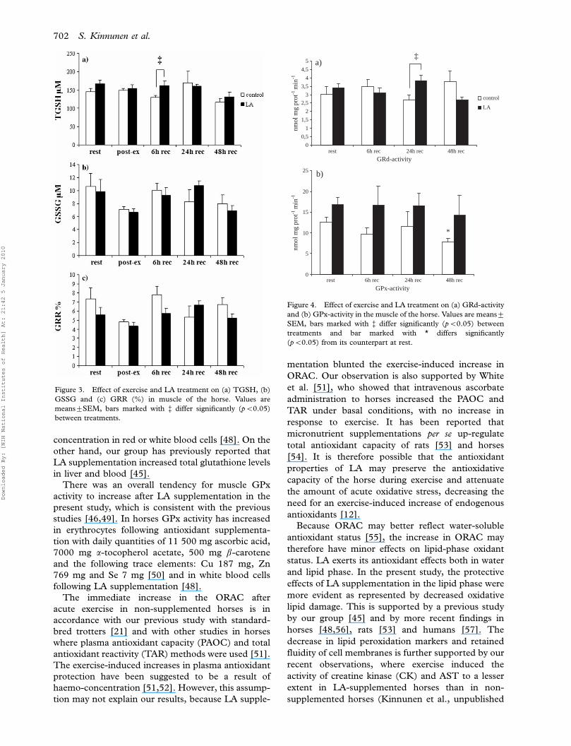
(Table II). Muscle GRd activity was significantly
higher in LA-supplemented horses than in non-
supplemented animals after 24-h recovery (Figure
4a). There was also a main increasing effect of LA
supplementation for muscle GPx activity (pB0.01,
Figure 4b).
In non-supplemented horses, intense acute exercise
increased the plasma ORAC (pB0.01, Table III)
immediately after the exercise. ORAC also remained
higher after 2 h of recovery (pB0.05). LA supple-
mentation attenuated the overall exercise-induced
ORAC response compared with controls (pB0.05).
LA supplementation had no statistically significant
effect on the muscle protein carbonyl levels
(Table III). Five-week LA administration decreased
the exercise-induced LPO in plasma compared with
control (pB0.05, Table III). Moreover, LA supple-
mentation had a main blunting effect for exercise-
induced plasma and muscle MDA concentrations
(pB0.05 in both, Table III). The overall trend in
plasma and muscle MDA levels was consistent with
those of plasma and muscle LPO.
Discussion
In the present study, we observed that 5-week LA
supplementation decreased free radical formation as
measured by EPR spectroscopy in response to
strenuous aerobic exercise in horse skeletal muscle.
This finding supports the earlier reports that both
LA and its reduced form DHLA directly scavenge
ROS [15]. In addition, 5-week LA supplementation
A
B
10G
0
200
400
600
800
1000
1200
1400
control
LA
*
EPR
sig
nal i
nten
stiy
rest post-ex
(b)(a)
Figure 1. Effect of exercise and LA treatment on the amount of free radicals in the muscle of the horse. (a) The values are means9SEM,
bar marked with * differs significantly (pB0.05) from its counterpart at rest. (b) EPR spectra taken ex vivo at 77 K showing steady-state
native free radical concentration of LA supplemented horses muscle tissue taken before (A) and after (B) exercise. Arrows on the spectrum
indicate the signal of Mn/MnO internal standard.
Figure 2. Effect of exercise and LA treatment on (a) TRx-activity
and (b) TRxRd-activity in the muscle of the horse. Values are
means9SEM, bars marked with % differ significantly (pB0.05)
between treatments.
700 S. Kinnunen et al.
Downloaded By: [NIH National Institutes of Health] At: 21:42 5 January 2010

up-regulated muscle TGSH levels and increased the
activities of TRxRd and GRd in muscle during
recovery, but had no effect on muscle TRx activity.
TRx and TRxRd play an essential role in cell
function and protection by limiting oxidative stress
directly via their antioxidant effects and indirectly
by protein�protein interactions with key signalling
molecules [27]. The protective role of TRx system
against oxidative insults is further supported in the
present study by the negative correlations between
TRxRd activity at rest and post-exercise free radical
formation. In addition, we observed positive correla-
tions between muscle protein carbonyl levels and
TRx and TRxRd activities after 24-h recovery in
LA-supplemented horses. Induction of antioxidant
defences to acute stress can be interpreted as a
protective response and the extent of this induction
may reflect the levels of oxidative stress [9]. Other
studies have reported decreases in the levels of
protein carbonyls after LA supplementation in
response to exercise training, but these studies did
not examine acute responses to exercise-induced
oxidative stress [28,29].
We found no significant changes in GSSG or the
GSSG-TGSH ratio in response to exercise or during
recovery. The glutathione redox ratio is considered
to be an indicator of tissue redox status [30] and
oxidative stress [7]. The changes in redox ratios
occurring with certain types of exercise have not
always been consistent among studies, even when
using similar protocols [31�34]. This holds true with
horses too [21,35�43]. Changes in glutathione status
are short-lived, likely due to rapid conversion of
GSSG back to GSH by GRd, which is an important
determinant of GRR and cellular protection against
oxidative stress [31,44].
LA supplementation increased GRd activity after
24-h recovery, suggesting increased protection
against exercise-induced oxidative damage. It has
been reported previously that 8-week LA supplemen-
tation combined with treadmill training and exhaus-
tive exercise had no effect on muscle GRd activity
immediately after one bout of acute exercise until
exhaustion [45]. On the other hand, supplementation
of LA together with carnitine increased the activity
of GRd in skeletal muscle in the resting state in aged
rats [46].
In the present study, the increase in TGSH
concentration in muscle during recovery can be
attributed to the enhanced regeneration of GSH
through increased GRd-activity and to the GSH-
sparing effect of LA [45,47]. In LA-supplemented
horses higher TRx activity at rest was associated with
lower post-exercise GRR%, supporting the hypoth-
esis of TRx protection against oxidative stress. It has
been reported earlier that LA had no effect on GSH
Table II. Significant correlations between different biochemical variables.
Control/LA r P
Resting TRxRd vs Post-ex EPR LA �0.900 B0.05
Post-ex TGSH vs Post-ex EPR Control 0.900 B0.05
Post-ex GSSG vs Post-ex EPR LA 1.000 B0.001
TRx 48-h rec vs TGSH 48-h rec Control 0.900 B0.05
TRx 24-h rec vs TGSH 24-h rec LA 0.900 B0.05
TRx 48-h rec vs TGSH 48-h rec Control 0.980 B0.01
TRx 48-h rec vs TGSH 48-h rec LA 0.980 B0.01
TRx 48-h rec vs AST 48-h rec LA �0.975 B0.01
Muscle PCarb 48-h rec vs TRxRd 48-h rec Control 0.900 B0.05
Muscle PCarb 24-h rec vs TRxRd 24-h rec LA 0.900 B0.05
Muscle PCarb 24-h rec vs TRx 24-h rec LA 0.900 B0.05
Plasma PCarb 24-h rec vs TRxRd 24-h rec LA 0.900 B0.05
Plasma PCarb 6-h rec vs Resting TRx Control �0.900 B0.05
Plasma PCarb 24-h rec vs Resting TRx Control �0.900 B0.05
Plasma PCarb 24-h rec vs TRx 24-h rec LA 0.900 B0.05
Plasma PCarb 48-h rec vs TRx 48-h rec LA �0.975 B0.01
Resting GSSG vs Resting TRx Control 0.900 B0.05
Resting GRR vs Resting TRx Control 0.900 B0.05
TGSH 24-h rec vs Plasma ORAC 24-h rec LA 0.900 B0.05
Post-ex EPR vs Plasma LPO 24-h rec Control 0.900 B0.05
Resting TRxRd vs Muscle LPO 24-h rec LA �0.900 B0.05
Resting TRxRd vs Muscle MDA 24-h rec LA �0.900 B0.05
CK 6-h rec vs Muscle LPO 6-h rec Control 0.829 B0.05
CK 24-h rec vs Muscle LPO 24-h rec Control 0.829 B0.05
AST 24-h re vs Plasma MDA 24-h rec Control �1.000 B0.001
AST 6-h rec vs Muscle LPO 6-h rec LA �0.900 B0.05
Abbreviations: muscle thioredoxin (TRx) and thioredoxin reductase (TRxRd), electron paramagnetic resonance (EPR), concentrations of
muscle total glutathione (TGSH) and oxidized glutathione (GSSG), glutathione redox ratio (GRR), plasma aspartate aminotransferase
(AST) and creatine kinase (CK), protein carbonyl (PCarb) concentrations in plasma and muscle, oxygen radical absorbance capacity
(ORAC) of plasma and lipid hydroperoxides (LPO) and malondialdehyde (MDA) in plasma and muscle.
LA attenuates exercise-induced oxidative stress 701
Downloaded By: [NIH National Institutes of Health] At: 21:42 5 January 2010
concentration in red or white blood cells [48]. On the
other hand, our group has previously reported that
LA supplementation increased total glutathione levels
in liver and blood [45].
There was an overall tendency for muscle GPx
activity to increase after LA supplementation in the
present study, which is consistent with the previous
studies [46,49]. In horses GPx activity has increased
in erythrocytes following antioxidant supplementa-
tion with daily quantities of 11 500 mg ascorbic acid,
7000 mg a-tocopherol acetate, 500 mg b-carotene
and the following trace elements: Cu 187 mg, Zn
769 mg and Se 7 mg [50] and in white blood cells
following LA supplementation [48].
The immediate increase in the ORAC after
acute exercise in non-supplemented horses is in
accordance with our previous study with standard-
bred trotters [21] and with other studies in horses
where plasma antioxidant capacity (PAOC) and total
antioxidant reactivity (TAR) methods were used [51].
The exercise-induced increases in plasma antioxidant
protection have been suggested to be a result of
haemo-concentration [51,52]. However, this assump-
tion may not explain our results, because LA supple-
mentation blunted the exercise-induced increase in
ORAC. Our observation is also supported by White
et al. [51], who showed that intravenous ascorbate
administration to horses increased the PAOC and
TAR under basal conditions, with no increase in
response to exercise. It has been reported that
micronutrient supplementations per se up-regulate
total antioxidant capacity of rats [53] and horses
[54]. It is therefore possible that the antioxidant
properties of LA may preserve the antioxidative
capacity of the horse during exercise and attenuate
the amount of acute oxidative stress, decreasing the
need for an exercise-induced increase of endogenous
antioxidants [12].
Because ORAC may better reflect water-soluble
antioxidant status [55], the increase in ORAC may
therefore have minor effects on lipid-phase oxidant
status. LA exerts its antioxidant effects both in water
and lipid phase. In the present study, the protective
effects of LA supplementation in the lipid phase were
more evident as represented by decreased oxidative
lipid damage. This is supported by a previous study
by our group [45] and by more recent findings in
horses [48,56], rats [53] and humans [57]. The
decrease in lipid peroxidation markers and retained
fluidity of cell membranes is further supported by our
recent observations, where exercise induced the
activity of creatine kinase (CK) and AST to a lesser
extent in LA-supplemented horses than in non-
supplemented horses (Kinnunen et al., unpublished
0
0,5
1
1,5
2
2,5
3
3,5
4
4,5
5
control
LA
nmol
mg
prot
-1 m
in-1
‡a)
0
5
10
15
20
25
nmol
mg
prot
-1 m
in-1
*
rest 6h rec 24h rec 48h rec
rest 6h rec 24h rec 48h rec
b)
GRd-activity
GPx-activity
Figure 4. Effect of exercise and LA treatment on (a) GRd-activity
and (b) GPx-activity in the muscle of the horse. Values are means9
SEM, bars marked with % differ significantly (pB0.05) between
treatments and bar marked with * differs significantly
(pB0.05) from its counterpart at rest.
Figure 3. Effect of exercise and LA treatment on (a) TGSH, (b)
GSSG and (c) GRR (%) in muscle of the horse. Values are
means9SEM, bars marked with % differ significantly (pB0.05)
between treatments.
702 S. Kinnunen et al.
Downloaded By: [NIH National Institutes of Health] At: 21:42 5 January 2010

observations). Concentrations of muscle LPO and
plasma MDA were strongly associated with the
plasma activities of CK and AST, suggesting a strong
association between lipid peroxidation and muscle
damage. These findings are supported by the earlier
studies in young and older men [58].
In summary, we show that LA supplementation
controlled strenuous aerobic exercise-induced oxida-
tive insult in skeletal muscle, improved glutathione
redox status and enhanced thioredoxin reducing
capacity. These results give new insight on the role
of thiol antioxidants to decrease the risks of strenuous
physical exercise. The expression of antioxidant
enzymes and up-regulation of other defence mechan-
isms appears to be induced by ROS generated during
exercise, suggesting that the generation of ROS is an
essential signal in the adaptation to exercise [2,8,59]
and the adaptive responses of skeletal muscle to
unaccustomed contractions [2,12]. It has been
suggested nearly two decades ago that for maximal
performance an optimal level of ROS is needed [60].
Therefore, the over-supplementation of nutritional
antioxidants may decrease the maximal performance
by attenuating essential ROS production.
Our results suggest that LA-supplementation may
reduce the indices of exercise-induced oxidative
stress directly by augmenting intracellular protective
mechanisms against oxidative insult or indirectly
by decreasing ROS production during exercise.
Nonetheless, one should still bear in mind that,
although supplementation with particular micronu-
trients decreases exercise-induced oxidative stress,
there may also be a risk of attenuating the normal
physiological response of tissues to exercise and
blunting the training-induced adaptations.
Acknowledgements
The skillful technical assistance of Ms Taina
Vihavainen, Ms Taija Hukkanen and Ms Satu Mart-
tila is gratefully acknowledged. The authors would
like to thank David Alan Carlson, PhD, for valuable
suggestions, DVM Kristiina Palttala, Ms Marjatta
Lehtisaari and MSc Marja-Leena Hannila for their
valuable help and support during the study; and the
staff and the students of Yla-Savo Vocational School
for the facilities.
The study has been supported by the grants from
Erkki Rajakoski Foundation, Helsinki to S.K., the
Finnish Ministry of Education and COST actions
B35 and BM0602.
Declaration of interest: The authors report no
conflicts of interest. The authors alone are respon-
sible for the content and writing of the paper.
References
[1] Vollaard NB, Shearman JP, Cooper CE. Exercise-induced
oxidative stress:myths, realities and physiological relevance.
Sports Med 2005;35:1045�1062.
[2] Jackson MJ. Free radicals generated by contracting muscle:
by-products of metabolism or key regulators of muscle
function? Free Radic Biol Med 2008;44:132�141.
[3] McArdle A, van der Meulen JH, Catapano M, Symons MCR,
Faulkner JA, Jackson MJ. Free radical activity following
contraction-induced injury to the extensor digitorum longus
muscles of rats. Free Radic Biol Med 1999;26:1085�1091.
Table III. Effects of a-lipoic acid (LA) on oxygen radical absorbing capacity (ORAC) and the concentrations of lipid hydroperoxides
(LPO), malondialdehyde (MDA) and protein carbonyls (PCarb) in plasma and in middle gluteal muscle of the horse at rest, immediately
after intense aerobic exercise and during recovery.
Recovery
At rest Post-exercise 2 h 6 h 24 h 48 h
Plasma
ORAC (mmol/L) con 35.792.03 43.291.40* 43.591.23* 34.391.43 33.092.03 34.491.69
LA 33.493.28 35.391.68% 34.793.11% 33.895.45 33.591.56 32.892.37
LPO (mM) con 7.091.26 12.992.63* 8.091.46 7.191.98 5.792.09 3.691.43
LA 5.990.95 8.791.82% 5.890.95% 3.391.31 5.690.88 2.790.61
MDA (mM) con 2.190.09 3.190.67* 2.790.57 2.490.28 2.490.69 1.890.22
LA 2.190.23 2.690.15 2.290.20 2.090.15 1.990.14 1.790.16
PCarb (nmol/mg protein) con 0.9990.050 1.0290.035 0.9890.063 1.0090.026 0.9690.035 1.0190.061
LA 1.0590.880 1.0390.056 1.0090.065 1.0490.042 1.0090.045 1.0390.030
Muscle
LPO (mM) con 134.9915.37 N.A. N.A. 108.396.43* 103.193.45* 99.197.02
LA 110.293.00% N.A. N.A. 105.396.87 108.693.72 107.294.16
MDA (mM) con 3.790.54 N.A. N.A. 3.390.64 2.890.14 2.590.21*
LA 2.790.30% N.A. N.A. 2.990.38 2.390.17 2.290.11
PCarb (Arbitrary units) con 1.090.08 N.A. N.A. 1.490.19* 1.190.09 0.99 0.06
LA 1.290.07 N.A. N.A. 1.290.03 1.090.13 0.990.09
Values are means9SEM, level of significance was set at pB0.05. Values marked with * differ significantly from the resting counterpart and
on LA-treatment row the values marked with % differ significantly from the non-treated horses at the same time point. N.A. (not available).
LA attenuates exercise-induced oxidative stress 703
Downloaded By: [NIH National Institutes of Health] At: 21:42 5 January 2010

[4] Stadler K, Jenei V, Bolcshazy G, Somogyi A, Jakus J.
Increased nitric oxide levels as an early sign of premature
aging in diabetes. Free Radic Biol Med 2003;35:1240�1251.
[5] Sun Y, Guo H, Yu H, Wang X, Wu J, Xue Y. Bioaccumulation
and physiological effects of tetrabromobisphenol A in
coontail Ceratophyllum demersum L. Chemosphere 2008;70:
1787�1795.
[6] Jones DP. Radical-free biology of oxidative stress. Am J
Physiol Cell Physiol 2008;295:C849�C868.
[7] Jones D. Redefining oxidative stress. Antioxid Redox Signal
2006;8:1865�1879.
[8] Niess A, Simon P. Response and adaptation of skeletal muscle
to exercise*the role of reactive oxygen species. Front Biosci
2007;12:4826�4838.
[9] Sen CK, Packer L. Thiol homeostasis and supplements in
physical exercise. Am J Clin Nutr 2000;72:653S�669S.
[10] Bera T, Lakshman K, Ghanteswari D, Pal S, Sudhahar D,
Islam MN, Bhuyan NR, Das P. Characterization of the redox
components of transplasma memebrane electron transport
system from Leishmania donovani promastigotes. Biochim
Biophys Acta 2005;1725:314�326.
[11] Sheline CT, Wei L. Free radical-mediated neurotoxicity may
be caused by inhibition of mitochondrial dehydrogenases
in vitro and in vivo. Neuroscience 2006;140:235�246.
[12] Atalay M, Lappalainen J, Sen CK. Dietary antioxidants for
the athlete. Curr Sports Med Rep 2006;5:182�186.
[13] Atmaca G. Antioxidant effects of sulfur-containing amino
acids. Yonsei Med J 2004;45:776�788.
[14] Cakatay U. Pro-oxidant actions of alpha-lipoic acid and
dihydrolipoic acid. Med Hypotheses 2006;66:110�117.
[15] Petersen Shay K, Moreau RF, Smith EJ, Hagen TM. Is alpha-
lipoic acid a scavenger of reactive oxygen species in vivo?
Evidence for its initiation of stress signaling pathways that
promote endogenous antioxidant capacity. IUBMB Life
2008;60:362�367.
[16] Meyer H. Pferdefutterung. Berlin: Blackwell Wissenschafts-
Verlag; 1996.
[17] Henneke DR, Potter GD, Kreider JL, Yeates BF. Relationship
between condition score, physical measurement and body fat
percentage in mares. Equine Vet J 1983;15:371�372.
[18] Persson SGB. Evaluation of exercise tolerance and fitness in
the performance horse. Cambridge: Burlington Press; 1983. p
441�457.
[19] Sen CK, Roy S, Khanna S, Packer L. Determination of
oxidized and reduced lipoic acid using high-performance
liquid chromatography and coulometric detection. Methods
Enzymol 1999;299:239�246.
[20] Lindholm A, Piehl K. Fibre composition, enzyme activity and
concentrations of metabolites and electrolytes in muscles of
standardbred horses. Acta Vet Scand 1974;15:287�309.
[21] Kinnunen S, Hyyppa S, Lehmuskero A, Oksala N, Maenpaa
P, Hanninen O, Atalay M. Oxygen radical absorbance
capacity (ORAC) and exercise-induced oxidative stress in
trotters. Eur J Appl Physiol 2005;95:550�556.
[22] Arab K, Steghens JP. Plasma lipid hydroperoxides measure-
ment by an automated xylenol orange method. Anal Biochem
2004;325:158�163.
[23] Gerard-Monnier D, Erdelmeier I, Regnard K, Moze-Henry
N, Yadan JC, Chaudiere J. Reactions of 1-methyl-2-
phenylindole with malondialdehyde and 4-hydroxyalkenals.
Analytical applications to a colorimetric assay of lipid
peroxidation. Chem Res Toxicol 1998;11:1176�1183.
[24] Sen CK, Marin E, Kretzschmar M, Hanninen O. Skeletal
muscle and liver glutathione homeostasis in response to
training, exercise, and immobilization. J Appl Physiol
1992;73:1265�1272.
[25] Atalay M, Oksala NK, Laaksonen DE, Khanna S, Nakao C,
Lappalainen J, Roy S, Hanninen O, Sen CK. Exercise training
modulates heat shock prtein response in diabetic rats. J Appl
Physiol 2004;97:605�611.
[26] Oksala NK, Paimela H, Alhava E, Atalay M. Heat shock
preconditioning induces protein carbonylation and alters
antioxidant protection in superficially injured guinea pig
gastric mucosa in vitro. Dig Dis Sci 2007;52:1897�1905.
[27] Lillig CH, Holmgren A. Thioredoxin and related
molecules*from biology to health and disease. Antioxid
Redox Signal 2007;9:25�47.
[28] Saengsirisuwan V, Kinnick TR, Schmit MB, Henriksen EJ.
Interactions of exercise and lipoic acid on skeletal muscle
glucose transport in obese Zucker rats. J Appl Physiol
2001;91:145�153.
[29] Saengsirisuwan V, Perez FR, Kinnick TR, Henriksen EJ.
Effects of exercise training and antioxidant R-ALA on glucose
transport in insulin-sensitive rat skeletal muscle. J Appl
Physiol 2002;92:50�58.
[30] Schafer FQ, Buettner GR. Redox environment of the cell as
viewed through the redox state of the glutathione disulfide/
glutathione couple. Free Radic Biol Med 2001;30:
1191�1212.
[31] Goldfarb AH, Bloomer RJ, McKenzie MJ. Combined
antioxidant treatment effects on blood oxidative stress after
eccentric exercise. Med Sci Sports Exerc 2005;37:234�239.
[32] Goldfarb AH, Garten RS, Chee PD, Cho C, Reeves GV,
Hollander DB, Thomas C, Aboudehen KS, Francois M,
Kraemer RR. Resistance exercise effects on blood glutathione
status and plasma protein carbonyls: influence of partial
vascular occlusion. Eur J Appl Physiol 2008;104:813�819.
[33] Goldfarb AH, Patrick SW, Bryer S, You T. Vitamin C
supplementation affects oxidative-stress markers in response
to a 30-minute run at 75% VO2max. Int J Sport Nutr Exerc
Metab 2005;15:279�290.
[34] Bloomer RJ, Goldfarb AH, Wideman L, McKenzie MJ,
Consitt LA. Effects of acute aerobic and aerobic exercise on
blood markers of oxidative stress. J Strength Cond Res
2005;19:276�285.
[35] Balogh N, Gaal T, Sz. Ribiczeyne P, Petri A. Biochemical
and antioxidant changes in plasma and erythrocytes of
pentathlon horses before and after exercise. Vet Clin Pathol
2001;30:214�218.
[36] Hargreaves BJ, Kronfeld DS, Waldron JN, Lopes MA, Gay
LS, Saker KE, Cooper WL, Sklan DJ, Harris PA. Antioxidant
status and muscle cell leakage during endurance exercise.
Equine Vet J Suppl 2002;34:116�121.
[37] Hargreaves BJ, Kronfeld DS, Waldron JN, Lopes MA, Gay
LS, Saker KE, Cooper WL, Sklan DJ, Harris PA. Antioxidant
status of horses during two 80-km endurance races. J Nutr
2002;132:1781S�1783S.
[38] Marlin DJ, Fenn K, Smith N, Deaton CD, Roberts CA,
Harris PA, Dunster C, Kelly FJ. Changes in circulatory
antioxidant status in horses during prolonged exercise. J Nutr
2002;132:1622S�1627S.
[39] de Moffarts B, Kirschvink N, Art T, Pincemail J, Michaux C,
Cayeux K, Defraigne JO, Lekeux P. Impact of training and
exercise intensity on blood antioxidant markers in healthy
standardbred horses. Equine Comp Exerc Physiol 2004;1:
1�11.
[40] de Moffarts B, Kirschvink N, Art T, Pincemail J, Lekeux P.
Assessment of the oxidant-antioxidant blood balance in a field
exercise test in standardbred and eventing horses. Equine
Compar Exer Physiol 2005;2:253�261.
[41] Williams CA, Carlucci S. Oral vitamin E supplementation
and oxidative stress, vitamin and antioxidant status in
intensely exercising horses. Equine Vet J Suppl 2006;
36:617�621.
[42] de Moffarts B, Kirschvink N, Art T, Pincemail J, Lekeux P.
Effects of exercise on blood oxidant/antioxidant markers in
704 S. Kinnunen et al.
Downloaded By: [NIH National Institutes of Health] At: 21:42 5 January 2010

standardbred horses: comparison between treadmill and race
track tests. Equine Vet J Suppl 2006;36:254�257.
[43] Maranon G, Munoz-Escassi B, Manley W, Garcıa C, Cayado
P, Sanchez de la Muela M, Olabarri B, Leon R, Vara E.
The effect of methyl sulphonyl methane supplementation on
biomarkers of oxidative stress in sport horses following
jumping exercise. Acta Vet Scand 2008;50:45.
[44] Hayes JD, McLellan LI. Glutathione and glutathione-
dependent enzymes represent a co-ordinately regulated
defence against oxidative stress. Free Radic Res 1999;
31:273�300.
[45] Khanna S, Atalay M, Laaksonen DE, Gul M, Roy S, Sen CK.
Alpha-lipoic acid supplementation: tissue glutathione
homeostasis at rest and after exercise. J Appl Physiol
1999;86:1191�1196.
[46] Kumaran S, Savitha S, Anusuya Devi M, Panneerselvam C.
L-Carnitine and DL-alpha-lipoic acid reverse the age-related
deficit in glutathione redox state in skeletal muscle and heart
tissues. Mech Ageing Dev 2004;125:507�512.
[47] Packer L, Witt EH, Tritschler HJ. Alpha-lipoic acid as a
biological antioxidant. Free Radic Biol Med 1995;19:
227�250.
[48] Williams CA, Hoffman RM, Kronfeld DS, Hess TM,
Saker KE, Harris PA. Lipoic acid as an antioxidant in
mature thoroughbred geldings: A preliminary study. J Nutr
2002;132:1628S�1631S.
[49] Chae CH, Shin CH, Kim HT. The combination of alpha-
lipoic acid supplementation and aerobic exercise inhibits
peroxidation in rat skeletal muscles. Nutr Res 2008;28:
399�405.
[50] de Moffarts B, Kirschvink N, Art T, Pincemail J, Lekeux P.
Effect of oral antioxidant supplementation on blood
antioxidant status in trained thoroughbred horses. Vet J
2005;169:65�74.
[51] White A, Estrada M, Walker K, Wisnia P, Filgueira G, Valdes
F, Araneda O, Behn C, Martinez R. Role of exercise and
ascorbate on plasma antioxidant capacity in thoroughbred
race horses. Comp Biochem Physiol A Mol Integr Physiol
2001;128:99�104.
[52] Surmen-Gur E, Ozturk E, Gur H, Punduk Z, Tuncel P. Effect
of vitamin E supplementation on post-exercise plasma lipid
peroxidation and blood antioxidant status in smokers: with
special reference to haemoconcentration effect. Eur J Appl
Physiol Occup Physiol 1999;79:472�478.
[53] Skibska B, Jozefowicz-Okonkwo G, Goraca A. Protective
effects of early administration of alpha-lipoic acid against
lipopolysaccharide-induced plasma lipid peroxidation.
Pharmacol Rep 2006;58:399�404.
[54] Avellini L, Chiaradia E, Gaiti A. Effect of exercise training,
selenium and vitamin E on some free radical scavengers in
horses (Equus caballus). Comp Biochem Physiol B Biochem
Mol Biol 1999;123:147�154.
[55] Cao G, Alessio HM, Cutler RG. Oxygen-radical absorbance
capacity assay for antioxidants. Free Radic Biol Med
1993;14:303�311.
[56] Williams CA, Kronfeldt DS, Hess TM, Saker KE, Waldron
JN, Crandell KM, Hoffman RM, Harris PA. Antioxidant
supplementation and subsequent oxidative stress of horses
during an 80-km endurance race. J Anim Sci 2004;82:
588�594.
[57] Zembron-Lacny A, Szyszka K, Szygula Z. Effect of cysteine
derivatives administration in healthy men exposed to intense
resistance exercise by evaluation of pro-antioxidant ratio.
J Physiol Sci 2007;57:343�348.
[58] Sacheck JM, Milbury PE, Cannon JG, Roubenoff R, Blum-
berg JB. Effect of vitamin E and eccentric exercise on selected
biomarkers of oxidative stress in young and elderly men. Free
Radic Biol Med 2003;34:1575�1588.
[59] Gomez-Cabrera M-C, Domenech E, Vina J. Moderate
exercise is an antioxidant: upregulation of antioxidant genes
by training. Free Radic Biol Med 2008;44:126�131.
[60] Reid MB, Shoji T, Moody MR, Entman ML. Reactive oxygen
in skeletal muscle. II. Extracellular release of free radicals.
J Appl Physiol 1992;75:1805�1809.
This paper was first published online on iFirst on 20 June
2009.
LA attenuates exercise-induced oxidative stress 705
Downloaded By: [NIH National Institutes of Health] At: 21:42 5 January 2010
Copyright © 2022 FDOKUMEN