Modulation of cell cycle control during oocyte-to-embryo ...
Global Veterinaria 12 (1): 80-90, 2014ISSN 1992-6197© IDOSI Publications, 2014DOI: 10.5829/idosi.gv.2014.12.01.76185
Corresponding Author: Suzanah Abdul Rahman, Department of Biomedical Science, Faculty of Allied Health Sciences,International Islamic University Malaysia (IIUM), Jalan Sultan Ahmad Shah, Bandar Indera Mahkota,25200 Kuantan, Pahang Malaysia. Tel: +609-5705203/5204, Fax: +609-5716776.
80
Effects of Thymoquinone Supplementation on Cyclophosphamide Toxicity of Mouse Embryo In vitro
Saheera Kamarzaman, Azantee Yazmie Abdul Wahab and Suzanah Abdul Rahman1 2 1
Department of Biomedical Science, Faculty of Allied Health Sciences,1
International Islamic University Malaysia (IIUM), MalaysiaFertility Centre, Faculty of Medicine, 1
International Islamic University Malaysia (IIUM), Malaysia
Abstract: Thymoquinone is the major active component derived from the traditional medicinal plant Nigellasativa, which has been shown to exhibit antioxidant property through different mechanisms in animal models.This study evaluates the prophylactic effect of thymoquinone supplementation on culture medium to amelioratecyclophosphamide-induced alterations in cellular differentiation and proliferation during embryo developmentin vitro. Male and female mice were exposed to cyclophosphamide via a single intraperitoneal (i.p.) injectionat 200 mg/kg. Sperms and oocytes were collected at day 33 and day 10 respectively, for insemination andfertilization in medium supplemented with thymoquinone (1µM, 10µM and 100µM). The stages of fertilization,embryo division, morphological effects and fragmentation were examined and compared between groups, 24hours post-fertilization. Thymoquinone supplementation improved fertilization rates, significantly reduced thepercentage of defects blastomeres of Type C (p<0.001) and significantly decreased the percentage of embryofragmentation Grade IV (>50%, p<0.05) following paternal and maternal exposures to cyclophosphamide. Thegood quality embryos of Type A and Grade I fragmentation were not observed in the group withoutthymoquinone supplementation. The findings of this study showed that thymoquinone is a suitable exogenousantioxidant for preserving fair-quality embryos which can result into full term pregnancy.
Keywords: Cyclophosphamide Thymoquinone In-vitro Fertilization Blastomeres Embryo Fragmentation
INTRODUCTION factors can lead to the generation of oxidative stress, from
Reactive oxygen species (ROS) are oxygen-derived the spermatozoa, oocytes and embryo) or externalfree radicals that are formed during the intermediate steps sources (e.g. Culture media, oxygen concentration,of oxygen reduction [1]. They can cause direct oxidation visible light, freeze-thawing and ART procedures) in theby combining with other molecules which can lead to in-vitro fertilization (IVF) setup [7]. Pathological effectsstructural and functional changes and result in cellular of increased level of oxidative stress in the IVF settingdamage [2,3]. In the event of excessive ROS production are associated with the low cleavage rate, low blastocystexceeding the antioxidant defense mechanism of the cells, rate, high embryonic fragmentation, low fertilizationit results in oxidative stress accompanied by other rates, impaired embryo development and higher rates ofadverse effects [4]. pregnancy loss [8].
Oxidative stress has been reported as one of the Cyclophosphamide (CPA), a commonly usedleading factors negatively affecting assisted reproductive anticancer drug, is one of the most damaging alkylatingtechniques (ART) outcome due to the lack of embryo agents that affects the DNA of replicating cells andprotection by oxygen radical scavengers [5,6]. Various rapidly multiplying cells especially in the gonads and
either internal sources (e.g. Endogenous production of

Global Veterinaria, 12 (1): 80-90, 2014
81
Fig. 1: Cleaving embryo scoring according to Baczkowski et al., [24].
pituitary which results in miscoding, cross-linking and hypotensive effects [26,27]. Thymoquinone is the activeDNA breakage by the transfer of alkyl groups to the compound of the essential oil with anti-oxidative effectguanine compound of the DNA [9]. Cyclophosphamide that works as a scavenger of various radical oxygentreatment causes significant systemic toxicity due to the species including superoxide radical anion and hydroxyloverproduction of reactive oxygen species (ROS) that radicals through different mechanisms [28-30].cause oxidative stress [10-12]. In male rodents, several The present study, therefore, was undertaken tostudies have reported that administration of evaluate the prophylactic effect of thymoquinonecyclophosphamide once a week for 5 weeks cause supplementation on culture medium to ameliorateoligospermia, azoospermia, testicular damage and germ cyclophosphamide-induced alterations in fertilization rate,cell toxicity [13-15]. This drug has been shown to cause blastomere structure and grade of fragmentation duringovarian toxicity and induce depletion in the primordial preimplantation embryo development.follicular (PMF) reserve of female mice [16].Cyclophosphamide has also been reported to be MATERIALS AND METHODSteratogenic in rodents. Single intraperitoneal injection of20mg/kg at 10 day of gestation was found to induce cleft Animals: A total of 40 Balb/c mice (20 males andth
palate, spina bifida, exencephaly, skeletal anomalies and 20 females), aged 8 to 15 weeks, weighing aboutsternum defects in mice foetuses [17]. 20-30g, were obtained from the local laboratory
Antioxidants provide a defense mechanism through (Kuala Lumpur, Malaysia) and provided with food pelletsthree levels of protection; (i) prevention, (ii) interception and water ad libitum.and (iii) repair [18]. In vitro antioxidants and scavengerssupplementation are able to minimize the development of Treatment Protocols: Cyclophosphamide, CPAa state of imbalance in oxidants and antioxidants. Various (Sigma-Aldrich) was dissolved in normal saline andanimal studies indicate that supplementation of culture administered intraperitoneally by a single injection ofmedium with antioxidants, vitamin C and E, amino acids, 200 mg/kg. Mice (n=5/group) were assigned randomly intoROS scavengers, disulphide reducing agents and divalent 4 experimental groups: (i) Group 1: Vehicle-treated controlchelators of cations can reduce oxidative stress and be (normal saline) (ii) Group 2: Vehicle-treated positivebeneficial to embryo survival and blastulation rates [19]. control (culture media was supplemented with
Nigella sativa, from the Ranunculacea family, has thymoquinone) (iii) Group 3: Male mice pre-treated withbeen one of the most widely used herbal medicines for the CPA and (iv) Group 4: Female mice pre-treated with CPA.treatment of various diseases. The pharmacological The culture media for in-vitro fertilization in groups 2, 3properties of the oil have been reported to include and 4 was supplemented with 1µM, 10µM and 100µM ofanti-cancer [20], anti-diabetic [21], anti-microbial [22], thymoquinone. Male and female mice were sacrificed onanti-viral [23], anti-bacterial [24], anti-clastogenic [25] and day 33 and day 10 post-CPA exposure,respectively.
Global Veterinaria, 12 (1): 80-90, 2014
82
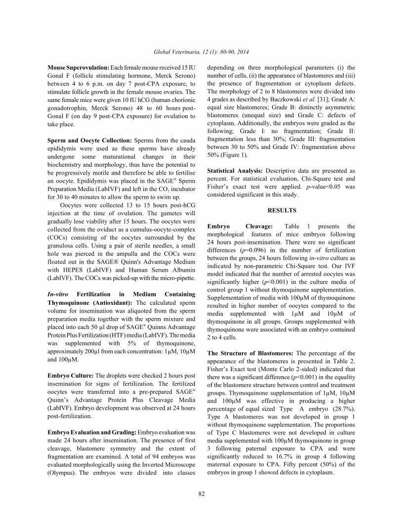
Mouse Superovulation: Each female mouse received 15 IU depending on three morphological parameters (i) theGonal F (follicle stimulating hormone, Merck Serono)between 4 to 6 p.m. on day 7 post-CPA exposure, tostimulate follicle growth in the female mouse ovaries. Thesame female mice were given 10 IU hCG (human chorionicgonadotrophin, Merck Serono) 48 to 60 hours post-Gonal F (on day 9 post-CPA exposure) for ovulation totake place.
Sperm and Oocyte Collection: Sperms from the caudaepididymis were used as these sperms have alreadyundergone some maturational changes in theirbiochemistry and morphology, thus have the potential tobe progressively motile and therefore be able to fertilisean oocyte. Epididymis was placed in the SAGE Sperm®
Preparation Media (LabIVF) and left in the CO incubator2
for 30 to 40 minutes to allow the sperm to swim up.Oocytes were collected 13 to 15 hours post-hCG
injection at the time of ovulation. The gametes willgradually lose viability after 15 hours. The oocytes werecollected from the oviduct as a cumulus-oocyte-complex(COCs) consisting of the oocytes surrounded by thegranulosa cells. Using a pair of sterile needles, a smallhole was pierced in the ampulla and the COCs werefloated out in the SAGE® Quinn's Advantage Mediumwith HEPES (LabIVF) and Human Serum Albumin(LabIVF). The COCs was picked-up with the micro-pipette.
In-vitro Fertilization in Medium ContainingThymoquinone (Antioxidant): The calculated spermvolume for insemination was aliquoted from the spermpreparation media together with the sperm mixture andplaced into each 50 µl drop of SAGE Quinns Advantage®
Protein Plus Fertilization (HTF) media (LabIVF). The mediawas supplemented with 5% of thymoquinone,approximately 200µl from each concentration: 1µM, 10µMand 100µM.
Embryo Culture: The droplets were checked 2 hours postinsemination for signs of fertilization. The fertilizedoocytes were transferred into a pre-prepared SAGE®
Quinn’s Advantage Protein Plus Cleavage Media(LabIVF). Embryo development was observed at 24 hourspost-fertilization.
Embryo Evaluation and Grading: Embryo evaluation wasmade 24 hours after insemination. The presence of firstcleavage, blastomere symmetry and the extent offragmentation are examined. A total of 94 embryos wasevaluated morphologically using the Inverted Microscope(Olympus). The embryos were divided into classes
number of cells, (ii) the appearance of blastomeres and (iii)the presence of fragmentation or cytoplasm defects.The morphology of 2 to 8 blastomeres were divided into4 grades as described by Baczkowski et al. [31]; Grade A:equal size blastomeres; Grade B: distinctly asymmetricblastomeres (unequal size) and Grade C: defects ofcytoplasm. Additionally, the embryos were graded as thefollowing; Grade I: no fragmentation; Grade II:fragmentation less than 30%; Grade III: fragmentationbetween 30 to 50% and Grade IV: fragmentation above50% (Figure 1).
Statistical Analysis: Descriptive data are presented aspercent. For statistical evaluation, Chi-Square test andFisher’s exact test were applied. p-value<0.05 wasconsidered significant in this study.
RESULTS
Embryo Cleavage: Table 1 presents themorphological features of mice embryos following24 hours post-insemination. There were no significantdifferences (p=0.096) in the number of fertilizationbetween the groups, 24 hours following in-vitro culture asindicated by non-parametric Chi-Square test. Our IVFmodel indicated that the number of arrested oocytes wassignificantly higher (p<0.001) in the culture media ofcontrol group 1 without thymoquinone supplementation.Supplementation of media with 100µM of thymoquinoneresulted in higher number of oocytes compared to themedia supplemented with 1µM and 10µM ofthymoquinone in all groups. Groups supplemented withthymoquinone were associated with an embryo contained2 to 4 cells.
The Structure of Blastomeres: The percentage of theappearance of the blastomeres is presented in Table 2.Fisher’s Exact test (Monte Carlo 2-sided) indicated thatthere was a significant difference (p<0.001) in the equalityof the blastomere structure between control and treatmentgroups. Thymoquinone supplementation of 1µM, 10µMand 100µM was effective in producing a higherpercentage of equal sized Type A embryo (28.7%).Type A blastomeres was not developed in group 1without thymoquinone supplementation. The proportionsof Type C blastomeres were not developed in culturemedia supplemented with 100µM thymoquinone in group3 following paternal exposure to CPA and weresignificantly reduced to 16.7% in group 4 followingmaternal exposure to CPA. Fifty percent (50%) of theembryos in group 1 showed defects in cytoplasm.

Global Veterinaria, 12 (1): 80-90, 2014
83
Table 1: The morphological features of mice embryos following 24 hours in-vitro cultureGroup No. of oocytes No. of arrested oocytes No. of fertilized oocytes Cell cleavageGroup 1: Control 27 17 10 >9 cellsGroup 2 : Positive Control:1µM TQ 13 2 11 2 to 4 cells10µM TQ* 0 0 0 0100 µM TQ 17 4 13 2 to 4 cellsGroup 3: CPA-treated male:1µM TQ 8 0 8 2 to 6 cells10µM TQ 11 1 10 2 to 4 cells100µM TQ 14 1 13 2 to 5 cellsGroup 4: CPA-treated female:1µM TQ 10 2 8 2 to 3 cells10µM TQ 5 2 3 2 to 3 cells100µM TQ 20 2 18 2 to 4 cells*Group 2 supplemented with 10uM TQ was excluded from the statistical analysis due to no oocytes.Non-parametric Chi-square test was not significant, p=0.096
Table 2: The percentage of the structure of mice blastomeres following 24 hours in-vitro cultureGroup Type A (equal size) Type B (Unequal size) Type C (defects of cytoplasm) TotalGroup 1: Control 0 50% (5) 50% (5) 100% (10)Group 2 : Positive Control:1µM TQ 81.8% (9) 9.1% (1) 9.1% (1) 100% (11)10µM TQ* 0 0 0 0100 µM TQ 30.8% (4) 61.5% (8) 7.7% (1) 100% (13)Group 3: CPA-treated male:1µM TQ 50% (4) 50% (4) 0 100% (8)10µM TQ 20% (2) 70% (7) 10% (1) 100% (10)100µM TQ 7.7% (1) 92.3% (12) 0 100% (13)Group 4: CPA-treated female:1µM TQ 0 100% (8) 0 100% (8)10µM TQ 100% (3) 0 0 100% (3)100µM TQ 22.2% (4) 61.1% (11) 16.7% (3) 100% (18)Total Count 27 56 11% within treatment groups 28.70% 59.60% 11.70% 100% (94)*Group 2 supplemented with 10uM TQ was excluded from the statistical analysis due to no oocytes.Data is presented in percentage.The number of embryo is presented in parentheses.Fisher’s Exact Test (Monte Carlo Sig 2-sided) was significant at p<0.001.
Table 3: The percentage of mice embryo fragmentation following 24 hours in-vitro cultureGrade I Grade II Grade III Grade IV
Group (No fragmentation) (Fragmentation <30%) (Fragmentation 30% -50%) (Fragmentation >50%) TotalGroup 1: Control 0 0 50% (5) 50% (5) 100% (10)Group 2 : Positive Control:1µM TQ 45.5% (5) 36.4% (4) 9.1% (1) 9.1% (1) 100% (11)10µM TQ* 0 0 0 0 0100 µM TQ 30.8% (4) 38.5% (5) 23.1% (3) 7.7% (1) 100% (13)Group 3: CPA-treated male:1µM TQ 37.5% (3) 37.5% (3) 25% (2) 0 100% (8)10µM TQ 20% (2) 60% (6) 20% (2) 0 100% (10)100µM TQ 30.8% (4) 38.5% (5) 15.4% (2) 15.4% (2) 100% (13)Group 4: CPA-treated female:1µM TQ 0 62.5% (5) 37.5% (3) 0 100% (8)10µM TQ 0 100% (3) 0 0 100% (3)100µM TQ 27.8% (5) 50% (9) 5.6% (1) 16.7% (3) 100% (18)Total Count 23 40 19 12 94% within treatment groups 24.50% 42.60% 20.20% 12.80% 100%*Group 2 supplemented with 10uM TQ was excluded from the statistical analysis due to no oocytes.Data is presented in percentage.The number of embryo is presented in parentheses.Fisher’s Exact Test (Monte Carlo Sig 2-sided) was significant at p<0.05, p=0.017.

Global Veterinaria, 12 (1): 80-90, 2014
84
Fig. 2: Cumulus-oocyte-complex (COCs)
Fig. 3: Embryos of Group 1 (Vehicle-treated control without thymoquinone supplementation) following 24 hours in-vitroculture showing (A) arrested oocytes and (B) Grade IV fragmented embryos (>50%, arrows)
Fig. 4: Embryos of Group 2 (Positive control) supplemented with (A) 1uM of thymoquinone and (B) 100uM ofthymoquinone, at different cleavage stages following 24 hours in-vitro culture.
a-Equal size blastomeres (Type A) with no fragmentation (Grade I).b-Equal size blastomeres (Type A) with <30% fragmentation (Grade II).c-Unequal size blastomeres (Type B) with no fragmentation (Grade I)d-Defects of cytoplasm (Type C) with no fragmentation (Grade I).e-Equal size blastomeres (Type A) with no fragmentation (Grade I).f-Unequal size blastomeres (Type B) with no fragmentation (Grade I).g-Unequal size blastomeres (Type B) with <30% fragmentation (Grade II).

Global Veterinaria, 12 (1): 80-90, 2014
85
Fig. 5: Embryos of Group 3 (CPA-treated male mice) supplemented with 100µM of thymoquinone at different cleavagestages following 24 hours in-vitro culture.a - Equal size blastomeres (Type A) with no fragmentation (Grade I).b - Unequal size of 3-cells blastomeres (Type B) with no fragmentation (Grade I).c - Unequal 2-cell blastomeres (Type B) with no fragmentation (Grade I).d - Unequal blastomeres of more than 3 cells (Type B) with <3-% fragmentation (Grade II).e - Arrested oocyte.
Fig. 6: Embryos of Group 4 (CPA-treated female mice) supplemented with 100uM of thymoquinone, at different cleavagestages following 24 hours in-vitro culture.a-Unequal size blastomeres (Type B) with no fragmentation (Grade I).b-Equal size blastomeres (Type A) with <30% fragmentation (Grade II).c-Unequal size blastomeres (Type A) with <30% fragmentation (Grade II).d-Arrested oocyte.e-Unequal size blastomeres (Type B) with 30%-50% fragmentation (Grade III).
Global Veterinaria, 12 (1): 80-90, 2014
86
Embryo Fragmentation: Table 3 presents the percentage IVF setup. We expected thymoquinone supplementationof embryo fragmentation following 24 hours in-vitro to (i) provide a bed of nutrients for embryos to fertilize inculture. The percentage of embryo fragmentation between the laboratory, (ii) protect in-vitro produced mousegroups are significantly different (p=0.017, p<0.05) as embryos from oxidative stress and (iii) improve conditionsdetermined by Fisher’s Exact test (Monte Carlo 2-sided). for embryonic development by elevating thymoquinoneThe overall proportion of Grade I non-fragmented antioxidant action, against the alkylating effects ofembryos were 24.5% with thymoquinone supplementation cyclophosphamide.and was not developed in group 1 media without In this study, a decreased rate of fertilization, a higherthymoquinone. Most of fragmented embryos in media number of arrested oocytes, a higher percentage ofsupplemented with thymoquinone were found to be of unequally cleaved blastomeres and defects in theintermediate grade, i.e. Grade II (42.6%). Supplementation cytoplasm as well as a higher percentage of Grades III andof 1µM and 10µM thymoquinone showed effectiveness IV embryo fragmentations were observed in the culturein protecting the embryo from the development of grade media without the supplementation of thymoquinone asIV fragmentation in groups pre-treated with compared to effects seen when thymoquinone was added.cyclophosphamide, 0% in groups 3 and 4, compared to Agarwal et al. [5] reported that pathological levels of ROS50% in group 1. Culture media supplemented with 100µM have a negative impact on embryo quality and may lead tothymoquinone showed low percentage of grade IV early embryonic developmental block and retardation.fragmentation with 15.4% and 16.7% following paternal Similar findings were reported by Bedaiwy et al. [33]and maternal exposures to CPA, respectively. which showed that elevated day 1 ROS levels in culture
Morphology of the Embryos: Figure 2 shows the fragmentation and reduced formation of a morphologicallycumulus-oocyte-complex (COCs) collected 13 hours normal blastocyst which ultimately leads to a lowerpost-hCG injection. In Figure 3, representative clinical pregnancy rate. Furthermore, Goto et al. [34]photomicrographs of arrested oocytes and highly found that ROS production was increased in embryosfragmented embryos (Grade IV) of group 1 are shown. cultured under in vitro conditions compared with thoseIn Figures 5, 6 and 7, embryo blastomeres of group 2 cultured in vivo. These results could be due to the(positive control), group 3 (CPA-treated male mice) and increased levels of oxidative stress produced by thegroup 4 (CPA-treated female mice) respectively, embryo or the environment and techniques employedsupplemented with 100µM thymoquinone at different during IVF culture. The embryo can produce ROS viacleavage stages following 24 hours in-vitro culture are several pathways, i.e. oxidative phosphorylation, NAPDHshown. The majority of the embryos of groups 3 and 4 and xantine oxidase systems. The embryo is a fastshowed an unequal size of blastomeres (Type B) with a developing organism with high energy needs that are metlow grade of fragmentation (Grade I and Grade II) as by generation of adenosine triphosphate (ATP) throughopposed to group 1. mitochondrial oxidative phosphorylation and glycolisis.
DISCUSSION and metabolism changes and these will lead to alteration
Identification of embryos that are most likely to result ROS occurs at certain critical points, such as duringin full term pregnancy are crucial to the success of in vitro embryonic genome activity, embryonic compaction andfertilization (IVF). In most cases of IVF procedure, multiple hatching, due to increased in energy demands [36]. Theembryos are generated and cultured. However, selecting nature of fertilization procedure in ART techniques cangood quality embryos for implantation is highly also contribute to ROS generation [6]. In conventionalsubjective and the procedure can end in IVF failure or IVF, the oocytes, cumulus cell mass and spermatozoa canmultiple pregnancies. Non-invasive methods of embryo all generate and contribute to the ROS levels in the media.evaluation are useful in reproductive medicine. They help Centrifugation of spermatozoa during preparationassess embryos without damage by observing the techniques has been shown to increase ROS productionmorphology and dynamics of embryo development [32]. and oxidative stress in male gametes [37]. The media used
This study was designed to determine the in the IVF setting can directly influence the quality ofdevelopmental competency of mouse embryo in the oocyte and embryo [6]. Some commercially availablepresence or absence of antioxidant supplementation in the culture media can contain metallic ions (i.e. Fe and Cu )
media were associated with slow development, high
As the embryo develops from the zygote stage, its needs
to its redox state [35]. Therefore, excessive generation of
2+ 2+

Global Veterinaria, 12 (1): 80-90, 2014
87
that can incorporate into the cells and independently Previous studies of IVF media used for bovine andaccelerate ROS generation within these cells during ARTprocessing [35]. Supplements added to the media mayalso increase the oxidant load. Supplementation of serumcontaining amine oxidase can lead to the increased of ROS(H O ) production. Concurrent with this, the in vitro2 2
formed embryos have a lower antioxidant mechanismwhen compared to in vivo generated embryos as shownby the work of Feugang et al. [38]. Fragmented embryoshave a limited developmental potential and rarely result inimplantation [39]. Thus, all the observations above haveled us to investigate the extent of the antioxidative effectof thymoquinone in a media environment in the protectionof the developing embryo against either intrinsic ROSlevels or that generated in the external environmentthroughout the process of IVF.
In vitro studies on the effect of cyclophosphamideon fertilization and cleavage demonstrated a dose-relatedinhibition [40]. Previous studies have shown that germcells are specifically sensitive to cyclophosphamidetreatment. Our previous study reported that 200mg/kgcyclophosphamide treatment when observed at 32 dayspost-treatment, kills up to 52% of spermatogonia and47.6% of elongated spermatid in male mice [15]. In femalemice, cyclophosphamide administration of 75mg/kgdestroyed more than 50% of primordial follicle(PMF) pool [16]. A recent study on the consequencesof maternal cyclophosphamide treatment (75mg/kg) inpre-implantation mouse embryos reported a reduction inthe oocyte fertilization rate, a reduction in 8-cell stageembryos formation from 48 to 72 hours post-fertilizationand increased aneuploidy compared to controls [41].Similar findings were reported by Pydyn and Ataya [42]that showed reduced oocyte fertilization and earlycleavage rates when female mice were injectedintraperitoneally with 100mg/kg cyclophosphamide.Treatment of oocytes with increasing concentrations (1 to1000µg/ml) of cyclophosphamide for 48 hours resulted indose-response inhibition of the rate of maturation of pigoocytes in vitro [43]. The particular sensitivity of thereproductive tissues to cyclophosphamide is due to thehigh proliferating activity [44]. It is also reported thatexposure to single high dose of cyclophosphamide canproduce DNA fragmentation in mouse testicular cells [15],probably resulted from cross-linking of DNA. It istherefore possible that cyclophosphamide can inducethese defects during embryo culture in vitro and may playa role in the progeny outcome. These observations areindicative of the effects of 200 mg/kg cyclophosphamideused in this study on the reproductive functions andexpected fertilization rates.
mouse embryo culture supplemented with antioxidantssuch as vitamins C and E, taurine, hypotaurine, thiols andB-mercaptoethanol showed great success of embryodevelopment. Our results demonstrated that embryoscultured in medium containing thymoquinone duringfertilization, following paternal and maternal exposure tocyclophosphamide resulted in the improvement offertilization rates, low level of arrested oocytes, as well asa low percentage of cytoplasmic defects andfragmentation. A similar observation was reported whenculture media was supplemented with 100µM vitamin Ethat resulted in significantly more expanded blastocyststhan embryos cultured in control medium [45]. Vitamin Eis a natural antioxidant that inhibits NADPH oxidase-mediated generation of superoxide anion and protects cellmembranes against oxidative stress [46]. Similarly, vitaminC has been reported to protect DNA from exogenousoxidation [47], prevents apoptosis in rat and mousefollicles, improves blastocyst production in mice [48] aswell as improves the developmental competence ofporcine embryos in vitro [49]. It can reduce embryooxidative stress by inducing hypotaurine and taurine inthe oviduct which can neutralize hydroxyl radicals andprevent sperm lipid peroxidation even at lowconcentrations [35]. Exogenous antioxidant such as
-mercaptoethanol ( ME) has been frequently used toincrease antioxidant capacity of embryos via increasingintracellular levels of ROS scavengers such as glutathione(GSH) [50,51]. A recent study by Hosseini et al. [52]reported that ME was found to increase the cleavagerates and improve both hatching rate and quality ofhatched embryos.
These results diverge from those obtained byAlhimaidi [53], who demonstrated that treatment ofthymoquinone at 10 mg/ml into sperms and embryoculture media reduces the sperm motility, fertilization rateand embryo development in vitro. The defects could bedue to the action of thymoquinone on the enzymes of thesperm or on the antigen of the cell membrane of the sperm.
In this study, media supplemented withthymoquinone were observed to allow the developmentof 4-cell embryos 24 hours following culture in vitro. Agood quality embryo should have at least 4 cells on thesecond day and at least 8 cells on the third day of culture[31]. The defense mechanism of thymoquinone againstROS does not only act as a superoxide anion scavengerbut also as a general free radical scavenger [54].Thymoquinone is also known as an effective inhibitor oflipid peroxidation [55].

Global Veterinaria, 12 (1): 80-90, 2014
88
CONCLUSION 8. Agarwal, A., S.S. Allamaneni, K.P. Nallella,
The results of the experiment indicated that the reactive oxygen species levels with the fertilizationinfluence of cyclophosphamide exposure on pre- rate after in vitro fertilization: a qualified meta-implantation embryo development can be ameliorated by analysis. Fertil Steril, 84(1): 228-231.supplementing the culture medium with thymoquinone. 9. Becker, K. and J. Schoneich, 1982. Expression ofThe addition of thymoquinone in the culture media plays genetic damage induced by alkylating agents in germa role in increasing the developmental competency as well cells of female mice. Mutat Res., 92(1-2): 447-464.as in increasing the resistance of mouse embryos cultured 10. Pryor, J.L., C. Hughes, W. Foster, B.F. Hales andin vitro to ROS. From these results, further investigations B. Robaire, 2000. Critical windows of exposure forare needed to determine the appropriate concentrations of children's health: the reproductive system in animalsthe antioxidant, as an excess of antioxidant compounds in and humans. Environ Health Perspect, 108 Supplthe medium may have deleterious effects on the embryo. (3): 491-503.
ACKNOWLEDGEMENT radiotherapy and chemotherapy on female
This study was supported by a grant from the 12. Alenzi, F.Q., S. El-Bolkiny Yel and M.L. Salem, 2010.International Islamic University Malaysia (IIUM) Protective effects of Nigella sativa oil andResearch Endowment Fund. The authors thank Mdm thymoquinone against toxicity induced by theNorhafizah Ab Manan from Faculty of Medicine, anticancer drug cyclophosphamide. Br J. Biomed Sci.,Cyberjaya University College of Medical Sciences 67(1): 20-28.(CUCMS) for her assistance with statistical analysis. 13. Elangovan, N., T.J. Chiou, W.F. Tzeng and S.T. Chu,
REFERENCES impairment of sperm and its fertilizing ability in mice.
1. Agarwal, A. and S.S. Allamaneni, 2004. Role of free 14. Tripathi, D.N. and G.B. Jena, 2008. Astaxanthinradicals in female reproductive diseases and assisted inhibits cytotoxic and genotoxic effects ofreproduction. Reprod Biomed Online, 9(3) : 338-347. cyclophosphamide in mice germ cells.
2. Guerin, P., S. El-Mouatassim and Y. Menezo, 2001. Toxicology, 248(2-3): 96-103.Oxidative stress and protection against reactive 15. Kamarzaman, S., M. Sha'ban and S. Abdul Rahman,oxygen species in the pre-implantation embryo and 2013. Effects on Mouse Spermatogenesis andits surroundings. Hum Reprod Update, 7(2): 175-189. DNA Fragmentation Following Exposure to
3. Agarwal, A., S. Gupta and R.K. Sharma, 2005. Role of Cyclophosphamide and Thymoquinone. Europeanoxidative stress in female reproduction. Reprod Biol. International Journal of Science and Technology,Endocrinol, 3: 28. 2(7): 119-136.
4. Park, S.H., H.W. Lee and W. Cao, 2010. Screening of 16. Farokhi, F., R. Sadrkhanlou, S.H. Hasanzadeh andnitrosative stress resistance genes in Coxiella F. Sultanalinejad, 2007. Morphological andburnetii: Involvement of nucleotide excision repair. morphometrical study of cyclophosphamide-inducedMicrob Pathog, 49(6): 323-329. changes in the ovary and uterus in the Syrian mice.
5. Agarwal, A., R.A. Saleh and M.A. Bedaiwy, 2003. Iranian Journal of Veterinary Research, 8(4): 337-342.Role of reactive oxygen species in the 17. Khaksary, M.M., H.N. Varzi and E. Bakhtiari, 2012.pathophysiology of human reproduction. Fertil Steril, The Teratogenicity of Cyclophosphamide on79(4): 829-843. Skeletal System and Neural Tube of Fetal Mice.
6. Agarwal, A., S. Gupta, H. Abdel-Razek, N. Krajcir and World Applied Sciences Journal, IDOSI Publications,K. Athayde, 2006. Impact of oxidative stress on 16(6): 831-834.gametes and embryos in an ART laboratory. Clin. 18. Sies, H., 1993. Strategies of antioxidant defense. Eur.Embryol, 9(3): 5-22. J. Biochem, 215(2): 213-219.
7. Du Plessis, S.S., K. Makker, N.R. Desai and 19. Taylor, C.T., 2001. Antioxidants and reactive oxygenA. Agarwal, 2008. Impact of oxidative stress on IVF. species in human fertility. Environ Toxicol Pharmacol,Expert Rev. Obstet. Gynecol., 3(4): 539-554. 10(4): 189-198.
A.T. George and E. Mascha, 2005. Correlation of
11. Meirow, D. and D. Nugent, 2001. The effects of
reproduction. Hum Reprod Update, 7(6): 535-543.
2006. Cyclophosphamide treatment causes
Toxicology, 222 (1-2): 60-70.
Global Veterinaria, 12 (1): 80-90, 2014
89
20. Wafaa A.A., S.A. Hassan, F. M. Galeb, M. A. El 30. Mahgoub, A.A., 2003. Thymoquinone protectsTaweel and F. A. Abu-Bedair, 2008. The In vitroPromising Therapeutic Activity of Thymoquinone onHepatocellular Carcinoma (HepG2) Cell Line. GlobalVeterinaria, IDOSI Publications, 2 (5) : 233-241.
21. Ali Hmza, A.J., E. Omar, A. Adnan and M.T. Osman,2013. Nigella sativa Oil Has Significant RepairingAbility of Damaged Pancreatic Tissue Occurs inInduced Type 1 Diabetes Mellitus. Global Journal ofPharmacology, IDOSI Publications, 7(1): 14-19.
22. Shohayeb, M. and E. Halawani, 2012. ComparativeAntimicrobial Activity of Some Active Constituentsof N. sativa L. World Applied Sciences Journal,IDOSI Publications, 20(2): 182-189.
23. Kawther, S.Z., W.M. Ahmed and S.N. Zerizer, 2008.Observations on the Biological Effects of BlackCumin Seed (Nigella sativa) and Green Tea (Camelliasinensis). Global Veterinaria, IDOSI Publications, 2(4):198-204.
24. Halawani, E., 2009. Antibacterial Activity ofThymoquinone and Thymohydroquinone of Nigellasativa L. and Their Interaction with Some Antibiotics.Advances in Biological Research, IDOSIPublications, 3(5-6): 158-152.
25. Wagdy, K.B., K.F.K.H. Abdel-Gawad, N. Belattar, A.Senator and M.A. Abdel-Wahhab, 2011. ProtectiveEffects of Nigella sativa extract against Chromiumvi-Induced Genotoxicity in Nile Tilapia (Oreochromisniloticus) and Zebrafish (Danio rerio). GlobalVeterinaria, IDOSI Publications, 7(3): 283-293.
26. Mukhallad, A.M., M. J.M. Mohamad and D. Hatham,2009. Effects of Black Seeds (Nigella Sativa) onSpermatogenesis and Fertility of Male Albino Rats.Research Journal of Medicine and Medical Sciences,4(2): 386-390.
27. Alenzi, F.Q., S. El-Bolkiny Yel and M.L. Salem, 2010.Protective effects of Nigella sativa oil andthymoquinone against toxicity induced by theanticancer drug cyclophosphamide. Br J. Biomed Sci.,67(1): 20-28.
28. Mansour, M.A., M.N. Nagi, A.S. El-Khatib andA.M. Al-Bekairi, 2002. Effects of thymoquinone onantioxidant enzyme activities, lipid peroxidation andDT-diaphorase in different tissues of mice: a possiblemechanism of action. Cell Biochem Funct,20(2): 143-151.
29. Badary, O.A., R.A. Taha, A.M. Gamal el-Din andM.H. Abdel-Wahab, 2003. Thymoquinone is a potentsuperoxide anion scavenger. Drug Chem Toxicol,26(2): 87-98.
against experimental colitis in rats. Toxicol Lett.,143(2): 133-143.
31. Baczkowski, T., R. Kurzawa and W. Glabowski, 2004.Methods of embryo scoring in in vitro fertilization.Reprod Biol., 4(1): 5-22.
32. Tesarik, J., A.M. Junca, A. Hazout, F.X. Aubriot,C. Nathan and P. Cohen-Bacrie and M. Dumont-Hassan, 2000. Embryos with high implantationpotential after intracytoplasmic sperm injection canbe recognized by a simple, non-invasive examinationof pronuclear morphology. Hum Reprod, 15(6): 1396-1399.
33. Bedaiwy, M.A., T. Falcone, M.S. Mohamed,A.A. Aleem, R.K. Sharma, S.E. Worley, J. Thorntonand A. Agarwal, 2004. Differential growth of humanembryos in vitro: role of reactive oxygen species.Fertil Steril, 82(3): 593-600.
34. Goto, Y., Y. Noda, T.Mori and M. Nakano, 1993.Increased generation of reactive oxygen species inembryos cultured in vitro. Free Radic. Biol. Med.,15(1): 69-75.
35. Guerin, P. El-Mouatassim and Y. Menezo, 2001.Oxidative stress and protection against reactiveoxygen species in the pre-implantation embryoand its surroundings. Hum Reprod Update,7(2): 175-189.
36. Ollero, M., E. Gil-Guzman, M.C. Lopez, R.K. Sharma,A. Agarwal, K. Larson, et al., 2001. Characterizationof subsets of human spermatozoa at different stagesof maturation: implications in the diagnosisand treatment of male infertility. Hum Reprod,16(9): 1912-1921.
37. Lampiao, F., H. Strijdom and S.S. Du Plessis, 2006.Reactive oxygen species measurement in humanspermatozoa by flow cytometry using thefluorescent probe, 2’,7’-dichlorofluorescein-diacetate(DCFH-DA). Medical Technology, 20(2): 7-8.
38. Feugang, J., M.A. Van Langendonckt, H. Sayoud,J.F. Rees, S. Pampfer ,A. Moens et al., 2003. Effect ofprooxidant agents added at the morula/blastocyststage on bovine embryo development, cell death andglutathione content. Zygote, 11(2): 107-118.
39. Erenus, M., C. Zouves, P. Rajamahendran, S. Leung,M. Fluker and V. Gomel, 1991. The effect of embryoquality on subsequent pregnancy rates after in vitrofertilization. Fertil Steril, 56(4): 707-710.
40. Ataya, K.M., E.F. Pydyn and A.G. Sacco, 1988. Effectof "activated" cyclophosphamide on mouse oocytein vitro fertilization and cleavage. Reprod Toxicol,2(2): 105-109.

Global Veterinaria, 12 (1): 80-90, 2014
90
41. Barekati, Z., H. Gourabi, M.R. Valojerdi and P.E. 50. Takahashi, M., T. Nagai, N. Okamura, H. TakahashiYazdi, 2008. Previous maternal chemotherapy by and A. Okano, 2002. Promoting effect of beta-cyclophosphamide (Cp) causes numerical mercaptoethanol on in vitro development underchromosome abnormalities in preimplantation mouse oxidative stress and cystine uptake of bovineembryos. Reprod Toxicol, 26(3-4): 278-281. embryos. Biol. Reprod, 66(3): 562-567.
42. Pydyn, E.F. and K.M. Ataya, 1991. Effect of 51. Mori, M., T. Otoi, P. Wongsrikeao, B. Agung andcyclophosphamide on mouse oocyte in vitro T. Nagai, 2006. Effects of beta-mercaptoethanol andfertilization and cleavage: recovery. Reprod Toxicol, cycloheximide on survival and DNA damage of5(1): 73-78. bovine embryos stored at 4 degrees C for 72 h.
43. Chen, W.Y., J.G. Yang, S.H. Huang and P.S. Li, 1998. Theriogenology, 65(7): 1322-1332.Effects of cyclophosphamide on maturation and 52. Hosseini, S.M., M. Forouzanfar, M. Hajian, V. Asgari,subsequent fertilizing capacity of pig oocytes P. Abedi, L. Hosseini et al., 2009. Antioxidantin vitro. Chin J. Physiol, 41(2): 75-83. supplementation of culture medium during embryo
44. Jarrell, J.F., L. Bodo, E.V. Young Lai, R.D. Barr and development and/or after vitrification-warming;G.J. O'Connell, 1991. The short-term reproductive which is the most important? J. Assist Reprod Genet,toxicity of cyclophosphamide in the female rat. 26(6): 355-364.Reprod Toxicol, 5(6): 481-485. 53. Alhimaidi, A.R., 2005. Thymoquinone treatment of
45. Olson, S.E. and G.E. Jr. Seidel, 2000. Culture of in intracytoplasmic sperm injection (ICSI) compared tovitro-produced bovine embryos with vitamin E the in vitro fertilization (IVF) of mice oocytes andimproves development in vitro and after transfer to their development in vitro. Adv. Mol. Med.,recipients. Biol Reprod, 62(2): 248-252. 1(3): 119-123.
46. Pascoe, G.A., M.W. Fariss, K. Olafsdottir and 54. Mansour, M.A., M.N. Nagi, A.S. El-Khatib andD.J. Reed, 1987. A role of vitamin E in protection A.M. Al-Bekairi, 2002. Effects of thymoquinone onagainst cell injury. Maintenance of intracellular antioxidant enzyme activities, lipid peroxidation andglutathione precursors and biosynthesis. Eur. J. DT-diaphorase in different tissues of mice: aBiochem, 166(1): 241-247. possible mechanism of action. Cell Biochem Funct,
47. Fraga, C.G., P.A. Motchnik, M.K. Shigenaga, H.J. 20(2): 143-151.Helbock, R.A. Jacob and B.N. Ames, 1991. Ascorbic 55. Houghton, P.J., R. Zarka, B. De las Heras andacid protects against endogenous oxidative DNA J.R. Hoult, 1995. Fixed oil of Nigella sativa anddamage in human sperm. Proc Natl Acad Sci U S A, derived thymoquinone inhibit eicosanoid generation88(24): 11003-11006. in leukocytes and membrane lipid peroxidation. Planta
48. Eppig, J.J., M. Hosoe, M.J. O'Brien, F.M. Pendola, A. Med., 61(1): 33-36.Requena and S. Watanabe, 2000. Conditions thataffect acquisition of developmental competence bymouse oocytes in vitro: FSH, insulin, glucose andascorbic acid. Mol. Cell Endocrinol, 163(1-2): 109-116.
49. Michel, K., C. Siriboon, L. Neng-Wen andJ. Jyh-Cherng, 2012. Ascorbic Acid Improves theDevelopmental Competence of Porcine Oocytes AfterParthenogenetic Activation and Somatic Cell NuclearTransplantation. European Journal of AppliedSciences, IDOSI Publications, 4(4): 182-190.
Copyright © 2022 FDOKUMEN