Modulation of cell cycle control during oocyte-to-embryo ...
13
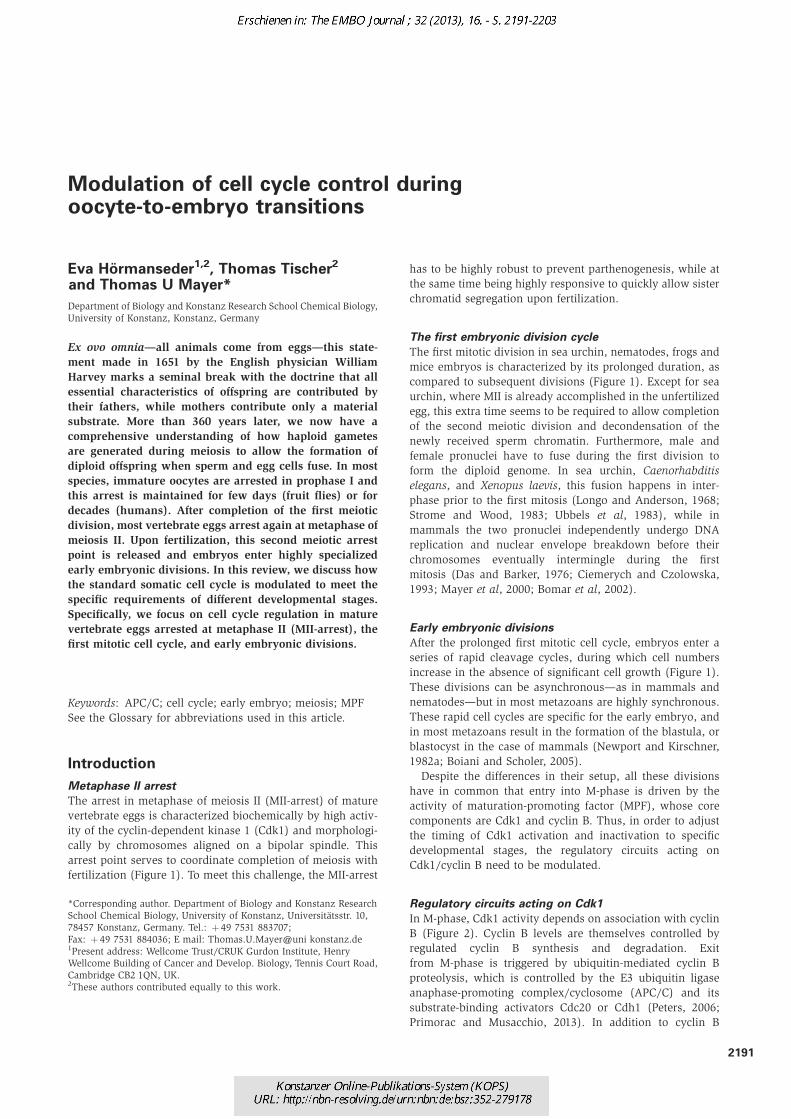
Modulation of cell cycle control during oocyte-to-embryo transitions Eva Ho ¨ rmanseder 1,2 , Thomas Tischer 2 and Thomas U Mayer* Department of Biology and Konstanz Research School Chemical Biology, University of Konstanz, Konstanz, Germany Ex ovo omnia—all animals come from eggs—this state- ment made in 1651 by the English physician William Harvey marks a seminal break with the doctrine that all essential characteristics of offspring are contributed by their fathers, while mothers contribute only a material substrate. More than 360 years later, we now have a comprehensive understanding of how haploid gametes are generated during meiosis to allow the formation of diploid offspring when sperm and egg cells fuse. In most species, immature oocytes are arrested in prophase I and this arrest is maintained for few days (fruit flies) or for decades (humans). After completion of the first meiotic division, most vertebrate eggs arrest again at metaphase of meiosis II. Upon fertilization, this second meiotic arrest point is released and embryos enter highly specialized early embryonic divisions. In this review, we discuss how the standard somatic cell cycle is modulated to meet the specific requirements of different developmental stages. Specifically, we focus on cell cycle regulation in mature vertebrate eggs arrested at metaphase II (MII-arrest), the first mitotic cell cycle, and early embryonic divisions. Keywords: APC/C; cell cycle; early embryo; meiosis; MPF See the Glossary for abbreviations used in this article. Introduction Metaphase II arrest The arrest in metaphase of meiosis II (MII-arrest) of mature vertebrate eggs is characterized biochemically by high activ- ity of the cyclin-dependent kinase 1 (Cdk1) and morphologi- cally by chromosomes aligned on a bipolar spindle. This arrest point serves to coordinate completion of meiosis with fertilization (Figure 1). To meet this challenge, the MII-arrest has to be highly robust to prevent parthenogenesis, while at the same time being highly responsive to quickly allow sister chromatid segregation upon fertilization. The first embryonic division cycle The first mitotic division in sea urchin, nematodes, frogs and mice embryos is characterized by its prolonged duration, as compared to subsequent divisions (Figure 1). Except for sea urchin, where MII is already accomplished in the unfertilized egg, this extra time seems to be required to allow completion of the second meiotic division and decondensation of the newly received sperm chromatin. Furthermore, male and female pronuclei have to fuse during the first division to form the diploid genome. In sea urchin, Caenorhabditis elegans, and Xenopus laevis, this fusion happens in inter- phase prior to the first mitosis (Longo and Anderson, 1968; Strome and Wood, 1983; Ubbels et al, 1983), while in mammals the two pronuclei independently undergo DNA replication and nuclear envelope breakdown before their chromosomes eventually intermingle during the first mitosis (Das and Barker, 1976; Ciemerych and Czolowska, 1993; Mayer et al, 2000; Bomar et al, 2002). Early embryonic divisions After the prolonged first mitotic cell cycle, embryos enter a series of rapid cleavage cycles, during which cell numbers increase in the absence of significant cell growth (Figure 1). These divisions can be asynchronous—as in mammals and nematodes—but in most metazoans are highly synchronous. These rapid cell cycles are specific for the early embryo, and in most metazoans result in the formation of the blastula, or blastocyst in the case of mammals (Newport and Kirschner, 1982a; Boiani and Scholer, 2005). Despite the differences in their setup, all these divisions have in common that entry into M-phase is driven by the activity of maturation-promoting factor (MPF), whose core components are Cdk1 and cyclin B. Thus, in order to adjust the timing of Cdk1 activation and inactivation to specific developmental stages, the regulatory circuits acting on Cdk1/cyclin B need to be modulated. Regulatory circuits acting on Cdk1 In M-phase, Cdk1 activity depends on association with cyclin B (Figure 2). Cyclin B levels are themselves controlled by regulated cyclin B synthesis and degradation. Exit from M-phase is triggered by ubiquitin-mediated cyclin B proteolysis, which is controlled by the E3 ubiquitin ligase anaphase-promoting complex/cyclosome (APC/C) and its substrate-binding activators Cdc20 or Cdh1 (Peters, 2006; Primorac and Musacchio, 2013). In addition to cyclin B *Corresponding author. Department of Biology and Konstanz Research School Chemical Biology, University of Konstanz, Universita ¨tsstr. 10, 78457 Konstanz, Germany. Tel.: þ 49 7531 883707; Fax: þ 49 7531 884036; E mail: Thomas.U.Mayer@uni konstanz.de 1 Present address: Wellcome Trust/CRUK Gurdon Institute, Henry Wellcome Building of Cancer and Develop. Biology, Tennis Court Road, Cambridge CB2 1QN, UK. 2 These authors contributed equally to this work. 2191
-
Upload
khangminh22 -
Category
Documents
-
view
2 -
download
0
Transcript of Modulation of cell cycle control during oocyte-to-embryo ...
Modulation of cell cycle control duringoocyte-to-embryo transitions
Eva Hormanseder1,2, Thomas Tischer2
and Thomas U Mayer*
Department of Biology and Konstanz Research School Chemical Biology,
University of Konstanz, Konstanz, Germany
Ex ovo omnia—all animals come from eggs—this state-
ment made in 1651 by the English physician William
Harvey marks a seminal break with the doctrine that all
essential characteristics of offspring are contributed by
their fathers, while mothers contribute only a material
substrate. More than 360 years later, we now have a
comprehensive understanding of how haploid gametes
are generated during meiosis to allow the formation of
diploid offspring when sperm and egg cells fuse. In most
species, immature oocytes are arrested in prophase I and
this arrest is maintained for few days (fruit flies) or for
decades (humans). After completion of the first meiotic
division, most vertebrate eggs arrest again at metaphase of
meiosis II. Upon fertilization, this second meiotic arrest
point is released and embryos enter highly specialized
early embryonic divisions. In this review, we discuss how
the standard somatic cell cycle is modulated to meet the
specific requirements of different developmental stages.
Specifically, we focus on cell cycle regulation in mature
vertebrate eggs arrested at metaphase II (MII-arrest), the
first mitotic cell cycle, and early embryonic divisions.
Keywords: APC/C; cell cycle; early embryo; meiosis; MPF
See the Glossary for abbreviations used in this article.
Introduction
Metaphase II arrest
The arrest in metaphase of meiosis II (MII-arrest) of mature
vertebrate eggs is characterized biochemically by high activ-
ity of the cyclin-dependent kinase 1 (Cdk1) and morphologi-
cally by chromosomes aligned on a bipolar spindle. This
arrest point serves to coordinate completion of meiosis with
fertilization (Figure 1). To meet this challenge, the MII-arrest
has to be highly robust to prevent parthenogenesis, while at
the same time being highly responsive to quickly allow sister
chromatid segregation upon fertilization.
The first embryonic division cycle
The first mitotic division in sea urchin, nematodes, frogs and
mice embryos is characterized by its prolonged duration, as
compared to subsequent divisions (Figure 1). Except for sea
urchin, where MII is already accomplished in the unfertilized
egg, this extra time seems to be required to allow completion
of the second meiotic division and decondensation of the
newly received sperm chromatin. Furthermore, male and
female pronuclei have to fuse during the first division to
form the diploid genome. In sea urchin, Caenorhabditis
elegans, and Xenopus laevis, this fusion happens in inter-
phase prior to the first mitosis (Longo and Anderson, 1968;
Strome and Wood, 1983; Ubbels et al, 1983), while in
mammals the two pronuclei independently undergo DNA
replication and nuclear envelope breakdown before their
chromosomes eventually intermingle during the first
mitosis (Das and Barker, 1976; Ciemerych and Czolowska,
1993; Mayer et al, 2000; Bomar et al, 2002).
Early embryonic divisions
After the prolonged first mitotic cell cycle, embryos enter a
series of rapid cleavage cycles, during which cell numbers
increase in the absence of significant cell growth (Figure 1).
These divisions can be asynchronous—as in mammals and
nematodes—but in most metazoans are highly synchronous.
These rapid cell cycles are specific for the early embryo, and
in most metazoans result in the formation of the blastula, or
blastocyst in the case of mammals (Newport and Kirschner,
1982a; Boiani and Scholer, 2005).
Despite the differences in their setup, all these divisions
have in common that entry into M-phase is driven by the
activity of maturation-promoting factor (MPF), whose core
components are Cdk1 and cyclin B. Thus, in order to adjust
the timing of Cdk1 activation and inactivation to specific
developmental stages, the regulatory circuits acting on
Cdk1/cyclin B need to be modulated.
Regulatory circuits acting on Cdk1
In M-phase, Cdk1 activity depends on association with cyclin
B (Figure 2). Cyclin B levels are themselves controlled by
regulated cyclin B synthesis and degradation. Exit
from M-phase is triggered by ubiquitin-mediated cyclin B
proteolysis, which is controlled by the E3 ubiquitin ligase
anaphase-promoting complex/cyclosome (APC/C) and its
substrate-binding activators Cdc20 or Cdh1 (Peters, 2006;
Primorac and Musacchio, 2013). In addition to cyclin B
*Corresponding author. Department of Biology and Konstanz Research
School Chemical Biology, University of Konstanz, Universitatsstr. 10,
78457 Konstanz, Germany. Tel.: þ 49 7531 883707;
Fax: þ 49 7531 884036; E mail: Thomas.U.Mayer@uni konstanz.de1Present address: Wellcome Trust/CRUK Gurdon Institute, Henry
Wellcome Building of Cancer and Develop. Biology, Tennis Court Road,
Cambridge CB2 1QN, UK.2These authors contributed equally to this work.
2191

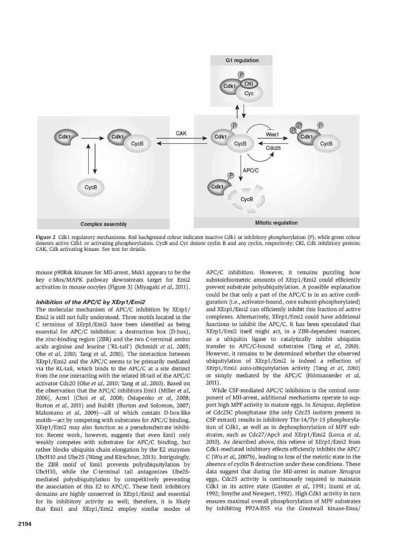
association, full Cdk1 activation also involves phospho-
rylation events. Stimulating phosphorylation of a conserved
threonine in the activation loop (Krek and Nigg, 1992) by Cdk-
activating kinases occurs in an unregulated, constitutive
manner (Fisher and Morgan, 1994; Tassan et al, 1994). On
the other hand, phosphorylation of Cdk1 in the ATP-binding
pocket (Thr-14 and Tyr-15) by vertebrate Wee1 and Myt1
kinases results in inactive MPF, and this negative effect on
Cdk1 activation is antagonized by the Cdc25 family of
phosphatases (Dunphy and Kumagai, 1991) (Figure 2).
Following exit from M-phase, Cdk1 activity remains sup-
pressed during G1 phase by the association with Cdk-inhibi-
tory proteins, a regulatory mechanism that will not be
discussed further here (for a review see Vidal and Koff, 2000).
M-phase onset requires both MPF activation and
inactivation of MPF-antagonizing phosphatases
At the onset of M-phase, Cdk1 is activated in a switch-like
manner via an auto-amplification loop, in which Cdk1
inactivates its inhibitors (the Wee1 and Myt1 kinases) and
activates it’s the counteracting Cdc25 phosphatases
(Figure 2). In addition to swift MPF activation, timely entry
into mitosis, however, also requires effective inactivation of
phosphatases that would counteract Cdk1-mediated phos-
phorylation events. In particular, recent studies revealed
that Cdk1 itself mediates inactivation of its antagonist
protein phosphatase 2A (PP2A). Specifically, Cdk1 activates
Greatwall kinase, which in turn phosphorylates and primes
Ensa/Arpp19, a protein inhibitor of the B55 subtype of PP2A
(Yu et al, 2006; Mochida et al, 2009; Vigneron et al, 2009;
Gharbi-Ayachi et al, 2010; Mochida et al, 2010). Thus, Cdk1
activation ultimately flips the switch from a state of MPF
substrate hypophosphorylation to one where MPF substrates
are maximally phosphorylated.
MII-arrest: Putting the cell cycle on hold
The proto-oncogene c-Mos and the MAP kinase
pathway
MII-arrest of mature vertebrate eggs depends on cytostatic
factor (CSF), an activity initially described as being present in
the cytoplasm of mature Rana pipiens eggs and capable of
inducing a metaphase-like arrest when injected into two-cell
embryos (Masui and Markert, 1971). Based on their studies,
Masui and Markert (1971) framed three criteria that any
candidate factors proposed to be CSF would have to fulfill
the following conditions: (i) accumulation during oocyte
maturation, with maximal levels in MII, (ii) ability to cause
a metaphase-like arrest when injected into embryonic
blastomeres and (iii) inactivation upon fertilization.
The first factor found to meet all three criteria was the
proto-oncogene c-Mos, which is newly synthesized in matur-
ing Xenopus oocytes, causes a CSF-like arrest when expressed
in embryos, and is degraded shortly after fertilization (Sagata
et al, 1989a,b). Further elegant biochemical and
pharmacological studies demonstrated that the serine–
threonine protein kinase c-Mos triggers a MEK/MAP kinase
(MAPK) signalling cascade that is essential for both the
establishment and maintenance of MII-arrest in Xenopus
oocytes (Sagata et al, 1988; Haccard et al, 1993; Kosako
et al, 1994; Abrieu et al, 1996; Bhatt and Ferrell, 1999).
Subsequent work identified the p90 ribosomal S6 kinase
(p90Rsk) as the key MAPK substrate relevant for CSF arrest,
indicating that MII-arrest is mediated by a strictly linear
c-Mos/MEK/MAPK/p90Rsk signalling cascade in Xenopus
eggs (Gross et al, 1999). The situation seems to be more
complex in mice, since c-Mos-deficient eggs, although
eventually undergoing parthenogenic activation, initially
manage to establish a transient CSF arrest (Colledge et al,
1994; Hashimoto et al, 1994; Choi et al, 1996; Verlhac et al,
1996). Here, c-Mos may, therefore, not be strictly essential for
establishment of the second meiotic arrest, but may only be
required for its maintenance. Furthermore, mouse oocytes
lacking all three mammalian homologues of p90Rsk exhibit a
normal CSF arrest (Dumont et al, 2005), suggesting that
mouse c-Mos functions to impose a robust MII-arrest in a
manner independent of p90Rsk kinases. Instead, mitogen-
and stress-activated protein kinase 1 (Msk1) may be the
relevant downstream MAPK target in mouse oocytes
(Miyagaki et al, 2011).
The APC/C inhibitor XErp1/Emi2 and the c-Mos/MAPK/
p90Rsk signalling cascade
How then does the c-Mos/MAPK pathway transmit its in-
hibitory signal to the APC/C? Data from Xenopus implicated
components of the spindle assembly checkpoint (SAC) as
links between p90Rsk and the APC/C, since the SAC kinase
Bub1 was identified as a p90Rsk substrate, and since deple-
tion of Bub1 as well as SAC components Mad1 and Mad2
appeared to interfere with the CSF arrest (Schwab et al, 2001;
Tunquist et al, 2002). However, how the SAC would
mechanistically mediate CSF arrest remained elusive, since
(i) there is no conclusive explanation of how SAC silencing
could be coordinated with fertilization, (ii) a robust CSF
arrest can be mounted in Xenopus egg extract lacking DNA
and, therefore, also lacking kinetochores from which SAC
signals could originate and (iii) the SAC is completely
dispensable for the MII-arrest in mouse oocytes (Tsurumi
et al, 2004). The gap between the c-Mos/MAPK/p90Rsk
pathway and the APC/C was filled by the identification of
XErp1 (Xenopus Emi1-related protein 1) or Emi2, whose
function requires activation by p90Rsk (Figure 3). XErp1/
Emi2, a highly conserved F-box protein, accumulates in
Glossary
APC/C anaphase promoting complex/cyclosomeCAK CDK activating kinaseCdk1 cyclin dependent kinase 1CKI cyclin dependent kinase inhibitorCPE cytoplasmic polyadenylation elementCPEB cytoplasmic polyadenylation element
binding proteinD box destruction boxICM inner cell massMII meiosis IIMII arrest arrest in metaphase of meiosis IIMBT mid blastula transitionMPF maturation promoting factorp90Rsk ribosomal S6 kinasePlk1 Polo like kinase 1PKA protein kinase APP2A protein phosphatase 2 ARL tail C terminal arginine leucine dipeptide in
XErp1/Emi2SAC spindle assembly checkpointUPS ubiquitin proteasome systemZBR zinc binding region in XErp1/Emi2 and
Emi1ZGA zygotic genome activation
2192

Cdk1
APCIC
XErp1
Mos
Cdk1-pY
Emit
SAC
CSF arrest Mil
Fertilization
• Cycle 1
M
'G2'
s
Time after fertilization (20°C) o
Mid-blastula transition
• Cycle2-12 Somatic cycles M M
'G1' G2 G1
s s
1.5 h 7h
Figure 1 Schematic representation of cell cycle regulation during Xenopus early development. illustrated are specialized cell cycle types, major developmental transitions and oocyte/embryo stages, as well as oscillations of Cdkl cyclin Band APC/C activity. Bars in the lower half depict activity levels for XErpl/Emi2, c Mos/MAPK, Emil and the spindle assembly checkpoint (SAC), as well as inhibitory Thr 14/1\'r 15 phosphorylation of Cdkl (Cdkl pY). See text for details.
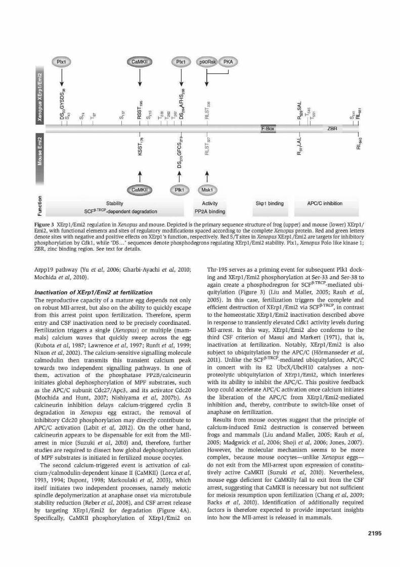
maturing oocytes (CSF criterion 1), mediates the Mll-arrest in mature eggs by directly inhibiting the APC/C, and causes a cell cycle arrest when injected into Xenopus embryos (CSF criterion TI) (Schmidt ec al, 2005). XErpl/Em12 Is phosphorylated by p90Rsk on Ser-335. Thr-336, Ser-342 and Ser-344, and subsequently associates with the B'56 subtype of PP2A (PP2A-B'56), which in turn removes Cdklmediated inhibitory phosphorylation from amino- and carboxy-terminal regions of XErpl/Emi2 (Inoue et aL. 2007; Nlshiyama et al, 2007a; Wu et al, 2007a,b; Isoda et al, 2011) (Figure 3). Phosphorylation of the XErp1/Emi2 C-terminal region interferes with its ability to bind and inhibit the APC/C (Figure 4A) (Wu et al, 2007a), similar to the situation of the activator Cdc20 that can also only associate with the APC/C when dephosphorylated (Labit et al, 2012). Phosphorylation in the amino-terminal Cdkl site cluster, on the other hand, controls XErp1fEmi2 stability (Figure 4A), by serving as priming event for the recruitment of Polo-like kinase 1 (Plkl) . Further phosphorylation by this kinase creates a phosphorylation-dependent recognition motif (or phosphodegron) for the Ubiquitin ligase SCfJl·TRCP• Which then Ubiq\fr tylates and targets XErp1/Erni2 for proteasomal degradation (Wu et al, 2007a; Isoda et al, 2011). Thus, p90Rsk-mediated
recruitment of PP2A-B'56 downstream of the c-Mos/MAPK pathway activates XErpl/.Erni2, both by stabilizing it and by directly impinging on its APC/C inhibitory properties.
Although these data explain the essential CSF-arrest role of c-Mos, it would appear somewhat paradoxical that Cdkl inactivates CSF, a factor that serves to impose robust Milarrest in the presence of high Cdkl activity. However, the cybernetic regulation of Cdk1-mediated XErpl/Erni2 inactivation (APC/C activation) and PP2A-B'56-mediated XErpl/ Emi2 activation (APC/C inactivation) provides a mechanism to maintain Cdkl activity at a high but constant level during Mll-arrest, despite continuous eyeliD B synthesis (Kubiak et al, 199 3; Ledan et al, 2001; Yamamoto et al, 2005); once cyclin B levels exceed a certain upper threshold, Cdkl transiently activates cyclin B degradation by the APC/C, to decrease cyclin B amounts to an optimal plateau with sufficiently strong MPF activity for mounting a stable CSF arrest and sufficiently low cyclin B levels to allow rapid release upon fertilization (Figure 4A). In mice, the essential function of both Erni2 and PP2A for CSF arrest seems conserved, as their knockdown or pharmacological inhibition results in parthenogenesis (Madgwick et al, 2006; Shoji et al, 2006; Chang et aL, 2011). Consistent with the dispensability of
2193
2194
G1 regulation
t~ ___ c_AK __ • ~ ~e1 ,.
~ ~CycB Cdc25
l·~ CycB
CycB
Complex assembly Mitotic regulation
Figure 2 Cclkl regulatory mechanisms. Red background colour indicates inactive Cdk1 or inlllbitory phosphorylation (P), while green colour denotes active Cdkl or activating phosphorylation. CycB and Cyc denote cyclin B and any eyeliD, respectively; CKI, Cdk inhibitory protein; CAK, Cdk activating kinase. See text for details.
mouse p90Rsk kinases for MIT-arrest, Mskl appears to be the key c-Mos/MAPK pathway downstrearo target for Emi2 activation in mouse oocytes (Figure 3) (Miyagak:i et al, 2011).
Inhibition of the APC/ C by XErp1/Emi2 The molecular mechanism of APC/C inhibition by XErpl/ Emil is still not fully understood. Three motifs located in the C terminus of XErpl/Emi2 have been identified as being essential for APC/C inhibition: a destruction box (D-box), the zinc-binding region (ZBR) and the two C-terrninal amino acids arginine and leucine ('RL-tail') (Schmidt et al, 2005; Ohe et al, 2ffi0; Tang et al, 2010). The interaction between XErplfEmi2 and the APC/C seems to be primarily mediated via the RL-tail, which binds to the APC(C at a sire distinct from the one interacting w ith the related IR-tail of the APC/C activator Cdc20 (Obe et al, 2010; Tang et al, 2010) . Based on the observation that the APC/C inhibitors Emil (Miller et al, 2006), Acml (Cboi et al, 2008; Ostapenko et al, 2008; Burton et al, 2011) and BubRl (Burton and Solomon, 2007; Malureanu et al, 2009)- all of which contain D-box-like motifs- act by competing with substrates for APC/C binding, XErpl/Emi2 may also function as a pseudosubstrate inhibitor. Recent work, however, suggests that even Emil only weakly competes with substrates for APC/C binding, but rather blocks ubiquitin chain elongation by the E2 enzymes UbcH10 and Ube2S (Wang and Kirschner, 2013).1ntriguingly, the ZBR motif of Emil prevents polyubiquitylation by UbcHlO, while the C-terminal tail antagonizes Ube2Smediated polyubiquitylation by competitively preventing the association of this E2 to APC/C. These Emil inhibitory domains are highly conserved in XErp1/Emi2 and essential for its inhibitory activity as well; therefore, it is likely that Emil and XErp1/Emi2 employ similar modes of
APC/C inhibition. However, it remains puzzling bow substoicbiometric amounts of XErp1/Emi2 could efficiently prevent substrate polyubiquitylation. A possible explanation could be that only a part of the APC/C is in an active configuration (i.e., activator-bound, core subunit-phosphorylated) and XErpl/Emi2 can efficiently inhibit this fraction of active complexes. Alternatively, XErp1/Emi2 could have additional functions to inhibit the APC/C. It has been speculated that XErpl/Emi2 itself might act, in a ZBR-dependent manner, as a ubiquitin ligase to catalytically inhibit ubiquitin transfer to APC/C-bound substrates (Tang et a.l, 2010). However, it remains to be determined whether the observed ubiquitylation of XErpl/Emi2 is indeed a reflection of XErpl/Eml2 auro-ublqU1tylatlon acttvlty (Tang et al, 2010) or simply mediated by the APC/C (Horrnanseder et al, 2011).
While CSF-mediated APC/C inhibition is the central component of Mil-arrest, additional mechanisms operate to support high MPF activity in mature eggs. ln Xenopus, depletion of Cdc2SC phosphatase (the only Cdc25 isoform present in CSF extract) results in inhibitory Thr-14jTyr-15 phosphorylation of Cdkl, as well as in dephosphorylation of MPF substrates, such as Cdc27/Apc3 and XErpl/Emi2 (Lorca et al, 2010). As described above, this relieve of XErp1jEmi2 from Cdkl-mediated inhibitory effects efficiently inhibits the APC/ C (Wu et al, 2007b), leading to loss of the meiotic state in the absence of cyclin B destruction under these conditions. These data suggest that during the Mil-arrest in mature Xenopus eggs, Cdc25 activity is continuously required to maintain Cdkl in its active state (Gautier et al, 1991; Izumi et al, 1992; Smythe and Newport, 1992). High Cdkl activity ln turn ensures maximal overall phosphorylation of MPF substrates by inhibiting PP2A-BSS via the Greatwall kinase-Ensa/
! ! ! ~ til I!
Ul en' 0 Ul :!l it til >-(!j
~ .... ., .,
Ill ., .. lii u)' M J ~ Oi lil &l Iii en' ...J
O UJ~ ur a: Ul t-!"t-Nt-2' 0 a: I I I I I I I I I I I I
...J
~ ., ,. j "' ... rf 1-10 ~
t-"' c/fa: I I I I I
F-Box ZBR I l I I!! !;) ~ ...:- Ul
~ Ul (.) Ul u. -:.:: (!j a:
- I I ...J 0
:l _j 1ft a:
a: "' cl'
0
t t t c: ..2 Stability Activity u Skpl binding APC/C inhibition c: SCFil-TRCP -dependent degradation PP2A binding :::1 u.
Figure 3 XErp l/Erni2 regulation in Xenopus and mouse. Depicted is the primary sequence structure of frog (upper) and mouse (lower) XErp 1/ Emi2, with functional elements and sites of regulatory modifications spaced according to the complete Xenopus protein. Red and green letters denote sites with negative and positive effects on XErpl's function, respectively. Red S(fsites in Xenopus XErpl/Emi2 are targets for inhibitory phosphorylation by Cdkl, while 'DS ... ' sequences denote phosphodegrons regulating XErpl/ Emi2 stability. Plxl , Xenopus Polo like kinase 1; ZBR, zinc binding region. See text for details.
Arppl9 pathway (Yu et al, 2006; Gharbi-Ayachi et al, 2010; Mochida et al, 2010).
Inactivation of XErp1/ Emi2 at fertilization The reproductive capacity of a mature egg depends not only on robust Mil-arrest, but also on the ability to quickly escape from this arrest point upon fertilization. Therefore, sperm entry and CSF inactivation need to be precisely coordinated. Fertilization triggers a single (Xerwpus) or multiple (mammals) calcium waves that quickly sweep across the egg (Kubota et al, 1987; Lawrence et al, 1997; Runft et al, 1999; Nixon et al, 2002). The calcium-sensitive signalling molecule calmodulin then transmits this transient calcium peak towards two independent signalling pathways. In one of them, activation of the phosphatase PPZB/calcineurin initiates global dephosphorylation of MPF substrates, such as the APC/C subunit Cdc27 1 Apc3, and its activator Cdc20 (Mochida and Hunt, 2007; Nishiyama et al, 2007b). As calcineurin inhibition delays calcium-triggered cyclin B degradation in Xerwpus egg extract, the removal of inhibitory Cdc20 phosphorylation may directly contribute to APC/C activation (Labit et al, 2012). On the other hand, calcineurin appears to be dispensable for exit from the Milarrest in mice (Suzuki et al, 2010) and, therefore, further studies are required to dissect how global dephosphorylation of MPF substrates is initiated in fertilized mouse oocytes.
The second calcium-triggered event is activation of calcium-/calmodulin-dependent kinase II (CaMKII) (Lorca et al, 1993, 1994; Dupont, 1998; Markoulaki et al, 2003), which itself initiates two independent processes, namely meiotic spindle depolymerization at anaphase onset via microtubule stability reduction (Reber et al, 2008), and CSF arrest release by targeting XErpl/Erni2 for degradation (Figure 4A). Specifically, CaMKII phosphorylation of XErp1/Erni2 on
Thr-195 serves as a priming event for subsequent Plkl docking and XErpl/Erni2 phosphorylation at Ser-33 and Ser-38 to again create a phosphodregron for scFil·TRCP_mediated ubiqui tylation (Figure 3) (Liu and Maller, 2005; Raub et al, 2005) . In this case, fertilization triggers the complete and efficient deStrUCtiOn Of XErpl /Emi2 via SCfjl-TRCP, in COntrast to the homeostatic XErpl,fEmi2 inactivation described above in response to transiently elevated Cdk1 activity levels during Mil-arrest. In this way, XErpl,fEmi2 also conforms to the third CSF criterion of Masui and Markert (1971), that is, inactivation at fertilization. Notably, XErpl/Erni2 is also subject to ubiquitylation by the APC/C (Hormanseder et al, 2011). Unlike the SCFil-TRCP_mediated ubiquitylation, APC/ C in concert with its E2 UbcXjUbcHlO catalyses a nonproteolytic ublqUitylatton of XErplfEmi2, which interferes with its ability to inhibit the APC/C. This positive feedback loop could accelerate APC/C activation once calcium initiates the liberation of the APC/C from XErpl/Erni2-mediated inhibition and, thereby, contribute to switch-like onset of anaphase on fertilization.
Results from mouse oocytes suggest that the principle of calcium-induced Emi2 destruction is conserved between frogs and mammals (Liu andand Maller, 2005; Raub et al, 2005; Madgwick et al, 2006; Shoji et al, 2006; Jones, 2007). However, the molecular mechanism seems to be more complex, because mouse oocytes- unlike Xerwpus eggsdo not exit from the Mil-arrest upon expression of constitutively active CaMKil (Suzuki et al, 2010) . Nevertheless, mouse eggs deficient for CaMKIIy fail to exit from the CSF arrest, suggesting that CaMKIT is necessary but not sufficient for meiosis resumption upon fertilization (Chang et al., 2009; Backs et al, 2010) . Identification of additionally required factors is therefore expected to provide important insights into how the Mil-arrest is released in mammals.
21 95

2196
a Meiotic regulation ~
j_ ciciD oo ___ ~ 0 ____ _.. -1
00 0 ll
1 Fertitiutio"
l SCFfl-TRCP -dependent degradation
~~ Mitotic regulation
0 _____ __. -1
00 0
'----------------= -------'-1
1 Metoph.,e-to-a"'ph"e '""''"•"
OQ. 00 0
Figure 4 XErpl / Emi2 and APC/ C regulation in meiosis (A) and mitosis (B). Bars indicate XErpl/Emi2 primary structure and sequence elements as in Figure 3. Inhibitory and activating phosphorylation is depicted by red and green circles, resi?fctively. Inactive and active APC/C or PP2A are indicated by red and green background colours, respectively. See text for details.
The first embryonic division
In most metazoan organisms, the first cell cycle following fertilization is characterized by its prolonged duration com-
pared to subsequent divisions. In Xenopus, the first cell cycle takes about 90min, whereas the following 11 cleavage cycles
each last for only about 30min (Hara et al, 1980; Newport and Kirschner, 1982a). In mouse, the length of the first and
second mitotic division is comparable, but metaphase duration is shortened from 120min in the first cell cycle to 70min in the second one (Ciemerych et al, 1999). An
explanation for this extension may lie in the fact that the first division cycle has to fulfill an array of specialized functions. Among them is the completion of the female genome's second meiotic division, accompanied by extrusion of the second polar body. Additionally, the

specialized chromatin of the incoming sperm has to be
adjusted to support embryonic development. The process
starts with incorporation of the sperm nucleus into the egg,
and the breakdown of its nuclear envelope. Following
decondensation of the highly compact sperm chromatin,
associated with replacement of protamine by somatic
histones (Rodman et al, 1981; Santos et al, 2002), the
nuclear envelope reassembles to form swollen male
pronuclei (Longo, 1985; Katagiri and Ohsumi, 1994). DNA
replication eventually initiates after decondensation of both
male and female chromatin (Luthardt and Donahue, 1973;
Bouniol-Baly et al, 1997; Ferreira and Carmo-Fonseca, 1997).
Inhibitory phosphorylation of Cdk1
While XErp1/Emi2, which following its complete degradation
upon fertilization re-accumulates during the first mitotic
division cycle, could in principle be well-placed to contribute
to its increased length, there are currently no experimental
data to support this hypothesis. Instead, the extended dura-
tion of the first division in Xenopus can at least in part be
attributed to delayed activation of Cdk1 caused by inhibitory
Tyr-14/Thr-15 phosphorylation. During the first mitotic divi-
sion, the responsible inhibitory kinases Wee1 and Myt1
appear not to be constantly antagonized by Cdc25 phospha-
tases (Figure 2), this shift in the balance possibly due to an
active c-Mos/MAPK pathway in the first cell cycle (Murakami
and Vande Woude, 1998). The c-Mos/MAPK pathway has
been reported to activate Wee1 (Murakami and Vande
Woude, 1998; Murakami et al, 1999; Walter et al, 2000),
and strong MAPK pathway activation can also trigger
destabilization of Cdc25A by targeting it for SCFb-TRCP-
dependent degradation (Isoda et al, 2009). Since the c-Mos/
MAPK pathway remains partially active during the first
cycle (until Xenopus c-Mos degradation B30min after
fertilization), it is therefore possible that it mediates both
Cdc25A degradation and Wee1 activation, with the net result
of prolonged inhibitory phosphorylation of Cdk1 and an
increased length of the first cell cycle. As the first division
progresses and c-Mos levels decline, the balance could then
tip in favour of Cdk1 activation, a process further promoted
by Plx, which is able to activate Cdc25 (Abrieu et al, 1998;
Qian et al, 1998; Toyoshima-Morimoto et al, 2002) as well as
to—specifically during embryonic M-phase—bind and inhibit
Myt1 (Inoue and Sagata, 2005). Thus, Plx and Cdk1 in
concert are able to start the autoamplification loop of Cdk1
activation.
A full mitosis-competent state further depends on inactiva-
tion of the Cdk1-antagonizing PP2A phosphatase. It is likely
that the mechanism of Cdk1-mediated inactivation of its
antagonist PP2A-B55 via the Greatwall-Ensa/Arpp19 path-
way (Gharbi-Ayachi et al, 2010; Mochida et al, 2010) acts not
only in MII-arrested eggs (Yu et al, 2006; Hara et al, 2012) and
during somatic divisions (Burgess et al, 2010; Voets and
Wolthuis, 2010), but also functions during early embryonic
divisions.
In mouse embryos, Cdk1 activation during the first mitotic
division does not appear to be significantly delayed; on the
other hand, mouse embryos exhibit prolongation of the first
mitosis compared to the second division cycle (Ciemerych
et al, 1999). This mitotic delay is however not mediated by
the SAC (Sikora-Polaczek et al, 2006), and further studies are
therefore required to elucidate the mechanisms impeding
timely Cdk1 inactivation in the first mitotic cell cycle in mice.
Early embryonic cell division cycles
After the prolonged first cell division, embryos enter a series
of specialized early embryonic cell division cycles. Their
length varies between species, from 15min per cycle in
zebrafish to 30min in frogs and 12 h in mice. Early cell
divisions in mammalian embryos are set apart not only by
their extended duration, but also by their marked asynchrony
between sister cells, resulting in frequent stages with odd
numbers of cells instead of exponential cell number increases
(from two- to four- to eight-cell stages) (Piko and Clegg,
1982). In Echinoderms Xenopus and zebrafish, these early
embryonic cell cycles last until the so-called mid-blastula
transition (MBT) towards somatic cell cycles, which is
initiated after 13 cycles in Xenopus and is marked by
switching of gene expression from maternally supplied to
newly transcribed zygotic mRNAs, referred to as zygotic
genome activation (ZGA). In Xenopus, ZGA is initiated after
13 cell cycles (Kimelman et al, 1987), in zebrafish after 10
divisions and in mouse at the two-cell stage (Tadros and
Lipshitz, 2009). Unlike MII-arrest and the first prolonged
division, the subsequent early embryonic divisions display
a high degree of variation between species, likely reflecting
the divergent metazoan strategies for generating offspring.
Indeed, embryos released into the environment without
parental protection, like those of Xenopus, display faster
early embryonic cell cycles than, for example, mammalian
embryos protected in the womb (Strathmann et al, 2002).
Rapid divisions allow fast developmental progression and
may therefore be favoured in less predictable environmental
conditions, while slow divisions might be generally more
favourable for the proper embryo development as long
as parental protection ensures decreased effects of
environmental stress. Therefore, adaptive cell cycle
mechanisms could have evolved to adjust cell division
length to the developmental strategy of a given species.
Cdc25 phosphatases
The rapid early embryonic cell divisions of Xenopus embryos
lack gap phases, surveillance mechanisms such as DNA
replication-, DNA damage- and spindle assembly checkpoints
(described in Musacchio and Salmon, 2007, Zegerman and
Diffley, 2009), as well as the APC/C inhibitor Emi1, and are
hence referred to as ‘minimal cell cycles’ (Figure 1, S–M
cycles) (Masui and Wang, 1998). The pacemaker underlying
the highly synchronous divisions in early Xenopus embryos is
again the Cdk1 activity, which accordingly has to peak
and drop every 30min. In contrast to the lengthened first
mitotic division, there is no inhibitory Thr-14/Tyr-15
phosphorylation on Cdk1 during these rapid divisions
(Ferrell et al, 1991; Hartley et al, 1996), which could be due
to either decreased Wee1/Myt1 kinase activity or increased
Cdc25 phosphatase activity. In support of the latter
possibility, embryos express Cdc25A in addition to the
Cdc25C isoform from fertilization until MBT (Kim et al,
1999), which may be sufficient to constantly keep the
balance tipped towards unphosphorylated active Cdk1 and
thus could contribute to the rapidness of early divisions.
2197

Regulated translation of cyclin B
In the absence of direct Cdk1 regulation via posttranslational
modifications, regulated synthesis and destruction of cyclin B
assumes the key role for early embryonic cell cycle regula-
tion. Since no transcription takes place during the first 12
divisions in Xenopus embryos, gene expression is solely
regulated at the posttranscriptional level (Newport and
Kirschner, 1982b). Indeed, sequence elements present in the
30-untranslated region of cyclin B mRNA and other
maternally inherited mRNAs seem to regulate their stage-
specific and/or a cell cycle-dependent translation. One of
these elements is the cytoplasmic polyadenylation element
(CPE), which recruits the CPE-binding protein (CPEB) that in
turn mediates polyadenylation and translation of the mRNA.
Consequently, injection of inhibitory CPEB antibodies into
one-cell embryos results in decreased cyclin B protein levels,
defective cell cycle progression and aberrant cell divisions
(Groisman et al, 2000). Cyclin B translation is coordinated
with the cell cycle phase by activating phosphorylation on
CPEB, mediated by the cell cycle kinase Aurora A (Mendez
et al, 2000; Groisman et al, 2001, 2002). Furthermore, cyclin B
expression is silenced upon exit from mitosis by the protein
maskin, which binds CPEB to inhibit mRNA translation.
Consistently, maskin protein levels have been found to
oscillate in a cell cycle-dependent manner in cycling
Xenopus egg extracts (Groisman et al, 2002), although the
underlying mechanism as well as the full extent of cyclin B
translation control via maskin specifically in embryonic
interphase remain to be determined.
Regulated cyclin B degradation
Regulated cyclin B synthesis is only half the story underlying
oscillating cyclin B levels during the cell cycle, equally im-
portant is the regulated degradation of cyclin B by the APC/C.
Xenopus early embryos lack APC/C inhibitory components,
such as the SAC or Emi1 (Gerhart et al, 1984; Ohsumi et al,
2004), and the only APC/C inhibitor present in early dividing
Xenopus embryos is XErp1/Emi2. Its depletion causes the
untimely destruction of APC/C substrates, ultimately
resulting in embryonic lethality (Tischer et al, 2012). Before
undergoing apoptosis, XErp1/Emi2-depleted embryos exhibit
a notable increase in cell cycle length, suggesting that the
APC/C inhibitory activity of XErp1/Emi2 contributes to the
short periodicity of early embryonic divisions, a notion
confirmed also by recent mathematical modelling studies
(Vinod et al, 2013). XErp1/Emi2 levels decline at the MBT,
when cell cycle length increases and gap phases first become
apparent, and its function as APC/C inhibitor is taken over by
Emi1, the SAC (Clute and Masui, 1995) and additional
regulatory mechanisms (Figure 1).
Control of XErp1/Emi2 activity during early embryonic
divisions
In the mature egg, XErp1/Emi2 mediates cell cycle arrest until
fertilization triggers its complete destruction and, thus, ana-
phase onset. During the first mitotic interphase, XErp1/Emi2
is rapidly resynthesized and its levels remain constant until
MBT, and the XErp1/Emi2 activity during pre-MBT divisions
is regulated on the posttranslational level via Cdk1 (nega-
tively) and PP2A-B’56 (positively). A current simplified
model posits that entry into mitosis is facilitated by XErp1/
Emi2-mediated APC/C inhibition. PP2A-B’56 prevails over
Cdk1 in early mitosis, resulting in sustained APC/C inhibition
and hence increasing cyclin B levels. Once the Cdk1 activity
reaches a certain threshold, the balance tips towards XErp1/
Emi2 hyperphosphorylation and subsequent inactivation, al-
lowing APC/C activation, cyclin B degradation and ultimately
exit from M-phase (Figure 4B). Therefore, the antagonistic
regulation of XErp1/Emi2 by Cdk1 and PP2A-B’56 in early
Xenopus embryos reflects the regulatory mechanism active
during meiotic MII-arrest, however, with two crucial adapta-
tions: first, PP2A-B’56 recruitment to XErp1/Emi2 is not
mediated by p90Rsk, whose upstream regulator c-Mos disap-
pears about 30min after fertilization and does not reappear
during subsequent embryonic divisions (Sagata et al,
1988, 1989b), but instead by protein kinase A (PKA) in
Xenopus. Intriguingly, PKA phosphorylates the same residues
in XErp1/Emi2 (Ser-335, Thr-336, Ser-342 and Ser-344) that are
also targeted by p90Rsk during the CSF arrest. Consequently,
embryos depleted of endogenous XErp1/Emi2 (which undergo
apoptosis at the time point of gastrulation) cannot be rescued
by the expression of XErp1/Emi2 versions that fail to recruit
PP2A-B’56 due to mutations in these phosphorylation sites, nor
by XErp1/Emi2 mutants unable to inhibit APC/C.
The second major adaptation of the Cdk1/PP2A-B’56 an-
tagonism is related its objective; while this mechanism serves
the role of a rheostat that balances continuous cyclin B
synthesis with transient APC/C activation during MII-arrest,
its key function during early embryonic divisions is as a
switch that controls entry into and exit from M-phase. The
demand for a switch-like regulation is especially evident at
exit from M-phase, as any XErp1/Emi2 reactivation upon
decreasing Cdk1 activity would interfere with the complete
destruction of cyclin B required for mitotic exit. An additional
layer of cell cycle-dependent control could be imposed by the
XErp1/Emi2 activator PKA, whose activity has been found to
oscillate in cycling egg extracts (Grieco et al, 1994, 1996);
however, studies in dividing embryos found global PKA
activity levels to remain constant throughout early divisions
(Tischer et al, 2012). In this respect, cycling extract may not
faithfully recapitulate early embryonic divisions, but rather
mimic the situation of the first mitotic division, a notion also
supported by the observation that XErp1/Emi2 itself is
dispensable for mitotic progression in cycling extracts (Liu
et al, 2006). Therefore, it is unlikely that PKA regulation helps
to prevent XErp1/Emi2 reactivation when Cdk1 activity
declines during M-phase exit. As an alternative scenario,
we speculate that maximal Cdk1 activity in late metaphase
might trigger PP2A-B’56 inactivation, similar to Cdk1-
induced PP2A-B55d via the Greatwall-Ensa/Arpp19 pathway
at the entry into M-phase (Gharbi-Ayachi et al, 2010; Mochida
et al, 2010), thereby causing permanent XErp1/Emi2
inactivation. Should such a mechanism indeed exist, it
would have to be explained how XErp1/Emi2 is reactivated
in the subsequent cell cycle and how PP2A-B’56 activity
could be kept constant during CSF arrest in the face of high
Cdk1 activity.
APC/CCdc20 activity regulation by Cdk1-mediated
phosphorylation
While XErp1/Emi2-mediated APC/C inhibition is central to
cell cycle regulation in early Xenopus embryos, it is likely that
additional regulatory mechanisms are active during pre-MBT
divisions to reinforce faithful cell cycle progression.
2198

Experiments in somatic cells and Xenopus cycling egg extract suggested the existence of a negative feedback loop, in which Cdkl-cyclin B complexes activate their own antagonist APC/ C by phosphorylating several APC/ C core subunits (Kraft et al, 2003) . Indeed, the APC/C subunit Cdc27/APC3 is phosphorylated in a cell-cycle-dependent manner in Xenopus embryos. This positive Cdkl effect on APC/C activity is however opposed by the inhibitory effect on the APC/C activator Cdc20 (Yudkovsky et al, 2000; D'Angiolella et al, 2003; Labit et al, 2012), whose Cdkl-mediated phosphorylation reduces its affinity for the APC/ C. Consequently, Cdc20 mutated at five Cdkl sites bound efficiently to and activated the APC/C even in the presence of high Cdkl activity. Degradation of APC/C substrates at anaphase onset therefore requires Cdc20 dephosphorylation, which may be mediated by PP2A in early dividing embryos (Labit et al, 2012) . While more research is required to fully dissect the underlying molecular mechanisms and their physiological relevance for early embryonic divisions, it seems likely that differences in the phosphorylation and dephosphorylation kinetics of Cdc20, APC/C and XErpl / Emi2 may all contribute to transitions from phases where the APC/C is inactive- due to XErpl/Emi2 hypophosphorylation and Cdc20 hyperphosphorylation- to phases where the APC/C is fully active- due to APC/ C and XErpl/Emi2 hyperphosphorylation and Cdc20 hypophosphorylation (Vinod et al, 2013).
Cell cycle adaptations in mouse earl y embryos 1n mice, early embryos divide asynchronously and asymmetrically to give rise to the blastocyst, comprising the inner cell mass (ICM), which will subsequently form the embryo proper, and the trophoblast, a surrounding layer of cells that will form a major part of the placenta (Figure 5). Similar to the situation in amphibians, mammalian early embryonic cells exhibit specialized cell cycles and are subject to cell cycle control mechanisms distinct from those operating in somatic cells (Yang et al, 2012) . Specifically, trophoblast
Blastocyst EIT)bryo
• Proliferation .._Trophoblast~ • Differentiation
• Endoreduplication
~ ~
Placenta
cells differentiate and undergo endoreduplication to amplify their genomes more than 500-fold (Yang et al, 2012). In contrast, embryonic stem (ES) cells derived from the pluripotent cells of the ICM exhibit, like their cell of origin, rapid cell cycles without fully accentuated Gl and G2 phases, reminiscent of Xenopus early pre-MBT divisions (McAulay et al, 1993; Stead et al, 2002). Apparently, constitutively high Cdk2 activity and elevated levels of both cyclin A and E underlie these rapid divisions with truncated gap phases (Stead et al, 2002), with cell-cycle-dependent Cdk regulation restricted to Cdkl- cyclin B and primarily aChieved by inhibitory 1:yr-15 phosphorylation of Cdkl as well as oscillating cyclin B levels (Stead et al, 2002). Another reported characteristic of mouse ES cells is Emil-mediated suppression of APC/Ccdhl activity during late M- and early Gl-phase (Ballabeni et al, 2011; Yang et al, 2011). Emil is highly homologous to XErpl/Emi2 and similarly is able to directly inhibit the APC/ C. Consequently, ES cells display elevated levels of interphase APC/ C substrates, such as cyclins and geminin (Fujii-Yamamoto et al, 2005; Yang et al, 2011), and depletion of Emil leads to geminin and cyclin A degradation due to unopposed APC/C activity. Interestingly, Emil depletion in ES cells does not only result in DNA rereplication, as seen in somatic cells (DiFiore and Pines, 2007, Machida and Dutta, 2007), but also in differentiation and giant cell formation (Yang et al, 2011, 2012). This phenotype may be linked to the dual ES cell functions of the APC/ CCdh 1 target and replication licensing inhibitor geminin, which is important for the inhibition of endoreduplication and for the maintenance of pluripotency alike (Yang et al, 2011 ) (Figure 5). Consistently, geminin mutant embryos fail to form the pluripotent cells of the ICM, but commit to the trophoblast cell lineage (Gonzalez et al, 2006). Likewise, wild-type trophoblast cells exhibit low geminin levels, allowing them to undergo endoreduplication, that is, multiple rounds of replication in the absence of cell division, and differentiation (Gonzalez et al, 2006). Collectively, these adaptations of the early embryonic cell
m ES cell cycle
M
s
B Endoreduplication
Gap
s
Emi1
1 APC/Ccdh1
Emi1
APC/CCdh1
• Cyclln E~ • Cyclin B • Rapid cell cycles • Geminin • Pluripotency • CycUnA
• Cyclln E} • ~ydl1 8 • Endoreduplication • 3en ntn • Differentiation • Cychn A
FigureS APC/ cf.dhJ regulation in mouse embryonic cell cycles and trophoblast endoreduplication cycles. (A) Mouse blastocyst with the JCM surrounded by the trophoblast. ES cells are derived from the ICM, andES cells as well as the cells of the ICM can proliferate and differentiate to form the embryo. Cells of the trophoblast initially proliferate, then stop and undergo endoreduplication and differentiation to give rise to the placenta. (B) Pluripotent ES cell cycles are. rapid and lack accentuated Gland G2 phases (please note that 'Gl. and 'G2' phase.s are not drawn to scale here) . Emil inhibits APC/ C activity for an extended duration, leading to increased eyeliD levels that drive rapid cell cycle progression. Geminin stabilization in ES cells prevents DNA re replication and maintains pluripotency. (C) Endoredupticating cells of the trophoblast lineage show high APC/CCdhl activity due to low levels of Emil, resulting in th.e degradation of geminin, cyclin A and eyeliD B. Cyclin E as the only remaining S phase promoting Cdk partner drives endoreduplication, facilitated by degradation of the replication re licensing inhibitor geminin .
2199

cycle are crucial, both for embryonic development and
placenta formation in mice.
Beyond the essential Emi1 role in protecting geminin from
degradation in ES cells, it remains to be determined whether
Emi1-regulated APC/C activity also matters for entry into and
exit from mitosis in these cells. Emi1 is inactivated in
prometaphase in somatic cells via sequential phosphoryla-
tion by Cdk1–cyclin B and Plk1, and subsequent SCFb-TRCP-
mediated targeting to the ubiquitin–proteasome system
(Hansen et al, 2004; Moshe et al, 2004, 2011), but
preliminary data suggest that such degradation may not
occur during ES cell mitosis (Yang et al, 2011). On the other
hand, Emi1 activity in somatic cells is subject to regulation by
Cdk1 and possibly PP2A (Moshe et al, 2011), as also seen for
XErp1/Emi2. Specifically, the fact that Emi1 stabilized upon
Plk1 inhibition does not result in the expected mitotic arrest
(Di Fiore and Pines, 2007) has lead to the proposal that Emi1
may, similar to XErp1/Emi2, additionally be inactivated by
Cdk1-mediated phosphorylation decreasing its affinity for the
APC/C (Moshe et al, 2011). It is therefore tempting to
speculate that similar Emi1 regulatory mechanisms operate
in mouse embryonic cell cycles to allow progression through
mitosis.
Conclusion
There is a remarkable degree of plasticity underlying the
basic mechanisms of cell cycle regulation, which allows
adjusting the control and timing of cell cycle progression to
the specific requirements of distinct developmental stages in
different species. This flexibility is based on the modular
organization of cell cycle regulation, where individual mod-
ules such as inhibitory Cdk1 phosphorylation, amplification
loops or stage-specific APC/C inhibitors can be activated,
attenuated or completely inactivated. Clearly, future research
efforts are required to understand the regulatory circuits of
cell cycle regulation at the molecular level. Especially, eluci-
dation of the molecular network underlying early embryonic
divisions with their much higher degree of interspecies
variations as compared to MII-arrest and the first mitotic
division remains a significant challenge, which will likely
require multidisciplinary approaches combining biochemis-
try, cell biology, live cell microscopy and mathematical
modelling.
Acknowledgements
We apologize to all colleagues whose work was not cited due tospace constraints. We thank JB Gurdon, R Laskey and the membersof their groups, as well as the Mayer lab, for critical commentson the manuscript and support. This work was supported bythe Collaborative Research Center 969 of the German ResearchFoundation (DFG).
Conflict of interest
The authors declare that they have no conflict of interest.
References
Abrieu A, Brassac T, Galas S, Fisher D, Labbe JC, Doree M (1998)The Polo like kinase Plx1 is a component of the MPF amplification loop at the G2/M phase transition of the cell cycle inXenopus eggs. J Cell Sci 111(Pt 12): 1751 1757
Abrieu A, Lorca T, Labbe JC, Morin N, Keyse S, Doree M (1996)MAP kinase does not inactivate, but rather prevents the cyclindegradation pathway from being turned on in Xenopus eggextracts. J Cell Sci 109((Pt 1)): 239 246
Backs J, Stein P, Backs T, Duncan FE, Grueter CE, McAnally J, Qi X,Schultz RM, Olson EN (2010) The gamma isoform of CaM kinaseII controls mouse egg activation by regulating cell cycle resumption. Proc Natl Acad Sci USA 107: 81 86
Ballabeni A, Park IH, Zhao R, Wang W, Lerou PH, Daley GQ,Kirschner MW (2011) Cell cycle adaptations of embryonic stemcells. Proc Natl Acad Sci USA 108: 19252 19257
Bhatt RR, Ferrell Jr. JE (1999) The protein kinase p90 rsk asan essential mediator of cytostatic factor activity. Science 286:1362 1365
Boiani M, Scholer HR (2005) Regulatory networks in embryoderived pluripotent stem cells. Nat Rev Mol Cell Biol 6:872 884
Bomar J, Moreira P, Balise JJ, Collas P (2002) Differential regulationof maternal and paternal chromosome condensation in mitoticzygotes. J Cell Sci 115: 2931 2940
Bouniol Baly C, Nguyen E, Besombes D, Debey P (1997) Dynamicorganization of DNA replication in one cell mouse embryos:relationship to transcriptional activation. Exp Cell Res 236:201 211
Burgess A, Vigneron S, Brioudes E, Labbe JC, Lorca T, Castro A(2010) Loss of human Greatwall results in G2 arrest and multiplemitotic defects due to deregulation of the cyclin B Cdc2/PP2Abalance. Proc Natl Acad Sci USA 107: 12564 12569
Burton JL, Solomon MJ (2007) Mad3p, a pseudosubstrate inhibitorof APCCdc20 in the spindle assembly checkpoint. Genes Dev 21:655 667
Burton JL, Xiong Y, Solomon MJ (2011) Mechanisms of pseudosubstrate inhibition of the anaphase promoting complex by Acm1.EMBO J 30: 1818 1829
Chang HY, Jennings PC, Stewart J, Verrills NM, Jones KT (2011)Essential role of protein phosphatase 2A in metaphase II arrestand activation of mouse eggs shown by okadaic acid, dominantnegative protein phosphatase 2A, and FTY720. J Biol Chem 286:14705 14712
Chang HY, Minahan K, Merriman JA, Jones KT (2009) Calmodulindependent protein kinase gamma 3 (CamKIIgamma3) mediatesthe cell cycle resumption of metaphase II eggs in mouse.Development 136: 4077 4081
Choi E, Dial JM, Jeong DE, Hall MC (2008) Unique D box and KENbox sequences limit ubiquitination of Acm1 and promote pseudosubstrate inhibition of the anaphase promoting complex. J BiolChem 283: 23701 23710
Choi T, Fukasawa K, Zhou R, Tessarollo L, Borror K, Resau J, VandeWoude GF (1996) The Mos/mitogen activated protein kinase(MAPK) pathway regulates the size and degradation of the firstpolar body in maturing mouse oocytes. Proc Natl Acad Sci USA93: 7032 7035
Ciemerych MA, Czolowska R (1993) Differential chromatin condensation of female and male pronuclei in mouse zygotes. MolReprod Dev 34: 73 80
Ciemerych MA, Maro B, Kubiak JZ (1999) Control of duration of thefirst two mitoses in a mouse embryo. Zygote 7: 293 300
Clute P, Masui Y (1995) Regulation of the appearance of divisionasynchrony and microtubule dependent chromosome cycles inXenopus laevis embryos. Dev Biol 171: 273 285
Colledge WH, Carlton MB, Udy GB, Evans MJ (1994) Disruption ofc mos causes parthenogenetic development of unfertilized mouseeggs. Nature 370: 65 68
D’Angiolella V, Mari C, Nocera D, Rametti L, Grieco D (2003) Thespindle checkpoint requires cyclin dependent kinase activity.Genes Dev 17: 2520 2525
Das NK, Barker C (1976) Mitotic chromosome condensation in thesperm nucleus during postfertilization maturation division inUrechis eggs. J Cell Biol 68: 155 159
Di Fiore B, Pines J (2007) Emi1 is needed to couple DNA replicationwith mitosis but does not regulate activation of the mitoticAPC/C. J Cell Biol 177: 425 437
2200

Dumont J, Umbhauer M, Rassinier P, Hanauer A, Verlhac MH(2005) p90Rsk is not involved in cytostatic factor arrest inmouse oocytes. J Cell Biol 169: 227 231
Dunphy WG, Kumagai A (1991) The cdc25 protein contains anintrinsic phosphatase activity. Cell 67: 189 196
Dupont G (1998) Link between fertilization induced Ca2þ oscillations and relief from metaphase II arrest in mammalian eggs: amodel based on calmodulin dependent kinase II activation.Biophys Chem 72: 153 167
Ferreira J, Carmo Fonseca M (1997) Genome replication in earlymouse embryos follows a defined temporal and spatial order. JCell Sci 110(Pt 7): 889 897
Ferrell Jr. JE, Wu M, Gerhart JC, Martin GS (1991) Cell cycle tyrosinephosphorylation of p34cdc2 and a microtubule associated proteinkinase homolog in Xenopus oocytes and eggs. Mol Cell Biol 11:1965 1971
Fisher RP, Morgan DO (1994) A novel cyclin associates with MO15/CDK7 to form the CDK activating kinase. Cell 78: 713 724
Fujii Yamamoto H, Kim JM, Arai K, Masai H (2005) Cell cycle anddevelopmental regulations of replication factors in mouse embryonic stem cells. J Biol Chem 280: 12976 12987
Gautier J, Solomon MJ, Booher RN, Bazan JF, Kirschner MW (1991)cdc25 is a specific tyrosine phosphatase that directly activatesp34cdc2. Cell 67: 197 211
Gerhart J, Wu M, Kirschner M (1984) Cell cycle dynamics of anM phase specific cytoplasmic factor in Xenopus laevis oocytesand eggs. J Cell Biol 98: 1247 1255
Gharbi Ayachi A, Labbe JC, Burgess A, Vigneron S, Strub JM,Brioudes E, Van Dorsselaer A, Castro A, Lorca T (2010) Thesubstrate of Greatwall kinase, Arpp19, controls mitosis by inhibiting protein phosphatase 2A. Science 330: 1673 1677
Gonzalez MA, Tachibana KE, Adams DJ, van der Weyden L,Hemberger M, Coleman N, Bradley A, Laskey RA (2006)Geminin is essential to prevent endoreduplication and to formpluripotent cells during mammalian development. Genes Dev 20:1880 1884
Grieco D, Avvedimento EV, Gottesman ME (1994) A role for cAMPdependent protein kinase in early embryonic divisions. Proc NatlAcad Sci USA 91: 9896 9900
Grieco D, Porcellini A, Avvedimento EV, Gottesman ME (1996)Requirement for cAMP PKA pathway activation by M phasepromoting factor in the transition from mitosis to interphase.Science 271: 1718 1723
Groisman I, Huang YS, Mendez R, Cao Q, Richter JD (2001)Translational control of embryonic cell division by CPEB andmaskin. Cold Spring Harb Symp Quant Biol 66: 345 351
Groisman I, Huang YS, Mendez R, Cao Q, Theurkauf W, Richter JD(2000) CPEB, maskin, and cyclin B1 mRNA at the mitoticapparatus: implications for local translational control of celldivision. Cell 103: 435 447
Groisman I, Jung MY, Sarkissian M, Cao Q, Richter JD (2002)Translational control of the embryonic cell cycle. Cell 109:473 483
Gross SD, Schwab MS, Lewellyn AL, Maller JL (1999) Induction ofmetaphase arrest in cleaving Xenopus embryos by the proteinkinase p90Rsk. Science 286: 1365 1367
Haccard O, Sarcevic B, Lewellyn A, Hartley R, Roy L, Izumi T,Erikson E, Maller JL (1993) Induction of metaphase arrest incleaving Xenopus embryos by MAP kinase. Science 262:1262 1265
Hansen DV, Loktev AV, Ban KH, Jackson PK (2004) Plk1 regulatesactivation of the anaphase promoting complex by phosphorylating and triggering SCFbetaTrCP dependent destruction of the APCInhibitor Emi1. Mol Biol Cell 15: 5623 5634
Hara K, Tydeman P, Kirschner M (1980) A cytoplasmic clock withthe same period as the division cycle in Xenopus eggs. Proc NatlAcad Sci USA 77: 462 466
Hara M, Abe Y, Tanaka T, Yamamoto T, Okumura E, Kishimoto T(2012) Greatwall kinase and cyclin B Cdk1 are both criticalconstituents of M phase promoting factor. Nat Commun 3: 1059
Hartley RS, Rempel RE, Maller JL (1996) In vivo regulation of theearly embryonic cell cycle in Xenopus. Dev Biol 173: 408 419
Hashimoto N, Watanabe N, Furuta Y, Tamemoto H, Sagata N,Yokoyama M, Okazaki K, Nagayoshi M, Takeda N, Ikawa Yet al. (1994) Parthenogenetic activation of oocytes in c mosdeficient mice. Nature 370: 68 71
Hormanseder E, Tischer T, Heubes S, Stemmann O, Mayer TU(2011) Non proteolytic ubiquitylation counteracts the APC/C inhibitory function of XErp1. EMBO Rep 12: 436 443
Inoue D, Ohe M, Kanemori Y, Nobui T, Sagata N (2007) A direct linkof the Mos MAPK pathway to Erp1/Emi2 in meiotic arrest ofXenopus laevis eggs. Nature 446: 1100 1104
Inoue D, Sagata N (2005) The Polo like kinase Plx1 interacts withand inhibits Myt1 after fertilization of Xenopus eggs. EMBO J 24:1057 1067
Isoda M, Kanemori Y, Nakajo N, Uchida S, Yamashita K, Ueno H,Sagata N (2009) The extracellular signal regulated kinase mitogen activated protein kinase pathway phosphorylates and targetsCdc25A for SCF beta TrCP dependent degradation for cell cyclearrest. Mol Biol Cell 20: 2186 2195
Isoda M, Sako K, Suzuki K, Nishino K, Nakajo N, Ohe M, Ezaki T,Kanemori Y, Inoue D, Ueno H, Sagata N (2011) Dynamic regulation of Emi2 by Emi2 bound Cdk1/Plk1/CK1 and PP2A B56 inmeiotic arrest of Xenopus eggs. Dev Cell 21: 506 519
Izumi T, Walker DH, Maller JL (1992) Periodic changes in phosphorylation of the Xenopus cdc25 phosphatase regulate its activity. Mol Biol Cell 3: 927 939
Jones KT (2007) Intracellular calcium in the fertilization and development of mammalian eggs. Clin Exp Pharmacol Physiol 34:1084 1089
Katagiri C, Ohsumi K (1994) Remodeling of sperm chromatininduced in egg extracts of amphibians. Int J Dev Biol 38: 209 216
Kim SH, Li C, Maller JL (1999) A maternal form of the phosphataseCdc25A regulates early embryonic cell cycles in Xenopus laevis.Dev Biol 212: 381 391
Kimelman D, Kirschner M, Scherson T (1987) The events of themidblastula transition in Xenopus are regulated by changes in thecell cycle. Cell 48: 399 407
Kosako H, Gotoh Y, Nishida E (1994) Requirement for the MAPkinase kinase/MAP kinase cascade in Xenopus oocyte maturation. EMBO J 13: 2131 2138
Kraft C, Herzog F, Gieffers C, Mechtler K, Hagting A, Pines J, PetersJM (2003) Mitotic regulation of the human anaphase promotingcomplex by phosphorylation. EMBO J 22: 6598 6609
Krek W, Nigg EA (1992) Cell cycle regulation of vertebrate p34cdc2activity: identification of Thr161 as an essential in vivo phosphorylation site. New Biol 4: 323 329
Kubiak JZ, Weber M, de Pennart H, Winston NJ, Maro B (1993) Themetaphase II arrest in mouse oocytes is controlled throughmicrotubule dependent destruction of cyclin B in the presenceof CSF. EMBO J 12: 3773 3778
Kubota HY, Yoshimoto Y, Yoneda M, Hiramoto Y (1987) Freecalcium wave upon activation in Xenopus eggs. Dev Biol 119:129 136
Labit H, Fujimitsu K, Bayin NS, Takaki T, Gannon J, Yamano H(2012) Dephosphorylation of Cdc20 is required for its C boxdependent activation of the APC/C. EMBO J 31: 3351 3362
Lawrence Y, Whitaker M, Swann K (1997) Sperm egg fusion is theprelude to the initial Ca2þ increase at fertilization in the mouse.Development 124: 233 241
Ledan E, Polanski Z, Terret ME, Maro B (2001) Meiotic maturationof the mouse oocyte requires an equilibrium between cyclin Bsynthesis and degradation. Dev Biol 232: 400 413
Liu J, Grimison B, Lewellyn AL, Maller JL (2006) The anaphasepromoting complex/cyclosome inhibitor Emi2 is essential formeiotic but not mitotic cell cycles. J Biol Chem 281: 34736 34741
Liu J, Maller JL (2005) Calcium elevation at fertilization coordinatesphosphorylation of XErp1/Emi2 by Plx1 and CaMK II to releasemetaphase arrest by cytostatic factor. Curr Biol 15: 1458 1468
Longo FJ (1985) Pronuclear events during fertilization. In Biology ofFertilization, Metz CB, Monroy A (eds). Vol. 3, pp 251 298, NewYork: Academic Press
Longo FJ, Anderson E (1968) The fine structure of pronucleardevelopment and fusion in the sea urchin, Arbacia punctulata.J Cell Biol 39: 339 368
Lorca T, Abrieu A, Means A, Doree M (1994) Ca2þ is involvedthrough type II calmodulin dependent protein kinase in cyclindegradation and exit from metaphase. Biochim Biophys Acta1223: 325 332
Lorca T, Bernis C, Vigneron S, Burgess A, Brioudes E, Labbe JC,Castro A (2010) Constant regulation of both the MPF amplification loop and the Greatwall PP2A pathway is required for
2201

metaphase II arrest and correct entry into the first embryonic cellcycle. J Cell Sci 123: 2281 2291
Lorca T, Cruzalegui FH, Fesquet D, Cavadore JC, Mery J, Means A,Doree M (1993) Calmodulin dependent protein kinase II mediatesinactivation of MPF and CSF upon fertilization of Xenopus eggs.Nature 366: 270 273
Luthardt FW, Donahue RP (1973) Pronuclear DNA synthesis inmouse eggs. An autoradiographic study. Exp Cell Res 82: 143 151
Machida YJ, Dutta A (2007) The APC/C inhibitor, Emi1, is essentialfor prevention of rereplication. Genes Dev 21: 184 194
Madgwick S, Hansen DV, Levasseur M, Jackson PK, Jones KT(2006) Mouse Emi2 is required to enter meiosis II by reestablishing cyclin B1 during interkinesis. J Cell Biol 174: 791 801
Malureanu LA, Jeganathan KB, Hamada M, Wasilewski L,Davenport J, van Deursen JM (2009) BubR1 N terminus acts asa soluble inhibitor of cyclin B degradation by APC/C(Cdc20) ininterphase. Dev Cell 16: 118 131
Markoulaki S, Matson S, Abbott AL, Ducibella T (2003) OscillatoryCaMKII activity in mouse egg activation. Dev Biol 258: 464 474
Masui Y, Markert CL (1971) Cytoplasmic control of nuclear behaviorduring meiotic maturation of frog oocytes. J Exp Zool 177: 129 145
Masui Y, Wang P (1998) Cell cycle transition in early embryonicdevelopment of Xenopus laevis. Biol Cell 90: 537 548
Mayer W, Smith A, Fundele R, Haaf T (2000) Spatial separation ofparental genomes in preimplantation mouse embryos. J Cell Biol148: 629 634
McAulay AD, Wang J, Xu X (1993) Optical perceptron learning forbinary classification with spatial light rebroadcasters. Appl Opt32: 1346 1353
Mendez R, Hake LE, Andresson T, Littlepage LE, Ruderman JV,Richter JD (2000) Phosphorylation of CPE binding factor by Eg2regulates translation of c mos mRNA. Nature 404: 302 307
Miller JJ, Summers MK, Hansen DV, Nachury MV, Lehman NL,Loktev A, Jackson PK (2006) Emi1 stably binds and inhibits theanaphase promoting complex/cyclosome as a pseudosubstrateinhibitor. Genes Dev 20: 2410 2420
Miyagaki Y, Kanemori Y, Baba T (2011) Possible involvement ofmitogen and stress activated protein kinase 1, MSK1, in metaphase II arrest through phosphorylation of EMI2 in mouse oocytes. Dev Biol 359: 73 81
Mochida S, Hunt T (2007) Calcineurin is required to releaseXenopus egg extracts from meiotic M phase. Nature 449: 336 340
Mochida S, Ikeo S, Gannon J, Hunt T (2009) Regulated activity ofPP2A B55 delta is crucial for controlling entry into and exit frommitosis in Xenopus egg extracts. EMBO J 28: 2777 2785
Mochida S, Maslen SL, Skehel M, Hunt T (2010) Greatwall phosphorylates an inhibitor of protein phosphatase 2A that is essentialfor mitosis. Science 330: 1670 1673
Moshe Y, Boulaire J, Pagano M, Hershko A (2004) A. Role of Pololike kinase in the degradation of early mitotic inhibitor 1, aregulator of the anaphase promoting complex/cyclosome. ProcNatl Acad Sci USA 101: 7937 7942
Moshe Y, Bar On O, Ganoth D, Hershko A (2011) Regulation of theaction of early mitotic inhibitor 1 on the anaphase promotingcomplex/cyclosome by cyclin dependent kinases. J Biol Chem286: 16647 16657
Murakami MS, Copeland TD, Vande Woude GF (1999) Mos positively regulates Xe Wee1 to lengthen the first mitotic cell cycle ofXenopus. Genes Dev 13: 620 631
Murakami MS, Vande Woude GF (1998) Analysis of the earlyembryonic cell cycles of Xenopus; regulation of cell cycle lengthby Xe wee1 and Mos. Development 125: 237 248
Musacchio A, Salmon ED (2007) The spindle assembly checkpointin space and time. Nat Rev Mol Cell Biol 8: 379 393
Newport J, Kirschner M (1982a) A major developmental transitionin early Xenopus embryos: I. characterization and timing ofcellular changes at the midblastula stage. Cell 30: 675 686
Newport J, Kirschner M (1982b) A major developmental transitionin early Xenopus embryos: II. Control of the onset of transcription. Cell 30: 687 696
Nishiyama T, Ohsumi K, Kishimoto T (2007a) Phosphorylation ofErp1 by p90rsk is required for cytostatic factor arrest in Xenopuslaevis eggs. Nature 446: 1096 1099
Nishiyama T, Yoshizaki N, Kishimoto T, Ohsumi K (2007b)Transient activation of calcineurin is essential to initiate embryonic development in Xenopus laevis. Nature 449: 341 345
Nixon VL, Levasseur M, McDougall A, Jones KT (2002) Ca(2þ )oscillations promote APC/C dependent cyclin B1 degradationduring metaphase arrest and completion of meiosis in fertilizingmouse eggs. Curr Biol 12: 746 750
Ohe M, Kawamura Y, Ueno H, Inoue D, Kanemori Y, Senoo C, IsodaM, Nakajo N, Sagata N (2010) Emi2 inhibition of the anaphasepromoting complex/cyclosome absolutely requires Emi2 bindingvia the C terminal RL tail. Mol Biol Cell 21: 905 913
Ohsumi K, Koyanagi A, Yamamoto TM, Gotoh T, Kishimoto T(2004) Emi1 mediated M phase arrest in Xenopus eggs is distinctfrom cytostatic factor arrest. Proc Natl Acad Sci USA 101:12531 12536
Ostapenko D, Burton JL, Wang R, Solomon MJ (2008)Pseudosubstrate inhibition of the anaphase promoting complexby Acm1: regulation by proteolysis and Cdc28 phosphorylation.Mol Cell Biol 28: 4653 4664
Peters JM (2006) The anaphase promoting complex/cyclosome: amachine designed to destroy. Nat Rev Mol Cell Biol 7: 644 656
Piko L, Clegg KB (1982) Quantitative changes in total RNA, totalpoly(A), and ribosomes in early mouse embryos. Dev Biol 89:362 378
Primorac I, Musacchio A (2013) Panta rhei: the APC/C at steadystate. J Cell Biol 201: 177 189
Qian YW, Erikson E, Li C, Maller JL (1998) Activated polo likekinase Plx1 is required at multiple points during mitosis inXenopus laevis. Mol Cell Biol 18: 4262 4271
Rauh NR, Schmidt A, Bormann J, Nigg EA, Mayer TU (2005)Calcium triggers exit from meiosis II by targeting the APC/Cinhibitor XErp1 for degradation. Nature 437: 1048 1052
Reber S, Over S, Kronja I, Gruss OJ (2008) CaM kinase II initiatesmeiotic spindle depolymerization independently of APC/C activation. J Cell Biol 183: 1007 1017
Rodman TC, Pruslin FH, Hoffmann HP, Allfrey VG (1981) Turnoverof basic chromosomal proteins in fertilized eggs: a cytoimmunochemical study of events in vivo. J Cell Biol 90: 351 361
Runft LL, Watras J, Jaffe LA (1999) Calcium release at fertilizationof Xenopus eggs requires type I IP(3) receptors, but not SH2domain mediated activation of PLCgamma or G(q) mediatedactivation of PLCbeta. Dev Biol 214: 399 411
Sagata N, Daar I, Oskarsson M, Showalter SD, Vande Woude GF(1989a) The product of the mos proto oncogene as a candidate‘initiator’ for oocyte maturation. Science 245: 643 646
Sagata N, Oskarsson M, Copeland T, Brumbaugh J, Vande WoudeGF (1988) Function of c mos proto oncogene product in meioticmaturation in Xenopus oocytes. Nature 335: 519 525
Sagata N, Watanabe N, Vande Woude GF, Ikawa Y (1989b) Thec mos proto oncogene product is a cytostatic factor responsiblefor meiotic arrest in vertebrate eggs. Nature 342: 512 518
Santos F, Hendrich B, Reik W, Dean W (2002) Dynamic reprogramming of DNA methylation in the early mouse embryo. Dev Biol241: 172 182
Schmidt A, Duncan PI, Rauh NR, Sauer G, Fry AM, Nigg EA, MayerTU (2005) Xenopus polo like kinase Plx1 regulates XErp1, a novelinhibitor of APC/C activity. Genes Dev 19: 502 513
Schwab MS, Roberts BT, Gross SD, Tunquist BJ, Taieb FE, LewellynAL, Maller JL (2001) Bub1 is activated by the protein kinasep90(Rsk) during Xenopus oocyte maturation. Curr Biol 11:141 150
Shoji S, Yoshida N, Amanai M, Ohgishi M, Fukui T, Fujimoto S,Nakano Y, Kajikawa E, Perry AC (2006) Mammalian Emi2 mediates cytostatic arrest and transduces the signal for meiotic exitvia Cdc20. EMBO J 25: 834 845
Sikora Polaczek M, Hupalowska A, Polanski Z, Kubiak JZ,Ciemerych MA (2006) The first mitosis of the mouse embryois prolonged by transitional metaphase arrest. Biol Rep 74:734 743
Smythe C, Newport JW (1992) Coupling of mitosis to the completion of S phase in Xenopus occurs via modulation of the tyrosinekinase that phosphorylates p34cdc2. Cell 68: 787 797
Stead E, White J, Faast R, Conn S, Goldstone S, Rathjen J, DhingraU, Rathjen P, Walker D, Dalton S (2002) Pluripotent cell divisioncycles are driven by ectopic Cdk2, cyclin A/E and E2F activities.Oncogene 21: 8320 8333
Strathmann RR, Staver JM, Hoffman JR (2002) Risk and theevolution of cell cycle durations of embryos. Evolution 56:708 720
2202

Strome S, Wood WB (1983) Generation of asymmetry and segregation of germ line granules in early C. elegans embryos. Cell 35:15 25
Suzuki T, Suzuki E, Yoshida N, Kubo A, Li H, Okuda E, Amanai M,Perry AC (2010) Mouse Emi2 as a distinctive regulatory hub insecond meiotic metaphase. Development 137: 3281 3291
Tadros W, Lipshitz HD (2009) The maternal to zygotic transition: aplay in two acts. Development 136: 3033 3042
Tang W, Wu JQ, Chen C, Yang CS, Guo JY, Freel CD, Kornbluth S(2010) Emi2 mediated inhibition of E2 substrate ubiquitin transfer by the anaphase promoting complex/cyclosome through aD box independent mechanism. Mol Biol Cell 21: 2589 2597
Tassan JP, Schultz SJ, Bartek J, Nigg EA (1994) Cell cycle analysis ofthe activity, subcellular localization, and subunit composition ofhuman CAK (CDK activating kinase). J Cell Biol 127: 467 478
Tischer T, Hormanseder E, Mayer TU (2012) The APC/C inhibitorXErp1/Emi2 Is essential for Xenopus early embryonic divisions.Science 338: 520 524
Toyoshima Morimoto F, Taniguchi E, Nishida E (2002) Plk1 promotes nuclear translocation of human Cdc25C during prophase.EMBO Rep 3: 341 348
Tsurumi C, Hoffmann S, Geley S, Graeser R, Polanski Z (2004) Thespindle assembly checkpoint is not essential for CSF arrest ofmouse oocytes. J Cell Biol 167: 1037 1050
Tunquist BJ, Schwab MS, Chen LG, Maller JL (2002) The spindlecheckpoint kinase bub1 and cyclin e/cdk2 both contribute to theestablishment of meiotic metaphase arrest by cytostatic factor.Curr Biol 12: 1027 1033
Ubbels GA, Hara K, Koster CH, Kirschner MW (1983) Evidencefor a functional role of the cytoskeleton in determination of thedorsoventral axis in Xenopus laevis eggs. J Embryol Exp Morphol 77:15 37
Verlhac MH, Kubiak JZ, Weber M, Geraud G, Colledge WH, EvansMJ, Maro B (1996) Mos is required for MAP kinase activation andis involved in microtubule organization during meiotic maturation in the mouse. Development 122: 815 822
Vidal A, Koff A (2000) Cell cycle inhibitors: three families united bya common cause. Gene 247: 1 15
Vigneron S, Brioudes E, Burgess A, Labbe JC, Lorca T, Castro A(2009) Greatwall maintains mitosis through regulation of PP2A.EMBO J 28: 2786 2793
Vinod PK, Zhou X, Zhang T, Mayer TU, Novak B (2013) The role ofAPC/C inhibitor Emi2/XErp1 in oscillatory dynamics of earlyembryonic cell cycles. Biophys Chem 177 178: 1 6
Voets E, Wolthuis RM (2010) MASTL is the human orthologue ofGreatwall kinase that facilitates mitotic entry, anaphase andcytokinesis. Cell Cycle 9: 3591 3601
Walter SA, Guadagno SN, Ferrell Jr. JE (2000) Activation of Wee1 byp42 MAPK in vitro and in cycling xenopus egg extracts. Mol BiolCell 11: 887 896
Wang W, Kirschner MW (2013) Emi1 preferentially inhibits ubiquitin chain elongation by the anaphase promoting complex. NatCell Biol 15: 797 806
Wu JQ, Hansen DV, Guo Y, Wang MZ, Tang W, Freel CD, Tung JJ,Jackson PK, Kornbluth S (2007a) Control of Emi2 activity andstability through Mos mediated recruitment of PP2A. Proc NatlAcad Sci USA 104: 16564 16569
Wu Q, Guo Y, Yamada A, Perry JA,Wang MZ, Araki M, Freel CD, TungJJ, Tang W, Margolis SS, Jackson PK, Yamano H, Asano M,Kornbluth S (2007b) A role for Cdc2 and PP2A mediated regulationof Emi2 in the maintenance of CSF arrest. Curr Biol 17: 213 224
Yamamoto TM, Iwabuchi M, Ohsumi K, Kishimoto T (2005) APC/CCdc20 mediated degradation of cyclin B participates in CSF arrestin unfertilized Xenopus eggs. Dev Biol 279: 345 355
Yang VS, Carter SA, Hyland SJ, Tachibana Konwalski K, Laskey RA,Gonzalez MA (2011) Geminin escapes degradation in G1 ofmouse pluripotent cells and mediates the expression of Oct4,Sox2, and Nanog. Curr Biol 21: 692 699
Yang VS, Carter SA, Ng Y, Hyland SJ, Tachibana Konwalski K, FisherRA, Sebire NJ, Seckl MJ, Pedersen RA, Laskey RA, Gonzalez MA(2012) Distinct activities of the anaphase promoting complex/cyclosome (APC/C) in mouse embryonic cells. Cell Cycle 11:846 855
Yu J, Zhao Y, Li Z, Galas S, Goldberg ML (2006) Greatwall kinaseparticipates in the Cdc2 autoregulatory loop in Xenopus eggextracts. Mol Cell 22: 83 91
Yudkovsky Y, Shteinberg M, Listovsky T, Brandeis M, Hershko A(2000) Phosphorylation of Cdc20/fizzy negatively regulates themammalian cyclosome/APC in the mitotic checkpoint. BiochemBiophys Res Commun 271: 299 304
Zegerman P, Diffley JF (2009) DNA replication as a target of theDNA damage checkpoint. DNA Repair (Amst) 8: 1077 1088
2203