TERATOGENESIS OF THE DEVELOPING EMBRYO DURING ...
536
-
Upload
khangminh22 -
Category
Documents
-
view
0 -
download
0
Transcript of TERATOGENESIS OF THE DEVELOPING EMBRYO DURING ...


ACKNOIILÐGEMENTS
Tt¡e aubhor wishes to express her appreciatlon to the following people for
their assistance durlng the course of this proJect and preparation of this
the si s.
Dr. Ran Tr:1si for his supporü, encouragenent, friendshlp and exlensivenorphological expertise particularly in relatlon to the nentous systen.
Dr. Ivor Dreosti fon his biochenÍcal expertlse, excellent sclentÍficguidance, and continued encouragenent and advice.
Dr. Bob Barbour for his continued interest and for pernission toundertake this project ln the Anato4r and Histologr Department.
Mr. Chris Lelgh fon hÍs experü technlcal advlce.
Dr. Ian Record for assÍstance in the early part of ùhe project wÍthexecutlon of the abryo culture technique.
Dn Peter Baghurst for statistical advice, and developnent of a progranfor analysfs of the proJect data.
Mr. Reg Buckley for trace elenent analysls.
Mr. Rob Murphy and Mr Jeff Tonlfnson for pneparation of the photographicplates.
The sbaff of the Departnent of Anatony and HÍstology' the CSIRO Divlsionof Human Nutrition, and the Electron Optical Centre for their assistanceand advice.
Mr. Al1an Partridge for his excellent t,ypingr and continued support,frfendship and tolerance.
l,frs. HiIda Joschko for her unceasÍng noral support and encouragemenf .

ABSTRACT
Recently there has been a heighlening of interest in the underlying
causes of birth defects, and the vulnerability of mother and developing
embryo and fetus to nutritional and environnental factors, which are now
recognised as major areas requiring attention.
This thesis details Lhe results of in vivo and in vitro studies by the
author which provide new insight,s in relation to the effecLs of zinc
deficiency, hypervitaminosis A, alcohol, nicotine and sal-icylic acid, at
the morphological and ul-trastructural leveIs in the developing enbryo. AJ-I
these agents have been previously suspected or proven to exert teratogenic
effects on the offspring of animals and humans.
The major aÍms of the thesis were first to examine mainly Lhe
morphological abnormalities and associated cytological and ultrastructural
changes particularly of the neural tube about the period of neurulation'
followÍng exposure to the aforementioned agents. Secondly, attention was
aLso focused on the contribution that concomitant exposure to several of
these teratogens nakes to the overalL aetiolory of malformation at both
the morphological and ultrastructural 1evels, whÍch to daùe only limit'ed
studies have explored.
Studies were perforned on rat embryos i¡ vivo, as well as -i¡ vitro using
the recently deveJ.oped enbryo culture technique. Growth and norphological
features of the embryo Ìüere examÍned using a dissecting microscope and the
results were statistical.ly analysed. Exanination of the enbryonic neural
tube was perforned principally with scanning and transnission electron
nicroscopy, and light microscopy vras also utilised to exanine neural cell
densi ti es.

Because of difficutties with inducing a teralogenic zinc deficiency in
culture, a 1ow dietary zinc status and an excess of vitanin A were induced
;ln vivo prior to the critical period of neurogenesis which Ín the rat
occurs between days 9.5 and 11.5 of gestatÍon. The results revealed that
when adninistered alone, zinc deficiency and hypervitaminosis A induced
severe neural dysnorpholory which was accompanied by extensive cell death
within the neuroepithelium and mesenchyrne respectively.
A furLher study was performed in w hich zinc deficiency and
hypervitarninosis A were adninistered concurrently which Ied to increased
numbers of malformed enbryos with neural tube defect,s including
exencephaly, as well as severe neural cell death, whlch were far greater
than the effect of the individual agents. Statistical analysis indicated
that concurrent exposure to these agents exerted an interactive effect on
neural tube defects which was reflected in the extensive ultraslructural
danage observed in the cranj.al neural tube.
J¡ vÍtro culture of 9.5 day rat enbryos was used to study the effects of
nicotine, alcohol and salicylic acid on embryonic growth and norpholory
over a range of concentrations. Examination of day 11.5 embryos revealed
that all three conpounds led to growth retardation and dysnorphology of a
number of structures including the neural tube, demonstrated by the
presence of hypoplastic forebrains, nicnocephaly, and/or open neural
tubes. These abnormalities were often acconpanied by a dose-dependent
cetlular disruptÍon and necrosis predominantly within the neuroepithelium
which consisted of the disruptÍon of t,he cytoplasmic contents, and in
addition, alterations ín nuclear structure in nicotine-treated enbryos.
The present study also found that ceIl death in the neural tube was not
always Iinked with neural dysnorpholory as exencephalfc enbryos exposed to

saticylic acid did not exhibit neuroepÍlhelial necrosis at Iower doses.
The author then investigated whether concurrent administration of alcohol
and nicotine would lead to an exacerbation of teratogenesis The results
showed that growth retardation and the frequency of dysmorpholory in the
combined treatment group was additive for a number of structures incLudlng
neural tubes, which suggests that the two agents exerted their teratogenic
effects independently. This result was reflected in the level of severity
of cellular disruption and necrosis in the neuroepit,helium whÍch also
appeared additive.
In the final study, the author investigated the possibÍIity of an
alcohol--saticylic acid inberaction The frequency of abnornalities when
the two treatnents were combined indicated that these two agents exerted
their teratogenic effects independently for a number of structures
examined,' including the neural tube. Many of the embryos denonstrated a
collapsed bub closed anterior neural tube particularly in the forebrain-
midbrain region. For embryos that failed to complete neuruLation when the
treatnents were combined, the frequency of this defect demonstrated an
interactÍon, which vJas aneliorative, producing fewer and possibly less
Iife threatening abnormalitÍes. These observations suggest that alcohol
and salicylic acid nay interfere with neurulation and thus may exerb their
neurotoxlc effects through similar nechanisms. Cell death and ceII Loss
which occurred mainly in the neuroepithel ium accom panied t'his
dysnorphology and was preceded by a breakdown of mitochondria and a
reduction in the intensity of polyribosomes while the nucLei generally
renained intact. The level of necrosis induced by concomitant exposure to
aLcohol and salicylic acid appeared to be far in excess of that observed
with the individual teratogens, suggesting an intenactlon at t'he

uI trastructural 1evel.
From these studies it can be concluded that al-I five agents investigated
act as nervous systen teratogens alone or in conbinatÍon, and depending on
the agents they exert eÍther an interactive or addÍtive effect on neural
tube dysmorphology and cell necrosis. The presence of extensive neural
necrosis in sone experimental enbryos appears to be an additional
teratogenic effect of the agent, rather than an underlying cause of failed
neurulation, as it was not consistently linked wÍth exencephaly and open
neural tubes. These neural abnornal-ities may arise as a result of severe
blebbing of the neuroepitheliun at the sites of neural fold fusion, and/or
by other mechanisns yet to be elucÍdated. The present observatÍons
strongly implicate neural tube necrosis as an underlying cause of
nicrocephaly, and suggests that it may also be linked with long term
intellectual and behavioural deficÍts reported Ín aninaÌs and hu¡nans.

CONTENTS
CHAPTER 1 : INTRODUCTION
INTRODUCTIONB ]BL IOGRA PIIY
CHAPTER 2: MATER]ALS AND METHODS
ANIMAL SDIETSTISSUE REI'OVAL AND EI'ßRYONIC ASSESSMENT
IN VITRO CULruRE TECHNIQUES
.1 Explantation of embrYos
.2 Preparation of culture medium
.3 Culture chanbers and gas mixturesBIOCHEMICAL ANALYSIS
.1 Protein estination
.2 Trace elementsMICROSCOPY
.1 LÍght and transmission electron microscopy
.2 Scanning electron nicroscoPYSTATISTICAL CONSIDERAT IONSBIBLIOG RAPHY
CHAPTER 3: IN UTERo GRoI'{TH AND DEt/EtoPMu\IT 0F ZINc DEFICIENIEMBRYOS DURING NH]ROGn{ESIS.
INTRODUCT ION.1 Zinc metabolism.2 Zinc requirenents.3 Absorplion and secretion of zinc.4 Biochenical roles of zinc.5 Aetiolory and clÍnical nanifestations
of zinc deficiencY.6 ZLnc and hunan develoPment.T Zínc and develoPment in animals
MATERIALS AND METT{ODS
.1 Ani.mals and dietsNESULTS
.1 Growth and norphological develo@ent
.2 CeIIuIar develoPrnenl
.3 UI trastructural observationsDISCUSSIONCONCLUSIONBIBLIOGRAPHÏ
4.1 INTRODUCTION4 .1 .1 Hisbory4.1 .2 Metabol isn
1
1
1
2
1-11-15
2-12-22-32-52-52-72-T2-82-82-92-92-g2-102-102-14
2.12.22.32.42.42.42.42.52.52.52.62.62.62.72.8
3.13.13.13.13.13.1
3-13-13-23-23-3
3.13.13.23.23.33.33.33.33.43.53.6
3-53-73-103-1 43-143-153-153-253-303-363-4 5
3-47
CHAPTEN 4: GROV{TH AND MORPHOLOGICAL EFFECTS OF HYPEMIITAMINOSIS A'
IN UTERO.
4-14-14-2

4.1 .34.1.44.1.54.1.64.1 .7
4.1.8
Biochenical roles of retinoidsRetinoid toxicity and cell functionDÍetary all-owances of vitamin A
Clinical assessment of hypervitaninosis A
Vitanin A and retinoid toxicity in enbryonicdevelo¡ment in animal sVÍtanin A and retinoid t,oxÍcity in enbryonic
4-54-74-84-9
4-11
4- 184-214-234-244-244-254-264-264-264-324-344-424- 51
5-15-35-35-35-45-45-85-145-1I5-27
6-16-16-36-1 06-1 6
6-196-206-226-226-276-3 1
6-3 46-426-49
development in humans.9 Mechanims of vitanin A action.10 Justification for vilanin A study
MATERIALS AND METHODS
.1 Aninals and diets
.2 Tissue preparation and analytical nethods
.J Statistical analysisRESULTS
,1 Gnov¡th and norphological development.2 CelluLar abnormal ities.3 Ultrastructural observafÍons
DTSCI'SSIONB TBL TOGRA PHY
CHAPTER 5: ZINC-VITAMIN A INTERACTIONS AND TERATOGMJBSIS.
INTRODU CT IONMATERIALS AND METHODS
. 1 Anina1 s and diet s
.2 Tissue preparation and analytical- net,hodsREST]LTS
.1 Growth and morphological developrnent
.2 Microscopic observations
.J Ultrastructural observationsDTSCUSSTONBIBLIOGNAPHY
INTRODUCTION.1 Ethanol netabolisn.2 Human studies.3 Ani:¡aI studies.4 Mechanisms of aÌcohol teratogenesÍs.5 Ains of the study in relationship t,o alcohol
t,eratoge ne si sMATffi IALS AND I'ÍEÍTIODSNESULTS
.1 Growth and norphological development
.2 Scanning electron nicroscopy
.3 Cellul-ar observations
.4 Ultrastructural observati.onsDTSCUSSTONBIBLIOGRAPHY
CHAPTER 6:THE EFFECTS OF ETHANOL IN VTTRO ON THE DEVELOPTNG NERVOUSSYSTEM.
4.14.14.24.24.24.24.34.34.34.34.44.5
5.15.25.25.25.35.35.35.35.45.5
6.16.16.16.16.16.1
6.26.36.36.36.36.36.46.5

CHAPTER ?:THE EFFECTS OF NICOTINE IN VITRO ON THE DEVELOPING NERVOUS
SYSTEM AND SOI.fE OTHER STRUCruRES.
INTRODU CT ION.1 History.2 Conponents of cigarette smoke.3 Pharmacological actions of nicotine.4 Metabolism of nicotine.5 Clinical characteristics of nicotine.6 fuoking and embryonic development in humans.7 Nicotine and embryonic development in animals.8 Aims of the PresenL studY
MATERIALS AND METHODS
.1 Aninal s
.2 ftrbryo culture and teratological screening
.3 StatisticsRESTJL TS
.1 Growth and norphological deveLopmenf
.2 Scanning electron microscope observations
.3 CeIluIar observatÍons
.4 UI trastructural obser¡¡ationsDISCUSSIONB ]BL IOGRA PHY
INTR ODU CT IONMATENIALS AND METT{ODS
RESULTS
.1 Growth and norphological observations
.2 Ul-trastructural obsenvationsDISCUSS IONBIBLTOGRAPHY
CHAPTER 9: ITIE IN VITRO ACTION 0F SALICYLIC ACID 0N RAT EÌ"ßRYOS.
7.17.17.17.17.17.17.17.17.17.27.27.27.27.37.37.37.37.37.47.5
T-17-17-2T-3T-3T-67-87 -187 -227 -237 -237 -237 -237 -247 -247 -zl7-307 -327-387 -46
CHAPTER 8: THE EFFECTS OF CONCURRENT ADMINISTRATION OF ALCOHOL AND
NICOTINE ON GRO.¡TTI AND DEI/E.OPMB¡T OF RAT EMBRYOS.
8.18.28.38.38.38.48.5
9999
8-18-48-58-58-148-218-30
1
1
1
1
.1
.2
.3
.1
.2
.3
9-19-19-3
INTRODU CT TON
Hi storyMetabol Ísm of sa1 icylate conpoundsPhamacological and biochenical actionsof the salicylatesThe effects of aspirin on embryonic developrnentin aninalsSalicylate compounds and embryonic developmentin hunansMATERIALS AND MEIT{ODSAnimaL sfubryo cuLture and teratological screeningSta t,i st i csRESIJL TS
Grow thMorphological dev eloprnentCeltular observationsUl tra structural observations
9.1.4
9.1 .5
9-6
9- 10
9-169-20g-209-20g-209-219-219-219-259-zl
9.29.29.29.29.39.39.39.39.3
.1
.2
.3
.4

DTSCU SSIONB IBL IOGRA PHY
1 0.1 ]NTRODUCTTON
10.2 MATERIALS AND METHODS
1 0.3 RESULTS
10.3.1 Growth and norphological observatÍons10.4 DISCUSSION10.5 BIBL]OGRAI'I{Y
CHAFTER 1 1: GHIIERAL DISCUSSION.
9.49.5
9-339-42
10-11 0-31 0-41 0-410-231 0-33
11-111-511-281 1-4011-481 1-50
CHAPIER 10: THE TERATOGÐ{IC EFFECTS 0N CULIIJRED RAT EMBRYOS 0F CONOIIRRENT
EXPOS'RE TO ALCOHOL AND SALICII,IC ACID.
11 .111 .211 .311.411 .511 .6
INTRODU CT TON
General morphologicaL observationsCeII death and neural teratogensTeratogenesis and the hunan conditionConcl usionsB TBL IOGRA PHY
APPENDIX 1: PUBLICATTONS AND ABSTRACTS

DECT.ANATION
This thesis conbains no naberial which has been accepted for the a$¡ard of
ar¡y institution, and to bhe best of the candidatets knowledge contains no
naterial prevlously published or written by any other personr except where
due reference is given
ldarlon A. Joschko.
Novenber 1991.
ilArÍß
lllGl{EDr
*¿t*ßÆil/*-þfrss++o courr' **f/ô--D --.{Heeæ*gÀ.r $n md:ai þ str eny ct þ trr, nta cqqff ln ü! I¡Úrruftr lfu¡I. r*qs r¡dHnË h tornatdpüump¡hg.

1-1
CHAPTER 1
INTTODUCTION
In recent years, there has been an upsurge of interest in t,he aetlology
and prevention of birth defects, probably partly due to t,he thalidonide
dÍsaster and partly because of the decllne 1n lnfant mortality fron other
more preventable causes such as infectious and nutnitional diseases. Fron
the analysis of many epidemiological studies the incidence of obvÍous
gross malfornations in tr'Iestern countries has been estÍmated at 3í aL
birth, (Shepard 1979t Kalter & Ïlarkany 1983), with one third of these
being Iife threaLening (Shepard 1979). The percentage of congenital
defeets is doubled aE2years of age through the further recognftion of
cardiovascular, renal and central nervous system anomal-ies (TuchDântr-
Duplessis 1977). Mental retardation which occurs in approximately 3l of
school age children is connorùy of congenital onigin and frequently is not
associated wlth a defÍnable structural change in the nervous systen
( Shepard 1986).
It has also been recognised that there ane a number of correlations
between pre- and peri-natal death and malfunctlon in hunans. Nornal1y,
embryonic and fetal loss occur in approxÍmately one of every two
pregnancles, whiLe around 75Íl oî structurally abnornal enbryos and fetuses
never reach the vfable stage (Shepard & Fantel 19TÐ. Anong sponüaneously
aborted ernbryos and fetuses, the rate of structural abnornalÍties varfes
fron 7-24f (Fantel et al 1980), whÍle maJor nalformatlons l¡ere reported fn
141 of stillblrths and 3Tí of neonatal deaths (Drew et al 1978). In terns
of wasted pregnancies due to defective fetal developnent nore than 5601000
Iives a year are clalned Ín the Unlted Süates t,hrough lnfant death,
spontaneous abortÍon, st,illbirth, and nlscarriage (Natlonal Foundation,

1-2
March of Dines 1g7Ð. In the aetiolory of congenitat malfornations (Table
1.1) around 251, of abnormalities are caused by genetic díseases or
chroroosonal abnormalitÍes, 10í can be attributed to exogenous factors such
as drugs and environnental agents, while the rnaJorlty (65%) have no known
cause (BeeIey 1981). It islikely bhat the aetiology of sone of these is
nultifactorial in origfn and nay be induced by an interplay of genetÍc and
environmental components (Fraser 1977).
Table 1.1 Aetiolory of hurnan nalformations.a
Suspected causedl0
Autosomal genetic dÍsease
Chronosomal abnormalltle s
Maternal conditÍonsdÍabetesendocrÍnoPathiesnutrltion def icienciesdrug addictions
Miaternal Ínfecfions
Mechanlcal def ormities
Chenicals, drugs, radiatÍon, hyperthernia
Unknown? polYgenic? Multifactorial
( gene- env Íronnent interactlons)? spontaneous? synergistic action of tenatogens
))
))
4
15-20
5
3
1-2
65
)))))
(a) Aften Beckman & Brent (1984).
So far only a handful of drugs and envÍronnental chemlcals have been
established as known hunan teratogens (TabIe 1,2), although the nunbers
lncrease when agents under suspiclon are also considered (Table 1.3). It
should be noted that nof all workers classify these agents in hunans in
the sane $¡ay as Shepard (1986). The nunber of agents demonstrated in

1_3
humans as having teratogenic activlty is surprisingly small, 1n contrast
to the hundreds of dysnorphogenic agents which have been recognised in
animals (Shepand 1989). ltre potential hunan developmental toxicants and
theÍr experÍmental effects in a nunber of species have recently been
reviewed by Schardein and KelIer (1989).
Table 1.2. Known Teratogens in Humans.b
RadÍationIhera peuticRadioiodineAtonic weapons
InfectionsRubella virusClt,onegal ov lrusHerpes simplex virus I and IIToxo plasmo sisVenezuelan equine encephal itis virusSy phttis
l,laternal metabolic ÍnbalanceEndemic cretÍnfsnDi abe tesPhenylke t,onuriaVirilízing tunors and netabollc conditfonsAI cohol I snHypertherniaRheunatic disease and congenital heart block
Drugs and enviromental cheuicalsAndrogenic hornonesAninoptenin and nethyl aninopterinCycl ophospharnideBusulfanThal idonideMercury, organicChl orobÍ ph enyl sDle tlyl s t Í1 be s tr oIDiph erryIhyda ntof nTrinethadione and ParanethadioneCounarf n anticoagul antsPer¡lcillanine ( possibly)Valproic acldGoftrogens and antÍthyrofd drugsTetracyclfnes13-cls- retinoic acid (fsotretirnin, Accutane)Li thfumMethfnazole and scalp defects
(b) Adapted fron Shepard (19S6).

1-4
Table 1.3 Possible and unllkely Teratogens in Humans.c
Possible teratogensCigarette srnokingDlazepan (Valtun)Zinc deficiencyHigh vitanin A intakeVaricella infectionBÍnge drinkÍngOrganic solvents (laboratory workers)
Unlike1y tenatogensAspi rl nBirth control pillsUl tra soundSpernicidesBendect,in (antf nauseant conbinatlon producb)Illiclb drugs (Marijuana, LSD, cocaine)Vldeo display teminalsAspartaneAnestheticsRubella vaccineMetronldazol eAgent Orange
(c) Adapted from Shepard (1986).
The apparent discrepancy between experinental and clinical observations
is probably due to the complexÍty and multiplicfty of factors Ínvolved in
the induction of congenital abnornalitfes. These include factors such as
appropriate anounts of agent, adninistered at the preclse monent in
morphogenesis, toget,her wilh the appropriate genetic susceptibility to
react. The tenporal coÌnponent in the inductfon of teratogenesis is of
extreme inportance. In order for an agent to induce dysnorphology ft nust
be adnlnistered during enbryological differentation or rthe crlbicalperlod of organogenesisn which varles between specles depending on the
length of gestation In rats Ít extends fron days 9-17, and fron days 20-
55 fn humans (Schardein 1985). The conparative tine periods for the majon
developnental events in hunans and rats are given in TabIe 1.4.
Mosf organs are usually highly susceptible to the effects of teratogens
for short tines within the perlod of organogenesJ.s (Ffg 1.1¡. For exanple,

1-5
the critical perlod for neurogenesls occurs between days !-11 in rats. An
acute dose of a teratogen on day 10 of gestation j.s 1lkely to have an
optinun effect on brain development, ¡.¡it,h lesser effects on other organs.
I{hi1e these conditlons may be readily net in laboratory aninals, it nay be
assumed that they are present only exceptionally durÍng the development of
the human concepfus.
Table 1 .4 Developnental events in ht¡mans and rats. d
Species age ( in days)
Hrman Rat
Neural plateFirst somiteBranchial arch, firstHeart-first beatsPronephros0ra1 plate perforationAnterior neuropore closedMesonephnosOtocyst closed10 sonÍtesThree branchial arehesMesonephric duct to cloacaThyroid appearsUpper lí.mb budPosterior neuropore closed20 somitesMetanephric bud appearsLung bud appearsCrovlr¡runp length, 5nnLower linb bud appearsSplral septun beginsHerniatfon of gubEle pigmenlDigÍtal rays-upper extrenityCrq¡r¡-rt¡mp length, 1Onr¡0ssification beginslfuLlerian duct appearsCIoaca dfvided by urorectal septunTestes, histological differentiationDlgit separatfonHeart septatÍon conpleteEle 11ds closedPalate closed conpletely
1 8-2020-212022222424-25252525-2626262727-2826-27zt-28282829-3029-30343434-35353T40-4340434343-4746-4756-5856-58
9.5101010.2101010.511-1211 .310 .511 .5121010.511 .311 .312.312 .11311 .2?11.512.5
13 .41517 -1813.517
14.5
15.51816-17
d Aoapted fron Shepard (1989).

1-6
40 eE
A Enef Pulse of Terotogenlc Treclmenl on
lhe lOlh Ooy of Geslol¡on Would Resullin lhe Followlng lncrdence of Molformo'ltons
35"/" Brorn Def ec ts33% Eye 0ef ecls24Y" Heorl 0ef eclsI B% Skelelol Oefecls
6 "/. Urogenito I OeleclsO"/o Po lole Defe cl s
Po lo leUro enllol
.9(!
Eoo
=èe
30
20 .tst
Broin
Heorl on ó
A¡r olelelon
Aor I i--..Arches
ro
ltii
89 ro ll t2 13 t4
Days of Ge¡tation in Rat
15 16
Fig. 1.1. Group of curves representing the susceptibÍI1ty of particularorgan systems Ín rabs to a hypothetical teratogenlc agent given ondifferent days of gestatlon (I,tllson 1973).
In order to consider the pathogenesis of neural tube defects, it fs
necessarV to consider the nornal process of neuroernbryological developnent
or prfmary neurulatÍon which beglns in the rat on day 9.5 and ln hunans on
days 16-18 of gestatlon tJhile there are sone varlalfons in the stages of
neural tube formation anongst species, generally primary neurulation or
closure of the neural tube can be considered to begin with the appearance
of the neural plale as a thickening of the dorsal ectoderm (0tRahitly &
Gardner 1979), followed by elevatlon of the lateral narglns of the neural
plate to forn the neural folds, neural cnest cell nigrati.onr and fusion of
the neural folds to forn a neuroepithelium discrete fron the surface
ectodern (Canpbell et aI 1986). In nannalfan enbryos lthas been shown
(Morris & Solursh 1977, Morriss & New 1979, Copp et aI 1990) bhat neural
tube fusÍon occurs initially ln the cervical reglon conüÍnulng in rostral
and caudal dÍrections. Closure contlnues to the upper hindlinb (otic

1_7
Ievel) whÍIe cephalic neural tube closune occurs by fusion of the rostral
tip of the neural folds and an fnterrnedfate point in the region of the
posberior forebrain forming the large prlnary forebrain vesiclq This is
followed by complete closure of the cephalic neural tube and then
progressive fusion caudally from bhe cervical region. Fusion of the
anterior and posberior neuropores is conpleted by gestation days 26-30 Ín
hunans (LemÍre 1982) and 11.5 in rats (Shepard 1989). Following cl-osure of
the posterj.or neuropore, secondary neurulation occurs in the caudal region
which fs nore relevant in species such as rodents where nearly half of the
vertebrae are caudal (Copp et al 1990). The events comprising secondary
neurulation, which has been associ.ated with spinal malformatlon (Hughes
and Freeman 1974), occurs sonetime following the period during which
embryos were exa¡nined in this thesis.
While the aetiology of nost teratology Ís unknown, the vulnerability of
mother and developing fetus to nutrftlonal and environmental exogenous
factors has been recognÍsed. Marry, if not nost of these substances readÍly
cross the placenta and/or are excreted into the breasü nilk, and nay even
accumulate Ín the fetus, which does not yet have the well developed
netabolic and excretory nechanÍsns of the adulb (Mirniran & DeBoer 1988).
The author has chosen to exanÍne the teratogeni.c effects of five of these
exogenous factors to whÍch young pregnanf vJornen are frequently exposed as
part of their diet and/or Llfestyle. I'Ihile they all fall outside the
category of known hurnan teratogens (see Tabte 1.3)r alt fÍve agents have
been lnpllcated in sone studies in both humans and aninals as havÍng a
detrímental effect on the developlng brain, anong other slructures. Hlgh
levels of these agents n¡ay accunulate fn the brain which is not adequately
protected by a blood - brain banrler early in developnent. This study will
be concerned nalrùy with an assessnent of the risk to brain developnent by

1-8
detailed exanÍnabion and evaluation of neural tube dysnorphology at gross,
histological and ultrastrucbural 1evels.
Although it has been recognised that wonen are exposed to a whole range
of exogenous agents whether voluntarily or involuntarily during pregnancy,
mosl experinental studies so far have exanined bhe dysmonphogenic effects
of a particular toxicant in Ísolatlon rather than conbined with ot,her
agents. Hence the contribution that conconitanf exposure to two or more
teratogens nakes to the overall aetíolory of hunan malfornation Ís still
unknown. It, is likely that studles of this kind wÍIl lead to the
recognition of nore human teratogens since sone compounds nay only er¡er be
ingested/admlnisbered at sub- or low teratogenlc IeveÌs alone, and Ít ls
orùy when several agents are present sinuLtaneously thaü potentiat,ion nay
occur, thus raising the threshold such that an obvious teratogenic
response ensues or is enhanced, resulting in a change in the frequency or
severity of a defect. This may occur eÍther as an interactive or addltive
(independenü) process possibly depending on whether the two teratogens
exert their effect on the enbryo lhrough the same or different nechanlsms
respectively. For example, si.nce compounds 11ke alcohol and salicylic aeid
cause simÍIar dysnorpholog¡r such as neural tube defects in rats, it is
likely that administration of both concurrently may lead to an lncrease in
frequency of dysnorphology which when assessed statistÍcalIy can establish
¡¡hether the two teratogens exert t,heir effect on the neural tube
independently or intenactively. Hence, Íf pregnant !¡omen expose
thenselves to several noxious agents I ike al cohol and tobacco
slnultaneously, the potential fs there for nore serÍous consequences to
develop fn theÍr unborn chÍldren than if an agenl is ingested alone.
Ihe dietary nanipulatlons irvestlgated for their teratogenÍc potential Ín

1_9
this study were a deficÍency of the trace el-ernent zinc and an excess of
the vitanin A. Optimal nutritional status requíres that at least 10 trace
elements including zinc, which are known to be essential for higher
animals (Moghissl 1981) are represented in the dlet. In the human setting,
diet' or lifestyle can lead to marginal supplies of trace elenents, which
Ís of particular relevance as Oberleas & Caldwell (1981) have poÍnted out
t'hat mild deficiency states nay be equally as criticaL Ín produclng
effects as severe malnutrition Recent studies from our laboratory (Record
et aI 1985a) have reported that nany prenenopausal women are nildly zinc
deficient, and nauseated pregnant r{omen may also suffer frorn zinc
deficiency (pfeiffer & Barnes 1981). Of particular interest Ís the
observat,ion that in areas of the world where zinc deficiency is prevalent,,
nalfornations of the central nen¡ous systen are especÍa1ly high (Sever &
Emnanuel 197Ð and include anencephaly (Cavdar et al 1982). It nay also be
no coincidence that children in these areas with a dietary insufficÍency
are slow learners (PfeÍffer & Barnes 1981). Furthermore, ÌJor¡en who suffer
fron acrodermaüitis enteropathica, a dtsease which 1nüerferes with
adequate dietary zfnc absorption, have also been delivered of anencephalic
lnfants (Hanbidge et al 197Ð.
Despite the unlversal use of various vltanln preparations by pregnant
wonen, only 18 cases have appeared over the past 25 years that rerate
excess vÍtanÍn A lnt'ake to adverse effects on the progeny (SchardeÍn &
Keller 1989). Ihe fads wlth rrnegavitaninn consumption however may change
this sltuaü1on as the so-called health giving benefÍts of large doses of
vltanins dÍseussed 1n sone popular health nagazines rnay glve way to less
than healthy consequences 1n wonen and thelr unborn chfldren Vftanin A
and analogues such as isotretÍnoin have been frequently ingested for the
treatnent of serious acne, often with serlous slde effects (Stange et al

1-10
1978, Stutt,gen 1975¡ l.lindhorst & Nigra 1984), and when used by young,
pregnant women have been shown to Iead to central nervous systern (CNS)
abnornalities, craniofacial, cardiovascufan and ear defect,s in their
unborn children (Rosa et, al 1986).
In contrast to these nutritionally derÍved teratogens, a nunber of agents
that are used voluntarily for personal or social reasons, or for
therapeutic benefit will also be considered for their potentÍal as neural-
tube teratogens. these Íncl-ude tobacco use through smoking, alcohol
consunption and the ingestionof analgesics. Since ¡nost or aIl- of these
factons enlail sone risk whether to pregnancy or to other aspects of
health, there is concern over their use, and nany reports have suggested
that these controllable factors in t.he envinonnent nay account for a
significant nunber of reproductÍve failures.
It has been estimated that fron 20 Lo 501l of women smoke during pregnancy
(AbeI 1980) which is an alarming proportion as smoking presents particular
dangers to the offspring, particularly in relabion to substantial
diminution of birthweight (l'feredÍth 197Ð. Smoking nothers were also BOf
nore likely to have a spontaneous abortion than were non-smokers, while
of 870,000 perinatal deafhs in the U.S, 4600 or over 5Í were attributed
directly to this cause (Epstein 1 978). Vlhether smoking has any association
with congenital abnormalities 1s stilL uncertain, although an increased
risk of congenital nalfornations has been reported (Kelsey et al 1978,
Christianson 1p80, Van den Eeden et aI 1990) anong offspring of wonen who
snoked nore than one pack of cigareütes per day. Neural üube defects such
as anencephaly (l'laeye 1978), along w ith impaired learning ability (Butler
& Goldsteln 1973, Streissguth 1978), have been found to be markedly
increased ln fhe offspring of women who are heavy snokers .

1-11
Alcohol, whÍch has been referred to as the major teratogen of the decade
(Schardein 1985) has been reported by one group (HiII eb al 1977) to be
fngested by 68f of wonen during pregnancy, wifhTl of them consunÍng it
daily. In another studyr gl of pregnant wo¡nenwerereported to be heavy
drlnkers (Rosett et aI 1978). Jones & Smith (.l973) descrÍbed a distinct
dysnorphic conditlon associated w ith maternal, gestational aI cohol ism
which they terned the fetaL alehol syndrone (FA,S) and whieh included CNS
defects such as nicrocephaly and hydrocephaly, and facial anonalÍes.
MenbaI retardation is one of the nost conrnon and serÍous problens
assoclated with the FAS, and alcohol has been reported to be its most
frequent cause in the western world (Clarren & Smith 1978) with between
3000 and 6000 babies born mentally retarded 1n the U.S. annually. 0n1y
linited neuropathological studies have been performed to date in humans
and these have indfcated cerebellar dysplasla and helerobopÍc cel1
clusters as consistent anonalies. Aninal studles have denonstrated all
aspects of bhe FAS reporfed in humans, and several have reported cel1
death in the developing neural tube (flebster et al t983; Bannigan &
Cottell 1984; Sullk et al 1988) which may be llnked with the gross neural
dysnorphologr frequently reported fn these studÍes.
SalÍcylic acid, or aspirin, the final agent exanined Ín this süudy, 1s
the nost widely used drug ln the world and anong its nar:y uses it acts at
hfgh doses as an anti-inflannatory agent in condÍtions such as arthrÍtis,
or as an analgesic, where in sone cases when taken in excessive quantÍtfes
it can be considered as a substance of abuse. The knowledge that two-
ühinds of pregnant wonen are reported to have taken aspirin durlng
pregnancy (H111 1973, Forfar & Nelson 1973) is sonewhat alarnlng as a
nunber of reports have associated salicylates wlth the fnduction of
congenltal abnornalltfes ln the offspring of sone pregnant women, and the

1- 12
observation that these drugs are active teratogens in animals has also
heighbened concern over this potentíal. As is the case for so nany
potentially teratogenic agents, there are some reports which demonstrate
thaf a high analgesic intake can lead to the birth of infants with a
raised incidence of CNS defects and other abnornalities (Ne1son & Forfar
1971), while other studies have found no assocÍation between the use of
these drugs in pregnancy and congenital malfornations (Slone et aI 1976),
stillbirth, neonatal death and reduced birthweight (Shapiro et al 1976).
For many of the agents which induce dysmorphogenesis in humans and
animals, the mechanisns whtch underlÍe these abnormalitÍes are yet to be
elucidated, and will probably require a multidisciplinary approach
involving electron microscopists, molecular biologists, geneticists and
others. Some clues towards an eventual solution may be found in
hisbological and ultrastructural anaLysis, since these techniques can
reveal pathologicat information such as the sibe and severity of cell
death and the types of organelles affected, which may provide some insight
inbo bhe kinds of cell functions that the terafogenlc agent may inberfere
w 1th.
Although zínc deficiency, hypervitaminosis A, nicotine, alcohol and
salicylic acid have alL been previously examÍned for their teratogenic
potent,ial in humans and animal studies, few have assessed the pathological
effects of the agents on the developing conceptus and especially on the
neural tube, hence nany questions renaln unanswered. Most studies have
examined drug and environmentally-induced dysnorphology at the end of
gestation, far fron the critical period of organogenesis when most
dysnorphogenic compounds exert theÍr effect. Furthernore, nost
observations have been u¡ade predoninantly on fetuses extracted from the

naternal environmentr rather than
direc! effecb of the agent on the
fron embrYo cu1ture
1_13
whÍch a1lows bhe
conceptus to be determined.
The first aÍn of this thesis is bo examine the effects j-¡0 vivo or in
vitro on the development of rat embryos exposed to each of fiveagentst
during bhe criticar period of neurogenesls (from days 9'5 to 11'5) in
order to gain furüher insight into the norphologícal abnormalities and
associated cytological and ultraslructural changes, with particular
emphasis on the neural tube. A conparisonwill then be made between the
neural abnornalitÍes induced by these agents at alI these Ievels' From
these observalions the author hopes to assess whether a link could exist
between any overt neural tube dysnorphology and pathologÍcal changes
observed.
The second aim of this thesis is to study the effects of several
combÍnations of the agents on enbryonic growth and development on day 11.5
of gestatÍon following t,he closure of the neural tube, and to assess both
quantitatively and qualitatively whether simultaneous exposure to two
agents witl bring about an additive or synergistic mode of action'
parbÍcularly in relatÍon to morphological and ultrastructural changes in
the developing neural tube.
Throughout this study, an animal model was used to exanine the enbryos
nidway in pregnancy Ímmediately followíng the crÍtical peniod for
neurogenesis. This is of particular relevance for studies of neunal bube
dysmorphology as it enables assessrnent of early ulbraslructural and
cytological aberratÍons often not evident later in pregnancy, but which
nay be relevant to the presence of nental retardatlon and intell-ectual and
behavioural dysfunction postnatally. It also provides a more accurate
determination of the frequency of nalfornabi.ons as abnormalities in ter¡n

1-14
studies may be nasked by early resorptions.
In order thab zinc deficiency and hypervitamÍnosis A could be studied
individually and in conbination these studies were performed ;Ln vÍvo since
embryos grown j.n zinc deficient nedium do nob demonstrate comparable
dysnorphologr wlth those developed j.n vivo (Record et aI 1985c). Hence in
order to nake the condltÍons of treatment of zinc defÍciency and
hypervitaninosis A equivalent for the combined study, the viüamÍn A study
was also perforned in vlvo. The remaining agents were administered in
vitro which ensures that ar¡y enbryonic dysnorpholory was a direct
consequence of the teratogenic properties of the agent rather than a
secondary effect of some disturbance to the naternal organism. The
technÍque also allowed developnent to be followed throughout the period of
neurogene si s.
Because lhe author wished to provide a conprehenslve background on the
llteratwe pertainlng to each agent examlned, which was often extensive,
Iiterature reviews for each agent w111 be contained in separate
introductions at the beglnning of the appropriate chaptens, with
correspondlng bibllographies at the end. Because of the number of agents
exa¡nined and the diversity and conplexity of the Íssues involved the
author found 1t necessary t,o provide exbensÍve dÍscussions of results
within the respectlve chapters of this thesis. Therefore the general
discusslon which constitutes chapter 11 will prwlde an overview of alt
discussions brÍngÍng together the najor thenes presented 1n the indivÍdual
chapters. FuI1 papers, and abstracts of conmunfcat,lons containing work
fron thls thesis which have been presented at cor¡ferences Ís docunented 1n
Appendix 1.

1-15
1.2 BIBLIOGRAPITY.
Abel, EL on growth
and develBanniga mice: an
electron George, RToxicol. 24, 483-500.
of drugs in the first trinester of261-214.moking in pregnancy and subsequent57 5.
canpbelI, LR., Dayton, DH. & SohaI, GS. (1986) NeuraI tube defects:a review ofhunan ãna aninal studies on the ebiology of neural tube
defects. Teratolory 34, 171-187 'Cavdar, AO., Bala;;, 8., Arcasoy, A. & Erten, U. ( 1982). Ef fect of
nubrÍtion on serun zÍnc concentratio¡ during pregnancy in lurkish wonen'
An. J. cltn. Nutr. 33, 542-544'Christtanson, n. iiggo). The relationship betw een naternal smoklng and
the incidence of congenital anonalies. An. J. EpldenÍol' 112' 684-695'
crarren, sK. ¿ J;ät, D!ü. (19?8). Fetal alcohor svndrome, N' Engl' J'
Med. 298, 1063-1067.Copp, AJ., Brook, FA., Estibeiro, JP', Shun, ASIJ' & Cockroft' DL' (1990)
The enbryonic dévelopnent of mannalian neural tube defects' Progr'Neurobiol . 35, 3$3-403.Drew, JIL, lueír, DA: & Beischer, NA. ( 197 8). Congeni tal mal formatj'ons of
abnornal glucose tolerance, and estriot excretÍon in pregnancy' 0bstet'Gynecol. 51, 129-132.Epstein, ss. írgiAl. The polilics of Cancer. sferra Club Books' san
Francisco.Fantet, AG., Shepard, TH., Vadhei¡n-Roth, C., Stephens, TD. & Coleman, C.
(1g80). Embryoniqand fetal phenotypes: Prevalence and other assoclatedfactors in a large study of sponta eous abortions. In: 9th BÍrth Defecbs
Synposfum of theNew york State Birth Defects Instítute on Hurnan Embryonic
and Fetal Death. Porter, IH. & Hook, m. (eds)' Acadenic Press' New York'
Forfar, JO. & Nelson, I.'íU. (1973) Epideniology of drugs taken by pregnanb
lronen: Drugs that nay affect the fetus adversely. cIÍn Pharnacol' Ther'
14,632-635.fi'""ã", Éõ. t 1g77). Relationshíp of aninal studies to naru In: Handbook
of Teratology, VoI 1. Ïlilson, JG. & Fraser, ¡'C. (eds). Plenum Press, New
York.Hanbfdge, KM., Neldner, KII. & ÏIalravens, PA. (19?5). Zinc, acrodernatitis
enteropathfca and conger¡ttal malfornations. Lancet 1, 5TT-5'18'-iift,'nn (1973) orugs ingested by pregnant women CI1n Pharnacol. Ther'
14' 654-6fl' ¡{ c .Ip-- ehânev, LM. & Mccurrey, LB. ( 19??)'Hitf, RM., Craig, JP., Chaney, MD', Tennysont
utir izatf on of ñ""- lhe-counter drugs during pregnancy. crln. obstet'Gynecol. 20, 381-394
Hughes, AF. a ï"ãér"n, RB. ( 19?4) Conparative renarks on the development
of the tailcord amon8 higher vertebrates. J. Enbryol. Exp' Morphol' 32'
3 55-363 .
Jones, KL., Snith, Dl,l., UIleIand, CN., et al (1973). Pattern ofnalformabtons in offspring of chronic aIcohoIlc nothers' Lancet 2'
1267 -1Zl 1 .
KaIter, H. & I{arkany, J. (19s3). congenftal Malfornations' New Engl' J'

1-16
Med. 308, 424-431.Kelsey, JL., Dwyer, T.' Holford, TR. & Bracken, MB. (1978). Maternal
smoking and congenital nalformations: an epideniological study. J.Epidemiol. Commun. HIth. 32, 102-1OTLenÍre, RJ. (1982) Neural tube defects: CIinicaI correlations. In:
Proceedings of the Congress of NeurologÍcaI Surgeons, Toronto, Canada.t{Ítlians & }Iilkins, Baltimore. pp 165-177.
Meredibh, HV. (197Ð, Relation between tobacco smoking of pregnant wonenand body sÍze of their progeny: a compilation and synthesÍs of publishedstudies. Human Biol. 47, 451-472.MÍrrniran, M. & De Boer, S. (1988). Long term effects of chemicals on
developing brain and behaviour. fn: Teratogens - Chemicals which causeBirth Defects. Meyers, VK. (ed). ELsevier, New York. pp27 1-314.Moghissi, KS. (1981). Risks and benefits of nutritional supplements
during pregnancy. Obstet. Gynecol. 58, 68-78.Morriss, GM. & New, DAT. (1979) Effect of oxygen concentration on
morphogenesj.s of cranial neural folds and neural crest in cultured ratembryos. J. Embryol. Exp. Morph. 54, 17-35.Morrlss, GM. & SoIursh, M. (1978) The role of primary nesenchyne in
normal and abnorrnal morphogenesis of namnalian neuraL folds. Zoon(Formshaping Movements in Neurogenesis) 6' 33-38.Naeye, RL. (1978). Relationships of cÍgarette smoking to congenital
anomalÍes and perinatal death. Am. J. Path. 90, 289-293.National Foundation (1975). Natlonal Foundation. March of Dirnes: Facts.
New YorkNelson, MM. & Forfar, J0. (1971). Associations between drugs adnÍnistered
during pregnancy and congenital anomalies of the fetus. Br. Med. J. 1t523-527 .
Ober1eas, D. & Caldwell, DF. (1981). Trace Minerals in pregnancy. fnt. J.Envlron. Stud. 17, 85-98.0'Rahilly, R. & Gardner, E. (1979) The initial developnent of the human
brain. Acta Anat. 104, 123-133.Pfeiffer, CC. & Barnes, B. (1981). RoIe of zinc, nanganese, chromium,
and vitanin deficiencÍes in blrth defects. Int. J. Environ. Stud. 17, 43-56.Record, IR., Record, SJ., Dreostí, IE. & Rohan, TE. (t985a). Dietary zinc
Íntake of pre-nenopausal wonen Hun. Nutr. Appl. Nutr. 394, 363-369.Record, fR., Dreosti, IE. & Tulsf, RS. (t985b). J.n vitro development of
zinc deficient and reptete rat embryos. Aust. J. Exp. BÍo1. Med. Sci.63,65-7 1 .Rosa, FtJ., Wilk, AL. & Kelsey, F0. (1986).Teratogen update: vitamin A
congeners. Teratotogy 33, 355-364.Rosett, HL., OuelIetLe, EM., I{einer, L. & 0wens, E. (t978). Therapy of
heavy drÍnkÍng during pregnancy. Obstet. Gynecol. 51, 41-46.Schardein, JL. (1985). ChenicalJ.y Induced Birth Defects. Marcel Dekker,
New York.Schardein, JL. & Kel1er, KA (1989). Potential human developnental
toxicants and the role of aninal testÍng Ín t,heir ldentificalÍon andcharacterisation Crit. Rev. Toxicol. 19, 251-339.Sever, LE & Ennanuel, I. (1973). Is there a connectÍon between naternaL
zinc deficlency and congenftal rnalfornations of lhe central nervoussysten in man? TeratoLogy 7, 117-119.Shapiro, S., Monson, RR., Kaufnan, DW., SiskÍnd, V., Heinonenr 0P. &
Slone, D. (1976). Perinat,al nortality and birth-wefght in relation toaspÍrin taken during pregnancy. Lancet. 1, 1375-1376.Shepard, TH. (1979). Teratogenicity of therapeutic agents. Current

1-17
problens in pediatrics. year Book Medical publishers. chicago.shepard, TH. (1986). Human Terat,ogenicity. Adv. pediatr. 3t, 225-26g,shepard, TH. (1989). A catalog of reratogenic Agents. 6tr¡1on The JohnsHopkins University press, Balti¡nore.shepard' TH. & Fantel, AG. (1979). Embryonic and early fetal Ioss. Clin.Perinatot. 6, 219-243.Slone, D., Siskind, V., Helnonen, Op, et aI. (1976). Aspirin andcongenital malformations. Lancet 1, 1373-1375.st'ange, L., Carlstron, K. & Erikkson, M. (197g). Hypervitaminosis inearly human pregnancy and malfornations of the centraL nervous system.Acta Obstet. Gynecol. Scand. 57, Zgg-291.streissguth, Ap. (1986) smoking and drinking during pregnancy andoffspring learnÍng disabilities: A review of the literature anddeveloprnent of a research stralegy. rn: Learning disabilities and prenatalrisk. Lewis, M. (eo). university of rllinois Press, rltinois. pp e-g-62.stuttgen,G.(1 gT s).oral vitanin A acid therapy.Act,a Dernatoi. venerol.(Suppl). 24, 174-179.surik, KK., cook, cs. & Ï'lebster, vls. ( 19gg). Teratogens and craniof acialnaLformations: Relationships to cell death. Developnent, 103 (suppl).
213-232.Tuchnann-DupIessis, H. (19TT). Drug Effects on bhe Fetus. Adis press,
New York.Van den Eeden, SK., Karagas, MR., Daling, JR., Vaughan, TL. (1990) Acase-control study of naternal snoking and congenital nalfornations.Paediatr. Perinat. Epideniot. 4, 147-155.ïlarkany, J. (1928). Terathanasia. Teratology 17, 1g7-192.Íüebster, WS., Ïf a1sh, DA., McEwan, SE. & Lipson, AH. ( 19S3). So¡oeteratogenic properties of ethanol and acetaLdehyde in c57B1/6J mice:rnplications for the study of t'he fetaL alcohol syndrone. Teratologr 2f,231-243.ÏJiIson, JG. ( l9T3). Envi.ronnent and Birth Def ects. Acadenic press, New
York.Windhorst, DB. & Nigra, T. (1982). General clinical toxlcology of onalretinoids. J. A¡n. Acad. DermatoL. 6, 675_692.

2-1
CHAPTER 2
MATERIALS AND METTTODS
2.1 . Animal s
The Sprague-Dawley straín of rat was used throughout these studÍes. They
w ere housed and naintained within the aninal facilfty of CSIR0
DivÍsion of Hunan Nutrit,ion, Adelaide, South Australla. AII animal
experinents were perforned according to the requÍnenenls of the
CSIRO/NHMRC guidellnes on the use of aninals for scientific purposes, and
$tere approved by the appnoprÍate ethics commÍttee prÍor to co¡nnencement.
Aninals were naintained on a 12 hour Iighf-dark cycle and were glven free
access to both colony diet and water untÍI the tine of nating. !{hen
enbryos $¡ere required, vlrgfn females of the appropriate age and weighf
wene placed overnight with suitable nales of the same strain kegnancy
was always established by the detectlon of spern in the vaginal snear the
following norning. CopulatÍon vras assumed to have occurred at mÍdnight,
nidway through the dark cycle. Hence the time of det,ection of vaginal
sperm vJas designated as day 0.5 of gestafion At the tfme of spern
detectÍon dams used in the i¡ vivo studies were randomly assigned to
appropriate experi.nental and control groups and were housed Índividually
f n plastlc and sbairùess steel cages wÍth staÍrùess steel mesh floors to
ensure that, faecal ingestion dld not occun The aninals were provlded with
glass disfÍlled water ad 1Íbltun, and food 1n accordance with the
requirenents of the lndlvidual experinent. Pregnant fenales used ln the ;i¡
vÍtro studies were housed comnunally in chronium plated wire cages with
free access to glass distilled $¡ater and colony diel unüil they $¡ere
sacrfficed on day 9.5 of gestatÍon

2-2
2 .2 Diet s
The colony diet that all animals were maintained on prior to matingrand
until the time of sacrifice for rats used in the in viÈro studies
consisted of a commercially available pelleted food (pno-nAt Milling
Industries, Adelaide, S.A.). During the experinental period, the rats used
in the in vivo studies received semi-synthetic soybean based diets which
were either zinc deficient or zinc replete according to the group to which
the animals were assigned.
Preparation of the semi-synthetic diet consisted of initially heating the
soy flour (SoYlArn, H.J. Langdon & Co, Melbourne, Victoria) in an oven for
one hour aË 105oC in order to inactivate the trypsin inhibitory factor. An
appropriate atrount (approximately f.5 Kg) of the soyflour was nixed with
hot 95i,, ethanol for 30 nins and filtered through a Buchner funnel to
remove the fat componenÈ of the flour. The trace elements including zinc
$rere removed by extracting divalent cations fron the flour by suspending it
in approximately 8 liÈres of water and 14g of Na EDTA and adjusting the pll
to the isoelectric point (pII 4.0) to prevent protein fron becoming ionised
and lost Ëhrough filtration. The mixture was stirred for 2 hours and
filtered. The EDTA step was repeated and Èhe flour was washed Èhree times
with metal-free distilled water to remove any Ëraces of EDTA. After a
further extraction with re-distilled hot ethanol, the flour was allowed to
air-dry and was stored at -20oC until required. The zinc content of the
soy flour was reduced fron 40-50 ug/g to less than 0.5 ug/g, which nas
deËermined by flame atomic absorption spectrophotonetry. Íthen required,
levels of zinc qrere adjusted by adding it in the forro of zinc sulphate
to a final level of 100 vglg. Each batch of the soy-based diet was
assayed for zinc and copper prior to use. Together with soy flour which
formed the basis for the semi-synthetic zinc-deficient diet, the

2-3
percentages of the najor components are listed Ín Table 2.1.
Table 2.1 ConposÍtion of t'he zinc-def icÍent dieb.
Conponent dþ
Soya-bean flour
Sucro se
Sunflol,¡er oil
SaIt nix
DL-methionine
Vitanin nix
43
44.2
8
4
0.7
0.1
The constituents of the seni-synthetic and comnercial diets are detailed
in Tabl e 2.2, where they are conpared with the nubrient requirements of
laboratory aninals of the Anerican InstÍtute of NutrÍtion (Natlonal
Academy of Sciences 1978).
Prevlously deternined growth rates (Record 1986, unpublished PhD thesÍs)
had demonstrated that animals fed the soy flour based dlet received the
essential nutrients provided by the conmercial diet when supplenented with
zinc, and that the onisslon of zinc Led to a deleterious effect' on growttt"
2.3 Tissue removal and embryonic assessmenf.
For the j¡ vivo experiments dans were sacrificed on day 11.5 of
gestation under dÍethyl ether anaesthesÍa and blood sanples were removed
by cardiac puncture and spun inmedialely to recover serum. The peritoneal
cavity ï¡as exposed and the uterus waa renoved. Each uberine horn was cut
longttudinally to the Ievel of the ovarÍes and the inplanted enbryos
encased ln decLdual tissue were excised and transferred to a petrl dish
containing Hanks Balanced SaIt solution (HBSS) pH 7.3.

2-4
Table 2.2 Conparison of commercial and semi-synthetic diets with
recommendations of the National Academy of Science'
ConstituenË Conmercial Semi-synthetic N. A. S.
ProteinCrude fatDigestible energyCrude fibreS ucrose
Cal ciurnPho s ph or ousZincCopperIronMangane seMolybdenumIodineSodiun chlorideMagne sium
23%(mixed protein) l9% (soy)3 .1"Á 87"
12800 rJ/Ke 16000 r.¡/Kg4.8i4 N.D.N. S. 44i(
121Á(ídeaL)57"
15900 x¡/Kg
.57.
.4/o
N. S.N.S.
I0gmg
mg
mg
00
T25
3550
ngmg
mgEg
I0
615
66L02
I20
42r
4000 ru4mg3rg
20 mg
8rg6rg
50 ugN. S.
1000 ru30 IU50 ugIrgN. S.N. S.N. S.
7 500 ru15 mg
5rg15 mg
25 mg15 mg
15 ug50 mg
750 IU60 IU12 .5 rog
0.3 mg
125 ug250 mg
.rÁ
.967"mg
r0g
.867.mg
0 .57"r.07"
100 mg12 og
190 mg
28 mg
N. D.7rg0.67"
470 ng
5mg
b
N. S.0 .15 mg
0.05"/"400 mg
Choline chlorideMeth ionine
Vitanin A 22500 rtJThiamine 58 ngRiboflavin 5 mg
Niacin 18 ngCalcium Pantothenate 20 ngPyridoxine 5 mg
Vitamin Bl2 120 ugAscorbic Acid N.D.Vitamin D3 4000 IUVitanin E 36 IUVitamin K 8.3 mg
Folic acid 2 ,gBiorin 110 ugInositol N.D.Para-amino benzoic acid N.D.
7004
500 mg
3.5 e
mg
.2g1000 mg
6s
N. D.N. S.
ab
: not deternined: not specified: sunflower oil: zinc was onitted frorn the zinc-deficient diets

2_5
Under the dissecting microscope, each embryo was then dissected free
of decidual Èissue using fine stainless steel forceps. The visceral
yolk sac was examined for the presence of viable blood vessels, opened
and the embryo exanined for the occurrence of fusion of the allantois
with the chorion Eo f orm the chorio-allantoic placenta which was then
renoved together with the amnion.
Several growth parameEers were assessed for each embryo using a
dissecting nicroscope. The crown-rump lengEh ÞJas measured with the aid of
a graticule inserted inËo the eyepiece and Èhe number of sonites was also
counted. In some cases protein estimations of t.he whole embryo were
determined by a modification of the method by Lowry et al (1951) after
storing the tissue al -20o C. Erobryos were also examined under the
dissecËing microscope for evidence of gross dysmorphology, which in order
to ensure uniformity in each litter rüas recorded on standard scoring
sheets based on those of Brown and Fabro (f9Bl). Some embryos were
photographed when required using Kodak Tri-X pan film, 400 ASA, while a
number from each dam were selected and prepared for scanning electron
microscopy (SnU). The anterior neural tubes of other embryos were prepared
for exanination at the cel1ular and ultrastructural levels using the
light and transmission electron nicroscopes (rnu) respectively.
2.4 In vitro culture techniques.
2.4.L Explantation of embryos.
The methods of explanting and culturing rat embryos in vitro in this study
is baged on those described by New (1971,1978), and were performed under
aseptic conditions. The uterus froro each 9.5 day pregnant animal was
removed and the uterine horns were opened longitudinally with fine

2-6
scissors, exposing the pearshaped rnasses of decidual tissue w hich
surrounded each embryo and its nenbranes . These decidua r¡ere carefully
renoved from their implantation sites with curved forceps, and
transferred to a petri dish containing HBSS. The embryo and its nembranes
(fig 2.1) vrere dissected free of the decidua by carefully teasing it
apart at the broad end of the decidual pear with fine watchmakers forceps
until it separated into trso halves. The headfold stage rat embryos and
surrounding membranes lay inEact. in one of these halves and could be
dissected free with relative ease. The delicate Reicherts menbrane with
attached trophoblast and parietal endoderm sras torn apart using fine
forceps, leaving intact the embryo, with ectoplacental cone and yolk sac,
Ectoplacenta I
Utcrine cone
Chorion
Yolk sac
rval I
Decidua
Amnion
Head fold
Reichert Allantoismembrane
Fig. 2.I. Section through head-fold stage rat embryo and embryonicmembranes of 9.5 days gestation. In the uterus (tett) and as explanÈed forculturing (rigbt). The total length of the conceptus at this stage, fronthe ectoplacenËal cone at one end to the developing enbryo at the other,is about I.5-2.0 nm. Later the embryo becomes surrounded by the amnion andthe yolk sac, and the allantois extends and fuses with the chorion andectoplacental cone to form Èbe placenta. After New (1978).

which was explanted with the conceptus' Removal
structure peculiar Èo rodents and insectivores,
can expand in cultu¡g (New 1973).
2-7
of Reicherts membrane, a
ensures ÈhaÈ Èhe yolk sac
2.4,2 Preparation of culture medium.
Blood vras collected by cardiac puncture from male and PregnanÈ female
raEs, and immediately centrifuged before clotting could occur so that
blood cells r{ere precipitated and a clear fibrin clot was formed
separa¡ely from the cells in the plasma layer above (New 1978). The clot
rdas squeezed using curved forceps releasing the serun and then discarded.
The blood was re-centrifuged and the serum was collecÈed using a glass
pipette and stored at -20"C until required. The serum was always
imnediately cenErifuged as this is considered to provide a betÈer growth
medium for headfold stage embryos than delayed centrifuged serum (Steele &
New 1974). Prior !o use, Ëhe serum sras heat inactivated by immersing it in
a rdater bath at 56"C for 30 minutes, a procedure which is also considered
to inprove its capacity to support embryonic growth and prevent
abnormalities in cultured embryos (Steele & New I97Ð., The heat treaÈnent
is a standard method for inactivating complement in serum when Reicherts
membrane which might act as a partial barrier to immune reactions between
rnother and embryo is no longer intacÈ (Steele & New L97Ð, although it has
also been suggested that the effects on embryonic growth of heat-
inactivated serum nay be concerned nore with nutrition than antigenicity
(New I 97 8).
2,4.3 Culture chanbers and gas mixtures
Three undanaged head fold sÈage enbryos were placed in sÈerile 60 ml
cylindrical glass botÈles together with 90% of imnediately centrifuged,
heat inactivat,ed, pooled rat serum made up to a volume of 3 nl with

2-B
sterile water, antibiotics, streptomycin-G-sulphate (00 ug/mI) and
penicillin-G-sulphate (6 ug/nl) (rlein et al 1980), and where appropriate,
Ehe Èreatment conpound. The culture bottles were placed in a water batb
aE 37 oC and equilibrated at the commencenent of the culture period with 5%
OZ, 57" CO2 and 902 N2 which was humidified by bubbling through water. The
bottles were Èhen laid horizontally on rollers in an incubator and rotated
at 30 r.p.m. f or 48 hours aE a tenperaLure of. 37"C. Rotating rnediurn
promotes oxygenation and assists respiration through the gentle swirling
action of the explants in the medium, and also ensures maximun and even
exposure of the embryo Eo the teratogenic agenÈ (New & Cockroft 1979).
The enbryos were regassed with progressively increasing concentrations
of. O, at 20 hours when the gas mixture contained 20i¿ 02, 57. CO2 and 757"
N2 and at 40 hours when it consisted of 407" 02, 57" Co2 atd 557" N2.
Rat embryos explanÈed at the headfold stage (9.5days) by this method and
cultured for 48 hours in 907" rat serun have been shown to be
indistinguishable from those in vivo (New et al L976). After 48 hours in
culture, the enbryos were removed from the incubator and examined for
the presence of a heartbeat and yolk sac circulation, and for growth and
morphological development under a dissecting microscope.
2.5 Biochemical analysis.
2,5,L Protein estination.
Determination of protein content of the whole embryo was based on the
nethod of Lorury et aI (1951). Embryos previously removed from the uterus
and frozen aÈ -20"C were thawed and digested in a solution containing
0.057" Triton X-100 in 0.1 M NaOH. After digestion, the protein content of
equal aliquots rùas determined exactly by this method including the
standard (crystalline bovine serun albunin, Sigma, St.Louis) w hich was

2-9
also dissolved in this solution.
2.5.2. Trace elements.
A Perkin-Elmer HG-500 Atomic Absorption Spectrophotometer (Perkin-
Elmer,Norwa1k, N.J.) was used Ëo determine zinc levels in sera, by the
direct aspiration flame-mode technique (Wilkins et al L972) following
dilution with glass-distilled water. Diet sanples were wet-ashed in a
mixture of nitric and perchloric acids. Residues were then diluted
appropriaËely w ith glass-distilled water prior to trace element
determination (Record eÈ a1 1982).
2.6 Microscopy.
2.6.L. Light and transmission electron microscopy.
Whole embryos were fixed for 24 hours at 4"C in 0.1M phosphate buffer
with 3% paraformaldehyde and 37" glttaraldehyde at pH 7.4. The tissue !ùas
then placed on a rotator, washed twice for 10 minutes in 0.lM phosphate
buffer (pll 7.4), and posÈ-fixed for I hour in 1% aqueous osmium tetroxide
to preserve unsaturaÈed lipids and phospholipids, after which it was
rewashed for a furtber 10 minutes in 0.lM phosphate buffer before being
dehydraÈed through a series of alcohols of increasing concentration from
307. to L00%. Specimens were then treated with two changes of. L007" alcohol
and copper sulphate in a 1:1 ratio, followed by inmersion in propylene
oxide (l12 epoxy propane) aloner and Èhen in conbination with increasing
proportions of resin (Spurrs resin, TAAB, Berks, U.K.), until iË eras
finally enbedded in pure resin aÈ ¡rhich stage it was allowed to
polymerise at 60oC for 24 hours.
Glass knives were used to cuE rtÈhickt'horizont.al sections of cranial
tissue (0.5 un-l.0 un) on a Porter-Blun MT2-B Ultramicrotome. The tissue
was collected on slides, stained with hot toluidine blue (0.025Ð - borax

2_ 10
(0.51), coverslipped, and exanined and phot,ographed using an orynpus BH
compound light mÍcroscope. Thin sections (60-90 nm) were cut using a
dianond knife ( Diatone, switzerland) and coLlected on 3nn, 2oo mesh
copper/rhodium grids and staÍned with uranyl acetate and Iead citrate.Uranyl acetate was dispensed through a nillipore filter into smaLl_ !ùax
nboatsr¡ in a petri-dish, surrounded by Zof alcohor. The grids were
floated in the nboatsn for 25 ninutes in the dark, then dipped in TO%
alcohol and washed in distilled water and dried with filter paper. Lead
citrate drops were pipetted into a partitioned petri dish contafnine 10-20
sodiun hydroxide pelrets. The grids were appried to the drops for 12
minutes, washed in glass dist,llled water and then dried on filter paper
before being examined wfth a JEOL 100S electron nicroscope at 60 KV, and
photographed as required.
2.6.2. Scanning electron microscopy.
Enbryonic tissue for scanning etectron microscopy Þras processed in a
similar nanner to that, used for transmission electron nlcroscopy except
that it was dehydrated through a graded series of acetones (30l,-1ool)
instead of a1cohols, and then inmediat,ery critÍcally point dried(Balzers-Union, LÍchtenstein) from iso-anyl acetate using carbon dÍoxfde
as the exchange fluid. SpecÍ.nens $¡ere nounted and secured on aluninlunstubs by double sided tape and sputber coated to a thickness of 10-20 nn
with carbon and gold-palladÍun using a Denton Vacuun Evaporator (DSM-1).
The tlssue ÞJas then examined wtth an ETEC scanning electron nicroscope at20 KV and photographed when required uslng Kodak 120 panatomfc X fitn.
2.7. Statisülcal consideratfons.
Continuous growth varj.ables such as proteÍn content, sonlte nunbers,
crosllFrunp lengthr âs well as naternal serun zlnc levels were analysed

2-11
using a standard anal-ysis of variance (ANOVA) technique which tested for
main effects of the teratogens, alone and in conbinat,ion w here
appropriate. Tukey tests for multiple comparÍsons between neans of Lhe
treatment groups (Zar 1984) were aLso used.
For discrete developnent data such as the number of embryos with
defects or $rith specific nalformations, a maxinun Iikelihood of estimation
of the nain effects of the teratogens alone or in conbination was used
which involved treating the number of affected embryos within each
treat¡nent group as being binomially distributed as X2 on the appropriate
degrees of freedom (Baker & NeIder 1978). An available progran was
modified to suit, the data by Dr. Peter Baghurst of the CSIRO, DivÍsÍon of
Human Nutrition, Adelaide.
In chapters 5r I and 10 where the combined effects of two treatments were
exanÍ.ned, in order to evaLuate the degree of independence which the two
teratogens exert when adminÍstered concurrently, a specific progran was
developed by Dr. Peter Baghurst. Five models were postulaled as likely
alternatives to fit the data describÍng the effect of the teratogens on
dysnorpholory of a parlicular enbryonic structure, when the treatnents
w ere adm inistered concurrently :
Model 1:p=Po (deviance 4) proposes that there is no effect of either
of the treatments.
Model 2:p=Po.rA (deviance 3) proposes that there is an effect of
treatment A only.
Model 3:p=Po.rg (deviance 2) proposes that there is an effect of
treatnent B only.
Model 4: P=Po.r¡.rg (deviance 1) proposes that there are independent
effects of both treatnents.

2- 12
Model 5z P= Po.rA. rB."RS (deviance 0) proposes that there is an
interaction between the two treatments'
The relationship of the model to probability theory is outlined below'
Po indicates probability of an evenb (eg. a defect) given no nexposuren'
t.¡r FBr rAB are relative risks associated wÍth nexposurert to A,B and AB
respectiv elY.
o (3df) Model 1, (dev. 4)
Po."B (ZOr¡ Model 3, (dev. 2)
P
Model l, (dev. 3)
.rA.rB (1df) Model 4-, (dev. 1)
.rA.rB."ts (0df) Model 5, (dev. 0)
Differences between deviances for the appropriabe degrees of freedom for
X2 indicate whether the model should be accepted or rejected'
Differences between devÍances of Model 1 (dev.4) and Model 4 (dev.1)
when signifieant indicate that Model 1 shouÌd be rejected.
Differences between Model 2 (dev. 3) and Model 4 (dev' 1)' and between
Model3(dev.2)andModel4(deu1)whensignÍficantindicateaneffect
of treatnent B and of treatment A respectively affer taking the
alternale treatmenb inbo account wÍthin the conbined treatnent Sroup'
Po'"A ( 2df)

Differences between ModeL 4 (0ev. 1) and ModeI 5 (dev. 0)
signifícant indicate an interactive effect of the two treatments. A
significant value for X2 indicates that the two treatnents
independently when conbined.
2- 13
w hen
non-
act
As the deviance value for Model 5 wiII always be nOtr since 1t contains
sufficÍent pararneters to describe any data set of this nature exactly, a
separate colunn for Model 5 has not been Íncl-uded in the appropriate
Tables in the text.

2-14
2.8 BIBLIOGRAPHY.
Baker, R.I. & Nelder, JA- (1978). The GLIM system. Release 3. GeneralísedLlnear Interactive ModellÍngs. Numerlcal Algorithns Groupr Oxford.Brown, NA. & Fabro, S. (1981). Quantitatlon of rat embryonic development
:Ln vitro: A norphological scoring system. Teratology. 24, 65'78.Lowry, OH., Rosebrough, NJ., Farr, AL. & Randallr nJ. (1951). Proteln
measurenent with the Folin-Pheno1 reagent. J. Biol. Chen. 193, 265-275.NatÍonaL Acadeny of Sciences. (1978) Nutrient requÍrements of donestÍc
anÍmals, No. 10. Nutrient requirenents of laboratory anfmals. llashingbonD. C. ' 7-37 .New, DAT. (1971). Methods for the culture of post-implantation rodents.
fn: Methods in Mamnalian Embryology. DanÍ.eI, JC. (ed). ÌJ.H. Freenan &
Co,. San Francisco. pp 305-319.New, DAT. (1973). Studies on nammalj.an fetuses in vÍtro during the period
of organogenesis. In: The Mammalian Fetus:h vitro. AustÍn, Cn. (ed).Chapnan & HaIl, London pp 15-65.New¡ DAT. (1978). llhole-enbryo culture and the study of mammalian
embryos duning organogenesis. Biol. Rev. 53, 81-122.New, DAT. & Cockcroft, DL. (1979). A rotating bot,tle culture nebhod with
continuous replacenent of the gas phase. Experlent'ia 35r 138-140.New, DAT., Coppola, Pf. & Cockroft, DL. (1976). Conparison of growth lI
vÍtro and in vivo of posl-inplantaion raü enbryos. J. Enbryol. Exp.Monphol. 36, 133-144.Record, IR., DreostÍ, IE., Manuel, S. &Buck1ey, BA. (1982). Interaction
of cadmlun and zÍnc 1n cultured rat enbryos. Life Sci. 31, Zl35-2143.Record, IX. (1986). Unpublished PhD thesis. Dept. Anatony and Histofogy'
Univ. of Adelaide.Steele, CE. & New, DAT. (1974). Serum variants causing lhe fornation of
double hearts and other abnornalitles in explanted rat embryos. J.Enbryol. Exp. Morph. 31 , 707 -7"19.Vlilkins, PJ., Grey, PC. & Dreosti, IE. (1972). P1asma zinc as an
indicator of zinc status in rats. Br. J. Nutr. 27, 113-120.Zar, Jlt (1984). Biostaülstical Analysls. Prentice-HalI Inc, New Jersey.

3- 1
CHAPTER 3
IN UTERO GRChITI aNd DEVELOPMENT OF ZINC DEFICIENT EMBNYOS DURING
NEUROGENESIS
3.1 INTRODUCTION.
3.1.1 . Zínc netabolim.
Recognit'ion of the trace eL em ent zinc as an essential- nutrient f or
tivÍng organisns began in 1 869 when RaulÍn deternÍned that, zlnc $¡as
required for the growth of the bacterlun Aspergillus niger (RauIin 1869).
Todd et aI (1934) further denonstnated its inportance Ín the growth and
naintenance of rats, and since then a dietary lnsufficiency has been shown
in nany species lncludÍng hunans, t,o lead to Ímpaired maintenance, growth,
reproduction and even death Ín the nost severe cases (Underwood 1977).
Because of the extensive literature in exfstence pertalning to the
netabollsm and blochenical roles of zÍnc the auühor will mairùy provÍde an
account of those aspects of zinc that are relevant to bhÍs thesis and
dlrects the reader to a number of comprehenslve reviews (Underwood 1977,
Nriagu 1980; Prasad 1976,1983; Sigel 1983; Cunnane 198S) dealfng wlththese topfcs.
The zinc content of the adult hunan body 1s approxinatery 1.5 to 2.oc
wfth the hfghest, concentrations (100 uelg - 200 uB/Ð occurring in the
rellnar male reproductÍve organs, halr and bone, with intermediate
concentratlons (50-ug/g) present tn kldney, nuscle, l1ver, and lesser
concentratlons of zJ,nc Ín nost other tissues of the body (Underwood
19TT). About 80f of the total zinc in blood occurs in the erythrocytes,
nainly assoclaüed wfth the enzyne carbonfc anhydrase (Gardiner 1984)

3-2
which is of inportance in the bransport of carbon dioxide from the tissue
to the lungs (Lindskov 1971), as well as wit,h the proteÍn superoxide
disnutase (Bannister eb a1 1971) which is involved in the protecbion of
tissue (and erythrocytes) from danage by superoxide ions (McCord et al
197ù. The nean plasma zinc level in hunans is around 90ug/100ml wÍth nost
bound to proteins especial-Iy albumin which loosely binds about half of the
zinc and represents zinc in transport, whiJ.e aldla 2-macroglobulins and
ganma-globulins tightly bind rnost of the remainder w ith orùy a smal-l
percentage (7Ð toosely bound to amino acids (Solomons 197Ð.
3 .1 .2 . Zinc r eq uÍ renent s.
Ib see¡ns that the ninÍnaI dietary requirement for zinc cannot be readily
deternined. Metabolic balance studies have suggested an approxinate zÍnc
requirenent value of 6 mglday based on observations in several dífferent
populations (Sandstead 1982), however experimental inductÍon of zinc
deficiency by Sandstead (1985) in adult nen on mÍxed diets set the mínimal
zinc requÍrement at 4ng/day. These values depend largely on the dietary
bioavailability of the trace elenent for absorption which is believed to
be reduced 1n plant foods by the intake of high levels of dietary fibre
and phytate (inositol hexaphosphate) (HaLsted et aI 19741, Reinhol-d eb aI
1976; Sandstead 1982). The possible public health significance of the
inhibibion of zinc retention is evident when one considers thal foods
derived fron cereal grains are the major food source for much of the
worlds population
3.1.3 Absorplfoh and excretion of zinc.
In humans onLy 2Ol Eo 30ll of avaflable ingested dÍetary zÍnc is absorbed
(Sandstron & Cederblad 1980)r but the site and nechar¡ism of absorpbfon
have not been fully resolved. It appears however that zÍnc absorption Ín

3-3
hunans occurs nainly in the duodenum (EVans 1976), while in rats (Cousins
197Ð and dogs (Naveh et al- 1988) the ileum and jejunum feature as well.
The nechanÍsm of absorption may invol-ve a zinc-binding ligand which
appears to facililate the uptake and active transport of zÍnc across the
Íntestinal border (Hurley et aI 1977). The zinc-binding Iigand has been
varÍously suggested to be of pancreatÍc origin Ín sone studies (Evans &
Johnson 1980a) bub not in others (Naveh et aI 1988), and to lnvolve
prostaglandins (Song et aI 1988), picolinic acid (Evans & Johnson 1980b)
and cÍtrate (Hurley et aI 19TT). Zínc absorption seems to be dependent
upon a nunber of factors (Underwood 1977) Íncluding thelevel of intake
and nutriüional status of the individual, with higher levels being
absorbed in persons with poor nutritional status (Spencer et al 1980).
The elimination of zinc fron the body occurs nainly by way of the faeces
(Miller et aI 1966; Robinson et aI 1973) with a minimal urinary zinc
loss frorn 0.1 to 0.7 ng./day (Underwood 1977), conpared with a dletary
intake of 10 to 15 ng (Robinson et aI 1973, Spencer et al 1978).
Menstrual losses are snall (approxinat,ely 65 ug/day) (Underwood 1977),
but nay be of signifÍcance in a zinc deflcient lndividual, whlle
sweat l-osses are also generally neglÍgibte but nay become Ínportant
unden climatic or other siüuations which induce excessive sweating (Prasad
et al 1963b).
3.1.4. Biochemical roles of zinc,
The bÍochenlstry of zÍnc 1s extensi.ve and w111 only be outllned in this
Introduction Several publfcations provfde very adequaüe accounts of the
biochenfcal roles of zÍnc (Prasad 1976, t983; Sigel 1983). Zinc plays an
important erzynlc role with over 24 metallo-enzynes 1n hunans known to
requfre zfnc for üheir functions (Parfsl & Va1lee 1970), and around 100
netallo-enzynes recognised across specles (Rlordan 1976). ZLnc enzy¡nes

3-4
play a role in Ímportant metabolic processes including carbohydrate'
tipÍd,probeinandnucleicacidsynthesisanddegradation,andarefound
as menbers of all major classes of enzymes (Kirchgessner & Roth 1980)'
Both DNA and RNA synthesis require the presence of zinc as a component of
enzymes and the macromolecules themselves' In nutritionally zinc deficient
animals, a reduced leveI of tritiabed thymidine is incorporated into DNA
(foasad & Oberleas 19?4). lhese workers have shown that decreased activity
of the netallo-enzyme, deoxythynidine kinase may have caused this early
reduction in DNA synthesis. zinc deprivation has also been shown to Iead
to reduced revers of the zinc dependent enzymes required for synthesis of
RNA, and increased leveIs required for RNA catabolis¡n (Terhune &
Sandstead 1972; Prasad & Oberleas 19?3)' Hence a primary effect of zinc
is on several zinc-dependent enzy¡nes which regulate the biosynthesis
and catabolic rate of RNA and DNA'
z'nc is also involved in the progression of the cerl cycre' Zinc
deprivation interferes with the biochenical processes essential for
cellstopassfrononepartofthecycletothenext.Aninvitro
study of the eukaryote Euglena gracilis which undergoes growth phases
similar to those Ín prokaryobes has shown that a deficiency in zinc
adversely affects all the stages of the cel1 cycle (Riordan 1976)
including nitosls (Eckhert & HurIey 1977i Chesters 1978' 1982' Record et
aI 1 985a).
other functions include a role in the structure, function and
stabÍlisation of biological menbranes (Chvapil 1976; Bettger & 0tDelI
1981), and zinc deficiency nay lead to free radical danage inthe fetus
(Dreosti
though t
1 9 87 ; Dreo sti & Par tick 1 9 87) '
to be the PrÍmarY initiator of
The superoxide radical is
cellular oxidabÍve injurY'

3-5
Ieading to Iipid peroxidation and changes in membrane structure and
functÍon (Slater et aI 1987), Zínc also appears to be involved in many
aspects of imnune function, especially T-Iymphocyte mediated actÍvity
(Prasad 1 983), as w eII as a number of other physiological
processes including hormone netabolism and neuroendocrine interactions
(Kirchgessner & Roth 1980), tissue growth and sexual developnenf
of males (Sandstead et aI 1967; Underwood 1977), taste acuity (Henkin
1984) and behaviour (HaIas 1983).
3.1.5. Aetiolory and clÍnical manifestations of zÍnc deficiency'
Inadequate zinc Levels in humans nay arise through a varÍety of
different causes described by Prasad (1979b), although dietary factors are
the najor determinanb of zínc status in the géneraI population A
comprehensive review of the likely mechanisns involved can be found in
Sandstead et aI (1976) and Prasad (1976). Inadequate dietary zinc has been
neported in infants and children receiving diets otherwise sufficient
(Sandstead 1923; Hanbidge 1977) as wel-l as in institubionalised or
hospitalised patients, people of poor econonic status, and those receiving
total parenteral nutrition not supplenented with zinc (Hauer & Kaninski
19?B). The likely outcome of this latter condibion is severe zj.nc
deficiency (Van Rij & McKerøie 1977).
As well as a nutritionally derived zinc insuffÍciencyr conditÍoned
zinc deficiencÍes are also of extreroe inportance and nay occur as a
consequence of factors other than a primary lack of the nutrient in the
dieb (Sandstead et aI 1976). Cereal protein based dlets such as those
regularly consumed in Egrpt and Iran have been shown Lo provide a
predispositlon to zinc deficiency even lhough dietary tntake of zinc
appeared to be quite adequate (Sarran et aL 1969, Prasad 1983). The

3-6
availability of zinc Ín these diets Ís poor however because of the high
IeveIs of phytate and fibre which bind zÍnc and prevent its adequate
absorpbion (Reinhold et aI 1976) and which probably contribute to
the development of a nubritional zinc deficiency. Adolescent patÍents
suffering from zinc nalabsorption in the Middle East were characterjzed
by dwarfÍsn, hypogonadism Ín maIes, poor appetite, mental lethargy and
severe skin diseases (hasad et aI 1961' 1 963a, b' Prasad 1966). All these
dysfuncbÍons were shown to be reversed with zinc supplementation
Zinc avaiLability 1s also reduced in infanb formula conpared with
breast nilk (Johnson & Evans 1978), while zinc deficlency has also been
reported in patÍents wibh malabsorption syndrome, leading to an intesüfnal
loss of zinc, and Ís often associated wlth other deficiencies (Prasad et
aI 1 963b). Genetic dÍsorders such as sickle cel1 anaenia and
acrodermatftfs enteropathica are also associated w ith zinc deficiency
probably caused by impaired intestinal- zinc absorption (NeIdner et al
1975). The cIinlca1 manÍfestations of the deficÍency, which are
reversible upon zÍnc supplenentation conslst of skin lesions and alopecia,
diarrhoea, nental letharryr and depression as well as eye lesionst
although untreated acrodermatitis enteropathÍca is usually fatal (Barnes &
Moynahan 197Ð.
Excessfve losses of zinc which occr¡r through hyperzlncuria can lead to
eventual zinc depletÍon Thfs ls synptomatlc of patients with cirrhosis of
the lfver, nephroüic syndrome and sickle cell disease (Prasad 1982).
IÌypem incuria has also been shown to occur fn condÍtions associafed wilh
burns, surger!¡, multfple ir\junles and major fracüures, as well as diabetes
nellitus , protein deprlvation, stan¡ation (Klingberg et aL 1976; Spencer
et aI 1976), alcoholisn (Prasad 1983) and the use of chelating

3-7
agents as therapeutic a8enls for disease (eg' penicilJ-amine for ÌJilsonfs
disease) (Kltngberg et al 1g76). Certain physiologicat conditions such as
pregnancy & lactation require an increased zinc intake (Prasad 1983) to
accornmodate the growing bissue nass. Plasna concentratiOns of zínc
decrease in hunan pregnancy (Henkin et aI 19?1). Presumably the decrease
reflects in part the uptake of zinc by the fetus and the other products of
conceptioru It has been estimated that the pregnant I'f olnan nust retain
approxÍmately 750 ug of zínc per day for the growth of producbs of
conceptÍon during the tast two thirds of pregnancy (sandstead 1973)' Thus
when zinc defÍciency occurs Ín pregnancy' a condibioning factor is the
denand of the fetus for zLnc. studies in the rab suggest that' the
placenta actively provides zinc to the fetus (sandstead et al 1970)'
zínc deficiency syndrone in pregnancy is characterised by íncreased
maternar morbidity, abnornar taste sensations, prolonged gestationt
inefficient labour, atonic bleeding and increased risk to the fetus'
Íncluding congenibal malfornations (Jameson 1980).
3.1.6. Zinc and hunan development'
Intheprevioussection,thecausesandclinÍcalmanifestationsofzlnc
deficiency were described, and specifical-Iy a naternally-mediated effect
on the fetus was alluded to. In bhis seclion the linited data available
which describes the likely implications of zinc deficiency in utero on
enbryonic developnenb in hunans wiIl be outlined' CIinically recognisable
zinc defÍciency in humans was nanifested as dwarfisn and hypogonadlsn in
adolescent youths, and was shown to be prevalent in Erypt (Prasad et aI
1gSla,b), rural lran (Ronagty et af 1968; Halsted 1973) and Turkey (Sever
and Ennanuel 19?3). These aræs have also been reported to have very high
rates of anencephaly (stevenson et aI 1966; Danyanov & Dutz 1971; Cavdar
eta].1980),whichtedtothesuggestionofallnkbetweenzinc

3-8
deficiencyandcentralnervoussystemanonalies(Prasad1979b).In
some of these studies serum zlnc leveIs were significantly lower in
poonlynourishedthaninwell-nourishedpregnantwonenandinmothers
of anencephalics compared with control-s (cavdar et al 1980)' In several
other studies (Janeson 1976; Janeson et aI 1 977)' increased maternal
morbidity and congenital malformations, including nyelomenÍngocoele could
be correlated with 1ow naternal serum zinc 1eveIs, which supported the
notion of a teratogenic effecb of zrnc deficÍency. Furthermore'
mothers who Írere zj¡c deficienb early in preSnancy and who $¡ere
supplementedwithzíncsulphatethroughout,deliverednorenormal
infants than those whose zÍnc leveIs renained low (Janeson 1982)'
Further evidence of the invoLvement of zinc in fetal devel0pment was
obbained from pregnant women with acrodernatitis enteropathica' which
is characterized by severe skin Iesions and diarrhoea (Moynahan
1973), synpt,ons which are exacerbated during pregnancy (Verbug et aI
19?4),andreversedbyzlncsupplementation(Barnes&Moynahanl973).In
three pregnant women with this disease who were not supplemented with
zinc, or who receÍved only diodoquinr âD earlier treatnent (Dillaha et
aI 1 953), seven pregnancies ended in abortion and congenibal
nalformations, nainly anencephaly and achondroplastic dwarfÍsm (Hambidge
et aI 1g75). However, Ín Wonen supplemented with zinc sulphate throughout
pregnancy nc congenital abnormal-ities were observed (Brenton et aI 1981)'
lfomen with acrodermatitls enleropathica which is known to produce
abberrant zinc metabolisn also had lower than normal plasna zinc levels
in the thlrd trlnester of pregnancy (Moynahan 19?4; Nerdner & Hanbfdge
1975).
stnilar abnormalitles were described earlier by Hurley and swenerton

3-9
(1966) in the offspring of zinc deficient rats, where reduced birthweÍght
andcongenitalabnormatitieswerelinkedwithmaternalserunzinclevels
at term. In wonen however, this association is not so cleart since a low
zj_nc sbatus at tern was correlated with gnowth retardation and fetal
dysnorpholory in some studies (Cavdar et at 1980; Soltan & Jenkins 1982)
bub not in others (Janeson 1976; Dreosti et aÌ 1982)' Raised maternal
zinc Ievels have been observed in the blood of mothers whose infants
were born with neural tube defects (McMichaet eb al 1982) and in the
hair of nothers of infants with spina bifida (Bergnann et aI 1980)' It
appears thaf in experimental studies and in poorly nourished
populations a correlation exists between inpaired fetal brain
development and low maternaf zinc status. However, adequately
nourished women nay also demonstrate low serum zinc Ievels if there
is above average t,ransfer of zinc to a large fetus' Alternatively'
nothers with growth retarded infants may show raised serun zinc IeveIs
as a smaller fetus has lower zinc requirenenbs (Dreosti et al 1982)'
Hair zinc content has been shown to be reduced in young healthy pregnant
women (Hanbidge & Droegemueller 19?4) and 1n children with mil-d or
moderate zinc deficiency (Hambidge et aI 1 9'12)' In contrast, raised
levels of hair zinc have been observed in cases of extrene zi-nc
deficiency (Neldner & Hambidge 1gTÐ, and in nothers of Ínfanbs with
spÍna bifida which was accourpanied by significantly lower birthweight
and infant lengfh 1n this group (Bergnann et aI 1980)' These authors
suggested that the dlfferenees Ín zínc and growth paraneters
lndicated an abnornality of zinc availability or metabolism in bhe
mothers of infants with spina btfida. Hence, haír zÍnc nay prove to be a
nore reliabLe marker for the detection of congenital abnornalities fhan
plasna zinc in the zínc deficient syndrone'

3- 10
Adequate zinc levels are also requÍ.red postnalally for continued brain
development. During the first I to 4 weeks in rat neonates when the
cerebellun and hippocampus are developing (Aftnann & Das 1965), zinc has
been shown to accumulate in intrahippocampal mossy flbre pathways
(Crawford & Connor 1975; Dreosti et aI 1981). The hippocampus has a role
in nenory, cognÍtion and enobion (Saghal 1980) and the involvement of zinc
in this region suggests that low levels of zinc at this tine ín neural
development nay lead to neuropsychiatrÍc dysfunction (Henkin eb al 1975i
llalravens et aI 1978), including irritability, lethargy and depression,
as we1l as rnental retardation (Pihl & Parkes 1977i Krischer 1978).
3.1 .7. Zínc and development in animals.
The inportance of zinc for intrauterine development has been fÍrm1y
established since Turk et aI (1959) found that naternal Íngestion of a
zinc deficient diet led to weakness and death in chicksr âs well as to
brain and skeletaL defects (Blamberg 1960; Kienholz et al 1961). SÍnce
ühen a reduced naternal dietary zinc intake has been shown to cause a
severe teratogenic response in several- other species including rodents
(Hurley & Swenerton 1966; HurIey & Shrader 1972, Hackman & HurIey
1983; Record eb al 1986), and nonkeys (Swenerton & Hurley 1980)'
Ieading to a range of undesirable consequences including impaired
intrauterine growth, severe congenital nalformations and enbryonic death.
Maternal dletany deficÍencies, even of a transilory nature (Hur1ey et aI
1971), have been demonstrated Lo cause congeniLal abnornalities over a
wide range of organ systens lncludÍng the brain¡ €Vêr palate and skeleton'
as well as cardiovascular, resplratory and urinogenital sysbens (HurIey &
Swenerton 1966; M111s et' al 1969i Hurley & Schrader 197 2; Warkany &
Petertng 1972; Record et al 1985b; Rogers & Hurley 1987; Harding et al

3-11
1988; da Cunha Ferreira et aI 1989; Joschko et aI 1989). While zinc
deficlent dysmorphology ranged over a number of ongan systens the
central nervous system (CNS) seened to be the nost seriously affected.
CNS abnormalities occurred when a dietary zinc insufficiency was
induced durÍng the period of peak neurogenesis (Record et aI 1985a;
Joschko et aI 1989), which in the rat occurs between days 9 and 11 of
gestatÍon (Beaudoin 197 9). Gross dysmorphology of the anterior brain is
thought to arise from cl-osure of the aqueduct of SylvÍus leading to
hydrocephalus (HurJ-ey & Shrader 1972; Adeloye & I'larkanV 197 6), and f rom
linited closure of several regions of the developing neural tube midway
through gestatÍon (Joschko et aI 1989). The inadequate neural tube closure
and acconpanyÍng celluLar necrosis observed in our laboratory (Record et
aI 1985b; Joschko el al 1989) nay underlie the exencephaly, anencephaly
and spina bifida reported in tern aninals (Warkany & Petering 1972; HurIey
& Schrader 1972; Dreosti 1983).
It has been proposed (Record et aI 1985b) that the nechanlsm underlying
the extensj.ve celluLar necrosis which occurred mainly in areas of norrnal
rapid proliferation in the neuroectoderm may be due to damage to
intracellular membranes, since zinc is known to play a role as a nenbrane
stabitiser (Chvapit 1976; Bett,ger & 0'Dell 1981). Zinc Ís also involved
in antioxidant and free radical defence ¡nechanisns (Dneosti 1986, l{illson
1987), hence a zLnc deficient status during fet,a1 Iife when free radical
prolective nechanisns are poorly developed (Dreosti & Partick 1987), lnây
lead to cellular oxidative damage (Dreosti 1986, 1987), and subsequent
ne¡nbrane dÍsnuption, as observed in the neural tube of zínc deficient
enbryos (Record et aI 1985b).
Biochenical sfudfes have suggested addÍtfonal or alternative nechanisns

3'12
of action of zinc deficiency in the developÍng embryo. A reduced uptake of
tritiated t,hymidÍne in developing neural tissue suggested that DNA
synthesis was reduced (Eckhert & HurI ey 1977; Record & Dreosti 1979i
DreostielaIlgso),whileanincreasedmitot'icindex(Hurley&
Schrader 1972i Eckhert & HurIey 1977) nay indícate ühat mitosis fs
blocked, or is compensatorily rsped upn when maternaÌ serun zinc Ievels
increased (Record et a1 1985b).
Post-natal effecbs of zinc deficiency in rats have also been
investigated and although tenatogenesis is nost evident in rats exposed
to zinc deficiency during organogenesis, zLnc depletion during the
perinatal perÍod has been shown to lead to changes in behaviouraL patterns
in rats (Lokken et al- 1973i Sandstead et al 1975) and in monkeys
(Sandstead 'l 9?7), as weII as reducing short term memory (Lokken et al
1973). A full description of these and other studies including the
possible aetiology, and biochemistry of the brain areas involved are
reported by Dreosti (1983). It should be noted that parallel
obserr¡ations have been made in humans as psychological disturbances
lncluding depression and mental Iethargy have been reporLed in
patients suffering fron dietary Índuced zinc deficiency in bhe MiddIe East
(prasad 1966; Sandstead et al 1967), as well as from the inherited zinc
deficiency dlsease acrodermatitis enteropathica (tlalravens et aI 1978).
InterpretatÍon of the adverse effects of zinc deficiency on the
developing enbryo may be confounded by several naternally-nediated
mechanisns such as the possible mobiLisation of naternal zinc stores or
hormonal influences, w hich suggests use could be made of the technique of
enbryo culture to dÍstÍnguish these effects. However, in the present
study, attention was focused on the j¡ vivo effects of zinc deficiency

3- 13
during neurogenesis in preference to the enbryo culture technique in the
J-ighb of observations rnade by Record et aI (1985c) who showed that normal
day 9.5 embryos were not affected adversely when grolrn on zinc defícíent
serum. Since the tíne when the experiments described in thÍs bhesis were
performed, Mieden et aI (1986) have reported successfu]-Iy growing
dysmorphic embryos in vitro, however while they demonstrated growth
retardatÍon, the Sroup did not report malformations of the CNS
characteristic of zínc deficiency in utero in rats. The uncertaÍnty
associated with Ín vitro studies on zinc deficient enbryos may arise
because of the ability of the yolk sac of enbryos in culture to endocytose
macroglobul-in-bound zinc and util ize it'. Such zinc stores are not
avaÍlable to the enbryo j¡ vivo, possibJ.y because of the presence of
Reicherts nembrane which may function as a filterr preventing
macroglobulins from reaching the enbryo, and which is removed prior to
embryo cultwe.
That abnormal cel1 death in developing neuraf tissue had not been
reported in previous histological studies until recenlly (Record et al
1985b; Harding et al 1987; Joschko et aL 1989), demonstrates the relevance
of performing tissue exaninations in developÍng enbryos close to the tine
of neurulation, so that the full extent of zinc deficíent neural tube
dysmorphology can be recognlzed. In this study, while gross and cellufar
dysnorphology were investigated, the ulbrastructural changes Índuced by
zinc defÍciency at the time of neurogenesls were a naion focus of
examination in an effort to gain further information as bo the site of
actíon and also the possible roechanisrû/s underlying lhe serious neural
tube dysnorphologr induced by this teratogen

3- 1 4
3.2 MATERTALS AND MEÏI]oDS.
3.2.1 Animals and DÍets.
Virgin fenale Sprague-Dawley rats (180-210e) were placed overnight wlth
nales of the sane strain, and pregnancy was established by the detectÍon
of spern 1n the vaginal snear the following norning (gestation day 0.5).
hegnant dans were housed Índividually in stainless steel and plastic
cages and were assÍgned lo an EDTA-extracted soybean based diet (tlilkins
et aI 1972) which was eiLher zlnc-deficient, containing less lhan 0.5 ug
Zrdg or was supplemented wÍth 100 ug oî Zn/g as zi.nc sulphate. Both zlnc-
deficfent and replete groups $rere fed dÍets in a cyclical pattern (Fig
3.1) nodified fron Record eü al (1985a), in order bo ensure high dietary
intakes on days 8 and $ of gestatÍon, with correspondingly low serum zfnc
levels fn bhe zinc-deflcient anÍnals whÍch was shown by thÍs group to
enhance neural tube teratogenesis.
Df etarylntake
(e)
25
20
15
10
5
II
II
IIIIIIIIIIIIII
IIII
0 8 10 12
Days of gestation
Fig. 3.1 Patt,ern of feeding of zinc replete and zlnc defÍcfenb dams toinduce peak consunption and hence lotr senum zinc levels on days I and 9of gesüat1on.
AnÍnals 9¡ere sacnificed on day 11.5 by ether anaesthesia and blood was
obtained by candiac puncture for serun zlnc analysls by flane atonic
absorption spectroscopy (Record et aI 1982). Enbryos l¡ere renoved fron

3- 15
the uterus into Hanks Balanced Salt Solution (PH 7.3), and examined under
the dissecting nicroscope for growth and norphological deveLopment.
SeveraL representative embryos fnom each dam were selected for
exanination by light and transmission electron microscopy (Record et al
1985b). Embryos prepared for scanning electnon microscopy were fixed
in o.1M sodiun cacodylate, 2% paraformaldehyde and 3f glutaraldehyde
(PH 7.3) al 4'C overnight, dehydrated through a serÍes of graded
aceLone solutions and crÍticaIly point dried (BALZERS UNION'
LÍchtenstein). The embryos were mounted on stubs, coated with carbon
and gold-palladium and examined with an ETEC scanning electron
mÍcroscope at 2o KV. The renaÍning enbryos within each litter were
stored at -20'C for proteÍn determination (Lowry et al 1951).
Continuous growth variabLes such as protein and somibe numbers were
analysed using standard analysís of variance. For discrete varÍables such
as number of dead enbryos or nunber of embryos with any kind of defect, a
naximum tikelihood nethod was used, assuning the data to be binomially
distributed as X2 on the approprÍate degrees of freedorn (Baker & Nelder
1978).
3.3 RESULTS.
3.3.1. Growth and morphological development.
The enbryos in the zinc-deficient group were recovered from dams fed Iess
than 0.5ug Zrúe in bhe diet, conpared r¡ith more than 100 ug Znlg îor
controLs and whose serun zinc levels were correspondingly Lower (0.70
uglml) than levels in the serum of zÍnc replete dams (1.24 ug/nl) fron
whon control enbryos were oblained (Table 3.1). The established
relardaüÍon of zinc deficiency on lnbrauterine growth (HwIey & Schrader
197Zi Record et aI 1985a, 1986; Harding et al 1988) was also observed

3- 16
in this study. A conparison of the control embryo Ín Fig. 3.1a and the
zínc deficient enbryos in FÍg. 3.1b photographed aü the same
magnification shows this cIearly. Ab 11.5 days of gesbation the zinc
def icent enbryos !ù ere generally up to 25l shorter, wit'h 5 f ew er
somÍtes and whole body proteÍn levels which were about 40Í less
bhan their zlnc replete counterparts (TabIe 3.1).
Table 3.1 The Effecl of Zinc Status on hbryonic Growth in Rats.1
Zinc replete : ZÍnc defÍcient
No. of embryos (Aans)
l,laternal serum zinc (ug/nt)
Crowr¡rump Ienglh (mn)
Nunber of somites
tubryonÍc protein (uB)
61 (5)
1 .25+0.01
3.68lo .08
22.58-0.33
245 .4011 8 .6
66 (5)
0.f010.08b
2.83!O .lZc
17.4611 .o1c
1)as.00g13 . Oìa
1v"lu"" are neans +SEM.
a'b'c Significantly ress than control values. (ANovA). ap(O.02, bp(O.01,
cp(O .005.
There was however no difference Ín lnplantation rates between zinc-
replete and zÍnc deffcfent enbryos (Table 3.2). It is possible t'hat a
dietary zinc insufficíency had eibher not been induced or was , orùy
narginal at the ü1ne of inplantation (day 6 of gestation) in the
present experÍnents, as self-lnduced cyclfcal feeding by the animals nay
have rafsed plasna zinc to more normal levels at this tlne. Studles by
Record (1986, unpublished PhD thesÍs) also fafled to find a speciffc
effect of zfnc deficlency on fhe implantatlon process. llhlle enbryonic
loss was also not signiffcantly affecfed by a dietary zLnc deficÍency,
the rate of dysnorphologr Ín zÍnc deflcÍent preplacental embryos Ìlas over
50Í for all organ systens as well as for several lndlcators of ovenal-l

3- 17
Dev elo pmentTabIe 3.2 The effect of Zinc Status on Morphological
in rats.1
Zinc Replete z ZLnc Deficient
No. of eobryos-dams.
fubryonic 1oss.
Total øbryos with defect.
Neural tube defects.
Ele defects.
Ear defects.
BranchiaL arch defects.
Heart defects.
LÍnb defects.
Incomplete flexÍon.
ImpaÍred chorlo- allantoic fusíon.
61-5
3(4.e)
3(4.e)
2(3.21)
0( 0)
0( 0)
0( 0)
0( 0)
0( 0)
2(3.2)
3(4.8)
66-5
6(e.1)
34( 51 .5)c
26(39.4)c
9( 13.6)c
7( 10.6)b
7( 10.6)c
4(6.1)a
18(2t.3)c
1 9( 28.8) c
31(47.0)c
1 Percentage of embryos in parentheses.
a p(0.05, b p(0.01 , c p(o.oo1 .
development such as dorso-lateral flexion and chorio-allantolc fusion
( TabI e 3.2).
By day 11.5 of gestation, control embryos exanlned under the dissecting
rnÍcroscope vJere observed to have rotated lnto the dorso-convex posiflon
(Fíe 3.1a), whÍle the allantois had fused with the chorlon üo form the
chorio-allantoic placenla which provides the rat with lts nutrlüÍon after
day 11 of gestation, at the tine when the enbryo 1s too large to depend
only on the yolk sac for this function (New 1 976). In the control enbryos
neurulablon was also conplete at thfs tfne, optlc and otic placodes had
developed togethen wÍth 3 sets of branchial arches, a well forned heart
tube and forelinb buds (FÍe 3.1a). Nearly 471 of the zinc deficient group

3-18
Fig. 3.1a. Zinc replefe 11.5 day enbryo. 0P, optÍc placode" 0T, oticplacode., 11213, branchial arches., H, heart', S, sonltes', F, forelinbbud.
Ffe. 3.1b. 11,5 day zinc-deficient embryos removed fron the same dan
wittr varying degrees of growth retardation and dysnorphology. Tkre twoenbryos on the rÍght hand side denonstrate Íncomplete or partial lack ofrotation into the dorsally convex positfon and severe neuraldysmorpholog. Note the presence of the unfused allantois (A) Ín three ofthese embryos.
Fie. 3.1 c. 11 .5tube anonalies.tubes fncluding(arrowheads).
day zÍnc-defÍcient enbryos showfng1,3, hydrocephalY (asterisks).' 2
exencephaLy (arrows) and 213,
a
,4,6,
range of neural516, open neural
cranial cysts
Fig. 3.1d. Higher magniffcatlon of enbryo 2 in Fig. 3.1.c shows fusedbut dimÍnished forebrafn vesLcles (asberisk), open midbrain and a largecranlal cys! on the rlghb side of the craniun (arrowhead) with a snallerone on the left side whlch can be nore easily dlslinguished in s
Fig. 3.1e. Dorsa1 and ventral aspects of early 10.5 day zinc repleteembryos showing lhe cranial neural fold (arrows) and caudal neural folds(arrow heads) as well as synneürical, unfused forebraln vesÍcles(asterlsks) and first branchial arches (1).
Fig. 3.1f. Lateral aspect of mfd to late 10.5 day zlnc-replete enbryosshowing nornal relationshÍp between growth and development. At ühis stageof normal developnent the neural tube f s orùy open in the nidbrafr¡-hindbrain region (arnow) and at the caudal end of the enbryo (arrowhead).
Bars= 1 mn.

ç
ï

3-19
demonstrated inconplete or tot,a1 lack of fusion of the chorlon and
allantois (TabIe 3.2). In Fig 3.1b, the unfused allantoÍs is shown in
these 4 littermates removed fron a zinc-deficÍent dan. The photograph also
denonstrates the wlthÍr¡-lifter variation frequently associated with zínc
deficiency. Although the two ernbryos on the lef t were much less affecbed
and larger than those on the righb, none had developed a viable placenta,
hence without Ít even the nore nornal ernbrryos were urù1kely to have
reached tern. Failure of the zinc deficient enbryos to rotate into fhe
dorsally convex positÍon occurred Ln 29l of the embryos and was often
associated with the absence of placental formation The two embryos on the
rÍght in Fig 3.'lb denonstrate incomplete or partial lack of rotation
These enbryos were also often snaller than enbryos vrhich had rotated. The
developing nervous systen of the zinc defÍcÍent rats demonstrated the
hlghesb rate of anonalies (39f) of all the organ systems exanined (Table
3.2). Fig 3.1c reveals a typical range of antenior neural tube anonalles
associated wfth zinc deficient enbryos. These six embryos were lftter
mates and abnornalÍ ties included trydrocephaly ( 1 ,3), open neural tubes
(exencephaly) (2r4r5,6) and cranlal cysts (216). The latter abnormality
cor.úd not be denonstrated by scanning electron mf crosco py as the cysts
collapsed during critfcal polnt drying. Higher nagniffcatlon of embryo 2
(Fig 3.1d) shows that the forebrain vesfcles have closed but are
diminlshed in sÍze. The nfdbrain region is open and a very large
cyst occuples the Lateral rfght side of the cranium. The snaller
menlngocele on the left side Ís Iess evidenl due to the angle on which the
enbryo was pholoþraphed. A conpanison of the abnornalities in Figs.
3.1b,c and d, with typical 11.5 day (ffg 3.1a) and late 10.5 day (Fies
3.1e,f) control enbryos shows that the abnornalities of the cranlal
neural üube are nob nerely reflectfons of a reduced rate of growth due to

3-20
zinc deprÍvation but rather teratogenically-induced anomalies.
Special süructures 11ke the developíng optic placodes and branchial
arches r.Iere also susceptible to zÍnc related teratogenesis (TabIe
J.2, Fig. 3.2), Figure3.2 presents some of these abnorrnalities, and
indÍcates the range of dysnorpholory induced by zínc deficlency both
between and within litters. In a typÍcal zinc replete day 11.5 enbryo
(Fig. 3.Za) rotation has been completed, the neural folds have fused,
the forelimb buds are clearly visible and the optic placodes, heart
and branchial arches are well forned. The otic vesicLes whieh lie
dorso-lateral to the second branchial on hyoÍd arches are not vfsible at
thÍs orlentation (see Fig. 3.1a). FÍg. 3.2b shows an enbryo at an
earller stage of developnent. At nid to late day 10.5 of geslatÍon
rotaüion Ís inconplete and the neural folds have not fused at the
cranial and caudal extremities. Nearly 14f of the zinc deficient
enbryos exanined exhfbited eye defe ct s w hi ch included
nÍcrophthalmia or anophthalnla, and 11f displayed dysnorpholory of the
nandibuLar arches (Figs. 3.2crdrerf) which were abnormally shaped and
ellher fused together at the nidline and/or to the perÍcardium (Fies.
J.2c,e,f), or s¡ere abnornally dimínÍstred in sÍze (Fig. 3.2d). Ttre hyofd
and thÍrd branchÍal arches were also absent in some enbryos (Figs. '3,2c-
f). Absence of forellmbs also occurred (Figs. 3.2c,d) and pericardia were
oflen enlarged when renoved fron the dan but frequently collapsed during
processÍng. In the enbryo shown ln Fig 3.2e the perÍcardlun had been
renoved from the heart, üo reveal a well developed heart tube.
Sone zinc deficient, enbryos denonstrated neural tube defects whereby the
forebrain and nldbraÍn were closed but the enbryos ÌJere frequently
nlcrocephalic wfth severely reduced forebrains (Flg. 3.Zc). Many of the

3-21
FÍg. 3.2a. In this normal 11.5 day zinc-replete embryo, the aninal has
rotated into the typical dorso-convex orientation The forebrain (Fb),
nÍdbraln (Mb) and hindbraÍn (gU) are clearly defined and the neural foldshave fused along the dorsal nldlÍne (arrows). At thÍs stage the enbryo has
developed optic placodes (OP), I sets of branchial arches (1rZr3'1 and oticvesicles which åre nornally located dorso-lateraI bo the second branchialarch, but are not visÍble at this orientation Tlre lÍne depicts theapproximate level at which the cranial neural tube was sectioned forhistologÍcal examÍnation H' heart., S' somites., F, forelinb bud.
FÍg. 3.2b. Dorsal view of a nÍd to late 10.5 day zinc'replete embryowiilianopen anterior (arrows) and posterior (P) neuropore. The embryohas not completed rotation S' somites.
Figs. l.2crd,e,f. Three severely affected 11.5 day zfnc defÍcÍentenbryos showing overall developnental retardation
e- The embryo is nicrocephalic wilh severely reduced forebrain vesicles(Fb). Ihe mandibular arches $¡ere fused in the n1dl1ne (1) and t'he second
and ühlrd branchÍal arches were underdeveloped. The optlc placodes had notdevetoped and the embryo had only partially rotated. H, heart.
d. In this zinc-deflcient enbryo bhe neural tube is open (arrowheads)inbhe cranial region and caudal to the hÍndbrain(Hb) where it is brieflyclosed. The nandibular arches (1) were underdeveloped, the pericardiunand heart (H) were enlarged, and the enbryo had not rotated into thedorso- convex posftion Fbr forebraÍn
e. A ventral vlew of this zinc-deficÍent embryo showfng severely reducedforebraln (Fb) wÍth mandibular arches fused together (1) and to theperfcardir:n (Pe), which had been partlally renoved to reveal the hearttube (H).
f. A different orientatlon revealed that the cranial neural folds(arrows) had not fused Ín thÍs enbryo and the forebrain (Fb) appeared tobe assynetrÍc. 1, fused mandibular arches, vsr ventrÍcular surface of theneuroepith eIiun.
Bars=20Oun.

Htl}
H
\.i
d
",r
ç'-Þr'.\,'';27-

3-22
more affected zinc defÍcient enbryos showed fncomplete closure of the
cranial and spinal neural tubes (Figs.3.2d,f). The spinal neural tube was
often open in its entire tength (Fig. 3.2d) while the rostral end was open
in the forebraÍn and midbrain regions but the hindbrain was sometj.mes
closed. The headfolds, despite their lack of fuslon were often observed to
be quite rigid and vertlcal as seen fn Figs. 3.2d and 3.2fr whÍch suggests
that the abnornality may derive fron the neural epithelial or crest cells'
rather than fron the underlyÍng supporting mesenchyme. Clearly a
conpari.son between the 10.5 day control enbryo and the zLnc deficient
enbryos in this figure shows that the abnornalities are teratogenically-
Índuced rather than simply a consequence of the reduced growth rate.
Apart frorn the obvlous structural abnornalitÍes whÍch may be attrlbuted
to enbryonic zinc deficiency, scannfng electron microscopy revealed that
these were often accompanied by changes Ín the surface epithelium as well
as the neuroepithelium of the anterior neural tube abufting the
ventrlcular lumen in nore severely affected enbryos. In a typical section
fron the forebraj.n region of the neural tube in an 1 1.5 day zinc replete
ernbryo (Fig 3.3a) the cells of the surface ectodern were relatively
snooth, except for a line of microvilll nainly concentrated around the
perineters of the cel1s, wtth a few scattered over the remafning
surface. Examination of the nídbraÍn region of a number of 11.5 day
control enbryos showed a simÍlar dlstribution of nÍcrovilli except that
there were usually fewer dlstrlbuted over the cell surface (noü shown).
In FÍg 3.3b, the surf ace ecf o'dern ín the f orebrain- ¡nldbrain region of
another zinc replete enbryo had been carefully renoved to reveal the
underlylng layers. The apical surface of the pseudostratified colunnar
neuroepfüheliaI cells whfch abuts the venünicular lunen was covered
with nar\y cytoplasmic extensions proJectfng fnto the lumen to forn a

3-23
Fig. 3.3a. Forebraln region of a day 1 1.5 zinc-replete embryo' The cellsof the surface ectoderr aru relatively srnooth except for a dense rayer of
microvilli (arrowhead) around bhe perÍmeters, and a few scattered over the
renaining surface.
Fig. 3.3b. Layers of the anterlor neural tube of an 11.5 day zincreplete embryo. I?¡e apÍcal surface of the neuroeplthellun (N) abuts the
ventricul-ar Lumen (L) and consists of cytoplasnic extensÍons, or blebsproJecting into the lunen (arrowheads). Itre basal end of these oells reston the basal lanina (nob visible) which apposes the stellate'shapednesenct¡rma] cells (M) with their nyriads of f ilopodia (arrows) whích make
connection between the cells.
Fig. 3.3c. Section of an open neural lube in the midbrain region of an
11.j day zinc deff cient enbrgo showing lhe neural cresb area (llC¡. Et
surface ectodern., L, Iumen
FÍe. 3.3d. Hieher nagnlfÍcatÍon of c, showing neuroeplthelíaI cells (N)
fn the neural crest regton which appear shrunken and less tlgbtly packed
than normal cells. The neural folds have not fused and the surfaceectodern (E) has shrunk away fron the underlying neuroepithelial ceIIs.Dense nicrovfLll cover the ectodermal surface (arrowheads).
Ffg. 3.3s MÍdbraln surface of 10.5 day zinc replete embryo showingAense distribulion of microvilll (arrow) around the perfneters ofectodermal cells (E) and large nunbers over the renairdng surface area'
athe
FÍe 3.3 f.enbryo wiùh(arrowheads).
Surfacelange,
of the ventrlcular lumen of 10.5 day zinc repletedense ni cr ov 111 Í ( arrow s) and nun erous blebs
Fig. 3.3g. Surface of the ventricular lumen ofembryo wlth nar¡y variabl+sized blebs coveredand with extended fibrillar network (arrowheads)'
1 1.5 day zinc-defícienbin nicrovilli (arrows)
Bars=1Oun arb,d; 50un c; 2un erfr8.

..*(,zt
I I,¡I ,,.
-<atf
a.t

3-24
series of snall nblebsn. The body of the neuroepithellal cells
appeared relatively snooth and slender and tíghtly packedr with the
basal ends lying on the basal lanfna, an extracellular supporting
layer whÍch abuts the nesenchymal ce1ls. These cells appeared stellate
shaped with nyrÍads of cytoplasmic projectlons or fj-lopodia running in
every direction giving then a tangled appearance. The nicrograph in Fig.
3.3d is fron the neural cresb area (see inset Fig. 3.3c) visible because
the neural folds in the midbrain of a zinc deflcienb enbryo have not
conplebely fused, and which in nornaL day 11.5 enbryos is the sÍte
where fusÍon of the neural folds occurs. The surface ectoderm is
covered in nicroviLll whlch appear dense and evenly distributed over
the entire cell surface, but are not heavlly concentrated around the
cell perimebers, ln contrast to sÍmfIar sites 1n both 11.5 day (Ftg
3.3a) and 10.5 day control enbryos (Fie 3.3e) which corroborates
observations ¡nade recently by Harding et aI (1988). The neuroepÍtheIiaI
cells of bhe zinc deficient embryo (Fig 3.3d) lrere also different from
control neuroepithelÍal cells as seen Ín Fig 3.3b. Tl¡ey appeared less
tÍghtly packed and possibly shrunken with more extensive cytoplas¡aic
projectlons compared wtth control neuroepitheliat cells. ExaminatÍon
of ühe ventricular surface of the neuroepithelfal cells revealed
that whiJ.e blebbing was a normal phenonenon in the neuroepÍtheliaI cells
of zinc replete day 11.5 enbryos, (Fig 3.36¡, and day 10.5 embryos (Fig
3.3f), exposure to zfnc deficiency 1n utero (Ffg 3.3t¡ appeared to
cause a vasl increase in t,he nunber and size of blebsr which were also
covered 1n microviffi. l?¡e nicrovilli however ¡¡ere considerably snaller
than ühose observed fn ühe neural tube of day 10.5 embryos (Flg. 3.3f).
Agatn lhe dÍfferences rer¡ealed at the scanning electron nlcroscope Ievel
suggests thaü the changes brought about by zinc insufff.ciency can be

3-25
attributed to nore than merely a reduction in growth.
3.3,2 Cellular develoPment.
Light nicroscopy of the crani.al region of grossly malforned zinc-
defÍcient embryos revealed thaf ühe dysmorpholory observed under the
dlssecting and scanning electron nicroscopes couLd be associated with
celluLar disorganisation In a typical 11.5 day zinc replet,e embtAo (fig
3.4a) sectioned horizontally through the cranium af the level of the optic
pits the neural folds have fused throughout the cranial reglon TL¡e
neuroepithelÍum is a thick layer of cells and the apical ends whÍch line
the venüricular lunen are essentÍally snoottt Outside the neuroepitheliun
lies the nesenchyne whÍch contains frregularly shaped ce1ls, and
developÍng blood vessels.
Higher nagnificablon (FÍg 3.4¡¡ of a sectÍon fron the nfdbrain of the
embryo in Fig 3.4a shows that, the neuroepitheliun consists of
pseudostratified columnar celIs (observed first under the scannlng
electron nicroscope, FiS 3.3b), which are arranged Ín a regular pattern
with mitotic cells abutting the ventrlcular lunen the columnar cells
contain oval straped nuclei with proninent nucleoli which occupry a
considerable portíon of the cells. Higher nagnification also revealed'that
the lumen end of these cells which appeared snooth In Fig 3.4a actually
for¡ned small cytoplasnic protrusions or nblebsn proJecüing into the lunen
of the cranial ventricles consistent with the irregular lumenal surface
observed under the scannÍng electron nfcroscope (Fig 3.lb)r while the
other end rested on a contÍnuous basal IanÍna adjacent to the nesenct¡tne
(not visfble at this nagnification).
Higher nagniflcation of the nesenchyne within the anterlor neural tube
(ffg 3.4b) shows thaü rnesenchynal cells are stellate-shaped and appose

3-26
Fig. 3.4a. Light nicrograph of the crar¡1aI region of an 11'5 day zinc-replete embryo sectÍoned horizontally at the leveI of the optic vesicles(Oi. The neuroepithelium (N) Ís bhick and snooth with dense mesenchyroe (M)
surrounding it. The dense regÍons in the nesenchyme are developing bloodvessels (tãrge arrowheads). The arrow poinüs to the site of fusion of bhe
neural folds. Snatl arrowheads nank the regfon seen ab highernagnlfication in b. Fbr forebraini Mb, nfdbrain; Hb, hindbrain, Et surfaceectodern.
Fig. 3.4b. Hlgher magnlfication of a section of the nidbrain neurallube of this 11.5 day zinc-replete enbryo. The pseudostratifled col-umnar
cells are arranged in a regular pattern with relatÍvely smooth apical ends
with only snall blebs (arrowbeads) projecting into the ventrÍcular lunen(L). Mltoflc figures (Mf) were also observed close to this sÍte. The basalends of lhe neuroepithelial cells are snooth and are located close to thenesenchyne (M) which contains stellate shaped cells with Ínüercellularcytoplasmic processes (arrows) and blood vessels (bv). N' newoepfthelium.
Fig. 3.4c. Section of forebrafn of 11.5 day zinc replete enbryoindicated by arrow 1n Ffg 3.4.a" kograrnned celt death (arrowheads) withinthe neuroepithelÍun occurs at the site of fusion of the neural folds. Note
the ceII debrls (arrow) Ín the lumen, whlch was probably extruded fron theneuroepfttretÍun (N); M, mesenchyne; surface ectoderm (E); blood vessel(bv).
Flgs. 3.4d,e. Conparable secüÍons through a 10.5 day zinc replete embryostrowtng siniLar organlsatfon The cranial neural folds (arrows) have notyeü fusid. The arrowheads fn d. nark the site seen at higher nagnificatlonln s. N, neuroepÍthelluni M, mesenctryne; bv, blood vesseli Mf' mitoticfigure; NC, ner¡¡al crest.
Bars=100un a, b, c, d; Bar=50un e.

CL
\
ñT
a
q,
\rt
at I
I
/
ôl ,vì
/ ,1 .l .¡ a
ì
'fa
a -!)./
ta
;
{l-
.A
l-
I It,
¿
)-t,
I f¡ tft,
!¡
-: .
,' i
,
I

3-27
each other through a dense fibrirlar network of cytoplasmlc projections
orpseudopodia,andarelocatedlnarnatrixoforganlcnaterialnot
visiblebynornalstalningmethods.Interspersedanongstthemesenchymal
ce1ls are developfng endothelial-lined blood vessels which often contained
nucleated erythrocytes. The neural tube is bordered by a single layer of
surface cells (Fig. 3'4c) which enables efficient dÍffuslon of nutrÍents
directly bo the underlying tissue during the time prior to placental
fornation There was very IÍttIe evldence of ceII disruption in the zinc
repleteembryosexaminedinthissludy'exceptatthemostrostralsite
where the neural tube had recently closed (Fig' 3'4c), and whlch
represenbs the sibe where nornal prosralnmed celI death occurs most
frequently during neurulabfon in heatthy rat embryos' At this slfe many
dense dark intraceLlular and extracellular bodies were obser¡¡ed' In order
to denonstrate that the abnormaj- zinc deflcient enbryos were not nerely
growth retarded, normal zlnc replete 10.5 day enbryos were also sectÍoned
and photographed at a comparable Ievel (FÍgs. 3'4d,e)' Although the
anberior neural tube has not yet closed in the forebrain and hindbrain
regions,thecellularconponentsarehíghlyorganisedlunlikesfnilar
sftes fn zinc deficient enbryos. To avoid unnecessarv repetition of
control photographs, Fig. 3.4 wtIl be referred to where appropriate in
subsequent chaPters.
HÍstological examination of 11.5 day zinc deficlent embryos as seen in
Fig. 3.5a revealed that While the rosfnal part of bhe cranlal neural tube
had closed, neurulatlon was fnconplete wlth the hindbraln region renaining
open due to the lack of fuslon of the neural'folds' Thts enbryo
denonstrated thaü the norphological distortfons thoughb to be
neningocoeres under the dissectÍng microscope (Flgs. 3.1c,d) observed Ín
nany zinc deficlent enbryos 1¡ere 1n fact fluid fllIed cysts as they did

3-28
Fig. 3.5a. Light mÍcrogr-aph of on of an 1 1'5 day zinc
deficlent enuryJ showing a large m cyst (asterisk) in the
lateral region of the crãnfum leading and distortion of the
neural tube. The neural folds have e hindbrain region and
there are condensed bodies within the neuroepithelium and the mesenchyme'
sone of which appear to be located on the ectodermal surface (arrow) inthe hindbrain á"u". Arrowheads nark the region seen at highernagnification in b. Fb, forebrain; Hb, hindbrain' E, ectoderm; bv' blood
vessel s.
FÍg 3.5b. Higher nagnification of a region in bhe forebrain-midbrainwhÍch shows that the basal surface of the newoepÍthelium (N) 1: disrupted
(arrowhead) and that large nunbers of inclusions and extracellu'lar debris
(arrows) are present in the neuroepithelium and nesenchyne (M)' Note the
cell debris within the blood u""""i (Uu) and ín the ventricular lumen (L)'
Figs. 3.5c,d. Severely díslorted neural '5 day zÍnc-
def icÍent embryo with neural folds (NF) fused' Snall
arrowheads in c, nark the siþe seen at hi in d' The
neuroepithelium (N) in d. is disorganised intra - and
extracellular bodies (large arrowhãads), and Iarge blebs. (sna[ arrows)
protrude Ínto the ventricular lumen (L)' The mesenchyme (M) also contains
some cerr debri; ii;;;; "r"ow")' Mibotic fisures (Mf) are vislbre' bv'
btood vessel.
Bar s= 50um.

??,
I
"^.' ¡'¡'-- r rt¡ -,/
Ç4
a\ùù
ùL
t
TI
Iiltl,',.f'
- a
-
ltiF
-
Fb a"r0'\
rU I tII
aL
f'1cJ
at\
-
2
-
.r!
:ç!
a
J
t L¡.¡tat.
iJta
I
¡l a-
'" ¡-
¿|
oa
a
b
.4..\
rD'
d ': \' 'i!;..:,c
b.\
a

3-29
noU involve the neural tube. Except for the presence of bhese cysts,
the abnornatities observed at the cellr:Lar IeveI ln thls anlnal generally
reflects the cranial dysmorphology of the embryo ln FÍ9. 3.2c viewed
under the scanr¡Ing electron nlcroscope. The cyst led to distortlon of the
neural tube on the sane slde of the drar¡1un causing conpressÍon of the
optÍc plt, neuroepitheliun and mesenchyne. Dense, dark bodies were also
noted at these sites.
HÍgher magnÍfÍcaüion of a sectlon of the neural tube (Fig. 3.5b) clearly
showed a considerable degree of disruption of the newoepithelÍun at
various sites ]n the forebrair¡-nidbrain regions which resulted Ín bhe
forrnation of large, extracellular spaces probably because of cell
shrinkage or death wlth cells appearing to be lost ínto the mesenchyne and
ventricglar lumen Many darkly stained bodies were observed 1n the
neuroepitheliat cells as well as in the Iu¡nen and the nesenchyme. These
bodies occurred either free in the üissue or were located wfthln cellS.
The nature of these dense bodles will be discussed later in this section
As w ell as dense, dark bodÍes wÍthin the mesenchyne, the ¡nesenchynal
celrs themselves were arso affected by zÍnc insufficíency (Fig' 3'5b)' The
cells appeared to be nore rounded than stellateshaped and with
few er inbercellular cytoplasnic connections. The crowded appearance
of the nesenchyne could probably be attrÍbuted to a nunber of features
including rounded cel1s, considerabLe numbers of intracellular and
extracellular dense, dark bodies sone of which probably originated in the
neuroeplthelium, and compression of mesenchynal contents due to the fluid
fil1ed cranlal cyst. The aggregation of condensed bodies observed in the
hindbrain region probably occurred as a resulb of nigration fron other
parts of lhe mesenchyme as well as the neuroepithellun. This site appeared

3-30
to provide an exit for the debris fron the neural tube as higher
magnlficaüion of this area (not shown) showed thaù naüerial was being
extruded from fhe neural tube through a break in the ectodern. Ïhis
procedure along wllh deposltion of naterial lnto the lumen tI¡ay provide the
enbryo with several mechanisms by whlch it nay rÍd lbself of pathological
materÍa1 durlng organogenesls.
The severely malforned anterior neural tube of another zinc deffclent
embryo shown fn Figs. 3.5c and d, probabl-y reflects the serlous neural
tube dysmorpholory seen Ín ühe enbryo 1n Fig 3.2d observed under the SEM.
Ihe neural folds of the forebraln and hlndbrain areas have nof fused and
are also severely dlstorted especlally in relation to the orlentation of
the forebrain neural folds. The lumenal- surface of bhe newoepithelÍal
cells showed severe blebblng and budding inbo the ventricul-ar space, whlle
the basal nargin of the neuroepitheliun was convoluted and 1n sone areas
the ner:¡oepithellal cells appeared disrupted at these sites. Despite these
abnormalities nitotic cetls vlere still visÍble at lhe lunen T?¡e ¡nesodern
showed sinilar abnormally rounded cells and an absence of cytoplasnic
connections as seen in Figs. 3.5a and b, however there was far less
evidence of intracellular and extracellular dense bodies. Hence the
disparities Ín dysmorphology and in the presence or absence of
condensed bodies may refLect a difference in efficiency between enbryos
to remove this debrfs, but is more probably related to the level of
zinc defÍciency and/or the stage of neurulatÍon when the deficiency is
nosf pronounced.
3.3.3 Ultraslructural observations
The transmission el-ectron microscope enables the ultrastructure of the
anterior neural tube of 11.5 day zinc replete embryos to be described

FiS. 3.6. Transmission electron micrographs of features ofcranlal neural tube cells.
3-31
normal 11.5 day
a. A region of the neuroepltheliurn (N) at, the apical end adjacent bo the
ventricularlumen(L)withcellsincloseproximitytoeachother.Thereare snall cell processes or nblebsn whÍch project, into the lumenal space
(arrow) with junótional complexes between apposing cells (ic). The nuclei(nu) are generalì.y oval in shape with dístincbive nucleoli (n) wibh
condensed heterochromatin (c) in a clockface arrangement around the
nuclear PeriPherY.
b. A rnitotic cellThe chronosomes (ch)
in late telophase adjacent to the ventricular lunenand nuclear envelope (arrow) are vÍsible. L, Iumen
c. Higher magnificatÍon of organelles in a normal neuroepithelial celIshowinj the cytoplasm fÍIled with free rÍbosones, nitochondria (m) withcharacteristic IàneIlar or plate-1ike cristae and rough endoplasmicreticutum (r) which were either straight or branched. The nucleus issurrounded by a nuclear envelope (arrow). Dr nucÌeolusi cr
heterochromatin; 8¡ Golgi apparatus.
d. The mesenchyme contains stellate-shaped celLs whose extensivecytoplasmic projections connecb the cel1s together (arrow),(confirned at much higher magnification than shown here), and aresuspended in an extracellular rnatrix (ECM). DUr nucleus; n, nucleoÌus'
e. Dorsal midline region of cranÍum at the most recent point of neuraltube closure showing norrnal programned cetL death. Dead cell components
were either tocateã within the extracellular space (open arrow) orphagocytosed by other cells (arrowheads), where in sone cases they
occupied most of the space within the host cell (asterisk)' Note t'he
vacuoles in severaL cells which appear to be phagocytosÍng extracellularnaterial (arrows). L' Iumen
f. Higher magnification of a sectÍon of the host cell marked by an
asterisk in e. cont,aining many phagocytosed, condensed cell remnants atvarious stages of digestion, contained within heterolysoso!¡es (arrows)
causÍng the ceLl components to be indistÍnguishable. A few membranes
probabiy of mÍtochondrial origin couLd stil1 be identified (largeàrrownea¿). The polyribosornes of the host cell were dispersed'mitochondria (m) were condensed and the cÍsternae of the rough endoplasnicretÍculum were vesiculated (open arrow). Note the abnormal shape of thenucleus (nu) and its outer envelope (small arrowheads).
Bars=2un.

'+'"
-'ú.
.-
' ;ìú
"{'
m o 3
. L.
.í1 "
'

3-32
(Fie. 3.6) and to be conpared with similar sites in 11.5 day zinc-
deficient embryos. The anterÍor neural tube consj-sts of a thÍck'
pseudostratified columnar epithelium with most of the neuroepithelial
cells extending fron the ventricular lurnen to the basal lanina. At the
lunenal surface (Fig.3.6a) of nornaL 11.5 day enbryos, snall apical ceII
processes referred to as nblebsn at the scanning electron nicroscope level
are closely apposed with conspicuous junctional complexes or terminal
bars, and nicrotubules are conmon in this region. Mitotic cells were found
at or near the lunen 1n the lateral regions of the anterÍor neural tubet
in various stages of the cell cycJ.e. Fig. 3.6b shows a mitotic ceIl in
late teLophase. Fig 3.6c denonstraLes a nu¡nber of features chanacteristic
of neural cells at this stage of developnenb. Thnoughout the
neuroepitheliun, the cytoplasnic membranes of neighbourÍng cells are
closely apposed but bhere are rþ junctional compl-exes. The interphase cell
body characterÍstically contains a J.arge nucleus with dense chronatin'
which is either concentrated in patches (heterochronatin) on finely
dispersed (euchronatin), and a nucl-eolus which is either centrally or
peripherally located. The cytoplasm contains free rÍbosomes, which at
hÍgher nagnification (not shown ) can be seen to be arranged polysomally,
which is indicative of protein synthesis. Rough endoplasnic reticulun
located in the cytoplasm varies fro¡n short fragnents to long, sometines
branching cisternae, whÍch nay even be dllated.
The cells often contain a prominent GoIgi apparatus, and nunerous
nÍtochondria with Ianellar crlstae. Fig. 3.64 shows the underlylng
nesenchyme which is situated adjacent to the basal surface of the
neuroepitheliaL layer and is separated fron it by the basal lanina (not
shown), which in nornal 11.5 day enbryos is intact. The cells which are
suspended in an extracellular natrix (ECM) are stellate-shaped r¡ith

3-33
extensive cytoplasmlc proJecttons or npseudopodlan which closely appose
neighbouring ce1ls as well as the basal lanlna by neans of tight and close
Junctions (Morriss 19TÐ. rhe organerles appear slntlar in structure to
those in the neuroepit,hel ÍaI cel1s. NonnaI progranned celI death isconcentrat'ed along the dorsal aspects of the anterÍor neural tube where
closure has ooourred nost recently (Fig 3.6.¡. Dead cell conponents were
either located in the extraceLlular space or phagocytosed by other
apparently hearthy cells and in sone cases occupied nost of ühe
space of the host cell. Ttre oelr debris usually varied ln densitv (Fig.
3.6f) depending on t,he ceLl conponenüs and the leve1 of dfgestion whÍch
had occurred. The highly dense Ínclusions in this nÍcrograph were
probably nainly nuclear rennants, with sone cytoplasnlc organelles
around the less dense perÍpheny. Sone membnanous remalns probably of
nitochondrÍa1 origln were also evident. Ser¡eral features of the host
cel1 whlch suggested that it too night succunb to eventual celldeath, included dÍspersed polyrfbosomes, condensed nitochondria and an
abnornally shaped nucleus wÍth Íts outer envelope no longer in close
proxfmfty.
Trans¡nission electron nicroscope exaninatlon of a number of zinc
deficient' enbryos showed extensive ultrastructural disorganisatlon
throughout the anterior neural tube, some aspects of which are
lllustrated in Flg. 3.7. The first nicrograph (FÍe 3.Za) shows largecytoprasnic protrusions or blebs at the apical end of the
neuroepithelial cells whÍch project into the Iunen of the ventriclegiving Ít the severely disrupted appearance first observed under the
SEM. Sone of these blebs were filled with electron dense cellremnants, seen as dark Ínclusions at the 1lght nicroscope level. The
accumulatlon of celLular naterial and lysoso¡ne-l1ke structures at this

ebs) proiecting into the ventricularcel-I remnants including mitochondria
ar structures are probably lysosomes
one of these processes appears to be
aring to extrude ceII remnants intodebris in the lumen (ttr:'n arrows)'
L, Iumen; Nr neuroePÍthel iun'
Fie.3.?.Ultrastructuraldisorganisationwilhintheanteriorof ã aãy 11.5 zinc def icient enbryo'
b.Neuroepithelialdeadcetlrennantstocatedextracellularly(arrowheads) and intracellularly (arrows), of varying density ranging
i"ot light fLocculent to dense bodÍes'
co nde nsedand other
3-34
neural tube
n itochondriaceII nemnantsc. A neuroepithel ial ceII ( N) containing n-any
with broken cristae (arrows), large vacuoles (v),
probably of nuclear origin (arrowheads)'
d. A neuroepithelial ceIl (N1) showing the early signs of cell disruptíon
including breakdown of the ceIl mernbrane (: rrows), dispersal of
cytoplasnic ribosornes, damagednitochondria (asterisks), and membrane
fragnents surrounded by rough "nooplasnic reticulum (1arge arrowheads)'
cornpare the sparse disbribution of polyribosones (smaII arrowheads)in the
cyLoplasrroftheaffectedcellwiththoseoftheadjacenthealthynäurðepithelial cetl (N2)' cv, coated vesicles'
tual fabe of neuroepithelial ceIIscomponents (arrow s)' The condensed
nucleus of the host ceII clearlYs; N' neuroePitheliun'
f.AsectionofthenesenchymewÍthdeadcellrennantslocatedclear origin, and extracelluJ'ar1yonganelles sbill disfinguishable'is are also visible and the large
e in the Process of engr:tfing ceII
components (oPen arrow)'
g. condensed dead cells and cell rennants (arrows) wlthin the nesenchymal
extracellular matrix (ECM)' The basal lamina was disrupled at several
sites (open arrows). N' neuroepithelium'
Bars=2um.

\sf,ÞTft/t
t'.
{
w Iv;
ECM
1
c:
)-r
,Æs{ì

3-35
site, and the dlsruptlon of the cell nenbrane suggesfs that the neural
tube disposes of dead naterial by extrusÍon into the ventricular
Iunen. Ffg. 3.7b shows a range of electron dense naterial situated
both lntra- and extra-cellulan1y in the walI of the neuroepithellun. Tlre
dark bodies varied considerably in content and were either phagocytosed
by apparently healthy neuroepÍt,hellal cells or were locaüed in the
extracellular spaces. Tt¡e dead ceII rennants ranged fron light flocculent
material to dense bodies, and appeared to represent all conponents of
,celIs both nuclear and cytoplasmic (Fie. 3.7c).
It appears 1Íkely that ühe ultimate fate of the apparently healthy
neuroepifhelial cells which phagocytose dead cell remnants in the
absence of nacrophages at this tine Ín enbryonic developnent fs death, as
denonstrated by the irregularly shaped nucleus and condensed cytoplasm of
the scavenging rhostr celI 1nFig.3.7d. Sone of the early signsof cell
disruption prior to condensation of the celI conponents are shown in Fig.
3.7e. The cell menbranes are disrupted, cytoplasn is dlspersed, bhe
cytoplasnlc ribosones have lost theÍr polysonal confÍguratlon, and nany
of the mitochondria are dÍsrupted. Sections of rough endoplasmic
retÍculun contain nembrane fragmenbs within the ci.sternae which nay be
digested by ribosonal enz ymes (Ghadially 1988). Dispersal of the
polysomes has also been previousl-y observed to be the firsb norphological
sign of actÍon of the toxic agent fluorodeoxy-uridine on embryonic
neuroepithellum (Langman & Cardell 1978).
The nÍcrograph in Fig. 3.7 f show s extracellular and j.ntracellular
remnants of varying density in the mesenchyme sinilar to cell
debris observed Ín the neuroepithelÍum, with sinilar organelles being
affected. Small vacuoles were frequently observed in nesenchynal cell-s

3-36
often containlng ceII remnanbg I?re large nunbers of dead cell rennanfs
observed in the mesenchyne close lo the basal lanlna 1n sone zinc
deficlent embryos nay be due to the extractlon of dead
neuroepithelial cells and ceLl rennants lnto the extracellular natrlx. Tt¡e
presence of a disrupted basal Laroina (fig. 3.79) Ín sone zinc deficient
embryos nakes this a llkeIy neans by which the neuroepitheliun rids ltself
of sone ceII debrls.
3.4. DISCUSSION.
The data pnesented in this study confirm previous observations (Hurley &
Swenerton 1966; Eckhert & HurIey 1979i Rogers et aI t985; Record et at
1986; Harding et aI 1988) ühat low dietary zlnc levels in pregnant dans is
highly correlated with growth refardatÍon in enbryonic and fetal raüs. Ihe
observation that embryonic growth is severely retarded by fn utero zinc
Ínpoverishnent denonstrates that growth retardation occurs at, the tlne
of neurogenesis (days 9-1 1).
Although nary embryos exposed to a zinc deficient envÍronnent displayed
severe nalfornations of the central nervous systen and craniofaclal
defects as reported previously (Hurley & Shrader 1972i Record et al
1986) the hfgh incidence of intrauterine deaths and resorptÍons
reported eLsewhere (Duncan & HurIey 1978b) was not observed Ín this
study. Thls difference may have arisen at Ieasf in part fron the variation
1n tine of renoval of the enbryos, which Ín previous studies was close to
term, whereas in the present investigatÍon occurred at mid-gestation, a
tine when the rab enbryo is not yet dependent on a functioning placenta.
It is shortly aften this period however, that the fuIl consequences of
the lack of chorÍo-allantoic fusion and of subsequenb placental fornation
becomes apparent as the embryo is dependent on ühis structure for adequate

3-37
growth and nutrition as it increases in size after day 11 or the 17 somife
stage in rats (New et al 1g76). Hence by renoving the embryos from the
uterus on day 11.5 followÍng neurulabÍon, the incidence of enbryonic
deaths and resorptions was mininised and the number of |tusefuln observable
malf ornabions wa s maxinised.
!Ihile nost embryonic structures were affected to some extent by zinc
deficiency, norphological and nicroscopic evidence revealed that the
developing nervous systen appeared to be most frequently and severely
affected. Biochemical studies supporb this organ selectivÍty as Eckhert &
Hurley (1977) reported a greater reduction in nuclelc acid synthesis in
brain compared with body regions fotlowing zÍnc impoverishnent which was
accompanied by an enhanced recovery in neural tissue following zlnc
replacenent. Hence the nore severe dinÍnution in neural tissue
demonstrated by reduced nucleic acid synthesÍs could account for this
Íncreased nervous systen vulnerability'
Dysnorphology of the brain and associated structures which occur as a
resul-t of in utero zinc inpoverish¡nent durÍng organogenesÍs including
abnormalities of the braÍn (Hur1ey & Swenerton 1966; Dreosti et al 1972'
Hurley & Shrader 1972; Record eb aI 1986; Harding et aI 1988; Joschko et
al 1989), craniofacial- processes (Dreosti eb al 1972i ÏJarkany & Petering
1972) and eye (Hurley & shraaer 1972i Record et al 1986; Rogers & Hurley
1987i Joschko el al 1989) was confirmed in this study. The vast range and
variation in intensity whÍch was characteristicalty seen in the devel-oping
nervous system is thought to occur as a consequence of the large changes
in serun zinc levels during organogenesis brought about by the zinc
defÍcient dans regulabÍng theÍr food intake in a cyclÍcal nanner of about
4 days duratfon (Record et aI 1985a), and hence the specific nalformation

3-38
is Iinked intinately to the timíng of the feeding and fasting stages of
the cycle (Record et al 1986). By fasting the dams on days 6 and 7 of
gestation, the author was able to induce Ìow serum zÍnc leveIs during
neurogenesis which was reflected in the high incidence of brain and eye
anomalies, since these organs are developing maximalJ-y (and are therefore
nost affected by zinc deficiency, see Fig. 1.1) between days 8 and 10 of
gesfation (Hur1ey et at 1g71). These differences in timing and degree of
zinc deficiency are likely to explain in part belweerrlitter variations ín
malfornati.ons observed in the neural- tube in this study, and previously
(Record et aI 1985a, Harding et aI 1988) where the 11.5 day enbryos in
some 1itters were so severely affected that, the neural tube was open from
the anterior end to the caudal nost regions, while in others the anlerior
neuraL tubes !ùere cÌosed or were slightly open in one region of the
cranium only. The gross neural tube abnormalities observed are
unlÍkely to arise from a delay in developnent but rather as a result of
abnormal development. This was supported by the cellular and
ultrastructural data, which showed serious damage associated with the open
neural tube and refuted bhe consideration thal neural tube abnormalities
were induced by a slowing of neurulation
Ultrastructural observations demonstrated that the major site of action
of z¡nc deficiency in the neural tube is on the neuroepithelial cells.
This action of a teratogen specifically on one kind of cell type has been
noted previously (Morriss 1972,1973; Joschko et al 1989) when excess
vÍtanin A was observed to preferentÍaIIy affecb tissues derived fron the
nesodermal gern 1ayer, with a ninÍnal effect on the neuroepithel-ium. Hence
it appears that zinc deficiency may be tissue specific exerting its affect
on t,he neuroepithellun which dlfferentiates into various brain
structures Iater in pregnancy (ÏIendell-Snith & ÏJillians 1984).

3-39
Tt¡e neural crest cells are anolher popr:lation of cells that appear to be
affected by ztnc inpoverishnent. Sfnce neural crest ceIls could nol be
readlly dlstinguished hlstologically fron other cells in the reglon of
closure of the neural tube, ühis observation could only be nade
lndfrectly in ühis study by exanination of neural crest ceII derlved
structures, such as branchial arches (Noden 1986). It is now well
established that nost of the faclal nesenctyme Ís derfved from the neural
crest cells (Pratt & Christiansen 1980; Noden 1982) This population of
enbryonlc cells 1s usually first identified in the neural ectodern
Ímnedfately before fusion of the neural folds in mannals (Verwoerd &
Oostran 1yl9) and migrates lnto the fÍrst and second arches by the 23
sonite stage (day 11) Ín rats (Sul1k et aI 1988). An absence or dimÍnution
of branchlaL arches at this tlne suggests that zÍnc deficiency
lnterferes with neural crest cells possibly by impeding crest cell
nÍgration. Hal1 (1984) previously concluded bhat defects of arch
derlved structures occurred through an interference with crest ceLL
migratlon fron their position between presumptive neural and epidernal
ectodern, and that this cor¡ld occur by inhiblting the Ínteraction between
the cells and the extracellular producls such as hyaluronfc acid through
whÍch they nigrate (Thorogood et al 1982). However, not all branchial arch
defects observed 1n this study nay be attributed to reduced crest cell
migrationr âs ín sone enbryos the nandibular processes were present, but
abnorrnal, often beÍng fused at the ¡nÍdlÍne and/or to the pericandiun
suggestlng lnstead that, the defect nay J.Íe Ín the incorrect positioning of
neural crest cells in these süructures. In contrast to facial
processes, zinc deficiency may exert its effect on optic placodes by
reducing the number of neural crest cells which derive fron the neural
plate, since evidence reported by Johnsbon (197Ð suggested bhat eye

nalfornations nay be due to an actual def,fclency of newal
3- 40
crest ceIIs,
raüher than to defective cell nigration
Itre nechar¡isn through which zinc deficiency exerts ibs teratogenic
effect on preplacental enbryos 1s as yet uneeitaÍn, however several
proposals have been forthconing and a nunber of studles have suggested
that zinc deficient teratogenesis 1s biochemically derived. Sone workers
(Swenerton et aI 1969t Grey and Dreosli 1972: Hurley 1976) have ascribed
the teratogenlcity of natennal zinc deficÍency to lmpaÍred nucleic acÍd
synthesis during enbryonic developnent resulting ln reduced cellular
proliferatfon, leading to inpaired cell and organ development. Decreased
celluLar proliferation nay be nediated by reduced activity of the zínc
dependent enzyme ttymidÍne kinase whlch is thought to lead bo a dfnÍnution
fn nitotic ativity (Prasad & Oberleas 1974; Dreosti & Hurley 1975i
Dreosti et aI 1980) since the enzyme is wÍdely recognised to be a raüe
J.fmibing step in DNA biosynthesis. The Ínvolvenent of thynidÍne kinase
ln reduced nitosls is further supported by the observafions thaf
3tt-ttynidlne incorporation has been reported by several groups to be
reduced 1n whole body DNA in zinc deficient fetuses (Swenerton et al
1969i Ecklnert & Hurley 1977, Dreosti et aI 1980)r with an even great,er
reductlon in the head regions of day 12 embryos of zinc deficient dams
compared with pair-fed controls (Eckhert & Hurley 1977). Other workers
(HurIey & Shrader 1972; Record et al 1985b) have proposed that the
zinc defícÍent induction of inpaired nucleic acld synthesis is related
to an increase in nitotic index, which suggesls that zinc de,ficiency
exerts its effecf by blocking or delaylng mibosis at a crÍtÍcal
stage of organogenesis (Record et al 1985b). !Ihile it was conflrned
in this study that nitosis occurs in areas where necrosis is severe,
Record et aI (1985b) suggested prwiously that zinc deficiency nay affect

only a specific Phase
able to accumulate
neighbouring ceII s.
3-41
of the celt cycle, or that perhaps sone ceLls were
and utilise zinc released as a result of death of
Ílhile bhere is strong evÍdence which suggests that zinc deficiency-
related teralogenesis may Ínvolve an action on the ce]l cycIe, the
appearance of the dysnorpholory of the neural folds and of the
ultrastructural nembrane defects, suggests lhat zinc impovenishment may
Ínterfere with neural tube closure directly by an action on cell
nembranes, principally those of the neuroepithetÍum' This was ilÌustrated
by an effecb on the internal Iiniting nembrane which $tas particularly
dislorted and disrupted by zinc inpoverishment, as well as the nembranes
of organelles, especially nitochondrial cristae and endoplasnic reticulum'
A menbrane-raediated action of zinc deficiency Ís considered to be
particularly 1ikely since an absence of zinc has been shown to compronÍse
the integrity of rnembranes (Chvapil 1976). Furthermore' in view of the
rapid onset of cell death and disruption observed only hours after
adninistration of a nunber of teratogens lncluding excess vitanin A
(Morriss 1973) and ethanol (Sulik et al 1988), and since the
uftrastrucbural defects and gross dysnorpholory which occurred with these
tvlo teratogens $¡as quÍte sinilar to observations made in zinc deficient
enbryos Ín this study, 1t is likely that ceLl death and disruptÍon may
rapidly folLow the inductÍon of a zlnc deficient state, and may underlie
the severe gross abnormalities characberistic of t'he zinc deficiency
syndrome.
Hence the present author proposes that the frequently reported
observation of reduced nucleic acid synthesis and cellular proliferation
is a consequence of the zinc deficient, state and related teratogenesis

3-42
nather than the cause of 1t, and that zinc deficiency exerts lts rapld,
prfmary node of action directly on ceLl and orgêne1le nembranes leading to
thefr disruptfon and destabllisatfon, and to the rapid and ser¡ere onset of
cell death. The dfsruptfon of the entire cell cycle fncluding nitosis and
DNA synthesis nay then ensue. I,lhile fhe present results support nenbrane
destruction and subsequent cell death as being closely correlated with
neural tube dysmorphologr lnduced by zinc defíciency, this study was the
first of a nunber perforned by the author, and further result,s descnibed
in later chapters casb sone doubt on the valldf ty of t,hisr associatlon,
causlng the aufhor to consider that neuraL tube necrosis nay be related
to functÍonal deficits nanifested after birth, rather than the underlying
cause of failed or inconplete neurulation
Membrane IÍpid peroxÍdation couLd be considered a likely nechanisn
through whfch membrane breakdown observed in developing enbryonic tissue
nay occur, since the involvenent of zinc Ín the free radical and cellular
antÍ-oxidant defence mechanisms has now been establlst¡ed (Dreosti 1986).
fubryonic tlssue nay be partlcularly vr.rlnerable to free radÍcal danage
slnce many of t,he mechanisms protecting the enbryo fnon fnee radical
damage are scarcely actlve in utero (Dreost,i & Partick 1987). Lipid
peroxidation has been shown fo Lead to disruptÍon of lysosomal menbranes
and subsequent release of Ìrydrolytic enzynes (l{ iIIs & I'liLkinson 1966)
and Ín view of the presence of lysoson+'like structures associated
with regions of subcelluLar disruption this nay Ín turn lead to the
extensi.ve cel1 death observed in the neural tube of zinc-deficlent
enbryos. Since lipid peroxidatlon has been reported t,o lncrease fn fetal
liver buü not in brain on day 20 of gestaùion (Dreosti & Partick
1987), nembrane lipid peroxidation would seen not, to be a likely
mechanisn of neural menbrane danage. Ho!,lever, the author suggests

3-43
that the difference 1n llpid peroxidatfon between the two organs
night be explained by the activation of a nbrain sparlngn effect later
in pregnancy when Iiver functlon nay be sacrificed by direcbing nore of
the available zfnc to the braln This proposal Ís further supported
by the absence of abnornal ceII death Ín neural tissue in the nost
extensive histological studies of older ztnc deficient enbryos and
fetuses (HurIey & Shnader 1972) as well as the observation that' both
serun and braÍn zfnc levels ín zlnc deficfent fetuses were unchanged
fron controls, whlle in Iiver values vtere sharply lower. These
observations poÍnt to the operation of an effective homeostatLc
nechanism for zinc 1n ùhis vital organ, leading to the rbraÍn-sparingn
effect. Record et a] (1985b) suggested that exposure of enbryos to waves
of zinc deficiency later 1n gestation is less Ilkely to cause gross
cellrùar necrosis because of a functional placenta allowing access to a
large volune of circuLating rnaternal zinc, and also because of the
regressÍon of decidual üissue which has a large nass conpared wlth the
enbryo, which probably contrlbutes to nut,rient requÍrenents of the enbryo.
The growth rate of the embryo decllnes with age fn nid-gesfation and thus
the proportion of cells for which extra zinc is required dimÍnist¡es
(Record et al- 1985b). Although large numbers of cells are involved' it 1s
also posslble ühat these cells acquíre sufficient zinc to naintaln fheir
integrity fron other quiescent celLs wilhin the embryo. Hence the enbryo
nay be selective about the organs which receive additÍona1 zinc and this
might explain wty brain is spared but Iíver is not. If it is aecepted that
the fetus has better access to circuLating zínc, and requires less zinc
later in pregnancy when the growth rate 1s reduced, this would explain why
roenbrane lipid peroxidation is not indicated at term, but it does not
disniss the possibility that the extensive cell death observed

preplacentallY occurs
radical danage.
3-44
as a dÍrect nenbrane effect nediated through free
The severe cell death or necrosis which was far in excess of the nornally
occurrÍng physlological or progralnned cell death (eCO¡ (Schwelchel &
Merker l¡g13i Geelen & Langnan 1977i WyIIie 1981)¡ wâs located throughout
the neuroepitheliun of the anterior neural tube. In contrastt
physÍoJ.ogical celÌ deabh was observed in control enbryos üo be nairùy
confined to the sites of neural tube cl-osure located dorsally along fhe
rostro.caudal axls, while the nitotically active Iateral regions displayed
only fhe occasj.onal dead or dying ce]l. Sulik et aI (1988) have
reported sinÍIar findings of extensive areas of cell death in the
developing neural. tube following exposure to a nunber of teratogens,
fncluding ethanol which demonstrated sone quite similar neural tube and
craniofacÍal dysnorphologl to that observed in zinc deficÍent embryos.
Nile blue stain was seen to be taken up specifically by necrotic cells in
regfons of the neural tube and other prolJferatÍng areas of the enbryo Ín
an as yet unknown manner at, sÍtes which corresponded to areas of normal
pCD and which had expanded from there. Hence Sulik et aL (1988) proposed
that the expansion of regions of progranmed cell death 1¡as a suÍtable
pathogenic nechanism underlying craniofacial malformations. While there
was sone expansion of ceII death fn the regions of PCD in the neural tube
of zÍnc deficÍent enbryos in the present study, histologÍcaI and
ultrastrucbural exanination revealed an extensive area of necrosis in bhe
nltotically active lateral regions of the neural tube, which suggesfs
that, the nechar¡lsm underlying necrosis night not be the same as that
underlying PCD. Hence it can be concluded that the extensive cell death
observed in experi.mental aninals in bhis study was not part of the
progranmed cell death seen routinely in control aninals, which has its

3-45
major function in the shaping of enbryonÍc tlssue and rcullingn of
unnecessary or unvíab1e cells, bul rather that, it refLected the
teratogenic outcome of zinc impoverishnent in rapidly proliferatÍng
regions of the preplacental embryo. I,lhile these observations point
towards a distinction between the aetiology of PCD (or apoplosis) and
necrosis, in the present study however, the author was unabLe to
differentiate between them uLtrastructunally. Although separate models
of cetl degeneratíon for apoptosis and necrosis have been proposed
(!,1y111e 1981)r all stages of both models trere observed in the present
study in regions of the neural tube where celI death has been designated
as either predoninantly necrotic or apoptotíc. Hence bhe author
proposes that if any distinction is to be made between apopbotic and
necnotÍc pathology, it should be based on a difference in locations
withÍn the neural tube rather than on rigidly defined norphological
criteria at the ultrastructural Ievel. It should also be considered
however that a possibte biochenical distinction and biochenÍcaL pathways
nay separate apoptosls from necrosis, mediated through different
lysosonal enzymes or as yet unidentified compounds being released by the
dying or dead cells.
3.5 CoNCT.USroN.
The presence of severe nenbrane danage and cell death predominantly in
the neuroepitheliun during neurulation nay underlie the Sross
dysnorpholory which included open neural tubes and poor branchÍal arch and
optic placode developnent in zÍnc deficient enbryos aE the tine of
neurulatlon in this study and exencephaly, aleft palate and eye defects 1n
term fetuses obsen¡ed previously. The nechanism by which zinc deficiency
exerts its teratogenlc effect is 1Íkely to be through a rapid and direct

3-46
action on ceII and organelle nenbranes Ieadlng to their destab1.lfsatlon.
This tnnediate aotlon nay be nedlated through free radfcal danage or
lysosonal enzy¡ne activtty, and nay ln turn lead to a secondary effect
lnterferlng wlth blochenlcal nechar¡lsns of the cell cycle lnoluding those
lr¡volved tn DNA synthesls and nlfosis.

3-47
3.6 B]BLIOGRAPHY
AdeIoye, Á. & Ì,larkany, J. ( 1976) Experinental congenital hydrocephalus: a
review with special- consideration of hydrocephalus produced by zincdefÍciency. Childs Brain 2, 325-360.AItmann, J. & Das, ED. (196Ð. AutoradÍographic and histological evidence
of postnatal hippocampal neurogenesis Ín rats. J. Comp. Neurol. 124r 319-
326.Baker, RJ. & Nelder, JA. (19?8). The GLIM systern. Release l. Generalised
Linear Interactive Modellings. Nunerical algorÍthms Sroup' Oxford.Bannister, J., Bannister, Í1. & Ïlood, E (lgll) Bovine erythrocyte cupro-
zinc protein: I. fsolation and general characterisation Eur. J. Biochen.18, 178-186.Barnes, PM. & Moynahan, EJ. (19?3) Zinc deficiency Ín acrodermatitis
enteropathica: MuItiple dietary intolerance treated with synthetic diet.Proc. R. Soc. Med. 66' 327-329.Beaudoin, AR. (19?9). Embryology and Teratology. In: The laboratory rat'
1íoL2. Research Apptications. Baker, HJ., Lindsay, JR. & lleisbrobh' SH.
(eds). Acadenic Press, New York.Beise1, WR. (1976) Trace elenents in infectious processes. Med. CIin.
Nth. Am. 60, 831-849.Berglund, T. (19S4). Zinc-Biological Effects. University of Stockholn.
fnstitute of Physics Report No. 84-12.Bergnann, KE., Makosch, G. & Tews, KH. (1980). Abnornalities of haír zÍnc
concentratlon in nothers of newborn infants wÍth spina bifida. Am. J.
Cl ín. Nubr. 33, 2145-2150.BetLgen, WJ. & OtDelt, BL. (1981) A cribical physiological rofe of z|nc
in the strucLure and function of biomembranes. Life Sci. 28, 1425-1438.Blamberg, DL., Blackwood, tIB., Supplee, WC. & Conbs, cF. (1960). The
effect of zinc deficiency in hens on hatchability and enbryonicdevelopment. Proc. Soc. Exp. Biol. Med. 104,217-220.Brenton, DP., Jackson, MJ. & Young, A (1981). Two pregnancíes in a
patient with acrodernatitis enteropathica treated with zinc sulphate.Lancet, Sept 5, 500-502.Cavdar, AO., Arcasoy, A. r Baycu, T. & Himnotoglu, 0' ( 1 980) ' Zinc
deficiency and anencephaly in Turkey. TeratologY 22, 141.Chesters, JK. (1978) Biochenical functions of zinc in aninals. Ïlorld Rev.
Nutr. Diet 32, 135-164.Cheslers, JK (1982) Metabolisn and biochemistry of zinc. In: CIinical,
biochenical and nutriLional aspects of trace elements. Prasad, AS. (ed).
Alan R. Liss, New Yonk. PP 221-238.Chvapi], JK. (1g76). Effect of zinc on cells and bionembranes. Med. Clin
Nth. Am. 60, 799-812,Cousins, Rl. (197Ð. Regulation of zinc absorption: role of intracellular
ligands. A¡n. J. Cl1n. Nutr. 32, 339-345.Crawford, IL. & Connor, JD. (1972) Zinc in maturing rat braln:
hippocampal concentratlon and localÍzation J. Neurochen. 19, 1451-1458.Cunnane,SC. (1988). Zinc: Clinical and Biochemlcal Significance. CRC
hess. Florida.da cunha Ferrei.ra, RM., Marquiegul, IM., & EI i-zaga, IV. (1989)
Teratogenlcity of zinc deficiency in the rat: study of the fetal skefeton.Teratology 39, 181-194.Danyanov, tr. & Dutz, I{. (1971). Anencephaly ln Shiraz, fran. Lancet 1t
82.D111aha, CJ., Lorincz, AL. & Aav ik, 0R. ( 1 953) Acrodermatitis
enteropathica: revlew of the literature and report of a case successfully

3-4I
treated with Diodoquin JAMA 152' 509:.512'Dreosti,IE. (19S3). Zinc and the central nervous systen. In: Neurobiology
of the Trace Elements. Vol 1. Dreosti, IE. & Snith, RM (eds)' Hunana
Press, New JerseY. PP 135-162.Dreosti, IE. (1986) Zinc - alcohol interaclions in brain development. In:
Alcohol and Brain development. l'lest, JR. (ed). 0xf ord Univ. Press, New
York. pp 373-405.nreosti, IE. ( 19BT). Micronutrients, superoxide and the fetus.
Neuroboxicol. 8, 445-450.Dreosti, IE., Grey, PC. & WÍ1kins, PJ. (1972) Deoxyribonucleic acid
synthesis, protein synthesis and teratogenesis in zinc-deficient rats.S. Afr. Med. J. 46, 1 585-1 588.Dreosti, IE. & HurIey, LS. (1975). Depressed thymidine kinase activlty in
zinc deficient rat embryos. Proc. Soc. Exp. BÍo1. Med. 150, 160-165.Dreosti, IE., Manuel, SJ., Buckley, RA., Fraser, FJ. & Record, IR. (1981)
The effect of late prenatal and/or early postnatal zinc defÍcÍency on thedevelopment, and some biochenical aspects of cerebellum and hippocampus inrats. Life Sci. 28, 2133-2141.Dreosti, IE., McMichael, AJ., Gibson, GT. et a1., ( 1 982) FetaI and
naternal serum copper and zinc levels in hunan pregnancy. Nutr. Res. 2t
591-602.Dreosti, IE. & Partick, EJ. (198?). Zinc, ethanol and lipid peroxidation
in adult and fetal rats Biol. Trace Element Res. 14' 179'191.Dreosti, IE., Record, IR. & ManueL, SJ. (1980) Incorporation of 3H-
thymidine into DNA and the activity of alkaline phosphatase in zincdeficient fetal rat brains. Biol. Trace Elenent Res. 2' 21-29.Duncan, JR & Hurley, LS. (1978a). Inüestinal absorption of zÍnc: a role
for zinc-binding ligands in milk. Am. J. Physlol. 235, 8556.
Duncan, JR & Hurley, LS (t987b). An interaction between zinc and vÍbaninA in pregnant and fetal raüs. J. Nutr. 108, 1431-1438.Eckhert, CD. & Hurley, LS. (1977). Reduced DNA synthesls 1n zinc
deficiency : regional dÍfferences in embryonic rats. J. Nutr. 107 ' 855-861 .
Eckhert, cD. & Hurley, LS. ( 19?9) Influence of various Ievels ofhypervitaninosis A and zínc deficiency on teratogenesis and DNA
synthesÍs in bhe rat. Teratology 19, 279-284.Evans, Gl,\I. ( 1g76). Zinc absorption and transport. In: Trace elements in
human health and disease. Vol 1, Prasad, AS. & Ober1eas, D. (eds).AcademÍc hess, New York. pp 181-187.Evans, Gll. & Johnson, PE. (lgAOa). Characterization and quantitation of a
zinc- binding ligand in hunan nilk. Pediatr. Res. 50, 50-57.Evans,Glí. & Johnson, EC. (1980b) Growth sbinulatory effect of plcolinic
acid added to rab diets. Proc. Soc. Exp. BioI. Med. 165t 457'459.Gardiner, pE. (1984) The distrlbution of zlnc Ín human erythrocytes. J.
Clin. Chen. Clin Biochen. 22, 159-163,Geelen, JAG. & Langnan, J. (1977) Closure of the neural tube 1n the
cephalic region of the nouse enbryo. Anat. Rec. 189' 625-640.GhadialJ.y, FN. (19S8) Ultrastructural pathology of the celI and natrix.
VoI 1, Buttemorths, London. pp. 1'65.Grey, PC. & Dreosti, IE. (1972). Deoxyribonucleic acid and proteln
netabolÍsm 1n zlnc-deficient rats. J. conp. Pathol. 82, 223-228.Hacknan, RM. & Hwley, LS. (1983) Interaction of dtetary zInc, genelic
slrain, and Acetazolamide in t,eratogenesls in mlce. Teratology 28r 355-368.Ha1as, ES. (1983) Behavioural changes in acconpanying zinc deficiency in
aninals. In: Neuroblology of the Trace Elements. VoI 1. Dreosti' IE. &

3-49
Smith, RM. (eds). Hunana Press, New Jersey. pp 213-243.HaIas, ES., Rowe, MC., Johnson, 0R., McKenzie, JM. & Sandstead, HH.
(1976) Effects of int,rauberÍne zinc deficiency on subsequent behavÍour.fn: Trace eÌements in human health and diaease. Prasad, AS. (ed). pg32T-343.Halsted, JA. (1973) Zinc deficiency and congenital malformations. Lancet
1, 13-23.Halsled, JA. & Smith, JC. Jr. (1970). Plasrna-zinc in health and disease.
Lancet 1, 322-324,HaI sted, JA., Smith, JC. Jr. & frw in, Mf. ( 1 97 4). A conspectus of
research on zinc requirenents of man J. Nutr. 104, 345-378.Hatl, BK. (1984). Genetic and epigenetic control of connective tissue in
the cranio-facial strucbures. Alan R. Liss, New York. March of DimesBirth Defects Foundation. Birth Defects: Original Art,icle Series. 20(3),1-17.Hanbidge, KM. (1977). Trace elenents in pediatric nutrition Adv.
Pediatr. 24, 191-231.Hambldge, & Droegemueller, W. (1974) Changes in plasna and hair
concentratÍons of zínc, copper, chromJ.un, and manganese duringpregnancy.Obstet. Gynecol. 44, 666-672.Hanbidge, KM., Hambidgê, C., Jacobs, M. & Baum, JD. (1972) Low levelsof
zinc in hair, anorexia, poor growth and hypogeusia in chlldren PedÍatnRes.6,868-874.Hanbidge, KM., NeIdner, IGI. & Ïlalravens, PA. (197il. Zinc, acrodermatitis
enteropathi.ca, and congenital malfornations. Lancet 1, 577-578.Harding, AI., Dreostí, IE. & Tu1si, RS. (1987) Teratogenic effect of
vitamin Eand zinc deficiency Ín the 11-day rat, enbryo. Nutr. Rep. Int.36,473-481.Harding, AJ., Dreosti, IE. & TuIsi, RS (1988). Zinc deficiency in the 11
day rat embryo: a scanning and transmission electron microscope study.Life Sci. 42, 889-896.Hauer, EC. & KamÍnski, MV. (1978). Trace metal profile of parenteral
nutrition solutlons. Am. J. CIin. Nut,r. 31, 264-268.Henkin, Rf. (1984). Zinc Ín taste functÍon A critical review. BioI.
Trace Elen. Res. 6, 263-270.Henkln, RI., Marshell, JR, & Meret, S. (1971) Maternal fetat metabolisn
of copper and zinc at term. Am. J.Obstet. Gynecol. 110, 131-134.Henkin, Rf., Patten, BM., Re, PK. & Bronzert, DA. (1975).4 syndrone of
acute zinc 1oss. Arch. Neurol. 32,745-752.HurIey, LS. (1976) Trace elements and teratogenesÍs. Med. Clin. N. Aner.
60' 771'778.Hurley, LS, & Cosens, G. (1974). ReproductÍon and prenatal developnent in
relation to dietary zinc level. In: Trace Element Metabolism in Aninals.TEMA 2. Hoekslra, JW, et aI (eds). University Park Press, Baltinore, pp516-518.Hwley, LS., Duncan, JR., Sloan, MV. & Eckhert, CD. (1977). Zinc bindlng
ligands Ín milk and lntest,ine. A role in neonatal nutrition? Proc. Natl.Acad. Sci. USA. 74, 3547-3549.Hurley, L$, Gowan, J. & Swenerton, l¡. (1971). Teratogenlc effects of
short-tern and transitory zinc deflclency in rabs. Teratology 4, 199-204.HurIey, LS. & Shrader, RE" (1972). Congenital malformatlons of the
nervous system in rats. Int. Rev. Neuroblol. Suppl. 1, Pfeiffer, CC.(ed) AcadenÍc Press, Nev¡ York. pp 7-51.Hurley, LS. & Swenerlon, tt (1966). CongenÍtaJ. nalformations resuJ.ting
fron zinc deflcfency in rats. Proc. Soc. Exp. Biol. Med. 123r 692-696.Janeson, S. (1976). Effects of zinc deficiency ln human reproductfon.

3-50
Acta Med. Scand. 197 (Supp1. 593)' 3-89'Janeson, S. ( 198-0) Zinó and pregnancy. In: Z inc in the env ironnent'
N"i"eu, ¡6 leo). John WiIey, New York' pp 183-197'
Jameson, S. (1982) Zínc status and pregnancy outcome in humans' In:
Clinical AppIfcãtions of Recent Advances in Zinc Metabolism' Vol' 7'
Prasad, AS., Dreosti, IE. & He!2e], BS (eds). Alan R. Liss, New York. pp
39-52.Jameson, s., Barany' s., ohl ssont A' & ursi-n-g' r'' (1977 ) Zinc def iciency
and pregnancy - results of lherapY' .HVS-Í9a' .86' 4t 8 (Abstr) '
Johnson, pE. l-rãn", cl,t. C iöf gl. Retative zinc avaÍlability in human
breast miIk, infant formulae "nA
oo*s milk. Am. J' Clin Nutr' 31' 416-
421.Johnston, MC. (1g7Ð. The neural crest in abnormalities of the face and
brain AIan R. Li.ssi llew York March of Dines Birth Defects Foundation
Birth Defects: Orieinaf Articles Series' 11 (7)' 1-18'
Joschko, tdA.; "Ureosti, I[ & Tul si, RS. ( 1 989) Zinclvita¡nin A
interactions and teratogenesÍs in rats: a light and electron microscope
study. Nubr. Res. 9, 205-216.Kienholz, EW., Turk¡ DE., Sunder ML. & Hoekstra, hlE' (1961) Effects of
zincdeficiencyinthedietsofhens.J.Nut,r,T5l2ll-222.Kirchgessner, M:-¿ iott, HP. (1980) Bioche¡nica1 changes of hormones and
meüallo-enzymes in zinc deficiency. In: zinc in the environmenL Nriagu'
J0. (ed). pp f2-103.KIÍngberg,WG.,_Prasad,AS&Oberleas,D.(1976).Zincdeficiency
following Penicillanine therapy. In: Trace elenenbs in human health and
disease. Vor 1. p";;J, ns. (edi. AcademÍc Press, New York' pp 51-65'Krischer, KN. (1 9?8) Copper and zinc in childhood behaviour'
Psychopharnacol. Bull. 14, 58-59'Langman, J. A C""O"ff, ni. (1978) Ultrastruclural observations on FUdR-
lnduced celL deabh and subsequent elimination of ceII debris' Teratology
17 ¡ 229-270.Lindskov, J. (197 1) Differences Ín concentration of galactose in
arterial, capillary and venouS blood following a single intravenousínjection. Scand. J. CIin. Lab' Invest' 26 ' 221'226'Lokken, PM., Halas, ES. & sandstead, HH (19?3)' Influence of zinc
deficiency on behaviour. Proc. soc. Exp. Biol. Med. 144, 680-682', JC., Maher, MM., Makuch, Rltl; Henklntities of zinc and copper during total
8.RandaII, RJ. (tgSt) ProteinJ. BÍot. Chem. 193, 265-275.
Mccord, JM., Kee1e, BB. Jr, & Fri (lgll) An enzyne-based theory
of obligate anaerobÍosis: The sÍological function of superoxidedisnutase. Proc. Nat. Acad. Sci' 68, 1024-1027 'McMlchaer, AJ:; DreostÍ, rB, Glbsôn, GT., Hartshorne, JM. et ar. (1982)
A prospective étuoy of serial naternal zinc Ievels and pregnancy' Early
Hun. Dev. 7, 59-69.MeadowS, N., Ruse, }J., Keeling, PI{N., Scopes, JW. & Thonpson' nPH. (1983)
peripheral blood íeucocyte zint depletion in babies wÍth intrauterineg"owin retardation Arch. Dis' Child' 58, 807-809'
Mieden, GD., Keen, cL., HurIeY, LS. & KIeÍn, NlJ. (19s6) Effects of whole
rat enbryos cultured on serum from zlnc- and copper-deficient rats' J'
Nutr. 116, 2424-2431.Mi1ler, I{J., Powel], Gt¡. & Hiers, JM. Jr. (1966) Influence of zÍnc
deficlency on dry natter digestibility in runinants' J' Dafry Sci' 49'
1012-1013.

3-51
, JK., Witliams, RB. & Dalgarro, AC'
. Clin. Nutr. 22' 1240'1249.f t,he malformations induced in ratA. J. Anat. 113r 241-250.ctural effecbs of excess malernaf
vitamin A on the primitive streak stage rat embryos. J. EmbryoI. Exp'
Morphol.30, 219'242.--M"i"J"i,'¡.1. if gZ,{1. Acrodermatitis enberopathica: a Iethal inherited
hunán zinc-deficÍency dÍsorder' Lancet 2' 399-400'Naveh, y., Bent,r", i. & Dianond, E. (1988) Site of zi-nc absorption in dog
smaII intestine. J. Nutr.118' 61-64'Neldner, KII. & Hambidge, KM. (19?5). zinc therapy of acrodernatitÍs
enteropathica. New Engl. J. Med.292,879-882'New, DAT., Coppol",}t. & Cockroft, DL. (1976) Conparison of growth in
vitro and in uiuo ãr post-implantatÍon rat enbryos. J. EnbryoI. Exp.
Morph. 36, 133-144.uoåen,
-nfu. liggzl patberns and organization of cranÍofacial- skeletogenicand myogenic nesenchyme: a perspective. In: Factors and nechanlsnsfnfluencing bone growth. Dixonr AD. & Sarnat' BG. (eds). AIanR' Lisst New
York pp 167-204.NoOen,' Dl,t. (1986). Origins and patterning of craniofacial mesenchymal
tissues. J. Craniofac. Gen. Devet. BioI. (Suppl 2), 15-31.ÑrÍagu, JO. (ed). (1980). Zíne in lhe environnent. Part II: Health
Effects. John Wiley, New York pp 183-197'Pallauf, J. & K:.råúgessner, K (1973). Zinkkottzentration des Rattenhaares
bei ZÍnk-Depletion uno - Repletion Zur Eignung des Haares als IndlkatorFur die Zinkversorgnung. ZbL. Vet. Med' A 20, 100-104'Parisi, AF. & ValIee, ¡1, (t9?0) Isolation of a zinc-containing alpha 2 -
nacroglobulin fron human serum. BÍochenistry 9, 2421-2426.pÍh1; RO. & parkes, M. (1g7il. Hair element content in learning disabled
children Science 198, 204-206,Prasad, AS. (1966) Metabotism of zÍnc and ibs deficiency in hunan
subjecfs. In: zínc metabolisn. Prasad, AS. (ed). charles C. Thonas,
Springf ield. pp 250-303.it"""-¿,¡,S. tìöZOl. Deficiency of zinc in nan and its toxicily' In: Trace
Elenents in Human Health and Disease. vot 1. Prasad, AS (ed)' AcadenlcPress, New York. PP. 1-20.prasåd, AS. (fgiéa). CIinical, biochenicaL and pharnacological role of
zLnc. Ann. Rev. Pharnacol. Toxicot. 20 , 393-426'Prasad, AS. (19?9b). Zinc in hunan nutrition. cRC Press, Boca Raton' pp
66-67.prasad, As. (1982). Clinical disorders of zÍnc defÍciency. In: CIinlcal
Applications of Recent Advances in Zinc Metabolis¡n. Prasad, AS, Dneosti'IE. & Hebzel, BS. (eds). Alan R LÍss, New York' pp 89-119'prasad, AS. (1g83). ClinÍcal, biochemicaL and nutritlonal spectrum of
zlnc deficiency in human subjects: an update. Nutr. Rev' 41, 197-208'prasad, A$, Halsted, JÀ & Nadini, M. (1 961). Syndrone of lron
deficiency, anaenia, hepatospleronegaly, dwarfi.sn, hypogonadism and
geophagia. An. J. Med. 31, 532'546.Prasad, AS., MiaIe, 4., Fanid, 2., Sandstead, HH. & Darby, I.¡J. (1963a).
Biochemical Studies on dwarfisn, hypogonadism, and anaemia' Arch'
Intern Med. 111' 407-428.Prasad, AS., 'l,f i"1", A., FarÍd, 2., Sandstead, HH., & schul ertr AR.
(1963b). Zinc netaboLism in patients with syndrone of lron deficÍencyanaemia, hepatosplernmegaly, dwarfism and hypoganadlsn. J.CIin Med. 61,
537 -549.

3-52
prasad, AS & Oberleas, D. (1973). Ribonucleases and deoxyribonucleaseactivities in zlnc-deficient tissues. J. Lab. ClÍn Med. 82, 461-466.prasad, AS. & Oberleas, D. (1974). Thyrnidine kinase activity and
incorporation of thynidine inbo DNA in zinc-defícient tissue. J. Lab.
CIin. Med. 83' 634-639.pratt, RM. & ChrÍstÍansen, m,. (1980) Cunrent research trends in prenatal
craniofacial development. Elsevier, New York.Raulin, J. (1869). Etudes CIÍniques sur Ia vegetatlon. Ann. Sci. Natl.
Botan. BioI. Vegetale. llr 93.Record, IR. (1986) PhD thesis. Dept. Anatony and Hisbolory' UniversÍty of
AdeI aid e.Record, IR. & Dreosti, IE. (1979). Effects of zinc deficiency on Iiver
and brain thynidine kinase in the fetal rat. Nutn ReÞ Int. 20,749-755.Record, IR., Dreosti, IE., Manuel, SJ. & BuckIey, RA. (1982) Interaction
of cadmiun and zinc in cuttured rat embryos. Life Sci. 31, 2735-2743.Record, IR., Dreosti, IE., Manuel, SJ., Buckley, RA., & Tulsi' RS.
(1985a). Teratological influence of the feeding cycle in zinc-deficlentrats.In:Trace El enenb l'letabol isn in Man and Aninal s. VoI. 5. Mil1s, CF.,Bremner, I. & Chesters, JK. (eds). Commonwealbh Agricultural Bureaux'Slough. U.K. pp 210-213.Record, IR., Dreosti, IE. & Tulsi, RS. (1985c) I¡ vitro development of
zÍnc-deficfent and replete rab embryos. Aust. J. BioI. Med. Sci.63,65-71.Record, IR., Dreosti, IE., Tulsi, RS & Manuel, SlI. (1986) Maternal
netabolism and teratogenesis in zinc-deficÍent rats. Teratology. 33, 311-317 .
Record, IR., TuIsi, RS., Dreosti, IE. & Fraserr FJ. (1985b) CeIlularnecrosis in zinc-deficient rat embryos Teratology 32, 397-405.Reinhotd, JG; Faradji, B.' Abadi, P. & Ismail-Beigi, F. (1976). Binding
of zinc bo fibre and other solids of wholeneaL bread: with a prelimlnaryexamination of the effects of cellutose consumption upon the metaboLlsnof zinc, calciun and phosphorous in Man In: Trace Elenents in HunanHealth and Disease. VoI.1. Prasad, AS. (ed). AcademÍc Press, New York. pp
163-180.Riordan, JF. (1976) Biochenlstry of zinc. Med. Clin North Am. 60, 661-
674.Rfordan, JF. & VaIIee, BL. (1976). Structure and function of zinc
netalleenzynes. Med. CIin North Am. 60: 227-234.Robinson, MP., McKerøie, J}{", Thornson, CD, & Van Rij, AL. ( 1973).
Metabolic balance of zinc, copper, cadmiun, iron, nolybdenum andselenium in young New Zealand women Br. J. Nutr. 30' 195-205.Rogers, JM. & HurIey, LS. (1987) Effects of zinc deficiency on
morphogenesis of the fefal rat eye. DeveJ.opment 99, 231-238,Rogers, JM., Keen, CL. & HurIeY, LS. (1985) Zinc deficiency in pregnant
Long-Evans hooded rats: teratogenicily and tlssue trace elenents.Teratology 31, 89-100.Ronaghy, HA., Caughey, JE. & Halsled, JA. (1968). A study of growth in
Iranian village ch1ldren. An. J. CI1n. Nutr. 21, 488-494.Saghal, A (1980). Function of the hippoeanpal systen. Trends Neuroscl.
3, 116-119.Sandstead, HH. (19?3). Zlnc nutrition in the Unlted Slates. An. J. CIln.
Nutr. 26, 1251-1260.Sandstead, HE (1982). Availability of zinc and lts requirenents in hunan
subjects. In: Current Topics in Nutrition and Dlsease. Vot. 6, Prasad,AS., (ed). Alan R. Llss, New Tork. pp 83-102.Sandsbead, HH. (1985). RequÍrenent of zinc ln hunan subjects. J. An.

3-53
CoII. Nutr. 4, 73-82.Sandstead, HH., Fosmire, GJ., McKenzie, JM. & HaIas, ES. (1975) Zínc
deficiency and brain developent in the rat. Fed. Proc. 34, 86-88.Sandstead, HH., Glasser, SR. & Gillespie, DD. (1970) Zinc deficiency:
Effect on fetal growth, zinc concentration and 6S-zinc uptake. Fed. Proc.29, 297 (Absbr).Sandstead, HH., Prasad, AS., SchuIert, AR. et, a1. (1967) Hunan zinc
deficiency: endocrine nanifestations and response to treatnent. An. J.Clin. Nutr. 20, 422-442.Sandstead, HH., Vo-Khactu, KP. & Solomons, N. (1976). Conditioned zinc
deficiencÍes. fn: Trace Elenents in Human HeaIth and Disease. Vol. 2.Prasad, AS. & 0ber1eas. D. (eds). Academic Press, New York. Ch.2.Sandstrom, B. & Cederblad, A. (1980). Zinc absorption from conposite
meals. II. Influence of main protein sources. Am. J. Clin Nutr.33, 1778-1783.Sarram, M., Younessi, M., Khorvash, P., Khoury, GA., & Reinhold, JG.
(1969). Zinc nutrition in hunan pregnancy in Fars province, fran. An. J.CIin. Nutr. 22, 726-732.Scrutton, MC., Ïfu, Cl{. & Goldthwait, DA. (1971). The presence and
possible role of zinc in RNA polynerase obtained from Escherichia coli.Proc. Natl. Acad. Sci. 68, 2497-2501.Schweichel, JU. & Merker, HJ. (1973) The norphology of various types of
cell death in prenatal tissues. Teratology 7, 253-266.Sever, LE & Emmanuel, L (1973). Is there a connection between maternal
zinc deficiency and congenital nalfornations of the central ner'r/oussysten in nan? Teratolory f, 117-118.Sigel, A (1983). Metal fons fn biological systens. In: Zinc and its role
Ín biology and nutrition. Sigel, H. (ed). Vo1. 15t Marcel Dekker, NewYork.SIater, TF., Cheesenan, KH., Davies, MJ,, Proudfoot, K. &Xin, !.I. (t987).
Free radical ¡aechanisms in reLaüion to tissue injury. Proc. Nutr. Soc.46, 1-12.Solomons, Nl,I. (1979). 0n the assessnenb of zinc and copper Ín nutriture
in nan. Am. J. Clin. Nutr. 32, 856-874,SoItan, MH. & Jenkins, DM. (1982). Maternal and fetal plasma zinc
concenlration and feLal abnornarity. Br. J. ObsteL Gynaecor. 89, 56-58.Songr MK., Lee, DBN., & Adhan, NF. (1988). Influence of prostaglandÍns
on unÍdlrectional zi-nc fluxes across the snall intestine of the rat.Br. J. Nutr.59¡ 417-428.Spencer, H,, Galza, CA., Kraner, L. & 0sís, D. (1980) ZÍnc netabolisn in
hunan beings. In: Zinc in the environnent. Nrlagur J0. (ed). John ÍlÍ1ey,New Tork, pp 105-119.Spencer, HD., Osis, D., Kraner, L. & Norris, C. ( 1 976). Intake,
excretj.on, and retention of zinc 1n Man In: Trace Elenents in HunanHeaIth and Disease. Zinc and Copper. Prasad, AS. & Oberleas, D. (eds).Academic hess, New York. Vol. 1, pp 345-361.Spencer, H., Osls, D., Kraner, L. & Norris, C. (1978). St,udies of zinc
netabolisn in Man In: Trace Substances fn Environnental HeaIth, 5.Henphill, DD. (ed). University of Missowl, CoIunbia. pp 193-203.Stevenson, AC., Johnston, HA., Steward, MIp. & GoIding, DR. (l 966).
Congenital malformations. A report of a study of a series of consecutivebirths 1n 24 centers. Bull. Ì,1.H.0. 34, 1-127.St,ewart, C., Katchan, 8., Cotlipp, PJ., CleJan, S., pudalov, S. & Chen,
SY. (1981). Zlnc and birt,h defects. Pedlafr. Res. 15 ¡ 515.sulik, KK, cook, cs., & I'lebster, tJS. (1988). Teratogens and crar¡iofacial
malfornatlons: relatlonships to cell death. Deveropnent 103 (suppl.),

3-54
213-232.Swenerton, !L & Hurley. LS. (1980). ZLnc deficiency in Rhesus and Bonnet
monkeys, including effects on reproduction J. Nutr. 110, 575-583.swenerton,H.'shrader,R.&Hurrey,LS'(1969)Zinc-deficientenbryos:
reduced thynidine incorporation ScÍence 166, 1014-1015.Terhune, M1'f & Sandstead, HtL (1972). Decreased RNA polymerase activity in
namnalian zinc defÍciency. Science 177r 68-69.Thorogood, P., Smith, t., Nicol, 4., McGinty, R. & Garrod, D. (1982).
Effects of vitamin A on the behaviour of migratory neural crest cells invitro. J. CeII Sci. 57, 331-350.
Todd, Ï,|R., Elvehjem, CA, & Hart, B. (1934). ZÍne in the nubrition of therat. Arn. J. Physiol. 1O?, 146-1 56.Turk, DE., Sunde, ML. & Hoekstra, WG. (1959). ZÍnc deficiency experinents
with pou.ì.try. Poultry Sci.38' 1256,Underwood, nI. (1gi7). Trace Elenenbs in Human and Aninal NutrÍtion. 4th
Edn Acadenic Press, New York. pp 196-242.Van Rij, AM. & McKerøie, JM. (1977). Zinc in parenteral nutrition hoc.
Nutr. Soc. (NZ). 2, 87-94.Verbug, DJ., Burd, LI., Hoxtell, EO, & MerriIl, LK' (1974)'
Acrodernatitis enteropathica and pregnancy. Obstet. Gynecol. 44' 233-237.Verwoerd, CDA. & Van Oostran, CG. (1979) Cephalic neural crest and
pJ.acodes. Adv. Anat. Enbryol. CelI Biol. 58, 1-75.l{alravens, PA., van DoornÍnk, t'IJ. & Hanbidge, KM. ( 197 8). Meta1 s and
nental function J. Pediat. 93, 535-541.!larkany, J. & Petering, HG. (19T2) Congenital malfor¡ûations of the
central nervous system in rals produced by maternal zinc deficiency.Teratologr 5r 319-334.hIendell-Snith, CP. & ViliI1Íams, PL. ( 1984). Basic human embryology.
Pitnan, Londontlitkins, PJ., Grey, PC. & Dreostir IE. (1972) Plasma zinc as an indicator
of zinc status in rals' Br' J' Nutr' 27r 113-120'Ïllllson, R. (198?). Vitarnj.n, seleniun, zínc and copper interactions
in free radicat protection against iJ.1-placed iron. Proc. Nutr. Soc. 46,27 -34 .ïlyllie, AII. (1981) Cell death: a new classification separating apoptosls
from necrosis. fn: Cell Death in Biology and Pathology. Bowen, ID. &
LockshÍn, RA. (eds). Chapman and HaII, London. pp 9-34.

4- 1
CHAPTER 4
GRCI¡TH AND MORPHOLOGICAL EFFECTS OF HYPERVITAMINOSIS A IN UTERO'
4.1 TNTRODUCTTON.
4.1 .1 HÍstorY.
Fron the time of the ancient Egptians synptoms of vitamin A def iciency
such as night blindness have been recognised as a disease which was
nutritionalty derived. In the mid to Iate 19th century endemic night
blindness observed in poorly nourished slaves, as weII as in orthodox
Russian catholics and in infants of mothers who upheld the Ientern fasts
was also alt.ributed bo a dietary inadequacy. It was not until 1913 however
that tw o groups (osborne & Mendel 1g13i McCoIlum & Dav is 1 91 3)
simultaneously reported that an essentiat Iipid soluble substance in
certain foods such as butter, mi)-k fat, egg yolk and cod liver oil
pronoted growth in experi¡nental rats, and that wibhout it xerophthalmia
and night blindness were frequently acconpanying symptons. The compound
was ab that tine referred to as fat soluble A and later as vitamin A The
experimentally induced symptoms of vitamin A deficiency were shown to have
a clinÍcal l-ink during !Iorld fiar 1 when xerophthalnia was reported in
soldiers deprived of dietary butterfat. The term vitanin A or retinoid is
the generic description of any substance, or mixture of subsLances
possessing vitamin-A like biological activity which is expressed in
internationaL units (ru) where 1 ru is equivafent to 0.3 ug retinol. 0ne
IU of vitamin A 1s also equivalent to 0.6 ug of the provitamin, Ècarotene
(WoIf 1984).

4-2
Vlhjl-e hypovitaminosis A has been, and continues to be, a najor public
health concern particularly in underdeveloped countries (See Underwood
1984 f or a comprehensi.ve review), hypervitaminosis A is a relatively
recent and infrequent problen with only 579 reported cases from 1850 to
1978 (Bauernfeind 1980), and fewer than 10 cases per year frorn 1976 to
1987 (Bendich & Langseth 1989). There is growing concern however that in
developed countries the prevalence of chronic hypervitaminosis A nay
increase in future years as the use of vitamin A' available withouL
prescription becones more widespread, particularly among sufferers of acne
and skin diseases, and health food faddists, with a further 10,000 new
prescriptions for the synthetic retinoid isobetrinoin, ( 13-cis-retÍnoie
acid, Accutane) written each nonth in the United States for s,omen of
reproductÍve age (Lammer et al 1985). This group is especially at rÍsk of
the consequences of excess vitamin A which include birth defects' since
young !{onen of chjl-d bearing age often suffer fron severe acne and
continue to ingest megadoses of vitanÍn A during the first trimester of
pregnancy. A recent review by Underwood (1989) describes the current
teratogenic status of vitamin A. AÌso at risk fron exposure to high
levels of vitamin A are infants and young children of welL meaning
parents overly concerned about the health of their youngsters and of
thmselves.
4 .1 .2 Metabol im.
Due to the extensive Iiterature available on Vitamin A metabolism, thÍs
section will onty outLine sone aspects which nay be relevant to thÍs
thesis. For a nore conprehensive report the reader is referred to a
review by Goodnan and Blaner (1984). Dietary vltanin A is derived from the
bfosynthesis of plant carotenoid pignenbs of which B-carotene has the
nost provitamin A activity and whlch occurs in significant amounts in

4_3
foods generally eaten by vertebrates (Bauernfeind 1972). Preforned vitamin
A can only be found in significant quantities in aninal tissues in the
form of long chain retinyl esters like retinyl palmitat,e. Mammal, fish and
fowl livers provide the richest supplies of preformed vitamin A because of
the speciaL role of the liver in uptake, storage and nelease of dietary
vitamin A (Bauernfeind 1980). Several reviews deal in greater detail with
the strucLures and functions of provitanin A and preformed vitamin A
(Underwood 1984; l,lolf 1984; 0lson 1986).
The biosynthesis of vita¡nin A occurs nainly in the intestinal- mucosa and
invoLves the cleavage of the provitanin A, B-carotene, to form two
molecuLes of retinaldehyde which are then reduced to retinol (Huang &
Goodman 1965; Fidge et al 1968; Goodnan & 0tson 1969) by the enzyme
retinaldehyde reductase which can also be found in the liver (Zachman &
Ol-son 1963) and in the eye (B1aner & Churchich 1980). The retinol so
forned is esterifÍed in the mucosal cel1 principally by pa1 mitic
acid by a specific enzyne reaction, and the reLinyl esbers enter
the lymph chylomicrons to be transported to the liver.
Preformed vitamin A in the forn of dietary retinyl esters are hydnolysed
in the inlestinal lunen to form retinol which is absorbed into the mucosal
ceIl, re-esterified with long chain maínly fatty acids, and the retinyl
esters fcrmed are aLso transported to the liver, the major storage site
for vitamin A, by way of lymph chylonicrons (Goodnan 1984b; Helgerud et al
1983). After uptake of the retinyl esters in the liver, a further
hydrolysis and re-esterj.fication occurs. Hepatic vitanin A usually
represents approxinately 90f of tot,a1 body reserves of vÍtamin A, where
both the hepatÍc parenchymal cells and the fal storing stellate cells,
seen to play a role in its storage (01son & Gunning 1983). Their reLative

4-4
roles in retinol storage under different conditions are yet to be
elucidated, however it is known that the parenchymal cell is the major
cell type involved in the hepatic uptake and initial metabolism of newly
absorbed retinot which is then transferred to the nottsparenchymal fat
storing cells after initiat uptake as retinol esters into lipid droplefs
for storage (Blonhoff et al 1982).
The parenchymal cel1 has also been identified as the celI type
responsible for the mobilisation of retinol from the liver (BIomhoff et
al 1982), white it has been suggested (McLaren 1981) that' the fat
storing cells act as a sump for excess vitamin A up bo a point, beyond
which hypervitaminosis A occurs. EVidence has been obtained that the fat
storing cells rnay play an important role in vitamin storage in the Iiven
under normaL as well as hypervitaminotic conditions (Knook et aI 1982).
Vitamin A is mobilised from the liver as the free alcohol- (retinol)
bound to a specific plasma transport protein, retinol binding protein
(nBP), which is responsible for the delivery of retinol to the extra-
hepatic sites of action of the vitamin
Retinol mobjLÍsation and delivery are highly regulated events that are
particulanty controlled by processes that regulate the rates of synthesis
and secretion of RBP by the liver (reviewed in detail by Goodnan 1984a
and VloLf 1984), and it seens that an adequate dietary zinc is a
requirement for the naintenance of mobilisation of vitanÍn A from the
liver (Fan et aI 1973, Smith et aL 1973i Smith 1982) since zinc
defÍciency has been shown to depress the plasma and liver mP leve1s to
about 1/4 and 1/2 of normal values respectÍveIy (SmÍth et aI 1974). The
interrelationship between zinc and vitamin A will be discussed further in
the folIow ing chapter.

4-5
DeIivery of retinol to peripheral target tissues may involve specÍfic
cell swf ace receptors that recognise RBP. Chen & HeIIer (977) show ed
that bovÍne pigment epithelial cells were capable of taking up retinol and
retinoic acid when these retinoids were presented to the cells as
complexes with serum RBP, but vitamin A was not taken up when presented to
the ce]Ls as serun albumin conplexes. The uptake of the retinoids was
accompanied by dissociation of the retinoid-HBP complex, with the retinoÍd
being taken up by the epithelial cells, while the protein renained
out side.
The eIÍmination pathways of vitamin A, retinol and retinoic acid
involve cleavage at several different sÍtes on the molecul-es to form a
nunber of metabolites which are excreted in the faeces having been derived
from the Iiver via the bjLe (Haenni & Big1er 1977)' Another rnajor
excretory rouLe of vitamin A netabolites is via the urine (Haenni et aI
1976). Under experimentat conditions (Roberts & Del-uca 1967; Varma &
Beaton 1972, Sundbloon & Olson 19S4) the anount of metabolites excreted in
the faeces is roughly twice that, in the urine during the first few days,
but in longer term experiments urinary excretion tends to predoninate.
4.1.3 Biochemical roles of retinoids'
Ttre best defined role of rebinoids is in visÍon where it functions as the
prosthetic group of visual pigrnents. The role is adequateJ-y covered in a
review by Bridges (1984) and is clearly different fron its general role in
epithelial and other tissues of the body, since the actÍon of eye vitamin
A Ís a passive or subsidiary one. Vitamin A probably has a number of
biochenical roles which Ís suggested by the presence of two functionally
active forrns of netabolic vÍtamin A, retinol and retinoic acid, which
are associated with t,wo ceIIuIar binding proteins, cellular retinol

binding protein (cmP) and cell-uIar retinoic acid binding
4-6
protein (CRABP).
The functÍons ascribed to retinol or retinoic acid include an action
on the cell nucleus to alter the expression of genetic information within
epithelial cell-s in lhe course of controlling bheir differentiation
(Chytil and Ong 1983; l¡olf 1984). Bashor et aI (1973) proposed that the
nucleus Ís responsibte for the ¡nolecular mechanisms underlying vitamin A-
dependent ceIIu]ar differentiation Two celluIar bindÍng proteins for
retinol and retinoic acid, distinct fron the serun RBP were shown to
interact with the nucleus (Ong & Chyt'il 1975; Chytil & Ong 1979) and to
transfer retinol specifically into the nucleus where it becomes bound to
the nuclear chromatin (Liau et aI 1981).
In the absence of sufficient vitamin A in the whole anÍmal, mucous
epitheliun has been shown to be transforned into squamous keratinising
epiLheliun (!lotf 1984), whereas Fritton-Jackson & FeII (1963)
transforned 12 day otd chick enbryo skin cultured originally in a
vitamin A-deficient medium fron squamous keratinising epithelium to form a
mucous secreting (ciliated) epithelÍun when the mediun contained
vitamin A. Further experinents perforrned by Fuchs & Green ( 1 981 )
showed that vitamin A controls a process that' determines Lhe amount
of different specific keratin nessenger ribose nucleic acid (nRNA)
present. How vitamÍn A directs the nRNA is uncertain, however it
denonstrates for the first time a controlling action of vitamin A on the
IeveL of one specific pRNA molecule coding for one specific proteirL
The other biochenical role in which vitamin A plays a parb is an action
on the ceII surface (ie. plasna menbrane). Deluca (1977) has shown changes
in glycoprotein synthesis, frequently in celL surface nenbranes Ín
response to vÍtanin A treatnent or deficiency, hence vitamin A may be

4_7
controlling the synthesis of specific glycoproteins. Most ceLl surface
proteins are glycoproteins and in fact it is possible that the action of
retinol on glycosylation in the endoplasmÍc reticul-um ultimately results
in changes in the cell surface, because the plasma membrane is forned
fron this structure llrus it appears like1y that a cell surface function
of vitamin A may be nediated by the principal cell surface menbrane
conslituents, the glycoprotei.ns, and that regulation of the glycosylation
of specific glycoproteins may affect certain cell surface phenomena such
as adhesion and fibronectin attachnent. There is a nultiplicity of results
on the decline of glycoprotein synthesis in vitamÍn A deficiency and the
stimulation of it by vitamin A administration A possible nechanÍsm for
the role of retinol in the formation of specific glycoproteins was
proposed by De Luca (1977) and involves the formation of phosphorylated
mannose derivatives from retinol. The evidence available in support of
this nechanism is described by I'loIf (1984).
4.1.4 Retinoid toxicity and cell function.
Although vÍtarnin A is essential for normal growth and function, excess
vitamin A has been shown to produce many pathological changes. lthile a
nunber of the early studies descrÍbing the action of vitamin A on cells
were performed in isolated systens the results are particularly reLevant
to the observations nade in the whole ani.mal, as many sÍnilarities v{ere
observed between the ceIl studies and in vivo dala in this thesÍs. Ïhe
additlon of retinol to various cell-s has led to serious disturbances in
ceI1 rnenbranes (Dingle & Lucy 1962; Kinsþ 1963) including those of
erythrocytes, where retinol caused isolated erythrocytes to lyse and be
destroyed in marly species.
It has been suggested (Lucy & Dingle 1964) that one of ùhe physÍological

4-8
roles of vitamin A was to stabilise membranes by acting as a
crosslinking agent between the Iipid and protein constÍtuents, since the
vitanin can be strongly bound to both molecular speci-es' LocaI
distensions (smal1 invaginations) in plasma membranes which finally
disintegrated were observed in mamnalian fibroblasts following the
addition of retinol (Dingle et al 1962). Nuclear membranes as well as
mitochondria were also distorted, with the latten becoming swollen and
internally disrupted. Endoplasmic reticul"ar membranes also demonstrated
disinLegrated cisternae, and an increase in the numben of free
ri-bosomes. Retinol also has an effect on lysosornes, causing a release
of lysosomal enzymes in both Iiving cells as well as isolated organelles
(FelI & Dingle 1g63) with the release of different enzymes depending on
the tissue from which the lysosomes have been isolated.
4.1 .5 Dietary allowances of vitamin A.
Several regulatory bodies have recommended daily dietary al-lowances of
vitamin A, since an inadequale intake of vitamin A means that the body
does not have a sufficienb IeveI of the nutrient for oplimal ceII
fornation and function, and depending on the degree of deficiency'
biochenical and clinical signs will develop. If excessive anounts of
vitanin A are ingested , biochenical and clinical abnormalities arise in
body tissues' many of which cause disconfort and pain
Several different regulatory authorities (cited in Bauernfeind 1983) have
set recommended dietary allowances (RDA) with considerable vari.ation
between them (TabIe 4.1). The recommended daily dietary intake set by
FAO/WHq19T4)ranees fron 825 IU for young children up to 4000IU/day for
lactating woneru Ttre National Instltute of Science (NAS/NRC/FNB 1980)
has set daily allowances higher in all calegories except for lactation

4_9
ThesevaLues range from 1320 IU in chiLdren 1-3 years, up to 4000 IU for
Iactating women. The U.S. RDA issued by the Food and Drug Association
(FDA 1983) requires 2500 IU for children under 4 years, and 5000 IU for
persons over this age. Pregnant and lactating women have RDA|s of 8000 IU.
TabIe 4.1 The recommended dietary allowance (RDA)a of vitamin A in humans.
FA0/!tH0 NTS FDA
1-3 years :
7-9 years :
16 years + (na1es) :
16 years + (fenales):
hegnant women :
Lactating wonen :
825
1320
2500
2500
2500
4000
1320
2320
333 0
26 40
3300
4000
2500
5000
(up to 4 years)
( 4 years +)
8000
8000
a n¡A values are given in international units (IU).
4.1.6 Clinical assessnent of hypervitaminosis A.
Urùike vitanin A deficiency, hypervitaminosis A was not reported bo be
a problen of nagnitude in public health until recently. Now there is a
growing concern that vitamin A intake in excess of 10 to 20 times the
recomnended daily al-lowance is becoming a clinical problem of increasing
frequency and risk because of extensive self-medication since vitamin A Ís
available comnercially without prescription in nraegadosen concentrations
of 25,OOO ru to 50,ooo ru. Hypervitaminosis A syndroroe which is
comprehensively reviewed by Kamm et al (1984) and Biesalski (1989), can be
either acute or chronic dependÍng on the age of the patientr and the dose
and duratÍon of ingestion (Aegerter & Kirkpatrick 1975; Lichtenstein
1975).
Acuüe toxicity was first observed in EskÍnos and arctic travellers who

4- 1 0
ingested polar bear or seal liver (Rodahl & Moore 1943; Southcott et aI
1g71). The nervous systero is usually severe]-y affected aften excessive
doses of vitamin A with symptoms in adults inctuding increased
intracranial pressure, pseudotumour cerebri and headache, fol-lowed by
effects on the gastrointestinal tract including nausea and vomiting
(Koerner & Voellm 197Ð as well as severe skin irritation After a single
dose symptoms may occur in adul-ts and proportionately in chiJ-dren when the
levels exceed 5OO'OOO IU (Bendich & Langseth 1989).
Chronic hypervitaminosis A was first described by Josephs (1944) in a 3
year old boy given halibut liver oil , 72 mg retinol equivalents/day for 3
months. Tk¡e chjLd presented with erùarged Iiver and spleen and abnormal
skeletal developnent. Sone of the nany symptoms of chronic vitarnin A
intoxication include cutaneous abnornalities, such as desquamation and
skin flaking, dryness of mucous membranes, bone changes in chÍIdrent
increased cerebrospinal fluid pressure' disturbed blood clotbing'
headaches, papillary edena, anorexia, weÍght 1oss, increased serum calcium
and alkaline phosphatase (Koerner & Voellm 1975) ' as well as inpaÍred
Iiver function following large daily doses for periods up to 9 years
(Meunter et al 1971; Babb & Kieraldo 1978).
Since then nany other reports of retinoid toxicity in children and aduLts
have been docunented and are cited in Bush & Dahns (1984). Most synptoms
of retinol ingestion have been observed to abate fotlowing cessation of
ingesüion with bony changes in chitdren the mosL persistent (Pease 1962).
A fatal case of hypervitamÍnosis A was reported ( Bush and Dahms 1984) in
a neonabe who ingesLed 90,000 IU (60 tines the suggested dose) for 11
days Among the nunerous synptoms contributing to the childs declÍne was
accelerated bone resorption, Ieading lo hypercalcemia and extensive
4- 1 1
calcification of respiratory alveolar septa and bronchioles, with
metastatic calcifications of kidneys, stonach, soft tissues and skÍn
DerÍvatives of vitanin A including tretinoin and isotretinoin ( 13 -cis-
retinoic acid) have al-so been reported Lo lead to cl-inical abnormalities.
patients treated with topical tretinoin, frequently prescribed in the
19Tors fon a wide variety of dermatoses including acne, experienced severe
skin irritations (Peachy & Connor 1971), and erythema (Bottag & Ott
1g7Ð, while oral tretinoin showed similar side effects to vitamin A.
Headache and other CNS problens, gastrointestinal and mucocutaneous
abnormalities were common symptoms (Stuttgen 197Ð, bul hepatotoxic
synptons were not observed presumably because oral tretinoin is not
stored in the liver. Isotretinoin, a Iess toxic synthetic derivative of
retinoic acid used successfully for the treatroent of dermatological
dÍsorders, has side effects greatly reduced in severity and frequency
compared with hypervitaminosis A. A review by Windhorst and Nigra (1982)
provides a summary of clinical assessnents of hundreds of patients treated
with this analogue of vitamin A-
4.1 .7 Vita¡nin A and retinoid ùoxicity in ernbryonic developrnent in animals
4.1 ,7 .a Vitanin A and embrryonic development.
Like zinc, vitanin A also plays a role Ín adequate growth and
dÍfferentiation of the ernbryo (Moore 1957) but urùike zínc il is both an
excess of the nutrient (Fell 1962; Glauert et al 1963; Morriss and
Thonson 1974) and a defieÍency (llarkany and Roth 1948; Kalter & Ïlarkany
1g5g, Duncan & Hurley 19?8) that are teratogenic Ín aninals. The
teratogenicity of excess vitamin A was firsü reported nore than 30 years
ago by Cohlan (1953,1954) who descrÍbed a devastatlng effect of the
vitamin on rat fetuses, which included a marked increase in resorptions'

4-12
stil-Lbirths and a number of gross congenital abnormalitíes of cranial and
facial stuctures in treated pregnancies compared with controls. The most
frequently observed anonalies following daily doses of 35'000 IU of
vitamin A were exencephaly, cleft palate, harelip, and eye defects with
less frequentJ-y observed abnormalities such as spina bifida
(nryeloschisis).
Since these early studies in rats, extensive reports have confirmed the
teratogenicity of vitamin A in a large nunber of species and have
added to then. Mice, guinea Pigs, hansters, rabbits and monkeys $¡ere
found to be susceptible to suprapt¡ysÍologicaL levels of vitanin A and
its analogues (studies up to 1979 are reviewed extensively by Geelen).
Ïlhile exencephaly and cranÍofacial defects are frequently reported to
occur in the offspring of vitamin A treated fetuses (Cohlan 1953r 1954;
Giroud & Martinet 1956; Kalter & Ïlarkany 1959; Langman & VleIsh 1966;
Morriss 1973, Theodosis & Fraser 1978; Padmanabhan et al 1981; Kochhar
et aI 1988), alnost all other organ systems can be affected. These
include respiratory (Mathews eb al- 1981; Singh et al 1983), limbs and
skeLeton (Marir¡.PadiLla & Ferm 1965i Shenefelf 1972; Kochhar et aI 1988)
and cardiovascular (Kal ter & I'Iarkany 1971; Fantel et al 1977), of w hich
the frequency and severity depends on factors such as the dose and
forn of vitamin A administered, the time and route of administration
during gestation and species susceptibiLity.
Since this study Ís concerned mainly with the teratogenic effects of
hypervitaminosis A on the development of the nervous system, brain
maLformations and craniofacial abnormalities will be described in nore
detail, while the reader is referred to Geel-en (1979) and Underwood (1984)
for further details on vitamin A induced anonalies in other organ systens.

4-13
Exencephaly, the rnost prevalent brain anonaly associated wifh excess
vitamin A is characterised in the early stages (day 11 of gestatÍon Ín
rats) by lack of fusion of the cephalic neural- folds which instead expand
lat,era1Iy and renain continuous with the surface ectodern' Thus neural
tube closure does not take ptace and the nalformed brain remains exposed
to the anniotic cavity. The prosencephalon and rnesencephalon remain opent
while the caudal part of the rhombencephalon is closed' Rathkets pouch'
the otic vesicles and the Gasserian ganglion are nornar (Geeren 1979)'
A number of other cNS abnormalities associated with hypervÍtaninosis
A, observed in human term fetuses have also been observed in rodents'
These incrude encephalocoere, anencephary and nicrocephary (Giroud
1 g60; Murakami & Kameyama 1 96 5), hydrocephaLus (cohlan 1 954;
Benito-Arranzi1964),aswel-lasmeningoceoleandrneningoencephalocoele
(Morriss 1g7Ð, and spina bifida, of which two distinct forms have been
described depending on the species examined (Coh1an 1954; Langnan &
llelch 1966; Shenefel E 1g72). These abnormalities have been reported fo
occur followÍng doses ranging frorn 15rO0O IU to 100'0oo IU of vitanÍn A
retinoL or esters, over various periods of gestation, bub always
including the tine of neurulation (days 8-11 i.n rats, days 7-10 in mice)'
central nervous systen defects were also reported Ín day 18 rat
embryos (pillans et aI 1988), when doses of vitamin A between 10'000 and
3O,OOO IU were administered during the earlier pre-inplantation periodt
which is thought to be relatively resistant to toxic inJury (Austin 1973i
Tuchnann - Duplessis 1gT7i Adams el al 1961). Since there $Ias no
indicalion that vitanin A adversely affected 81 hour blastocyst
viabiliby, cell nunber, nitobic index or chronosone structure, it
l¡as suggested that a persistence of vitarnin A in the nother or enbryo
for a number of subsequent days is the nost Likely explanation for the

Iater expression of toxic ir¡jury' which is characteristic of the
thatarenotedasaresultofexposuretotheteratogenduringthe
of organogenesis (PiÌlans et aI 1988)'
4- 1 4
effe ct s
period
Craniofacia].anoma]ieshavealsobeenwidelyassociatedwith
administration of excess vitamin a. one of the most comnon abnormalities
reported in a nunber of studies was cleft patate which was induced in nice
by administration of vitamin A (Takekoshi 1 964; Sauer and Evans 1980;
NeweIl and Edwards 1981) later in gestation (days 10-13) than that
required to induce exencephaly. Administration of a single dose of
20,OOO IU of vitamin A earlier in gestat,ion (day ?) (Marin-Padilla &
Ferm 196Ð led to cleft lip and palate as vrell as anencephaly in
ha¡osters. A high incidence of cleft palate has also been reported in
rats (Kochhar & Johnson 1g65i Nanda 1971; Morriss 1972, 1973i Lorente &
Millertg?8a,b)with50fofembryosexposedto60'000IUvitaninAfor3
days from day 9 or 10 of gestation resorbed, and Bof of those remaining
with cleft palate and high incidences of eye defects and exencephaly'
Nanda (g71) reported that the palafal processes of rab embryos exposed
to 4o,ooo IU of vitamin A frorn day g-11 of gestation showed
significantly fewer labelled ceIIs and milotic figures when injected
with tritiated thymidine into the amniotic sac, and suggested that
vitamin A might cause cleft palate by disturbing the proliferative abiltty
of the cells of the palataÌ process by interfering with bhe DNA synthesis
phase and generation tine.
palatal abnormarities were often accompanied by optÍc and otic defects'
Ear anomalies which often accompanied palatat defects included absence of
external ear ( Morriss 1973 Singh et aÌ 1977), anotia, microtia'
nacrotia, double ears and malposÍtÍons of the pinna (singh et aI 19'.17)'

4-15
Histological sectionÍng of the cranium on day 20 (Padmanabahn &
Singh 1980) frequently revealed malformations of bhe middle and ínternal
ear, often without abnornalities of the exlernal earr when doses of
10o,o0o to 200,000 IU of vitanin A palnitate were administered on days 8-
14 of gestalion Most studies examined have aLso reported eye defects of
varíed frequency, severity and abnormafities, including exophthalmia and
anophthalmia (Marin-Paditla & Fern 1965; Padmanabhan et aI 1981) (See
GeeIen (197Ð for sunmarY).
4.1.?.b Retinoic acid and enbryonic devel-opnenf .
Vitamin A in the alcohol and esler forns have been shown to cause severe
abnormalities in a nurnber of organ systems depending on the dose and tines
of adnÍnistration during prenatal development. lfhile all-Lrans retinoic
acid, a physiologically active metabolite of vitamin A' and 13-cis
retinoic acid ( Ísotretinoin, Accutane), a stereoj.somer of aJ-1- lrans
retinoic acid, have been shown to be more effective in the treatment of
skin disorders and have fewer side effects, they have al-so been reported
to act as potent teratogens and embryotoxins in a numben of species,
including the mouse (Kochhar 1973i Kwasigroch & Kochhar 1980; Webster et
aI 1986; Yasuda el aI 1986; Jarvis et aI 1990), rat (KistIer 1981),
hamster (Shenefe1L 1972t Ïfitey et al t983; lfillhite et aI 1986), chick
(Hart et a1 1990) and nonkey (Fantel et aI 1977:. Yip el 1980). Ïfhile lhe
teratogenic effects of rebinoic acids are very sinitar to bhose of vitamin
A, retlnoic acid appears to be twice as potent as retinoL and retinyl
esters in producing abnormalities since defects could be induced using
only half the former dose (Lorente & MiIler 1978a,b). It has been
suggested (Morrj.ss & Thonson 1974) that the greater teratogenicity of
retinoic acld may occur because vitamin A may not be teratogenic untiL the
capacity to store vitanin A in the ¡oabernal tissue is exceeded, whereas

retinoic aci.d cannot be stored in maternal tissue
4-16
(Dowling & lJald 1960).
A comparatÍve assessment of the toxÍcotogr of vitanin A and retÍnoids
has recentry been compiled by Biesarski (1989). The most prevalenf
anomal j-es attribut,ed to retinoic acid, depending on the timing of
administration of the compound are resorptions (Shenefelt 1972),
craniofacial defects (Willhit,e 1984; SkaIko & Kwasigroch 1985; Webster et
aI 1986; Abbott et aI 1990; Ritchie & !{ebster 1991), heart (TayIor et aI
19gO; Davis & SadIer 1981) Iinb abnormalities (SulÍk & Dehart 1988; Abbott'
et al 1990), and caudaL neural tube defects (Tibbles & lliJ-ey 1988; Alles &
SuI ik 1 gg0). Doses of 7 .5-10 nglkg in the pigtail monkey (Macaca
nemestrina) from day 20 to 44 of gestation resulted in muscufo-skeletaL
and,/or craniofacial defects, in virtuatly alL delivered offspring (Fantel
et aI 1gT7, Newetl-Morris et aI 1980, Yip et al 1980). These abnornalities
included nalformed ears, trypertolerism, exophthalmia, as well as
hypoplasia and apoplasia of facial bones. Doses of 25 nglkg retinoic acid
have led to extensive death in rats following gavaging on day 9 of
gestat,ion (Kist]er 1981), while 120 ng/kg on days 9 or 10 of gestation
have Ied to resorptions of up to 100f as well as craniofacial and
central nen/ous system defects in surviving enbryos treated on day 9 or
day 11 of gestation (Kistler 1981)'
AdminÍstration of retinoic acid on day 7,5 of gestation in hansters Ied
to a diverse range of anonalies covering narìy organ systens and including
spina bifida (Shenefe] f 1gT2). Differences were observed in the forns of
spina bifida between rodents and hamsLers foltowing 80 ng/kg all-trans
retinoic acid (libbres & ÌJiley 1988), whÍch these authors suggested may
reflect species differences in the position of the posterior neuropore in
the neuraxis and Ín the extent to which secondary neurulation contributes

4-17
to the development of the Iumbo-sacral cord segrnents' Treatment of mice
with 50-100 ng/kg have led to anonalies in 40-100f of embryos while
higher doses (Zoomg/ke) of retÍnoic acid on days 8, 9, and 10 of gestation
have 1ed to nearly 100f resorption of conceptuses (Kochhar 1967)' In a
more recent study, (AIIes & Sulik 1990) using lower Ievels (28 mglkg) of
all-trans-retinoic acid, resorptions occurred in 44f of conceptuses' while
7g% oî surviving term fetuses denonstrated spina bifida aperta, which the
authors aùtributed to celt death in subjacent non-neural tissues.
Administration of retinoÍc acid Iater in gestation (days 12-18) have
led t,o anomafies of skeleton, limbs and palate (Kisbler 1981) which have
alsobeenobservedfollowingadministrationofthestereoisomerof
retinoic acid, 13-cis-retinoic acid (1lcisRA, isotretinoin, Accutane) in
nice (sulik & Dehart 1988), while craniofaciat and cardiovascufar defecfs
were observed in chÍck enbryos on day 14 of incubat,ion (Hart et ar 1990)'
4.1 .7. c Behavioural abnormalities'
Many studies have shown that teratogenic agents administ'ered relatÍvely
early in pregnancy during the embryonic period produced Sross structural
mal_formations thaf nay be lethal to the offsprin8. These same agenfs
administered Iater in pregnancy may act as behavioural teratogens' which
may or may not be correlated with histological abnormalities of the brain
Hypervitaminosis A, rnaternal-Iy adninistered during fetal life has been
shown to read to behaviourar abnormarities (Marakhovski 1969; Butcher et ar
1g72i Hutchings & Gaston 1974; Vorhees 1974; Coyle & Singer 1975i Vorhees
etaI1978)andimpairedlearningandnemorisation(Butcheretal1972)
at doses rower than those required to produce congenital abnormarities
Iike exencephaly, cleft palate or linb deformitÍes in animals
(Cotùan 1g53i Giroud & Martinet 1956; Noden 1969).

4- 1 I
Although congenital anomalies need not be associated with behavioural
abnormalit.ies, Langman & Í'leIch ( 1967) have reported norphological def ects
in the cerebral- cortex associated with spasticity, tremors and
hyperactivity in nice, following the naternal administration of 10'000 IU
of vitamin A on days 16 to 18 of gestation There was a decrease in celL
density in the cortex 1O days postnatally, which Índicated either a
reduction in the nunber of cells formed or a degeneration of existÍng
cells. Ataxia has also been associated with histological abnornalities in
hamsters exposed to 2OO'0OO IU of vitamin A on days 15¡ 16 or 17 of
gestation (Robens 1970). In contrast, there was no evídence of
histological defects withín lhe brain Ín rats that showed hypoactivity and
reduced avoidance acquisition (Malakhowski 1969,1971) or swimming maze
deficits one month postnatal-ly after maternal- administraLion of 100,000
IU of vitamin A on days 9 to 11 of gestation (Butcher et al 1972).
Several other studies (Hut,chings & Gaston 1974, Vorhees 1974) have not
shown behavioural abnormalities to be linked with braÍn dysmorpholory
over a range of doses of vitamin A up to 1OO,0OO IU (Vorhees 1974).
More recently Sallenfait and Vannler (1988) denonstrated behavioural
inpairment in rats at the neonatal or adult stage in the absence of any
overt signs of toxicity in offspring following exposure of the fetuses
to 160,000 IU of vitamin A.
4.1.8 Vitanin A and retinoid toxicity in embryonic developrnent in hurnans.
Tl¡e confirnation of teratogenicity of vitanin A in a number of animal
species in nary studies since the original report by Cohlan (1953) has
províded the basis for the determination of abnornalities in hunans- Since
the recognition of the value of the applÍcation of high doses of vitanin A
(K1ignan et aI 1974) and its analogues (ShalÍta et aI 1983) for the

4_19
treatment of dernatological diseases such as acne and psoriasis, as well
as the practice of ingesting rrmega dosesn of vitamin A for possible health
benefits, (Paul-ing et al- 1986), the risk of fet,al abnornaliLies anong
women who use these compounds in the first trinester of pregnancy has
increased.
Only a few of the cases of human birth defects possibly attributed to
high maternal vitamin A ingestion had been published up to 1986, nost
however had been reported to bhe FDA by nedical practitioners and are
sumnarised by Rosa et a1 (1986). Doses aslow as 181000 IU vitamÍn A and
up to 150,000 lU daily before and during early pregnancy, or throughout
pregnancy have been shown to be teratogenic wit,h birth defects
enconpassing a wide range of organ systems Stange et al (1978) reported
that daily doses of 150,000 IU of vitanin A recornnended for acne
treatnent, were ingested by a woman unLil pregnancy was confirmed on day
40. TL¡e newborn infant was microcephalic with multiple malformations of
the CNS including cerebral and aqueductal abnormaLities, as well as small
adrenal glands. Renal abnormalities have also been reported (Bernhardt &
Dorsey 1974) r âs welL as heart defects and cleft palate (Morriss &
Thonson 1974), abnornalities of the external- ear and auditory eanal (Von
Lennep et, at 1985, unpublished reports to FDA), and skelelal anomalies
(unpublished report to the FDA). Accidental overdosing with 500,000 IU of
vitamin A during the second nonth of pregnancy led to a series of defects
including facial palsy, henifacial atrophy and bilateral oculomotor palsy
(Mounoud et aI 197Ð.
DespiLe bhe wÍdespread use of long term negadoses of vitanin A in fertile
wo¡nen, there are no epidemiologÍcal studies that, provide the data
necessary to quantitate the risk for najor malfornatlons following dally

4- 20
fetal exposure to supptements of any dose of vitamin A' Hence
striking findings of teralogenicity onty arose in 1983 following Lhe
narkefing of isotrelinoin' a vit,amin A analogue for oral treatment of
acne, psoriasis and keratotic dermatitÍs (ShatÍta et aI 1983)' The
teratogenic effect of isotrelinoin in hunan pregnancy is nolJ welL
established (Rosa 1983; Braun et aI 1984; Lamner et at 1985; Rosa 1986)
and its use in pregnancy is contraindicated Hence women inadverlently
exposed to it should be counselled for the possibility of abortion (f¡¡'
19g3). Nonetheless, despite the limited scale of information available on
vitanÍn A teratolory, bhe pattern of dysmorphologr reported in infanfs
exposed to isotretinoin during early preSnancy (Rosa 1983; Braun el aI
1984; Lamuer et aI 1985; Rosa et al 1986) is sÍnílar to that following
maternal negadose exposure of vitamÍn A, and includes craniofacial
defects such as cleft palate, nicrotia, central nervous systen
abnormalities includÍng hydrocephalus, microcephaly, cardiac and thymic
abnormalities (Fernhoff & Lammer 1984; Lott eL al 1984; Lamner et aI 1985;
Rosa et aI 19S6). Since isotretinoin and vitamin A (retinol and retinyl
esters) also induce sone quite similar patterns of dysmorphology in
aninals (see 4.1.?), it is probable that similar pathogenetic ¡nechanisms
are involved in inducing the malformations.
There have been reports in the lilerature where isotretinoin ingestion
durÍng the first eighl weeks of pregnancy Led to the delivery of a normal
infant (Kassis et aL 1985). However, white there were no visible
abnorrnalities it is Iikety that postnatally the infant may have suffered
inpaired long tern intellectual or psychotogical development and learning
disabilities, sÍnce behavioural anomalies were common postnatally in
roorphologically nornal rodents following retinoid adninlstration
(Hutchings & Gaston 19?4; Vorhees 19?4). Sínce it has been established

4 -21
that there are similarities in teratogenic outcone between humans and
animals, further observations by physicians nay establish the existence
of Iearning and psychological deficits in infants who were thought to
be exposed to excessive vitamin A or isotretinoin in utero'
The extensive reports of teratology in animals and humans following
exposure to a whole range of retinoids including megadoses of
vitamin A should provide strong motivaLÍon to the manufacturers and
distributors of bhese products to provide striking warning to their
restricted usage during pregnancy'
4.1.9 Mechanis¡ns of vitanin A action'
Teratological experiments using retinol and alI-trans-refinoic acid
clearly demonstrate the effects of the retinoids on a variet'y of enbryonic
populations, which has ted to a nunber of proposals as to the lesion
underlying the fail-ure of the anterior neural bube to close in association
with excess vitamin A. Several Sroups (Langnan & I'lelch 1966, 1967i
ïheodosis & Fraser 19?8; Yasuda et al 1989) have attributed the causes
to a fault derived fron the neuroepithelium. Langman & tIeIch (1966)
observed an accunulation of dividing neuroepitheriar ceÌrs adjacent to
t,he ventricul-ar surface of the anterior neural tube together with
disruption of the internal Limiting rnenbrane and forrnation of bleb-like
cytoplasmic protrusions. These irregularities v¡ere considered to lead to
the brain vesicle contÍnuing to Srow and differentiate so that a larget
everted brain rnass formed, and upon degeneration resul-ted in anencephaly.
These observations were then followed by biochemical studies (Langnan &
welch 1967) which showed that treat¡nent with excess vitanin A on 2 to 3
successive days in nice, inhibited mitosis and DNA synthesis in
neuroepithelial cells of the cerebral cortex, prolonged the cell cycle by

\ -22
\Tft, interfered with differentiatÍon of existing neuroblasts and resulted
in fewer, and abnormal cortical ceIIs. It is unclear how excess vitamin A
causes the accumul-ation of dividÍng neuroepithelial- ceIIs. Langman & lüeIch
(967) suggested that, it might occur through interference with one of the
¡nitotic phases or perhaps through accel-eration of lhe ceLl cycle resulting
in an increased number of cell-s going through mitosis at a given tine'
Iheodosis & Fraser(1978) presented evidence that suggests that vitamin A
affects the enbryonic tj-ssue involved in neurulation by inducÍng
uLtrastructural changes, in the nembranes of the enbryonic cells and their
organelles a few hours after maternal treatment. It is postulated that
these early cytotoxic changes are relevant to the ensuing failure of
neural tube closure and consequently to the development of exencephaly.
These observations and conclusions gain further support from studies
utitÍsing ret.inoic acid. The severe brain malformations observed in
hunans, and the uptake of 14C-a11-trans-retinoic acid by neuroepithelial
celLs (Dencker 1g7Ð, and of Nile blue stain by necroLic neuroepithelial
cells (Sulik et aL 1988) strongly suggested that these cells were
affected by excess vilanin A and its derivatives. In contrast to these
proposals however, several other workers (MarÍn-Padilta 1966; Gee1en 1973i
Morriss 197Ð, suggested that any stimulus which adversely affects
nesenchyrnal proliferation can secondarily affect closure by producing a
Ìack of supporting mesenchyne for the rising fo1ds. Ultrastructural data
(Morriss 1972, Joschko et aI 1989), have demonstrated a paucity of
mesenchymal cells, and the presence of ceII death in the nesenchyme nay
underlie this observation. It has also been suggested t'hat the
biochenical defect underlyÍng this necrosis nay be due to a deficiency of
glycoprotein synthesls essential for biosynthesis and/or naintenance of
menbranes (Geelen 19?3; Theodosis & Fraser 1978; Marir¡'Padilla 1966)'

)-23
llhile several proposals have been made as to the site of action of
excess vitamin A teratogenesis underlying failure of neural fube closuret
a deficiency of neural crest ceIIs is thoughü to underlie isotretinoin-
induced craniofacial defects in mice (tlebster et aI 1986), possibly
through an absence of neural crest cell activity and poor nigration
of branchial arch nesenchyme. In nanmalian embryos, neural crest cells
are initially located in the nargins of the neural folds (Tan & Morriss-
Kay 1985), and as the folds elevate to form the neural tube, the crest
cells migrate primarily under the surface ectoderm to new locatÍons in the
upper face and visceral arches (Nict¡ots 1981 i Johnston & Sulik 1985).
Cephal-ic neural crest cell abnornalities may also be responsible for some
of the nalfornations attributed to alcohol and thatidonide exposure (SuIik
et aI 1988). Hence the above considerations suggest that, the mechanisns
underlying retinoid-induced CNS malformations are currently still poorly
understood, inconclusive and probably differ from the mechanisms affecting
the development of neural crest cel-I-derived structures.
4.1.10 Justífication for vita:lin A study.
Although animal studÍes have demonstrated gross and ceIIuIar
dysnorphofogy, very few have extensively explored the effects of
hypervitaminosis A on the ceII menbrane and subcelLular components of the
neural folds during the critical period of neurogenesis. lIhile Morriss
(1973) has exanined the anterior neural tube during and after completion
of neurulatíon in the rat, to the present authors knowledge this study
reports for the first tÍrne, the ultrastructural effects on the neural
tube, j.mmediately following neurulation on day 1 1.5 of gestation. This
chapter crÍtically examlnes the developing neuroepÍthe1 ium and
underlying mesenctgrne, and attempts to draw conclusÍons as to the possible
sites and nechanisms of action underlylng the teratogenic assault in

relation to previously proposed mechanisms and to others which may
as a result of this investigation'
1I -24
ari se
Although the i¡ vitro culture technique has been shown to be a híghly
suitable system to examine vitamin A induced teratogenicity, in vitro zinc
deficient enbryos do not demonstrate the typicat pattern of dysnorpholory
urùess the enbryos are explanted from nothers who are already zinc
deficient(Recordeta]1985).Hencean.uvivoinvestigationwas
performed so that both manipulations were administered under similar
experimental conditions. This hlas necessary in order that the issue of
whether an interaction occurs between zinc deficiency and
hypervitaminosis A in anímals exposed to both teratogens simuftaneously
might be resolved. The outcone of this investigation is the subject of
chapter 5 of this lhesis.
4.2 MATERIALS AND MEÏ{ODS
4.2.1 Animals and diets.
virgin fenal-e sprague-Dawley rats (180-210g) were praced overnight with
nales of the sane strain, and pregnancy was established by the detectÍon
of sperm in the vaginal smear the following morning (gestation day 0'5)'
hegnant dams s¡ere housed individuatty in stainLess steel and plastic
cages and l.rere assigned to a soybean based diet (llilkins et aI 1972)
containing 100ug of Zn/8 as zinc sulphat'e'
The dams were fed diets in a cycrical pattern in order to ensure high
diet,ary intakes on days I and 9 of gestation, theneby fo]lowÍng the sane
protocol as for the aninats !n the zinc study. The anirnaLs vJere fed a
restricted zinc-replete diet of 129 fron day 0.5 to 5'5 of gestatíon which
l¡as then reduced to 2-3 g/day on days 6.5 and ?.5r and for the following

4-25
three days the dams had free access to both food and water. 0n day 8'5 of
gestation between 8am and 9am, (prior to presentation of ad lib dieLs)
half the animals received a supplement of 100,000 IU of the viÈamin A
ester, vitamin A palmitate in 1ml of sunflower seed oil, or the oil alone'
by stomach intubation. Timing and selection of the dose aduinistered was
based on previous teratological studies performed by Morriss (1973) and
Morris & Steele Qg77). Dams receiving vitamin A palmiEate or the vehicle
srere provided with diet ad libitum following dosing to ensure optimal
intestinal absorption (vtolf 1984).
4.2.2 Tissue preparation and analytical methods.
The dams were sacrificed on day 11.5 of gestation under ether
anaesthesia and blood was obtained by cardiac puncture for serum zínc
analysis by flame absorption spectrophoEometry (Dreosti eE a1 1982). The
embryos hrere removed from the uterus and stored in IIBSS (pI{ 7.3), and
examined for groh'th and morphological develoPment under the dissecting
microscope. The meÈhods by which Ehe embryos hrere examined for growth and
morphological developnent, and by light and electron microscoPy have been
outlined in detail in chapÈer 2. Embryos prepared for scanning electron
microscopy were fixed in 0.1M sodium cacodylaÈe, 27" paxaf.ormaldehyde and
3% glutaraldehyde (ptt Z3) at 4"C overnight and dehydrated through a
series of graded acetones and critically point dried as outlined in
chapter 2. The embryos vùere mounted on stubs, coated with carbon and gold-
palladium and examined with an ETEC scanning electron microscope at 20
KV. The remaining embryos v¡ithin each litter !rere stored at -20oC for
protein estinations (Lowry et al 195I).

4-26
\.2.3 Statisticat anatysis
Continuous growth varÍables and zinc l-evels were analysed by standard
analysis of variance for zinc replete and hypervitaminosis A groups. A
maxj-num l ikel ihood nethod f or discrete norphoJ-ogieal data was used
assuning the data to be binoniall-y distributed and the devÍance to be
approximat,ely dislributed as X2 on the appropriat,e degrees of f reedon.
4.3 RESULTS.
4.3.1 Gror¿th and norphological development.
Day 11.5 embryos exposed to naternaJ. hypervitaminosis A on day g ofgestation díd not show a signfficant reduction 1n any of bhe growth
paraneters examined compared with controLs of the sane age (Table 4.1).
There was al-so no difference i-nzinc levels between the two groups. These
factors were nefrected in the dissecting microscope photograph in Fig.
4.1a where a typical embryo exposed to 100,000 ru of vitamin A on day g of
gestatfon was sinÍlar Ín size to that of the control enbryo on the right.
Although excess vitanin A did not read to a reductÍon in growth
paramelers, norphorogÍcat developnent was impaired (Tabre 4.2). lJhire
dysnorpholory was evident in nearly half ( 46lø) of the hypervitaninosi.s A
group, enbryonic nortality was not affected (TabIe 4.2). Many of the
observed abnormaLlties (37Ð Ínvolved the anterior neural tube with the
nost prevalent consisting of a severe evaginatlon of the neural tube
part'lcularly in the midbraÍn and rostral hlndbrain regions and wÍth sone
forebrain fnvorvenent, in more affected enbryos. This patlern ofabnornality frequently referred to as exencephaly has been reported
prevlously in Wpervitaninosfs A enbryos by Morriss (1gTZ11gT3).

4 -27
Table 4.1. The effect of hypervitaninosis A on gronrth in rat embryos.
Zinc replete : 100'000 IU Vitanin A
Total enbryos-dans
Maternal serum zinc (ug/nl)
Crown-rump length (mm)
Nunber of somites
Embryonic protein (uB)
6t -s
1 .25+0.01
3.68fl.08
22 .5 8+0 .33
2'45 . 40+1 I .6 0
1 06-8
1 .27 70 .01
3.35f .17
21 .44+0.78
269.86+16.92
II Values are means + SEM.
Urilike zinc deficient embryos there was very little variation in the
kind of neural lube defect dísplayed within and between litters of embryos
exposed to excess vitamin A. This is seen in Fig. 4.1b where 3 embryos at
different orientabions from the sane 1Ítter all demonstrate defecbs
characteristic of exencephaly. Several other developmental anomalies were
found to occur more frequently in hyperviLamÍnosis A enbryos conpared
with controls (Table 4.2). These included a signifÍcant increase (8.5f)
in enbryos which failed to rotate into the dorsally convex position
typical of day 11.5 enbryos, while partial or total lack of fusion of
the chorlon and allantoÍs required for placental formation' occcurred in
1gf of treated enbryos and is denonstrated in Fig. 4.1c by the
presence of an unfused allantois. ThÍs enbryo is also representatÍve of
vitanln A enbryos with dysnorphfc eyes, (24.5í), where anophthalnia was a
connon anomaly, as was nicrophthalnia. It Ís posslble however that eye
defects 1.¡ere overestinated in enbryos exposed to excess vitanin A in
this study, as severe foldlng of the anterlor neural tube nay have
led to developed optic placodes being hidden during norphological
exanination. Dysnorphology of ühe branchial arches and oüic placodes
lnduced by hypervitanlnosis A (Table 4.2) were rarely seen in
thts study (1.9í and 2.81 respectively) urùlke prevlous observatÍons

he lefl which was exposed to 100,000
tion was not growth retarded when
ete) embryo of the sane age on theis exencePhal ic, with evaginated
neural folds (NF).
4-28
b. Different orientations of bhree day 11.5 hypervitaminosis A embryos
from the sane ritter of simirar appearance, all showing exencephary
i""*orheads). rnu embryo on the rÍght also has an open caudal neuropore
(p).
c. This day 11.5 byperviLaminosis A embryo shows an unfused al-lantois(A), and a typÍcat ðpen and evaginated cranial neural tube (arrowheads)'
The otic placode (0T) is correctrv *rigned wit'h the second branchial arch
(2) .
Fb, forebraÍni Hbt
f orelinb.
Bars= 10Own.
hindbraini OP, optÍc placode; H, heart; S' somites; F'

NF
Fb
H
F
12.3
\
A1
OT
#"
F
Hv
å
ß
.23

TabI e DE'1
4 -29
rvitaninosis A on norphological development4.2 The effect of hY
in rat enbrYos.
Zinc neplete 100,000 TU VitanÍn A
Total embryo s-dans
Tot,al dead
No. of embryos with any kindof defect
No. of eobryos with cranial-neural tube defects
Eye defects
Ear defects
Branchial arch defects
Heart defects
LÍnb defects
Incomplete flexion
Absence of chorieallantoicfusion
Poor yolk-sac ci.rculation
1 (1.2) :
85-6
6 (7.1)
3 (3.5)
0 (0)
1 (1.2)
1 (1.2)
1 (1.2)
1 (1.2)
1 (1.2)
1 (1.2)
0 (0)
106-8
5 (4.7)
49 (46.2)b
39 (36.8)b
26 (24.5)b
3 (2.8)
2 (1.9)
6 (5.7)
6 (5.7)
9 (8.5)a
20 (18.9)b
12 (11 .3)b
a,Peb
rcentages in parentheses.Significantly less than controls. ap(O.05r bp(O.001
.
(MorrÍss 1973, tlebster et aI 1986) where vitanin A compounds frequently
led to a reductlon j.n branchial arch size and otic placode misalignnent.
The poor representation of these ar¡cnalies in the present study could
possibly be attribuùed to differences in factors such as dosager tinÍng
and retinold compound used. llhile there lJas a trend towards poor heart
and forelinb deveLopment (bolh nearly 6fr) in hypervitaninosis A embryos
(Tabl e 4,2), the results were not signif icant.
Scannlng electron nicroscopy cor¡firned observallons made under the
dissecting nicroscope thaf enbryos exposed to hypervitaninosis A were
generally slnllar Ín size and in morphologlcal developnent to day 11.5

4_30
control enbryos, (see Fie.3.2a), except in the craniat region. Fig.4.2a
shows that the cranial neural- folds of a typical hypervitaninosis A embryo
appeared to develop to fuIl size but were severely evagÍnabed, and
had collapsed onto the cranlum particularly in the mÍdbrain with sone
involvement of the forebrain and rostral hÍndbnain regions. The
appearance of the neural tube suggests that the supporting mesenchyme was
affected, rathen than the neuroepÍthel1um. This observatÍon and suggestion
has been made previously by Morriss (1972). Because of the distortion of
t,he cranium ít was not possible to identify the optic placodes in a
nunber of hypervitaninosls A embryos as seen in Fig. 4.2a, although the
otic vesÍcles $¡ere usually visible. The branchial arches also did not
appear to be affected by hypenvitaminosis A when exanined under the
SEM, although the orientation of the enbryo in Fig. 4.2a does not show
this clearly. In a nunber of enbryos sinilar to FiS. 4.2a, the
presence of the allantois indicated that chorÍeallantoic fusion and hence
placental fornaüion had not occurred.
Neural tube dysnorphologr was often associated wtth changes Ín the
surface and neural ectodern of hypervitaminosis A embryos. Fig.4.2b shows
a region of the surface ectoderm in the nldbrain of a typical control 11.5
day embryo characterised by a relatively dense row of microvilli lining
the ceIl perimeters and a number of microvillf scattered over the surface.
In conparison, the density of microvÍ111 over the midbrain surface of an
experinental embryo (FÍ9. 4.2c) is considerably increased. The ventricular
surface of the neuroepÍthellum which Ís exposed dlrectly to the annlotlc
fluid in exencephalic enbryos also showed obvious abnornalities. Fig. 4.2d
shows that the ventricular surface of the neural tube was covered in very
large, irregular blebs which were nuch nore extensive than those observed
at a conparable slle 1n control embryos (see Fig. 3.3b). A cut wit'h a

Fig 4.2.ti ssue.
4_31
scanning electron micrographs of control and hypervitaminosis A
a. A typical 11.5 day WpervitamÍnosis A embryo showing severeryevaginated neural folds (arrowheads) parbÍcular1y in the midbràin regionwit'h some involvement of the f orebrain and rostral hindbraln The opti"placode does not appear to have developed but the otic pracode Ísunaffected and bhe allantois (A) has not fused. The branchial arches (1)are nomal (al though not cI early visibl e at this orientation) togetherwÍth the heart (H) and forelimb (F). Conpare this embryo wÍth the controlin Fig. 3.2a- Fb, forebrain; Hb, hindbraini S, so¡nites.
b. This nÍcrograph shows a typical region of the surface ectoderm (E) inthe midbrain of a control day 11.5 rat embryo with row s of microvilli(arrowheads) lining the perÍrneters of the ceLls and a few scattered overthe surface.
c. A similar regÍon of the surface ectoderrn (E) 1n the nidbrain of an11.5 day hyperviLaninosis A embryo shows that the density of themicrovilli over the celL surfaces has increased and blebs are alsopresent (arrowheads). Note the large bleb covered Ín nicrovillÍ(asferisk).
d. The ventrÍcular surface of the neuroepitheliun in t,he midbraÍn of an11.5 day hypervitaninosis embryo is covered in irregularly-shapedcytoplasnic projections (or brebs) of varying sizes (asterisks).
erf. The figures show newoepithelÍal (N) and mesenct¡rmal (M) ceIIs froslthe nidbrains of control and hypervitaminosis A unbryo respectively.WhiLe the neuraL tíssue is sÍnÍlar in'both enbryos, the stellate shapedce1I bodies (asterisks) and extensive cytoplasmic network of connectingcell processes or filopodia (arrows) observed in control embryos in e,vJere repraced by more rounded cells (asterisks) of varyÍngotientatlons Ín f, whÍch had l-ost their interconnecting f1lopoctia inthe treated embryo.
Bars=200un, a; 5un, b-f.

,
ì(
I
-- - -_-- -'' l
ì
E
i
,(
c
,
._a
t.
ç
D
G.
.r
.: -t l','Í.'
.,],4,'i,
I
Ill
I
I
l|r"
ry!
)\-
NIti\
I
i ,li
rY
tr
D
at I
h\
I

4-32
yazor bl-ade into lhe midbrain region of the craniaL neural Lube of control
and hypervitaminosis A embryos exposed the underlying mesenchyme and
newoepithelium. !thile the newoepithelial cells appeared sinilar in
control (Fie 4.2e) and treated (Fig. 4.21) embryos, the nesenchynal cells\
in the hypervitamÍnosis A embryo appeared smaller and with far fewer
Ínterconnecting cytoplasm ic proj ections.
4.3.2 CelluLar abnornalities
LÍght nicroscopy of a typical affected hypervitaminosis A ernbryo (Fig.
4.3a) showed that ùhe rostral part of the anterÍor neural tube was very
abnormal with neural folds which had failed to rise and fuse in the dorsal
midline of the cranial neural tube. Ttre hindbrain although fused was
irregular in shape being convoluted at both the lumenal and
neuroepithelial-nesenchymal margins. In the sane figure the
neuroepithelial surface exposed to the amniotic fluid showed some
evidence of cytoplasmic Ioss, while in fhe medial region of the
underlying mesenchyme the cells were very sparse, and in the lateral
region cl-ose to the surface ectodern where neural crest cells are
belier¡ed to migrate (Îan & Morriss-Kay 1985), the cells appeared dense
and dark. Higher nagnification (Fie. 4.3b) of the neural tube
close to the level seen in Fig. 4.3a showed that in sone areas of the
neuroepithelÍum which abutted the ventricular lunen the cytoplasm appeared
nwashed outn and lhe cells were shrunken with sone evidence of ceIl debris
being extruded lnto the Iunen In the nesenctyne particularly towards the
midlÍne anea, there vJere few cells and ÍnterconnectÍng cytoplasnic
proJectÍons, however the cells dÍsplayed the typical stellafe shape
chanacterisbic of mesenchyne cells at siniLan sites in control enbryos. In
the lateral reglons of the nesencþne the cells vrere rnore rounded with few
connections, and nargr dense, dark bodies were pnesent both Íntra- and

4-33
Fig.4.3.a. A horizont,al section through the craniun of a typicalaffected hypervitaminosis A embryo, with severely evaginated neural- folds(NF) which had failed to fuse in the dorsal nidtine of the rostral neuraltube, and a convoluted basal surface (arrow heads). The hindbrain hadfused but was irregul-ar ín shape being severely convoluted (arrowheads)along the basal- end of the neuroepitheliun. The underlying mesenchynalcel1 s w ere sparse in som e areas (asterisk) and appeared to havelost t,heir interconnecting filopodia, while Ín the lateral regions thecell-s appeared denseand rounded (¡lack arrows). The area marked by thearrows is seen at hÍgher nagnification in b.
b. Higher magnification of a section narked by arrows in a, but at asIÍghtly deeper 1evel. The neuroepitheliun was mainly unaffected by thetreatnent except in snal-l areas cLose to the ventricular lumen v¡here cellshrinkage (open arrow) was observed. Many of the mesenchymal ceLlsappeared to have lost their fílopodia albhough they maintained theÍrcharacterist,ic steLlate shape (arrows). Large nunbers of cells werelocated close to the surface ectodern (E)in the region where neuralcrest ceI1 mÍgration occurs, and were rounded with condensed fragments(arrowheads) located wibhin the cells or interspersed amongst them.
c. A deeper horizontal section of the craniun of an hypervitaminosis A
embryo at a level where the rostral part of the neural tube has closed.The neuroepitheliurn was smoother but with some distortions of theforebrain and cor¡volutions mainly Ín the nidbrain area (arrowheads), andthe mesenchyme (M) was aLso nore uniformly dense. Note the unusuaLnarrowing of the cranium in the hindbrain region (open anrows). The arrowsnark the area seen at highen magnification in d.
d. Higher nagnification of a section through the forebrairmídbrainregion of an hypervitamÍnosÍs A ernbryo shows that, the neuroepitheliun isrelatively unaffected except at t,he convoluted sites identified 1n c.There appears to be a disruption of cells at these sites (arrowheads).The cells in the nesenctyne showed a Loss of cytoplasmic connections andmany dense, dark bodies located intra- and extra-cellularIy (arrows),partÍcularly in the lateral areas adjacent to the surface ectodern.
N, neuroepithel iun;surface ectoderrn; bv,
M, mesenchyrae; Fb,blood vessef.
forebraini Hb, hindbrain; E,
Bars= 1 0Oun.

-.---+
)a
f - t
O {"-
\\\
-
t
-
c

4-34
extracell uIar1y.
!then hypervitaminosis A embryos which demonstrated exencephaly were
sectioned at a leveL at which the rostral part of the anterior neural tube
was closed (fig. 4.3c) the neuroepÍthelium displayed a smoothness and
unifornity not seen in sections cut through the exencephalic region Ihe
mesenchyrae was al so nore unif ornly dense, hower¡er there !ras some overall
distortÍon of the cranium in the hindbrain region At higher magnification
(Fig. 4.3d), the neuroepithelial cells appeared relatively unaffected'
how ever the mesenchyme !{as severely abnormal with even rnore densg dark
bodies Ín the laLeral regions than occurred in the evagÍnated areas of the
neural tube seen in Fics. 4.3a and b.
4.3.3 Ultrastructural observations.
Fig. 4.4 depicts a nunber of ul-trastructural features typically observed
in sections of the anterior neural tube of 11.5 day eontrol and
trypervítaminosis A embryos. Although hypervitamÍnosis A appeared bo
affecb the nesenchyne predominantly, some changes were also observed at
certain sites in the neuroepithel iun. At the apical end of the
neuroepithelial cells (Fig. 4.4a) in the mÍdbrain of the embryo prevÍously
seen in Fig. 4.3r cVtoplasmic projections were prominenl and tortuous'
and often had a condensed appearancs These cytoplasnj.c extensions were
much larger and nore irregular than those seen at similar sites in Lhe
neural tube of zinc replete enbryos (fig.3.3b),and are equivalent to the
large, írregular nblebsn visible on the ventricular surface under the SEM
(fig. 4.2d). Langnan and l,lelch (1966) noted sÍ¡¡llar structures and
atlributed them to a decrease in mitosfs wÍth mary neuroepÍthe1ia1 ce1Is
aggregat,lng in the region of the internal lfnitfng nenbrane. In the
present study however, a reduction in the nunber of nltotic cells was nol

4-35
Fig. 4.4 shows a number of representative ultrastructural featuresassocialed wÍth hypervitaminosis A-induced necrosis, within the anteriorneural tube of 11.5 daY embryos'
a. The neuroepithelium (N) aO¡acent to the ventricular lumen in thenidbrain show s extensive ceII disruption (arrows) with many blebsprotruding Ín¡o the ventricular space (arrowheads). CeIl rennants werealso observed in this space (open arrows). MitotÍc cells (asterisk) werevisible and appeared unaffected. L' Iunen
b. At the basat end of the neuroepitheliurn (N) there lras a depletion ofcytoplasmic ribosomes (arrow s) giving some of the celIs a washed outappearance, whiLe the mitochondria appeared condensed. The basal Iamina!üas broken at some sites (arrowheads) and fragnents of neuroepithelialcells are being extruded into the mesenchyne (M).
c,d. Mesenchynal cells (M) fron 11.5 day control and hypervitaminosis A
enbryos respectively. The stellate-shaped control cells inapproximately the sane orientation with interconnectÍng filopodia(arrows) in c, can be conpared with the more rounded, poorly orientatedceIIs which have lost their filopodia in d, and contain hetenolysosones(open arrows) with dead cell remnants. 0ther condensed cell debris(arrowheads) located in the extraceluLlar matrix close fo mesenchymalcell-s appear to be preparing to phagocytose this condensed materiaf.
e. A pyknotic cell (arrowheads) in the mesenchyne of the anlerior neuraltube of an 11.5 day hypervita¡nÍ¡rosis A-treated embryo. The cytoplasm and
nucleus (nu) are condensed, and the mÍtochondrial cristae are severelydamaged (arrows). TLre close proxinity of the necrotic ceÌl to anapparently healthy mesenchyrnal cell (l't) suggests that the dead ce11 Ísabou! to be phagocytosed. ECM, extracellular matrix.
f. A dyÍng mesenchymal cell which had previously phagocytosed a
considerable anount of dead cell debris (aruows) is surrounded by healt'Wcells (M). The cyloplasn, mitochondria (n) and nucleus (ny) are allcondensed and the Iatter 1s irregularly shaped. Abnornally Iargesections of rough endoplasnic retict¡lum are also present (anrowheads).The small, opaque structures nay be prinary lysosones (L). ECM'
extracellrrl ar m atrix.
Bans=2un.

Ll Q
vlrl '"ir 'i' iD
,lECM
¡ECM
o
tù,
I
\l¿¡.,
a
,.rà, d¡"J
a
.rt I
/.¡
ECM
¡
-

4-36
apparent, and mitotic cells (Fie. 4.4a) did not appear to be affected by
excess vitamin A. Despite these changes at the lumenal surface, higher
magnificatÍon (not shown) of newoepiLhelial cetLs near this site did not
reveal any abnormaJ-itÍes of ceIl organelles or nuclei.
In the basaL region of the neuroepÍthelium adjacent to the mesenchyme, in
hypervitaminosÍs A - treated embryos (Fig. 4.4b)r there was a depletion of
cytoplasmic ribosomes giving the cells a washed out appearance' while
the mÍtochondrÍa appeared condensed. At t,hese sites the basal Iamina
was also broken allowing sone of the contents of the neuroepilheliun to
spil1 into the mesenchyme.
Additionat to these lesser neuroepithelial effectsr IEM revealed that
trypervitaninosis A had a sÍgnificant effect on the nesenchyne. Conpared
with the stellate-shaped nesenchymal cells of control enbryos (Fig. 4.4c)
with their extensive connectÍng fÍlopodÍa or cytoplasmic projections,
hypervitaninosis A led to nore rounded cells with fewer fiLopodia naking
connections with neighbouring nesenctrynal cells (Fig. 4.4d)' and in some
secbi.ons t,here w ere aI so f ew er cells. FÍS. 4.4d al so show s that son e
apparently healtty nesenctynal cel-Is contained eLectron dense inclusions
simÍtar to structu¡es observed by Ghadially (1988) in sone pathological
states whlch he referred to as heterolysosones, and which contained lysed
ce1I rennants. This materÍal was probably phagocytosed and digested by
lysosomaJ. enzynes in the ceIl. Extracellular wholer condensed cells and
dead cell remnants were also frequently observed in the mesenchyne of
Ìyper"rritaninosis A enbryos as depicted Ín Fig. 4.4d. Fig. 4.4e shows part
of a typlcal whole dead or dying cell which can be recognised by the
presence of a condensed cytoplasm, condensed, shrunken nucleusr and
swollen nltochondrÍa with broken crfstae. The close proxlnfty of the

4-37
nornal- nesenchymal cells to the condensed cell suggests that Lhe forner
are IÍkely to phagocytose and lyse the dead cell. From the observation of
nunerous micrographs showing cell death in t'his study it appears that
deat,h is also the eventual fate of previously hea]-thy mesenchymal
cells similar to that seen in Fie. 4.4f, which had prevÍousry
engulfed other dead ceII rennants (heterolysosomes) and digested them to
some degree. Already this cell has becone surrounded by other healthy
mesenchymal cel1s and wilI most likely be phagocytosed by these
neighbouring cell s.
Fig. 4.5 shows a number of typÍcaI hypervitaninosis A-induced
ultrastructuraL abnornalities at higher nagnification in the mesenchyme.
Fig. 4.5a is a higher magnification of the two adjacent cells seen in Fig'
4.4e. The upper cell shows the norma.L distribution of cytop]asm with its
polysomal conflguration of ribosomes, whereas the affected ceII below
shows a very condensed state of the cytoplasn although the polysomal
arrangernent of ribosomes Ís still present. The nucleus is also condensed
and shrunken The condensed extracellular rennanls observed in Fig 4'5b
consist of vesiculaled RER which contains ceil naterial within the
cisternae. Condensed mitochondrÍa are also present, and several- prinary
lysosomes of varying sizes appear to be associated with the affected
organelles. tlhile these ce]1 fragments are Iocated in fhe
extracelLular natrix within the mesenchyme, they may condense further
and eventually be phagocytosed by apparently healthy nesenchyrnal cells
such as vras seen in Fig. 4.4d. Higher magnÍfication (Fig'4'5c)of t'he
heterolysoso¡nes Ín the condensed ceII in Fig. 4.4f show partially
dÍgested remnants of other dead cells which were phagocytosed by this
nesenct¡ynal ce]l which still shows sone heatthy cytoplasmic contents'

Fig. 4.5 features differenb aspects of organeller"*nchyme of the anterior neural tube of 11'5 day
ernbryo s.
4-38
disruption in thehypervitaminosis A
a. Higher magnification of a section of Fig. 4.4e, showing the upper cellwith nor¡nal cytoplasmic polyribosomal configuration (arrows) while in thelow er dead or dying cell, t,he cytoplasm and nuclear matrix (nu) arecondensed. The sub-nuclear region of t,he affected cel1 also appears tohave shrunk away from the outer nuclear envelope (asterisks) which isalso disrupted, and the mitochondria (m) have broken cristae.
b. Condensed cell- remnants in the extracelluLar matrix includesvesicuLabed rough endoplasmic reticulun (vr), vacuoles (v)' condensed
cytoplasm (c), nitochðndria (n) and probably nuclear rennants (nu).
Several primary lysosones (L) nay also be present associated with theaffected organelles.
c. Several heterolysosones (arrowheads) within an apparently healthynesenchymal cell containing partially digested remnants of other dead
cel1s. The cell conponents are so heavily condensed and lysed that theyare unidentifiable. m, nitochondria; r, rough endoplasmic reticulum; L'prinary lysosomes.
d. A mesenchymal ce1] at an advanced stage of necrosis showing extrenelycondensed cyboplasm in which the loose ri-bosones have Iost their polysomal
configuration The cytoplasm contains nany structunes (arrowheads) whichmay bã vacuoles, vesicuLated rough endoplasnic reticulum or mitochondrialrenains. Condensed mitochondria (¡n) are also visible.
e. Another nesenchyrnal cel1 at higher magnification shows many affectednitochondria (m). The large mitochondria on the right is about to be
ensheathed by segments of rough endoplasmic reticulum (arrows). A whorlednenbranous body (asterisk) derived fnon invotuting mitochondrÍa is presenb
on the 1eft. r' rough endoplasmic reticulun.
f. IlypervitaninosÍs A also affects cell nenbranes (cm) leading to theirdestruction and Ioss of cytoplasmic content (arrowheads). The mitochondria(m) Ín this cell are severely affected and the nuclear chronatin (arrows)appears nore dispersed.
Bars= 1 um.

I

4-39
The mernbrane components vüere so heavity digested that it was not possible
to distinguish individual organelles. Several other snaller membrane
bound structures present in the cytoplasn appear to be primary
lysosomes, as they are simÍlar to structures depicted as primary
lysosomes by Ghadially (1988). In other cells (Fie. 4.5d'e) exposed
to excess vitamin A, danaged organelles such as mitochondria and Rm
are Ídentifiable. Fig. 4.5d shows remnants of several mitochondria'
however many of the visible renaining sbructures are vesiculated RER'
which have a vacuofated appearance and whÍch in some cases have
sequestered cytoplasrnic remnants in their cislernae and probably lyse lhen
through enzymes released from the polyribosomes on the ensheathing
membranes (GhadialIy 1 988) I Possibty forming aubolysosomes. Loose
oytoptasmic ribosomes seem to have lost their normal polysonal
configuration and appear condensed at sone sites in this cell' In the
mesenchynal cell in Fig. 4.5e mitochondria are severely affected. The
large nitochondria on the rÍght is apparently beconing ensheathed by a
segnent of RER, whiLe a whorLed membranous body, probably derived fron
involutÍng mitochondria, comparable with features observed by Ghadially
(1g88) in cultured nonkey kidney cells can be seen on the left of the
nicrograph. In Fig. 4.5f, the cell rnenbrane has disinlegrated and the
cytoplasnic contents including damaged nftochondria are able to spill out
into the extracellular matrix.
As weLl- aa mesenchynal cells, several other cel-Is associated with the
mesenchyne were also affecled by hypervibaminosis À Erythrocytes in their
imnature nucleated form Iocated within the developing blood vessels v¡ere
often vacuolabed (Fi8. 4.6a), an observation in accord with Glauert et al
(1963). The close proxínÍty of the blood vessels to the disrupted sites in
the neuroepithelÍun suggests that so¡ne of the contents of these vessels

4- 40
Fig. 4.6a show s aff ected erythrocytes from an 1 1.5 day hypervitaminosis A
embryo at various sbages of vacuolation (arrows) witbin the developingblood vessels of the nesenchyme Some of the cell debris wÍthin thesevessels may be derived fron damaged sites in the neuroepÍthel iun(asterisks). Arrowhead marks the site seen ab high magnification in b. bv,blood vessel i Er, nucleated erythrocytes; M' nesenchyme; N'
neuroepithelium; ECM, extracelluJ.ar maLrix.
Fig.4.6b. Higher nagnificatÍon of part of a severely vacuolatederythrocyte marked by arrowhead in a, containing lanel-Iar cupshapedbodÍes (arrowheads). or nilochondria; Er, erythrocyte; vrr vesicuLatedrough endoplasnic reticulun.
FÍg. 4.6c shows several cranial surface ectodermal cells (E) from an11.5 day enbryo exposed to hypervitaminosis A filled with dead ceIIrennants located within heterolysosomes (arrows). Ihe ceI1 debris has beenso heavily lysed that the indÍvidual org¿neIIes are nostlyindÍstinguistrable. Note the presence of microviLli (arrowheads), menbraneand other celL remnants on the surface of, or outside of bhe embryo (openarrows). rmr nucleus; M, nesenchynal cells.
Fie. 4.6d. Another section of the cranial surfacehypervitamínosis A-treated embryo with ectodernal cells (E)
covered in dense mj.crovilli (arrows). Note the presenceremnants (arrow heads) close to the surface, and theheterolysoaomes. nu, nucleus; M, rnesencbynal cells.
Bars=4 tu.
from anwhf ch vI ereof membraneabsence of

:'/.(Dt
o
\*
+!r'ì
a.¡
it'EèM
þ
\
/lf.t
\r
,
I
)k

4- 41
may be of neuroepithelial origin Higher nagnification of one of these
affected erythrocytes (Fig. 4.6b) showed that as well as severe
vacuolation, Iamelfar cup-shaped bodies Ieading to the formation of
autolysosomes were aLso present. GhadÍaIly ( 1988) described these
structures as being derived fron endoplasmie reticuLum which wraps ibself
around organelles to be sequestered. AlternatÍvely, other structures
present in the erythrocytes suggesbs that organeJ-les have dropped into
dilated clsternae of RER again leading to intracisternal sequestration
and autolysosonal fornation, with lysing enzymes being produced by the
polyribosomes on the ensheathing membrane (Ghadially 1 988). The
surface ectoderm of the cranium r{as also affected by hypervifaninosÍs A.
Fig. 4.6c shows several of t,hese cel1s which were filled with
inclusÍons of various densíties. These inclusions consisted of celI
remnants which were probably being digested by lysosomal eraymes, and then
extruded onto the surface of the neural Lube. Sone ceIl debris which had
probably been extruded were located free on the surface of the craniun.
Despite the presence of these large J-ysosomes withln the cells, the
nucl-ear and cytoplasnic contents of the surface ectodermal cells do not
appear to be affected. In another region of the cranium (Fig. 4.6d)
where lysosones w ere not present in the surface ectodermal cells,
there were vast numbers of rnicroviLli projecting above the cranial
surfaces of hypervitaminosis A-treated enbryos. Ihese structures were
seen prevÍous1y under the SEM 1n Fig. 4.2c.

4-42
4.4 DISCUSSION.
ThÍs study is one of the f ew that' examines the ef f ect of t¡y ær-
vitaminosis A on growth, general morpholory and ultrastructure at the
tine when this teratogen specifically affects neural tube closure and
the early stages of developnent of craniofacial structures in the rat.
This is in conbrast to nany reports which examine the anirnals at term
when the full but more general features of hypervitaninosis A
dysmorpholory are evident, but the possible underlying patholog¡l may no
longer be apparent. To the best of the authors knowledge this is the first'
j¡ vivo exanination of the histologlcal and ultrastructural effects of
excess vitamin A imnediately following neural tube closure on day
11.5 of gestation in raüs. In some previous studies (Morriss 1972, 1973',
Morriss & Steele 1974 ,1977) lfmited hÍstological and r.¡Ltrastructural
examination of embryonÍc and extra-enbryonfc tissue in vi.vo and in
enbryo culture prior to neural tube closure has shown so¡ne similarities
in cel1 and organelte damage with those observed following neurulation in
the nuch more extensive present study.
Assessment of excess vitanln A-induced defects followíng neurulatlon
could provide a more accurate indication of the frequency of neural tube
and related abnornalÍties than at term since sone severely affected
animals are llke1y to be resorbed by the end of gestation This
consideratÍon is reflected 1n the low mortality rate observed in t'he
present study, and the conslstently higher lncidence of resorpbions and
fetal loss reported at term (CohIan 1953, 1954). This fs further
supported by the observaü1on that nearly on+f ifth of hypenrltanÍnosis A
enbryos 1n the present study falled to develop adequate fuslon of the
chorion and allantoig Since it ls ttkely that these enbryos would not
develop a vlable placenta, a necessity for adequate nutrltlon 1n older

4-43
these aninals woul-d most likely be lostembryos (New
before they
et aI 1978), nany of
could be exanined.
As in a number of earlier report,s (Kochhar & Johnson 1965, Eckhert &
Hurley 1g7gi Hayes et aI 1981) hypervitaminosis A did not lead to a
reductÍon Ín any of the growth related paraneters exanined in the present
study. This was Ín contrast to several other studies however (Morriss
1972i Morriss & Steele 1974) where excess vÍtanin A in varj.ous forns
Ied to a reduction in embryonic size and somite number, or fetal weight.
The most like1y explanation for the variations in growth outcome 1Íes in
different experlmental condÍtÍons between the studies lncluding animal
straÍns, dose and tlne of vitamin A ad¡ninistration and harvestingr as weIl
as variafions in the mode of vitamin A administration and solubility,
factors which are likely to alter absorption and availabflÍty of fhe
vitanin in utero. ClearIy, in view of the discrepancies between the
findings in different studies, further investigations are required to
establish whether the teratogen has an adverse effect on the growth of the
rat in ubero, and if so, by whal m'echanism(s) this nay occur.
This'study also provides a nore extensj-ve investigation than bhose
available in the pubtished literature fn relation to the celluLar and
subcellular abnornatitÍes of the neural tube inmediately followÍng the
period when neural tube closure is normally completed in rats.
Exencephaly which was obserrred to be the najor hypervltarnlnosls A-
induced abnornallty in this study is characterlsed by nor¡-closure of
the cephalÍc part of the neural tube and fs a confirned neural tube
anonaly of vitanin A teratogenesls, (Cohlan, 1 953, 1954; Morriss 1973).
ThÍs animal nodel provides a sultable corroboration of evenls ln hunans
since exencephaly can be consldered as an early stage in the developnenf

4-44
of human anencephaly (Hanaway & llelch 1970). lJhite few human specÍmens
of exencephaly have been reported, their scarcity mighü inply that the
malfornation is lethal, however Snith et al (1982) suggested that necrosis
of the malfornation occurs at an early gestational age resulfing 1n
anencephaly in berm feLuses.
Growth and development of the neural folds did not appear to be lmpaired
in hypewitaminosis A embryos in this study. Instead, the folds failed to
attaÍn the normal apposfng positions reached prior to fusion (0tShea
1986), spilling over the lateral regions of the cranium, particularly in
the midbrain regÍon wibh sone involvenent of forebrain and rostral
hindbraÍn whlctt prevented fusion of the surface and neural ectoderms and
the co¡oplet,ion of neurulaüion Similar observations have been reported
previously ln rats (Morriss 197Ð and in mÍce (Theodosis & Fraser 19?S).
ThÍs collapsed appearance of the neural folds suggested that the
neuroepitheliun vJas poorly orÍentated rather than extensively da.maged.
However, the lunenal surface of the neuroepitheliun whÍch was constantly
exposed fo high Ievels of vÍtarnin A 1n the amniotlc fluid displayed
extensive abnornal blebbing in the present study, and autophagocytosis has
been reported previously in the neuroeplthelium ln mouse enbryos prior to
neural lube closure (Theodosis & Fraser 1978). It, has been suggested that
the cytoplasrnÍc protruslons whlch distorted the neuroepitheltun occurred
as a consequence of the slowlng of the cel1 cycle leading to an
accumulatlon of nltotic celIs, and hence an increased surface area
(Langnan & ÍIelch 1966).
Ultrastructunal and histological observations 1n the present study did
nol however reveal an increase in nitotÍc cells, hence the author
considers that the abnornallty 1s nore likely to be ühe resul! of a

4-45
direct cytotoxic effect on the exposed neuroepithelial cel-1
menbranes. These nenbrane changes have also been seen in migrating
embryonic cells exposed to excess vitamÍn A for periods as shorf as
two bours (Morríss 19?3), which agaÍn suggests a direct cytoboxic
effect, and which has been associated wifh a loss of mÍgratory ability
(Morriss 1g73, Kwasigroch & Kochhar 1975i MorrÍss 1976). Siroilar
cytoplasnic blebbing has also been reporled in neural crest cells (llebster
et al 1986) following adninistration of isoLretinoin which shows some
simÍ1ar developnental abnorrnalttles lo excess vitamin Ar and also j.n
nesenchymal cells in anencephalic mouse embryos exposed to excess vitamin
A during organogenesis (ÏJaterman 1979).
Other possible direct membrane effects were seen on the surface ectoderm
where the density of mÍcrovilli in vitamÍn A-treated enbryos was in excess
of those in controls. Previously it had been suggested (Harding et al
1988) that increased surface ectodermal processes might be related to the
aninals attenpts to extract addifional zfnc from the environment. The
paucity of mesoderm-derived cells of the developÍng neural Lube
observed previously by Morriss (1973) prior to neunal tube closure was
also observed in this study following t'he period of neurulation,
together with a lack of cytoplasmic connections between renaining
nesenctrynal cells. Furthermore nany of lhe cells in this neural layer in
the midbraln w ere nore sev erely affected than those of the
neuroepithelium at a sÍnilar sile. The necrosis 1n the nesenchyme
consisted mainly of swollen and disorganlsed nitochondria and vacuolated
RER. In addltÍon, severe cell death, and auto- and heterolysosones ï¡ere
observed ln many of these cells in the present study. Although lysosones
cannot be conclusively identified without hlstochemical analysisr fhelr
sinflarity to those observed 1n many pathological tissues (Ghadfatly

1988), enables the author to be sufficiently confÍdent
these structures.
4-46
of the identity of
The reduction in density of nesenchynal celIs at sltes in the cranial
neural tube, where the neuroepithelium was mosl distorted, and the
presence of necrosis in these regions suggests bhat such changes Inay
have contributed to inadequacy of mesenchymal tissue in lhis regÍon Tt¡e
inportance of a sufficient number of mesenchynal cells for cranial neural
tube closure is indtcated by studies on vitanin A-induced exencephaly
(Marir¡Padilla 1966; Morriss 1972i Morriss & Steele 1974). In these
studles, the cranial nesenchymal ceIIs dld not increase their numbers
normaIly, and the neuroepithellum increased in size, bul appeared
irregr:lar and floppy, so that the neural folds did not attain their normal
height and the lateral borders did not nove together and fuser thus
inpeding the conplelion of neurulation. These observations suggesf that
the mesenchyne has a supportive function during lhe first stage of
cranial neurulation as the folds are rlsing, and that the deflciency of
the mesenchyne during this perlod prevents lhe second stage of cranÍal
neurulation fron proceeding nornal1y, hence preventing neural tube
closure.
Since Morriss (1973) reported that the onset of necrosls and cell
loss occurred as rapidly as two hours after excess vitamln A
ad¡nir¡lslration at ühe neural plate stage (day 8) in rat enbryos, this
suggests that excess vita¡nin A exerts a toxic effect directly on the
nesenchymal cells. Furthermore, poor cetl nigratlon (Marlr¡-Pad1l1a 1966i
Morrfss 1972) and a decrease Ín DNA synthesLs (Morriss & Steele 1974)
have also been lnpllcated as causes of poor cephalÍc nesenchyne
nunbers, bub their contributlon at thls level 1s still unresolvedt

4_47
although it does appear that these effects must follow as secondary
to the rapid onset and contÍnued cel1 death seen in this layer of the
neural tube.
The nechanisns associated with the CNS malfornations while poorly
understood, probably differ fron those affecting the neuraL crest ceLl-
derived craniofacial defects (Johnston 19Tù. Howeverr some of the cel1s
observed in the nesenctryme in the midbrair¡hindbrain region in this study'
were probably neuraL crest cells whose nigration to cranio-facial sites
was inpeded by hypervitaminosis A, an observaLion nade prevÍously by
Thorogood et al (1982) in neural crest cel1s i¡ vitro. The frontonasal
processes Íncluding eyes and frontal regions of the braÍn derive from
neural crest celLs in the nÍdbrain (DeMeyer 1975). Neural crest cells
whÍch v{ere identified in bhis study by the sinllarlty Ín the location of
their nigratory pathway to that described previously by Tan and Morriss-
Kay (1985) appeared to migrate between the surface ectoderm and the
primary mesenchyme w here necrosis w as frequently observed. The
presence of necrotic cells in the midbrain mesenchyme may provide a
basis for the large number of optie placode defects observed in the
present study, and may underlie optic dysnorphology observed in
older rat fetuses (Padnanabhan 1981), since brain-eye-face
nalformations are believed to involve deflcient crest cell formation
(Johnston 197Ð. It was not possible to determine whether the fronto-
nasal regions were affected by hypervitaminosis A at this early
stage in gestallon, however the necrosis observed in the mesenchyne nay
interfere with adequate neural crest cell mlgrationr whÍch may provide a
basis for the numerous fronto-nasal abnornalitles reported later in
gestation and in term fetuses (Coh1an 1954; Fantel et aI 1977i Kalter
1960). The absence of neural crest cells at sLnilar sites along a

4-4B
mÍgratory pathway in control embryos may be attrÍbuted to the
observation that Ín normal embryos crest ceJ-l nigration in the nidbraÍrr
hindbrain regÍon which is the last to close occurs early in neurulation
well before neural fold fusion is completed (Morriss & Thorogood 1978,
Nichols 1981), however theÍr lingering presenee in hypervitaminosis A
embryos suggests thaü the teratogen interferes with effÍcient nÍgration
possibly through an effect on the extracellular matrlx, which has been
shown to be involved in efficient crest celL nigration (Thorogood et, al
1982) .
The absence of hypervitaninosis A-induced ear and branchial arch
defects in this study was in contrast to previous observations made ab
approximately the sane time in gestation (Morriss 1972; Morriss & Steele
1977). Ihe dÍscrepancies might be attributed to a delay in the inducbion
of branchial arch and otic anomalies by the teratogen in the present
study' and/or to methodological differences between the studies
including route of adninistration, forn of vitamin A used and solubility
of the vitanin, thus preventing abnormalities from becoming apparent at
this early time Ín gestat,f on. Since the lat,eral structures of the f ace
such as the otemaxillo-nandibulo segnents derfve fron neural crest cells
that originate in the hindbrain reglons of the anterior neural tube and
migrate to the branchiaL arches (DeMeyer 1975), and since necrosis has
been observed in t,he hlndbrain regÍon of the mesenchyne Ín this study, at
a site considered to be in the neural crest celt migratory pathway, it is
likely that a developnental defect nay have been evident if examination
of these trypervit,aninosls A treated embryos had occurred at a tlne later
1n gestat'f on w hen the branchial arch-derived structures Ì{ ere
differentiating lnto their fetal forms.

4_49
In summary the present study supports previous observations wherein
impairment of neural tube closure may be at,tributed to a tack of primary
mesenchyne predominantly in the mÍdbrain, with degrees of involvement of
forebrain and nostral hÍndbrain mesenchyme as we11, leadÍng to a collapse
of the neural folds in this region hlhile the neuroepitheliun was mainly
unaffected, excepl along the apical end, the Írregular, large cytoplasmic
blebs which projected into the ventricular lunen may have interfered with
the second st,age of neurulation, thus preventing fusion of the neural
folds. !{Íth the observed diminution of supportÍng mesenchyne the unfused
neural folds can no longer support themselves and hence collapse, exposing
the ventrÍculan surface of the neuroepithelium thus leading to exencephaly
in rats embryos. Furthermore, the basÍs of craniofacial abnormalities
appear to involve the inpairnent of crest cell nigration and probably also
damage to the crest cells themselves. fn additi.on, light microscopSr has
reveaLed extensive disruption of the cells in the nesenchyne including
blood cells, and both prinary mesenchyrae and probably neural crest eells
which ultrastructurally were seen to be severely danaged with lysosomal
formation, extensive cell and organelle membrane dÍsruptlon, and in nar¡y
cases celI death. The extensive detaÍls at ultrastructural and general
morphological levels derived from the present study considerably advance
our understanding of the finer details associated with hypervitaminosis A.
As vÍtanin A and its analogues are fnequently ingested by young wonen of
child bearing age, these observations strongly potnt to the necessity of
cautlonlng wonen in ühÍs age group agalnst the use of t,hese agents. The
presenü study in rats denonstrates the serfous inpllcations of exposure to
retÍnoid compounds on the developing embryo durlng organogenesÍs.
Since both zfnc deficlency and hypervitanÍnosis A have been shown to

4-50
exert thelr effects on the monphology of the anterior neural tube and
other developÍng structures in the present study, and slnce zinc is
required for adequate mobÍ1lsation of vÍbamin A from the liver (Snith et
al 1973), a further study was perforned in order to establish whether
nanipulalÍon of these two Ínterdependent dÍetary agents causes an
interaction at norphologfcal and ultrastructural 1eveIs.

4-51
4 .5 B IBL IOGRA PTIY
Abbott, BD., Hi11, LG. & Birnbaum, LS. (1990) Processes involved inretÍnoic acid production of snall embryonic palatal shelves and linbdefects. Teratolory 41, 299-310.Adans, CE., Hay, MI'. & Lutwak-Mann, G. (1961) The action of various
agents upon the rabbÍt embryo. J. Enbryol. Exp. Morphol.9, 468-491.Adams, J. & BueIke-San, J. ( tggl) Behavioural assessnent of the
postnatal animal: testing and methods developnent. In: DevelopnentalToxicolory. Kimroel, CA. & Buelke-Sam, J. (eds). Raven press' New York. pp
233-258.Aegerber, E & Kirkpatrick, JA. (197Ð 0rthopaedic diseases. Physiolory'
pathology, radiology. Saunders, Philadel-phia. pp 448-450.Austin, CR. ( 197Ð Embryo transfer and sensÍtivity to teratogenesis.
Nature 244, 333-334.Babb, RR. & Kieraldo, JK. (1978) Cirrhosis due to hypervitaninosis A.
ÏlesL. J. Med. 128, 244-246.Bashor, MM., Taft, D0. & chytil, F. (1973) In vitro binding of retinolto rat-tissue components. Proc. NatI. Acad. Sci. 70' 3483-3487.
Bauernfei.nd, JC. ( 1 972) Carotenoid vitamin A precursors and analogs infoods and feeds. Agric. Food. Chen. 20, 456-473.Bauernfeind, JC. (1980) nThe Safe Use of Vitanin A.n A Report of the
International Vitamin A ConsuLtative Group ( IVACG). NutritionFoundation, New YorIcBauernfeind, JC. (1983) Vitanin A: Technology and applÍcations. WId.
Rev. Nutr. Diet. 41 , 1 1 0-1 99.Bauernfeind, JC. (ed). (1986) Vitamin A deficiency and its control.
Academic press, New ïork.Bendich, A. & Langseth, L. (1989) Safety of vitaminA. Am. J. C1in. Nutr.
49, 358-371.Benito-Arrarøi, S. (1964) Hidrocefol.ia experimental por hipervitaminosis
A. Bo1. Pathol. Med. 4 ' 70-75.Bernhardt, IB., and Dorsey, DI. (1974) llypervitaminosis A and congenital
renal anonalies in a hunan infant. Obstet. Gynecol. 43' 750-755.BiesaI ski, HK. ( 1 989) Com parativ e assessn ent of the toxicol ogy of
vitamin A and retinoids in nan Toxicology 57; 117'161.Blaner, VlS. & Churchich, JE. ( t ggO) fne nembrane bound retinol
dehydrogenase fron bovine rod outer segments. Biochem. Biophys. Res.Commun. 94, 820-826.Blonhoff, R., HeIgerud, P., Rasmussen, M., Berg, T. & Norum, KR. (1982)
Invivo uptake of chylornicron 3ll-retinyl ester by rat liver. EVidence forretinol t¡ansfer from parenchynal to non-parenchymal cells. hoc. Natl.Acad. Sci. 79, 7326-7330.Bo11ag, !1. & Ott, F. (1975) Vitamin A acid in benign and malignant
epithelial tunours of the skÍn. Acta. Dern. Venerol. (Suppl.) Z4' 163-166.Braun, JT., Franciosi, RA., Mastri, AR., Drake, RM, & 0tNeiIl, BL (1984).
Isotretinoin dysnorphic syndrome. Lancet 1, 506-507.Bridges, CDB. (1984) Retinoids in photosensitive systems. In: The
Retinoids. Sporn, MB., Roberts, AB. & Goodrnan, DS. (eds). Academic Press,New Tork. pp 126-176.Bush, ME. & Dahms, BB. (1984) Fatal hypervltaninosis A in a neonate.
Arch. Pathol. Lab. Med. 108' 838-842.Butcher, RE., Brunner, RL., Roth, T. & KinneL, CA. (1972) A learning
impairroent associated with nalernal hypervitaminosis A in rats. LifeSci.11,141-145.

4 -52
chen, cc, & HeIIer, J. (1977) uptake of relinol and retinoic acidfrorn serum RBP by retinoL pignent epitheLial cell-s. J. BioI. Chem. 252,
5216-5221 .
Chyti1, F. & Ong, DE. (1979) CelLular retinol and retinoic acid bindingproteins in vitamin A action Fed. Proc. 2510-2514.Chyti1, F. & Ong, DE. (1983) CelluIar retinol and retinoic acid-binding
proteins. Adv. Nutr. Res. 5, 13-29.cohlan, sQ. (1953) Excessive intake of vitamin A as a cause of
congenital anonalíes Ín the rat. Science 1 17 r 535-536.Coñlan, SO. (1954) Congenital anomalies in the rat produced by excessive
intake of vitamin A durÍng pregnancy. Pediatrics 13, 556-567.CoyLe, IR. & Singer, G. ( 1975) The interactÍon of post-weanÍng housing
conditÍons and prenatal drug effects on behaviour. Psychopharn. 41,237-244.
Dav is, LA. & Sadl er, TI¡t. ( 1981 ) Ef f ect,s of v itamin A endocardial cushiondevelopment in the nouse heart. TeratologY 24, 139-148.Deluca, S. (1977) The direct involvement of Vitamin A in glycosol
transfer reactions of nammalial membranes. Vitan. Horn. 35, 1-57'DeMeyer, !1. ( 192 5) Median f acial malformations and their irnplications
for Brain Mal-formatÍons. Birth Defects: Original Articles Series, VoI. 11.
Al-an R. Liss, New York. PP 155-181.Dencker, L. (1979) Embryonic-feta1 tocalization of drugs and nutrients.
In: Persaud, IVN (ed): Advances in the st,udy of birth defects. VoI 1: MTP
Press, London. pp 1-8.Dingle, JT., Glauert, AM., Daniel-, M. & Lucy, JA. (1962) Vitamin A and
menbrane systems. 2, Membrane stability and protein vitamin A Iipidj-nteracl j-ons. Biochen. J. 84, 7 6p.
Dingle, JT. & Lucy, JA. (1962) Studies on the node of action of excess ofvitamin A. 5. The effect of vitamin A on the stability of the erythroeyte.Biochen. J. 84, 6 1 1-621 .
DowlÍng, JE. & ltald, G. (1960) The role of vitanin A acid. Vit. Horm.
1 8: 515-541.Dreosti,IE., McMichael, AJ., Gibson, GT., Buckley, RA., Hartshorne, J. &
Colley, Dp. (1982) Fetal and maternal serum copper and zinc Ievels inpregnancy. Nutr. Res. 2,591'602.Duncan, JR & Hurley, LS, (1978) An interaction between zinc and vitanin
A in pregnant and febal rats. J. Nutr. 108' 1431-1438.EckherL, CD. & Hurl ey, LS. ( 1 9? 9) Influence of various I evel s of
hypervitaminosis A and zÍnc defÍcÍency on teratogenesis and DNA synthesis1n the rat. Teratolory 19, 279-284.Fan, FF., McDaniel, E0. & srnith, Jc. Jr. (1973) Zínc and vitamin A:
possible relationships. Fed. Proc. 332, 948.Fantel, AG., Shepard, TH., Newell-Morris, LL. & Moffett, Bc. (1977)
Teratogenic effects of retinoic acid in thepiglail nonkeys (Macacanemestrina). 1. General features. Teratol-ory 15r 65-72.
FDA (1983) Adverse effects with isotretinoin. Drug Bull. 13, 21-22.FeJ-1, HB. (1962) The influence of hydrocortisone on the netaplasticaction ofvitamin A on the epidermÍs of enbryonic chicken skin in organculture. J. Enbryol. Exp. Morphol. 10, 389-409.FelI, HB. & Dingle, JT. (1963) Studies on bhe rnode of action of excess
vitanin A. Lysosomal protease and the degradation of cartilage matrix.Biochem. J. 87, 403-408.Fernhoff, PM. & Lanmer, [I. (1984) CranlofaciaL features of isotretinoin
J. PedÍatr. 105, 597 -600.Fidge, NH., & Goodman, S. DeW. (1968) The erøynatic reductÍon of retinol
to retinal in rat intestine. J. BioI. Chem. 243¡ 4372-4379.

4-53
Frittor¡Jackson, SF., and FelI, HB. (1963) Epidermal fine structure Ínenbryonic chicken skin during a typicat differentiation induced by
vítamin A in culture. Dev. Biol. T, 394'419'Fuchs, E & Green, tt (1981) Regulation of terminal differentiation of
cul-tured hunan keratinocytes by vitarnin A. Cel-t 25, 617-625'Geel en, JAG. ( 197Ð Vitamin A- induced anomal ies in young rab embryos'
Acta Morph. Neerl. Scand. ' 11, 233-240.GeeIen, JAG. ( 1979) Hypervitaminosis A induced teratogenesis' Crit'
Rev. Toxicol. 6, 351-375.Ghadially, FN. (198S) Ultrastructural Pathology of the cel-1 and matrix.
VoI 1. Butterworths, London pp 1-65.Giroud, A. (1960) Causes and nophogenesis of anencephaly' fn: Ciba
Foundation Synposiun on Congenital Malfornations. WoIsLenho1me, GEll.
& OfConnor, CM. (eds). Churchill, London. pp 199-212.Gj-roud, 4., & Martinet, M. (1956) Teratogeneses pour hauLes doses de
vitamin A en fonction des stades du developpement. Archs. Anat. Microsc.Mor ptr" Exp. 45 , 77 -99.G1áuert, AM., Daniel, MR., Lucy, JA. & Dingle, JT. (1963) Studies on the
action of excess vitanin 4.7. Changes in the fine structure oferythrocytes during haernolysis by vitamin A. J. CeIl BioI.1T,111'121'CãoOman, DS. (1984a) Plasna retinol-binding protein In: The Retinoids.
Sporn, MB., Roberts, AB. & Goodman, DS. (eds). Vol 2. Acadenic Press, New
York. pp.42-88.Coo¿nãn, DS. (1984b) Vitamin A and retinoids in health and disease. N.
Engl. J. Med. 310: 1023-1031.Goodman, DS. & Blaner, WS. (1984) Biosynthesis absorption, and hepatic
metabolism of retinol. In: The Retinoids. Sporn, MB., Roberts, AB' &
Goodnan, DS. (eds) VoI 2. Academic Press, New York' pp'2-39'Goodman, DS. & Olson, JA. (1969) The conversion of all-trans B carotene
into retinol. Methods Enzynol. 15, 462-475.Haenni, R & Bigler, F. (1977) Isolation and identification of 3 maior
netabolites of retinoic acid from ral feces. Hetv' Chin. Acta 60, 881-887.Haenni, R, Bigler, F., Meíster, lrl. & EngIert, G. (1976) Isolation and
identification of 3 urinary metabolites of retinoic acid in the rat.HeIv. Chin. Acta 59, 2221-2227,
Hanaway, J. & !lelch, G. (1970) Anencephaly: a review and interpretationin terns of modern experinental embryorogy' Dis' Nerv' syst' 31' 5zl-533'Harding, AJ., Dreosti, IE. & Tutsi, RS. (1988) Zinc deficiency inthe 11-
day rat embryo: A scanning and transmission electron mi.croscope sLudy.Life Sci.42,889-896.Hart, RC., McCue, PA., RogIand, Wf., !Jinn, KJ. & Unger, E'(1990) Avian
nodel for 13-cis-retinoic acid enbryopathy:demonstration of neural creslrelated defects.Hassell, JR., Greenberg, JH. & Johnston, MC. (1977 ) lntlibÍtion of
cranial neural crest cell developrnenb in the embryo. J. Embryol' Exp'Morphol. 39, 267-271.Hayes, WC., Cobet-Geard, sR., Hanl ey, TR. et al ( 1 981 ) Teratogenic
effects of vitanin A palmitate in Fischer 344 rats. Drug Chen. Toxicol. 4,
283-295.HeIgerud, P., Peterson, LB. & Norum, KR. (1983) Retinol esterification
by nicrosones fron lhe mucosa of human snalÌ intestine. J. CIin Invest.71, T4T-753.
Huang, Hs¡ & Goodman, DS. (1965) Vitanin A and carotenoid. I Intestinalnetabollsn of 14C-l-abelled vitamin A alcohol and B-carotene in the rat.J. Biol. Chen. 240' 2839-2844.

4-54
Hutchings, DE. & Gaston, J. (1974) The effects of vitamin A excessadministered during the nid-fetal period on Iearning and development inrat offspring. Dev. Psychobiol. 7, 225-233.Jarvis, BL., Johnston, MC. & SuIik, KK.(t990) Congenital malformations
of the external, middle and inner ear produced by isotretinoin exposure innouse enbryos. OtolaryngoL. Head Neck Surg. 102, 391-401.Johnston,MC. (1975) The neural crest in abnornalities of the face and
brain. In: Morphogenesis and malformation of face and brain. BirthDefecbs: Original articÌe series. VoI 11, pp 155-181.Johnston, MC. & SuIik, KK. (1985) Development of the face and oral
cavity. In: 0rbanrs 0ral Histoi-ogy and Embryology. Bhaskar, SV. (ed). CV.Mosby, St. Louis. Ch 10. pp 1-23.Joschko, MA., Dreosti., IE & Tulsi, RS. ( 1989) Zinc/ vitarnin A
interactj.ons and teratogenesJ.s in rats: a light and electron mÍcroscopestudy. Nutr. Res. 9 , 205-216.Josephs, H.$1. (1944) Hypervitaminosis A and carotinemia. An. J. Dis.
child. 67,33-43.KaIter, H. (1968) Teratology of the Central Nervous System. Univ. of
Chicago Press, Chicago.KaIter, H. & Vüarkany, J. ( 1959) Experinental- producbion of congenital
nalformations in mammals by metabolic procedure Ptrysiol. Rev. 39, 69-115.Kal ter, H. & I,l arkany, J, (1961) Experimental- product,ion of congeni taI
mal-fornations in strains of inbred mice by maternal treatnent withhypervitaminosis A. Am. J. Pathol. 38, 1-21.Kamn, JJ., Ashenfeller, K0. Ehnann, Ctf. (1984) Preclinical and cLinical
toxicolory of selected retinoids. In: The Retinoids. Sporn, InfB.,Roberts, AB & Goodman, DS (eds). VoI 2. Academic hess, Orlando. pp 288-321 .
Kassi.s, I., Sunderj i, S., Abdul-Karim, R., ( 1 985) Isotretrinoin(Accut,ane) and pregnancy. Teratology 32, 145-146Kay, ED. ( 1987) CraniofaciaL dysmorphogenesis following hypervitaminosis
A in nice. Teralology 35, 105-117.Kinsky, SC. (1963) Conparative response of manmalian erythrocytes and
nicrobial protoplasts to polyene antibiotics and vit,auin A. Arch.Biochem. 102, 180-188.Kist1er, A" (1981) Teratogenesis of retinoic acid in rats: susceptibte
stages and suppressÍon of retinoic-acid-induced linb nal-fornations bycyclohexanide. Teratolog 23, 25-31.Kligman, AM., MilJ.s, 0H. &Leyden, JJ. (t974) Acne Vulgaris: a treatable
disease. Postgrad. Med. 55, 99-105.Knook, DL., Bronwer, A & Leernis, AM. (1982) Rol-e of fat-storing liver
cells in vÍtamin A met.abolism (Abstr). Proc.6th fnt. Congr. Liver Dis.Freiburg, !¡. Gernany. Fal-k Found. p.103.Koehhar, DM. (1967) Terabogente activity of retinoic acid. Acta Pathot.
Microbiol. Scandinav. 70, 398-404.Kochhar, DM. ( 1973) Linb deveLopnent in mouse embryos. I. Analysis of
the teratogenic effects of retinoic acid. Teratology f, Z8g-298.Kochhar, DM. & Agnish, ND. (1977) nChenical Surgeryn as an approach to
study norphogenetic events in ernbryonic rnouselinb. Dev. BÍol. 61, 388-394.Kochhar, DM. & Aydet otte, l'ß. ( 197 4) Susce ptibl e stages and abnormal
norphogenesis in the developing nouse linb analysed Ín organ cultureafter transplacental exposure to vitanin A (retinoic acid). J. Enbryol.Exp. Morphol. 31, 721-734.Kochhar, DM. & Johnson, EM. (1965) MorphologicaJ- and autoradÍographic
studies of cr eft parate induced in rat enbryos by naternal

4 -55
hypervitaminosis A. J. Embryo1. ExÞ Morph' 14, 223-238'i<åcnrrar, DM., penner, JD. & Satre, MA. (1988) Derivation of retinoic acid
and metabolites from a teratogenic dose of retinol (vitamin A) in mice'Toxicol. Appl. Pharmacol-. 96 , 429-441 .
Koerner, i4IF. & voellm, J. (1g7il New aspects of tolerance of retinol inhumans. Int. J. Vitam. Nutr. Res. 45, 363-372'
Kwasj-groch, TE. & Kochhar, DM. (1975) Locomotory behaviour of limb bud
ce1ls: effect of excess vitamin A in vivo and in vitro' Exp' CeIl Res''g5, 269-278.Kwasigroch, TE. & Kochhar, DM. (1980) Production of congenital linb
defects with retinoie acid: Phenom:nological evidence of progressivedifferentiation during limb norphogenesis. Anat. Embryol. 1612 105-113.
Kwasigroch, TE., Skalko, RG. & Church, JK. ( 1 984) Mouse I imb bud
developlnenb in subnerged culture: Quantitative assessment of the effectsof in vivo exposure to retinoic acid. Teratogen. carcinogen. Mutagen. 4'
311-326.Lanmer, F"J. r Chen, DT., Hoar, RM., Agnish, ND', Benket PJ" Braun'
JT., Curry, CY., Fernhoff, PM., Grix, All', LotI, IT', Richard' JM' &
sun, Sc. iiggsl Retinoic acid embryrpathy. N.Eng1. J. Med. 313' 837-841.
Lammer, EJ., Hayes, AM., Schunior, A. & Holmes, LB. (1988) Unusually highrisk for adverse outcomes of pregnancy following fetal Ísotretinoinexposure. An. J. Hun. Geneb. 43' 584.Làngman, J. & l,l e1ch, J. ( 1966) Ef f ect of vitanin A on the development of
the central nen/ous system. J. Comp. Neurol. 128, 1-16'Langman, J. & Ïlelch, Gll. ( 1967) Excess vitamin A and developnent of the
cerebral cortex. J. Conp. Neurol,131r 15-26.Liau, G., Ong, DE. & Chytil, F. (1981) Interaction of the retínol-cRBP
complex with iÀolated nuclei and nuclear compounds. J. CelI Biol. 91,
63-6 8.Lichtenstein, L. (19?5) Diseases of bone and joinLs. Mosby, st. Louis.
pp 84-95.-iorenle, CA. & MiIter, SA. (1978a) The effect of hypervitaminosis A on
rat palatal developnent. TeratologY 18, Zl7-284.Lorente, CA. & Miiler, SA. (19?8b) Vitamin A induction of cleft palate.
CIeft Pa1ate J. 15, 378-385.Lott, IT., Bocian, M., Pribam, Hll. & Leitner, M. (1984) FetaI
hydrocephalus and ear anomalies associated w ith maternal use ofisotretinoin J. Pediatr. 105, 597-600.Lowry, oH., Rosebrough, MJ., Farr, AL. & Randall, RJ. (1951) Protein
neasurement with ttre folin phenol reagent. J. Biol. Chem.,1931 265-275.Lucy, JA. & Dingle, JT. (1964) Fat-soluble vÍtanins and biological
membranes. Nature 204, 156-160.Malakhovskii, VG. (1g71) Antenatal effect of pyrimethamine and vitamin A
on behaviour of rat progeny. 8u11. Exp. Biol. Med. 71 ¡ 254-257.Marin-PadÍ11a, M. ( lgOO) Mesodermal alteralions induced by
hypervitaminosÍs A. J. Embryol. Exp. Morph. 15, 261-269'Marirrpadilla, ¡4 & Ferm, V¡[ ( 1965) Sonite necrosis and developnental
malfornations Índuced by vÍtamin a in the golden hanster. J. EmbryoI.Exp. Morphol. 13' 1-8.Matthews, LM.,'John"on, EM. & Newman, LM. (1981) Introducbion of late
gestational beråtogenesis in rat Iung by hypervibaminosis A. Teralology22, 253-258.McCollun, El/. & Davis, M. ( 1913) The necessity of certain lipins in the
diet during grolr t,h. J. Blol. Chen. 15, 167'175.Mclaren, ñS. f1981) The Luxus vitamins-A and 81r. Arn. J. CIin Nubr. 34'
161 1-16 16 .

4-56
Meunter, MD., Perry, HO., Ludwig, J. (197 1) Chronic vitarnin A
intoxication in adults. Am. J. Med. 50, 129-135'Moore, T. (1g57) vitamin R. ElsevÍer, New York. pp.3-16.Morriss, GM. (1gT2) Morphogenesis of the mal-formations induced in rat
enbryos by maternal hypervitaminosis A. J. Anat.113r 241-250.Morriss, GM. (1973) The ultrastructural effects of excess rnaternal
vitamin A on the primitive streak st'age rat embryos. J. Embryol. Exp.
Morphol. 30, 219-242,Morriss, GM. (1976) Abnormal ceIl migration as a possible factor in the
genesis of vitanin A-Índuced craniofacial abnormalities. In: New
Ãpproaches to the Evaluation of abnormal Embryonic Development. Neubert'D. & Merker, HJ. (eds). Thierne, Stuttgart. pp. 678-687.Morriss, GM. & Steele, CE. (1974) The effect of excess vitarain A on the
developmenl of rat embryos in culture. J. EmbryoI. Exp. Morph.32' 505'514.Morriss, GM. & Steele, CE. (tlll) Conparison of the effects of retinol
and retÍnoÍc acid on postimplantat,ion rat embryos in vitro. Teratology 15,
109-120.Morri.ss, GM. & Thomson, AD. (1974) Vitamin A and rat enbryos. Lancet 2,
899- 900 .Morriss, GM. & Thorogood, P\i. (1978) An approach to cranial neural crest
migration and differentiation in mammalian embryos. In: Development inmanmalian embryos. Johnson, MFI. (ed). VoI 3. North-Ho1land' Ansterdam. pp.
363-412.Mounoud, FL., Iflein, D. & I'Ieber, F. ( 1975) A propos dtun cas de syndrone
de Goldenhar: intoxication aigue a la vitamine A chez Ia nerfe pendant lagross esse. J. Gernett. Hun.,23,135-154.Murakami, U. & Kameyama, Y. ( 1 96 5) Mal-f ormations of the nouse f etus
caused by hypervitaminosis A of the nolher during pregnancy. Arch.Environ Health. 10, 732-741.Nanda, R (1971) Tritiated thymidine 1abellÍng of the palatal process of
rat enbryos with cleft palate induced by hypervilaminosis A. Arch. OralBiol. 16,435-444.
New, DAT. (19TS) Who1e-enbryo culture and the study of mamrnalían embryosduring organogenesis. BÍo1. Rev. 53, 81-122.Newell, DR. & Edwards, JRG. (lggl) The effect of vitamin A on fusion of
mouse palates If. Ret,iny1 palmitate, retinol and retÍnoic acid in vitro.Teratolory 23' 125-130.NewelI-MorrÍs, L., SÍriannÍ, JE., Shepard, TH., Fantel, AG' & Moffet,
BG. (1980) Teratogenic effects of retinoic acid in pigtail nonkeys.(Macaca nenestrÍna). II. Craniofacial features. Teratolory 22, 87-101.NichoLs, DH. (1981) Neural crest forrnation Ín the head of the mouse
embryo as observed using a nevl histological technique. J. Enbryol. Exp.
Morph. VoI. 64, 105-120.NoIen, GA. (1969) Variations in teratogenic response to hypervitaninosis
A in three strains of the albino rat. Food. Cosmet. Toxicol. T' 209-213.glson, JA & Gunning, D. (1983) The storage form of vitanin A in rat
liver ce]Is. J. Nutr. 1"13,2184-2191.Olson, JA (1986) PhysÍological and Mefabol-ic Basis of Major Signs of
Vitamin A Deficiency. In: Vitanin A Deficiency and its ControL.Bauernfeind, .lC (ed). Acadeni.c Press, New York pp 19-67.Ong, DE & Chytil, F. (1975) Retinoic acid binding protein in the rat
tÍssue. J. Biol. Chen. 250,6113-6117.Osborne, Its. & Mendet, LB. (1913). The relatÍon of growth to the chemical
constituents of the dÍet. J. Biol. Chen. 15,311-315.0rShea, KS. (tggO) Gene and tenatogen induced defects of early central

4-57
t. Microsc. 3, 1195-1213'5. ('l!81) Malformations of the eye
sis A during gesbation in the rat'
ger and feel better' W'H' Freenan &
n and arrestment of growth of bones
2,980-985.retinoic acid in the treatment of
62-466.ÁF. iìgegl rhe effects of in vivodoses of vitamin A durÍng theTerator osY 37 ' 7 -11 'G. (1970J rhe range of teratogenicusing a medicinal substance during
RS. (1985) rn vitro develoPment ofos. Aust. Exp. Biol' Med' Sci' 63' 65-
rameters determining isotretinoinTeratologV 43, 171-1 81 'ects of trYPervitaminosis A in bhe
ppl. Pharmacol. 16, 88-99'PathwaYs of rebinol and retinoic
,6oo-605.n A content and toxicitY of bear and
retinoin. Lancet 2, 513'efects with maternal exPosure to a
2, 13-17,yoPathY. N. Engl' J' Med' 315t 262-
Fo. (1986) vitanin A congeners'
1 988) Methodological ProPosa1 Ínassessment of propoxyphene' chlor-
ntrols. Teratolory 37, 185-199'itaminosis A and matrix alterationsembryos. Teratolory 21, 123-130'êrlr JJ., Pochi, PE', & Sbrausst JJ'and related disorders: an update' J'
of malformations in hamsters caused
nd stage at treatnent' Teratology 5'
G. & Kaur, G' (1983) Effect ofgestation on Pre- and Postnabal
BioI. 21 , 1 03- 1 07 'R. (1977) Malfornalions of the earat fetuses. Ind' J' Med' Res' 66' 661-
5) The use of subnerged Iímb culbureretinoic acid- induced dose-related115-120.
erns of hunan nalfornatÍons' genebic'. Saunders, PhitadelPhia'

4-58
ning electron microscoPY
32, 992-999.- Zinc: A trace element
iJ]-it3c'ooo'"n, Deooitt, s. (eds) (1984) rhe
rl srron,"'*.*îrl"ril""", M' ( 1,97 8) Hvpervitaminosis A ín
early hunan 0""äãï"v ánO r"f fo-"t"tions of th ¡ central nervous systen'
;i:îr'r'-""',ï:.:t"i:l;-1""1'"1,'å:,
"T;33i, *:, Grupper, c' ( 1 e? 5) T! e
therapeutic u""-o?-uitarnin A ãcid. Ãcta' Derm of iãnerot' (Suppl' 74)
ced Iinb rnalfornationsTeratologt 37, 5Zl -537'
and craniofacial103 (suPPl') 213-
232.Sundbloom'J.&Olson,JA.(1984)Effect-ofagingonthestorageand
cababolism of vitanin A in mice"gxp' Gerenlol" lg' 257-265'
Takekoshi, s (1964) tne mecúJrå- "r vitamin A induced terabogenesis'
nt and disbribution ofRes. 240, 403-416'ic acid-induced heart
97.
Theodo si s, DT' & Fr 'Iy change s in mouse
neuroepÍtheriun preced uv ttvp"*vit"tinosis A'
t;;::i;"îo]uio.lthtii;, t:, NÍcho', A., McG'nbv, R. & Garrod, D' (1e82)
Effects of vitamin A on the ¡lnavilur'of nignåio"y neurar crest cells'
J. (1988) A comparative study of retinoÍc acid
"r p""iáã for inducing spina bifÍda in mice and
113-125.
Tuchnanr¡-Duplessis,it(1g7n--Drugeffectsonthefetus.AdisPress'NewYork.Underwood,BA(tg84)VitaninAinanimalandhumannutritionln:The
Refinoids,vol 1. sporn, MB et J (eos). Aca¿"tio Press, New York pp 281-
392,Underwood,BA.(1989)TeratogenicityofvitaminA.Int.J.Vitam.Nutr.
til;J"ïonti.tl'ri1;?t"; cE (1.srz) QuanriratÍve aspects or the urinarv and
fecar excrebion of radj oactive-netauolites of viiamin A in the rat' can
j' - '"2;""ti:,-];:tr:ï"îïl: o" Anv' {¡., Rodesch' F' &
Re I case'-of partial sÍrenomelia and possible vitamin
; n"l,,irXtl"*u'"?l;li; or marernal hvpervitaminosis A
'i"îît":",t"ff::t;flr13;, *#.','
Tcoanier, c1.., & Butcher, RE. ( 1e? 8) rr':relabionship of gestationai- age to vitamin A Índuced postnatal

4-59
, 271-275'1948) iãngenitat ma'l-formations induced in rals
iciencY. J. Nutr' 35, 1-11'anning electron microscope studies of central
nervoussystemdevelopment.In:Recentadvancesinthedevelopmenbalbiolory of ""nt"åi'nã"uou"
system marfornations. Birth Defects: 0riginar
\*;i;i;"'îî.,'"l,;rli:råI,uu,ÍJ.', Lamv"": l'' n surik' KK' (1e86)
Isotretinoin embryopathy and the lranial neunaf c:est: An ;þ vivo and jB
vitro study. ;. ï""niofaciat Gen' Dev' BioI' 6' 211-222'
lüÍIey, MJ., Cauwenbergs, p. ;'î;l;", IM.. ( ig83) Ef f ects of retinoic
acid on the developnent of the facial skeleton in hansters: Early changes
involving craniJ neur¿ crest cells' Acta Anat' 116' 180-192'
l{ illhire, cc., Hirl, RB. & ;;;;;8, -D!{.
( 1 986) Isotretinoin- induced
craniofacia] nälro"r"tions in hunaãs and hamst >rs' J' craniofac' Genet'
Dev. Biol. 2,193-209'WiIkins,PJ.,Grey,PC.&Dreosti,IE.(197ÐPlasmazincaSan
indicatorolzincstatusinrats.Br.J.Nutr.2T,l13-120.fiillhite, cc. (1984) Dose response relationships of relinol Ín production
of the Arnold-cùiá"i malformations. Toxicol. Lett. 20, 257-262'
!{il1hire, cc.'îît";it, yF. ( 1984) Amet ioratÍon of enbrvotoxicitv bv
structural noaiiication-of the terminar group of cancer chernopreventive
retinoids. J. ÑatI' Cancer Inst' ?2' 689'695'
!l ÍI son, JG. ( 1 97 1 ) u se or pri."t"" _ in teratologicar research and
testing. In: Maiformabions Citg""ltales -des Manmiferes' Tuchmann-
;;;i;;"i", H. {ed)' Masson' Paris' pp'273-292'tJindhorst, DB. & Nigra, t
-ttggäf General ctinicat toxicolory of oral
retinoids. J. An' Acad' Dernatol' 6' 6'l.5-682'
1,lo1f, G. ( 1984) Multipl" r"n"tions 'or
vitarnin A' Physiol' Rev' 64' 873-
937 .Yasuda, Y., Okarnolo' M' t Konoshi' H' ' Matsuo' T' et a1' ( 1 986)
Developmental"no'aliesinducedbyall-trans-retinoicacidinfetalnice: 1. Macroscopic findÍngs' Teratolory 34' 37-49'yÍp, JE., Kokich, G. & Shepar¿,-in. Ctõ-AOj
- ttre effect of high doses of
retinoic acid on prenatar crar;ofacial development in Macaca nenestrina'
t;:ffit"""it tl: ti-t3i "on, rA: ( 1e6 3) uprake or 1 4c-B-carotene and its
conversion to retÍnol ester uv tteîsorale¿ perfused rab liver' J' Biol'
Chen. 238, 541-546'

5-1
CHAPTER 5.
ZINC-VITAMIN A INTERACTIONS AND TERATOGEX{ESTS.
5.1 fntroduction.
0vert similarÍties between the cranial and neural dysnorphology of
embryos exposed to zinc deficiency or hypervitanlnosis A, together with
thelr individual effects on cell viabllity raised the question whether
teratogenesis may be potentiated if the two conditions occurred
concurrently. An inleraction between zlnc Ímpoverishnent and
hypovitaminosis A has been clearly shown for a nunber of parameters
including lnpi.antatÍon sites, nalforned fetuses and plasna vitanin A
levels (Duncan and Hurley 1978). The observatlon that zlnc deficient
rats exhibited Iow plasma vltanín A levels despite adequate dietary
vÍtanin A ingestion and adequate llver stores of vita¡nin A (Smith et al
1973r Duncan and Hurley 197 8) led to the suggestion that an interaction
between zinc and vitanin A was I1ke1y, possibly at the level of the
llver RBP which was significantly reduced, resulting in Ínpaired
nobflisation of vitanin A fron this organ (Snith et aL 19T4). Since
large numbers of fetal abnornalities accompanied these low serun
vitamin A and zÍnc levels lt Ís likely that inadequate levels of bobh
nutrlents nay underlle the inductfon of these fetal anonalles.
Thus, whlle strong evidence pofnts to an Ínteractfon between zinc
deficiency and vltanin A when vftanin A leveIs are 1ow, the possibility
that an lnteractlon ¡nay occur between zinc inpoverishnent and excess
vitanin A has not been widely studled, although llnlted morphological
evldence so far suggests that it does not occur (Eckhert and Hurley
1979). Furtherrnore, although ft, has been proposed thaf vltamln A

5-2
exerts its terabogenic effect through interference with DNA synthesis
leading to altered tissue differentiation and abnormal developmenl
(Kochhar 1 968, Nanda 197 1), and a s1m11ar mechanisn has been
expressed for the teratogenic effects of zinc deflciency (Swenerton et
aI 1969, HurIey et al 1971, Eckhert and Hurley 19TT)' avallable
blochemical dala have failed to support an interaction between zinc
deficiency and excess vitanin A (Eckhert and Hurley 1 979)'
Nevertheless, the invoLvement of these nutrients in a number of
¡netabolic processes comnon to both, prompted the present investiSatÍon
to explore al the cetÌular and subcellular leve1 the posslbility of a
potentÍal interactíon Since adequate levels of zinc (Chvapll 1976) and
vitanin A (l,lo1f 1984) are known to be required f or the proper
naíntenance of menbranes, and several studíes have shown sinilarities
between cellular necrosis and severe menbrane disruption in enbryonic
brain tlssue of zinc defÍcient (Record et aI 1985ar Harding et aI 1988'
Brenert et al 1989) and hypervitaminosls A anÍmals (Morriss 1973,
Iheodosis & Fraser 19?8), these factors point to the likelihood of a
potential shared mechanisn of teratogenic action for the two nutrients.
previous studÍes have directed litt1e attenlion bo the brain, and the
questÍon of an ÍnteractÍon between zinc deflciency and excess vitanln A
affecting neural morphologl has not been examined. Tt¡e purpose of
conblning these two teratogens in the present study was to examine
nicroscoplcally a possÍble interactÍon between hypervitaninosis A and
zÍnc deficiency al the morphologlcal and particularly at the
ultrastructural leve}s 1n the enbryonic rat. The need for such an
investlgaü1on is hightlghted by the recent reports of bfrth defects ln
wonen lngestÍng vitamln A analogues for the treatment of skin disorders
(Stange et al 1978) and by the lncreastng use of vltanin A itself for the

sane purpose by young wonen who faII Ín an age group where
often appears to be marginal (Record eü aI 1985b).
5-3
zínc lntake
5.2 MATERIALS and METHODS.
5.2.1 Anínals and dfets.
Fenale Sprague-Dawley rats (180-2109) were mated overnighl and were
assigned the following morning (gestatfon day 0.5) to two Eroups receiving
either a zinc-defÍcient (less than 0.5 ue/Ð or zÍnc-replete (100 ue/e)
soybean based diet as described in chapter 2. Animals t{ere housed
individually Ín stainless steel and plastic cages and r.¡ere fed ln a
cyclfcal pattern as outlined in Chapter 2 to ensune hlgh diefary intakes
on days I and 9 of gestation, and subsequent neural tube teratogenesls in
the zlnc-deficient aninals (Record et aI 1986). 0n day 8.5 of gestat'ion
between 0800-0900h the zinc-defieient animals were dosed by stonach
intubatlon with 100,000 IU of vitanin A palnitaüe in 1 ml of sunflol{er
seed oi1, a dose prev iously reported to induce neural tube
teratogenesls in rats (Morriss 1972). The renafning anÍnals received the
oil alone.
5.2.2 Tissue preparation and analytical methods.
On day 11.5 of gestatÍon, dans were sacrifÍced, blood was collected for
serum zÍnc analysis by atomic absorption spectroscopy (Dreosti eb aI 1982)
and embryos were renoved for assessment of growth and developnentr and for
detalled exanination of fhe anterlor neural tube by tight and electron
mlcroscopy described in defaÍ1 in Chapter 2.
Continuous growth variables and naternal serun zinc levels l¡ere
analysed by standard analysis of varfance. For dfscrete developnent data'
such as the nunber of dead or nalforned enbryos, a naximun Iikellhood
nethod of estlnatlon was used, which lnvolved treating the nunber of

npositivesn wÍthin each treatnent Sroup as being
(Baker & Nelder 197Ð.
5-4
binomially distributed
5.3 RESULTS.
5.3.1 Grcr¡th and norphological development.
Both zinc deficienü embryos and those in the conbined zinc-
deficient/hypervitaninosis A group showed signÍficant retardalion of all
growth indices compared with controls (Tab1e 5.1). Zinc Ievels in bot'h
these groups w ere significantly different from control s, whil e the
combined treatment group did not differ in its zinc lei¡eIs conpared with
exposure to zinc deficÍency alone. No reduction of any paraneter occurred
in enbryos fron dans treated only with excess vitanin A. The present study
reports for the first tÍme the effect on embryonic gfowth of the
concurrent exposure to excess vitanin A and zinc deficiencyr at Ievels
which were individually teratogenic, but which together were not different
from the zinc defÍcíent levels, suggesting that even at teratogenic levels
acute hypervÍtaninosis A dld not potentiate the gnowth retardatÍon
associated wÍth zlnc deficiencY.
Tab1e 5.1 Effect of Zinc Status and Hypervitaninosis A on Growth in Rats.
Zínc- Zlnc-replete deficlent
Zinc-replete+ Zinc-deficient+IlypervitaninosÍs A If pervitamlnosis A
No. enbryos( dans) 6 1 ( 5) 66 ( 5)Ihternal sertmzinc (ug/nl) 1 .210.0 0.710.1 a
Crcn¡rprtmpleneth(nni 3.79.1 2.810.14
No. somites 22.6!0.4 1T .5¡1 .14tubryonlcproteln (us) 245.\113.6 145.0111a
106 ( 8)
1 .270.1
3.410.2
21 .4$.8
2f 0 .091 I
80 (6)
0 .610.1 a
2.7¡0.24
18.5+1 .1 a
a SignifÍcant effect of ztnc deficiency orùy. p(0.001
129.0+Z6a
1- Values are neans + SEM.

5-5
ÏJithin-litter and between-Litter effects were very much reduced when
anirnals were exposed to both dietary manipulatÍons concurrently' The three
embryos in FÍgs. 5.1a,b and c whÍch represent Lypical embryos in three
different Iitters demonstrate betweer¡Iitter similarities. The naJority
of abnormal enbryos in nost li|ters had this appearance wibh the cranial
reglon being nost affected. These animals were sinilar to the
hypervitaninosis A embryo in the centre of Fig. 5.1d, except t'hat the
zino-defÍcient/hypervitaninosfs A enbryos generally denonstrated nore
severe dysmorpho].ogf and were up Lo 30{l snaller than hypervibaminosis A
embryos. The renaining enbryos in Fig. 5.1d rvere litternates whlch had
been exposed to concurrent dietary nanipulations and which did not
demonstrate the nore typicat pattern of abnornalitÍes usually seen in this
group, but rather showed the variability and characteristic dysmorpholog¡
of zinc-deficient enbrYos.
No inberaction was evi.dent between hypervitaninosis A and zinc deficiency
with nespect to the rate of inplantation, surr¡ival of the conceptus and
certain teraba, although individual treatnents Ied to a raised incidence
of these anonalies in sone cases (TabI e 5'2)' However, with respect to
dysnorphogenesis in genenal, whÍch occurred fn nearly 52f and 46f, of
embryos in the zÍnc-inpoverished and excess vitanin A groups respectivelyt
bhe conblned effect of the nutrlent nanipulations Ied to 93f of enbryos
showing sone klnd of abnormality (Tabl e 5.2). In the case of neural tube
defects which were observed in over 80f of the enbryos, an increase in the
severity of neural dysnorphology often acconpanled this lncrease ln
frequency. Ele defects also were observed bo increase to nearly 39f as a
result of concurrent adminlstratlon, while the frequency of branohlal arch
defects dld nof dlffer fron those observed when zfnc deffcency alone Ì¡as
induced. The fnterference by zinc deficency with chorio-allanboic fuslon

5-6
Fig. 5.1 arb and c shows three 11.5 day embryos fron different littersexposed to zinc-deficiency and hypervitanÍnosis A concurrently, whichdemonstnate betweer¡-Iitter sinilarities. The embryos appear to be
dysmorphÍc in the cranÍa} region only. All three anlmals show exencephaly.The cranial neural folds have not fused (arrows) and have collapsed onfothe cranial surface in the midbrain and hindbrain regi.ons (arrowheads).The optic vesfcles are also not well developed. Fb, forebraÍni 1r2r3,branchial arches; OP, optic placodes; 0T otÍc vesÍcles; Hr heart; Ft
forelinb; S, somites.
Flg. 5.1 d. A day 11.5 hypervitaninosis A embryo (centre) can be conparedwith the three zÍnc-deficient-hypervitanÍnosis A aninals fn Figs.5.1 â,b, and c. Nobe thal dysnorpholory Ís very similan except that theenbryos exposed to both treabmenüs showed nore distortlon of the cranialneural folds. the renalning embryos 1n the figure are litternates exposedto concurrenf dielary manlpulatlons which show the range of dysnorphologrnore characteristic of zfnc-deficlenb titters. Unfused neural folds areindicated by arrows. 1, abnormal nandibular arches; P, pericardium; H,
heart.
Bars=500um.


Tab1e 5.2 Effect of ZÍnc StatDevelopnent in Rats
5-7
and Hypervitaninosis A on Morphologicalus1
zínc2RepI eLe
Zj-ncDef icient
Zfnc replete + Zinc deficient +
hypervitaninosis A hypervitaninosis A
No. enbryosdans.fubryonícloss.Deformedembryos.NeuraI lubedefect s.Eledefect s.Eardefect s.Branchi alarch defects.InconpletefI exion.Impaired chorio-allantoic fusion.
61-5
3(4.e)
3(4.9)
2(3.2)
0( 0)
1(1.6)
0( 0)
2(3.2)
3(4.8)
66-5
6(9.1)
35( 51.5)
26ß9.4)
9(13.6)
?(10.6)
7(10.6)
1 9( 28.8 )
31(46.9)
1 06-8
5( 4.7 )
49( 46.2)
¡g(:6 .8)
26(24.5)
3( 2.8)
2( 1 .9)
9( 8.5 )
20( 18.9)
80-6
4( 5)
7 4(92 .5)
65( 81 .3)
31(38.8)
2(2.5)
5(6.3)
11( 13.8)
21(26.3)
1 N,-b""" in parentheses indicate percentages'2 zíno nepletå group sane as in zlnc deficient study as both
experÍments were performed concurrently'
A
and dorso-Lateral flexÍon appeared to be ameliorated by hypervitaninosis
Table 5.3 displays the deviances or ngoodness of fitn for evaluatÍng the
inportance of the treatnents and thein interactions. tlhen the difference
between deviances (as indicated in Chapter 2) is greater than fhe X2 value
for the approprÍate degrees of freedon, this indicates that the associated
¡nodel does not fit the data we11, and thab further explanatory variables
are requÍred to descríbe the observed variation. Embryonic loss was the
only outcone which was not affected by either treatnent. Branchial arch
defects and inconplete flexÍon are explained by an effect of zinc-
deficiency only, although the data in Tabte 5.2 indicates an amelioration
of effects in the conbined group due bo hypervitaninosis A for the latter

5-8
defect. For other outcones however it nÍght be expected that the
dlfference between deviances would be less t,han X21,0.05 = 3.84 Íf the
zfnc-depletion and hypervitanÍnosis A were actÍng independently of one
another (Model 4 in Table 5.1). However, the other observed devLances
were substantialty higher than this, hence it must be concluded that
the treatments do fnteract wfth one another. These non-independent
(or interactive) effects were observed 1n relatlon to total nunbers of
deforned embryos, and neural tube defects (NTD) where the incidence of
these terata was 39í, 3Tí, and 81f respectiveJ.y. Hence model 4 in Table
5.3 denonstrates that bhe effect on NTD of concurrent treatnent wit'h
zinc defÍciency and excess vltamÍn A is nor¡independent or j.nteractive'
rather than independent or additlve.
Inconplete flexion and inpaired chorio-allantoic fusion also denonstrated
non-Índependent or interactive effects (Table 5.3). The observation thaü
the frequencÍes of impaired fusion when exposed to concurrent zÍnc-
deffciency and hypervitaminosis A (26.31) was much less than those when
only zlnc deflciency was acting (4Tfi), suggested that hypervftanÍnosls
A 1ed to an amelioration of bhese effects. A sinilar aneliorative
trend was indlcated in TabIe 5.2 for inconplete f}exlon, but was
analysed as an effect of zlnc only in Table 5.3b. Alühough previous
workers have denonstrated a reLationshfp between zinc status and vÍtamin A
meüabolisn (Snlth et al 19?3), the flndtngs of the present study appear to
be the first evidence of a possible reclprocal lnteraction
5 .3 .2 l{i.crosco pic observations.
l{hite morphological data ÍndÍcates that co¡nbined zinc deficlency and
excess vitanin A exert an inleractive effect leading to an increase 1n fhe
frequency of varlous developnental anonalles, nicroscoplc observations

5-9
TabIe 5.3 EValuation of the Degree of Independence of the EffecfsZinc Deficiency and l,lypervitaminosis A on MorphologicalDevelopment in Rats.l
of
Deviance2
!Ícdel 1(dev 4)No effectof eithertreatment
(3 df)
Idcdel 2(dev 3)Effect ofzi-ncdef ici encyonLy ( 2 df)
l4cdel 3( dev e) l4cdel 4( dev 1)Effect of Independent
hypervitanÍnosis effects ofA onJ-y both treatnents(2 df) (t df)
hbryonicI ossDef oraedembryos
NTDEledef ect sBranchÍ aIarch defectIncøpleteflexlonInpairedfusion
5.23
129.434
114.254
4o .47 a
9.724
26.614
46.goa
7 0.24b
65.fib
34.27b
1 .23
10.52
22.6gb
87.07c
74 36c
13 .4 Bc
I .95c
25.19c
46 .66c
12.OOe
g.57e
0.27d
1.67
9.76
16.36e
,|fSee chapber 2 for explanation of statistical model used.¿Deviance is assumed lo be distributed as X2
a ff dev 4-dev1>5.99 (critical value for X2 for 2df, p(0.05)r then rejectl,lodel 1.bri a"u l-dev 1>3.84 (critical value for X2 for 1df, p(0.05), indfcatessignificant effect of hypervf taminosis A' then rejecf }dcdel 2.cff dev 2-dev 1>3.84 indÍcates signÍficant effect of zinc defÍciency, then4eject I'l¡del 3.olf dev 1<3.84 accept Model 4 for Índependent effects of both treatments.e If: dev 1>3.84, reject ì4cdel 4, indicates interactive effects of bothtreatrnents, ie. l,fodel 5.
allow a nore precise assessment of the severity of these abnormalities
particularly |n relation to the cranial regÍon which appears to be nost
affected by the concurrent dietary rnanipulatfons. Scanning electron
nicroscopy of the surface features of a typlcal affected 11.5 day enbryo
exposed to zLnc deffciency and excess vltanin A concurrently (Flgs.
5.2a,b) showed that the neural folds $¡ere severely evaginatedr and
dlstorted, and had ' conpletely collapsed onto the remainder of the
craniun exposÍng the neural ectodern. The neural folds had

5- 10
collapsed throughout the midbrain, and in the rostral parb of the
hindbrain down to the level of the mandÍbular arch. The neural tube
caudal to this site was nearly always closed. The forebrain vesicles also
appeared to be underdeveloped, while the site of lhe optic placodes was
often hidden under the collapsed neural folds making il difficult 1n
sone enbryos to determine whether eye developmenb !{as affected by
the treatment. The severity of these abnormalitles appeared greater
than in embryos exposed to zinc-deficÍency or hypervitamlnosis A aIone.
The otic veslcles which in the embryo 1n Fig. 5.2a ÌJere correctly
allgned with the hyoÍd arches, Ì¡ere rarely dysnorphic fn enbryos exposed
concurrently to both treatnents. Flg. 5.2b shows a ventral view of
the same enbryo with the perlcardiun renoved to provide a full view of
the branchial arches. In thls enbryo and most others exposed
to z1-nc deficiency and hypervitamlnosis Ar these arches !üere weII
developed, however in a nunber of affected animals the hyold arches
were fused to the pericardiun. FÍg 5.2b atso denonstrates the distorted
and flattened prosencephalic region seen so frequently Ín exencephallc
enbryos.
Higher nagnification of reglons of the zinc defÍcient-hypervltanÍnosis A
embryo showed that both the exposed neural and surface ectoderms (Figs.
5.2crd, respectfvely) $¡ere very irregular conpared with sinflar surfaces
in controls (Figs. 3.3a,b) and none affected than 1n zinc deficient
(Figs. 3.3c,d,g) and hypervitaninosis A enbryos (Fig. 4.2c,d) at the sane
stage of developnent. The apical surface of the neural ectodern 1n the
nidbraÍn (Flg 5.2c) whlch was constantly exposed to the annfotic fluid
in these enbryos, was covered fn blebs of varlous sizes wfth
associaüed nicrovilll, and long cytoplasnÍc strands which appeared fo
Itnk the blebs logethen The cells of the surface ectodern (Fig. 5.2d)

5-1 1
Fig. 5.2arb. Scanning electron nicrographs of a dorsal and ventrelateralview of a typical day 11.5 zinc-defÍcient-hypervitamlnosis A embryo'showing unfused and severely evaginated cranial neural folds (arrows)completely exposing the ventricular surface (asterisk) to the anniotÍcfluÍd. The neural folds have collapsed onto the cranlal surface in theforebrain, nidbrain and hindbraÍn regions (arrowheads). The presence ofnornal optÍc prinordia cannot be determined because of the collapsedneural folds in that area. The otic vesicles (0T) have not closed, buü theanimal has rotated and has developed a forelimb bud (F). The pericardlunand heart which appeared normal were re¡noved to clearly show the welldeveloped branchial arches (1 rZ13¡. Note that even at thÍs lownagnification the surface ectoderm over the cranlun and branchlal archesappears abnormal. Fb, forebraini Hb, hindbrain; Sr sonftes.
Fig. 5.2c, Ventricular surface in the nidbrain of bhe same embryo showsthat the apical ends of the neuroepithelial cells have large irregularcytoplasmlc projectlons (asterisks) with extenslve branching blebs(asterisks), and occasional long cytoplasnic strands (smal1 arrows)belween blebs, and scattered mÍcrovilli (arrowheads).
FÍg. 5.2d. The cells of the surface ectodern of the midbrain in the sameenbryo while relatfvely snooth were interrupted by huge blebs (asterlsks)with scattered microvilli (arrowheads) and a network of interconnectingcytoplasmLc strands (arrows).
Bars=100un arb; 1oum c,d.

r
t,
'-f

5- 12
also shovred sinilar features, however the individual blebs were larger and
nore widely spaced with a more promÍnent network of cytoplasmÍc sLrands
between them.
When the cranlal neural tubes of embryos exposed to both zinc-deficiency
and trypervitanlnosis A were sectfoned and exanined under the llghl
nÍcroscope, celluLar abnornalities were so severe as lo support a
potentiation rather bhan an additfve effect of bhese two teratogens. Fig.
5.3 shows horizontaf secbÍons through an embryo sínilar to thab seen in
FÍgs. 5.Za and b. At low magnÍfication (Fig. 5.3a) bhe cranial neural
folds were distorted and flattened with nar¡y free neuroepithelial cells in
the region cotnparable to the ventrÍcular 1u¡nen in control enbryos, while
the underJ.ying mesodern appeared depleted of a few nesenchynal cel1st and
contained abnornall-y large, developing blood vessels'
Higher nagnificatÍon of a reglon through the forebrain (Fig. 5'3b) showed
bhat the cells of the neuroepÍthellum $rere no longer closely alÍgned as is
typical of these pseudostratified colunnar cells, but were severely
disordered, with nany loose, rounded cells together wÍth nÍtotic cells
lying free in the region of the ventrlcular lumer¡ The structures which
had been identifÍed as blebs at this site under the SEM (Fig' 5'2c) could
be Ídentified under the llght nicroscope as individual cells rather than
as extenslve cytoplasmlc cell projections. At the basal end of the
neuroeplthellum in the sane sectlon, neuroepÍthelial cells appeared bo be
extruded lnto the mesenchyme, where there lJas a severe paucity of cells
avallable to support the neural folds.
This absence of rnesenchymal cells was also seen at higher nagnlfication
in a sectlon through the midbrain (Fig. 5.3c)r where these cells were thln
and stnetched and had lost their lnterconnecting cyloplasmlc proJections

Fig. 5.3. Horizontal sections through the anteríor neural
day zinc-def icient-hypervitaminosis A exencephal ic embryo'
5- 13
tube of an 1 1.5
a. The neural tube is severely distorted and the neural fotds (NF) have
not fused and are evaginated. The neuroepÍthelium (N) is thÍn and bhe
orientabion of the cells at the apical end Ís highly irregular (arrows),
while bhe basal end is convoluted. There is a paucity of mesenchynal ceLls(M) throughouü the cranial regions and abnornalJ-y large develo-pÍng blood
vessels (bv) oo"upy rnost of the space in the hindbrain (Hb) ab t'hisIevel. Confirnation that these areas are occupied by blood vessels was
made at higher magnÍfication. The sites marked by arrowheads and
asterisks are seen at higher nagnification in b and c respectively'
b. Higher magnification of the area between the arrowheads in theprevious micrograph showing a thin neuroepithelium in which fhe cells have
lost their pseuáostratified colunnar orÍentation with cells and cellfragments (süort arrows) and mÍtotic figures (arrowheads) Iying freeon the surface of the ventricular lumen The convotuted basal end of the
neuroepitheliun appears to contain large empty spaces at some sites(Iong årrows), "nd
probably indicates regions where neuroepithelial cel1
debris has been extruãed info the mesenclryrne. The f ew mesenchynal ceIIs(M) present have a rounded rather Lhan a typical stellate shape and
filopodia are absent. bv, blood vessels'
c. Higher magnification of an area between the asterisks in a, in thenidbrain region of the neural tube showÍng sinilar disorientation withinthe neuroepithelium as was seen in b. In additlon, many dense inclusionsand extracellular debris were also visible (arrows). N' neuroepitheliun;M, nesenchynal celJ.s; Mf, nitotic figure'
d. A horizontal section at a deeper level where the neural folds in the
forebrain region had nearly fused. The neuroepithelium (N) is stillseverely disrupted wÍth free cells and ceI1 fragnents (arrowheads)spilline into the ventrÍcular lumen (L). The basal end of thenèu"o"pÍthelium is still highly convoluted, but the nesenchynalcells il¡l are extremely dense although nany condensed structures(arrows) were visible Ín the lateral regions'
Bars=50um.

-_- NF 1
¡\
ÐA
It
.)t ¡s. ?.
;l ¡'\
a
a
t
.+
d

5- 1 4
or filopodia. In addition bo the severe disruption of the neuroepithelial
cells, many of these cells contained dense inclusions similar to
strucbures identified in previous chapters as phagocytosed dead ceII
rmnants.
A horÍzontal sectÍon through the craniun of the same embryo at a deepen
level whene the neural folds had not collapsed (Fig. 5.3d), and where the
forebrain region had nearly closed, showed the typical disorganlsed
features of surface and neuroepithetÍal celts exposed directly to the
berabogens, however the underlying mesenchyme contained many cel1s at this
level together with nar¡y dark intr+and extracellular bodies, whlch
suggested that the teratogens had caused extensive cell death in this
Iayer of the neural tube.
5.3.3 Ultrastructural Observations.
The patt,ern of necrosis observed in enbryos exposed concurrenfly to
zinc-def iciency and hypervitaminosis A in bhe nesenctryne was sinÍ1ar to'
but nore extensÍve than that observed at comparable sites in zinc-
deficÍent and trypervitaninosis A ani.nals. Ihe transnÍssion electron
nicroscope rer¡ealed that the neuroepithelfal cells which l¡ere seen to be
spil1Íng out into lhe ventricular space under the light nicroscope (Fig.
5.3) had Iost theÍr colunnar orientation and theÍr apical junctional
conplexes (fig. 5.4a), tùhich are characteristic of healfhy
neuroepithelÍat cells (see tr'ig 3.6a). MarV of the nuelei were lrregular
1n shape, however the nucleoli and chronatin appeared norual, as did
the cytoplasn. I,lhile the nore superf Íciat regions of the neural folds
whlch had collapsed and fallen out over the renainfng areas of the
cranium were depleted of mesenchyne cells (see Ffgs. 5.3arbrc), the
section 1n Fig. 5.4b 1s from an area of the nesenchyme at a deeper

5- 15
Figs.5.4a.d.Electronnicrographsofsectionsoftheanberiorneura]-tube of the day 1i.s "i""
deficient-hypervitaminosis A embryo in Fig' 5'3'
a- A section of Lhe disrupted neuroepitheriun (N) where the cells have
lost bheir apicai junctional conplexes causÍng then to Iose their closely
spacedcolumnarorient,ationandtospillintotheventricularlumen(L).The nuclei (nu) were irregular in shape' however the nucleoli and
distribution of chromatin appeared normaf. M' nesenchyme. Mf, mitoticf igur e.
b. A sectÍon of the mesenchyne with many affected mesenchynal cells'The nuclei (nu) 1¡ere abnornally shaped in cells which contained
heterolysosones fiIled with hðavity condensed, lysed cel1 rennants(arrowheads). The extracellular matrix contained Iarge areas of dead cellrennants (asterisks) ranging from light flocculent to electron dense
debris, as weII as whole, dead onoensed ceIIs (d)' Part of the
nesenchymal cefi (¡f1) can be seen at higher magnification in d'
c. Higher nagnification of the extracellular debris seen in the extreme
top right, hand corner of Fig. 5.4b. Some of the identifiable features
include nenbrane bound vacuoles (v)r vacuolated rough endoplasmic
reticufum (vr), mitochondria (n), and cytoplasn (c)' The dense amorphous
structures nay be primary lysosones (Ly). ECM, extracellular matrÍx'
d. Higher nagnification of several heterolysosomes (arrowheads) seen inthe mesenchynai cell (Mr) shows that mosb organelles exposed to zinc-def iciency and lt*"iilaå'inosi.s A during neurulation have been digested'
The only recogãtïaute structures remaining are vacuolated rough
endoplasnic ""iiou:.,rt (asberisks) which probably participate in the lytic
process. nur nucleus.
Figs. 5.4e*f. Electron nicrographs of sections of the anterÍor neural tube
of a sinjl-ar zinc-deficient - hypervibamlnosis A embryo'
e. The nicrograph shows a condensed, vacuolated, dead cel1 (d)
extruded from the ieuroepithelÍum (N) into the extracellular natrixat a slte where the basal famina is disrupted (arrows)' Note the
vacuolation of this cell and the phagocytosed membranes contained
sone of the vacuoles (arrowheads)' BL, basal lanina; M' mesenchymal
f. This micrograph shows a range of ceIl rennants including part of a
condensed whoLe cell (d) in various stages of destruction wíthin fhe
extracellulan space ln the neuroeplthelÍun (N). Several anorphous'
erectron dense siructures which are probably primary rysosones (Ly) are inclose proximity to these cel-I rennants. Vacuolated rough endoplasmic
reticul-um appears to be digesting several nitochondrÍa (arrowheads)' The
broken basal- fånin" (arrõw) provides a site for extrusion of these
rennants lnto the extracellular natrix (ECM) of the nesenchyne' IDt
nitochondria.
g. Lysosones (Ly) wÍth li¡nitin isible (arrows)'
were closely """oti"l"d with cell hyne' vacuoLated
rough endoplasnrã reticutum (vr) ta (n) as well as
cytõprasniô ribosones (c) are also na1 cell'
belng(ECM)
severew lthincel I.
Bars=4un a,b; lurn crd,erfrg.

ECM
ECM

5-16
Ievel- in the mídbrain comparable to Fig. 5.3d' which was observed to
contain numerous cells and many necrotic cell remnants. The pattern of
necrosis observed in enbryos exposed concurrently to zinc defÍciency and
hypervitaninosis A in the mesenchyne was much nore extensive than that
observed at sinflar sites in zinc-defÍcÍent enbryos and even
hypervitaninosis A animal s at neurulation Mar¡y of the cells in the
mesenchyne, sone of which nay be neuraL crest ceIIsr contained
electron-dense material wlthln structures that were probably secondary or
heterolysosomes, as judged by their sinilarity with structures
identified by GhadiaIIy (1988). These lysosomes contained necrotic cell
material which had probably been phagocytosed by the host mesenchymal
cells. The extracelLular matrÍx was also fulI of dead cell rennants
ceLls and other unidentifiable ceIIuIar malerial. Higher magnification
(Fig. 5.4c) of sone of the extracellular remnants seen in Fig. 5.4b
showed that despite bhe severely condensed appearance of much of this
debrÍs, a few nenbranes and organelles had not been entirely digested
and were stÍII recognisable as RER and nltochondria. Digestion of
cell conponenbs 1n the nesenchymal cells by heterolysosomes seen at
higher nagnification Ín Fig. 5.4d, Ieaves nost onganelles
unidentÍfiable after exposure of the neural tube to zinc deficiency
and trypervitaninosÍs A throughoul neurulabion Itre only recognisable
organeJ-1es renaining appear to be vacuolated rough endoplasnlc
reticulun which have thenselves been reported to release lytic enzymes
derived fron ribosones (Ghadially 1988).
The probability thab sone of ühe cell debris Iocated fn the nesenchyne
v¡as derlved fron the neuroepitheliun is conflmed fn Ffg. 5.4e. It¡e
micrograph shows a condensed dead cell belng extruded into the nesenchyne
bhrough breaks Ín the basal lanina which suggests bhat sone of the

5-17
necroticnaterialinthiscelllayerisprobab}ytheremnantsof
neuroepithelial celIs. The micrographs of FÍgs'5'4f and g confirm the
involvement of Iysosones in the destruction of neural tube ceIIs' Fig'
5.4f shows a range of celI rennants in various states of destrucbion in
bhe neuroepitheLium near the junction with the mesenchyme' The loss of
basal Ianina at this site suggests that the dead cel1 remnantswill be
extruded into the nesenchyme. The etectron dense strucbures associated
with the area of extracellular debris are probably primary lysosomes
and are closely associated with sevenely depleted areas of cytoplasm' A
nunber of sections of vacuolated RHì surround some of the mitochondria
and appear to be digesting then. sinilar electron dense structures
are seen to be associated with cell remnants in the mesenchyme at hÍgher
roagnification in Fig. 5.4g. Several appear to be menbrane bound while
another appears to be only partially menbrane bound, whÍch suggests that
prinary lysosomes nay lose lheir menbranes when actively secretÍng theÍr
Iytic contents.

5- 1 I
5.4 DISCUSSION.
This is the first report of the combined effects of vitanin A excess and
zinc inpoverishment imposed at teralogenic levels on embryonÍc body size
as determined by total body protein content' crown-rump length and
somite nunbers. zj:nc deficiency arone at teratogenÍc leveIs has been
consistently reported to lead to reduced embryonic (Record et aI ,|986;
Joschko et al- 1989) and fetal (Eckhert and Hurley 1979; Duncan and Hurley
1g78; Rogers et al 1985) size, while embryonic size and fetal body weighl
were not affected by negadoses of vitamin A (Eckhert and Hurley 1979;
Joschko et aI 1989), nor by vitanin A insufficiency (Duncan and Hurley
1978). Theresultsinthe present study have shown LhaEzinc deficÍency
alone in rat enbryos led to reduced leveIs for all growth parameters
considered, and the conet¡rrent adminlstration of excess vitamin A did not
ameliorate this effect. similar observations were made by Eckhert and
Hurley (19?9) in rat fetuses. Peters et aI (1986) recently reported
sinilar findings in zinc deficient fetuses, but with only marginally hÍgh
(subteratogenÍc) levels of dÍetary vitamin A, while hypovitaminosis A at
narginal and decreased concentrations also had no influence on the reduced
body weight of zj.nc deficient fetuses (Duncan and Hurley 1978). Adequabe
zinc leveLs appear to be of far greater consequence for the efficient
growth of the organisn than vitamin A, which appears to have no apparent
effects on growth in excess or deficiencyt alone or cornblned with zínc'
although some studies referred bo in the previous chapters have
demonstrated a reduction in enbryonie and fetal size associated with
hypervitaminosis A (Morriss 19?2). Hence it appears that whÍIe zinc and
vitanin A nay interact at sone levels they do nob appear to do so at the
level which affects embryonic and fetal body welght'

5-1 9
Although previous workers (Smith et aI 1973) have denonstrated that zinc
deficÍency a.Lters vitanin A netabolism at' the level of the 1Íver by
preventing adequate mobÍlisation of Liver vitanfn A, probably by
Ínterfering with the synthesÍs of the specific retinol transport protein,
ttsP (Smith et aI 1974), this study appears to provide the first evÍdence
of a possible specific interaction between these two nutrients.
The data presented 1n bhis report for concurrent administratÍon of zinc-
deficiency and excess vitamin A appear to faI1 Ínto several categories
indicating differentÍal effects on the developnent of the varfous
morphological features exanÍned. In the first category, when zfnc
deficiency and hypervitaminosis A were induced together, they did not
exert Índependent effects on the embryo Ín relatfon to the anÍnalrs
fallure to rotate and its inability to achÍeve chorÍeallantoic fusion,
but rather concurrent adnÍnÍstration appeared to lead to an aneliorat,lon
of effects which denonstrated a staüistically signÍficant teratogenic
interaction. In the second group, the facial structunes of eye and
bnanchial arches $¡ ere affected deleteriously by the concurrent
adninistration of both teratogens which however acted independently. In
the third category which included the t,otal nunber of malformed enbryos,
and frequency of embryos with neural tube defects the teratogens did not
exert lndependent effecüs buf rather demonsürated an enhanced
effect on the frequency of dysnorphology as well as severe
exacerbatlon of embryonlc anonalles, and when analysed in süatistical
terms there $¡as evidence of an interaction between the two teratogens.
This exacerbation of dysnorphologr was reflected in nany cases by cell
and subcellular dlsruption in bhe Iayers of the anterlor newal tube.
The nost interesting obsen¡atlon nade for both of the paraneters 1n the

5-20
first category is that the interference with enbryonic rotatlon and
chorÍo-allantoic fusion induced by zinc deficiency alone appeaned to be
ameliorated by hypervlbaninosis A leading to fewer embryos beÍng affecled'
This suggests that vitanfn A at high leveIs nay exert a protective
influence on the embryos from the deleberious effects of zlnc deficlency,
although this effect was nob observed for normal or sub-nornal level-s of
vitaminA(Duncan&Hur1ey1978).Partofthedeleteniouseffectofzinc
deficiency on these two parameters nay be associated with reduced plasna
vitanin A levels, since zinc deficency is known to reduce these levels by
50f even when normaf quantities of vitamin A are ingested (Duncan & Hurley
1978), probably because of Ínhibltion of the synthesis of RBP, retinol
being the active forn in which vitanin A fs transported in the plasna to
the tissues (Snith et aI 1974).
Desplte this inabillty of retlr¡oI to pass to bhe üissues, it 1s possible
that vitamin A nay exert a coúpensatory effect on menbranes and ceIl
differentiaüion through the retfnyl ester conponent which is known to be
stored in the llpoprobeÍn fractlon of the naternal clrculation and which
passes to the enbryo when the leve]s of vitamin A ingested are excessive
(Underwood 1 984).
Hence the present author proposes that in the case lJhen severe zinc
defÍciency Iowers plasrna retinol Ievels considerably, the retinyl
esters nay conpensate by ralsing the vÍtanln A to nore nornal levels
and hence nay act Ín bhe embryo on lipid bilayers of the chorlon
and altantois preventfng zinc deffciency fron destabillslng the
menbranes, leadlng to sinilar breakdown and subsequent death of the
cells and tÍssues, as has been observed in thÍs study ln other
strucüures foltowing concurrent exposure to z|nc def|clency and

5-21
trypervitamino sis A. ÌÍhíte this might be an approprÍate explanation f or
Ínteractions whÍch demonstrate a protective nechanisn it does not
appear to explain the independent or the potentiative effects observed
in other structures in this study. Hence the author can onfy suggest
that the diffenences may lie in the teratogens acting on dífferent
nechanisns as well, and that these structures may have different nutrient
requirements at different tines.
The second category of results which íncluded the neural crest derived
structures of eye and branchial arches (Johnston 1975¡ DeMeyer 1975)t
demonstrated that zinc deficiency and hypervitaninosÍs A exerfed
independent effects of both treatnents on the frequency of dysnorpholory
of the optíc placodes, while only zinc deficency had a significant
effect on branchial arches so when concurrent nutrÍent manipulations
were adninlsbered only the zinc conponent of the treatment contributed to
the teratogenic effecL In contrast, Eckhert and HurIey (1979) reported
that only vitanin A treatnent affected the nunber of fetuses with eye
anonalies, while cleft palate which appears to develop fron branchial
arch defects at a later stage in gestation, was affected by the
intake of vita¡oin A but not zÍnc. DespÍte these differences' both
studies determined bhat there was no sÍgnificant interaction between
zinc and vilanin A treafnents Ín relatÍon to the frequency of these
anonal ies.
The dffferences between these studies nay have arÍsen through the
exanination of these paraneüers at differenü tÍmes in gestaü1on. In
addition, the apparently snaller nunber of eye defects seen by Ecld:ert
and Hurley (1979) nay be abtrlbuted to the increased lfkellhood of
resorptlons and death fn teratogenlc lltters when harvested at ternt

5-22
rather than at mid-gestatlon and/or possibly also as a result of the
different treatment regimes. In additÍon, the maternal vitamin A intake
was considerably lower 1n the earlier study (Eckhert & Hurley 1979) and
the lnduction of zinc deficlency was linited to days I t'o 12 of
gestation tlhile plasna zinc Ievels 1¡ere s1¡nflar in both studies a
possÍb]e effecb of zfinc defieiency on enbryonic developnent prior to
the tine of prefnplantatÍon was not consÍdered in the previous study'
which night also contribute to dlfferences in the teratogenic outcones
between the two studies.
Defective branchial arch developnent is believed to result fron
defectlve crest cell migration (Johnston 19TÐ. This might occur through
mechanlsns such as destabíIísation of the extracellular matrlx which Ís
required for adequate crest ceLl migration (NichoIs 1981) or by
destabillsatlon of cytoplasuic projections between the cells which
nay interfere with crest cel} orientatÍon and nay prevent mlgration Ín
this nanner, or through a direct acblon on the crest cells along their
nfgratory pathway whlch was observed in the present study' Tt¡e additive
effect on eye defects whfch occurred as a result of eombining the two
teratogens suggests that zlnc deficlency and excess vltamin A can exert a
teralogenic effect on the sane organ probably by Ínterfering w 1th
adequate crest cell fornation and possibly through several different
nechanims.
In the thlrd group, an lnteracbion between zlnc deficiency and
trypervttamÍnosis A was evident tn relation to the total nunber of enbryos
affected, and also !n the frequency of abnornalltles ln the developing
braln when both nutrlents Ì¡ere adninistered concurrently, which was Ln
dlrect contrast to !he lndependent effecbs denonstrated by the

5-23
admÍnistrabion of the teratogens in the brain of term fetuses by Eckhert
and Hurley (1gTÐ. These workers reported that despite the high incÍdence
of fetal dysnorphologr in the brains of these anlmals no interaction vlas
apparent between the two treatnents. They also noted that apart fron the
brain, a distinction could be nade between the organs and paraneters
affected by the individual treatnent and concluded lhat 1L was unlikely
that the two treatnents acted through a conmon ¡nechanisn. Un1ike the
aforenentioned report, the present study was unable to draw a
distinction between the organ systens which were affecled by one or
other of the tenatogens, except Ín the case of branchlal arches. In
nost cases there appeared to be a contribution to the defect from both
teratogens which in sone structures led to a slrongly interactive
effect either of ameliorallon or potentiatlon. However nost of
these dÍfferences ane probably due to different paraneters being
examined in the two studies, and also that the aninaLs were exanÍned at
different times ln gestatfon. The use of enbryos at ühe tine of
neurulation ls highty relevant to the lnterpretation of the data' as
it reduced the conpltcating effects of conceptlonal loss and/or
conpensatory grolrth which nay have occurred lf fhe conceptuse s lJ ere
examined al the end of gestation This distfnction nay account at least
in part for the apparent Índependenü effect of each nutrient when
conblned, reported by Eckhert and Hurley (197Ð in rat fetuses at term.
Not only was the frequency of teratogenlcfty enhanced 1n the anterior
neural tube, but at the light nicroscope 1wel, enbryos exposed Lo zinc
deflciency and hyper"vltanlnosis A concurrently exhfblted more severe and
extensive necrosis within the cells of the neuroepfthellun of the
developfng neural üube than thaü obsen¡ed 1n enbryos exposed to the
indivfdual teratogens Thls supponts the other observatlons thaü

5-24
concurrent treatnent Ieads to a potentiative rather than an additive
effect on the developing neural tube. tlhereas the effect of zinc
deficÍency alone on the mesenchyme is not especially profound (Record et
at 1985; Handing et aI 1987), that of excess vitamin A in thÍs study and
previously (Morriss 1973) was shown to lead to a paucity of cephalic
nesoderm. When the two breatnents were conbined, a nore extensive and
severe necrosis aIso occurred in lhe nesenclynal ceII s than would be
expected if the two treatnents administered their effecbs independently'
suggesting an exacerbation of necrosis which was synergÍstic rather than
addÍtive. I,thile it appears that each teratogen exhibits a pref erence for
action at a specific site in lhe neural tube, concurrent treatnent
resulted in more extensive and more severe danage to both the nesenchyme
and the neuroepitheliun of the developing neural tuber leadfng to
conplete collapse of the anterior neural tube. This suggests that at the
cellular level- the two teratogens exert their effects synergistically
which accords wíth the observed developmental arpnalfes described in this
study. Also, sínce the ultrastructural data has shown that both nutrients
severely conpromise membranes ln the developÍng neural tube Ít seems
likely thab this night be the site of teratogenic action for these agenls'
eÍther through a conmon or indÍvidual mechanisns dependÍng on lhe
specificÍty of action of the two teratogens on nembrane.
!Jhether the nenbrane nedÍated effect 1s the pnincipal teratogenfc lesion
or fs secondary to sone other mechanisn preventlng neural closure requÍres
further ÍnvestigatÍon Howorer, bhe author feels that in view of lhe
sorerity of lhe ultrastructural damage, and fts appearance close bo the
time of nutrlent manlpulation, lhat at least at thÍs early period Ín
gesüatton, neunulation and organogenesis are sorerely compromised by a
direct menbnane effect, a suggestlon supported by Morriss (1973) who

5-25
obsen¡ed on day 8, two hours following vitamin A adninistrationr that
severe cellu1ar necrosis had occurred in the ectodernal and endodernaf
gern layers. Furthernore, Record et aI (1985a) have proposed from
ultrastructural observatfons in zinc-deficient enbryos that necrosls nay
be responsible for early, preplacental effects on the embryo and suggests
bhat Iater in developnent inhibitÍon of growth is responsible for some
of the more subtle effects. Scobt (1977) has also observed cell death
to be associated with teratogenic responses of a wide range of
compounds. Hence the ultrastructural observations point bo the
underlying nechanism for an interaction between zÍnc deficiency and
hypervilaninosis A to be prinarily related to a direct teratogenic
actlon on the nenbrane, and that nore subtle developnental anomalles
nay be due secondarily to lmpaired DNA synthesls, although Eckhert
and Hurley (19?9) found that reduced DNA synthesls on day 12 o1
gestatÍon appeared to be zlnc-related onlY, as hypervitaminosís A did
not affect the levels of DNA synthesÍsr and the conbined treatment did
not cause the Íncorporatlon of 3H-thymidine lnto DNA to fall beyond
levels assoclated w llh zinc deficiency alone. As their study has been the
only one lo neasr¡re DNA synthesls in enbryos exposed to these concurrent
nutrlent nanipulations, further confirnatory studÍes need to be performed.
In conclusíon, light and electron microscope studles suggest that ceLl
necrosis nay be the underlying defect associated wlth neural lube
dysnorphology 1n rat enbryos exposed to zlnc deficiency and
hypervftaminosts A. When adnlnistered alone, each treatnent appears to act
prinarlly on a separate germ layer of the developing neural tuber but when
admlnlstered concurrenüIy, bolh the neuroeplthellun and nesenchyrne are
affected wlbh an Íncreased severlty suggesting a posltive fnteractlon The
histological evidence 1s supported by gross developnental data, whlch upon

5-26
statisticalanalysisstronglypoÍntsboanon-lndependent,lnteractive
action of zínc deficlency and hypervitaninosls A on neural tube
teratogenesÍs. Ultrastructural studies have highlfghted the effect of bofh
nutrient manipulations on lhe inüegrÍly of membranes, which raises the
possibÍliüy that a significant conponent 1n the lndÍvfdual and comblned
teratogenfcity of zlnc deficÍency and hypervitanlnosis A nay lle in their
separate effects on cell nembrane stabillty'

5-27
5.5 BIBLTæRAI'HY.
Baker, F.I. & Nelder, JA. (1978). The GLIM system. Release 3. GeneralÍsedlinear interactive nodellings. Numerical Algorithms Group, Oxford.
Breuert, JC., DreostÍ, IE. & TulsÍ, RS. (1989). A teratogenic Ínberactionbetween dietary deflciencies of zfnc and follc acÍd in rats: anelectron microscope study. Nutr. Res. 9, 105-112.
Chvapil, M. (1976). Effect of zinc on cells and bÍome.mbranes. Med. CI1n.Nth. An. 60, 799-812.
DeMeyer, Í1. (1975). Medlan faciat nalformatlons and their implicationsfor brain nalfornations. BÍrth Defects: Original Article Serles. 11'155-181.
Dreosti, IE., McMichael, N., Gibson, GT. r Buckley' RA., Hartshorne, J. &
ColIey, DP. (1982). Nutr. Res. 2t 591-602.Duncan, JR. & HurIey, LS. (1978). An interactÍon between zinc and vitamin
A in pregnant and fetal rats. J Nutr. 108, 1431-1438.Eckhert, CD. & Hurley, LS. (1977). Reduced DNA synthesis in zj.ncdeficiency; regional differences Ín eubryonlc rats. J. Nutr. 107, 855-861 .
Ecklrert, CD. & Hurley, LS. (1979). Influence of various levels oftrypervitaninosÍs A and zinc deficiency on teratogenesís and DNA
synthesls in the rat. Teratologr 19, 2T 9-284.Ghadially, FN. (1988) Ultrastrucbural Pathology of the Cell and Mat,rix.Butterworths. Vol. 2' pp589-765.
Harding, 4,J., Dreosti, IE. & Tr:Isi, RS. ( 1987) Teratogenlc effects ofvitanin E and zÍnc deficiency ln the 11-day rat embryo. Nutr. Res. Int.36, 473-481 .
HardÍng, A.J. , Dreosti, IE. & Tr:J.sf , nS. ( 1988) . ZInc def iclency in the 11
day rat embryo: a scanning and transnission electron rnicroscopestudy. Lif e Sci. 42, 889-896.
Hurley, LS. , Gowan, J. & Swenerton, H. ( 1971). Teratogenic effects ofshort-term and transÍtory zinc deficiency in rats. Teratology 4, 199-204.
Johnston, MC. (1975) The neural crest in abnornalíties of the face andbraln. Birth Defects Origfnal Article SerÍes 11, 1-18.
Joschko, l'{4. , Dreosti, IE. & Tr:1si, RS. ( 1989) . Zinc/ vitamin A
interactions and teratogenesis Ín rats: a light and electron microscopestudy. Nutr. Res. 9, 205-216.
Kochhar, DM. (1968). Studies of vltanin A-Índuced teratogenesÍs: eff,ectson enbryonic rnesenctryne and epitheliun, and on ÍncorporatÍon of 3¡lthymidine. Teratolory 1, 299 -310.
Morri.ss, G.M. (1972). lúcrphogenesis of the malformations induced in ratembryos by naternal hypervitanÍnosis A. J. Anat. 113, 241-250.
l"lorriss, GM. ( 1973) The r¡ltrastructural effects of excess naternalvitanfn A in pregnant and fetal rats. J. Nutr. 108'1431-1438.
Nanda, R. (1971). Trttlated thynidÍne labelllng of the palatal processesof rat eobryos with cleft palate lnduced by hypervlüa.ninosis A. Archs.Oral Blol. 16, 435-444.
Nichols, DH. (1981) NeuraI crest fornaülon in the head of the nouseenbryos as observed using a new histological technÍque. J. Enbryol. Exp.Morphol. 64, 105-120.
Peters, AJ., Keen, CL., Lonnerdal, B. & HurIey, LS. (1986). ZÍnc -vitanin A interacfion in pregnant and fetal rats: supplaental vÍtanln A
does not prevent ztnc-deficiency-induced teratogenesis. J. Nuür. f16,1765-1771 .
Record, II. , Dreosti, IE., l,lanuel, St. , Buckley, RA. & Tulsf r RS.

5-28
(1985b). Teratological lnfluence of the feeding cycle in zfnc deficientrats. fn: Trace Elernent Metabotim in l,fan and Anina1s. Mills, et al(eds). Cornmon^¡ealth Agricultural Bureau, Slough. ! ' 210-213.
Record, fR., Dreosti, IE., Tulsi, RS. & Manuel, SJ. ( 1986). Maternalnetabolim and teratogenesis in zlnc-deficlent nabs. Terabology 33, 311-317 .
Record, IX. , Record, SJ. , Dreostir IE. & Rohan, IE. ( 1985c). Dfetary zlncintake of pr+'nenopausal wonen. Hunan Nutr. Appl. Nutr. lpA' 363-369.
Record, n. , Tulsi, RS., Dreosti, IE. & Fraser, FJ. ( 1985a). CellularnecrosÍs in zinc deficient rat embryos. Teratology 32, 397-405.
Rogers, JM., Keen, CL. & Hurtey, LS. (1985) Zínc deficiency in pregnantLong-EVans hooded rats: teratogenicfty and tissue lrace elenents.Teratolory 31, 89-100.
Scott, ILJ. (19TT). Cel1 death and reduced proliferative rate. In:Handbook of Teratology. VoI 2. flÍIson, JG. & Fraser, FD. (eds). P1enunkess, New York. pp 81-98.
Snith, JC. Jr., McDaniel, ¡8., Fan, FF. & HaIstead, JA. (1973). Zinc:a trace element essenti.al in vitanÍn A metabolisn. Science 181, 954-955.
Smith, JE., Brøvn, Ð., & Smith, JC. Jr. ( 1974) The effect of zincdeficiency on the netabollm of retfnol-binding protein in the rat. J.Lab. CIin. Med. 84, 692-697.
Stange, L., Carlstrcm, K. & Eriksson, M. ( 1978). tlypervitanirpsis A inearly hr.¡man pregnancy and nalforoatlons of the central nervous system.Acta Obstet. Gynecol. Scand. 57,289-291.
S¿enerton, H., Shrader, nE. & HurIey, LS. (1969). Zinc defÍcient øbryos:reduced t,hynidine incorporatÍon. Science 166, 1014-1015.
TheodosÍs, DT. & Fraser, FC. ( 1 978). Early changes in the mouseneuroepithelir:n preceding exencephaly induced by Wpervitaminosis A.Teratology 18, 219-232.
llolf, G (1984). Multiple Functions of vltanin A. Physiol. Rev. 64' 873-937 .

6-1
CHAPTER 6
THE EFFECTS OF ETHANOL IN VITRO ON THE DEVH'OPING NERVOUS SYSTEM'
6.1 INTRODUCTION
6.1 .1 Ethanol Metabolisn.
The netaboLisn of ethanol wilI only briefly be outlined as its nany
cornponents and the influences upon it are beyond the scope of this
Introduction For a more detailed account the reader is referred to Loomis
(1986), and to the numerous publications of Lieber, includÍng Lieber
(1g85). Ethanol is a relatively smalI' uncharged nolecule completeJ-y
miscible with waber, while slightly fat soluble. It is mainly absorbed
across the gastrÍc mucosa by diffusion across a concentration gradientt
however it can also be absorbed through the mucous nembrane of the nouth
into lhe blood. The portion of ingested alcohol- that enters the blood via
absorption fron the stonach depends on dilution factors such as the
quantity of food and water ingested as weII as the emptying time of the
stomach when alcohol is present. That part of the alcohol whÍch is not
absorbed fron the stomach is readily absorbed froro the upper srnall
intestine. Frorn the tine that lhe ethanol is transported in the blood to
the liver and kidneys, netabolic and excretory mechanisms begin, resulting
in a continuously shifbing state of equilibriun between the alcohol in the
blood, the tissues and the gastno-intestinal contents' The rate of
ethanol oxidalion has been shown to involve a nunber of factors including
enzyne systems, nutritional status, genetic factors (Vesel1 et aI 1971)'
ingestion of other drugs (Lieber & DeCarli 1970; Sotaniemi et aI 1972),
and liver disease (Da0ruz eb aI 1975i Dow et aL 197ù'
Most of the ingested alcohoL is metabolised through the nechanj'sns

6-z
operating within the liver hepatocyte, which contains 3 pathways for
ethanol- metabolism, each Located in a different subcellular compartrnent
(Lieber 1985), while about 5f is elininated unchanged Ín bodily fluids
such as sweat, breath and urine (Loomis 1986). In alcohol-naive animals
or humans, of the alcohol- metabolised by the Ii.ver, up t,o 98f is
metabolised nainty through the alcohol dehydrogenase (ADH) pathway of the
cytosol, an enzyme that catalyzes the conversion of ethanol to
acetaldehyde (Lieber 1985). This init,ial step is followed by several more
which utilize other enzynes (see Loomis 1986 for details) and which
ultimately leads to the fornation of C0, and Hr0. In the process of
converting each grarn of alcohol to COa and HrO, the body receives 7.1
Kca1,. Lhereby naking alcohol an effective source of energy.
A port,ion of al-cohol is also believed to be oxidised by a second type of
systen that is independent of the ADH catalysed systen (Lieber & DeCarli
1968). The microsonal ethanol-oxidising system (MEOS) located in the
endoplasnic reticulum also converts alcohol to acetaldehyde. Urùike the
ADH system, it is a membrane bound system that utilises a cytochrome
protein to oxidise ethanol, and has been shown Ín animals Lo increase in
activity folJ-owing chronic ingestion of ethanol (Lieber & DeCanli 1970;
Estes & Heinenann 1986). This increase in activiLy appears to be
associated with an increase in various constituents of the snooth
endoplasmic reticulum assocÍated with drug metabolisn including
phospholipÍds, cytochrom" P45O reductase and cytochrom" P45O in ani¡nals
(JoIy et aI 1972), and in alcoholics on after chronic alcohoL Íntake in
hunans (Mezey & Tobon 1971). The third pat,hway of ethanol ¡netabolism is
via the enzyme catalase which occurs primarily in the peroxisones
and nifochondria, but this pathway is not believed by sone to
play a signÍficant role in ethanol metabollsn (Feytmans & Leighton 1973).

6_3
rn the case of chronic alcohor consumpbion, the MEOS and possibry the ADH
andcata}asesystemsareinduced,withanincreasedcapacitytocopewith
a total alcohol load in patients with alcohol dependence (Lieber 1985)' In
the fetus, levels of ADH are low and alcohol elimination is consequently
about50fslowerthaninadults(rdanpaarrHeikkilaetallg72).Therefore'
alcohol is eliminated prinarily by passive dÍffusion back through the
placenta and is oxidised by the mother'
6.1.2 Hr:nan studies'
6.1.2a Introduction.
AlthoughtheassociationofalcoholwÍthdanagetothefetuswaSwell
known fo the Anclent Ïlorrd, with the early Greeks prohibÍting the use of
alcoholbynewlymarriedcouplestopreventtheconceptionofchildren
with defects (llarner & Rosell 1975)' it was not until Lemoine el aI (1968)
Ín France, and Jones and Smith (19?3) in England reported a distinct
pattern of abnornalifÍes associated with infants born of alcoholic
nothers, that it became internationally recognised as the Fetal Alcohol
syndrome (FAS). A patbern of anomalies comprised of prenatal and
postnatal growth rebardation, facial dysmorphology, ongan pathogenesis
and cNS dysfunction currently constitute crj'teria for a diagnosis of the
FAS (CIarren & snith 1978; Streissguth et aÌ 1980b; Clarren 1981)' l'lhile
considerable atlention has now been focused on the effects of rnaternal
intoxication, identification of FAS at birbh is still far from adequate
due to dlfficulties with recognition of some physical anomalies and nental
retardation whlch nay not become apparent until later in childhood'
Furthermore, available human studies designed to deternine the incidence
of FAS have not been consistent however, as many are fraught with
difficulties particularly with regard to assessing the quantity of
alcohol actually consu¡ned by the mobher durlng pregnancy' slnce nost

6-4
underestimate their intake (Lipson 1988)' AIso of concern Ís the lack of
specific criteria for distinguishing the syndrome' While solne
dysnorphologists have described a characteristic facÍal appearance' rnost
concede that they are fþn-specific and can be imitated by a number of
other syndromes (Lipson 1988). In addition, the presence of sorne or one of
the features which normally characterises the FAS in the offspring of
naternal alcohol abusers, or the occurrence of spontaneous abortion
have led to a further group of infants being classified as denonstrating
fetal alcohoL effects (FAE) (Jones & Smith 1g73i Rosett & Weiner 1984)'
A recent estimabe of fhe incidence of FAS is 3-5 per 1000 líve births
among women who consuned arcohor during pregnancy, and up to 90 per 1000
arnong alcoholics, whereas the incidence of FAE is 1-2 per 1000 live
bÍrths and 25 per 1000 for the lwo Sroups respectÍve1y (Abel 1984)'
6.1.2b AIcoho1 and pregnancy wastage'
Harlap & ShÍono (1980) perforned a prospective study involving over
30,000 women and reported that regutar ingestion of 1 or 2 drinks daily
during the first brimester had a significantly increased risk of rnid-
trinester abortion (relative risk 1.98) which increased to a relative risk
of 3.8 in women who exceeded 3 drinks da1Iy. Retrospective studies by
Hanson et aI (1978) and Kline et aI (1980) have also reported an increased
risk for spontaneous abortion prior to the 28th week of gestation i'n wonen
who were moderate or heavy drinkers, and this increased risk could not be
explained by age, parÍty, race' marital status' smoking' or the number of
previous spontaneous or induced aborllons sÍnce control for these
confounding variables dÍd not alter the associatÍon between drinking
during pregnancy and spontaneous abortion Hence KIine et al (1980)
estinated bhat nore tjhan 25l, of pregnant wonen drinkÍng twice a week or
nore are likely to abort, conpared with abouL 141 among women who drink

6-5
less often, and that considering beverages such as wine, beer, and spiribs
they suggested that the minimum harmful dosage l{as one ounce of absoLute
alcohol daity. In another study (Sokol et aI 1980)' an association was
also reported between nalcohol abusen and sponLaneous abortion' but the
Ievel of alcohol which these workers considered as abusive was uncLear'
Late fetar deaths in some instances appears to be pant of a biological
continuum represented in early gestation by spontaneous abortion An
association between maternal alcohol intake and stillbirth rates has been
reported in a prospective study by KaminskÍ et aI (1978). For light
drinkers, the stillbirth rate was 9.9/1000 births which Íncreased to
25.5/1OOO for heavy dninkers while risk factors such as smoking when
combined with heavy alcohol use resulted in even higher stillbirth rates
of 50 .5/ 1000.
6.1.2c Growth retardation and ethanol ingestion'
The average birthweight of children with FAS is 2000g (Abel 1984)
compared with a median birthweight in the United Stabes of over 3000S'
Although AbeI and sokol (19s6) have stated that reduced birthweight 1s the
nost reliable observed effect of prenatal alcohol exposure in humans and
animals, the literature revealed that the outcone of studÍes in hunans has
nob been consÍstent, pnobably due to a number of factors which include the
range of confounding variables which must be considered in epldemiological
studÍes (Mau & Netter 1974), the quantity of alcohol- consumed (Little
1g77i Kaninski et al 1978)' as vrell as the type of beverage (Kuzma & Sokol
1982) and the trimester when the alcohol was consuned (Tennes & Blackard
1978; Little 1977).
l,lhereas Oulebte et aI (1977) did not observe a reductlon in birth weight
in offspring of nothers who drank moderately throughout pregnancyt Mau and

6-6
Nett,er(1974)notedthatmoderatedrinkingledtoagrowthretardationin
newborns, however variabres such as age, parity and smoking $Jere not
conlrolled for in their studY'
In a recent prospective study (Day et al 1989) Iow birthweight and
decreased head circunference were found to be significantly correlated
with exposure to alcohol during the first two months of the first
trimesber. However, Abel and sokol (1986) concruded thaf exposure durin8
the Iatter part of preSnancy appeared to have a Sreater Ímpact on
birthweighl than exposure during early pre8nancyr âs wonen who stopped
drÍnking in rnid-pregnancy had children with lower birthweights and
lengths than those who stopped drinking before 13 weeks (Rosett et
aI 1978; Olegard et a] 1984). In contrast however, Tennes and Blackard
(1980) failed to demonstrate any relationship between drinking patlerns
in each trimester and birthweight'
The quantity of alcohol is also consj.dered to be of significance in
determining whether the birthweight in offspring of drinkÍng mothers 1s
affected. In a large study of over 9000 births, Kaminski et aI (1978)
found no relatÍonship between birthweight and daily alcohol consunption
below45Elnonwasthereasignificantreduclioninbírthweightandhead
circumference in a study by Davis el al (1982) invorvÍng 973 wonen'
although a trend r.¡as observed for these two paraneters in bhe 28 wonen who
drank over 20g of alcohol daily. The data however were not corrected for
confounding variables. In contrast, ÏJright et al (1983) reported an
association between birthweight, infant Iength, head circumference and
prenature delivery in a prospective study which involved 1122 preCnanclest
whereby an excess of 10g of alcohol daily before conception and early in
pregnancyapproxinatelydoubledtherlskofatightinfant.

6_7
Alcohotic beverages such as beer have been reported (Kuzma & sokol 1982)
toleadtosignificantlymoreweightreducedinfantscomparedtothose
borntowomenwhodrankotheralcoholicbeverages,especiallyinfrequent
drinkers.OtherepidemiologicalstudiesÍncludingKaminskietal(1978)
have arso implicated beer as a faclor affecting birthweight. This effect
may however be due to obher factors such as differences in socio-economic
status,orthat,thesenothersdrankmoreheavilythanwomenconsumÍng
othertypesofalcoholicbeverages.AddibionalstudiesarerequiredaS
differences in birthweight have not been related to different alcoholic
beverages in aninals (Abel et al 1981)'
postnatal growth was also reduced in children with FAS in sorne studies'
but not in obhers. smith (1980) has described postnatal catch up growth as
infrequent, although Nitowsky (1980) reported that in a substantial group
of children (25Ð acceleration of growth can be seen during childhood'
However, Ín a study by streissguth et aI (1985) when children identified
with FAS at birth were exanined 10 years later, they continued to exhibit
growthretardationintermsofr.¡eight,heightandheadcircumference.
similar observations were also recently reported in 3 year olds exposed
prenabally to alcohol (¡ay et aI 1991). Postnatal catch-up growth has
been reported to be related to the anount of prenatal alcohol exposure
(4be11984),ratherthantoimpairedfoodutilisation(AbeI1981)'
6.1.2d Ethanol and gross abnornalities'
The typical facial nalfornations have provided the means by which
patients with FAS can be nosb easily categorised and bherefore grouped
intoaratherspecificentity.ThefacialcharacteristicsofFASchildren
have been described by clarren and smith (1978), and include facial bone
hypoplasia with maxillary and nandibular hypoplasia, short palpebral

6-8
fissures,thinupperlipandhypoplasiaoragenesisofthefittrum.The
shortened palpebral fissures are believed to be a consequence of
microphthalmia. short nose with depressed bridge, epicanthar folds'
anteverted nares, cleft tip or palate and posteríor rotation of the ears
are additional but less common facíal defects. As welL as facial
anomalies, dysmorpholory associated with the FAS in humans cover a wíde
range including cNS defects such as microcephaly (Jones et al 1973;
Clarren & Smith 1978; Samson 1986), and skeletal, urinogenital' cutaneous
and cardiac abnormalities of which ventricular septal defects occurred in
up to SOf, of FAS children (Clarren & Smith 1978)'
6.1.2e Ethanol and CNS dysfunction'
Although cNS dysfunction represents one of the nost common features of
FAS, evidence that CNS damage can occur even j'n the absence of other febal
alcohol effects, suggests that the developing brain may be especially
susceptible to damage from alcohol (streissSuth 1986)' The literature
dealing with this topic is extensÍve and the reader is referred to !Jesb
(1986) and !fest and Goodlet (1990) for comprehensive reviews' Numerous
studies have demonstrated thal IQ scores were often well below average in
children born to chronically alcohotic women (Jones et aI 1 974;
Streissguth et aI 1976; Streissguth et aI 197ü. In a sample of 20
patÍents between 9 months and 21 yeans' the average IQ was 65, with 60f of
the patients with an IQ more than 2 standard deviations below the nean
nor¡nal val-ue (streissguth 19?6). Further, clarren & snith (1978) reporled
t,hat when standardised Íntelligence exaninatÍons were administeredr more
than851,ofchildrenborntoalcoholicmotherswereofsubnornal
intelligence. In anobher more recent study (conry 1990) children dÍagnosed
with FAS were found to perform significantly worse on a battery of
Ínteltectual and neurophysiological bests than nornal controls' The degree

6-9
of inpairment in neasured intelligence has been found to be associated
with severitY of the
(Clarren & Smith 1978;
physical anomal-ies associated with FAS and FAE
Abel 1984).
other manifestations of cNS dysfunction include hyperactivity (shaywitz
et aI 1980, Streissguth et aL 19?8), seizures in newborns, irritability'
tremuLousness and poor suck (Havliceck & Childaeva 1977; Clarren & Snilh
1978). l,lhile attention deficits (ShaywiLz eL aI 1980) and poor habi|uation
(Streissguth et aI 1983) were also reported in very young children' mental
retardation and sensory deficits resulting fron prenatal alcohol exposure
were often difficult to detect for several years after birth and fhe
majorÍty of these problems did not become evident untÍI the chitd entered
school (Pearson & Peckham 1g77 Streissguth et aI 1978). At 7 years of
age nany of these children were found to be educallonal-Iy subnornal with
sensory defects which were still present at 11 years, and were greatesL at
16 years (Pearson & Peckham 1977).
The neuropathological alterations ident,ified in children diagnosed with
FAS revealed severe CNS disorganisation which derived fron alterations in
the firsl trimester, the period of greatest neural migrabÍon, and included
cortical disorganisation, agenesÍs of corpus callosum and anterior
commissure, and !¡ere presumably caused by focal areas of cellular
deslruction in the germinal matrices that gÍve rise to these tissues
(ct-arren et a] 1978; Pfeiffer et al 1g7gi clarren 1981; ÏIÍsniewski et aI
1983). In addition, sone of these brains (clarren et at 19?8) had areas of
leptomeningeal neuroglÍal heterotopias-(vÍable ectopic clusters) which
suggested that alcohol nay not only be neurally toxic, but nay also
specifically interrupt the migration of cells fron the germinal rnatrix to
their proper destination (CIarren 1986). Gross neural dysnonphologies

6-10
obser,vedinthesebraj.nsineludednicrocephaty,hydrocephalus,andless
frequentlyanencephalyandmeningomyelocoele(Clarren&Smíth197B),and
ithasbeensuggestedthatalcoholmayinterferewithneuraltubeclosure
(CIarren 1 979).
Clarren (1986) has stated that a simple relationship between the
díagnosisofFAsorFAE,theseverityofbrainrralformatÍonsandthe
consunption pattern of the nother does noL exist. However, it renains
a strong clinical impression that children wÍth aII the phenolypic and
growth deficient features of FAS are likety to be significantJ'y inpaired
in behaviour and intellectual function' whereas children
few clinicaL features of FAS have far nore mild central
dysfunction, if anY at all'
who disPI aY
nervous sYstem
6.1.3 Animal studies'
6.1.3a Acube and chronic effects of ethanol on developnent'
since most alcoholic women are chronic drinkers il has been assumed that
longdurationofexposureboalcoholisaprerequisitetoFAS.Hencemany
experi.nents designed to reproduce FAS in aninals took this into account
and concentrated nostly on chronic alcohoL treatment' The major features
of FAS including growth relardation, craniofacial defects and brain
abnormalities v{ere observed in a number of species (TabIe 6'1)' It is
arso necessary however to consider that drinking patterns anong wonen
vary'andasizeablepercentageofthenareactuallynbingedrinkersn'
a condition which has also been reported to lead to FAS in children of
these wonen (Madden & Jones 1g72i Clarren et aI 19?8; Hermann et al
1g80). These observations have led a nunber of workers bo exanlne the
effects of acute doses of alcohol durÍng the critical early periods of
developrnent in aninals which often resulted in abnornallties of offspring

6-11
Table6.lTeratogenesisofChronicalcoholingeslioninanimalsinvivo.
Species : Major Outcomes : Aulhors
;;;;";-;;;-; ;;;-;;;il ";;;;;-p*;;;;;;;;;-;;;-;- ;;";;-l;il;;;coordination; abnornal flattening of Telford 1957
gyri & shallowness of fissures'
Rats : Low birtrn^reight; decreased litter : Tse & Lee 1975
size; neural tube defects(NTD);abnornal skin.
Mice : CNS, cardiac and eye defects' : Chernoff 1977
il- - - - - - ;;; "; ;;- i ;;;;; ";; ";
- ;;- ;il;; ;;; ";;;l ;il-
-
Rats : Increased resorptions; increased fetal: Hendersen et aInortality; decreased body & organ 1979
weights.
Míce:Fetalwastage;CNS,skeletal,:Randall&Taylorand cardiovascular anomalies' 1979
Rats : Low body and brain weights; reduced : Ïloodson & Ritcheybraín RNA, DNA levels and brain 1979
cell numbers.
Beagles : Reduced litbersize; decreased pup : EIIis & Pick 1980
w eight.
Pigs Potter et al 1980
Pigtailed : Growth retardation; facial anomalies; : clarren et aI 1988
¡nacaques CNS dYsfunction.( Macaca
:::::::ïl--------whichwere also characteristic of the human FAS (Tab1e6.2)' The type of
anomalies induced in the offspring have been observed to depend on the
gestational day of teratogen exposure (sulik et aI 1984). These animal
nodels are especially useful in bhe eLucidation of stages susceptible
to the deleterious effects of alcohol and their pathogenÍc mechanisns.
6.1.3b. Ethanol and cultured embryos.
Sone of the features typical of alcohol-induced teratogenicity observed
in offsprÍng in utero have also been reported in cultured embryos (Brown
: Brain dysmorphology often withoutexternal signs of FAS.

6-12
TabIe 6.2 Teratogenesis of acute alcohol ingestíon in animals j¡ vivo.
SpecÍes : Major teratogenic outcomes : Reference
Chickseggs
Growth retardation; CNS & skeletalabnornalities leading to increasedchick norbaliLy. CeIl necrosis inneural lube and chaotÍc neuroblastformation in pnimitive spinal cord.
Sandor 1 96 8; Sandor &
Etias 1968.
Rats : CNS abnormalilies including : Nakanura & Suzukí 1967
microscoPic degeneration ofbrain tissues.
Rats & : Increase in fetal mortatity; : Sandor & Anels 1971
mice caloboma of iris; ectodactYlY;CNS abnornalibies.
Mice Increased death and malformationsincluding f etal neurologicala bnormal i tie s.
: Chernoff 1977
Rats Felal growth retardation;skeletal retardation
Kronick 1976, KennedY& Persaud 1979.
Mice Exencephaly & other head defects;brain abnotmalÍties ; craniofacialanomalies & Iimb def ects.
: llebster et al 1 980, 1 983;Padnanabhan et aI 1984.
Mice Increased resorptions; craniofacial : Sulik et aI 1981.abnormalities; poor heart development;abnormalities of neuroecloderm.
Mice Open neural tube; degenerating cells: Bannigan & Burke 1982;& necrotic fragments. Bannigan & cottell 1 984.
Mice Exencephaly ; reduced brain weight. SuLÍk et al 1983a' 1984'1 988.
l.'.ice Retardation of mesodermal celL layer: Nakatsuji & Johnson 1984.
Rats Fetal nortality; growth retardation; :
cranioschisis with exencePhalY ;
naxillary hypoplasia ; exophthalnia ;
dÍgital anomalÍes.
Padnanabhan & Muawad
1985.
Rats Reduced febal viability; decreasedbraÍn weight.
: Bonthius & lfest 1989
Rats Thinning and malfor¡nations of thefetal cerebraL cortex
Kotkoskie et at 1 990
et aL 1g7 g, tlynter et al t 983; Beck et al 1984;
vitro culture technique is partÍcularly useful
Priscot,t 198S). Thej¡
in determining whether

6_13
alcohol alone has teratogenic properties, and Íf so whether it exerts its
teratogenic effect through maternally mediated mechanisms, or by an effect
directly on the enbryo itself.
llhen Brown eb aI (1979) cultured rat embryos in ethanol from day 9.5 to
11.5 (the period approximately equivalent to days 20 to 30 of human
embryonic development), growth and developnent were retarded by 5 Lo 7
hours. Developnental reLardation was consistent with the najor features of
FAS including microcephaly and the aut,hors suggested that these structural
deficiencies may be the result of reduced cellular proJ.iferation in bhe
organogenesis phase due to a direct action of ethanol. An effect of
ethanol on embryonic developnent independent of maternal netabolisn was
inplied by the results reported by l{ynber et al (1983) who cultured rat
enbryos in ethanol for specific periods of organogenesis. Marked growth
retardation, panticularly of the head region was observed, and depending
on the dose and in which 6-hour period of organogenesis the embryos were
exposed to ethanol, open neural tubes were also observed in sone anirnals.
Beck et a1 (1984) further denonstrated a direct, debrimenbal effect of a
brief exposure of al-cohol when 9.5 day rat enbryos were cultured for the
finst four hours Ín the posb-drÍnk serun of scotch consuners. The results
demonstrated the growth retarding and teratogenic effects of alcohol
ingestion including non-closure of the anterior neural tube and
mlcrocephaly. Open neural tubes and a range of dysnorphologies which
Íncluded dorsally concave enbryos were observed when I'lÍsbar rats lJere
cultured in ethanol from day 10 to day 12 of gestation (Priscott 1985).
Since embryos at this period of gestation have not yet developed alcohol
dehydrogenase, the aubhor suggested that the abnormalities observed
nay be a direct action of ethanol on the enbryo.

6-14
6.1.3c. Alcohol and CNS dysfunction'
Structural abnormalities of the central nervous system which have been
observed both in vivo and in embryo culture have been associated with
histological changes in sorne studies. Exposure of chick embryos to ethanol
(sandor 1 968) led to cell necrosis in the neuraL tube and chaotic
neuroblast formation in the spinal cord. sulik et aI (1981) have also
reported histological abnormal-ities in the neuroectodern of the developing
brain in mice shortly after exposure to ethanol on day J of gestation when
nice undergo gastrulation or mesodernal fornation which is responsible
for induction and naintenance of the neuroepithelium. Furthermore the
obse¡r¡ed decrease in development of the neural ptate and its derivatives
nay account for the craniofacial malformations observed Ín their study. In
a later study, Sulik eb aI (198S) observed a depositÍon of NiIe blue stain
in regions of neuroepithelium in embryos which were affected by maternal
ebhanol åd'ninistration
BannÍgan & Burke (1982) also neported cel-luLar changes in mice up to 50
hours following ethanol injection on day 9 of gestation, includÍng
degenerating cells and necrotic fragments, whÍch accompanied open defects
of the cranial neural tube. Nakatsuji & Johnson (1984) also reponted
retardation of mesenchynal cell migration in the rnesodermal cell layer
in ¡oouse enbryos following ethanol injections on days 6.5 to 7 of
gestatioru These workers suggested that t,he inhibitfon of morphogenetic
novements during gastrulation may be the primary effect of ethanol in
causing major craniofacial malfornations of FAS.
CNS dysfunction, now routinely descrlbed as a characteristic conponent of
bhe FAS Ín humans remains the most devastating consequence Ín fetuses that
survÍve heavy naternal alcohol consunption (Strelssguth et al 1980a).

6-15
Learning defects and other behavioural impairnents (see Streissguth 1986
for review) are considered to be the nost obvious form of aberrant
brain dysfunction (Abel 1 979;Streissguth et all 980a). Afcohol-induced
effects on learning in aninals have been noted by a number of other
investigators (Vincent 1958; Bond & DiGiusto 1977 i Martin et al 1977:'
Riley et aI 1979) and included inferior learning ability' learning
avoidance behaviour, as well as difficulty with response inhibition and
reversal learning, which Streissguth et aI ( 1 97 8) suggested w ere
behaviours analagous to the inhibited behaviours observed in some FAS
chitdren Hyperacbivity in newborn rats, determined by a wide varieby of
activity neasurements, has also been reporled by several groups as a
consequence of maternal ethanol adninistration (Bond & DiGiusto 1977;
Martin et aI 1978). More recently, long tern behavioural effects
persisting into adulthood following prenatal alcohol exposure have been
demonstratdd in a number of species, and are reviewed extensively by Riley
(1990).
Ethanol- induced CNS dysfunction has also been nanifested as
neurochemÍcal, hormonal and neural anonalies, Ieading to a profound
effect on offspring functÍon even in the absence of external physical
abnornalities (Papara-NÍcholson & Telford 1957i Barnes & llalker 1981;
West et al 1981; Abel et al 1983; West & Hodges - Savola 1983). This is
denonstrated in guinea pigs whose neural development is complete at birbh'
where shallow fissures, abnormalÌy flat ryri and cel-lular lesions in fhe
cortex and basal ganglia were found 1n the offspring of nothers given oral
doses of ethanol, in spfte of the absence of external sbructural ano¡nalies
(Papara - Nichotson & Telford 1957). Delayed myelination of the fetal
brain was also observeO (Rosman & Malone 1976i Jacobsen et al 1979) as
well as structural alterations of specific regions of the brain such as

6-16
neuronal ceII loss (Barnes & Walker 1981) and alteration of the mossy
fibres in the hippocanpus and cerebellun (Bauer-MoffetL & AItman 1977 i
together with alterations in circuitry (llest et
1985) and developmental delays at synapses (VoIkPhilIiPs
aI 1981;
& Cragg 1982),
!lest & Hanre
1984) and in dendritic spÍnes (Davies & Smith 1981; Hammer & Scheibel
1g81) of the pyranidal neurons of the somatic sensori-notor cortex
(Hamner & sheibel 1981). Other studies displayed decreased proteÍn
synthesis (Rawat 1g7Ð and reduced brain serotonin levels (Krsiak eb aI
1977)intheoffspringofa]-coholfedrats.Thesereducedserotonin
levels in brains of the offspring may be particularly relevant' as Brase
and Loh (1975) have Linked reduced levels with hyperactivity 1n hunans'
6.1 .3d Teratogenesis of acetaldehyde'
Although alcohol is certainly the necessary core to lhe productlon of FAS
and the brain anomalies described in humans, it is not' crear if ethanor
itself, or netabolites Iike acetaldehyde and acebate or a conbination are
t,eratogenic Ín hunans. Aninal studies have been performed in an effort to
clarify thÍs uncertainty but with linÍted success' A nunber of j¡ vivo and
j¡ vitro studies (Table 6.3) investigated the effects of acetaldehyde on
developnenbandhavesofararguedbothforandagainstarolefor
acetaldehyde in the genesis of FAS'
6.1.4 Mechanisns of alcohol teratogenesis'
lJhereas the nechanj.sns by which ethanot exerts its widespread teratogenic
effects are not crearry understood, and have not yet been furly
deternined, tt is likely that a nunber of mechanisns are involved' which
are far too extensive to be dealt with fully 1n this report' sone of these
will be briefJ.y considered in thts section, and the reader is referred to
a nunber of cornprehensive reviews (Abel 1984; West et aI 1986; Webster

6_17
TabIe 6.3 Teratogenesis of Acetaldehyde in vivo and in vitro in animals.
Species : Major teratogenic outcomes Reference
CFT. P
mice.T-npaired fetal growth and protein :
content. I-mpaired developnent with NTD
a frequenl anonalY. AcconPanyinghisLological and ultrastructural defects.
0r shea & Kaufman1979, 1981.
Hoodedwistarrats.
Reduced body weight and reduced DNA
synthesis especiallY in fetal CNS;
Increased febal mortalitY.
Dreosti et aI 1 981 .
catn].ce
Significant incnease in NTD andabnormal CNS develoPnent; Reducedsomj.te count; Reduced DNA synthesis.
Thonpson & FoIb 1982.(:Ln vitro)
Sprague-DawIeyrat s.
Decreased growlh and developmentalretardation including total proteincontent and head length. ReducedDNA content above 75 uM.
Canpbell & Fantel1983. (¡a vitno)
Al binowistarnat s
No significant effect on total protein': PriscottDNA, somites, viabÍlity or morphologicalaberrations at 100 & 260 uM. fubryotoxicat 800 uM causing rapid death & necrosis.
1 985.
Sprague-Dawleyrals.
Decreased embryonic head length. No
significant effect on morphologicalscore and crown rump length.
: AIi & Persaud 1988.
1989; RandaLl et aI 1990; Schenker et al 1990) for additional infornation
One of the mechanisms through which ethanol nay exert its teratogenic
effect is inpaired placental transport of nutrients. ïlhile essential
amino acid transport was reduced in feLuses ln some studies (Henderson et
aL 198?; Fisher & Karl 1988) following maternal ethanol exposure, no
reduclion in nutrient transfer was reported by Jones et aI (1981). J¡
vitro sludies (fJynter et al 1983) have also denonstrated reduced growth
and dysmorpholory in embryos assocÍated with inpaired yoJ.k sac circulation
but not exclusively, hence it is unllkely that a nutrÍent defíciency
solely underlÍes aLcohol teratogenesÍs, and thus a direct effecb of
alcohol on enbryonic tissue, possibly through mechanlsms such as ionic
changes within enbraonic menbranes (Michaelis et al 1978) should also be

6-18
considered.
Fetalhypoxiahasalsobeenreportedtobeaconsequenceofethano].
ingeslion because of the excessive demands fon oxygen made by the liver
during the metabotism of ethanol (Ugarte & Val-erøuela 1971) Ieaving other
tissues deprived of necessalxr oxygen Ethanol also leads to a release of
catecholamines fron the adrenals (Pernan 1958) causing vasoconstrictÍon
(Abel 1g82) which exacerbates the oxygen deficiency' A further
contribution to bhe hypoxic effect vJas reported Ín pregnant monkeys given
Iarge doses of ethanol which led to transient collapse of the unbilical
vasculature (Mukheriee & Hodgen 1982). This effect nay also underlie
the abnornally high incidence of spontaneous abortÍon reported during
first and second trimesters in binge drinkers (Yang et al 1986)' Ïfhite it
is likety that hypoxia causes deficits related to the cNS (MÍchaelis
lggo), it is stilI uncertain whether aIl the defects associated with FAS
and FAE can be attrÍbuted to this mechanism'
Maternal ethanol ingesbion also led to increased prostaglandin revels in
the fetus foIIowÍng the release from varÍous tissues (Anton et al 1983;
CoIIieretaIlgs5)'aswellasinterferencewithprincipalcatabolic
enzynes (Pennington et aI 1983). Both actions lead to increased levels of
prostaglandins Ín the fetal circulaLion, which in turn has been shown to
stimulate cAMP productlon which reduced the role of cell division (Pastan
et al 1g,lÐ. Increased levels of brain eAMP couÌd affect the development
of the cNS, as Penníngton (1988) reported that increased prostaglandÍn E
and cAMP Ievels were positively correlated with decreased brain weighf
afber in utero ethanol exposure. Furthermoret the admlnistration of
acetylsalicylic acid, a prostaglandÍn synthesis inhÍbitor' prior to
naternal ethanol administration led to a 5Ol reduction in birfh

6-19
defecbs in newborn mice (Pennington 1988)' Hence' increased prostaglandin
levelsmayalsounderliesomeofthedefectsassocíatedwithFAS.
Ithasalsobeensuggestedthatethanolmayelicitdeleteriouseffects
inuberobyalteringmaterna}andfetalhornonelevelsduringpregnancy.
Halmesnaki ef aI (1987) reporLed that women who Save birth to FAS
children had }ow estradiol and estriol Ievels throughout pregnancy, and
Low levers of progesterone accompanied by high proJ-actin concentrabions
duringweeks16to24,whiledrinkÍngrnotherswhogavebÍrthbonormal
infantsshowednochangesinhormonelevels'However'asyetitisnot
knownwhetherdeviabionsfronnornalnaternalhormoneconcentratlons
duringpregnancycontributetoFAsEthanolhasa]-sobeenshowntoleadto
allerationsinfetalsteroidhormonelevels,whichledtochangesin
hypothalamicnucleiinma].erabsbutnotinfemalesexposedLoethanolin
ulero (Barron et aI 1988). More research is required in order to elucidate
thecontributionofa].terationsofvarÍoushornonesintheaetiologyof
FAS and FAE.
6.1.5. Aims of the study in relation to alcohol teratogenesis'
The ains of the present study were firstry to confirm previous
observations thab ethanol has a direct dose-related effect on embryonic
growth and dysmorphology, and secondly to confirm and extend' for the
first time in the rat å¡ vitro model, the limited studies of the cytotoxic
effectsofethanolinthecranialneuraltubepreviouslyexaminedinthe
mouse embryo ;!¡ vivo. To the best of the authors knowledge, the
cellularabnornalitiesobservedinnicehavenotbeenexaninedatthe
ultrastructurallevelinarryspecies.Hencethethirdandmosbinportant
aim was to describe the alcohol-induced subcellular changes seen in the
cranial neural tube following neurulatlon, which accompany the cytological

6 -20
and morphologÍcal anonalies observed in this study' The likely underlying
mechanismsreSponsibleforthisteratogenicactionwÍlIaIsobe
considered.Theoutcomeoftheseinvestigationsmightenabletheauthorto
specurate as to whether the observed neurar tube patholory is associated
withtheneuraldysmorphology,lowbrainweighLandCNsdysfunctions
reported as features of the FAS in anÍnal-s and humans'
I,lhile the rat in vivo nodel has not proven to be as effective an animal
nodelforalcoholleratogenesisasthenouse,previousinvitrorat
studies have shov¡n considerable sinilarit'ies in ethanol-induced
dysmorpholorytothoseinthemousemodelj¡vivo,aswellasinhunans.
Hence, the use of explanted enbryos in the present and subsequent studies
inthisreportprovÍdesasysteminwhÍchtheembryonÍcenvironmentcan
be controlled, and fetal growth and development examined visuall-y with a
precision not possible in j¡ vivo studies when the time of adninistration
of the agent 1n relation to or8anogenesis Ís criticat in order to induce
maximal dysnorphology during neurulation. In addition, j¡ vÍtro rnodels
enable direct assessment of the teratogenicity of the curtured conpound to
be nade in isolabion, without bhe confounding aspect of maternal
influenees such as metabolite fornation
6.2 MATERIALS AND METTIODS.
Enbryos were removed fron pregnant sprague-Dawley rats for explantation on
day 9.5 of gestation (ttre ¿ay on which spern was delecled in the vaginal
snear was designated day 0.5 of gestation). Aninals were anaesthetised
with dÍethyl ether and maternal blood was collect'ed by cardiac puncture
and imnediately centrÍfuged. The ulerÍ were renoved from the dam' a slÍt
was made along the anti-mesonetrial borders and individual inplantalion
sites $¡ere carefutly excised. Enbryos were explanted under a stereoscopic

dissecting nicroscope by the method
parietal yolk sac and ReicherÈs membrane vJere renoved, leaving intact the
visceral yolk sac, ectoplacental cone and Èhe annion'
embryos at the early headfold stage were added t o cu1 t.ur e
6-2L
of New (1978). Decidua, Erophoblast,
containin g 2.7 m1 of heat inactivaEed rat serum' together
of antibiotics, streptomycin G-sulphate (OO ug/ml) ' and
Groups of 3
bot tIe s each
w irh 100 ul
penicillin-G-sulphate (6ug/m1), made uP to volume with sterile water'
I,lhere appropriate eÈhanol (of the grade used in the liquor industry) Í'sas
diluEed with sterile vrater Eo give a final concenEration of 200 (43'4
mr"r),400 (g6.8 mM),600 (130.2 mM) and 800 (173,6 mM) mg/100nl of serum'
Culture bottles containing media and embryos hlere placed in a water bath
at 37oC and flushed witb a nixture of 5% 02, 57. COZ and 90% N2, after
which they were placed in a roÈaEing incubator set aË 37oC' Following 20
and 40 hours of incubaÈion, botEles were renoved and flushed wíth 207' 02'
57" co. and 7 5% NZ, and 407' 02, 57' co2 a¡ð 557" N, respectively'
After a total culture period of 48 hours, embryos were microscopically
examined for viability, which was judged by the Presence of a contracting
heart, and by blood vessels vrith circulating red blood cells in the yolk
sac. viable ernbryos $rere examined for several neasures of growth
including yolk sac size, crovtn-rumP length and sotoiEe numbers' The
embryos were also assessed for the degree of developroent aÈtained and
abnormalities $¡ere noted quantitatively v¡hile overall assessmenÈ of
embryonic development was performed by use of Ehe scanning electron
microscope. cellular and ultrastructural morphology specifically of tbe
neural tube were examinecl by light and transmission electron microscopy
respectively. Continuous growth variables and discrete development data
were statistically analysed as described in detail in chapter 2'

6 -22
6.3 RESULTS.
6.3.1 . Gro,¡th and morphological development.
Embryos explanted at the headfold stage of development (9.S Aays) (Fig.
6.1a) and culbured Ín rat serun for 48 hours (Figs. 6.1b,c) showed
sinilar development to 11.5 day embryos which had developed in utero
(c.f. Fie.3.1a). Ìlhen conpared with ix vivo control enbryos (see Table
3.1) developnent was not affected by j¡ vitro culture, although there
was a slight reduction in growLh in in vitro controls (TabIe 6.4)
reflected in shorter crov¡n-rump J-engths and reduced somite nunbers.
Embryonlc viability, was rarely affected by ethanol:h vifno even in
embryos that were severely growth retarded and developnentally
compromised.
In the present study a significant effect of dose $ras found for crown-
runp Iength (F 4,76 = 38.0. p(0.001), somite numbers (F4,t6= 38.0 p(0.001)
and yolk sac diameter (f 4,?0 = 23,1. p(0.001). Generally embryos
exposed lo 200 ngl of ethanot did not differ frorn controls in terns
of growth parameters such as crown-runp length and yolk sac dianeter,
although there vùere sígnificantly fewer somites even at this low dose
(TabIe 6.4). At concentrations greater than 200 mBít all 3 determinants
of growth denonstrated a dose-dependent significanl decrease. Conpared
with controls cror"rrFrtunp lengths were reduced by 13fi at 400 mgl ethanol
and by over 40f at 800 mgf. Tt¡ere were also significantly fewer somites
at each concentration of ethanol up to 800 ^g%
where the number of
somites was 12 less than in control enbryos. Yolk sac dÍaneters were not
slgnifÍcantly differenü from controls at the Iowest concenlraLion of 200
ne% elhanol but decreased in a dose-dependent nanner as the
concentratlon of ethanol lncreased from 400 r,9fit with a 25fi reductlon

6-23
1
Tab]e 6.4. Effects of alcohol on Srol.rth on day 11.5 embryos j¡ vitro.
Ethanol Concentration (mef )
20 200 400 600 800
Total no.embryos 16
Crowrrrunp1 ength( nrn) 3.22+0.07No.somites 23.75!0.28YoIk sacdian( rnm) 4.06+0.06
13
3 .23t0 .07
20.31t0.65
4.06+0 . 12
19
2.32t0 .t1,3
16:63!0.96
3.þ?10.{f
20
2.48s0.13
13.1010.87
3.08g0.09
14
1 .80$.17
9.3111 .35
2.92+0 .12
1' Values are neans + SEM.t Crow n-runp length & somite nunbers are only approximate at thisconcentration due to small size of embryo and the failure of the embryosto rotate into a dorsally convex confornation
in diameten at 8OO nef. tÍh1le growth of theyolk sac was significantly
conpromised in ethanol embryos, the vasculature of thÍs structure dÍd not
appear to be affected except ab the highest concentnation
GeneraJ.Iy, morphological data demonstrated that the frequency and/or
severity of abnormalibies w ere dose-related such bhat Íncreasing
concentrations of ethanol led to an increase in dysmorpholory (Table 6.5,
Figs. 6.1d-g). Furthermore, nany affected embryos denonstrated nore than
one dysnorphic feature. The neural tubes of embryos exposed to
concentrations of 400 mg% ethanol and upwards were frequently affecbed
conpared to control day 11.5 enbryos (Table 6.5). These neural anonalies
which included open neural tubes, obserrred in sone enbryos fron 400 to
8OO neí alcohol (FÍgs. 6.1d-e) were sonetimes confÍned to the cranial
reglon 1n sone anÍmals, while in others they extended caudally. Cranial
anonalies also included Iarge fluld-fÍlled vesicles (Fig 6.1d)' as well
as abnornally- shaped cnanla with severely dininished forebraln or
nidbrain reglons, mainly in enbryos cultured in 600 ng! and 800 mgf,
(ftes. 6.1e,f,g)r which !{ere typical of nicrocephaly which has been

6 -2t+
Fig. 6.1. a.
untreated dam.
allantois i An'
Headfotd stage (g.¡ day) rat enbryos removed fronEc, ectoplacental conei Ch, chorion; Vsr yolk sac;
amnion; hf, headfold.
anA,
brc. Lateral and dorsal views respectively of an 11.5 day embryo
cuilured in rat serum for 48 hours. Note the neunal folds have fused along
the entire length of the neural tube. Fb, forebrain! Mb, midbrain; Hb,
hindbrain; OP, optic vesiclesi OT, otic vesicles; 1 1213, branchial arches;
H, heart; F, forelinb buds; Sr sonites.
d. Three 11.5 day enbryos fron the sane culture bottle exposed to 400 mgf
alcohol. The embryo in the centre appears developnental.ly normal-, whereas
the other two show underdeveloped forebrain and nidbrain regions, and
fluid filled vesicles (arrowheads). The neural folds have not fused fromthe hindbraÍn to bhe caudal end of the embryo (arrows indicate rostralpoint of neural tube closure). The enbryos are anophthalmic' themandibular arches (1) are abnornal, while the second and third branchialarches are poorly developed or absent, and the animals have not rotated'
ê,f. Dorso-ventral and dorsal views respectively of three 11'5 day
embryos cul!ured in 6oo n8Í," alcohol. The anlmals show overaLldevelopmental retardation and are microcephalic with abnormal neuraltubes, open either cranially (open arrow), and/or from the hindbrain tothe caudal ends of the embryos (the arrows indicate the rostral-mostpoint of closure). The embryos are also anophthalnicr otic vesíc1es are
not present, and the mandibular arches (1) are fused to the pericardium(arrowhead), while the second and third branchial arches are under-developed. The anÍnals have only partially rotated. Mx, naxillaryprocess; A, allantois; H, heart.
g. Enbryos cultured in 8OO ng9l alcohoL l^Iere even nore severelydãvelopnentally retarded, particularly Ín the cranial region The animalsappeared anencephalic, and sone showed unfused cranial neural folds(árnows) and anóphthaimia, with severely affected nandibuLar arches (1)
fused to the pericardium (arrowheads). The second and t'hird branchialarches were absent and the anÍmals had not rotated. H, heart.
Bars=50oun.


6-25
Table 6.5 Effects of ethanoln on morphological developnent in 11'5 day rat
- :1::I:-"- i * - -- - - - - - -Ethanol- concentration (mgf) Significance of
do se- reL atederfect (p)
0
Total no. enbrYos 16
No. with anY kindof defect 2(12.5)
Arry kind of NTD 1(6 .3)
Open N. T. ar¡rwhere 1 ( 6 .3 )Open cranialN. T. orùy 0( 0)
Ele defects 0( 0)
Ear defects 1(6.3)Branchial- archdefects 1 ( 6.3)
200
13
400
'19
600 goo 2
Heart defectsAbsence offorel imbs
9(69.2)
2(15.4)
2( 15 .4)
0( 0)
0( 0)
0( 0)
0( 0)
0( 0)
0( 0)
6(42.2)
5(38.5)
3(23.1)
1?(89.5)
10ß2.6)
5(26.3)
3( 15.8)
5(26.3)
2( 10.5)
5(26.3)
0( 0)
11(63.2)
1?(89.5)
7(36.8)
6(31.6)
18(90.0)
1 5(75 .0)
8(40.0)
3( 15.0)
11(55.0)
8(40.0)
9(45.0)
2( 10 .0)
16(80.0)
18( 90.0)
10(50.0)
15(75.0)
14
14( 100)
14(100)
7(50.0)
6(42.9)
9(64.3)
7(50.0)
7( 50.0)
7(50.0)
13(92.9)
14(100)
7( 50.0)
11(75.6)
<<0 .00 1
<0.001
<0 .03
<0 .003
<<0 .00 1
<<0 .00 1
<<0.00 1
<<0 .00 1
<0 .00 1
<0.001
<0.002
<<0 .00 1
20
0( 0)
1(6.3)
Incomplete flexion 2(12.5)Inpaired chorio-alLantoíc fusion 0(0)YoIk saccirculation 1 ( 6.3)
1 Percentages gj.ven in parentheses2 F""qr"nJ:.es are only approxÍmate due to dif iculties in idenlifying
anonalies in these very snall embryos'
observed Ín experimental animals at term and in hunan offspring as a
result of in utero alcohol exposure. Many of these enbryos also showed
open caudal neural tubes characteristic of 'spina bifida.
Ear and eye defects were also observed in some enbryos at concentrations
from 400 ngf ethanol¡ aPProachÍng 65Í of embryos in the case of eye
defects and up to 50f for ear defects (TabIe 6.5, Figs. 6.1d'e,f)' The
prinary optic and otic defects observed at this early stage in the

6 -26
deveLopnent of these structures were usually the absence of lhe
respective placode either unilaterally or bilaterally, and narrowing of
the optic stalks. There was also a dose-dependent increase Ín branchial
arch defects with abnormalities occurring in26% of embryos at 400 mgf
and up to 50l, aL 8OO me% of ethanol (Table 6.5). Dysnorpholory occwred
mainly Ín the first branchial- arches which were often underdeveLoped and
smaller than in control enbryos and vJere frequently fused to the
pericardium (Fig. 6.tg). In some cases the second and third arches were
either underdeveloped or absent (Figs. 6.1e,f). Heart defecbs were not
readily apparent at this stage in morphogenesis, with orùy 10f detected
at 600 ngf, which consisted of poor development of bhe primitive heart
tube and possibJ.y up to 50$ affected at 800 tg%. However bhe snall size of
the embryo prevented an accurate determination of heart abnornalities at
this concentration
A number of control enbryos denonstrated inconplete flexion which
suggested that they may have been cultured at a slightly earlier
stage in developnent Lhan other enbryos in this group (Table 6.4). This
was also indicat,ed by fewer sonites in these enbryos after 48 hours Ín
culbure than previously reported. Compared with control s (12.5%) there
was an increase in the number of ethanol-cultured enbryos that failed to
rotate into the dosatly convex conformation, with 43f of enbryos failing
to rotabe at 200 ngf ethanol, and 90f at 400 and 600 mgf' while none had
rotated when exposed to 800 ne%. Enbryos at different stages of
rotation are tllustrated fn FÍgs. 6.1d,9. Impaired chorio-allantoic
fusion was also observed in 39f to 50l of enbryos through the full range
of concentrations of ethanol.

6 -27
6.3.2 Scanning electron microscopy.
Fig. 6.Za is a scanning electron nÍcrograph of a typical j-u vitro 11.5
day control- embryo, w hich al though slightly small er than its ;!-n vivo
counterparts was structurally sinilar to them (c.f. Fig. 3,2a). The embryo
had rotated Ínto bhe dorso-convex posifion, the neural tube had fused for
Íts entire length except in some cases for the caudal-most region of the
tube, and the forebrain vesicles and midbrain were wel-I developed. Three
pairs of branchial arches had developed, and the heart and forelímb buds
$¡ere well- forned. The optic placodes were also present at thÍs stage, and
the otic placodes (not seen in this nicrograph) were aligned with the
second branchial or hyoid arch cLose bo the dorsal midlÍne. Surface
features of control -Ln vitro enbryos de¡nonstrated by a section fron the
midbrain in Fig. 6.2b showed similar characteristics to those observed in
day 11.5 j¡ vÍvo control enbryos (Fig. 3.3a). The indivÍduaI
ectodernal cells were surrounded by a thick border of microvilli along
the perimeters with others scattered over the remaining cel1 surfaces.
Characteristfcs of the cranial neural tube of an in vitro day 10.5 contnol
embryo can be seen ín Fig. 6.2c. At, this stage in development the neural
tube is open at the rostraL and caudal extrenities (latter not seen in the
nicrognaph) with the intervening part of the tube closed. The craniat
point of closure 1n the hindbraÍn can be seen to be V-shaped and the
neural folds are upríghf, snooth and extending t,owards each other. Higher
nagnification of a sectÍon of the midbratn (Fig. 6.2d) of thls 10.5 day
ernbryo shows the Ínterface belween surface ectodern and neuroepithellum.
fn control embryos aü this stage of developnent there Ís a snooth
tnansltion between surface and neuroepÍtheliun which represents the
neural crest regfon where fusÍon of the two sldes of the neural plate
occurs Ín the do¡'sal mldline of the anlnal. At even hígher nagnification

6 -28
Fig. 6.2. a. Scanning electron micrograph of a typical 11.5 day embryo
cultured in rat serum for 48 hours. The embryo is structurally simi-Lar toits;Ln vivo counterpart in Fig. 3.2a. The asterisk marks the site seen athigher magnification in b. Fb, forebrain; Mb, midbrain; 11213, branchialarchesi OP, optic placode; H, heart; F, forelÍmbs; S, somites'
b. A section of midbrain surface ectoderm (E) at the site marked by theasterisk j.n a, showing sinilar featunes to those observed at comparablesites in in vivo enbryos of the same age (see Fig. 3.3b). The cells weresurrounded by a dense perimeter of nicrovilli (arrowheads) with additionalrnicnovilli scattered over the celI surfaces.
c. The micrograph shows a dorsal vj.ew of the cranium of a 10.5 day i4vitno embryo w ith unfused neural fotds (arrowheads) and exposed
ventricular surface (asterisks). The Iarge arrow indicates the most recentpoint of closure of the anterior neuraL tube. The regionnarked by thesmall arrow is seen at higher magnification in d. Mb, midbrain
d. The neural crest region (arrowheads, NC) in the midbrain of the 10.5
day embryo in c, which lies between the surface ectodern (E) and theventricular surface of the neuroepithelium (N) is very snooth whichprobably facíIitates fusion of the neural folds. The asterisk marks theapproximate site seen at higher magnification in e.
e. A seetion of the mÍdbrain surface ectoderm (E) from a
embryo marked by an asterisk in d, showing smaller celIsdist.ribution of microvilli interspersed with blebs (arrowheads).
10.5 daywith a
f. Higher magnification of the neuroepithelial surface of the ventrieularl_umen frorn a 10.5 day enbryo near (N) in d, shows the surface covered invery long roicrovjfli (arrows) and bLebs of varying sizes (asterisks).
g. Dorso-laberal view of an abnorrnal 1 1.5 day enbryo cultured in 600 ngfalcohol for 4B hours denonslrating craniorachischisis. The neural tube had
not fused cranially fron between the forebrain vesicles to the caudal end
of the embryo (arrowheads). Conpare this embryo with the limited neuraltube opening in the controf 10.5 day embryo in c. The asterisk marks theapproxinate site seen aL higher nagnification in h. Fb, forebraini Mxt
maxillary prominence; H, heart.
h. Section of open neural tube fron the mldbrain region of the 11.5 day
enbryo in g, which was exposed to 600 ngÍ alcohol in culture. The neuralcrest area (NC) is very irregular, being covered 1n blebs of varying sizes(asterisks) w hich nay prevent neural tube closurs Large blebs(arrowheads) were aLso located on the ventricular surface of theneuroepÍtheliun (N). Cornpare ùhis figure with a sinilar site in the 10.5
day controf embryo in d. The arrow indÍcates the approximate site of thesection seen at higher nagnificabion in j. E' surface ectodern.

6-29
i. HÍgher magnification of a region of the surface ectoder¡n (E) of the
midbrain in h, fron an 11.5 day enbryo cultured in 600 mgfr, which shows
that the cells are covered in nÍcrovilli which appear to be sínilar tothose at conparable sites in controJ- day 11.5 enbryos in b.
j. Higher magnifícation of the ventricular surface of the neuroepitheliu¡noi an enbryo exposed to 600 mgf ¿coho] in culture, shovrn in h' The
surface in the open neural tube of the midbrain was covered in blebs ofvarious sizes (asterisks), but dÍd not show the long nicrovil-Li observed
at a similar site in the open neural lube of a 10.5 day contro] enbryo inf.
k. A severely affected 11.5 day enbryo cultured Ín 800 mef alcoholshowing overall developmental rebardatÍon and craniaL features which are
indistinguishable. The aninal is microcephalic and the forebrain vesiclesare absent. The neural folds remain unfused (arrows) and the nandibulararches (1) are abnormal and fused to the pericardium (arrowheads),
although the hyoid or second branchÍal arches (2) have developed. The
enbryJ has no¡. rotated. The vertical arrow also narks t,he approxinatepositlon of the section seen in e. Hr heart.
I. A section from approximately bhe nidbrain region of bhe enbryo in kt
cultured Ín 8OO mgf alcohoL showing dense nicrovil-Ii on the surfaceectodermat cells (E), a severely disrupted neural cnest area (NC)' and
I arge blebs ( arrowheads) on lhe ventricular surface of theneuroepithetium (N).
Bars= 1OOum, a,c,g,k;1Oum, drhrli 2tun, brerfriri'

trlt't !¡'
,
tç
_a

6-30
of the midbrain surface ectoderm of this 10.5 day control enbryo (Fig.
6,Ze), the epiLhelial cells were srnaller with dense microvilli around the
perineter and over the ceII surface, which was interspersed with a few
bIebs, while the neuroepithelÍal- surface (Fig. 6.2f) was covened in
large microvílli with marry associated blebs which projected into the
ventricular lumen
Fig. 6.2C shows a typical severely malformed ethanol-treated enbryo
cultured in 600 tg% with sinilar external features to those seen j.n a
number of embryos exposed to 400 mgf. The general ethanol-induced defecùs
have already been described in Fig. 6.1, how ever the scanning
electron micrographs denonstrate more specific aspects of these
anonalies. In many of these enbryos which had not rotated, the neural
tubes remained open from between the forebrain vesicles to the caudaL
end of the enbryos, a defect typical of craniorachischisis observed
previously in ethanol-treated enbryos ;i.-n vivo, and which was unlike the
appearance of less developed 10.5 day embryos (e.f.Fig. 6.2c). At this
stage in developnent ühe optic placodes and the forelimb buds were
frequently not evident, alfhough the branchial- arches had developed in
sone cases. A portion of the neural folds in the nldbrain of the Éne
enbryo (Fie 6.2h) showed the site of fusion of t'he folds to be
heavily blebbed, unlike the relatively smooth appearance of this site Ín
the 10.5 day embryo in Fig. 6.2d.
Higher magnification of a region of the surface ectoderm of t,he
nidbrain (Fig.6.2i) of the sane 11.5 day enbryo cultured in 600 ngl
ethanol showed that the cells were covered Ín large nunbers of
nícrovilli which were i.n excess of those seen at slmflar sltes In control
enbryos of the sane age (FÍ9. 6.2e). The neuroepitheliat cells located

6-31
$rithin the open neural tube (Fig. 6.ej) were covered in Iarge blebs'
with very few microvilli, which once again was very dÍfferent to the
sane surface in control embryos with open neural- tubes at an earlier
stage Ín development (Fig. 6.2f).
FÍgure 6.2k shows a severely affected microcephalíc enbryo typical of
those cuLtured in 800 mgf. The forebrain region is severely abnormal, the
midbrain-hindbrain areas are grossly reduced and the cranial neural tube
had not completely fused. The enbryo had also not developed optic and
otÍc placodes or linb buds and had not rotated. The abnornal- mandibular
anches were fused to the pericardium as were the snaller hyoid arches, and
the third vi.sceral arches were absent. Fie. 6.2I revealed that the
nidbrain surface of the enbryo was also eovered in extensive
nicrovillÍ while the site of fusion of neural folds rì¡as even more
irregular than that observed in 600 mgf ethanol-treated embryos, and the
neuroepithelium was covered in large blebs. The differences j.n structural
and surface appearance of treated enbryos compared with 10.5 day
contnols Índicates strongly that development was not nerely delayed as
a result of ebhanol exposure but thaü the differences could be
attributed to teratogenesis.
6.3.3 CelluIar observaüions.
LÍght microscopy of malformed etharpl-cultured embryos reflected the
dysmorphology observed under the dÍssectlng and Lhe scanning electron
nÍcroscopes. Sections through the closed cranial neural tube of a typical
11.5 day crrltured control enbryo (ftgs. 6.3a,b) were sinflar bo those of
control enbryos of the sane age which developed fn utero (c.f Figs.
3.4a,b). Fig. 6.3a shows a thÍck, smooth neuroepithelfun, wlth an
abundance of surrounding nesenchynal cells wlth snall, developing

6 -32
Fie. 6.3. a,b. HorÍzonlal sections through bhe craniun of an 11'5 day;¡¡vitro embryo showing a thick neuroepithelium (N) of pseudostratÍfÍed
colunnar cefls (long-arrows). At the apical end of the neureoepithelium
which abuts tne vent,ricular Iumen (L), snalL blebs (short arrow s) and
nitotic figures (Mf) can be seen The mesenchyne (M) contaj-ns mar¡y
sterrate-shaped cells with interconnecting firopodia (arrowheads)
supporbed in an extracellular natrix (EcM) which also contains developing
uiããa vessers (bv). E, surface ectodtrm; Fb, forebrain; Hb' híndbrain
c. Horizontal- section through the craniun of an 1 1.5 day embryo cultured
1n 600 ncf alcohol showing a thin, severely affected neuroepithelium (N)'
but a healtkry mesenchyne except for a number of blood vessels (bv)' which
appeared to be abnormllly dilated. The neural folds in the rostral part of
the cranium have fused but are severely distorted, while the neural tube
in the hindbrain region remains open The open neural tube continued tothe caudal end of thã ernbryo, typical of craniorachischisis' Many denset
dark structures were observed w-ittrin the ventricles (shorb arrows), as
well as in the neuroepÍthelium (arrowheads) and adjacent to Ít 1n bhe
nesenchyme (asterisks). Fb, forebraini Hb' hindbrain
d. Higher magnification of a sectíon of neuroepithelium denarcated by
Iong anrolJs in c. The neuroepithellun is severely disrupted with largeextracellul-ar spaces cOntalning ltany condensed ceIl rennants (short
arrows), which are atso locateO witf¡in the cells (Iong arrows)' Many other
condensed particles are located in the nesenclryme (asterÍsks) aL siteswhere the basal ends of the neuroepithelial cells are rþ longer closelyapposed (arrowheads). Some cell debris (open arrows) is also present lnthe ventricular tunen (L). bv, blood vessel'
Bars=50un.

la
a
t.â
\', . t,l.l,J',t"../IsI
ì'..
'l !-
¿
I
,
)
{>
l¡aa" .a
¡)t' ìt, l'
tl
¡a
a!
..,
I
t'¡.)
IaC
tt-
oa.
a. I
/aa
i
I
.¿ -t\t
o
a
kfrÁ'..
b
\\.
¡'i'-l
ttt(
1a'\
f
dÕ.ta
,
B\it
I
\r.-
P
I
l-*.-t
1.. lìt l
ìf
.ty.
- t-
I
.h

6-33
blood vessels interspersed amongst then.
At higher magnification (Fie. 6.3b), the neuroepithel ial cells are
arranged in a negular pattern with very little extracellular space between
them. The surface of the ventricular lumen is relatively smooth and
mitotic cells are Located ab the lunenal- border. At the other end of the
neuroepitheliun, but separated fron it by the basal lamina' the nesenchyme
consísts of stellate-shaped celts with cytoplasmic projections which forn
a dense fibrillar network with which the cells nake contact with each
other and the basal Iamina. SnaII endothelial-lined blood vessels can be
seen lo be interspersed anongst the mesenchymal ceLls.
HorÍzontal secbions through the craniun of ethanol-cuLtured embryos
demonstrated a strong dose-dependent effect of alcohol on cellu1ar
abnormal-ities fro¡n 400 mgf,, which supports a simíIar dose-dependent
rnorphological effect observed under the dissecting and scannÍng electron
nicroscopes llhile enbryos were hardly affected at 200 ng% ethanolt
cellular abnormalities occurred in nore extensive areas of the
neuroepitheliun with increasing ethanol concentrations, while the
mesenchynal celIs did not appear to be affected. Fig. 6.3c shows a
horizontaÌ section through the anterior neural tube of a representative
1 1.5 day enbryo cr:L tured in 600 nC% al cohol. The rostral part of the
anterÍor neural tube has closed but is disborted, whÍle the caudal end
remains open. The nesenchyne is abundant and appears unaffected by the
treatment, except for bhe presence of dark bodles close to the junctlon
with the neuroepithelÍun. Slnllar dark bodies are present throughout the
neuroeplühellum which appeared narrower and cor¡voluted at the junctlon
with the nesenchyne as $¡ell as at some sites in the ventrfcular lunen
Hlgher nagnÍfication of a site in the forebraÍn-nldbrain region (ffg.

6-¡+6.3d) revealed a severely shrunken, thinner neuroepithelial waLl withlarge extracelLt¡lar spaces which often contained ce11 debris, as weII as
dense, dark, cellular inclusions which r{ere consistently seen atconcentratÍons of 400 mgf and above in this study.Neuroepithelial celIrennants appear to be extruded into the ventricular lumen, as well as intothe mesenchyrne. Apart fron this cellutar material, the mesenchynal cel1
nunbers of ethanol-cultured enbryos appeared relatively unaffected
although it is like1y t'hat there were fewer intercellular connections.
ft appears that the consequences of an intact mesenchyne were that
the neural folds although open had not, eollapsed and fallen over the
cranium as was seen in previous chapters in this thesis. At
concentrations of 600 ne% (fig. 6.3d) and g00 mg%, developÍng blood
vessels within the nesenclryme appeared enlarged.
6.3.4 Ultrastructural observations.
J¡ vitro 11.5 day control enbryos denonstrated sfnilar neural tube
features to those observed previously Ín control in vivo animaLs of the
same age (Fis. 3.5). Tt¡e anterior neural tube consists of a thick,pseudostratified columnar epithetÍun (fig. 6.4a) with nost of the
neuroepithelial cells extending fron the ventricular lumen to the basal_
lamina. In the lateral regÍons of the waLls of the anterior neural- tube atthe lumenal- surface where the apical cells and their processes are
nornally closely apposed, mÍtotÍc cells are prevalent (FÍg. 6.4a). Along
lhis surface the apieal cell processes have conspicuous junct,ional
conplexes or ternÍnal bars at their bases, and a tenninal web of ftlaments
extends fro¡o,junctÍon to Junction (not shown). At other sltes along the
neuroepÍthellal cells including the basal end adjacent to the basal Iamina
(Fie. 6.4b) the cytoprasmic nenbranes are closely apposed but are not
held together by junctional conplexes. Mesenclrynal cells nay often 1ie

6_35
Figs. 6.4a-f. El-ecbron micrographs of norrnal neuroepit'helium and
nesenchyme from the anterior neural tube of an 11'5 day cultured embryo'
Nobe the sinilarities in the ceÌl-s and organelles between this in vÍtroembryo and an j¿ vivo animal of the sane age (FiS' 3'6)'
a. The interphase cells of the thick pseudo-stratÍfied columnar
neuroepitheliun (N) are closely apposed with oval shaped nucleÍ (nu)
and dense nucleoli (n). The apícal ends of these cells which abut bhe
ventricul-ar lunen (L) consis! of cytoplasmic processes or blebs(arrowheads) which project into bhÍs space and are held together byjunctional conplexes- (ig). Mibot,ic f igures (Mf) are prevalent in bhisarea.
b. The basal end of the neuroepithelium is supported by a basal Lanina(arrows) but the cells while closely apposed are not connected by
junctÍonal conplexes. Ihe adjacent nesenchynal cells show t'he t'ypicalstellate shape with long cytoplasnic processes (large arrowhead), and
are suspended in an extracelltùar matrix (ECM)'
c. Structures such as nuclei (nu), nitochondrla (m), Gol8l apparatus (g)
and rough endoplasnic reticulun (r) which are surrounded by cytoplasnlcribosomeã (cr) in polysomaL arrangements, all show the characteristics ofnornal in utero interphase organeJ-les observed in the anterfor neuraltubes of untreated enbryos -ü Jivo.
d. occasional dead or dying cetls (d) and phagocytosed cell rennants(arrow) are located in the lateral regions of the neuroepitheliurn (N) ofthe anterlor neural tube of 1 1.5 day jn vÍtro control enbryos'
q Extensive prograrnned cell death or apoptosis (arrows) occurs at fhenosü recent site of closure of the anterior neural tube along the dorsalnidline, fron the sane 1 1.5 day i4 vitre control enbryo featured 1n d.
sone of the dead ceII conponents (arrowheads) appear to have been extrudedinto the ventricuLar lunen (L). lhe asterisk narks the site seen at highermagnification in f. N, neuroepithelir:n; E, sgrface ectoder¡n.
f. Higher nagnÍflcation of apopbotic area narked by asterÍsk in et fron an
11.5 day j.u vitro control enbryo showing dead cell rennants locatedintracelLr.rlarly (arrowheads) and extracellularly (arrows), consisling ofmembranes, cytoplasn, nuclear naterial and organelles in advanced sfagesof degeneratlon Ly, prinary lysosone.
g. E[ectron micrograph of a section of neuroeptthelfun (N) fron fhelateral antenior neural tube, at the interface with the nesenchyne (M)t
fro¡o an 11.5 day enbryo cultured 1n 200 mgf ¿cohol. Tt¡e basal lanina(arrowheads) is intact at this concentrabÍon, however the neuroeptthelialce1ls (N) are shrunken Ieadlng to extracelluLar spaces containing sone
nembrane rennants (arrows). The close proxiniüy of the cytoplasmic vacuole(v) to the extracellular nenbrane remnanls suggests that the ceII ls about

6-36
to phagocYtose this naterial'
h. Electron micrograph of a section of neuroepithelium (N) from the
lateral region or tne anterior neural lube of an 11.5 day enbryo cultwedin 400 lqgf alcohol. CeII shrínkage and death has occurred throughoub thisarea leading to extensive extracellular debris (arrows) as well as so¡ne
ptragocytosed ceII rennants (arrowheads). The asterisk .marks the site seen
at higher magnification in Í'
i. Higher magnification of extracellular debrls indicated by asterisk Ính. The structure on the right appears to be a whole dead on dying cell (d)
with severely condensed cytoplasm and organelles, and vacuoles (v)
containÍng cell remnants. The cLose proxirnity of the discontinuous ceLl
neu¡brane of the dying cell to the ex;racellular rennants (arrows) suggests
that the dying -ce1-1
may be phagocytosing some of these remnants' m'
condensed nitochondria; r, condensed rough endoplasnic reticuluni vrrvesicuLated rough endoplasnic retÍcuLum'
Bars=1Oum, a,d,e,h; 1un, b,crf,gri'

"u!,
üþ'
/(Û
I

6 _37
close to this membrane, and their characteristic pseudopodÍa which nay be
extensive, often contact t,he basal fanina (not, shown), as well as other
rnesenchymal cells which often gives the appearance of an extensive
fibrill-ar network of cells when viewed under the scanning el-ectron
microsco pe.
High nagnification of an interphase celI body of the neuroepithelium (Fig
6.4c) revealed that it characberisticatly contains a Iarge nucleus with
dense chronatin finely dispersedr or sometimes concentrated in patches.
Ihe cytoplasm is f illed with free ribosomes in a polysomal aruangenent
indicative of an active state of proteÍn synthesis, while rough
endoplasmic reüiculum usually consist,s of only short fragments, although
branching cisternae nay be presenb and may even be dilated. The cells also
typically have a prominent Golgi apparatus and coated vesicles can be
seen in fhe cytoplasn nean t,he GoIgÍ, from which Lhey are derived
(Ghadially 1988), but were aÌso found near or fused with Lhe plasma
nembrane (not shown). The mi.tochondria Ín neural tube cel1s generally
displayed cristae with a Iamellar configuratÍon which extend across the
organell e.
Occasionaldead or dying cells and phagocytosed material were observed
1n the Iateral regions of the neuroepitheliun of control enbryos (Fig.
6.4d), and are consídered to be part of normal progra¡uned cell death,
which however, 1s nalnly concentrated along the dorsal aspects of the
anterior neural tube where closure has occurred most recently. This
necrotic naterial nay consist of rnany dead cells or phagocytosed rennants
of celIs contalned 1n ot,her neuroeplthetlal cells (Fig 6.4e). Sone
of these remnants appear to have been extruded lnt,o the ventricular
lunen. The mÍcrograph in Fig. 6.4f shows sone of these cell rennants

6-38
which include membranes, cytoplasm, nuclear material and organelles in
advanced stages of degeneration, and located either Ín vacuol-es within
other cells or free in the extracellulan space. Note that sone of these
bodies range in degrees of condensation The very large osmiophilic
granules associated with an area of degeneration is most likely a primary
Iysosone.
Although sections through the anteri.or neural tube of enbryos cultured in
200ne|/ alcohol showed few cytological abnormalities ab t'he light
microscope level, similar sections observed under the bransmission
eLectron rnicroscope showed some initial signs of cellul-ar disruption
at this concentratlon when compared with control sections. Fig. 6.49
shows a region of the neuroepitheliurn which lies close bo the junclion
with the mesenchyme. I,Ihile the basal famina is unaffected the cells
show evÍdence of shrinkage causing them to puJ.l away fron
neighbouring cel-ls at sites along t,heir length Ieading to the
developnent of large extracellular spaces between the columnar ceI1s.
Vacuolated nembrane fragnents which were vÍsible in these spaces nay be
remnants of ceII organelles extruded by neighbouring necrotÍc ceIIs. There
was also an increase in bhe number of electron dense granules (possible
prirnary Iysosomes) conpared with control enbrlos.
At 400 mg! alcohol (FÍgs.6.4h,Í), necrosÍs slmllar to the cell death
observed malnly in the dorsaL regions and occasÍonally in the lateral
areas of control enbryos, was observed to be nuch ¡nore severe and
extensive in the lateral regions of ethanol-treat,ed enbryos. Urùlke
simllar sltes in controls hower¡er (FÍe. 6.4d), the neuroepithellal cel]s
of treated enbryos (Ffg. 6.4h) were shrunken and hence no longen closely
apposed, with nany of the extracellular spaces filled with necrotic

6_39
material which consisted of recognisabl-e ceI1 organelles (Fie. 6.4i) at
various stages of electron density. This increase in necrosis was similar
to lhe effect seen in zinc-deficient embryos in Chapter I of this thesis.
The anterior neural tube of embryos cultured in 600 ngf alcohol showed a
large variety of cell destructÍon which is lllustnated in Fig. 6.5' and
is representative of the increasÍng severify of necrosis observed in
embryos with increasing concentratíons of ethanol, leading ultimately to
skrinkage and in some cases alnost conplete destruction of the
neuroepithelium particuÌarly in lhe forebrain of the anterior neural tube
(FÍgs. 6.5a,b). Many of the cellular remnants lay free wit'hin the
exbracellular spaces, or were phagocytosed by neighbouring apparently
healthy celIs. Some of these dead celI components $¡ere eventually extruded
into the ventricular lumen (Fig. 6.5a), or Ínto the mesenchyne at sites
where the basal tanina was disnupted (Fig. 6.5c). Highen magnificatfon
(fig. 6.5d) of a site adjacent to the venlricular Iunen in Fig.6.5a
revealed thab extrusion of dead cell naterial inbo the ventricular lumen
occurred at sites where the junctional complexes between the cytoplasnic
processes of the neuroepithelial cells were disrupted. CeIIs in this
region whÍch appeared vacuoÌated at lower magniffcation conlained areas
where the cyloplasn was depleted of organelles and bhe free ribosones had
lost theÍr polysomal configuration
Many neuroeplthel iaI celL s contafned heterolysosones (fig. 6.5e)
described as incluslons under the light microscopêr ÌJlth naüerial that
ranged from Iight membrane fragments to dense bodies confalning
degeneratlng nuclear rennants and cytoplasnlc organelles which
appeared to be phagocytosed by apparently healthy neighbourlng cells.
In thls nicrograph, most of the lndividual onganelles were no longer

Fie. 6.5tube fronhours.
6-40
shows electron micrographs of sections of the anberior neural
an 11.5 day embryo expãsed to 600 ngf ¿coho1 -Ln vitro for 48
a,b. Sections of the neuroepibheliun (N) at two different sites showÍng
extensive cel-I disruption and shrinkage with many extracellular remnants
iop"n arrows) throulhout the neuroepithelium, and phagocytosed ceIIdeùris (long arrows) within obher neuroepithelial ce1ls 1n b. It appears
that bhe basal lanina (short arrows) has been forced away from fhe
neuroepithelium by the vast anount of cell debris which has accumulated
at this site in a. Sorne neuroepithelial rennants Ín a. (asterisk) alsoappear to have been extruded into the ventricular lumen (L)' The arrowhead
in u. narks the region seen at hÍgher magnifÍcation in e.
c. Basal end of the neuroepÍthelium in the same embryo (exposed t'o 600
ßgfi) seen in a. and b, showing extrusion of neuroepithelÍaI rennants intothe extraceLlul-ar matrix (ECM) of the mesenchyme at the site of disruptionof the basal lamÍna (arrows). Note the considerable quantity of celldebris in the nesenctryne (arrowheads), and the large exbracellular spaces
within the neuroepithelium (asterisks)'
d. Higher magnification of the area near the asterisk in a, which shows
a site at the apical end of the neuroepitheliu¡n (N) where a condensed dead
ceII (d) containing heterolysosmes (smaII arrowheads) and other extra-cellular debris (arrows) are being extruded into the venbrÍcularIumen (L) at a site where the junctional complexes of the neuroepithelialcyboplasnic processes (cp) or blebs have been disrupted. Large arrowheads
indicate dead cell remnants in the venbricufar fumen
e. Hlgher magnÍfication of a slte Ín the neuroepÍtheliun narked by the
arrowhead in b. This neuroepithelial ce]] contains numerous
heterolysosones (arrows) filled with dead celL debris of varying densÍty.The orgânelles are so condensed and severely lysed as to be nostlyunrecognisabls Note the presence of danaged mitochondria (m) in the hostce!l, wrrictt suggests thaf it too nay face destruction Dür nucleus.
f. Extrusion of neuroepitheliat cel-I rennants Ínto the extracellularnatrÍx (gcli,t) of the nesenchyne at a site where the basal lanina isOi"""pt"d, (arrowheads). lhe cell debris included vesiculated rough
endoplasmic reticulun (vr)r nitochondria (¡n) and nuclear rennants (nu).
Note the poor integrity of neuroepÍthelÍaI cell nenbranes Ín this region(arrows). Nr neuroepitheliuni BL, basal Ianina'
g. Extraceltular remnants with a flocculent appearance within theneuroepitheliun (N) consisttng of cytoplasmic ribosones (cr) which appear
to be relatively unaffecteã (c.f. cytoplasn in adjacent cells),vesiculated rough endoplasmic reticuLum (arrows), swollen ¡nitochondriawith broken crÍs-tae (ni and probably prlnary lysosones (LV). nu, nucleus.
tr Other indlcatlons of celL necrosls in the neuroepithellun includedswollen and dispersed cyuoplasnlc ribosomes (cr), condensed cytoplasm (c),
nuclear rennanbs (nu), nitochondria wlfh broken cristae (n), fragrnents ofmicrotubules (aruows), and danaged GoIgi Uodles (g)'
Barg=1Oun a,b, c; 2un d, erfrgr h.

f t,;
:l¡ltù
k'ã.
:ri
T;il
/ tr'
\Ì:
,'i
,*o
fn o 3o
r- 1â)
0d
t,
) 1 ì./l a¡
i¡*
JÉ
-::
.1..
I

6-41
identifiable, being severely condensed and rysed. whire the int,egrity of
these phagocytosing neuroepither ial cerr s appeared good at row
magnification, higher magnÍfication revealed that the mitochondrla were
often swollen with broken cristae.
Figure 6.5f shows the junction between the neuroepitheliun and
rnesenchyne where a range of extracellular materÍal is being extruded
through a disrupted basal Lani.na into the extracellular matrix of the
adjacent mesenct¡rme. Ident,ifiable cel1 organelles lncluded condensed
mitochondriar vesiculated RER, and nuclear rennants. CeII remnants
which Iìt ere frequently observed in the extracellular spaces withÍn the
neuroepithelium (Fig. 6.59) usually Íncluded organell-es such as free
ribosomes, vesiculated endoplasmic reticulun often containing celldebris within the cisternae, and swollen nitochondria with broken cristae.
The electron dense structure fn close proxirnity to these necrotic
organelles may be a prlmary lysosome.
Other features of interest relating to cellu1ar necrosis can be seen Ín
Fig. 6.5h' and include loss of the polyribosomal configuration within the
cytoplasn, fragments of mÍcrotubules, and Golgi bodies whÍch were in the
process of being extruded fron a condensed, dead ce1l. Dispersed nuclear
naterÍal and damaged niüochondria r'rere also present 1n thls nf crograph.

6 -\2
6.4 DISCUSSION.
Use of the fu vitro enbryo culture technique demonstrated that ethanol
has a direcb teratogenÍc effect, reflected in impaired growth and
development, in the absence of confoundíng factors such as altered
maternal functi.on, nutrition or metabolisnr. A direct effect by ethanol
on the enbryo would make it particularly vulnerable in utero since at this
stage of development enbryos do not possess ethanol dehydrogenase or
ethanol-oxÍdising acbivitÍes (Pikkarainen & Raiha 1967). Hence the
concentration of ethanol in the enbryo renains high and at any tine may
be higher than in bhe mother, since it lacks the ability to dispose of
ethanol itself, although the MEOS which converts l-ess than 2% of ethanol
to acetaldehyde (Lieber fi DeCarIi 1970) may be induced followíng chronic
exposure to atcohol. However, since previous observations (ïIynter et aI
1983) have shown that short term exposure (6 hours) to ethanol during
neurogenesis leads to slnilar open anterior neuropores in 11.5 day rat
embryos as seen in the present study' it is unlÍkely that these
abnornalities are due to ethanol being netabolised to acetaldehyde. In the
event thaü some acetaldehyde was forned, it wouLd probably be insufficient
to contribuLe significantly to the extensive neural tube defects induced
by direct alcohol exposure.
Use of the i¡ vitro nethod in this study also allowed the
contlnuous observatlon of embryos affected by ethanol at specific
tÍnes such as during and around the time of neurogenesis. J¡ vltro
studies are also parbicularly useful in inducing ethanol
teratogenlcfty slnce the concentrations requlred to lnduce
dysnorpholory 1n rat offspring 1n utero are frequenlly lethaL to both
nother and embryo (unpublished observation) whereas rat enbryos

6_43
tolerate them well in vitro, and they are comparable bo levels observed
in heavy drinkers. Blood ethanol- concentrations vary wideJ-y in humans
depending on the amount of ethanol consumed and serum alcohol levels as
high as 500 to 780 neft have been reported in alcoholics (Hanmond et
a1 1973; Lindblad & Olsen 1976).
ComparÍson of ethanol dysmorpholory in rat embryos ;Ln vitro in present
study, with effects in vivo in a pilot study perforned by author
(unpublished data), as weIl as by others in mice (Sulik et aI 1981;
Bannigan & Burke 19821 Bannigan & Cottell 1984) have demonstrated a
similar pattern of abnormalities which included ce1IuIar and
ultrastructural defects under both experinental conditions. The results of
the present study have shown that ethanol leads directly to growth
rebardation of enbryos in culture, and consequentJ-y may be the agent
responsible for developmental anonalies in alcohol teratogenesis in vivo.
Growth retardation occurred in a dose-dependent manner over a range from
2OO nB% to 800 ^g%
alcohol, which confirmed the work of Wynter et aI
(1983). Hence growth retardation demonstrated in this study by a reduction
in a number of paraneters occurred during the period of organogenesis, and
nay parallet the snall-for-gestational- age phenomenon often reponted in
tern infants witb FAS (Jones et al 1973i Hanson et a1 1976).
The dysmorphologr reported in this study ft vitro which included cranial
abnorrnalities such as exencephaly and nicrocephaly, as weII as oculan and
branchial arch irregularities extends tinited observations reported
previously in aninalsiX vitro (tlynt,er et aI 1983) and similarities in
aninals in vivo (Bannigan & Burke 1982;!Iebster et a1 1983;SuIik 1984a'b;
SulÍk et aI 1988). The particular vulnerability of the developing head
reported in these studies is important because of the frequency of

6-44
craniofacial dysnorphology of the human FAS child' which it has been
suggested, could be a sensitive indicator of Lhe level of alcohol exposure
in utero (Rostand eb aI 1990). Many of the rat embryos wÍth facial
defects observed in the present study also demonstrated open neural
tubes and microcephaly, the latter invol-ving variable reduction of the
rostral parts of the brain
These observed cranial abnormalities correspond with those reported in
humans where clinical correlation of head size with subsequent brain
function has suggested that microcephaly is strongly related to mental
retardatÍon (Crome 197 2). Brown et aI (1979) ha0 previously estinated
that, day 11.5 rat enbryos treated with 300n89l atcohol have a deficiency
of 8.9 x10⁵ cella conpared with controls, and that treated enbryos
were retarded by 5-7 hours gestat,ion This finding together with the
observation Ín the present study of a severe loss of neuroepithelial
cells durÍng neurogenesis suggests that reduced cell nunbers in the
developÍng neural tube may underlie, or at Ieast, contribute to the
often severe nental retardation and behavÍoural abnormalities reported in
children with FAS (AbeI 1984). llhether necrosis and celI Ìoss are also
Ilnked wibh the failure of the neural tube to close is unclear, as not aII
embryos with extensive neuroepithelial cel1 danage denonstrated open
neural tubes and in fact the neural tube was found to be closed in some of
these severely affected embryos even in cases when t'he ethanol
concentraiÍon was 800 ngfr. Hence the nechanism through which neural tube
closure Ís prevented by ethanol exposure during neurogenesis may be
unrelated to the severe necrosis observed in the anterior neural- tube.
To the best of the authorfs knowledge this represents the
whÍch describes the cellulan and ultrastrucbural changes 1n
firsf study
the neural

6-45
tube which occur as a result of ethanol exposure during organogenesis in
the rab in vitro. BannÍgan & Burke (1982) previously described cellular
abnornalities in the anterior neural tube of the nouse embryo in utero
from 6 to 50 hours after ethanol iqjection Both studies demonstrated
sinilar abnornalitÍes however the absence of dead cel-I material in the
neuroepithelium at 50 hours post-ir¡jection suggested that all the cell
debris had been renoved by this tine. A similar absence of dead cells was
observed by the present author Ín an;i-n vivo píIot study when embryos
were examined 96 & 120 hours post-ethanol intubation (unpublished data).
In both cases bhe neuroepithelial wall contained large extracellular
spaces and was severely depleted of cells. In a later study by this
group (Bannigan & Cottell 1984) simÍlar ultrastructural abnorrnaliLies to
those seen Ín the present study were observed up Eo 24 hours following
ebhanol exposure. Dying and fnagnenting cel-1s were observed in the
neuroepithelium of all nouse embryos exanined, and the debris from this
necrosis was also phagocytosed by neighbouring healthy cells.
Extrusion of dead cell naterial from the neuroepitheliun in the present
study probably occurred through fhe large apical cytoplasnic
extensions of the neuroepithelial ceLls abutting the ventricular lumen
of the anberior tube. These cells which contained heterolysosones with
lysed dead neuroepithelial ceII fragnents, appeared to extrude this
material into the lumen probably by a process Like exocytosis. In
addition, the r¡Ltrastructural observations revealed that extracellular
debris in t,he neuroepithelium located closer to the junction with the
mesenchyne appeared to be extruded into it through breaks in the basal
lanina of the neuroepitheliun. The fate of this dead ceII material is
still not cLear, however there was some evidence of necrotic naterial
within the developing nesenchynal blood vessels.

6-46
TheultrastructuraldataobtainedinthisreporthaSenabled
clarification of some of the abnormal cellufar structures idenlified as
cytoplasmic vacuoles reported previously at the Iight microscope leveI
inmice(Banni8an&Burke1982),andwhichhadtedthoseauthorsto
suggesbthatethanolisprimarilytoxictosomeconponentofthecytoplasrn
orthecellmenbrane,withlittteeffectonthenucleus.Sinilar
sLructures to those seen by Bannigan and Burke (1982) were observed in
thepresentstudyatthecellularlevel,howeverfollowing
ullrastructuralexamÍnationthesestructureswerefoundtobe
abnormal organelles such as swollen nitochondria with damaged cristae
and RER with dilafed cisternae. A sinilar observation was made 1n
Iiver tissue followÍng alcohol exposure (Lieber 1982), where vesiculated
RER often contained cell debris and mitochondria were enlarged with
abnormal cristae following alcohol exposure'
Bannigan and Burke (1982) have proposed that the neural tube failed to
close because of growth retardation resulting from the loss of large
nunbers of neuroepithellal cells This nay not be the underlying factor of
impaired neural fold fusion however' as the neuroepithelium in the present
study was severely affected even in embryos where the neural tube had
closed.Analternativeexplanationmaybethattheneuraltubeis
prevented from closing as a result of a very disrupted lunenaL border in
the neural crest region, where fusion of neural folds occurs' since
ethanol nay exert an effect on the extracellular nacrornolecules (surface
coat naterial), which are thought to play a role in neural tube closure
(Sadler 1978, Lee et aI 1976). This explanation is further supported by a
snoother appearance of the lumenal border at the sile of fusion along the
dorsal aspect of lhe craniun in enbryoswith closed neural tubes in bhe
presenb study. These considerations ted bhe present author to propose that

6_47
lhe mechanism by which ethanol interferes with neural tube closure Ís
Índependent of its necrotic cell-depleting action on the neuroepithelium,
which in turn may be linked with the severe nent,al retardation associated
with children of alcoholic mothers (Jones et al 1974; Streissguth et al
1978).
It is difficult to pinpoint a site of acLion and mechanj-sn for ethanoL
teratogenicity from histological and ultrastructural- studies alone. The
effect on mitochondria observed in this study suggests that ethanol nay
interfere wibh oxidative phosphorylation, the neans by which the ceIl
provides enerry for functions such as celI nembrane maintenance and
int,egrity. However, many other mechanisns have been proposed and anong the
celluLar effects considered are increased peroxidative activity
(Dreosti 1984) and free radical generatÍon (Zidenberg-Chenr et al 1988),
interference w ith cytoskeletal cornponents (Hassler & Moran 1 986) '
dininished DNA synthesis and reduced rate of ceLl division (Dreosti et aI
1981; Webster 1989), direcb effects on nenbranes resultfng in excessive
flufdity (Daniel & Evans 1982), and reduced yolk sac developnent (ïüynter
et al 1983). The observation however by (Wynter et aI 1983) that some
enbryos with curvature defects and open anterior neural tubes grew within
yolk sacs w it,h w elL developed circul-ation inpl ies instead that ethanol
exerts its dysmorphic aclion directly on embryonie tissue. This is
supported by the observation that ethanot did not affect the yolk sac of
the embryonic rat aL nid gestation (Jollie 1990). Studies showing
inberference w ith ce11 nigration and subsequent effects on
gastrulation and neural crest populabions also suggest inhlbition of
morphogenetic movement as a slgnÍficant effect of ethanoL on developing
embryos (Nakatsuji 1983; Nakatsuii & Johnson 1p84; Hassler & Moran 1986).
Since there is considerable evidence in support of a nunber of likeJ.y

6-48
mechanisms of action, it is probabJ.e that ethanof teratogenicity is not
mediated by a single factor, but rather t'hab this agent has a
nuttifactorial effect on the developing embryo, which would nake the
organism especially vutnerable to other teratogenic influences relating
to nutritional inbalance or environmental toxins and drugs.
The results of t,his study which relate to the pathogenesis of the FAS
indicate thab alcohol exerts a direct norphoJ.ogical and cyloloxic effect
on the developing mammalian embryo, irrespective of maternal rnetabolism'
and consequently is a teratogenic agent, if not the teratogenic agent
responsible for FAS, and that alcohol exert,s a severe effect on the
developing neural t,ube leading to open neural tubes and nicrocephaly.
Ultrastructurally, neuroepitheliat cell death is a prominent feature of
affected embryos with severely disrupted mitochondria. In addifion' open
neural tubes may be linked with the disruption of basal lanÍna and J.arge
cytoplasnic blebs, while necrosis nay underlie the frequently observed
nicrocephaly and may be the principal factor Ín behavioural abnornalities
and nental retardation, connon features of the FAS
Another agent which is frequenbly associated with alcohol ingestion is
nicoLine. The effects of cigarebte smoking often confound epidemiological
data, and like alcohol this agent has been shown to exhibit growth
rebarding effects and microcephaly in the fetus, as well as behavloural
abnorrnalit,ies and mental rebardation in children. Because smoking and
alcohol consunption often go rrhand in handn in this socÍety, it seened
pertÍnent that the morphological and ultrastructural outcome of nicotine
exposure inrabs be investÍgated and bhis is descrÍbed in the foJ.Iowing
chapter.

6-49
6.5 BBL]OGRAPHY.
AbeI, EL. (1978) Effects of ethanol on pregnant rats and their offspring'
of alcohof on Iearning in rals'
o beer, wine, whiskeY, and elhanol:nd waber consumption' Neurobehav'
Toxicol. Terator' 3' 49-51'AbeI, H.. ( 1984t f'ãtaf al cohol syndron e and f et'al aÌ cohol ef f ects' HL enum
the outcone of maternal alcoholcol. Teratol. 6, 373-377,
981) Effects of beer, wine, whiskeYoffsprÍng. Teratoloey 23, 217-222'
Abel, EÍ.., Jacobson, s., sherwin, BT. (1983) rn utero alcohor exposure
permanentlyreduceslhenumberofpyranidalneuronsinrathippocampus.Dev. Brain Res. 1 ,333-340'AbeI & Soko], RJ. (1986) Mabernal and fetal characterÍstics affecbÍng
alcohol" teratoeåni"ifv. Neurobehav' IÏîåïl; illXl"iiå;"t'n";trti;r", rore21.not teratogenicitY in nÍce: A Iight-254.thanol teratogenicÍty: an electron
Effect of Prenatal alcohol
."irti: trï: t;ä ","r"," u"nluil "
natal ethanol exposure permanently
reduces the number of pyranidar neurons in rat hippocampus. Develop' Brain
Res. 1, 333-340.Bauer-Moffett, c. & AItman, J, (197?) The effect of ethanol chronically
administered-to pre-w eanling r tlin-"r?r:"""bel1ar development' A
' AP. (1984) Growbh of rat embryos inSyrn" 105, 218-233.of Prenatal alcohol consumPtion
41, 1269-1270.alcohol in restricting
atol. 11,135-143.bIe role of 5-hydroxytryplamine in16, 1oo5-1016.
, si ( 1 97 9) Ethanol enbrYotoxicitY:ence 206, 573-575.togenicÍLY of acetaldehYde in
:lif; Tï';'::i f"'"ÍfJ5'Í31;'.Teratolory 15, 223-230'clarren, sK. (19fp) Neural tube defects and fetal alcohol syndrone' J'
Pediatr.95,328.clarren, sK. (1981) RecognitÍon of fetal alcohol syndrone' JAMA' 245'
2436-2439.clarren, sK. (1986) Neuropatholory 1n fetal alcohol syndrone' In: Alcohol
and Brain levetopment. ÏJest, ¡¡. (ed). oxford univ. Press, New York'

6-50
Clanren, SK & Smith, Dl,l. ( 197 S) The Feta1 Alcohol Syndrome' New EngI' J'
Med. 298, 1063-1067.CIarren, SK., A1v'ord, Ec. Jr., Sumi, SM., Streissguth, AP. & Smith' DW.
(1978) Brain malfornations related to prenatal exposure to ethanol' J'
Pediatr. 92, 64-67 ,
clarren, sK., Astley, sJ. & Bowden, DM. (1988) Physical anomalies and
developnental delay" in nott-human prinate infants exposed to weekly doses
of ethanot during !estation' Teratolory 3? ' 561-569'collier, HoJ.' ¡1"¡onard-Gibson, l^IJ' & saeed' sA' ( 1975) stinuration of
prostaglandin blosynthesis by capsaicin, ethanol and tyramine' Lancet 1,
702.'conry, J. (1990) Neuropsychological deficits Ín fetal alcohol syndrone
and fetal alconoi effecis. Alcohol. Clin. Exp. Res. 14, 650-655'C"or", L. (19T2) In: The brain in uncfassified mental retardation'
Cavanagh, JB. (ed). Churchill-Livingstone' London'Da Cruz, AG., Correia, JP. & Menzies, L (19?5) Ethanol metabolisn in
Iiver cirrhosis and chronic aLcohorism. Acta Hepato. Gaslroenterol. 22,
36 9-374 .Danielr MA.
ebhanol and m
& Evans, MA. (1982) Quantitative comparison of maternalaternal tertiary butanol diet' on postnatal dev elopnent. J.
Pharnacol. Exp. Ther. 222, 294-300'Davies, DL. & Smith, DE. (1981) A GoIgi study of mouse CAI- pyramidal
neurons following prenataL ethanol exposure. Neurosci' Lett' 26r 49-54'Davis, PJM., pa-rtriOge, Jl,l. & Storrs, CN. ( 1982) Alcohol consumpbion in
pregnancy. How much is safe? Arch. Dls. child 57' 940-943.Day, NL., Jasperse, D., Richardson, G., RobIes, N. et aI. (1989) Prenatal
exposure to alcohol: effect on infanb growth and morphologÍccharacteristics, Pediatrics 84, 536-541 'Day, NL., RobIes, N.r Richardson, !', Geva, D' Scher, M' et aI' (1991)
The effects of prenatal alcohol use on the growth of children at threeyears of age. ALcohol. CIin. Exp. Res. 15¡ 67'71'Diaz, J. I Sanson, HH. (1980) Inpaired brain growth in neonatal rats
exposed to ethanol. Science 208, 751-753'Dãw, J., Krasner, N. & GoIdberg, A. (1975) Relation between hepatic
alcohol dehydrogenase activity and the ascorbic acid in Ieucocytes ofpatients with liver disease. Clin Sci. MoIec. Med. 49, 603-608'-¡rãost:.,
IE. (.¡984)Interactions between trace elemenls and alcohol inrats. Ciba. Found. Symp. 105r 103-123.Dreosti, IE., Bali""O, J.r Belling, GB', Record, IE', Manuel' SJ' &
Hetzel, BS. (1981) the effect of ethanol- and acetaldehyde on DNA
synthesÍs in growing cells and on fetal developnent Ín the rat'ALcoholÍsm: CIin Exp. Res. 5¡ 357-362.ElIÍs, Fl¡. & Pick, JR. (1980) An aninal modeÌ of the fetal alcohoL
syndrone in beagles. ALcoholisn: clin Exp. Res. 4, 123-134.Êriksson, Crp. 11980) Problems and pitfalls in acetaldehyde determination
Alcoholisn 4, 22-29.Eriksson, cJP. (lggo) MelabolÍsn of acetaldehyde in bhe Iiver and
gastrolntestinal tract. INSERM Synp. 95t 1 1 1-130'Estes, NJ. & Heinenann, RN. (eds). ( 1 986) AIcohoI isn: Developrnent,
consequences, and interventions. c. v. Mosby co. r st. Louis.Feytnans, E & LeighLon, F. (1973) Effects of pyrazole and 3-anino-1r2,4-
triazole on ¡nebhanol and ethanol rnetabolisn by the rat' Biochen'Pharmacol. 22, 349-360.Fisher, SE. & iarl, PI. (1988) Maternal ethanol use and selectivefetal
nalnutrition. Recent Dev. Alcohol. 6, 277-289.Ghadially, FN. (1988) Ultrastrucbural pathology of the cell and matrfx.

6 -51
VoI. 1. Butterworths, London' pp' 1-65'Halmesmaki, E., Autti, I', Granstron, UH', Stenman' UH' & YlÍkorkala'
(1gg?) Estradioi, estriol, progesterone, prolactin and hunan chorionicgonadotrophin in pregnant women with alcohol abuse. J' CIin Endocrinol'
Metab. 64, 153-156.Hammer, Rp. & Jcheiber, AB. (1981) Morphologic evidence for a deray of
neuronal- maturation in fetar arcohol exposure. Exp. Neuror. 74r 587-596'
Hammond, KB., Rumaek, BH., Rodgerson, DO'(t973) Blood ethanol' A reportof unusually high levels in a 1Íving patient. JAMA 226' 63-64.
Hanson, Jï¡., Sireissguth, AP. A Sn1tn, D!'1. ( 197 8) The ef f ects of moderate
al-cohol consumption during pregnancy Ín fetat growth and morphogenesis' J'
Pediatr. 92, 457-460.HarIap, S. & Shiono, PH. ( 1 980) AIcohol, smoking and incidence of
spontaneous abortions in the first and second trinester. Lancet 2'173-176.HassIer, JA. & Moran, DJ. (1986) Effects of ethanol on the cytoskeleton
of migraling and difierentiating neural crest cefls: possible role interatogenesis. J. Craniofac. Genet. Dev. Biol' 2, (Suppl') 129-135'
HavlÍõek, V., ChiJ-diaeva, R & Chernick, V. (1977) EEG frequency spectrum
characteristic of sI eep rates in infants of al cohol ic mothers'Neuropediatr. 8' 360-373.Henderson, C.1.,
-tteitnan, DhI. & Schenker, S. ( 1987) Ann. New York Acad'
Sci.492' 224-232.Henderson, GI:, Hoyumpa, AM., McCIain, C' & Schenker' S' ( 1979) The
effects of chronic and acute alcohol administration on fetal- development
in the rat. Alcoholism: CIin. Exp' Res' 3, 99-106'Henderson, GI., Heitnan, Dll. & schenker, s. (1987) Effect of ethanol on
rat placental and fetal hepatocyte transport of amino acids' Ann' Acad'
Sci. 492, 224-232.Hermann, J., pãrrioster, PD. & Opitz, JM. (1980) letraectrodactyly and
obher skel-etal manifestations in the fetal alcohol syndrome' Eur' J'
PedÍatr. 1 33, 221-226,Idanpaan-HeikkiIa, J., Jouppila, P', AkerbIom, HK'' Isoaho' R' et al
(1g72) Elinination and metabolic effects of ethanol in nother, fetus and
newbo¡nn infanb. Am. J. Obstet. Gynecol' 112, 387-393'Jacobson, S., Rich, J.' & Tovsky, NJ' (1979) Delayed myel-ination and
l-amination in lhe cerebral cortex of the albino rat as a resulb of thefebal alcohol syndrone. In: Currents in Alcoholism. Galanter, M.(ed)' vol'5. Grune & Stratton, New York. pp 123-133'Jacobson, S.r SetrgaI, P.r granson, R., Door, B't & Burrop' J' (1980)
Conparisons between an oral and an Íntravenous method to demonstrate the
ü vitro effects of ethanol in the monkey. Neurobehav. Toxicol. 2,253-258.JoIlie, WP. ( 1 990) Defecbs of sustained dietary ethanol on the
ultrastructure of the visceral yolk-sac placenta of the rat. Teratology42, 541-552.Joly, JG., Ishii, H. & Lieber, cs. (1972) Microsomal cyanide bindlng
cytochrome. Its roie in hepatic ethanol oxidabion Gastroenterof. 62, 174'
Jones, KL., & smith, Dtl. (19?3) Recognition of the fetal- alcoholsyndrome in early infancy. Lancet 2' 999-1101'Jones, KL., Snith, DW., Streissguth, AP. & Myrianthopoulos' NC' (1974)
Outcone in offspring of chronic alcoholic vronen Lancet 1, 1076-1078'
Jones, KL., snìtn,-DI,l., urLeland, cN. & streissguth, AP. ( 1973) Patternof nalfornation in offspring in chronic alcohol wonen. Lanceb 1, 1267-
1268.Jones, PJH., Leichler, J. & Lee, M. (1981) Upbake of zinc' folate and

6 -52
analoSsofglucoseandaminoacidbytheratfetusexposedtoalcoholinuterol Nutr' ReP' rnt' 2\' 75-83'Kaminski, M., Rumeau, c, schwartz, D. ( 19TB) Arcohor consumption in
pregnant women and the outcone of pre8nancy. AIcohol CIin' Exp' Res' 2'
155-163.Kennedy, LA. & Persaud, TVN. (19?9) Acute alcohol intoxication in the
pregnant nat. In: Advances in the Study of Birth Defects, Vol 2,
i""ã¡ofogical Testing. Persaud, TVN. (ed). MTP Press, Great Britain pp'
223-238.KIine,J.,Shroud,P.rStein,Z',Sussev,M"Wareburton'D'(1980)
Drinking during pregnancy and spontaneous aborlion, Lancet 2: 176'179'
Kobkoskie, LA. A- lto"ton, s. ( 1990) Acut,e response of the f etaltelencephalon to short-term maternal exposure to ethanol in the rat' Acta
Neuropathol . 7 9, 513-519 .
rronict<, JB. ( ßnü Teratogenic effects of ethyl alcohol administered tomice. Am. J. Obstet. Gynecof. 124, 676-680'-r"ou", HF. ( lggl) Fetal alcohol syndrorne: A dilemma of maternalalcoholism. In: Pathol. Annual VoI 16' Pt 1' pp 295-311'Krsiak, M., EIis, J., Poschlova, N. & Masek, K. (1977) Increased
aggresiveness and lower brain serotonin IeveIs in offspring of mice given
alcohol during gestation. J. Stud. Alcohol 38, 1696-1704.
Kuzma, J. & sor.äi, RJ. (1982) Malernar drinking behaviour and decreased
intrauterine g"ortt. Al-cohol: clin Exp. Res. 6, 396-402.Lee,H.,Sheffield,JB.,NageIe,RG'&KaIrnos,GW'(1976)Theroleof
extracelLul-ar material in chick neurulation 1' Effects of concanavilin A'
J. Exp. MorPh. 198, 261-266.Lemoine, P., H"""ou"""u, M.' Bortreyu, JP" & Numuet' JC' (1968) Les
enfants de perents alcoliques. Anonalies observees a propos de 127 cas'
Quest Med. 25¡ 476-482'Lieber cs, ( 1982) Disorders of alcohotisn: pathogenesis and treatment'
WB. Saunders Co, PhiladelPhia'ii"U.", CS. tfégSl AIcohòI Metabolism. In: Alcoholic Liver Disease' Hall'
P. (ed). pp 3-40.Lieber, CS. À O" CarIi, LM. ( 196 8) Ethanol oxidation by hepatic
microsomes: AdapLive j.ncrease after ethanol feeding. science 162, 917-918'
Lieber, cs. a éeCarri, LM. (1970) Effect of drug adninistrabion on theactivÍty of the hepatic rnicrosonal ethanot oxidising sysbem' Life Sci' 9'
267 -27 6 .
Lindblad, 8., & 01son, R. (1976) Unusually high Ievels of blood alcohol'JAMA 236, 16oo-1602.Lipson, A. (1988) Fetal alcohol syndrone. Aust. Fanily Phys. 17, 385-386'liïtfé, RE. (19?7) AleohoI consumption during pregnancy and decreased
birthweÍght. Amer. J. hrblic Health' 67, 1154-1155'Loomis, TA. (19s6) Tt¡e Pharnacology of ALcohol. In: Alcoholism. Estes, NJ'
& Heinenann, ME. (eds). c.v. Mosby co, st. Louis. pp 93'102.Ma d de n, J s. & Jo ne s, D. (1 g7 2) Bo ut and co ntinuo us drinking i n
al-coholisn. Br. J. Alc. 67, 245-250'Martin, JC., Martin, DC., SÍgnan, G. & Radow, B' (1977 ) 0ffspring
survival, development and operant pe:fornance following naternaf ethanol
consumpbion Dev. PsychobioJ-. 10, 435-446'Martin, JC., Martin, DC., Sigman, G' & Radow, B' (1978)' Maternal ethanol
consumption and hy-peractivity in cross-fostered offspring' Physiol'PsychoI. 6, 362-365.Mau, G. a ¡¡uitãr, p. (1974) Are coffee and alcohol consunption risk
facbons in pregnancy? Geburtsh. Frau:r¡heilkd. 34' 1018-1022.Mezey, E A foUon,' f. (ß71) Rates of ethanol clearance and activities of

6-53
theethanol-oxidisingenzymesinchronicalcohoticpatients.Gastroenterol. 61 , 7 07 -7 15'
Michael is, EK. ( rbg'ol Fetal arcohol exposure: cerrular toxicity and
molecular eve,,t'" inuolved in toxicity.AlcohoL CIin. Exp. Res. 14, 819.
826.Michaelis, EK., MuIvaney, MJ. & Freed, hIJ. (19?8) Effect of acute and
chronic ethanor int"r." on synapbosornal grutamte binding activity' Biochem'
Pharmacol . 27 , 1685-1691 'Mukherjee, AB,-;-Ho;;en, cD. (1982) Maternal ethanol exposure induces
transient irnpairment- of' urnbilical circulation and fet'a1 hypoxia inmonkeys. Science 218, 7 00-702'.
Nakamura, K. ;-i;;uki, A. (1967) Experiments concerning congenítal
nalformations or ¿i," encepharon induced by cerLain agenfs. Arch' Anat'
Path. 15, 116-121.Nakatsuj i, N. ( 1983) Craniofacial malformations in xenopus laevis
tadpolescausedbytheexposureofearlyenbryostoethanol.TerafologyEffects of ethanol on the primitive29, 369-375.
e and the study of mannalian enbryos81 -122.ffects of ethanol in hunan beÍngs'
Neurobehav. Toxicol. 2, 151-155' M., Sabel, KG. & SandÍn, B. (19g4)
e at birth in relation to l-afergical sYmPtons. In: Children ofÚniv. Goteborg, Sweden'1 , 8-29'e teratogenic effect of acetaldehyde:etal alcohol sYndrome' J' AnaL' 128'
1) Effect of acetaldehYde on theos. J. Anat. 132, 107-118'n, NP., l'leiner, L. (1977 ) Adversebuse during PreBnancY' N' Engl' J'
Med. 297, 528-530.padnanabnan,'-R., Haneed, MS. & sugathan, TN. (1984) Effeets of acute
doses of ebhanol on pre-and postnaial development in the nouse' Drug
AIcohol DePend. 1 4' 197 -202'padmanabhan, R. & Muawad, l¡JM. (1985) Exencephaly and axial skeletal
dysmorphogenesis induced by acufe doses of ethanol in mouse fetuses' Drug
AIcohoI DePend. 16, 215-227.papara-Nicholson, D. & Telford, IR. (1957 ) Effecls of alcohol on
reproduction and fetal development in the guÍnea pig. Anat' Res' 1Zl ' 438-
439.Pearson, R. & Peckhan, C. (1g77) Handicapped children in secondary
schoors. From bhe nationar chird De 'elopment, stu¿y. tubI1c Hrth' 91, 296-
304.PeÍffer, J., Majewski, F.' FÍsbach, H., Bierich, JR. & Vo}k, B. (1979)
AlcohoL enbryo and fetopatw neuro athotory of 3 children and 3 febuses'
J. Neuro1. Sci. 41, 1 25-137.Pennlngton,SN.,Boyd,Jll',Kalnus,GW'&Ïlilson'Rll'(1983)The
nol-ecular mechanlsm of fetal aicohol syndrome (FAS)' I Ethanol-lnducedcol' Teratol' 5' 259-262'uced grow th inhibition: The rol-e of
1ln. ExP. Res. 12, 125-129'hyl alcohol on the secretion fron the

6 -54
Scand. 44, 241-247 'change in suscePtibilitY of ratalcohol during fetal, neonatal and
oI. 8, 441-454.(196?) DeveloPment of alcoholiver. Pediatr. Res. 1' 165-168'& HetzeI, BS. (1980) ExPerimental
he sheep fetus following exposure to
aI ALcohol Syndronen 1n: Fetal braÍnroblen of nental deficiency' Hetzel'-HoItand bionedlcal Press'cetaldehyde and 2r 3-butanediol onem. Pharnacol. 34' 529-532'
, RF. (1990) PersPectives on thendrome. AIcohoI. Clin' Exp' Res' 14'
r ena tal ethanol expo sur e i n n i'ce :
5'312.nal ethanol consumPtion on foetal. Acad' sci' zl3' 175'178'
avioural- effects of prenatal alcoholRes. 14, 670-673.
R (19?9) Lack of response inhibitionI. PsychoPharnacol. 62, 47-52'
Rosett, HL. (1980) A clinical perspecbive of the fetal alcohol syndrone'
AIcohol: Clin. ExP. Res' 4, 119-122'Rosett, HL., Ouïette, Eú., We.lner, L. & Ow ens, E. ( 19? 8) Therapy of
heavydrinkingduríngpre8nan.cy.An.J.Obsteb.Gynecot.51,41-46.Rosebt, HL. & 1Ù;i;;;,'L.î1984) AIcohol and the fetus' Oxford Universitv
An exPerimental studY of the fetal
, Dehaene, P. et aI. (1990) AIcohols, and f etal grow ttr' J' Epidemiol'
Commun. Htth. 44, 302-306 'SadIer, T1il. (1978) Dislribution of surface coat naterial on fusing neural
folds of mouse-enuryos during neurulation Anab' Rec' 191' 345-350'
Sanson,HH.(1986)Microcephalyandfetalalcoho].syndrome:Hunanandaninal studies. In: Alcoho1 and Brajn Developnent' lÍest' JR (ed)' Oxford
1-alcohoL on the develoPnent ofCytol. Ser. EnbrYol' 5, 167-172'
ion of ethanol on the Prenatal. EnbrYol. CYtol. Ser' 8, 105- 1 18'
influence of ethYI-alcohol on ühe
developnenb of lhe chick enbryo. I. Rev. Roun' Enbryol' CyloI' Ser'
, RE. (19?8) Teratogenic potential oftologY 18, 385-392.
BA. (1980) Behavior and learninglllgence born to alcohotic nothers'
J. Pediatr. 96, 97 8-982.smÍth, DIitI. (1980) Alcohol effects on the fetus' In: Drug and chenlcal
rÍsks to the fetus and newborn. schwar'z, RH' & Yaffe, sJ' (eds)' Alan
R. Liss, New York. PP 73-82'

6-55
sokol, RJ., MiJ.Ier, sI., Reed,_ G. (1980) Alcohol abuse during pregnancy:
An epidemioLogic lt"OV. AIcoho-I Clin Exp' Res' 4: 135-145'
Sotaniemi, n.,- i"oài", R., Huhti, Í1., Huikko, M. & Koivisto, (197 2)
ïncreased clearance of ethanol from the bl00d of asthmatic patients'Ann. Allergy 30, 254-257'Streissguth, lp.-(tgiO) Psychologic handicaps ín children with fetal
alcoholsyndrome.Ann.NY.Acad.Scí.273'140-14Streissgutn, lpl lr ggO) The behav ioural teratology of al cohol:
performance, behavÍoraI, and inteLlectual deficits in prenatally exposed
children. In: Alcohol and Brain Developnent' l|lest' JR' (ed)' oxford univ'Press, New York. PP 3-44'Streissguth, ¡,p.1-¡ãrr, HM., Martin, Dc. & Herman, cS' (1980a) Effects of
maternal alcofroí, nicotine and caffeine use during pregnancy on infantmental and rnotor devetopment at 8 rnonths. Alcohol' clin' Exp' Res' 4'
152-164.Streissguth, AP., Barr, HM. & MartÍn, Dc. ( 1983) Maternal alcohol use and
neonatal habituation assessed with the Brazelton Scale' ChÍId Dev' 54'
1109-1118.streÍssguth, AP., cJ-arren, sK & Jones, KL. (19s5) Natural history of the
fetal alcohol "yld"ot": A 1g-year foIIow-up of eleven patients' Lancet 2'
85-91 .Streissguth, AP., Herman, CS. & Smith, Dt'¡. ( 19?8) Intelligence, behaviour
and dysnorphogenesis in the fetal alcohol syndrone: A report of 20
patients. J. PedÍ alr. 92, 363'367 'Streissgulh,AP.,Landesnan-Dwyer,S''Martin'JC'&SmÍth'DI¡I'(1986)
Teratogenic effects of alcohoL in huroans and laboratory animals' science'
2Og¡ 353-361.-sriir.,-xr. (1984a) craniofaciat defects from genetic and teratogen-induced deficiencies in presonite enbryos. Birth Defects: original ArbicleSeries. 20, 7 9-98.-i,ri if., KK. ( igã4U) Cribical perÍods f or al cohol teratogenesis in mice'
wilh special reference to the gastrulation stage of embryogenesis. In:Mechanisns of alcohol danage in utero. ciba foundation symposium, Pitman'
London, 105, 124-141.sul ik, rr., coot, cs. & l,l ebster, 1'¡S. ( 1988) Teratogen and craniof acial
nalfornabions: relationships to celI death. Development 103, (Suppl') 213-
232.Sulik, KK. & Johnston, MC. (1983) Sequence of developnental alterations
following acube ebhanoL exposure in mice. craniofacial features of the
fetal al-cohol syndrone. An. J. Anat' 166, 257-260'Sul1k, KK., Johnston, MC'' Schanbra, U', Peiffer' RL" Zaytoud' HS' Jr' &
Dehart DB. (1983a). Brain and eye nalformations following acute naternalethanol exposure of gastrulation stage mouse enbryo* Teratology 7r 78-79'
Sul ik, KK., Johnsion, MC., ïlebb, MA. ( 1 981 ) Fetal al cohol syndrome
enbryogenesis in a nouse model. Science 214, 936-938.sulik, KK., Lauder, JM. & Dehart, DM. (1984) Brain malfornations in
prenatal nÍce following acute naternal ethanol adninistration' Int' J'
Dev. Neurosci. 2, 203-214.surgeon Generarrs Advisory on Alcohor and Pregnancy (1981) FDA Drug
BulJ.etin, VoI. 11' No. 2.Tennes, K. & B'tackard, C. ( 1 980) Materna alcohol consumptlont
birthweÍght and nÍnor rhysical anonalies. An' J' Obsbet' Gynecol' 1' 774-
780.Thompson, PAC. & FoIb, PI. (tggz) An iJr vitro model of alcohol and
acetaldetryde teratogeniclty. J. Appl. Toxicol. 2' 190-195.
Tse, I,lJ. A lee, -14. i lglÐ ïdu"*"" effects of naternal alcohol consunption

6 -56
Nature 2ff, 479-480'Mechanisns of liver and Pancreasf Alcoholisn. Israel, Y. & Mardones'
3-161 .
Veghelyir PV., Osztovics, M', Kardos, G', Lesztner' Vr E.'
tgaii, s & Imrei, J. (1g?g) The foetal alcohol synd and
pãtnogenesis. Acta Pediatn Hung. 19, 17.1-189''Vese-ll,Es.,Page, JG. & Passananti, GT' ( 19? 1) Genet entalfactors affectine ethanol metabolisn in nan. Clin. r. 12,
192-201.íãff., n. (1984) Feta1 alcohol syndrone Ín the rat. Neuro'histological and
neurobiologicaÌ aspects. In: Neurobehavioural Teratology' Yanai, J' (ed)'
Elsevier, Amsterdan. PP 163-193.llallgren, H. & Barry, H. IfI. ( 19?O) Acbions of afcohol, VoI' I'
ELsevi.er, New York. 35-60.Warner, R. & Rosett, HL. (19?5) The effects of drinking on offspring: An
historical survey of the Anerican an I British Literature. J. St'ud' Alcohol
36, 1395-1420.-t¡;bs;;;, WS. (1989) AIcohol as a teratogen: A teratological perspective
of the fetal alcohol syndrone. In: Hunan Metabolism of Alcohol. Babt' RD'
& Crol, K. (eds). CRC Press, New York' pp 134-155'!lebster, WS.r Walsh, DA.r Lipsonr AH', McEwen, SE' (1980) Teratogenesis
after acute alcohol exposure in inbred and outbred mice. Neurobehav'
ToxÍcoI. Teratof. 2, 227-234.ÏJebster, WS., Wâfsn, DA., McEwen, SE., Lipson, AH. (1983) Sone
teratogenic próperties of elhanol and acetaldehyde in C5TB1/6J mice:Inplieations for the study of the fetal alcohol syndrone' Teratolory 27'
231-243.-í¿""t, JR. (ed) (1986) AIcohol and brain development. Oxford UnÍversiLy
kess, Oxford.I¡Jest, JR. & Goodlet, cn. (1990) Teratogenic effects of alcohol on brain
developrnent. Ann. Med. 22, 319-325.!Iest, JR., Hodges, cA. & Black, Ac. Jr. (1981) Prenatal exposure to
ethanol alter" ffr" organisation of hippocanpal nossy fibers in rats'Science 21 1 ' 957 -959.Ïfest, JR & Hodges-SavoIa, CA. (1983) Permanent hippocampal mossy fiber
hyper-devel-opr"ñt following prenatal ethanol exposure. Neurobehav'
Toxicol. Teratol. 5z 139-150.llest, JR. & Hanre, KM. (1985) Effects of al-coho1 exposure during
dífferent periods of development: changes in hippocanpal mossy fibers'Dev. Brain Res. 17, 280-284.tlisniewski, K., Danbska, M., Shar, JH. & QazÍ, a' (1983) A cIinical
neuropathological' study of the fetal alcohol syndrome. Neuropediatrics1 4, 197-201.ÏJoodson, PM. & Ritchey, sJ. (1979) Effect of naternal alcohol consulnption
on fetal brain ceII number and size. Nutr. Rep. Int' 20, 225-228'ÏIright, JT., !Íaterson, J. r Barrison, IE', TopI is, P' et aI' ( 1 983)
Alcohol consumption, pregnancy and lor¿ birth rate. Lanceb 1, 663-665'Wynter, JM., ltalsh, DA., I'lebster, !lS', McEwen, SE., & Lipson' AH' ( 1983)
TeratogenesÍs after acule alcohol exposure in cultured rat embryos'Teratogãn Carcinogen Mutagen 3, 421-428'yang,HY., Shurn, lrc., Ng, HT. & Chen, cF. (1986) Effect of ethanol on
human unbilical artery and vein in vitro. Gynecol' Obstet' Invest' 21t
131-135.Zidenberg-cherr, s., Benak, PA., Hurley' LS. & Keen, cL. (1988) Altered
nineral metabolism: A nechanisn underlying the fetal alcohol syndrone in

6-57
rats. Drug Nutr. Interact' 5, 257'Z14'

7 -1
CHAPTER 7
THE EFFECTS OF NICOTINE TN VITRO ON THE DEVELOPING NERVOUS SYSTEM AND
OTHER STRUCTURES.
7.1 INTRODUCTION.
7 .1 .1 HistorY.
The development of the tobacco habit has been briefly sunnarised from the
account by Ashton and stepney (1982)r which shouÌd be consulted if further
information is required. The use of tobacco snoke prior to the late 15th
century, probably initially for relígious and ritual purposes' but later
also for pleasure played an important role Ín Mayan civiLÍsation and
ultimately spread throughout North and South America. Tobacco smoking
was first observed by Europeans in 1492 in their travels to the New lIJrId
with Columbus. During the 16th century, tobacco cultivation and the
medÍcinal use of the Ieaf became widespread in Europe, although the
recreational use of bobacco was at first largely confined to American
colonists, sailors and inhabitants of the naritine ports' It was thought
by many to be prophylactic against the plague and to cure a variety
of complaÍnbs including headaches, asthma, gout, scabies, ulcers, labour
pains and ironically, even cancer. It was the belief in the medicinal
properties of tobacco whích caused Jean Nicot, the French anbassador to
portugal, after whom the plant was named Nicotiana tabacun, in 1570' to
Íntroduce the leaves to France and to encoura8e their culbivatÍon
In t,he late 16th century, English colonlsts in Virginia began smoking
tobacco and the hablt spread to England. During the 17th century smoking
exlended throughoul Europe, Russia and many other countrles
includlng the Far East, and despite harsh penalties in a nunber of
countries such as Turkey, Persia, Russla and Japan, the habib could nob be
checked. Nicotine was first Ísolafed fron the leaves 1n 1828 by


7-1I
the incidence of nmininal brain dysfunctionrt(Dunn et aL 1977; Mau 1980).
In contrast, in a careful- follow up study of offspring of a large and
homogeneous population of snokÍng mothers, Hardy & Mellits (1972) failed
to detecl any significant difference in eibher physical neasure¡nenbs or
intellectual functioning in 4 and f year olds as compared with non-snoking
rnobhers. Ïlhile nicotine is only one component of cigarette smoke
whÍch may contribute bo the increased incidence of congenÍtaI
abnormalities, a number of biochemical studies described in the following
section implicale it in having a detrimental effect in neural tissue,
which may contribute to the increased incidence of neural tube defects
and behavÍouraL deficits associated with cigarette s¡noking reported in
some human and ani-mal studies.
7 .1.7 Nicotine and enbryonic development in aninals.
7 ,1.7 a Birthweight and perinatal mortality.
The pattern of stillbirths, fetal wastage, neonatal- deaths' reduced
birthweight and slowed developnenb observed in human offspring followÍng
nabernal smoking during pregnancy has been replicated under controlled
conditions in sone animaL models which employed nicotÍne as the agent
(Becker et aI 1968; Becker & Martin 1971; Hamner & Mitchell 1979).
Administration of nicotine between 0.5 and 5nglkg twice daily throughout
pregnancy has been reported bo Iead to increased gestation length and
postnatal mortality, as well as reduced litter size, maternaL food intake
and weight gaÍn (Becker et al 1968; Becker & Martin 1971; Hudson & Tiniras
1g7Z). In contrast, AbeI et al (1979), found no significant effects on
gestatlon length, litbersize or birthweighl for rats injected with 1.5
ng/kg dally when pair-fed controls were incLuded ln bhe experinent'
althougÌr lt was suggested lhat the discrepancy may lie in the lower dosage
used in hÍs study. Abel et al ( 1979) also falled to observe any

1-19
significant differences in nost organ weights of adult rats whose mothers
received nicotine throughout pregnancy, conpared with pair-fed offspring'
which was in accordance with the observations that body weÍghl of
nicotine-exposed offspring are reporled to catch up to thaf of non-exposed
offsprÍng by lhe time of weaning (Becker et al 1968)'
T.l.TbTeratogeniceffectsofcigarettesmokingandnicotine.
In severaL animaL studies a deliberate attenpt has been nade to control
for many of the variables which have previously led to inconsistent
findings with hunans, bub no clear picture has yet energed, aS anÍnal
experiments of cigarette or nicotine exposure and malfornations have
produced confJ-icting resulbs. Nicotine injected into chick enbryos in
doses equivalent to the amount absorbed Ín humans fron smoking one
cigarette has induced vertebral anornalies (Landauer 1960; Upshall 1972),
while skeletal abnormalities have also been reported in mouse neonates
(Nashinura & Nakai 1958; Paulson et aI 1988), with a naxÍmum nalfor¡nation
rate occurring when the aninals were Lreated with nicotlne between days 9
and 11 of gestation in the early study, which suggested that the conpound
interfered with osteogenesis. Ingestion of tobacco plants by pregnant
cattle and pigs also regularly led lo musculo-skeletal anomalies in
the offspring (Menges et aI 1970; Crow & Pike 1973; Keeler et aI 1981)'
In contrast, pregnant rats exposed to smoke equivalent to 30 cigarettes
twice daily did not denonstrate an increased frequency of skeletal
anonalies followÍng fixÍng and staining of fetuses (Reckzeh et aI 197Ð,
nor were there any signlficant teratological effects when pregnant rats
were subjected to varlable doses of nicotine (Lindenschnldt &
Persaud 1980; Persaud 1982).
Atthough studies in chicks (Landauer 1 960; Gatling 1964) reported

7 -20
several abnornalities including cephalic lesions foLlowing injections of
nicotine fnto the embryos, there were no reports of anencephaly or other
nen/ous systen anonalies in these animals, nor in rats exposed to heavy
snoking or nicotine throughout pregnancy (Geller 1959i Becker et aI 1968)'
albhough craniofacial abnornalities have recently been observed in mouse
fetuses (Saad et aI lggù. whereas a number of animal studies reported a
nÍcotine and snoking effect during organogenesis, the effects of which
are fhought t,o be mainly atLributable to nicotÍne inpairment of
placental function, (Becker & King 1966; Hudson & Timiras 1972), several
studies also indicate that fhe embryo is susceptible to the effects
of nicotine prior to and during inplantation and well before the
establishnent of the placenta. Adminislration of nicotine to rats during
the first 5 days of pregnancy has been reported lo suppress embryo growth
(Card & MitchelI lglgi Hanrner & Mitchell 1979; Hanner et aI 197Ð, and ib
has been suggested thab this effect nay in part occur from reduced uterine
blood flow and decreased intrauberÍne oxygen tension (Harnmer et aI 1981;
Mitchell et aI 1983). As well as reduced lubal btood flow' lhis Sroup
(Mitchell & Hanmer 1985) also reponfed that exposure to nicotine in the
rat led to reduced enbryonic cel1 proJ-iferation. Hence iü appears thab
nicotine exerts an effect on the embryo even during the preinplantation
period, and the consequences of delayed developnent persisl beyond the
period of nicotine exposure. Balling and BeÍer (1985), in an ix vifro
study of preimplantabÍon embryos, observed a sÍnilar decrease in
development fron the sÍngle cell stage Ín rabbits, as welf as a decrease
tn DNA synthesis, hence denonstrating t,hat nicotine exerts an effect on
embryonic development which is at least in part not mediated by naternal
nechanisns. These observations occurred at concentratlons of nicotine
hlgher than those that may be expected in the blood of hunans considered

7 -21
Lo be nnor¡naI snokersrf.
Despite lhe absence of overt neural dysnorpholory in rodents, in utero
exposure to nÍcotine or tobacco has been correlated with reduced ceLl
numbers in fetal rat brain and other organs (Haworth & Ford 1972), as wel]
as lower fetal and brain weight (Younoszai et aI 1969; Haworth & Ford
1972). Rat studies that showed reduced DNA and protein leveLs in
developÍng brain suggested developmental delays which persisted lnto the
postnatal perÍod (Hudson et al 1974). Slot,kin et al (1986) showed
equivalent impairment of brain and body weÍght in rat offspring following
maternal nicotine injections during pregnancy ralher Lhan the normal
rrbrain-sparingr situation following non-specific toxic insults (Reunis &
Goldman 1980), which suggests that there is a nore selective action on
braln üissue development in nicotine-exposed group. This was reflected ín
the nicotine group showing greater abnormaliLies Ín ornithine
decarboxylase (a known najor regulator of macronoLecule synthesis during
replication and differentiation ) activity in fetal brain than in other
rat bissues ( slotkin et ar 1 984). A prolonged elevation of
ornithine decarboxylase has been shown previously to be associated with an
overall delay 1n the course of ceLlular naturatlon (Slotkin el al 1984),
and this pattern of ornithine decarboxylase persisted well intopostnafal 1Ífe, days lo weeks after terninatÍon of nicotine exposure
(Slotkin et al 1986). Hence it has been postulated ( Al-Hachim & Mahmoud
1985; Lajtha & Sershen 1986; Seidler et aI 1986) that nicotine exposure
during early fetal developnent regulates growth and developnent of the
central nervous sysüen, eventually translating into various functional and
slructural abnormalities.
These biochenlcal abnormalities nay be tinked to the long-tern effects of

7 -22
snoking and nicotine in pregnant rats whÍch have been reported to lead to
impaired responses in the offspring (Martin & Becker 1972) where a daily
injection of 3mg of nicotine throughout pregnancy led to a learning
deficit in the offspring, a finding similar fo that observed in
epidemiological studies wÍth humans (gutler & Goldstein 1973, Dunn et aI
1976). fn contrast, BertoLini et al (1982) reported a significant Íncrease
in the rate of learning at 60 days postnatally in rat offspring whose
mothers !vere exposed to smoke daily throughout gestation conpared with
controls. No other changes Ín parameters including brain weight were
affected however. The improved avoidance acquisition of the offsprÍng of
these cigarette smoke-exposed dans was considered by these workers as not
due to nícotine, as nicotine exposure during pregnancy had no significant
effect of any kind (Bertolini et aI 1982), and hence Ít was suggested that
the behavioural effect nay be attributed to a nunber of ot,her factors.
7 .1 ,8. Ai-us of the present study.
The conflícting evidence frorn hunans and aninals as outlined above with
respect to the norphological effects of nicotine, particularly in relation
to the development of the central nervous system Ied the author to exanine
whether nicotine is a nervous system teratogen, and whether it nay exert
its effect at the time of neunulation. These questions were investigated
in cult,ured rat embryos as this systen provides a means by which lhe
direct terafogenicity of nicotine per se can be gauged rather than of its
netabolltes which need to be consldered when the naternal environ¡nent is
present.

7 -23
7.2 MATERTALS AND METHoDS.
T .2.1 Aninals.
virgin fenale sprague-Dawtey rats (200-250Ð were placed overnight with
nares of the same strain AnÍnals with positive vaginal smears were housed
individually and the tine of detection of sperm was designated as day 0'5'
of gestation.
7.2,2 Enbryo culture and beratological screening'
A detailed account of the embryo culture technique can be found in
chapter 2 of this thesis. On day 9.5 of gestatÍon, dams were anaesthetised
with diethyl ether, uteri were renoved and enbryos were cleared of
decidual tissue and Reicherts membrane for enbryo curture. Groups of 3
early head-fold embryos were placed in 60mI culture bottles along with
3mI of prewarmed nedium and gassed with oxy8en' nltrogen and carbon
dioxide af, the concentrations previously described. where appropriate
nicotine (freebase, 98- 1OOl,, Signa) was made up to the required
concenbration in sterÍle water in the range of 1ooug/nl to 400u9/mI' and
added to bhe culture bottles. The appropriate range of concentrations
was deternined iniLially in a pilot study where embryos were cultured in
nicotine at levels of O, 25t 1OO' 250 and 500 ugln]'
After forty-eight hours in culture, enbryos were removed and exarnined
under the dissecling nicroscope for signs of impaired growth and gross
dysmorphology. Representative enbryos $tere processed for Iieht and
electron microscopy as described ln chapter 2'
7.2.3 Statisfics.
Continuous growth variables such as crolJIFrulnp length and sonite nunbers
Fon discrete develoPnent
tube defects a naxÍmun
were analysed by standard analysls of variance'
data such as number of enbryos wlth neuraL

T -24
likeLlhood method was used assuning the data to be binonially dfstributed
and the devlance to be approxinately dlst,rÍbuted as X2 on t,he appropriate
degrees of freedon (Baker & Nelder 1 978).
7 .3 RESULTS.
7 .3.1 Grcn¡th and norphological developnent.
Deternlnatlon of appropriate concentrat,ions from an j¡ vitro pilot study
(Fig. T.1a) revealed lhat at a concentration of 25ug/mI, enbryos !ûere
growth reduced conpared with controls, but did not denonstrate ar¡y
dysnorphology. At concentratlons of 100 and 250 uglmL dysnorphology was
dose-dependent whlle at a concentration of 500 uglnl the enbryos $rere
severely growth and developnentally retarded with only a rudlmentary heart
tuber and a heartbeat was nearly always absent. A representatlve enbryo
cultured in 250 ue/nI of nicotine (Fí9. 7.1b) $¡as growth reduced with a
diminished forebrain characteristic of nfcrocephaly. The abnormal
branchial arches were fused to the perlcardium and the poorly
developed caudal region was typical of enbryos which had not rotaüed into
the dorsi-convex position. FolIow ing norphological and ultrastructural
exanination of enbryos Ín the pllot study, concentrations of nicotine fron
100u9/nI to 400 uglnl were considered a suit,able range for use in the
presenb study.
Enbryos explanted on day 9.5 of gesüation and cultured for 48 hours inraf serun containing nÍcotine at varÍous concentratlons showed
sÍgnificant retardatlon of several growth indÍces conpared wilh controls
(Table 7.1) there was a clear and sÍgnlficant dependence on dose for both
crolrrr-rulnp length (r' 4,g1=47.14 p(0.001) and sonite nunber (r 4,g1=67.99
p<0.001). Ttre yolk sac dÍaneter of enbryos cultured ín 100 uglnl was not
different fron conüroIs, hower¡er a sinilar dose-dependent reductj-on 1n

7 -25
slze occurred at doses grealer than 200 ug./nI (F 4, g.1=27.96 p<0.001).
As well as a reductlon in yolk sac sfze, nany enbryos also showed poor
developmenb of the yotk sac vasculature at higher concentrations of
nicotine, when conpared wibh controls (Figs. 7.1crd). The yolk sac !s the
princlpal route of nutrient uptake for pre-placental rat enbryos (Bect
1967), and is equÍvalent to the syncytlotrophoblast in fhe human (I{endell-
Snith & l{illians 1984).
A nunber of the morphological paraneters exanined when the enbryos were
cultured in nÍcotine showed a concentration-related dysmorphologr whlch
Ìras statislÍcally highly significant (Tabte 7.2). 0f particular interest
was the ÍncÍdence of overt anterior neural tube dysnorphologr whfch rose
lron 20l in enbryos treated wÍth nlcotine at 100 uglnl to nearly all
enbryos affected at 300 ug/n].
The nost signifÍcant anonalies of the CNS included narked under-
developnent of the nidbraln and forebrain regÍons with frequent distortion
of the latter. Ele defects were orùy observed at higher concentratlons,
with anonalies including nlcrophthalnÍa and anophthalnÍa, while branchial
arches were frequently distorüed and/or reduced in size 1n up to 86f of
Table 7.1 Effect of nicotÍne on grorth of rat embryos;L¡ v1tro.1
Nicotine Concentration (ug/nI serun)
0 100 200 300 400
Total enbryosCrowtrrunpIength (nn)
No. sonitesYoIk sac dian.( nn)
21
3.3x0.1
22.5¡0.5
4.1to.t
15
I .0g0. 1
20.59.9
4.1t0. t
21
2,6¡0 .1
16 .o$ .9
3 .8t0.1
15
2.2¡0.1
1 1 .4+0.5
14
1 .7$.1
7 .7¡0.6
3.ofl.1
.lI Va1ues are neans + SEM
3.39.1

7 -26
Fig. 7.1. a. Ttre photograph shows the degree of teratogenesis associated
with 11.5 day enbryos cultured for 48 hours at different levels ofnicotine fron O (LHSj, zS, 1oo , 250 to 50O ug/ ml (nHS)'
b. VentraL view of a representative severely dysnorphlc enbryo culturedin 250 ug/mf showing a reduced forebrain (arrow) and abnormal nandibulararches (1) whlch are fused to the pericandlum (arrow heads). H, heart.
crd. yolk sacs (ys) of 11.5 day j¿ vitro embryos cultured 1n normal ratserum and Ín serun containing 100 uglmt nicotlne respectlvely. The
treated enbryos and corresponding yolk sacs were bolh growth retardedand the latter failed to develop the vasculature indicated by the arrowsin the control enbrYo fn c.
Bars=50oun.


7 -27
embryos at 400 ug/nI. Retarded devel-opment of the pnimitive heart tube
was rarely observed except at the highest level- of nicotine, however all
embryos exhÍbited heart beabs. fmpaired dorso-lateral flexion and chorio-
alLantoic placental fonmation were often observed in nÍcotine treated
embryos throughout the range of concentrations.
T .3.? Scanníng electron microscopic observations.
Fig 7.2 demonstrates the typical dysnorphology observed in embryos
exposed to incneasing concentrations of nicotÍne during neurulation.
!lhile the cranial neuropore was nearly a1w ays cl-osed in nicotÍne-
cultured 11.5 day enbryos, they were often microcephalÍc with oddly
shaped rostral portions including under developed nidbrains and forebrains
even at, the lowesü concentration of 100 uglmI (Fig. 7.2a).
Culture of embryos in 200 ug/nl nÍcotine often led to cranial anornali.es
which consisted of hypolasia of the forebrain and in sone cases of the
mÍdbraÍn as weI1. In addition, nandibular arches had developed abnormally
whereas hyoid and third branchial arches appeared to denonstrate
developmental delay. Optic placodes were also absent while otic vesicles
appeared moderately erùarged at this concentration suggesting otic placode
delay. A section of the forebrain surface ectoderur (Fig. 7.2c) revealed an
abundance of short microvilli extending over the entire surface of the
ectodernal cells, with occasional long nicroviLli interspersed anong then.
The densÍty and length of the mlcrovÍIli appeared to increase with
increasing concentration of nicotine (Flg 7.Ze). The surface ectodern of
affected enbryos can be conpared wÍth a sinilar sÍte fn a control enbryo
in FÍg. 6.2b.
As the concentration of nicotine fncreased to 300 ug/nt (Fie.7.2d) tt¡e
craniun was even nore abnornal in appearance, and in sone embryos the

7 -28
Table 7.2 Effect of- nicotine on norphological developnent 1n rat embryos
ån vitro. 1
Significance ofconcentration-related effect
(P)
Total embryos 21 15 21 15 14
Any kind ofdefect5(21,7)9(60.0)16(76'2)15(100'0)14(100'0)<<0'001Neural tubedefecrs o(o) 3(20) 11(52.4) 14(93.3) 14(100'0) <0'001
Ele defecrs 0( o) 0( o) 2(9 .Ð 4(26 .T) 13(92.9) <0.001
Ear def ects o( o) o( o) 2(9.Ð 1(6 '7 ) 4( 28 '6) <0 '024Branchial arch
def ects o(0) 2( 13.3) 6(28.6) 6(40 '0) 12(85 '7) <<0'001
Heart defects o(o) 1rc.7) 3(14.3) 1rc'7) 7(50'0) <0'001
Absence offoreLinbs o(o) 1(6.7 ) 2(9.5) 8(53'3) 13( 92'9) <0'001
Inconpleteflexion 1(4.4) 3(20.0) 8(38.1) 15(100'0)13(92'9) <0'001Impairedfusion 5(21 .7) 9(60.0) 13(61.9) 10(66.7) 12(85'7) <0'003YoIk saccircularÍon 3(14.3) 5(33.3) 13(51.9) 15(100.0)14(100.0) <0.001
;--------I Percentages given in parentheses.
forebraín vesicles had collapsed. AbnormalÍtles of the branchlal arches
were also nore severe at this concentration and included dislortion of the
hypoplastic mandibuLar arch and maxillary component as well as fusion of
the arches to the pericardÍun. Once again the second and third branchial
arches denonstrated developmental de1ay. At bhis concentration oti.c
vesÍcl-e fornatlon also appeared to be delayed, as lndÍcabed by the
perslstence and apparently enlarged opening of thls structure, which
howeven appeared to be correctly aligned with the hyoid arct¡. Anonalles of
the obic vesLcle and branchial arches are demonstrated at hlgher
nagnlflcalion tn Fig.7.2h and can be compared wtth these features in a
control enbryo (FÍg. 7,2e) of the sane age.
Nicotine concentration (ug/nI serum)
o 1OO 200 300 400

Fig. 7.2. a. Ani¡ vÍtro day 11.5 embryo cultured in 100 uglml nicotlnewhich appears rerativerv welL developed except for an underdeveloped
forebrain and midbrain
b. An i¿ vitro day 11.5 enbryo cultured in 200 uglnl nicotine showing
hypoplastic foreUrãtn (asterisks), diminished nidbrain' abnornal
mandÍburar arches and derayed olic vesicle formation (arrow). The opticplacode does not appear to be present. Note that fhe neural foldshave fused, except for the posterior neì'rropore (arrowheads)' The asteriskmarks the approxl¡nate region seen at higher nagnification ln c.
7 -29
c. A section of surface ectoderm fron the forebraÍn regfon narked by an
asterisk in b. The surface was covered in an abundance of nicrovilli inexcess of the numbers observed at a comparable site in control ;iA vitroenbryos (c.f. Fíg. 6.2b).
d. An i¡ vitro day 11.5 embryo exposed to 300 uglml nicot'ine 1n cultureshowing bhe unãerOevetoped forebrain and midbrain characteristic ofnicotine-cultured enbryos. In addÍüion the forebrain in this enbryo has
collapsed aL the síte where the neural folds have fused (arrow). The
nandibular arches and naxillary processes were also frequently severelydysnorphlc and otic veslele fornatlon was delayed (arrowhead). Tt¡e
asterist< narks the appnoxÍnate area seen at hÍgher magnifÍcation in e'
e. A section from the forebrain suface ectoderm narked by an asterÍsk ind. At 3OO ug/ml of nicotine the surface is covered in very long extensÍvenÍcrovillf' and interspersed with blebs (asterisks)'
f.AnÅ¡vltroenbryoculburedin400ug/mlofnicotÍne.Atthisconcentratlon the enbrVo shows severe overall devetopmental retardationThe cranial- regions are indisbÍnguishable fron each other, the site ofneural fold fuãion appears to have collapsed (arrowheads) and t'he
mandibular arches are reduced and very abnormal. The stonodeun (arrow)
has become readily vlsible as a result of trypoplasia of the forebrain and
¡nandlbular arches.
g,h. Higher nagnificatlon of the branchial arch regions of 11'5 day
nõrnal (wf,or" enbryo not shown) and 300 uglml nlcotine-culüured enbryos
respecllvely showlng fusion of the abnornal mandlbular arch to thepenicarOtum (arrowhÀads) of the treated anÍroal, as well as lncompletedevelopnent of the second and third arches. The nicrographs also show thatottc vestcle (arrows) fornaffon appears to be delayed indlcated by thepresence of the erùarged opening of this strucfure in h, compared with theconbrol.
Fb, forebralni Mb, nidbrain; Hb, hindbraini 11213, branchlal archesi Mxt
naxÍllary process; H, hearb; F, Fonelinb bud; S' somLtes'
Bars=10Oun, arb, d, frgr h; 2umr cr ê.


7-30
At the highest, concentration of nicotine (400 ue/mL) the embryos showed
considerable developnental retardation (Fig. 7.2f) and often appeared
microcephalic or anencephalic, with complete absence of the forebrain, and
reduced midbrain and hindbrain regions. ThÍs was often accompanÍed by
collapse of the neural folds at fhe site of fusion along the dorsal aspect
of the neural tube. Hypoplasia of the forebrain and ¡nandibular arches Led
to exposure of the stomodeum or primilive mouth, whÍIe the second and
third branchÍal arches wene frequently absent in bhese embryos.
7 .3.3 CeIluIar observations.
Light microscopy of embryos cultured 1n varying concentrations of
nicotine revealed that the severity of cellulan abnornalitÍes
increased with increasing concentrations of nicotine. Histological
examination of a horizontal section through the cranial- neuraL tube of
a typical 11.5 day conlrol enbryo (Fig. 7.3a) and an embryo of similar
age cultured Ín 300 ueln1 (FÍ8.7.3b) revealed that whÍle fusion of the
anterior neuropore had occurred in both, the neuroepiühelium of the
nicotine cultured enbryo was diminished in thickness with fewer cells
partÍcularly in the lateraL regions of the neural tube and many
large intercellular spaces. These celluLar abnornalities were observed
even at the Iowest concentration of nÍcotine but the effect was l-ess
severe, The nesenchyme was also affected with nany of the celLs in this
Iayer aggregated close to the surface ectoderm, such that nost of the
mesenchynal area appeared enpty at this magnÍfication
Higher magnification (Fig. 7.3c) of a seclion of the neuroepÍthelÍum in
Fig. 7.3b showed a relatively snooth apical surface with snall blebs' and
many large dark intra- and extracelluLar bodies present throughout the
neuroepithelial wall of the forebrain and nidbrain, which suggested that

7 -31
Fig. 7.3. â,b. Horizontal sections through the cranium of an 11'5 day j¡vítro contror enbryo and an embryo curtured in 300 ug/mr nicotinerespectively. The neuroepithelium (N) of the treaLed embryo is dininishedin thickness particularly in the lateral regions of the neural tube' withmany extracellul"" "p""""
in this area (arrows). The mesenchyme (M) was
also affected, as most ceIls appeared to have aggregated close to the
surface ectodern (E), which appeared to consÍst of thicker cells in the
nicotine-treated "rï"yo, while most of the space in this region of the
neural tube wa" oooupi"d by large, abnormal, developing blood vessels(bv). The arrowheads and arrol^rs in b, mark the regions seen at highernagnification in c and d respectively'
c. HÍgher magnification of an area in the neuroepithelium marked by
arrowheads in b, showing severe ceII disruption and shrinkage throughout
the width of ih" n"u"oãpitheliun (N) as well as along the basal end
adjacent to the mesenchyne where the ceLl-s no longer appear to be restingon a basal lamina (openärrows). Despite evidence of severe necrosis which
Íncluded dense extraäellular debris and cell inclusÍons, nitotic figures(Mf) were frequ"ntry visible adjacent to the venlricul-ar surface which was
relatively smooth with only srnall blebs (arrowheads)' bv, blood vessef'
d. Higher magnification of an area ín the mesenchyrne marked by arrows inb. Many of the aggregated mesenchymal cells (M) appeared to have losftheir stellate snifr, orientation, and interconnecling filopodia' Some
intracellular and extraceLlular condensed structures (arrows) can also
be seen. The thickened surface ectoderm (E) contained a nunber ofvacuolated ceIIs and dense inclusions (arrowheads)' N, neuroepit'heliun;bv, blood vessels'
Bar s= 50um.

ç
aa
{{(:
\.'ì:' l
'\.=
;:1i'.r=
1í
tt,
t,
I.
Y'
\-1t*--. i ' ,:,i
'tt'-
t\t t¡'{..
(,

T -32
extensive cell death had occurred at these sites.
Higher nagnificatÍon of the underlying mesenchyrne (Fig. 7.3d) showed that
interspersed arnongst the aggregated nesenchymal cells were fragnents of
extracellulan debris, and nany of the ceLls contaÍned dense dark
inclusÍons, sÍmilar to those seen in the neuroepithelÍun. The regÍons in
the nesenchyme which appeared enpty at low magnificatÍon vüere Ín fact
occupied by extrenely lange abnormally developing blood vessels, whose
endothetial cells also showed evj.dence of necrosis, although not, visible
Ín this secbion. These large vesseLs were seen in many of the
nicotine-cultured enbryos. The ce1Is of the surface ectoderm adjacent
to the nesenchyne were also affected by nicoüine and showed evidence of
patholory.
7.3.4 Ultrastructural observations.
Transnission electron microscopy of representative enbryos cultured Ín
nlcotlne for 48 hours during the period of neurogenesls extends bhe
observations of celluIar disruptlon seen under the 1Íght microscope, and
confirms that the anterior neural tube is affecüed by nicotine 1n a dose-
dependent manner at the ultrastructural level, with extensive cel1
deabh in the neuroepithelÍum and with a lesser effect in the nesenchyne.
The mlcrograph of Fig. 7.4a st¡ows a section of neuroeplthelium and
adjacent mesencl¡yne from a lateral regfon of the anterlor neunal tube of
an enbryo cultured 1n 300 ug/nl nicotlne. Many fragnents of dead or dying
cell components were observed 1n the large extracellular spaces
lndlcatlng that cell loss and shrinkage had occurred. Sone of this cell
debris appeared to be extruded inbo the nesenchyne at sltes where the
basal lanlna was broken Dead celL remnants were also presenb Ín other
healtty neuroepithelÍaI cells. At t,his low nagniff catlon the cytoplasn

7 -33
appeared to be vacuolated and the nucleÍ contaÍned very abnornal
nucleoli. Despite the extensive necrosis 1n the lateral neuroepithelium,
mltotic cells were abundant and dÍd not appear to be affected by
nicotine. Higher magnificatlon of neuroepiLhelial celIs fron Lhe sane
region of t,his nÍcotine-cultured enbryo (Fie. 7.4b) showed that the
nvacuolesn seen in the cyloplasm in Fig. 7 .4a Ír ere damaged
mitochondrÍa whose cristae w ere severely disrupted by the
treatnent. Conlrol- chromatin (Fig. 7.4c) denonstrated a clunped appearance
whereas the chronatin in the nicotine-cuLtu¡ed enbryo was dispersed,
a feature indicative of increased protein synthesis. The nucleoli also
had a ribbor¡-lÍke appearance and were sinilar to structures described
as nwanderingrf nucleolÍ observed in germ ceII tumours and Ís suggestive
of overactivity of DNA synthesis (GhadÍal-Iy 1988).
Cellt¡lar disruption was not conf ined to the nesenchymal end of the
newoepltheliun. Tlre apical reþÍon of the neuroeplthelÍal ce1ls whÍch
abuts the ventricular lumen stas also affected in embryos exposed to
300 ug/ul nicotine (Fig. 7.4d), and compared with a sirnilar site in a
control enbryo (Fig. 7.4e) showed extensive disruption of the
mÍtochondria, with sone vaeuolation, whÍle the ceI1 conplexes and the
renaining cytoplasnic organelles did not appear to be affected.
Necrosis was also evÍdent in the nesenchyne of nicotÍne-cultured embryos
(Ffe. 7.4f). tJhile the ¡nesenchymal ceIIs in control embryos (Fig. ?.4e) at,
a similan site, are normalty stellateshaped wlth extensive cytoplasmic
projections, those ln experfnental animals were often condensed and
contained sinilar danaged nitochondrial cristae as seen 1n
neuroepithelial cells, and nany lysosone-Lfke structures assoclated wÍth
cell debris lyfng wlthln the extracellular maürix. At the lower end of the

T -34
Fig. 7.4. a. Electron erior neural tube
of a day 11.5 embryo showing severe
disruption of bhe ne acellular spaces
often contained dead hich was extruded
into the extracellufar natrix (ECM) ) ab sites where
bhe basal lanina was broken (open arrows)' Some apparently healthyceIIs contaÍned dense inclusions (arrowheads). Despite the severenecrosis, nitotic figures (Mf) were still visible. Note the apparent
vacuolation of nany oi th"s" cells. The asterisk marks the site seen athigher magnification in b.
brc. Higher nagnification of several neuroepithelia-[ cells from an
"r'bryo crrltured iñ gOO ug7ml nicotÍne, (region narked by the asterisk in
a), and fron a similar site in an in vitro control animal respectively'Several ultrastructural abnormalitíes which can be attributed to thenicotine treatment include the presence of ribbon-like rrwanderingrl
nucleoli (n), dispersed chronatln and nunerous nitochondria (n) withdanaged cristae. conpare these w ith the normal nucleoli (n),nÍtoc-hondria (m) and clunped chronatin (ch) of control neuroepithelialcells fn c. vtîr vesÍculated rough endoplasmic reticulun.
d, e. Electron micrographs of the apical end of the neuroepÍtheliun froman 11.5 day embryo cullwed in 300 ug/nI nicobine, and conparabte controltlssue respectÍïely. In treated embryos, there was usually Iess ce]lshrÍnkage and extracellular spaces belween the cells at this end of thenewoepitheliun (N), which was also only mitdly blebbed. The mitochondria(n) however were severely affected. The opaque circular structureslocated in areas close to danaged nitochondria may be prinary lysosomes(Ly). Conpare these featunes with conparable structures in e, and note
the absence of nicrofilanents (nf) in d. L' Lurneni Jct junctlonalconplexes; Br blebs.
frg. SectÍons of the nesenchyne (M) from the anterior neural tube of invitro day 11.5 embrryos exposed lo 300 ug/ml nlcotÍne or to nornal- serum
respectivêfy. The nicotine-treated tissue in f, Iies close to thedisrupted basal Lamlna (open arrows) (seen at low nagnificatÍon in a).The extracetlu.Lar matrix (ECM) contained cell debris (arrows) which was
often electron dense, and which was sonetimes assocÍated with lysosonal-tikeslructures(asLerÍsks).Mar¡yofthenitochondria(n)alsocontaineddanaged cristae. Compare with nornal nesenchyne in g. BL, basal lamina;bv, blood vessels.
Bars=4un.

Dr
tI
ECM
í\i+Þli
aI
ECM
ECM
Þ,-

7 -35
nicrograph, the origin of some of these cell rer¡nants becomes apparent, as
neuroepithelial cell debris is extruded into the mesenchyme through breaks
in the basal lamina.
Fig. 7.5 shows the range of celLular necrosis typical of enbryos
exposed to 300 ug,/mI nÍcotine observed at higher nagnification Some of
the Íntracellular and exbracellular material (Fig. 7.5arb) whÍch was
seen particularly in the forebrain and mÍdbrain regions of the anterior
neural tube, was often so severely condensed that it was difficult t,o
identÍfy the individual celI conponenfs, although some nuclear and
organelle structures can be recognised Ín Fig. 7.5b. The large size of
the structures contained within the neuroepithelial cells and the healt,ty
appearance of the eells suggests that they are heterolysosomes
phagocytosed by the host cell, rather than autolysosomes, however the
presence of vacuoles ingesting cytoplasn of the host cell suggests that,
this structure may also act âs an autolysosome leading to eventual
destructlon of the host cell as welL.
In some cases entíre whole, dead condensed cells were also observed(Fig.
7.5c). the nuclear and mitochondríal components are readily identifiable,
and the circular opaque nembrane-bound structures are probably
lysosones. Another feature of uIürastruclural- pathology was the
presence of aggregations of cytoplasnic organelles particularly
nitochondrfa and cÍrcular RR (Fle. 7.5d). These organelles appeared to
be rounded up, and surrounded by adjacent apparently healthy
neuroepfthelial cells whfch nay be preparing to phagocytose then. I?¡e
nftochondria nay be the remnants of cells such as $¡ere seen 1n Ffg.
7 .4d.

T -36
Fig.T.S shows a range of nicotine-induced ultrastructural pathologl fronthe anterior neu""I tube of an 11.5 day enbryo cultured i'n 300 uglm}nicotine for 48 hours.
a. Neuroepithel ial cells eontainÍng either cell rennants Iocatedextracellularly (arrowheads), or lntracel-Iular1y within heterolysosones(anrows). Lysls within these structu¡es was often so extensive that theÍndividual cell conponents could not be recognised'
b. Some rnenbrane (arrow) and nuclear conponents (nu) of what nay have
been a condensed whole celL could still be recognised in thisheterolysosome. Severa] peripherally located vacuotes (v) appear to be
ÍngestÍng cytoplasn of the host cel1, suggesting thab this heferolysosonemat also function as an autolysosome leading to the eventual digestion ofthe host celJ.. nu, nucleus.
c,d. WhoIe condensed dead cell and cell organelles locatedextracelluJ.arly withÍn the neuroepitheliun, showing a range of organellesincludÍng nuclei (nu) mitochondria (m), vesiculated rough endoplasnicreticulum (vr) and free cytoplasmic ribosonesr (cr). Note the nenbranebound opaque structures associ.ated wÍth the necrotic naterÍal in c' v¡hichane probably prÍ"nary lysosones (Llr).
e. The extracellular matrÍx (ECM) of the nesenchyne contained a range ofce1l components at various stages of condensation from light f[occulent bo
very dense, and which included cytoplasmfc ribososmes with soneinterspersed organelles (arrows) to whole dead cells (d). These condenseddead cells appear to be preparing to ingest ceII remnants from theextracellular natrÍx eÍther through vacuoles (v) or through disrupted cel1menbranes (arrowhead). bv, blood vessel.
f. HÍgh magnification of a dying mesenchymal cell which appears to have
ingested a nunber of whole, condensed dead cells (arrows) in which then,.,õlet (nu) are cLearly visible, and is phagocytosÍng severely lysed celldebris (arrowheads). Note fhat the mitochondrÍa (m) of the host cell arealso affecfed.
Bars=2um.

oI I
tl .'r
-J
e .r .'
1
î
I ¿

T -37
Ihe mesenclryne also contained a range of dead cell rennants. Fig. T.5e
shows cell debris at varj.ous stages of condensation from lfght flocculent
to electron dense. The close proxÍmity of much of bhis debrÍs which has
already been seen to be broken in many enbryos exanined suggests t,hat, its
origÍn is within the neuroeplthelium. The structure in Fig. 7.5f appears
to be a condensed nesenchynal cell which is acbively phagocytosing
dead ceII remnants. TT¡e cell appears to have previously phagocytosed
other whole condensed cells.

7-38
7 .4 DISq]SSION.
The present study reports for the first time the i¡ vitro effect of
nicotine on embryonic growth and morphological and ultrastructural
developnenb at the tÍme of neurulation Because of the short duration of
the action of this drug (Lichtensteiger et aI 1976, 1982; Svensson &
Engberg 1980; Taylor 1985), the continuous exposure obtained by smoking
can hardly be nimicked by indivídua.L ir¡jections. ü vltro exposure
provides an atternative to nicotine administratfon to whole anÍmals by
way of mi.ni-osmotic pumps, and to exposure in drÍnking water (Peters et
al 1979i Peters 1984) which has been proven to be unpalatable to rats
(Murrin et al 1987; Slotkin et a1 1987). LeveIs of nicotine in
steady smokers (BenowÍtz & Jacob 1984) have been observed to be raised
considerably to peak blood levels of over 50 ng./ mI for a 12 hour period
tn 24 hours and were never completely reduced. Although the leveIs of
nfcotlne required to induce growth retardatÍon and abnornalities in vitro
1n rats were considerably higher in the presenl study than was found Ín
the serun of heavy smokers, the potential of nicotine to act as a nervous
systen terabogen inhumans should not be overlooked. !fhlle the rat does
not appear as sensftive to nicobine as the hunan, at this elevaled scale
of exposure growth retardation was first observed at the lower levels of
the dose range and at slightly higher 1evels, teratogenesfs was lnduced'
and included mÍcrocephaly, branchial arch defects and ceLl death. It 1s
likely that a similan pattern of teratogenesls may follow the growth
retardatlon also seen in humans, bub at a lower dosage level. The
observatfons made in anfnals:Lg vitro 1n fhe present study nay süill be of
direct appllcation to hunans, as 1n cases such as cancer, substances
consldered to be carcinogenic and potentially hazardous nust be less than
1000-fold the leveI shown to be carcinogenlc 1n aninals 1n order to be

7 -39
considered a safe Ievel in humans (Dreosti, personal communication).
Furthermore, while the j¡ vitro systen enables a pattern of nicotine-
induced teratogenesis to be clearly defined' in vÍvo studies have shown
that growth retardation is observed at a sinilar order of magnitude in
animafs (Murrin et aI 198?) to that which has been reported in humans to
induce gnowth retardation. Hence it is possible that differences in t'he fu
vitro systen itself may underlie the lower sensitivity to nÍcotine in
rats, and nay possibly be related to the absence of the maternal
metabolism, ralher than a subsensitivity effect of nicotine in rats. Since
this study has reported a direct teratogenic effect of nicotine per se
on the developing rat neural tube further studies will be perforned which
exanine t,he norphological effects of nicotine Ín vivo.
The in vitro system was utilised in the present study as it aIIows the
investigation of a possible direct teratogenic effect of nicotine. In
addition, since alcohol teratogenesj.s mimics the hunan conditÍon more
readily -h vitro than in vivo in rats, and sj-nce the author also wished to
investígate the concurrent effects of alcohol and nÍcotine described in
the subsequent chapter, this required that the teratogens be examined
under the sane experinental conditions.
The welI established link between intrauterine growth retardafion
(present at tern) and heavy snoking associated with high nicotine
levels Ín humans (Heinonen et aI 1977; Mau & Netter 197\; Haddow et aI
1987i Bosley et aI 1981) and in rodents (Mau & Netler 1974; Dunn et al
1977, Saad et aI 1991) was confirned in this study, and was shown for the
first time to occur as early as the time of neurulation in rats' the
severlty of the effect being dose-related. It has been proposed
previously that nicotine nay exert Íts effect of fet,al growth by neans

7-40
of vasoconstriction, leading to decreased perfusion of fetal tissue
and hypoxia (Mochizuki et al 1984)r or by a growth-inhibiting action
directly on fetal tissue (Suzuki et al 1974). In this context it is
relevant to consider that while the vasculature of the anterior
neural tube is neither adequately developed nor innervated at this
stage of embryogenesis, the yolk sac vasculature however is well
developed. Sínce ib has been established that the yolk sac is the main
nutritive organ of the rodent embryo during much of the period of
organogenesis (Beck et aI 1967) any agent harnful to this membrane can
also be expected to affect fetal growth and developnent.
The present study has denonstrated that yolk sac size was severely
compronised by the presence of nicotÍne in the culture medium. As nicotine
is highly Iipid-soluble, it may exert a direct effect on the
developnent of the yolk sac leading bo inadequate digestion and absorption
of macronolecules vital for adequate nutrient supply of the embryo. A
study usÍng pregnant rhesus monkeys (Suzuki et aI 197 4) supports the
suggeslion that nicotÍne nay exert a growth-inhibiting action directly
on fetal tissue, which is supported by hlgher concentration, longer
half-life and specific fetal organ accumulation In addifion, since
the vascular development of the yolk sac which Ís crÍtica1 for adequate
and efficient oxygen circulation prior to the fornation of a functÍonal
placenta (New 1978) was also compronised, it fs possibJ-e that nicotine
nay interfere with the formation of an adequate vascular transport
systen within bhe yolk sac by an unknown nechanisrn. Hence, lnadequate
nutrition and perfusion which have been observed in rats in culture,
nay be a basis for the very high incidence of snoking-related
spontaneous abortions observed in hunans (Ktine et aI 1977), where bhe
syncyüiotrophoblast (llendell-Snlth & l{illians 1984) ls the human analogue

7 -41
of the endogenously functioning yolk sac Ín rodents.
llhile previous epideniogÍcal studies were unable to determfne with
certainty whether smoking leads to congenltal abnornalities, possibly
because of nethodoLogical and analytical differences betw een studÍes
(Ìferler et aI 1985), the present data have shown considerable nicotin+
related dysnorpholory particularly within the developing nervous systen.
Consequently, it is possible that the incidence of neural tube defects
and cleft palate observed previously in humans and rodents although
1ow, nay have been significant, but have passed undetected because
these anomalÍes r{ere nasked by the hígh incidence of spontaneous
abortion, and early fetal deabh occurrlng in heavy snokers.
llhereas nÍcotine-nedÍated embryonic dysnorphologr appears to affect a
number of organ systems, the teratogenlc effect exerted by this agent, on
the developing nervous system, futher supported by the cellr:l-ar and
ultrastructural observations, may be especially severe, since prevÍous
observations (TJalve et aI 1968), have shown brain tissue in rodents to be
one of fhe principal accumulating regions for thÍs conpound. tlhile
thÍs accunulatlon has previously been acconpanied by reduced leveIs of
brain DNA and proleln during neurogenesls, this suggests that the
growth and the consequences of developnental delays which occur aE that
stage may persist postnatal.Iy (Hudson et, aI 1974). In the present study
the observation of dispersed nuclear chromatin in nicotine-treat,ed enbryos
suggests that, DNA activily 1s erùanced. In additlon, the nucleoIl whlch
are the sites of rfbosonal RNA synthesis, assuned a ribbon-like appearance
1n treated enbryos, whlch had beenobserved previously ín actfve gern cell
tunouns (Ghadially 1988), and which may lndicate an enhanced level of Rl'lA
productlon as weII. It has been postulated that, nicotfne adnlnlstration

7 -42
during early fetal development interferes with early biochemical events
whlch regulate growth and developnent of the central nervous system, and
nay underlfe the various structural and functional abnormalities (41-
Hachim et al 1985; Seidler et al 1986).
Although narry of the surviving off spring of hearry snoking nothers appear
physically normal, psychological and intellectual deficits are apparent in
humans (Rantakiltio 1983; Naeye & Peters 1984) and aninals (Peters eü al
1979i Peters & Tang 1982; LÍchtensteiger & Schlunpf 1985), studies
clearry implicate nicotine as a behavioural teratogen. The
behavioural abnormallties seen in children of mothers who smoked during
pregnancy resembLe the symptons of mÍninal brain dysfunction, which
include short attention span, hyperactivity, poor reading ability,
sensorX¡ deficits and difficulties in social adjustment. Alterations in
central catecholanine neurons may play a role in t,his syndrone (Shaywitz
et al 1976; Raskin et aI 1984), and these systems are typical targets for
nicotÍne fn the adult brain (Lichtensteiger et aI 1988). SpecÍfic
nicotinic binding sites begin to develop as early as gestabfonal day 12 at
sone brain sitesr and are associated wfth catecholanlne nerve celLs
(Lfchtensteiger et al 1982). Hence, the present author suggests that, in
view of the severe nicotine-induced ultrastructural necrosis observed inthe anterior neural lube in thls study close to the t,ine of comnencemenü
of nicotinfc receptor developnent, this destructlon may fnterfere
with the proper developnent of the catecholamine neunons and
assoclated nicotlnlc receptors. ThÍs 1n lurn nay be tinked with the
functlonal abnornalitles characterised by minlmal brain dysfunctÍon
l¡hile 1t is tenpüing to consfder such an assocÍatfon, an
interpretatlon of data along thfs Ilne is stlll difftcult as othen
consl.derations such as the relative role of other neurotransnitter

systems such as central dopamine and
1988) is not yet cIear.
7-43
norepinephrine (Lichtensteiger et al
Although nicotÍne-induced neural tube anomal ies observed in this
study differed morphologically from the open neural tubes associ.ated with
zinc and vÍtanin E deficiencies and vitanin A excesses (Record et aI 1985;
Harding et aI 1988; Joschko et aI 1989), the treatment did nevertheless
induce severe forebraÍn and branchial arch defects. Furthermore, at a
cellular and ultrastructural 1evel, nicotine caused neuroepithelial and
mesenchymal cell death and disnuption sinilar in sone hrays to that
induced by these other nen/ous systen teratogens TL¡e observatlon that the
underlying neural tube cel1 death induced by various agents Ín previous
chapters was sl.milar, while norphologically the neural üubes appeared
very dífferent between the studies, suggests that celL necrosis may
not be the underlying factor responsible for the lack of neural fold
fusÍon observed 1n the other studies. Thus, while nicotine may not
interfere directly wÍth the nechanisn affectÍng neural tube closure, it
may share some conmon mechanisms of action with zJ-nc deficlency,
hypervÍtamj.nosis A, and alcohol,and strengthens the suggestÍon nade above
that these changes nay underlÍe behavioural and leanning disabilÍt1es, as
these features r,Jere aLso reported to be associated wÍth offspring
exposed to these three aforementloned teratogens.
Hence the biochenical defects, reporled prevlously and the
ultrastructwal changes seen in the presenl study whieh reflect nicotine-
induced biochemical changes, not necessarÍly 1n ühe sa¡ne directlon as
reported by Hudson et al (1974)r ¡nây be associated with the abnormal
growth and developnent observed during enbryonic 1ife, and nay be
¡nar¡lfesbed postnatally as lntellectual and behavloural lnpairnent.

7-44
Sone of the nechanlsms underlyÍng bhis nicotine-induced cellular necrosis
shor.ù-d now be considered. Of particular interest is the possibllity that
nicoting-¡nediated neural cell necrosis nay also involve oxidative menbrane
damage which occurs as the resutt of superoxide and hydroxyl radical
formation, (Joschko et aL 1989, Dreosti & Partick 1987) both of which are
known to be hlghly danaging t,o biological tissue including the developÍng
fetus (Dreosti 1986). Producbion of these free radicals could occur in the
rat, embryo at 10.5 days gestation when the electron transport system
becones functional (New 1978), and when the antioxÍdant defence nechanisms
involved in free radical removal are ninimally developed (Dreosti &
Partlck 198?). In addition, free radical nelated injury nay arise
because of the generation of superoxide radicals via the cytochrome P450
enzyne systems, through which nicotine appears to be netabolised in the
ceI1.
Also relevant is the findÍng that post-ischaernic ir\jury followÍng hypoxia
appears to arise from superoxÍde radicals generated upon avaílabi1ity of
molecular oxygen accompanying reperfusion (Granger et al 1981). TttÍs nay
be of fnportance in relatÍon to nicotÍne as although the vascular changes
brought about by nicotine are conplex, anlnal studÍes indicate that the
initial coronarT vasoconstrictÍon leading to hypoxia 1s followed by
inereased coronary blood flow and dilatlon of perÍpheral blood vessels
when the stage of sympathetic ganglionic stinuLation is succeeded by
paralysis (Goodnan & Gitnan 1985). ThÍs sequence of events nay underlle
lhe poor developnenb of yolk sac vasculature observed Ín this study
Ieading to nenbrane liptd danage of cell nenbranes of the neural tube
ceIIs as well as mitochondria which appeared to be nore affected than
oüher organelles, and nay underlle nlcotine-nedÍated neural tube
teratogenesis at the tfne of neurulatlon lÍhile a nunber of suggesbions

have been made as to the rÍkely mechanism underlylng cetl necrosis
on observations made in this study, experimental assessnent
validÍty of these suggestions ls beyond the scope of this study.
T -45
ba sed
of the
llhat the present study has shown is thab reduced intrauüerine growth
assoclated with nicotlne in rats in vivo can also be tnduced in ratenbryos in culture. It has atso provided the first evidence that a
specific component of cigarett,e smoke has a dlrecü teratogenic effect, on
the deveroping newal tube j-n vitro, whÍch ineluded cellular and
ultrastructural abnormalitÍes, and that the severe necrosis observed at
these levels rnight underlie the norphological denonstratlon ofnicrocephaly. The observation that:Ln vivo growth effects can be ninicked
fu vÍtro suggests that the l¡ viüro dysnorpholory night be reproduced l¡JÅre. This suggestion will be investigated shortly by the author. l?¡e
neans by which nicotlne lnterferes wlth normal growth and developnent isas yet unclear, but the influence of nicotlne on many enbryonic
conponents seen 1n the present study suggests a multffactorial orlgÍn
fn sumnary, the findings outlined in thls st,udy denonstrate the rlsk tot'he erobryo assocÍated w ith intraulerine exposure to nicotlne, and
when extrapolated to humans, hfghlight the need for hronen üo exerclse
cautÍon wfth regard to snoking during pregnancy.

7 -46
7.5 BIBLIOGRAPITY
Abel, EL. (1980). Snoking during pregnancy: A review of effects on growth
and developnent in offspring. Human Biot. 52, 593'625'Abel, Et., Dinteheff, BA. & Day, N. (1979). Effects of in ufero exposune
to aIcohol, nicoti-ne, and alcohol plus nicotine, on growth and developmentin rats. Neurobehav. Toxicol. 1, 153'159.AC.O.G. Technicat Bultetin: (1979). Cigarette smokÍng and Pregnancy. No'
53.A1-Hachim. GM. & Mahmoud, FA. ( 1985) Prenatal nicotine and CNS
development. Epitepsy 26, 661-665.Andrews, J.& McGarrity, JM. (1972). A conmunity study of smoking in
pregnâncy. J. Obsbet. Gynecol. Br. Commonw. 79, 1057-1073.Ashton, H. & StepneVr R. (1982) Smoking, Psychology and Pharmacology.
Tavist,ock Publications, London.Baker, RJ. & NeIder, JA. (1978) The GLIM systen. Release 3. Generalised
Linear Interactive l4odellings. Nunerlcal Algoritlms Group' Oxford.BalIing, R.. & Beier, HM. (1985). Direct effects of nicobine on rabbit
inplantatÍon enbryos. Toxicology 34' 309-313.Beck, F., Lloyd, JB. & Griffiths, A. (1967 A historical and biochemical
study of some aspects of placental funct,ion in the rat using maternalinjection of horseradish peroxidase. J. Anat. 101
'461-478.Becker, RF. & King, JE. (1966). StudÍes on nicotine absorption duringpregnancy. II. The effects of acute heavy doses on nother and neonates.An. J. Obstet. Gynecol. 95, 515-522.Becker, RF., Little, cRD. & King, JE. (1968). Experimental studies on
nicotine absorption in rats durÍng pregnancy. A¡n J. Obstet. Gynecol. 1 80t
957-96I.Becker, RF. & Martin, JC. (1971) Vitat effects of chronic nicotine
absorptÍon and chronic hypoxic stress during pregnancy and the nursingperiod. Án. J. Obstet. Gynecol. 110, 522-533.Benowitz, NL. (1983)The use of biologic fluÍd samples in assessing
tobacco smoke consumptÍon Natl. Insb. Dnug Abuse Res. Monogr. Ser. 48, 6-26.BenowÍtz, NL. (198S) Pharmacokinetics and pharmaco-dynanics of nicotine.
In: Pharnacology of nicotine. Rand, MJ. & Thurau, K. (eds). ICSU Press,New York. pp 2-18.Benowitz, NL. & Jacob, P. (1984) Nicotine and carbon monoxide intake fron
high-and Iow-yield cigarettes. Clin. Pharmacol. Ther. 36, 265-210.Bènowitz, NL., Kuyt, F.' Jacob, P., Jones, RT. & 0snan, AL. (1983)
Cotinine dÍspositÍon and effects. Clin. Pharoacol. Ther. 34' 604-611.BertolÍni, 4., Bernardi, M. & Genedani, S. (1982) Effects of prenatal
exposure to cigarette smoke and nÍcotine on pregnancyr offspringdevelopnent and avoidance behaviour in rats. Neurobehav. ToxÍcol. Teratol.4, 545-548.Bosley, ARJ., Sibert, JR. & Newcorobe, RG. (tgAt) Effects of maternal
soking on fetal growth and nutrition. Arch. Dfs. ChÍId. 56,727-729.Bottoms, SF., Kuhnert, DD., Kuhnert, PM. & Reese' AL. (1982) Maternal
passive smoktng and fetal serum thlocyanate levels. An. J. 0bstet.Gynecol. 144 , T8T-791.Buneau, MA., Monette, D., Shapcoft, C. et al. (1982) Carboxyhernaglobln
concentration in fetal cord blood and in blood of nothers who snokedduring labor. PedÍatrics 69' 371-373.Busacca, M., BaIconi, G., Pletra, A. et aI (1984) Maternal snoking and
prostacyclin productlon by cultured endothelial cells fron unbillcalarterLes. Am. J. Obst,et,. Gynecol. 148 , 1127- 1130.

7 -47
ButIer, N. & GoldsteÍn, H. (1971) SmokÍng in pregnancy and subsequentchitd development. Br. Med. J. 4, 573-575.Butler, N., Goldstein, H. & Ross, E. (1972) Cigarette smoking in
pregnancy: Its influence on birth weight and perinatal mortality. Br. Med.
J. IcIin. Res]. 2, 127-130.Card, Jp. & Mibchell, JA. (197Ð The effects of nicotine on implantatlon
in the rat. Biol. Reprod. 20, 532-539.Chase, HC. (1969) Infant morLality and weight at bÍrth: 1960 United
Stales birLh cohort. Am. J. Publ. HIth. 59t 1618-1628.ChristÍanson, R (1980) The relationship between maternal snoking and the
Íncidence of congenital anomalies. Am. J. Epideniol. 112, 684-695.Cnat,tingius, S., Axelssonr 0., EkIund, G. & Lindmark, G. (1985) Smoking,
maternal age, and fetal growth. Obst,et. Gynecol. 66' 449-452.Cnaft,ingius, S., Haglund, B. & Meirik, 0. (1988) Cigarette snoking as
risk factor for late fetal and early neonatal death. Brit. Med. J.297,258-261.Constock, KG., Shah, FA., Meyer, MB. & Abbey, H. (1971) Low birthweight
and neonatal mortality related to maternal- smoking and socj.o-economicstatus. An. J. Obstet. Gynecol. 111, 53-59.Cope, f., Lancaster, P. & Stevens, L. (1973) Snoking in pregnancy. Med.
J. Aust. 1, 73-77.Correa, P., Fonthan, E., Kickle, Ltl, Lin, Y. & HaenszeI, Í1. ( 1983)
Passíve moking and lung cancer. Lancet 2, 595-596.Crosby, tlM., Metcoff, J. Costiloe, JP. et aI. (1lft) Fetal malnutrition:
An appraisal of correlated factors. Am. J. Obstet. Gynecol . 128, 22-31.Crowe, Ï1. & Pike, HT. ( 1973) Congenital arthrogryposis associated with
Íngestion of tobacco stalks by pregnanl sows. J. An. Vet. Med. Assoc. 162,453-455.Dadak, G., Leithner, C.' Sinzinger, H. & Silberbauer, K. ( 1981)
Dinlnished prostacyclin fornation in unbilical arteries of babies born towoúen who smoke. Lancet 1, 94.Dreosti, IE. (1986) NutritÍon: wÍth special reference to the trace
elenent zinc. In: AIcohol and Brain Development. Ïfest, JR. (ed). OxfordUniv. hess, London. pp 373-405.Dreosti, IE. & Partick, EI. (1987) Zinc, ethanol and Llpid peroxidation
in adult and fetal rats. BioI. Trace Elenent Res. 14' 179-191.Dunn, HG., McBurney, AK., Ingran, S. & Hunter, CM. (1976) Maternal
cigarette snokÍng during pregnancy and the childs subsequenb development.Can. J. hrblic Health. 67, 499-605.Dunn, HG., McBurney, AK., Ingran, S. & Hunter, CM. (1977) Maternal
cigarette snoking during pregnancy and the cbflds subsequent development.II: Neurological and fnteLlectual maturation to the age of 6.5 years. CanJ. Public Health. 68, 43-50.Ericson, 4., Kallen, B. & tlesterholm, P. ( 1979) Cigarette snoklng as an
etiologÍc factor in cleft lip and palate. Am. J. 0bstet. Gynecol. 135r348-351 .Evans, D., Newconbe, R & Canpbell, tt (1979) Maternal smoklng habits and
congenital malfomations. Br. Med. J. 2, 171-174,Fabro, ll. & Sieber, SM. (1969) Caffeine and nÍcotine penetrate the
preimplantation blastocysb. Nature 223, 410-41 1.Fedrick, J., Albernan, ED. & Goldsteln, H. (1971) PossÍble teratogenlc
effect of cigarette smoking. Nature 231, 529-530.Frazier, TM., Davis, GH., Goldstein, H. & Gotdberg, ID. (1961) Cigarette
snokfng and prenaturlty. A Prospective study. An. J. Obstet. Gynecol. 81,988-996.Garn, SM., Johnston, M., RldelIa, sA. & Petzold, As. (1981) Effect of

7-48
maternal cigarette smoking on Apgar scores. An. J. Dis' Child 135' 503-
506 .-GatIing, nR. (t964) Effect of nicotÍne on chick enbryo. Arch. Path.78r
652-657 .
Geller, Ll,f. ( 1959) Failure of nicotine to af f ect development of of f springwhen adninistered to pregnant rats. science 129, 212-215.Gennser, GK., Marsal, K. & Brantmark, B. (1975) Maternal snoking and
fetal breathing movenents. Am. J. Obsbet. Gynecol. 123' 861-867.Ghadially, FN. (1988) Ultrastructural Patholory of the cel1 and natrix.
VoI. 1. Butteruorths, London. pp 1-55.GiIman, A. , Goodman, LS., Ra]1, T!'¡. & Murad, F. ( 1985) Pharmacological
Basis of therapeutics. MacmiIlan, New York.GoldÍng, J. & Butler, NR. (1983) Maternal smoking and anencephaly Br.
Med. J. IClin. Res.] 287, 533-534.Goldstein, It (1gT2) Cigarette snoking and low birthweight babies. Am. J.
Obstet. Gynecol. 1 14 , 570-571.Granger, DN, Rutil i, G., McOord, Jl"f. ( 1 981 ) SuperoxÍde radical s in f el ine
intestinal ischenia. Gastroenterology. 81' 22-29.Haddow, JE., Knight, GJ., Palomaki, GE., K\oza, EM. & llald, NJ. (1987)
Cigarette consumptÍon and serum cotinine in rel-ation to birthweÍght. Br.
J. Obstet. Gynecol . 94, 678-681 .
Hanner, RE. & Mitchell, JA. (1979) NÍcotine reduces embryo growLhdelays, inplantation and retards parturition Ín rats. Proc. Soc. Exp'Biol. Med. 162, 333-336.
Hammer, RE., MitchelI, JA. & Goldman, H. (197!) Effects of nicoti.ne onconceptus ceLl prolÍferatÍon and ovÍducal/uterine blood flow Ín the rat.In: Cellr:Iar and Molecular Aspects of Inplantation GIasser, S. & Bullock,D. (eds). Plenum Press, New York. pp 439-442.Hansson, E & Schmttenlow, CG. (1962) Physiological disposition and fate
of C14-I"b"Iled nicotine 1n mice and rats. J. Pharm. Exp. Ther. 137, 91-102.Hardy, JB. & Mellits, ED. (1972) Does maternal smoking during pregnancy
have a long-te¡m effect on the child? LanceL 2, 1332-1336.Harrison, G., Branson, RS. & Vaucher, YE. (1983) Association of naternal
sqoking with body composition of the newborn. Am. J. Clin. Nutr. 757-762-Haworth, JC. & Ford, JD. (1972) Comparlson of the effects of maternal
undernutrition and exposure to cigarette snoke on the cellul-ar growth ofthe rat fetus. An. J. Obstet. Gynecol . 112, 653'656.Hearey, LD., HarrÍs, MS., Usatin, DM., et aI. (1984) Investigation of a
cluster of anencephaly and spÍna bifida. An. J. Epidemiol.120r 559-564.Heinonen, OP., SIoane, D. & ShapJ.ro, S. (1977) Birth defects and drugs in
pregnancy. fublishing Sciences Sroupr Acton' lhss.Henninki, K., Mutanen, P. & SolonienÍ, I. (1983) Smoking and the
occurrenceof congenital naLfornations and spontaneous abortions:Mr:ltivarÍate analysis. Am. J. Obsbet. Gynecol. 145¡ 61-66.Herriot, 4., Billewicz, WZ. & Hytten, FE. (1962) Cigarette snokÍng in
pregnâncy. Lancet 1, 771-773.Hinmelberger, D., Brown, B. & Cohen, E. (1978) Cigarette snoking during
pregnancy and the ocurrence of spontaneous abortfon and conBenitalabnormality. An. J. Epideniol. 108, 470-479.Hudson, DB., Merrill, BF. & Sands, LM. (1974) Effects of prenatal and
postnatal nicotlne adninstratlon on biochenlcal aspects of braindevelopnent. Adv. Behav. Biol. 8, 243-256.Hudson, DB. & Timlras, PS. ( 1971) Effects of prenatal adminlstration of
nicotlne on blochenical developnent of the rat braln. Trans. An. Soc.Neurochem. 2, 83.

7 -49
Hudson, DB. & Timirias, PS. (1972) Nicotj.ne injection durÍng gestationimpairment of reproduction, fetal viability and development. Biol. Reprod.
7 , 247 -253,Jaffe, JIt (1985) In: The PharmacologicaL Basis of Therapeutlcs. Gil-nan'
AG., Goodman, L. & Gilman, A. (eds). MacMilIan, New York pp. 535-560.Johnston, C. (1981) Ciogarette snoking and the outcone of Human
PregnancÍes: A status report on the consequences. CIin Toxicol. 18, 189-209.Joschko, MA., Dreosti, IE. & Tulsi, RS. (1988) The teratogenic effects of
nicotine in the developing central nervous systen in rats: an in vitrostudy. J. Anat. 161, 259.Joschko, MA., Dreosti, IE. & TuIsi, RS. (1991) The teratogenic effects of
nicotine in vÍtro in rats: A llght and electron mlcroscope study.Neurotoxicol. Teratol . 13 , 307-3 16 .
Joschko, MA., Dreosti, IE. & Tul si, nS. ( 1 989) Zinc/ v itarn in A
interactions and teratogenesls in rats: a light and electron microscopestudy. Nutr. Res. 9, 205-216.KeeIer, RF., Shupe, JL. Crowe, MI'l. et aI (1981) Nicotiania glauco-induced
congenital deformities in calves: Clinical and pathoJ.ogÍc aspects. An. J.Vet. Res. 42, 1231-1234.KeIsey, J., Dwyer, T., Holford, T. & Bracken, M. (1978) Maternal snoking
and congenitaL nalfornations : An epidemiologic study. J. Epidemiol.Con¡nun. HIth. 32, 102-107 .Kleinman, JC., Pierre, MB Jr., Madans, JH., Land, GH. & Schranm, lIF.
(198S) The effects of maternal snokÍng on fetal and infant nortality. An.
J. Epidemlol. 127, 274-282.Kline, J., Stein, 2., Susser, M. & Ïtarburton, D. (1977) Smoking: A rfsk
factor for spontaneous abortion. N. EngI. J. Med. 297, 793-796.KuIlander, S. & Kallen, B. (197 1) A prospectlve study of snoking and
pregnancy. Acta Obstet. Gynecol. Scand. 50, 83-94.Laj tha, A. & Sershen, H. ( I gg0) Effects of naternal nlcotine
adninlsbratÍon on fetal proteÍn lurnover. Trans. An. Soc. Neurochen. 17,253.Landauer, W. (1960) Nicotine-induced nalformations of chick enbryos and
their bearing on the phenocopy problen. J. Exp. ZooI.143, 107-122.Landesman-Dwyer, S. & EnnanuelrI. (1979) Smoking during pregnancy.
Teratology 19 t 119-126.Larsson, PS., Haag, I{8. & Silvetter H. (1968) Tobacco, Experinental and
CIinical Studies. Supplenent I. Willlans and I'Iilkins, Baltinore.Larsson, PS., Haag, HB. & Silvette' H. (1971) Tobacco, Experimental and
ClÍnical Studies. Supplement II. Ìlillians and Wilkins, Baltinore.Lichtensteiger, Í1., FeI Íx, D., Lienhart, R. &Hefti, F. ( t gt0) I
quantÍtative correlatÍon between singLe unit activlty and fluorescencelntensity of dopanine neurones in zona conpacta of substantia nigra, asdenonstrated under the i.nfluence of nicotine and physostigmine. BrainRes.117,85-103.LÍchtensteiger, W., Hefti, F., Felix' D.r Huwyler' T.' Melamed' E. &
Schlunpf, M. (1982) Stimulation of nigrostrÍatal dopanine neurons bynicotine. Neuropharmacology 21, 963-968.LÍchtensteiger, lJ. & Schlumpf, M. (1985) Prenatal nicotine affects fetal
testosterone and sexual dimorphisn of saccharin preference. Pharmacol.Blochen. Behav. 23, 439-444.LÍchtensteiger, W., Ribary, U., Schlumpf et aI (1988) Prenatal adverse
effects of nlcotine on the developing brain. hog. Brain Res. 73, 137-111.LÍndenschnidt,Rn. & Persaud, TVN. (1980) Effect of ethanol and nicotine
in the pregnant rat. Res. Connun. Cheur. Pathol. Pharmacol. 27 , 195-198.

7-50
Lowe, CR. (1959) Effect of nothers snoking habÍts on birth weight oftheir children. Br. Med. J. 2, 673-676.Luck, l,i. & Nau, H. (1984) Nicotine and cotinÍne concentratlons in serum
and milk of nursing snokers. Br. J. CIin. Pharmacol. 18, 9-15.Luck, t1l., Nau, H., Hansen, R. & Steldinger, R. (1985) Ext,ent of nicotine
and ootÍnine transfer to the hunan fetus, placenta and amni.otic fluid ofsnoking nothers. Dev. Pharnacol. Ther. 8, 384-395.MatIoy, MH., KleÍnman, JC., Bakewell, JM., Schramm, WF. & Land, GH'
(1989) Mafernal snoklng during pregnancy: no association with congenital-malformations in Missouri 1980-83. An. J. tubIic HIth. 79, 1243-1246.MannÍng, FA. & Feyeraband, c. (1976) CiSarebte smoking and fetal
breathing movenents. Brit. J. 0bstef. Gynecol. 83' 262-270.Martin, J. (1982) An overview: Maternal nÍcobine and caffeÍne consumplÍ.on
and offsprÍng outcone. Neurobehav. Toxicol. Teratol. 4, 421-427.Martin, JC. & Becker, RF. (1972) The effects of chronic naternal
absorption of nicotine or hypoxic episodes upon the lifespan of the rat.Psychon. Sci. 29, 145-146.Martin, JC., Martin, DC., Lund, CA. & StreÍssguth, AP. (1977) Maternal
alcohol ingestion and cigarebLe smoking and their effects on newbornconditÍonÍng. Alcohol. Clin. Exp. Res. 1, 243'247 .
MartÍn, DC., Martin, JC., Streissguth, AP. & Lund, CS. (1979) Suckingfrequency and anplitude in newborns as a function of drinking and smokingIn: Currents 1n Alcoholism. Galanter, M. (ed). 5, 359'366.Mau, G. ( 1980) Smoklng in pregnancy - effect,s on enbryo and fetus. Trends
Pharmac. Sci. 1, 345-346.Mau, G. & Netter, P. (1974) The effects of paternal cigarette snoking on
perinatal nortality and the incidence of malfonnations. Deutsch. Med.
Ïlochenschr. 99, 1113-1 1 1 8.McKennÍs, H., Turnbull, EB., Bowman, ER. & Ílada, E. ( 1959) Denethylation
of cotinine in vivo. J. An. Chem. Soc. 8t' 395'l-3954.Menges, Rl¡¡., Selby, LA. Marienfeld, CJ., Ave, !JA. &Greer, DL. (t970) A
tobacco related epidenÍc and congenital linb defornltles in swine.Environ. Res. 3, 285-302.MeredÍth, tfv. (197Ð Relation between tobacco snoklng of pregnant wonen
and body size of their progeny: A conpllation and synthesis of publishedstudies. Hunan Biol. 47, 451'472.Meyer, I'fB. ( 1978) How does naternaf smoking aff ect birthweight and
naternal $¡eight gain? Am. J. Obsteb. Gynecol. 131 ' 888-893.Meyer, J., Jonas, BS. & Tonascia, JA. (1976) Perinatal events associated
with naternaL mokÍng durÍng pregnancy. A¡n. J. Epideniol. 103' 464-476.Meyer, MB. & Tonascia, JA. (1977) Maternal snoking, pregnancy
conplicatlons and perinatal nortality. Am. J. 0bstet. Gyneco1.128' 494-502.MÍtchel], JA. & Hanner, RE. (1985) Effects of nicotine on oviducal blood
flor¿ and enbryo development in lhe rat. J. Reprod. Fert. 74, T1-76.Mitchell, JA., Hanner, nE. & GoIdnan, IL (1983) Conconitant reduction in
uberine blood flow and Intrauterine oxygen tenslon in the rat followingnicotine ad¡nlnistration. In: Oxygen Transport to Tissues IV. Bicher, tt &
Bruley, D. P1enun Press, Ner¿ York. pp 251-241.Mochizuki, M., Maruo, T., Masuko, K. & ohtsu, T. (1984) Effects of
snoking on fetoplacental-¡naternal systen during pregnancy. An. J. Obstet.Gynecol. 149, 413-420.Mosier, HD. & Jansons, RA. (1973) Distribution and fate of nicotine in
the ral fetus. Teratology 6' 303-311.Mulcahy, R. & Knaggs, JF. (1968) Effect of age, parity and cigarette
moking on outcone of pregnancy. A¡n. J. Obstet. Gynecol. 101 ' 844-8119.

7 -51
(1987) NrcotineLife Sci. 40: 1699-Murri n, LC., Ferrer' J R'' Zeug' I.I' t Har ey' NJ'
admÍstration to rats: Methodological considerations'
1708.'ilä""r", RL. ( 19?8) Fetat and neonatal df sonders related to maternal
a and Placenta Previa: Frequency'king. Obstet. Gynecol' 55, 701-704'aternaÌ cigarette smoking during. Obstet. GYnecoI . 57 ¡ 18-21 'J. (197'l) AbruPtio Placentae and
Am' J' obstet' GYnecol' 128' 740-
746.Naeye, RL., Harkness, ÏI. & utts, J. (197g) The duration of maternal
cigarebte snor<inË, reÉal and placentar disorders. Early Hum. Dev. 3, 229-
'T1;r", RL. & peters, EC. (19g4) Mentar deveropnent of _chirdren whose
motherssnokedduringpregnancy.Obstet.Gynecol.64,60l-607.Nasarat,HA., tr-Haãnir,GM. & Mahmoud, È1. (1986) PerÍnatal effects of
nicotine. BioI. Neonate. 49 , 8:,14'Nash, JE A p""lãi,äl-Wn.'-(f g88) EmbryopathÍc risks of cÍgarette snokÍns'
Exp. PathoL. 33, 65-73Nashinura, tL '& N"üi, K. (1958) Developmental anonalies in offspring of
cÍence 12?, 87f-878.e and the study of mannalÍan enbryos
-122.& Reroington, t[.,. ( 1985) The f etal
tobacco syndrone. 253, 2998-299'Niswander, rnl aã#a"í,
-u-i-r9?2) The women and their pregnancles' voI
L.' Ismail, M. & PauIson, J' (1988)
e develoPnenb of the CD-1 mouse'
3.on the interaction of ethanoL and
onmun. Chen. Pathol' Pharmacol' 37 '313-316.Peters, DAV. (1984) Prenatal nicotine exposure increases adrenergic
receptorbindingintheratcerebralcortex.Res.Comnun.Chem.PaLhol.Pharn. 46, 307-31'1 .peters, DAv. a"iäne, s. (1982) sex-dependent bioroglcaJ' changes following
prenatal nicotine î*po",r"" in the rat. Pharnacol. Biochen. Behav' 17,
197Ð Postnatal effects of maternal1, 221-225.
reider, DF. ( 1 96 5) Snoking a nd
based on studY of 777 0 caucasians'
, M. (19S4) Effects of snokfng on
stef' rnvest' 17 t 179'182'r of naternal srooklng to norbldit'y and
mortaliby of the chtld up to the age of five' Acta Paedlatr' scand' 67 '
621-631.Rantaklllio, P. (1983) A follow-uP
n. & Finger, S (eds)' Acadenic press'

7 -52
OrLando. pp 111-125,Reckzeh, G., Dontenwill, tl. & Leuscher, F. (1975) "festing of cigarette
snoke for teratogenicÍty in nats. Toxicol. 4, 289'295'Record, IR., Dreosti, IE. Tul si, RS. & Manuel, sJ. ( 1 986 ) MaternaL
netabolisn and teratogenesis in zinc-deficient rabs. TeratologY 33' 311-
317 .
Russel, MAH. & Feyeraband, C. (1975). BIood and urinary nicotine 1nnonmokers. Lancet 1, 179-181.Russel, CS., Taylor, R. & Law, CE. (1968) Snoking in pregnancy maternaL
blood pressure, pregnancy outcome, babyweight, growth and other relatedf actors. A prospective study. Brít. J. Prev. soc. Med. 22, 119-126.saad, AY., Gartner, LP. & Hiabt, JL. (1991) Teratogeníc effects of
nicotine on palate fornation Ín mice. BioI. Struct. Morphog. 3r 31-35.Schmiberlow, CG., Hansson, 8., Applegren, LE. & Hoffnan, PC. (1967)
Distribution of nicotine in central nervous systen. Ann. N.Y. Acad. Sci.142,2-14.Schwartz, D., Gouiard, J., Kaninski, M. & Runeau-Rouquette, C. (1972)
SnokÍng and pregnancy. Results of a prospectÍve study of 6989 women Rev.
Eur. Ebudes. CIin. BioI. 17, 867-879.SeidIer, F., Greer, N., Cho, H.r Faust, J. & SlotkÍn, T' ( 1986)
Impairnent of brain developrnent caused by maternal nicotine injections.Trans. An. Soc. Neuroche¡n. 17 ' 221 .Shaywitz, BA., Yager, RD. & Klopper, J[ (1976) SelectÍve brain dopanÍne
depletion in developing nats: an experinent,al nodel for ninÍmal braindysfunction. Science 191, 305-307.Shiono, PH., Klebanoff, Ma. & Rhoads, GC. (tgg0) Srnoking and drinking
durÍng pregnancy: Their effects on pretern birth. JAMA 255, 82-84.SilvèUte, H., Hoff, EC., Larson, PS. & Haag, HB. (1962) The actÍons of
nicotine on central nervous systen functions. Pharnacol. Rev. 14, 137-173.SÍmpson, l,¡J. (1957) A preliminary report of cigarette snoking and the
incidence of prematurity. Am. J. Obstet. Gynecol, 73, 808-815.Slotkin, TA., Greer, N., Faust, J., Cho, H. & Seidlen, FJ. (1986) Effects
of maberna.L nicotine iqjections on brain developnent Ín the rat: OrnfthÍnedecarboxylase activity, nucleic acÍds and proteÍns in dÍscrete brainregions. Brain Res. BuII. 17, 41-50.Siobkin, TA., Orband-Miller, L.r Queen, KL.r I'lhitmore, WL. & Seidler' FJ.
(1987) Effects of prenatal nicotine exposure on biochemical development ofrat brain reglons: Maternal drug Ínfusions via osnobic ninipumps. J.Phamacol. Exp. Ther. 240, 602-611.slotkin, TA., Persons, D., stepetÍs, RJ., Taylor, D. & Bartoloner J.
(19S4) Control of nucleic acid and probein synthesis in developing tlssuesof bhe neonatal rat: Effects of alpha-difluononethylornithine, a specificirreversibre inhibltor of ornithine decarboxyLase' leratol0gy 30' 211-224'Slotkin, TA. & Thadani, PV. (1980) NeurochenicaÌ terafology of drugs of
abuse. In: Advances in the study of birth defects. VoI.4. Persaud, TVN.
(ed). MIP Press, Lancaster. pp 199-234.Sontag, LI,l. & fJallace, RF. (1935) The effect of cigarette snoking during
pregnancy upon the felal heart rate. An. J. Obstet. Gynecol.29r 77-83'-Spira, 4., SpÍra, N., Goujard, J. & Schwetz, D. (1975) Snoklng durÍngpregnancy and placental weight. A nultivariate analysis on 3759 cases.Perinat. Med. 3, 237-241.Surgeon General (19?9) Srnoking and health. Departnent of HeaIth'
Education and ÏleIfare. Publicatlon No. (PHS) 79-50066, US. GovermentPrinling Office, ïJashinglon.Surgeon General (1981) The Health Consequences of Snoking: The changing
clgarette. Departnent of Health and Human Services. Publlcatlon No.

7 -53
(Plts). 81-50156, US. Government Printing office' l{ashlngton.suzuki, K., Horiguchi, T., Comas-Urrutia, c. et a1. (1974) Placental
transfer and distri¡ution of nicotine in the pregnant rhesus monkey. An.
J. Obstet. Gynecol. 119, 253-262-Suzuki, K., Minei, LJ. Johnson, EE. (1980) Effect of nicotine upon
uterine blood flow in the pregnant rhesus monkey. Am. J. Obstet. Gynecol.
136,1009-1013.Svensson, TH. & Engberg, G. (lggO) Effect of nicotine on single cell
activity in the noradrenergic nucLeus locus coerul-eus. Acta. PhysioI.Scand. Suppl. 479' 31-34.Taylor, p(1980) In: The pharmacological Basis of therapeutics. GiIman'
AG, Goodnan, LS. & Gilman, A. (eds). vracnillan, Ner¿ York. pp 211-220.Taylor, p. (1985) Ganglionic stimulation and blocking agents. In: The
Pharmacological Basis of Therapeutics; Gilnan, AG., Goodman, LS., Rall'TI{., Murad, F. (eds). Macmillan PubIÍshing co. New York. pp 215-221,1699.Tjalve, lL, Hansson, E & Schmiterlow, CG. (1968) passage of 14C-nicotine
and its metabolit,es into nice foetuses and placentae. Act,a Pharmacol.ToxicoL. 26, 539-555.Tokuhata, GK. (1968) Snoking Ín relabion to lnfertilÍty and fetal Ioss.
Arch. Environ. HIth. 17t 353-359.UIikorkala, 0., Viinikka, L. & Lehtovirta, P. (1985) Effect of nicotine
on fetal prostacyclin and thromboxane in hunans. Obstet. Gynecol. 66, 102-
105.underwood, P., Kester, K., OtLane, JM. & CaIIagan, D. (1967) Parental
snoking enpirÍcally related lo pregnancy outcorne.Obstet. Gynecol. 29, 1-8Upshail, DG. (gTZ) Correlation of chick embryo leratogenicity with the
nicotinic activity of a serles of tetrahydropyrinidfnes. Teratology 5'287 -294,van den Eeden, sK., Karagas, MR., Daling, JR. & Vaughan, IT.. (1990) A
case-conbrol study of naternal snoking and congenital nalformations.Paediatr. Perinat. Epidemiol. 4, 147-155.llendell-Smith, CP. & Williams, PL. (1984) Basic hunan enbryology.
Pittman, London. pp 58-60.hrerler, MM., Pober, BR. & HoInes, LB. (1985) Snoking and pregnancy.
Teratology 32, 473-481.I'Iingerdl J. & Schoen, HI. (1974) Factors influencing length at birth and
height, at fÍve years. PediatrÍcs 53, 737-741.yerushalmy, J. (1971) tt¡e relationship of parents cigarette smoking to
outcome of pregnancy-implicatÍons as to the problem inferring causationfrom observed aÀsociatÍons. An. J. Epideniol. 93r 443-456.Younszai, MK., Pelosa, J. & Haworth, JC. (1969) Fetat growth retardatlon
in rats exposed to cigarette smoke during pregnancy. Am. J. Obstet'Gynecol. 104, 1207-1214.Zuckerman, BS., Parker, SJ., Hingson, R., Albert, JJ. & Mitchell, J.(1986) Maternal psychoactive substance use during pregnancy. In: Advances
Ín PerÍnatal medicine, Vol. 5. Milunsky, 4., Friednan, EA. & Gluck' L.
(eds). pp 130-135.

8-1
CHAPTER 8
THE EFFECTS OF CONCURRENT ADMIN]STRATTON OF ALCOHOL AND NICOTINE ON
cRqrlni AND DEI/EIOPMEX\IT 0F RAT EMBRYOS.
8.1 INTRODUCTION
Alcohol and cigarette srnoking have been shown to be deLeterious to the
developíng enbryo as reviewed and described in the previous two chapters.
It has been well established in both animal-s and in humans that alcohol
exerts a teratogenic effect during the period of organogenesis which leads
to abnormalÍties collectÍveIy described as the FAS and whÍch as weIl as
growth retardation and cranÍofacÍal defects also includes mental
retardation. In a previous chapter the author has denonstrated for the
firsb tine the ethanol-induced cellular changes in the rat enbryo in
vitro, and bhe accompanying uLtrastructural changes not previously
reponled in ar¡y species. Ethanol exposure during the critical period of
neurogenesis was shown to tead to reduced numbers of neuroepithelial
cells which the author suggests nay lead to inadequate differentiation
later in neuronal developnent and hence nay underlle the nental
retardation and behavioural abnormalities observed in chiLdren born of
alcoholÍc mothers, as welI as in offspring of aninals treated wÍth
ebhanol during gestation
Snoking durÍng pregnancy has also been reported to exert a detnimental
effect on offspring leading to reduced birthweight as well as a higher
Íncldence of fetal and perinatal death. There is also sone evidence that
naternal snoking may lead to an increased Íncfdence of congenltal
malformations (LandesmarrDwyer & Enanuel 197Ð. The previous chapter
demonstraüed nÍcotine-induced growth reduction and norphological
abnormalftles l¡ vitro in rat enbryos, particularly of the craniun and

8-2
neunal tube. Abnornalities included branchial arch defects which lead to
craniofacial dysmorphology including cleft palater âs weLl as
anophthalmia and nicrocephaly. Cellular and ultrastructural
abnormal"ities were observed within the neural tube, particularly in the
neuroepithelium where there was a deficit of ceIIs which nay
differentiate into neurons later in development. This paucity of cells
led bo the suggestion by the author that the intellectual
abnormalities recently reported to be related to maternal smoking may be
due to a paucity of neurons brought about by severe cell death of the
precursor cells during neurogenesis.
Ïlhereas the dangers of alcohol ingeslion or of cigarette snoking during
pregnancy are now wídely recognised, a relationship between them has
rarely been considered despite the report that as many as 24ft of pregnant
women consune alcohol and smoke cigarettes regularly (Coolidee 1984). The
wonen who smoked during pregnancy were generally found to be young, of
middle or lower social classes, less educated and consumers of ¡nore
alcohol than those who did not snoke (Rubin et aI 1988). Atthough several
studies have reporLed a significant relationship between snoking and
drinking behav ior during pregnancy (Kuz na & Kissinger 1 981;
Streissguth et al 1983), Iinited information is avajl-able in humans as to
how a conbination of these agents influences the incidence of
dysnorphologl reported for each agent alone. Martin et al (1977) obser¡¡ed
that noderate rnaternaL alcohol intake coupled wit,h moderate to heavy
cigarette snoking exerted an Ínteractlve and deleterious effect upon
Iearning in newborn infants, which was not predicted when the two drugs
were taken separafely. Malernaf aLcohol and nicotine consumption in
combination resulted in poorer neonatal perfornances on day 2 of life. In
addition, the combined effect of maternal drÍnking and snoking has also

been reported t,o have a greater effect than that
Íncreasing the time newborn infants sleep in
(Landesman-Dwyer et aI 1977).
8-3
of drinking alone in
an atypical position
lJhile a number of animal studies have been performed, dffferences in Lhe
varying doses of ethanol and ni.cotine, different treat¡nent periods, and
duration of exposure (chronic or acute) have led to differÍng results so
far. AbeI et aI (1979) using doses of ethanol and nícoline previously
reported to lead to fetal wastage in animals (Becker et' al 1968; Abel &
DinLcheff 1978), could not confirn an interaction between these agents, an
effect which they attributed to the use of sub-teratogenic levels of
nicotine. Lindenschmidt and Persaud ( 1 9 80) reported no significant
effect on embryonic development and fetal viabilfty following a sÍngle
ir¡jection of ethanol and nicotine on day 9 of gestation in pregnant
Sprague-Dawley rats. In contrast, Peterson et aI (1981) administered
ethanol intraperitoneally to pregnant nice exposed to tobacco s¡noke on
gestational days 6 to 16, which resulted in a signifícant decrease
in fetal weight and length, as well as a highly significant resorption
frequency of 67% in the group exposed to ethanol and tobacco smoke
conpared with frequencj-es of 5l and 3 1% íA the ethanol and tobacco groups
respectively. Hence these workers concluded that the effect,s on fetal
vÍabiJ.ity were potentiated by tobacco smoke, and that ethanol and
tobacco smoke may interact to produce fetotoxiclty. The study however dld
not denonstrate that ethanol was teratogenic which the authors suggesfed
mÍght be at,tributed to a lack of sensitivity of swiss albino mÍce. In
a nore recent study (I,Ioo & Persaud 1988) ethanol also fafled to induce a
teratogenic effect Ín rat embryos exposed to this agent on days9-12t
whlle those in ühe nicotÍne and nÍcotine/alcohol groups demonstrated a
range of abnormalities which were significantly dlfferent from controls,

although there was no difference between the groups.
concluded that the concentration of alcohol used in
inadequate to influence embryogenesis.
8-4
These authors
this study was
The following study was performed to examÍne whether a potentiating
effect of ethanol and nicotine occurs in relation to embryonic growth
and developnent during neurogenesis. The author was pronpted to
investigate this issue in view of the often similar teratogenic effects
attributed to ethanol and nicotine both norphol ogÍcalIy and
ultrastructuraJ-Iy described in the previous two chapters, and the
inconcl-usive evidence available in relation to the combined effects of
these agents in previous animal studies, as well as reports of an
association between snokj.ng and drinking behaviour in hunans.
8.2 MATERIALS AND I"ÍETIIODS
The procedures followed in Lhis study for embryo cultivation and recovery
as well as tissue processing were previously outlined in section 7.2 of
this thesis and described in detait in Chapter 2. The culture procedure
also did not deviate from the previous study except bhat 400 nef of
alcohol and 200 uglml of nicotine were added to the cuLture nedlum.
Alcohol was added at the last noment imnediately following the lnitÍal
gassing to avoid loss of this agent through evaporation
As seen in the previous two chapt,ers, the large variation in frequencies
of different abnornalities wfthin the different organ systens nade it
difficult to select levels of t,he two agents whfch would enable a
potential interaction to be lnvestigated for all organ systems. Hence
levels were chosen which were partÍcularly suited to investigate the
effects on the neural tube. The frequency of neural anonaliesr as well as
the severity of cellular dlsorganisaLf on I.¡ ere considered when selectlng

8-5
appropriate concentrations of alcohoL and nicotine for this study.
Continuous growth variables such as crown-rump lengthr somite numbers
and yolk sac diameber were analysed by standard analysis of variance,
and Tukey tests for nultiple comparisons between means. For discrete
developnent data, a maximun likelihood nethod of estimation vùas used
which involved treating the number of affected embryos wÍthin each
treatment group as being binomÍally distributed.
8.3 RE.SULTS
8.3.1 Growth and morphologicaJ- observations.
Table 8.1 shows that there was a significant effect of nicoline and
alcohol on cro$¡n-rump Iength (t3,90=40.37 p(0.001), so¡nite number
(F3,69=64.09 p(0.001) and yolk sac dianeter (F3,90=14.36 p(0.001). The
means of all treatnent groups f or crowtFrump length and somite nunbers
were significantly different fron the control Sroup and from each other.
Eor crown-runp length there was a 289l and 14f reduction for alcohol
and nicotine groups respectively which decreased to 42í for the
conbined group, while for somite numbers, there was a 28ll and 15f,
decrease for these two Sroups, which decreased by 47fi in the conbined
group. These val-ues suggest that alcohol and nicotine exerb an
additive effecl on growth retardation when administered concurrently.
In contrast, comparisons between neans indicates that for yolk sac
diameter only the alcohol group had a signiflcant effect on this
parameter as only alcohol was signtficantly different to controls
(15í reductlon), but was nob different lo the combined alcohol-
nicotine group (191), Hence 200 ug/ml nÍcotine did not have a significant
effect on the yolk sac at t,his concentrabion either alone or in
comblnation w ith alcohol.

8-6
Table 8.1 Effect of a.Lcohol and nicotine on embryonic growth in rats.1'2
Control s +Al c +Ni c +Alc/+Nic
No. of embryos
Crown-runp Iength
No. of somibes
Yol-k sac dian.
12
3.22+0.114
22.67 +0 .[Oa
3.p0+0.134
20 17
2 .I210 . o 8b 2.77 lo .o6c
16.47+0.84b 19.41fl.65o
3.33$.0?b' o 3.64+o.07ar c
45
1 .87+0.06d
1 1 .49J0 .43d
3.16+0.07b
2Values are neans t SE:M.
Means are significantly different from aLl others not sharingthe same superscripts. (Tukey tesbs for multiple conparisons p(0.05).
TabIe 8.2 demonstrates the frequency of dysmorphology of a nunber of
structures exposed individually and concurrently to 400 ng$ aLcohol and
200 ug/nJ- nicotine. The nunber of affected enbryos over aII organ systens
wasvery hlgh in aLI 3 treatrnent groups. Since theleveIs of alcohol and
nicotine v¡ere selected in order to examine the combined effects of these
agents firstly on the neural tube, these doses were not always the nost
suitable for the exanination of other structures. The small incidence of
ear and heart anonalies observed in all groups (Table 8.2) led to no
significant effect of either alcohol or nicotine alone or in combination
(Table 8.3). AIt,hough the frequency of heart defects was very low when
exposed to the individual teratogens, combining the treatments appeared Lo
lead to an exacerbation of effects, which however could not be adequately
Ínterpreted when analysed statistÍcally (Table 8.3).
Neura] tube defects occurred ín65l and 4TÍ of embryos in the alcohol
and nfcotine groups respecüively, and 1n T1l of enbryos in the combined
group. These values indicate that there was an effect of both drugs on
neural tube defects, which was confirned by significant differences
between deviances in Table 8.3¡ which also showed t,hat, alcohol and
nicotfne have an additive effect when adnÍntstered concurrently.

TabI e 8 .2
8-7
dev elo pmentEffect of alcohol and nicotine on morphologicalin rat embryos. l
Control- s +Al- c +Nic +Al c/ NÍc
No. of embryosNo. of affectedenbryosNo. with NTD.No. with openneural tubesNo. with opencranial neuraltube onlyEye defects
Ear defectsBranchial archdefect s
Heart defectsAbsent/ reducedforel Ínbs
fncomplete flexionAbsence of chorio-allantoic fusÍonPoor yolk-sacci rcul ation
2
3(25.0)0( 0)
1( 8.3)
0(0)
0( 0)
0( 0)
0(0)
0( 0)
1( 8.3)
2(16.7)
2(16.7)
1(8.3)
20
19(95.0)13(65.0)
9( 45.0)
6(30.0)
6(30.0)
4( 20 .0)
5(25.0)
1(5.0)
1 9( 95 .0 )
18( 90.0)
8( 40.0)
16(80.0)
1T
12(70.6)B(47.1)
1(5.9)
0( 0)
2( 11.8)
1(5.9)
2(11.8)
1(5.9)
3( 17.6)
1 0( 58 .8)
5(29.4)
6( 35.3)
45
44( 97.8)32(71 .1)
3(6.7)
3( 6.7 )
23$1.1)
6( 13 .3)
14(31.1)
11(24.4)
44( 97.8)
41(91 .1)
34(75.6)
42( 93.3)1
^ Nunbers in parentheses indicate percentages.¿ NTD refer to open neural tubes, microcephaly, bnormal and/or collapsed
forebrain vesi.cles and flabtened midbrains.
Open neural tubes were observed in 45l of alcohol enbryos and only 6l of
nicot,ine enbryos. The dissecting nicroscope photograph in Fig. 8.1a shows
a dorso-lateral view of an alcohol enbryo on the left hand sÍde with an
open neural- lube from behind lhe midbrain to the caudal end of the
embryo typical of craniorachÍschisis. This embryo also exhibited
forebrain vesÍcles which were splayed thus compressing the optic pits. Ttre
two embryos on the rlght hand side cuLtured in 200 uglnt nicotine, have
neural folds which had fused, although the enbryo on the rÍght l¡as
nicrocephalic. The nicotÍne-treated ernbryo in the centre was retatively
unaffected, although the allantois had failed to fuse to lhe chorion.
llhen the treatnents were combined, there appeared to be an amelÍoratlon

8-8
of alcohol-induced open neural tubes, although this effect could not
be shown statistically (Table 8.3), probably because of convergence
probrens wÍthin the program rerating to the lange alcohol and very
smal-l nicot'ine effects. Similar difficulties arose with the analysis of
data relating to embryos with open cranial neural tubes only.
Figs.8.1b and c, show ventral- and dorsal views respectively of
microcephalic embryos cultured in alcohol and nicotine concurrently with
hypoplastic forebraÍns, abnormal nandibular arches and enlarged
pericardia. The dorsal regions of the neural tube appear normal however,
and the neural folds have fused along Lhe entire length of the enbryo.
The enbryos have only partially rotated into the dorsi-convex positÍon
Alcohol and nicotine also exerted teratogenic effects on chorieallantolc
fusion, dorsi-convex flexion and yolk sac circulat,ion and, when combined,
showed an incneased Íncidence above that seen for the individual groups.
Thís was reflected in the statisticar analysis (Tabre g.3), which
denonstrated that the individual treatnents exerted a signÍficant effect
of both agents on dysnorpholory which was additive when aLcohol- and
nicotÍne were adninistered concurrently.
EVe' branchial arch and forelinb defects could be nainly att,rfbuted toarcohol with respective frequencies of 30Í, 25% and 95{n, and 12fi, 1zl and
18l of aninals respectively denonstratÍng these terata in the nicotine
group. The co¡nbined effect of alcohol and nicotÍne led to an increase lnincidence of eye defects (51%), branchlal arch defects (31f) and forelinbdefects (98f). These observaüions were confÍrned statistÍcalIy Ín
TabIe 8.3. DÍfferences between devlances denonstrated a strong alcohol
effect fon all three terata, with orùy a trend towards an effect on eye
defects by nlcotfne at this concentratfon

8-9
Fig. 8.1. a. Day 11.5 embryos cultured in 400 ngf alcohol (tert) and 200
"ä7;i nicotine (äentre and right). The neural tube of the alcohol- breated
embryo is open caudal to the midbrain (arrowheads). The forebrain is also
abnormal leading to conpression of the optíc vesicles (arrow) and theanimal has not rotated. The nicotine-treated embryo on the right has a
closed neura]. tube throughout its entire length, however it isnicrocephalic and has not rotated, while the embryo Ín the centre isrelatively unaffected despite the lack of placental formatÍon indicated by
the presence of the unfused al-Iantois (A)'
b, c. Thnee day 11.5 æbryos at different orientations exposed
concurrently t,o 20ó ue/m1 nicotine and 400 ngf ¿cohol in vitro' A dorsal
view in b, shows that the neural folds had fused throughout the length ofthe neural tube (asterisks). A ventral víew in c, shows thaL the embrlos
were microcephalÍc wíth severely reduced fronto-nasal regions (arrow)
and abnorrnaL nandibutar arches which were fused to the pericardium(arrowheads). OT, otic vesicles; 1, mandibular arch; H' heart'
Bars=1rnm.

t
t

TabIe I .3 Evaluation of the dealcohol and nicotine.
8- 10
çree of independence of the effects of
Deviance2
I4cdel 1
(DevÍance 4)No effect ofAIc or Nic.
( ¡or)
I.4odeI 2(Deviance 3)Effect of Alconly.
( 2df)
l',lcde1 3(Deviance 2)effect of Nicon1y.
( 2df)
I'{c del 4(Deviance 1 )Inde pendenteffects ofAIc and Nic.
( 1df)
24.064
15.06
11 .37
1g.07a
4.14
g.354
9 .16
7 2.484
30.764
21 .7 6a
45.154
9.93b
12.68
5 .81
4.16
1 .85
4.78
0.87
5.51b
8. 14b
5.45b
ß.29c
5.35
6 .93
14.52e
7 .12c
3.7 u
7o .6 gc
26 .46c
13.14c
39.45c
1 .06
*
*
0 .97 d
0.05d
1.72
o.1gd
0.83d
2 .3od
o.g2d
dNo. with NTD.No. with openneural- tubesNo. with opencranial N. T.
Ele defects
Ear defectsBranchial archdef ect s
Heart defectsAbsent/ reducedfor eI imbsIncmpletefl exionAbsence ofchorio-aIIanb.fusÍonPoor yolk-sacci rcul ation,|
]See chapber 2 for explanatÍon of statistical nodel used.
loevianoå is ássuned to be distributed as X2.-Convergence problems assocÍated with zero effect of nicotine or large
effect of aIcohol.aIf Dev 4-Dev 1 >5.99 (critical vafue for X2 for zdf, p<0.05), then. rejecf l.'l¡de1 1.brr ¡"i l-Dev 1 >3.84 (critical value for X2 for 1df, p(0.05), índicates
sÍgnjJicant effect of nfcotine, then reject lbdel 2.c1f Dev 2-Dev 1 >3.84,indicates signifieanl effect of aIcohol, reject. I,lodel 3.dtf Dev 1 <3.84, accept Model 4 for independent effects of
trea tnent s.
Sone of the morphological features are reflected 1n the scanning electron
micrographs in FÍg. 8.2. A typical enbryo exposed to 200 uglmI of nicoflne
(Fig. 8.2a) demonstrated fused neural folds and was nlcrocephallcr with
reduced forebrain vesicles and flattened midbrains. The enbryo falled to

8- 1 1
rotate into bhe normal- dorsi-convex positíon and showed a dysmorphic
appearance overaLl. The hyoid and third nandibular arches had not
developed, and there was no evidence of the optic placode. lfhile a few
of the enbryos exposed to 400 nCÍ alcohol had open cranial neural tubes,
more conmonly at t,h1s concentration the neural tubes demonstrated
craniorachischisis (FÍ9. 8.2b), wÍth the neural folds failing to fuse
from behind the midbrain. In addition, the forebrain vesicles of sone
embryos were curiously splayed. At, higher nagnification (Fig B.2c) the
forebrain vesicles of eubryos with this defect appear to have collapsed
and separatedr !tith the surface ectoderm falling into the ventricular
space between the vesicles.
The enbryo Ín FiS. 8.2d is fypical of those exposed to alcohol and
nicot,ine concurrently. The craniun was severely dysnorphíc, but the
neural folds had fused bhroughout the length of the neural tube,
branchial arches w ere abnorrnal and the embryo had not rot,ated. Higher
magnification (Fig. 8.2e) showed thaf the cranium was nicnocephalic with
an absence of forebrain vesicles and reduced mÍdbrain The mandibular
arches were fused t,o each other and to the pericardium, whire bhe
second and third pairs had not differentiated. The positÍon of the
forebrain vesicles in relation to the mandibular arches suggests that
palatal defects may have occurred if the embryo had been examined later
in gestation. Other cranÍal defects lncluded the absence of optÍc
placodes and Ín some embrXlos, inconplete closure of the otic vesieles.
The surface ectodern 1n the forebrain region were conpared for each
treatment group and were also found to be affected by alcohol and nicotine
aIone, and when conbÍned. Dense short nicrovÍIIÍ and sone blebs are
frequently obse¡¡¡ed in day 11.5 control enbryos on a snooth surface in

8- 12
Fíg.8.2. a, A dorsal view of a typical invitro 11.5 day embryo culbured
in 200 ug/m] of nicotine. The animal is generally dysmorphic' and although
bhe neural tube is closed, fusion did not appear to have occurred smoothly
"i "or" sites (arrowheads). The animal is also nicrocephalic with reduced
foretlrain (arror¿s) and flattened rnidbrain, and has not rotated inbo the
normal dorso-convex position. l'lhereas the mandibular arch ( 1) appears
normal, the second ànd third branchiat arches have not developed' H'
heart.
b. Dorso-Iateral view of anin vitro 11.5 day embryo cultured Ín 400 ngf
a1cohol. The neural folds have not fused from the hindbrain to the caudal
end of the embryo (arrows). The cranial neural tube is also abnormal withdistorted forebrain vesicles which appear to be severely splayed (curved
arrows), and a flattened midbrain (Mb). The enbryo has not rotated'
c. Dorsal vier.¡ of bhe craniu¡n of the salne enbryo. The nsplayedn
appearance seen in b, can be. attributed to collapse of the neural folds inthe forebraÍn region (arrows). The open caudal neural tube is indicated by
the arrowhead. Mb' midbraini H, heart'
d,e.Int,hisembryowhichwasexposedtonÍcotineandalcoholconcurrently at Ievels of 200 ug/¡rt and 400 ngf, respectively, the animal
was growth retarded with a severely dygrorphic cranium. The enbryo was
nicrocephatic with severe hypoplasia of the forebrain (asterisks) ' Despite
these neural tube abnormalities, the neural- folds had fused throughout the
enbryo. The mandibular arch ( 1) !'ras also affected while the second and
third arches are not prorninent, and the embryo had not rotated'Higher magniffcation of the cranial region in e' shows that thenandibular arch is fused to the pericardium (arrowheads)' compare the
branchial arches with those of a normal embryo in Fig. 6.2e- Note thatthe obÍc vesicle (OT) is normal and aligned with the second branchialarcn (Z) which like bhe third branchial arch (3) had onJ-y comnenced todifferentiate in mar¡y of these embryos. Fb, forebraini Mb, midbrain; Mx,
maxillary processi OT, otic placode; ys' yolk sac'
f,g,h,i. surface features of bhe forebrain of 11.5 day æbryos culturedin normal serum (f), 200 ug/rnl nicotine (g), 400 nSÍ alcohol (h)' and the
two treatments 'conbined
aÍ). The surface of control enbryos (f) was
covered j.n microviifi (arrows) with a few blebs Ínterspersed (asterisks)'whereas the forebrain of nicotine treated embryos showed sinilar bub nore
abundanb nicrovilli (arrows) located on irregular ectoderrnaL ceLls'In atcohol-treated embryos the ectodernal cells $¡ere nore regularr but
were covered 1n long thin nicrovilli (arrows). llhen the treatnentswere conbined, the fòrebraÍn surface v¡as covered in vast numbers of veryIong, thick nicrovÍIIi (arrows).
Bars= 10Ourn. a, br cr d, e; 3um. f r gr hr i.

d
{
{ {
i
t
>¿
I
3
tF {J
!ì1t

this area of the
exposed to 200
nyriads of short
relatively smooth
8- 13
craniun (FÍg 8.2f). The forebrain surface of enbryos
ugln1 of nicotine (FiB. 8.29) were often covered with
microvilli on irregular raised cells, while
cells with large blebs seen in embryos cultured in
replaced by very long and dense microvilli on
the
400
theng% aIcohol, were
surface ectoderm of enbryos exposed to both treatments.
Fig. 8.3 shows light microscope secbions of the craniun of embryos
exposed lo 200 ug/ml nicotine (a'b)' 400 mg$ al-cohol (c'0) and 200 ug/ml
nicotine-400 ngft alcohol (e,f). The levels at which the horizontal
sections were cut was slightly different in each treatment group and also
in relation to conbrol sections (Ffe. 6.3) due t,o the difficulties in
determining conparabJ-e depths of cutting because of the dysmorphic shapes
of these enbryo s.
Figs. 8.3a and c demonstrate the typical appearances of horizontal
sections through the craniuns of embryos exposed to 200 ug/mI nicotine and
4OO neí atcohol respectively. In the nicotine embryos al this
concentration (Fig. 8.3a), there was a dininution of the forebrain and an
absence of welI defined optic vesicles. Even in deeper sections (not
shown) the optic vesicles were not apparent. The mesenchyne
contained Iarge areas which appeared devoid of cells as seen
prevÍously in Fie 7.3b. In contrast, the alcohol-treated enbryo (Fig
8.3c) appeared microcephallc with conpressed optic vesicles and unfused
neural folds in the hÍndbrain region In both sections the shape of the
neural tubes reflected the reduced forebrafn areas observed 1n scanning
electron ni.crographs. Hlgher nagnificatÍon of sectfons of the cranlal
neural tube in Flgs 8.3b and d, revealed thaü nicotine (Fie8.3b) and
alcohol (Fig 8.3d) exposure led to extracellular spaces and dense, dark

8- 1 4
cell debris wit,hin the neuroepitheliuur, with sone remnants in the
ventricular Iumen, especially in embryos exposed to nicotine. While the
nesenchymal cells appeared unaffected in embryos exposed to either
treatment, abnormally large blood vessels Iô¡ ere present in bhe
extracellular matrix of the nicotine-cuLtured embryos (Fies. $.Ja,b)r
whÍch appeared to force the compression of mesenchynal celLs into the
space adjacent to the surface ectoderm.
The overallshape of the cranium in embryos exposed to alcohol- and
nicotine simul-taneously (Figs. 8.3 e,f) showed that the forebrain vesicles
were narrow conpared with controls, and the neural tube in the mÍdbrain-
hindbrain region !tas abnornal in shape, while the basal end of the
neuroepithelium was convoluted and the thickness of the neuroepitheliun
was i.rregular. Ttre degree of cell shrinkage and intra -and exbracellular
debrls was j.ncreased in enbryos exposed to both treatments and occurred
to a lesser degree Ín the nesenchyme as well (FÍ9. 8.3f).
8.3.2. Ultrastructural observations.
Figs. 8.4a and 8.4b depict the general features of lhe neuroepitheliun
of enbryos exposed to 200 ug/ml of nicotine in culture at the
lumen end and at the mesenchymal end respectively. The principal.
feature is a narrowlng of the neuroepithelial wall with considerable
celt shrinkage and l-oss Ieading to Iarge extracelluLar spaces and
Ín sorne cases intracellular inclusions which are probably condensed dead
cell rennants. lhe nucleoli denonstrate the ribbor¡-Ilke wandering nucleoli
sinilar to structures seen in the nucleus of gern cell tunours by
Ghadlalty (1988), and which v¡ere descrÍbed in Chapter 7. Desplte the
cell disruption, nltotic cells were stilt observed close to the lumen or
apical end of the neuroepfthetlun (not shown).

8- 1 5
FÍg. 8.3 shows horizontaL sectÍons through the cranium of nicotine (a,b),
atcõfrot (c,d), and nicotine-al"cohoI (e,f) treated 11'5 day ¿4 vitroenbryos. sections shown at higher rnagnification in b,d,f are narked by
arro$¡s in ar cr e.
a,b. Horizontal sections through the cranium of an 11,5 day embryo
exposed t,o 200 ug/nI nicotine. Light microscopy reveals that there is an
abÃence of optic vesicles in the forebrain region. The thickness of theneuroepitheliun is reasonably comparable to controls (see Fig' 6'3) aL
this concentration of nicotine, however b, shows that there is ceIIshrinkage and loss Ieading to extraceLlular spaces (asterisks) which oftencontained dense ceII remnants (arrows) which were also observed ín otherhealthy cells. The mesenctrymal cells vJere aggregated lowards the surfaceectoderm (E), and much of Lhis neural tube layer appeared empty, althoughhigher magnificalion Í.n b, revealed that these spaces were occupied by
targe, developing blood vessels. There appeared bo be sone loss ofintercellular cytoplasmic connections betl¡een the mesenchynal celIs.
c, d. fubryos exposed to 400 mgf alcohol appeared microcephalic withcompressed optic vesicle area (o) and nininal ventriculan space. The
neurat folds (¡W) frave not fused in bhe hindbrain region except at therostral end whÍch 1s marked by the arrowhead. The neuroepithelium was
convoluted at the basal surface (thin arrows) with large extracellularspaces (asterisks) and sone cell debris located mainly with other ceLls(ùnict arrows). The mesenchymal ce1ls appeared to have lost some of LheÍrinterconnecting filopodia, buL did not appear to be dying.
erf. The cranial shape of enbryos cultured in 200 ug/n1 nÍcotine and 400
ngi alcohol concurrently, was abnotual as the forebrairmidbrain regionwas reduced in nagnitude (doubLe-ended arrow). The cranial neural foldshave fused in thÍÀ ernbryo although there is extensive disruption of theneuroepitheliun at its basal end (arrowheads) and large extracellulanspaces (ast,erisks) and nany dense extracellulan and intracellular ceIIremnants (arrows). Despite the severe breakdown of the neuroepitheliun'nitotic figures are stll1 visible. The surface ectodern has been tornaway fron the mesenchyne during processing (arrowheads).
bv, blood vessel-si L, ventricular luneni B, blebsi Fb, forebraini Hb'
hindbrain; N, neuroepitheliun; M, mesenchyne; Mf, nitotÍc figures.
Bar s=50um.

oo
!¡
a ?€
--
-..d
-
$ra. i.
õlå9
I
¡t-
¡'>
n:,'.
.|¡¿
I¡
- o -gl. I
ä,r1
I
atl¿)
h ¡I
t-1 \-.
cr?
Ð i-2q
v¿
ìI
-)
aI
I
¡
a .t
I
o'
3E --ì
l-=
:l ,
at-
: *r
YcT
-''I
F

8-16
Comparable observatíons were made in enbryos exposed to 400 n8%
alcohot. Fig. 8.4c shows a region of the neuroepithelial wall at
the apical end from an ethanol-treated embryo, where cells abut the
ventricular lumen of the anterior neural tube. Sone cell debrÍs was
present in the extracellular spaces, which once again were considerable.
At the basal end of the neuroepithel ium (Fig. 8.4d) where the
neuroepithelial ceIIs abub the nesenchyne, cell debris was found within
this layer and within the large extracellul-ar spaces seen more often at
this end of the neuroepithelial wal-l. Condensed ¡nitochondria and circular
endoplasnic reticulum were slill discernable amongst' the cell debris.
Higher nagnification of the extracellulan matrix in the region adjacent to
the neuroepithelium (Fig. 8.4e) shows that the basal Iamina has been
disrupted and the neuroepithelial cell remnants have been extruded into
the nesenchyme. The neuroepithelium of enbryos cuLlured in alcohol-
and nicotine concurrently (figs. 8.4f,9) showed cel1 shrinkage and
loss leading to the fornation of many large extracellufar spaces as
r.J ell as extracellular and intracell-uIar dead celI remnants, w hich
appeared to be phagocytosed by heatthy cells. The apical surface of the
neuroepithelial cells projected into the venbricular l-umen' which also
contained extruded dead ceIl debris (Fie 8.4f). Ribbon-lÍke nucleoli were
also observed in the nuclei of sone of these cells. At the basaL end of
the neuroepithelium (Fie 8.4e) the cel1 borders were highly irregular
and dead ceII materÍal was present in nesenchynal ceLls close to bhis
j uncfÍon.
Higher nagnÍfication (Fig 8.5a) of an area of neuroepÍtheliun seen Ín
Fig. 8.4f, shows extracellular debrfs with remnants of cells which
consLst of danaged nitochondria which have lost their cristae'
polyribosones, REn and probably condensed nuclear components. The

Fig. 8.4 show sneural tube of(a,b), 4oo mgf(f,e).
8_ 17
sone of the ultrastructural features of the anteriorll.5dayinvitroembryosculturedin200ug/mlnicotÍneafcãnoi 1c,O,e), and 200 uglml nicotine + 400 mgf alcohol
arb. A section of Lhe neuroepithelir.un abutting the ventricular lwen (L)
in a, and adjacenb to the extracellular natrix (ECM) of the mesenchyme'
Large extracellular spaces were observed bhroughoul the neuroepithelial
""rT t""terisks) which were particularly extensive al the basal end of the
nueroepitheliun in b, as well as some intracelluLan Ínclusions (arrows) 'The basal lamina (SL) appears to be disrupted at some sites (arrowheads),
and ribbon-like, wandering nucleoli (n) are also visible.
c,d. ALcohoL - induced cell necrosis in the neuroepitheliun showÍng the
apicat and basal ends respectively. Both regions showed ceII shrinkage and
disruption leading to the formation of extracelluJ.ar spaces (asterisks)which often contaiñed condensed cell remnants (arrows). Phagocytosed celldebrls (arrowheads) was also present within other neuroepithelial cells.
e. This electron micrograph shol'¡s a range of ceII debris tocated in bhe
extracellu.Lar mabrix (ECM) adjacent to a site where bhe basal lamína has
been disruptecl (arrowhead) and some cytoplasmic ribosomes (cr) are beingextruded from the neuroepithelium. he condensed vacuolated ce1I (arrows)
nay be preparing to phagocytose surrounding organelles'
îrg. Features of the neuroepitheliun of nicotine-alcohol cultured embryos
shõwing the apical or lumenal, and basal ends respectively. The neuralcell_s shov¡ed extensive cell destruction and hence extracell-uIar space
beyond that observed for the indÍvidual treatnents and which oftencontained condensed, dead cells (Iong arrovs). Some apparently healthycells contained nunerous heterolysosonest which were filled withcondensed, Iysed naterial (arrowheads). Note the distortion of the nucleiin these celLs (short arrows) r âs well as the presence of wandering
nucleoli (n) in others. CeIl remnants were also present in the ventricularIunen in f (open arrow) adiacent to the apical blebs (B). TLre basal end ofthe neuroepitheliunwas highly convoluted (maII arrq¿head), and the basal
Iamina was also disrupted at bhese slles. ftre asterÍsk in f, and gr mark
the sites seen at higher nagnificalion in FÍg. 8.5. L,Itmen; ECM'
extracellular matrix; M, nesenchyrnal cell.
Bars=1Oun a, b, c, f,g; 2um dr e.

ECM
...,1.1,'d
üt
a-
s'
llECM
:,t:.1,. -
e
L
{2G
ù
ìif¡
a
I\1,'.
,3

8-18
heavily condensed structure in this nrÍcrograph appears to consÍsb of
an aggregate of ceII organelles, which are probably mitochondria, while
the large, circular opaque structure is probably a Iysosome. 0f
partÍcular interest is the close proxinity of this extracelluLar debris
to the ventricular Iumen. It appears to be separated from it only by a
junctional complex. In a sinilar micrograph in FÍ9. 8.5b cut at a
sIÍght1y different depth, the junctional- conplex appears to have parted
and has now becone associated with the membrane surrounding the
exbracell-ufar material- i.nstead, allowing the extracellular debris to
be extruded from the neuroepithelÍun into the lumen. Note the
affected mitochondría in the adjacent neuroepithelial cells. Higher
nagnification (Fig. 8.¡c) clearly shows the remaÍns of the nitochondrÍa
which have lost their cnistae, and outer membranes. Interspersed
anongst these is cytoplasm with vÍsible polyribosomes. The dense
structure consists of very condensed cel1 organeÌ1es and the amorphous
region of this nay be a lysosome.
The nicrograph in Fig. 8.5d depicts part of a healthy neuroepithelial
ceII which has phagocytosed the celluIar remains of other cells As with
much of the phagocytosed naterial it was not possible to distinguish
individual celI components whict¡ suggesls that nuch of this cell debris
nay have been digesled by the host cell, or the lysosonal systen of the
engulfed cell. At the basal end of the neuroepitheliun in Fig.8.5er the
basal lanÍna is disrupted and neuroepithelial cel-1 rennants located in the
large extracelLular spaces are aboub to be extruded into the nesenchyme.
The nicrograph in Fig. 8.5f reveals the nore typical differences between
cytoplasn and nuclei in affected and non-affected cells exposed to
nicotlne and alcohol concurrently. The cell on the left appears nornal

B- 19
Fig. 8.5 shovls a range of ceLlular necrosis at higher magnificationwithin the neuroepittreliunm of 11.5 day in vitro embryos cultured in 200
uglnt nicotine and 400 mgÍ alcohol- concurrently'
â,b. Higher magnification of a site in Fig. 8.4f marked by Lhe asterisk'showing ã condensed, dead on dying ceII (arrows) containing a range ofdamageã cell organelles adjacent to the ventricular Iumen (L) at differentstagãs of tysis. The organelles are prevented from being extruded
into the lumen by the junctional conplex (arrowhead) between two apposing
neuroepithelial ceIIs in a. In b. however, which is from the same sitein the neuroepithelium, but at a different teveI, the neuroepithelialcells appear to have formed junctions (arrowheads) with membranes of the
dying celIs enabling its contents to be extruded into the ventricularfumen Note the severely affected nitochondria (n) in the neuroepithelialcells (N) in b.
c. organelles within the condensed, dying cell in b, can be clearlyidentified as nitochondria (m) and condensed cytoplasmic ribosomes (cr) inthis nicrograph. The cell also contaj-ns heterolysosomes (arrows) filledwith phagoèytosed, severely Iysed cel1 remnants. The structure on
the 1ef t may be a primary lysosome (LV) containing lytic erìzynes only'
d. A heterolysosome (arrows) contained within an apparently healthyneuroepitheliaL cetl (N), where cell organelles are mainly unrecognisable'The presence of cytoplasmic ribosomes within vacuoles (arrowheads) around
the perimeter of the lysosome suggests thaL it may also be digesting partof the host celI.
e. Higher magnification of a site in Fig. 8.4g marked by an asterisk.Neuroepithelial cell debris was also removed from the neuroepitirelium (N)
at the basal end by extrusion into the extracellular matrix (ECM) of themesenchyme at sites where the basal lamina was disrupted (arrows)' Note
the large extraceLluJ-ar space (asterisk) in lhe neuroepithelium'
f. High magnification of the typical appearanee of cytoplasm and nucleus(nu) of affected (N.r) and unaffected N2r Na neuroepithetial cells fnom a
nicotin+aIcoho1 tieated aninal. The-typi-cal polysomal configuration ofcytoplasmic ribosomes (arrø¡s) in N, has been Iost in N, and the cytoplasmhas becone condensed. The clunped aþ earance of the heterochromatin in thenucleus of the normal ceII (N:) (arrowheads) has also been replaced by
dispersed chronatin in N, whicJr is Índicative of a metabolÍcally activesta te.
Bars= 1un.

f.4', i :,L
t '¡
T, ECM I
T t \æ.

8- 20
with cytoprasm showÍng the typicar poryrÍbosonar confÍguration, whereas inthe affected cell on the right the ribosomes are condensed and have lostt'heir porysomaì. configuration There are al-so differences between thenuclei' fn the apparently normal neuroepithelial ce1l, the chronatin iscJ-umped which is indicative of netabolicaì-ly Ínactive cerls, whereas inthe affected cell the chnomatin is dispersed which is characterist,ic ofa net'abolically active state. At the top of this cerl is a section offragmented nucreorus, which is arso characteristic of a verymetabolically active stat,e.

8-21
8.4 DISCUSSION.
The results of the present study indicate thaü af the concentrations of
nicotine and alcohol used in this experiment, rat enbryos exposed to these
agents individually from day 9.5 to 11.5 of gestation showed significant
reductions in crovün-rump length and somite nunbers. When exposed to
ethanol and nlcotine concurrently there Ìùas a further reduction in these
parameters, which vJas additive. fn cont,rast, yolk sac dianeters were
significantly reduced by alcohol onIy, as the alcohol and alcohol-
nicotine groups Ìrere not different from each other. This suggesbed
that the concentration of nicotine used in this study was below the
threshold required to Índuce an effect on this growth parameter. ft was
difficult to make a direct comparÍson between the results observed in
the present study and the few which had previously examined the effects
on gnowth when these two agents wene combined, rnainly because of the
variatÍon between t,he studies in the species used, route of drug
adninÍstration, time of aninal examination and most important,Iy dosage
1evels.
I'Ihen Abel
(6e/Ke/ day)
et al (1979) exposed pregnant rats to high levels of ethanol
and low levels of nicotine (1.5 ng/Ke/day) aL the sa¡ne
t,he reduction in blrth weight in this group suggested that
alcohol contributed to this effect and thab the dose of nicotine used
lneffective in producÍng a growth effect. A similar observation
reported by the aubhor in the present study but only ln relation to
sac di.ameter, which suggesbs thaf nicotine does nol interfere with
sac growth al the sane level as it does wiLh enbryonlc growttr.
tin e,
only
}JAS
was
yol k
yolk
Murrin et aI (1987) also found a sinilar lack of effect on growth and
developnent at this level of nicotfne (1.5 ne/ke/ day) in rats, atthough Ít

8-22
was in the range received by humans smoking one pack of cigarettes per
day, and produced plasma leveIs of nÍcotine in rats similar to the
Ievels found in hunans (Benowitz & Jacob 1984), and which clinical
studies have shown leads to growth retardation in utero (Kullander &
Ka1len19T 1; Andrews & McGarry 1972; ). Hence it appears that the rat is
far Iess aensitive to the teratogenic effects of nicotine than are
humans. This concLusion was support'ed by the higher nicotine
ler¡eIs requÍred to induce growth retardation and dysnorpholory in the
presenb study and in one reported by lloo and Persaud (1988). In their
studyr êrbryos ÍJere exposed to intermediate IeveIs of nicotine (16.7
nC/Ke) and a low level of ethanol (I.t{ e/KÐ from days 9 to 12 of
gestation, the level of reduction in growth parameters this time suggested
that the concentrabion of alcohol was inadequate to influence
embryogenesis. Yet another dubious outcome of the concomitant exposure to
alcohol and nicotlne was reported by Persaud (1982), whose entÍre study
was composed of only an alcohol group and three alcohol-nicotine groups.
Although higher doses of nicotÍne of up to 28.4 ne/Kg daily for 7 days via
mini-osnobic punps and moderate ÌeveLs of alcohol in a liquid diet were
used, they did not observe growth retardation, or devel-opmentaJ-
dÍsburbances in term fetuses.
Persaud (1982) did however observe 21 f resorptions in the alcohol
group and up Eo 11.5% 1n the alcohol-nicotine group, which although not
signifÍcant Índicated that there was a trend t,owards amelÍoration due to
nicotlne, These observations did not support clinical studies which had
reported t,hat smokÍng potentÍat,es the effect of alcohol (Littf e et aI
1976; KaninskÍ et, al 1978), Ieading to an increase 1n sti[births and
a greater reduction in birthweight associated with maternal smoklng and
drinking conpared to naternal drinking alone. l{hile Peterson et al (1981)

8_23
observed a high resorption frequency and growth retandation in the
combined alcohol-nicotine group indicating a potentiation of effects
as was observed in the present study, and which supported clinical
data, the absence of a significant growth effect in al-cohol and nicotine
groups and the presence of only one malfornation in both of these groups'
suggests thaù the doses of these two agents were sub-teratogenic. As
discussed by these authors it is likely that extraneous physical factors
may have led to the reduced grovrth in the combined group. Peterson et al
(1981) suggested that the lolr growth effect in nice in alcohol (trend
onfy) and al-cohol-snoke groups was due to undernourishmentr âs al-cohol
causes animals to be conatose for 4 hours. This is support,ed by the
observabion ( Schoenaker et aI 1 980) that ethanol-treated animals
if adequalely nourished do not produce low birthweight fetuses.
Furthenmore, Peterson et aI (1980) suggested that the potentiation of the
Low growth effect in the alcohol-smoke group nay be atùributed to
asphyxiation from smoke inhalation, whereas animals in the individual
groups were exposed to only one of these factors. The associated
high leveI of resorplion in the alcohol group (311) and even higher
level in the alcohoL-snoke group (67%), but low leveL in the snoke group
nay also occur as the result of a stress reaction rather than fron an
effect of the agents thenselves.
In the present study where alcohol and nicotine are added to the culture
nedium, confounding issues such as a nalnourishnent and asphyxlation
are ellninated and hence the potentiatÍon of a grovlth retardation
in the alcohol-nicotine group can be nore confídently attrlbuted
to a direct effect of these two agents on the developÍng embryo.
Some suggestions relating lo the 1ikely mechar¡lsns underlying growth
retardatlon derive from the observations made by Brown et aI (197Ð that

8-24
ethanol retards gnowth in utero by inhibiting cell proliferation and that
tobacco smoke (nicotine), lowers the amount of oxygen available for fetal
glucose metabolisn, thus decreasing the anount of available enerry needed
for enbryonic growth and differentiation (Robkin & Cockroff 1978).
As the present study was primarily concerned with the dysnorphic and
ultrasfructural abnormalitÍes of bhe neuraL tube, the doses of nicotine
and alcohol were chosen in accordance wÍth this consideration Nicotine
level-s of 200 ug/mI were considered appropriate as although neunal
dysnorpholory assoclated wÍth nicotine rarely consisted of open neural-
tubes, nícrocephalic embryos with hypoplastic forebrains and sonetimes
reduced midbrains and hindbrains were frequently observed, together with
neuraL tube ceII death. ConcentratÍons of 400 ngÍ atcohol were chosen as a
suitable teratogenic level for aLcohoL as Ít appeared to be the threshold
dose for open neural fubes and neural necrosis. In utillslng
concentrations of these two agents appropriate to demonstrate neural tube
defects when administered al-one or in combination, these IeveIs were not
necessarily appropriate for exanining abnormalities of structures such as
forelimbs, and processes such as enbryonic rotation. The incidences of
anonalies of these systems r4rere so high at the chosen level- of aIcohol,
that iL complet,ely sr¡ramped the moderate effect of nicotÍne makÍng it,
dffficult to adequately assess the dysnorphic action of nicotine on these
para¡neters in the combined group. Since the affected embryos generally
demonstrated an absence of forelinbs rather than abnornalÍties of this
structure, and enbryos which failed to rotate into the dorsi-convex
positfon often had only achieved the rotational positlon sínilar to 10.5
day enbryos, the present aufhor suggests that these anonalies are more
lndicative of an alcohol-nicotine-induced developmental delay rather than
an enhanced teratogenlc susceptfbillty.

8-25
The high incidence of abnornalÍties and the leveI of dysmorphology of
the neurat tube and other developing structures $thich have been attributed
to conconitant exposure to alcohol and nicotine in the present study have
not been reported previously. The absence of very l-ow Ievels of
dysmorpholory in sone previous studies nust be atbributed to a number of
causes íncluding the use of sub-teratogenic I evel s of al cohol and
nicotine, and possibly also to the high nortality rates observed in some
treatment groups which nay mask a higher incidence of fetal abnormalitles.
This study also provided the first adequately illust,rated evidence
of dysmorpholory in nicotine-alcohol treated enbryos using t,eratogenic
doses of both agents. At the gross level and under the scanning electron
microscope, enbryos exposed to nicotine and alcohol concurrently appeared
to exhibit a dysmorphic effecb for a number of structures including
branchÍal arches and optic prinordÍa, which were often more severe than
that, observed when the treatnents were administered alone. These
observations are difficult to compare with obher studies once again
because of the bias towards an alcohol (Abel eb aI 1979) or nlcotine
(lloo & Persaud 1988) effect in the comblned groups, due lo t,he use of
subteraLogenic levels of one of these agenls in each study. In addition,
several other studies using either a single combÍned treatment of
ethanol and nicotine in noderate doses early in gestation (Lindenschnidl
& Persaud 1980), or a large dose of nicobine and noderate levels of
alcohol (Persaud 1982), did not lead to a significant deleterfous
effect on fetal development. Hence, the signÍficance of these findings
cannoü be deternined, because nelther the alcohol nor nÍcotine groups
dlffered signÍficantly fron the salÍne controls.
the data relating to neural tube defecbs Índicated a strong effect on

8-26
neural dysnorpholory for both agents, whÍch were shown to exert their
teratogenic effects independently when conbined. l'{hen an attempt was nade
to analyse neural tube defecbs according bo whether the embryos
demonstrated open cranial neuraÌ tubes only, or whether they exhibited
open neural- tubes arrywhere along the enbryo, Ít was not possible to do so
due to convergence problems associated wÍth zero or low levels of
nicotine, although the frequency of open neural- tubes in the nicotine
alcohol gnoup suggested that there v¡as a trend towards an anelioration of
an alcohol effect. In a study which examined specifically the effects of
these agents on the neural tube, Ìüoo and Persaud (1988) observed that
forebrain, midbrain and hindbrain defects in enbryos exposed to nicotine
were r¡ct significantly different to those exposed to alcohol-nicotine, but
differed from controJ.s, and since the ethanol group did not differ from
saline controls the results of exposure to the combination of alcohol and
nicotine were almost due solely to the nicotine. The differences between
the outcome in their experiment and the present one can probably be
attributed to exposure of the embryos to subteratogenic leveIs of alcohol
Ín the forner study.
The detrinental effects of alcohol and nicotine combined on neural tube
norphologr were acconpanled by changes to the cranial surface where there
was an incnease in the numbers of rnicrovÍ111 conpared wÍth controls and
with the individual treatnents. heviously this increase fn nicrovllli has
been observed in deficient states of nlcronutrients (Harding et aI 1988;
Brenert et al 1989) and was atüributed to a conpensatory nechanism to
extract nore of these nutrienls from the environment. However the
occurrence of the phenonenon in the presence of noxious agents such as
nicotlne and alcohol could also indicate thaü the cells are
extendfng thenselves i.n search of nutrients that are prevent,ed fron

8-27
reaching the embryo as a result of interference wÍth yolk sac
developnent which was observed in these anírnal- s. Alternatively, the
presence of additional microvilli in dysmorphic 11.5 day embryos, wbich
were of a similar density to leveLs observed in younger prenaLal rats
(TuIsi et al 1991), suggests thaL the mechanisns t,hat lead to normal
dÍminutlon of ecLodernal mícrovilli with progressing embryonic/fetal age
(HoIbrook & Odland 1975i TuIsi et al 1991) may be interfered with as a
result of exposure to embryotoxins such as nicobine and aLcohol.
This study al so denonstrated f or the f irst, time the cel-I ul-ar and
ultrastructural effects of combining nicotine and alcohoI. The independent
effects of the two agents reflected in dysmorphology of the neural tube
and ot,her systens was also reflected in neural tube cell death. The degree
of disrupt,ion observed in the neuroepithelÍun of enbryos exposed to
nicotÍne and alcohol simultaneously appeared to be addibive. This suggests
that nicotine and alcohol may induce neural cell death through different
mechanisms, possibly through hypoxia in the case of nicotÍne, and through
interference wj.th prostaglandin synthesis in the case of aIcohol.
AIt,ernatively, these agents may each exert their teratogenic effect,s
directly on the neuroepithellal cellg The nuclear effects of dispersed
chronatin characleristic of an active metabolÍc slate (Ghadialty 1988) and
meandening nucleolÍ seen in nicotine.-treated aninals was also observed
in alcohol-nicotÍne embryos, along with severe nltochondrial
disruption, also seen in alcohol-treated enbryos. SÍnce lhe neural
tubes were not exanined until 48 hours after the connencement of
culture, sone of these observallons may sÍmply be part of the general
pattern of necrosis of developing cel1s, rather than nechanlsns
specific to the lndividual teratogens, although the nuclear effects

8-28
observed may be nicotine-specific as they were only reported in embryos
exposed to t,his agent. The mechanisns by which the necrotic remains of
neuroepÍtheIial ceII s w ere removed from the neuroepithelium w ere
similar to t,hose observed in previous studies in this thesis. Dead cel-l
remnants Ì{ere extruded into the ventricular Iunen, probably following
disruption of the junctional complexes or inbo the nesenchyme through
breaks in the basal Lanina.
Neural tube celL death has been suggested as a likely mechanism
underlying open neural tubes by a number of workers (Scott 1977i Record et
aI 1985). However, several observabions nade by the present author appear
to refute this association fn parficular, day 11.5 enbryos exposed bo
nicotine always demonstrate closed neural tubes but often show severe
neuroepithelial necrosi.s. In addition, aLcohol-treated enbryos which may
have closed neural tubes in the forebrain region st,ill demonstrate severe
cel-I death at this si.te, as do others with completely cLosed neural
tubes, or tubes that appear to have collapsed following initial fusion
A possible explanation for the above alternatives nay be related to
the rapidity with which the individual teratogen causes celI death. If
lhe onset is rapid and occurs early fn neurogenesis, the neural folds fail
fo fuse and exencephaly is evident by day 11.5 of gestation. However,
if cell death occurs at a slower rate the lube may close initially and
when sufficient necrosÍs has occurred the closed tube collapses, and
finally if cell death does not occur until late fn neurogenesls neural
tube closure 1s unaffected as 1n lhe case of nicotine-treated enbryos.
AIternatlvely, the author suggests that neural tube cell death nay in
fact be an additlonal buü potent effect of the teratogen, unrelated to,
buf Ín some cases concomitant with open neural tubes, and thab necrosis

8-29
may be more related to the mental- retardabion and behavioural
abnormalities observed in offspring exposed prenatally to these agents,
either aLone or in combination Clinical studies have shown that infants
born to nothers who both drink and smoke have been reported to perform
significantly worse on a simple operant learning task (Martin et aI
1977) and to have a weaker suck (Martin et aI 1978) compared to those
born to mothers who either drink or snoke heavily but do not do both.
Functional abnormalities were also reported in rats (Martin et aI 1982)
where an i-nteraction was obse¡r¡ed in which the conbined effect of nicotine
and alcohol was greater than would have been predÍcted fron the effect of
either agent alone.
While there has been a lack of consÍstency between available studies
which examÍne the like1y polentiation of the effects of concomitant
exposure to alcohol and snoking on developnent in utero, the present study
strongly indicates that concurrent exposure of these agents leads to an
additional effect on the nervous systen which although not interactive
Íncreased the frequency of cranial dysrnorphology and neural cell death.
These observations lead to the concl-usion that the nechanis¡ns underlying
alcohol and nicotine teratogenesis act Índependently. Furthernore, fron
the present findings and previous Iimited clinical studies r¿hich indicated
a polentiaLÍon of poor perfornance on learning and behavÍour, Íf alcohol
and smoking are part of ühe J.ifestyle of the pregnant woman, thenearly
indicatÍons are that nornal prenatal development nay be further at rÍsk if
both practices are pursued during pregnancy. fn view of the profound
social and econonic inplÍcations of the conbined effects of snoking and
drfnking, further, carefully focused studies deserve conslderation

8-30
8.5 BIBLIOGRAPHY
AbeI, EL. & Dinbcheff, BA. (1978). Effects of prenatal alcohol exposureon growth and developnent in rats. J. Pharnacol. Exp Ther. 207 t 916-921.Abe1, 9.., Dintcheff, BA. & Day, N. (1979). Effects of in uteno exposure
to aLcohol, nicotine and alcohol plus nicotine on growth and developnentin rabs. Neurobehav. Toxicol. 1 ' 153-159.Andrews, J.& McGarrity, JM. (1972) A conmunity study of smoking in
pregnancy. J. Obstet. Gynecol. Br. Conmonw. 79, 1057-1073.Becker, RF., Little, CRD. & King, JE. (1968) Experinental studÍes on
nicotine absorption in rats during pregnancy. Am. J. Obstet. Gynecol. 100,
957 -968.Benowitz, NL. & Jacob, P. (1984). NÍcotine and carbon monoxide intake
from high-and 1ow-yieId cigarettes. CIin Pharmacol. Ther. 36' 265-210.Brenert, JC., Dreosti, IE. & Tutsi, RS. (1989). A t,eratogenic interactÍon
between dietary deficiencies of zÍnc and folic acid in rats: an electronmicroscope study. Nutr. Res. 9, 105-112.Brown, NA., GouIding, EH. & Fabro, S. (1979). Ethanol embryotoxicity:
effects on mammalian enbryos in vitro. Science 206, 573'575.Coolidge, JS. (1984). Prenatal risk factors. hegnant Ì¡onenrs awareness
of these and lheir behavior. Ugeskr. Laeg. 146, 218'222.GhadiaIly, FN. (1988) Ultrasbructural pathologl of the cell and rnalrix.
Vo1. 1. Butterworths, London. pp. 1-55.Harding, AJ., Dreosti, IE. & Tulsi, RS. (1988).Zinc deficiency in the 11
day rat embryo: a scannfng and transmission electron microscope study.Life Sci. 42, 889-896.HoIbrook, AK. & OdIand, GF. (1975) The fine structure of the developing
human epidernis: light, scanning and transnission electron nicroscopy ofthe perldern. Invest. Dermatol. 656, 16-38.Kaminski, M., Fumeau, C. & Schwartz, D. (1978). AIcohoI consumption Ín
pregnant wonen and the oulcome of pregnancy. Alcoho1 2, 155-164.KuIlander, S. & Kallen, B. (1971) A prospectlve study of smoking and
pregnancy. Acta Obstet. Gynecol. Scand. 50, 83-94.Kuzna, JhI. & Kissinger, DG. (1981). Patterns of alcohol and cigarette use
in pregnany. Neurobehav. Toxicol. Teratol. lr 211-221.Landesnan-Dwyer,S. & Enrnanuel, I. (1979). Snoking during pregnancy.
Teratologr 19¡ 119-126.Landesnan-Dwyer, S,KelIer, LS. & Streissguth, AP. (1977). Naturalísbic
observation of high and low risk newborng AIcoho1 2, 177-178.Lindenschnidt,RR. & Persaud, TVN. (1980) Effect, of ethanol and nicobine
in bhe pregnant rat. Res. Connun Chen Pathol. Pharnacol. Zlr 195-198.L1tü1e, RE., SchuItz, FP. & MandeIl, W. (1976). DrinkÍng during
pregnancy. J. Stud. AIc. 37, 375-379,Martin, JC., Martfn, DC., Chao, S. & Shores, P. ( 1 982) Interaclive
effects of chronlc matennal ethano] and nlcotine exposure upon offspringdevelopment and function Neurobehav. Toxicol. Teratol. 4, 293-298.Mart,ln, JG., MartÍn'DC., Lund, C. & Streissguth, S. (1977) Maternal
alcohol ÍngestÍon and cigarette smoking and their effects upon newbornconditfonlng. AIcohol. Clin Exp. Res. 1, 243-247.Martin, JC., Martin, DC., SÍgnan, G. & Radow, B. (1978) Maternal ethanol
consunption and hyperactÍvity 1n cross-fostered offsprfng. Physiol.Psychol. 6, 362-365.Morrlss, GM. (1973). The ultrastruclural effects of excess maternal
vitanln A on the prinitive streak rat embryos. J. EnbryoI. Exp. MorphoI.30, 219-242.

8-3t
Murrin, LC., Ferren, JR., Zeug, hr. & Hatey, NJ. (1987). Nicotineadministration to rats:Methodological considerations. Life Sci. 40, 1699-1708.Peterson, KL., Heninger, RW. & SeegmiIler, RE. ( 1981). FetotoxÍcity
following chronic prenatal treabnent of mfce with tobacco smoke andethanol. Bull. Environm. Contan. Toxicol. 26, 813-819.Persaud, TVN. ( 1982). Further studies on the interactÍon of ethanol and
nicotine Ín the pregnanf rat. Res. Conmun. Chen. Pathol. Pharnacol. 3T ¡
313-316.Record, IR., Tulsi, RS., DreostÍ, IE. & Fraser, FJ. (1985) CeIlular
necrosis in zinc-deficient rat embryos. Teratology 32r 397-405.Robkin, MA. & Cockcroft, DL. (1978) The effect of carbon monoxide on
glucose metabolism and growth of rat embryos. Teratology 18, 337-342.Rubin, DH., KrasilnÍkoff, PA., Leventhal, JM. et al. (1988) Cigarette
smoking and alcohol consumption during pregnancy by Danish wonen and theÍrspouses. A potential source of fetal morbÍdity. An. J. Drug A1c. Abuse.14, 405-417.Schoemaker, WJ., Koda, LY., !Jiener, SG. & B1oom, FE. (1980). Animal
nodels of fetal alcohol syndrone (FAS): Toxicity of teratogenÍcity.Teratolory 21, 694.Streissguth, AP., Darby, BL., Barr, HM. et a]. (1983) ComparÍson of
drinking and smoking patterns during pregnancy over a sfx year interval.An. J. Obstet. Gynecol. 145,716-724,Tulsi, RS., Harding, AJ., Joschko, MA. & Dreosti, IE. ( 1 991 ) A
quantitative and morphologfcal study of ectodermal microvilli in ten areasÍn the control and experimental prenatal rat. Teratolory ( in press).tloo, ND. & Persaud, IVN. (1988). Rat embryogenesis following exposure to
alcohol and nicotine. Acta Anat. 131, 122-126.

9-1
CHAPTER 9
THE IN VTTRO ACTION OF SALTCYLIC ACID ON RAT EMBRYOS.
9.1 INTRODUCTION
9.1.1 HistorY
Since the history of the salicylates Ís extensive and dates back to the
hippocratic era, only a brief account w ilI be presented in this
Introduction. Because many of the relevant references are original
publications of the late 19th, and early 20th centuries and are therefore
difficult to obtaln and many are also in foreign languages, they have not
been viewed by the authon. A comprehensive review of the history of
salicylates to the time of developnent of conmercial aspirin, together
with the relevant references can be found in Rainsford (1984).
It was known nearly 2500 years ago that various plant extracts including
poplar, willow and myrtle juices had analgesic and anfi-inflamnatory
properties, cbaracteristics of modern day sallcylate compounds, and were
a particularly useful renedy for the treatnent of eye diseases and pain
in childblrth. In the first century, the Ro¡nans and Greeks used
salicylate-containing preparations derlved from extracts of willow leaves,
or poplar bark infusions for the treatment of pain, whlle the ash of
wilLow bark was effective in the treatnent of corns, skin diseases, gout
and earache. In the second century AD the antiseplic properties of the
salicylat,es Ín willow Ieaves were recognised for the treatnent of various
skin conditions including wounds and ul-cers. Hence by tbis tlne there
were nar¡y therapeutic applications of salicylate-contalning plants, of
which a number are slill appropriate today. These plants were also used
from the early days of cÍvilisation in Europe, the Middle East, AsÍa and
America. For exanple, in China preparations of the bark of the poplar

9-2tree have been used for centuries for the treatnent of rheumatic fever,
coldsr haenorrhages, goitre and as a general antÍseptÍc for wounds and
abscesses, whiLe the North AnerÍcan rndians considered wintergreen as an
exceptional herb for therapeutic use.
The antipyret,ic properties of salicylate-containing wil1ow bark was
reported by Stone in 1763 following the first clinical trial of a
salicylate-containing preparation for the treatnenl of fever from maLaria
which was still endemic in Britain During the 19th century intense
interest üras shown in the therapeutic developnent of the saJ-icylates
including the preparation of salicylic acid finst fron natural sources of
salicylaLe, and raber by chenical synthesis, pioneered by Kolbe and
Lautemann in 1860. It was used extensively as an antiseptic to replace the
more noxious phenor, and as an anti-pyretic agent from 1g75, when Buss
used it on patients wiLh typhoid fever, and for the treatment of acut,e
joint rheunatÍsn, chronic rheumatism and gout. Buss also recognised the
synptoms of overdose fotlowing oral ingestion of up to 49 of sal_icylÍcacid, and t,he gastric irritancy accompanying rarge doses in rabbits.
Acetylation of salicylic acÍd to produce aspirin in an unpure form was
performed by Von Gerhardt in 1853, and purified by Von GiIm at, fnnsbruck
in 1860. commercial production of acetyl salicylic acid (ASA) was
initiated by the Bayer compar\y in Erberf erd, Germany, Ín 1g99 af ter itvJas recognised that the high doses of sodiun salicylate and salicylic acid
enployed at the time had noxious effects including nausea, vomiting and
serious gastro-int,estinal symptons. SÍnce the chemicaÌ name was
difficult to pronounce, and sounded sÍmilar to the salicylÍc acid itwas designed to replace, Dreser recalled that natural salícylic acid had
been prepared from plants of the spirea family, hence the term

9-3lrspirinn, to w hich he added the nArt f or acetyl. Aspirin was eventually
marketed in Ausbralia in 1915 by a pharmacist George Nicholas, whose
firm Nicholas Pty Ltd marketed aspÍrin under the welL known trade name
of nAspron. Today, aspirin rates next to alcohol as the nost consumed drug
in the world, with over 40,000 tons of aspirÍn consumed in the worLd each
year (Laurence & Bennet,t, 1 982).
9.1 .2 Metabolisn of salicylate compounds.
The term ttsali-cylatesn is used to describe a group of drugs which have incommon a 2-hydroxy benzoaLe radical, and sevenal reviews deal adnirably
with the subject of absorptÍon, netabolism and distribubion (Smith &
Snith 1966; Barnett et aI 1982; Rainsford 1 984). Aspirin and saticylicacid are both noderately weak acids and are fairly highly lipid soluble
with poor aqueous solubility (RaÍnsford 1984). Oral_ty admÍnislered
ASA is absorbed by passive diffusion partry in the stonach, buL to a
large extent in the smalr intestine (rtowen et ar 19g5). The gastro-
intestinal absorption of the salicylates Ís governed by a nunber ofproperties including the pH of the intestinal lunen and the surface area
of the gastric and intestinal tract available fon absorption (Rainsford
1984). Ihe weakly acidic properties of all the salicylates neans that they
are largely unionised in the acid environnent of the stonach, which
facilitat'es absorption because the uncharged molecules of weakly acidicdrugs are able to penetrate lipid roenbranes with relative ease (Rang &
DaIe 1987). lÍhen these drugs pass into the intestine in appreciabt_e
quantities, the anount absorbed witr be a large proportion of the
total fron the gastro-intest,inal tract because the surface area
available for absorption in the intestÍnal tract is nuch greater than
that in the stomach (Levy et al 1961), and at arnost neutrar pH lhegreater solubllity ln water aids in dispersal of these drugs on the

9-4
absorbing surface, thereby enhancing absorption (Rang & Dale 1987).
Most of the absorbed aspirÍn is hydrolysed to salicylic acid and acetic
acid by eslerases in the gastno-intestinal tract, blood' liver' kidney and
other organs (Spenney & Nowell 1979i Rainsford et aI 1980; Rylance &
Lrallace 1981). Aspirln esterases have been identified in the gastric
nucosa of humans (Trnavsky & Zaehar 197il, and have been isolated and
characterised from this tissue in rabbits (Spenney & Nowell 1979), rats
and pigs (Rainsford el aI 1979¡ unpublished studies). lüith low therapeutic
doses, nost of lhe salicylates in the plasma are protein bound, but with
higher concentrations nore is available for action in the tissues
(Rang & DaIe 1987). The plasma half-Iife of ASA Ís about 15 mÍnutes,
while that of salicylic acid Ís longer and dose-dependentr being 2-3 hours
after 600 ng, and 6-12 hours after Iarger doses, while at toxic doses'
the half-life of salicytic acid may increase to 15 lo 2O hours (Flower &
Moncada 1985).
The prÍncipal plasma constituent bindlng salicylate and aspirin is
albumin (Levy 1978) and there are two inportant consequences of the
drug bÍnding to this plasma protein Finslly, the binding capacity of
albumin varies with age and disease states, and is markedly reduced in
the eIderly, j.n neonates and in those suffering from rheumatoid arthritis'
al-cohol liver dÍsease, or acute or severe infection, as a consequence of
altered binding properties, and/or reduced plaslla concentrations of
albumin in the circulabion (Brodie & Boobis 1978; Levy 1978; ltindorfer
et al 19?8). Hence these Índividuals may have a much greater proportion
of rfreen (ie-unbound) drug relative to that which is bound, since the
ilfreen drug concentrations determine the occurrence of some therapeutic
effects as !reLl- as side-effects. The tendency for this to occur will

9-5
relative to bodY weightdepend upon the anount of drug which is ingested'
or surface area, since bhe capacity of individuats bo bind the drug to
plasma proteins is linited (Windorfer et aI 1978)' Secondly, when
saticylate binds to albumin, there is an associated nelease of tryptophan'
urate, snall peptides and cerbain non-esterified fatty acids (Dawkins et
aI 19?0; McArthur eL al 197 Ð, and it has been postulated that'
salicylate-induced displacenent of these peptides and tryptophan from
albumin couLd account in Partr for the anti-inflamnatory actions of the
drug (McArthur et at 1g7ü. Hence, these authors suggested that it is the
amount of drug bound to plasna albunin which determines the therapeutic
actions of salicylate (McArthur et aI 1971). Thus it appears that both
drug binding to albumin and the amount of free drug determine the
therapeutic effects and sid+effects of the salicylates.
A najor proportion of aspÍrin is hydrolysed to salicylic acid and acetic
acids in the gastro-intestinal tract, blood, 1iver, kidney and other
organs by the proposed aspirin esterases (Rylance & I'laIlace 1981;
Rainsford 1g80). However, aspirin also acetylates a variety of proteins in
various tissues and conçonents of the blood (Caterson et al 1978) and from
this acetylabion, salicylate is released. Sallcylabe is then netabolised
and hence renoved from the body by a number of parallel and competing
pathways (Thiessen 1982) involving a serles of liver microsomal and
mibochondrial enzynes (Rainsford 1984) to form salicyluric aeid, salicylic
phenolic glucuronides, acylglucuronide and gentisic acid (See RaÍnsford
1984 and Barnett et al 1982 for revlews). The general pattern of aspirin
and salicylate netabolism Ís slnilar Ín hunans and laboratory animals
albhough the relative proportions of the netabolites formed nay vary
somewhat in different species (fwanoto et aI 1982). I{hile sone biliary
excretion of sallcylate nebaboli.tes occurs in nost specles, the bulk of

9-6
the netabotites along with a fraction of the unchanged salicylic acid
excreted in bhe urine (Iwamoto el al 1982)'
are
placental transfer of salicyl-ate Is rapid, because of Ùheir high tipid
solubility coefficient (Boutos et aI 1972). However fetal blood
concentrations do not reach naternal blood levels for 6O to 90 mÍnutes
after intravenous admlnÍstration in humans at term (Noschel et aI 1972) or
earlj_er in gestation (Elis et aI 19?8). This slow equilibration may be
attributed to the observation thab almost 751 of the drug Ís bound to
plasma proteins and is thus not free for rapid diffusion fron naternal to
fetal plasna (BouJ.os et al 1g72). Furthermore' it was observed (PaLnesano
& Cassidy 1969; Levy & Garrettson 1974) that salicylate elininatÍon is
slow because of the immaturity of the glucuronídation and renal excretory
pathways. This results in a higher concentration in the fetus and
increases the length of exposure to the drug (PalmÍsano & Cassady 1969).
9.1.3 pharmacological and blochemical actions of the salicylates.
The prÍncipal pharmacological actions of the salicylate drugs are as
anti-inflannatory, analgesic and anti-pyretic agents. The literature
describtng these actions and others is extensÍve (see reviews Barnett et
aI 1982; Rainsford 1984) and wíll onty be summarised brÍefly in t'his
thesis. AspirÍn (and its hydrolysis product salicylic acid), is thought to
exert its naJor pharmacologÍcal effects by inhibition of prostaglandin
biosynthesis (Vane 1971; Flower & Vane 197Ð although by different
nechanisros (Laurence & Bennetb 1987). The najor effect of aspfrin is on
bhe cyclo-oxygenase enzyne system responsible for the primary step in the
netabolic conversion of bhe precursor arachidonic acid, to bhe unstable
cyclic endoperoxidases. Hence administratÍon of these agents can lead to
inhibition of the entire prostaglandin cascade and therefore

9-7
inhibition of the production of the unstable intermediary substance as
well as thromboxane and al-1 prostaglandins (Heymann 1986). Prostaglandins
and the synthetic pathways responsible for their production have been
identified in virtually all mannalian tissues, including fetaL tissues,
and their physiological effect depends on continued synthesis, since they
are rapidly catabolised and are not stored in tissues.
Aspirin has an analgesic effect particularly usefuL for 1ow intensity
pain, such as headache, myalgia, arthralgia and other pains arising fron
integumental structures. Chronic use of salicylate drugs does noL lead to
tolerance or addiction (FIower et aI 1985). The analgesic dose range is
between 0.3 and 19, 3-4 times daiIy. Pant of the analgesia arises from
actions on subcorüical siües of the CNS, probably the hypothalanus. In
addÍtion, salicylate may inhibit plasmin, and thereby bradykinin
formation Penipheral chemoreceptors which are stimulated by kinins to
produce paÍn are blocked by salicylates. hostaglandin synthesis is
blocked because of cyclo-oxygenase inhibltion, therefore the sensitivity
of the receptors to pain producÍng substances Ís greatly reduced (Moncada
et al 1985).
SaJ-Ícylate also has antÍpyretic properties, and lowers the body
tenperature in cases of fever by a direcf action on the hypothalanic
thermoreceptive region and the temperatune regulating centre concerned
with heat production and heab 1oss. The Íncreased heat loss occurs due to
perÍpheral vasodilation, and to Íncreased sweating. The nechanis¡n of
antlpyretic action while uncertain, appears to block pyrogen-induced
prostaglandin synthesis (lfoodbury et al 197Ð. In contrast, however large
doses of salicylates can actually induce a pynetic effect as oxidatÍve
phosphorylat,ion is lnhibited by large doses of salicylates, and the energy

9-8
nornally used for ATP production is dissipated as heat (l{oodbury eü al
1975).
salicylates when adninistered Ín doses of 5-89 daily are used for
the treatment of inflammabory diseases including rheunatoid arthrifis.
The increased capillary permeability which is characteristic of
the inflammatory state is reduced by salicylates, hence preventing
edema, cellular exudation and pain These effects appear to be due to a
number of nechanisms including inhibition of prostaglandin synthesis'
and of plasnin, a proteolytic erøyme which nay actÍvate kinin formation'
as w eI1 as inhibilion of leukocyte migration and phagocytosis
during which conpounds such as histanine, and 5-tVdroxy-tryptophan may
often be released, and stabilisabion of lysosomal merobranes' thus
preventing the escape of lysosomal enzymes into the cytoplasn and
damage to cell structwes (Moncada et al- 1985).
Aspirin exhibits a number of other physiological effectsl some of which
are not always advantageous, but will onty be briefly al-luded to in this
section. The reader is referred to Laurence & Bennett (1987), and GiInan
et aI (1985) for nore detailed accounts of these actions. fn medium or
large therapeutÍc doses, aspirin dÍrectly stimuLates the respiratory
centre leading to respiratory alkalosis which is nornally conpensated by
Íncreased urinary bicarbonate el imination. In toxic doses how orer,
salicylates cause respiratory depressÍon and a combination of
uncompensated respÍratory and metabolic acldosls. Salicylate also has an
effect on carbohydrate metabolism. At noderate doses salicylate can lower
the blood sugar, by increasing peripheral ulilÍsation of glucose and has
been used in the treatnent of diabetes mellitus. I'lÍth heavy doses howevert
hyperglycaenia can occur, perhaps due to depression of aerobic glycolysist

9-9
increased hepatic glycogenolysis and increased adrenal cortical activity'
At Iow doses (1ess Lhan 2g/day) salicylate causes the retention of uric
acid, whereas at high doses (S-8glOay) tne renal tubular reabsorption of
uric acid Ís reduced and salicylate is useful Ín depleting uric acid
leve1s in patients suffering from gout, although few patients with this
disease can tolerate the hÍgh doses required. SalÍcylate is also useful
in reducing blood plabelet adhesiveness which results in prolongation of
bleeding time, an action of aspirin vaLuabLe in occlusive arterial
disease, while at doses in excess of 58r aspirin or sodiurn salicylate can
induce hypo prot,hronbinaemia.
Aspirin also has an effect on endocrlne functlon In addition to
stinulating adrenaLin release, ASA increases the plasna adrenocortico-
steroid Ievels by sbimulabing the hypothalamus to increase the release of
adrenocorticotrophic hornone and also interferes with thyroid hornone
binding by competition for binding sÍtes. In addition, aspirin can l-ead
to epigastric distress, nausea and voniting, as well as to exacerbation
of peptic ulcer synptons, gastro-intestinal haenorrhage and blood Ioss in
some sensitive patÍents on prolonged salicylate therapy' Since
prostaglandins are essential to naintain cellular tntegrity in tbe
gastrointestinal tract, inhibition of prostaglandin synthesis nay
lead to danage of gastrointestinal epithelium.
There is no evidence that occasional use of snall doses of ASA durÍng
pregnancy is harmf ul-. How everr if taken negularly during t'he last
trinesler of pregnancy, aspirin may conLribute to prolonged gestation and
Labour as well as increased naternal blood loss during dellvery. ASA nay
also cause a variety of diffÍculties if ingested in Iarge enough doses
during criülcal periods of early geslation. Sone data show a strong

9- 1o
correlation between consunption of large doses of acetyl salicylic acid
during the fÍrst 16 weeks of pregnancy, and the incidence of fetal
nalfornations. This Íssue wÍ11 be explored nore fulIy in Lhe following
sections.
9.1.4 The effects of aspirin on enbryonic development in aninals.
9 .1 .4a. Gror¿th
Administration of ASA and related salicylate conpounds to pregnant rats
have led to a reductÍon in growth parameters (KoshhakjÍ & Schubert
1973i DePass & l,leaver 1982; Greenaway et al 1982; Cicurel & Schrnid 1988),
with a similar observation reported in cats (Khera 1976)r but not in
nice (Guy & Sueheston 1986). Maternal- administration of a dose of
450-500me/ke of aspirin or Na salicylate in rats led to a significant
reduction in fetal weÍght (Goldnan & Yakovac 1963; McGarrity et af 1981i
DePass & Ïleaver 1982) and length (DePass & tleaver 1982). A simil-ar
decrease in growth paraneters in vitro including length, somite number,
and protein content was shown to be dose-dependent at concentrat.ions of
400, 600 and 800 uelml of Na salicylate when embryos l¡ere cultured for 24
hours on day 10.5 of gestation (Greenaway et aI 1982). In another study,
while protein content and crown-runp Iength of cultured enbryos were
significantly decreased in aspirin-treated groups, exposure to sinilar
concentratÍons of its major met,abol ite, sal icylic acid, did not
significantly affect growth (Yokoyama et aI 1984).
9.1.4b Enbryonic and fetal wastage
SalÍcylate compounds have also been shown to lead to enbryonic and fetal
death, following daily dietary adninistration of doses of aspÍrin Ín rats
(Brown & l{est 1964) ranging fron 50 ng/Kg/day throughout pregnancy, whlle
all fetuses were killed by treatnent wfth 600ne/Ke/daily fron day 6 of

9-11
gestation (Obbink & DoLderup 1964). I,lÍlson et al (1977) administered
aspirin orally to rats and monkeys during the post-Ínplantation period and
reported an incidence of 34f dead or resorbed rat fet,uses on day 20 of
gestation at 150 mg/kg, but with a considerably lower level in nonkeys at
a comparable dose and time of gestation These differences Ín enbryonic
and fetal wastage between species appeared to be nelated to the hÍgher
levels of unbound salicylic acid in rab embryos, which matched unbound
leveIs in maternal plasna. Hence the rat embryos were exposed lo a
considerably greater level and duration of dosage of salicylic acid than
were nonkey enbryos ab equivalent maternal- doses of aspirin A nunber of
other studies have reporled an Íncreased incidence of resorptions
follov¡Íng asplrin adninistratÍon, including DePass & l,leaver (1982) who
observed a significant increase in the nunber of resorbed fetuses in
FÍscher and llistar rats at 625 nglkg and 500 ng/kg respectively,
conpared with controls, while a lower dose of 400 mglKg daily in early
gestat,ion in dogs induced similar effects (Robertson et al 197Ð. Goldman
& Yakovac (1963) observed that an injection of 500 ng/Kg of Na
salicylate, a nore soluble form of aspirin, on days p, 10, or 11 in
pregnant rats also 1ed to an lncrease in felal death, but they did not
report any sÍgnifÍcant dÍfferences in naternal morüality or early
resorptions. A higher incidence of fetaL death has also been reported when
aspirin administration was restricted to later in gestation (Eriksson
197 1) .
9.1.4c. Aspirln and dysnorpholory
The teratogenicity of the salicylates was first descrlbed by tlarkany and
lakacs (1959) who found that adninistration of nethyl or sodiun salicylate
to pregnant rats on days !, 10 or 11 of gestation resulted 1n a spectrum
of congenital abnornalitles extending over a nunber of organ systens.

9'1 2
These included malfornatÍons of lhe skeleton, viscera and palate, as weII
as the CNS, where anonalies included cranj.orachischisÍs, exencephaly,
hydrocephaly, hindbrain defects, ocular defects and spina bifida. Simil-ar
abnormalities have been reported in a nunber of other studies following
the naternal- adninistration of salicylate compounds in rats (GoIdman &
Yakovac 1963i Bertone & Monie 1965, McColL et, al 1965i McGarrity et al-
1981) as well as in guinea pigs (Kronka & Hoar 1973) and cats (Khera
1976). Larsson & Eriksson (1966) were abl-e to induce anonalies of the ribs
and vertebrae in fhe offspring of Ajax mice treated wit,h Na salicylate on
day 9 of gestation, as welÌ as vesseL abnormalities when treatnent was
delayed until day 15, while Trasler (1965) induced cteft tips in the
offspring of these aninals by administering 500 ng/Kg of asplrin over a
24 hour period between the 8th and 10th days of gestation TeratogenicÍty
was also reported in 50f of the offspring of dogs folJ-owing a large dose
of aspirin (400 nelk8lday) between 23 and 30 days of pregnancy,
which included cleft palate, micrognathÍa, and cardÍovascular and tail
anonalj.es. ÏIhile salicylateinduced teralogenesis has been observed
in nost species, congenital abnormalities were not observed in rabbits
in early studies (EarIey & Hayden 1964; McColI et aI 1967), which was
attrÍbuted to the different netabolism and rate of excrebion (Bray et al
1948) as weLl as the degree of plasma binding of salicylate in this
species (Kucera et aI 1968). More recent studies however (Rainsford 1984)
have reported teratogenic anonalies in bobh rabbits and ferrets.
A teratogenlc dose-response relationship has been reported by Kinnel et
aI (1971) following doses of aspirin in the range of 250 to 1000 nglkg inpregnant rafs at each treatnent tine between days 9 and 11 of gestatlon
An extenslve range of abnormallties was reported inctuding defecfs of the
cNs and face, including eye and palate, as well as anonalÍes which

9-13
occurred before lhe peak tirne of organogenesis for that organ such as
skelebal defects which included polydactyly of the hÍndfeet, and kidney
abnornalities which led the authors to suggest that the cel-lu1ar
precursors of bhese systems were affecbed at the biochemical l-eve1
resulting Ín abnormal early differentiation Fol1owÍng administration of
625 ng/kg of aspirin to pregnant rats on day 1 1 of gestatÍon, Klein et al
(1981) has reported a unique pattern of cytotoxicity in embryonic
hindlimb bud r¡ith ce1l death Iocal-ised in the pre-axial mesodern of the
developing rat hindlimb, and has subsequently speculated that thÍs
necrotic event Ís responsible for the observed polydactyly in this
structure. Cybotoxicity of aspirÍn has been previously reported in cells
of adult gastric mucosa of mice (Hingson & Ito 1971), rats (Rainsford &
Brune 1978) and ferret,s (Pfeiffer & lleibel 1973).
Several j¡ vitro studles in rats have reported a nunber of congenital
abnormalities similar to those jX vivo, particularly in relation to the
CNS where defects were described both at a gross and cellr¡Lar level. JI}
vitro salicylate-induced neural tube defects were fÍrst reporled by
McGarrity et aL (1981). In a more detailed study Greenaway et aI
(1982) observed nalformations of the developing nervous systen
following the culture of rat enbryos in 400, 600 or 800 uglnl of Na
salÍcylate between days 10.5 and 11.5 of gestation, as well as a dos+
dependent reducbion in gnowth paraneters. Ano¡nalies included open
neural tubes, necrosis of the mesenehyne and dilation or thinnÍng of fhe
neural ectoderm, which were suggestive of exencephaly in rat fetuses
exposed to salicylate conpounds in vivo. Yokoyama eü al (1984) also
reported salicylate-induced abnormalities foltow íng 72 hours of culture
with exposure Iinit,ed to less than 4 hourg The predoninant defects of the
fetuses on day 14.5 of gestation were edenatous anonaLies of the face and

9- 14
tait following exposure to aspirin and l-ocalised malfornations such as
clef t Iip and curly or short t,ail following exposure to t'he netabolite
salicylic acíd.
Several studies have denonstrated the Iong term effects of maternal
aspirin administration on offspring. Butcher et al- (1972) investigated
the learning abÍlity of rats exposed to subteratogenic amounts of aspirin
and concl-uded that thÍs compound which is known to be teratogenic to the
central nervous system of animals, Ied to psychological inpaÍrment in
offspring, evenwhen administered in anounts that do not lead to gross
malformations. Kinmel et al (1974) also reported íncreased activity in rat
offspring following exposure to doses of aspirÍn too low to produee
differential birthweight effects. In a more recenb study Vorhees et aI
(1982) have shown a psychoteratogenic effect postnatal-Iy fol1owÍng a
single hÍgh dose of aspirin on day 1 1 of gestation.
A nunber of studies have been performed in an atùempb to determine
the causative agent of aspirin teratogenicity. The observation that
berzoÍc acid, a widely used food preservative (Furia 1 968) significantly
Íncreased t,he frequency of maLformations when 250ng/kg of aspirin were
used (Kinnel et aI 1971), implicated salicylic acid in this role, since
benzoic acid nodified the metabolÍsn of aspirin and thus increased the
concentration and persistence of salicylic acid Ín plasnar preventing
further netabolisn to sallcylurlc acid (Levy et aI 1969). This was
further supporled by the observation that salicyJ.ic acid was the najor
netabollte that reached the enbryo after oral adnlnistration of aspirin
(wÍt,h or without benzoÍc acÍd) (KÍnnel et aI 1971). These observations
together wlth the knowledge that aspirin can readlJ.y undergo hydrolysls
to forn salicyllc acid (Snith & Snifh 1966), and that aspirin and

9-15
sal-icylic acid induced sÍmilar abnormalities Ín day 20 rat febuses
(Kost¡ajkÍ & Schulert 1973) also led these workers to conclude that' the
teratogenÍc effect of aspirin is due to its najor metabollte'
The teratogenic action of salicylate compounds was thought to occur
through maternally nediafed metabolic facbors, and was possibly rel-ated to
maternal stress (Goldman & Yakovac 1963)r oF to food restriclion during
pregnancy (Beatl & Klein 1977) as aspirin administration greatly increased
t,he formation of congenttal malformations in rats (96%) on restricbed
dÍets, when compared wibh nornally f ed control s Q\ft). Qther
experinents (Kímnel et aI 197 1) Ínvol-ving fluorÍnetrÍc technlques
where enbryoníc and plasna leveIs of salicylales were assessed showed
thaf salicylat,es crossed the rat placenta and were thus avaílable for
direct action on the enbryo. In addibion, more recent j¡ vitro culture
studies (McGarrity et aI 1981; Greenaway et at 1982; Yokoyana eb al
1984) have shown conclusively bhat this class of drug has a direct effect
on the rat embryo in the absence of any maternal influence.
Tk¡e rnajor netabolic consequences of salicylate poÍsoning capable of
acbing direcbly on the enbryo is the cytotoxic uncoupling of oxidative
phosphorylabion (Smith 1959i Bostrom et at 1964), which has been
reported to be linked wíth depressed synthesis of acid muco-
polysaccharides (Larsson et aI 1 963). Hence the teratogenic effect of
sal lcylates in nice has been suggested to be related to the
depression of acid rnucopolysaccharide synthesÍs (Larsson et af 1963i
Larsson & Bostron 196Ð, whleh is particularly relevant since the presence
of acid mucopolysaccharides in embryonic tissue nighf have an Ínportant
function in growth and differentiation (Balazs & Holmgren 1949)' and
abnornaLities have been reported in vascular and skeletal tissuest

which nornally conlain high concentratÍons of bhese
1956).
9- 16
compounds (Meyer el al-
Another possible mechanism underlying salicylate-induced dysnorpholory may
be rel-ated to the drugs effect on reducing RNA polynerase activify in 13
and 16 day fetuses leading to an inhibition of RNA synthesis (Janakedevi &
Srnith 1969) and wilh interference with biosynfhesÍs of nucleic acids and
protein synthesis (Janakadevi & Smith 1970). It has also been suggested
that the observed dysnorpholory nay be nediated through disturbed
prostaglandÍn synthesis (Vane 197 1) since aspirin, a prostaglandÍn
synthesls inhibitor interferes with fetal vascular physfology (Rudolph
1981 ) .
9.1 .5 Sal icylabe con pounds and embryonic deveJ.opment in humans.
!lhile the metabolic pathways for sallcylate netabolism ane the same Ín
rats and humans (Levy & Leonards 1966), data pertaÍning to human
studies do not indicate as cl-ear a correlation between aspirÍn
ingestion and unfavourable pregnancy outcome, although several
retrospective studies do suggest such a trend. Collins & Iurner (975)
and Turner & Coltfns (197il found significantly decreased birthweights
as weII as increased fetal wastage ln wonen who chronicaJ.Iy ingested
saIÍcylates throughout pregnancy. PerÍnatal mortality was observed to
be associated with these low birthweight infants. These findings Ied
these workers to conclude that the regular ingestion of aspirin by
these wonen was detrinental to the welfare of Lheir babies. In contrast
however, Shapiro et al (1976) using the collaborative perinatal project
previously described by Slone et at (t976)' could find no evldence
that, prenatal- aspirin is a cause of stillbirths, neonatal deaths, or
reduced birthweight.

9-17
In a retrospective study by Nelson and Forfar (971), aspirin ingestion
during the first 28 days of pregnancy occurred in a significantly higher
proportion of nothers of infants with congenital malformations than in
conLrols. Abnornalities included hydrocephalus and congenital heart
disease as well as talipes, achondroplasia, mongolism, congenital
dislocatÍon of the hip, and hydroceLe. Richards (1972) also perforned a
rebrospective study of 883 cases and found that significanbly nore
mothers with malformed infants had ingested salicylates during the first
16 weeks of pregnancy. I'Iomen w ere matched f or such variables as aget
parity, social class and area of residence. Ïlhi1e an overall lncrease of
congenital abnornalities was observed in the study, talipes was the only
anomaly whose incidence was significantly lncreased by aspirÍn ingestion.
Richards(1972) however, indicated that the study was not able to
separate the effects of aspirin fron Lhose of the underlying condltion
for which the drug was taken In a four-year retrospective study, Saxen
(1975) reported that the eonsumption of aspirin during the first
trinester occurred nearly three times as frequently in rnothers ofinfants
wfth oral clefts and nearÌy four tines (19.8í) as frequently in mothers of
infants with both cleft lip and palate' as in normal infants.
Several other reports have been published which point to a possible
association between congenital abnormalities and salicylate ÍngesbÍon
during pregnancy. McNieI (1973) report,ed on 8 individual cases which
inplicated aspirin ingestion durÍng pregnancy as a likely cause of
congenital nalformatÍons Ín hunans, with anornalies that encompassed a wlde
range of organ systeros and Íncluded skeletal defects especially
polydactyly, cardÍovascular ar¡ornalÍes and in one case anencephaly.
McNleI (1973) suggested bhat ingestion of aspirin Ín dosages of 650n9'
4 tines daily for 5 days, by wonen during a critical perlod of gestation

9-18
nay result in congenital defects. Since most of the organ sysbems
are differentiated and formed by 60 days of gestation (Millen 1963), it
is like1y that the critical period would occur wibhin this tÍne.
Although Turner & Cotlins (1975) did not report a significanb increase
in salÍcylat+induced abnornalities, major congenital nalformations were
observed in 6 of 144 infants of mothers who regularly took sallcylates
during pregnancy.
In contrasb, Crombie (1970) sall no difference between the nunber of
congenital abnormalities and nornal infants in women fssued with aspirin
prescriptions during early pregnancy. The authors concluded that any
relationship between drug consunption and a congenifal abnomality is
indirect and possÍbly more related to the norbld conditions for whÍch fhe
drugs were given A further prospective study (Slone et al 1976) involvÍng
50,000 mother-child paírs also found no evidence that aspirin ingestion in
conventional doses during pregnancy was associated with congenital
nalformations, however these workers dÍd not discounb the posslbillty that
grossly excessive exposure to aspirin nay be teratogenic. Many factors
have undoubtedly contrÍbuted to the discrepant results in hunan studiest
sone of which according to Streissguth et aI (1987) include differences in
the brimester of drug use, the dose studied, the type of statistical
analyses employed, and the degree to whlch other confounding factors
were controlLed. Recent evidence has also suggested that maternal aspirfn
ingestion during the first half of pregnancy was significantly relaled
to IQ and attention decrenents in 421 exposed children at 4 years of age
even at doses when structural anonalies or lntrauterine growth was not,
affected (Streissguth et al 1987), and continued to be signifÍcant even
when adJustnenüs were made for a variety of potentially confounding
factors. 0f particular interest was lhe observation that this asplrin

9- 19
effect was sÍgnificanbly greater for girls than boys. This study led the
authors to suggest that naternal aspirin use during pregnancy can have
long tern effects on exposed ehildren and bhat these effects reflectfunctional decrenents in the absence of physical growth decrenen¡s inexposed offspring. A much larger study however (Klebanoff & Berendes
1 988) which involved nearly 20,OO0 pregnancies refuted these
salicylate-nediated effects on the IQ of 4 year olds and in fact reported
that Lhe mean IQ of children exposed to aspirin was sÍgnificantlyhigher than that, of unexposed children, even after adjustnents were made
for rnultiple social, denographic and other confounders. The effect ofprenatal aspirÍn exposure also did not vary by Ínfant sex, and hence itwas concluded that an adverse effect of aspirin exposure on rQ was
urù ike1y.
The presenl i¡ vitro study was perforned to confÍrm and extend prevlous
observations thal salicylic acÍd has a direct effect on the developing
embryonic nervous systen, and to study for the first time the effect of
this aspirin metabolite on the ultrastructure of the nervous system at the
tine of neurogenesis. It, is hoped bhat fhe ultrastructural data nay
contrÍbute to a better understanding of t,he factors underJ-ying neural
dysnorpholory in the developing enbryo.

9 -20
9.2 MATMIALS AND MEITIODS.
9 .2 .1 Ani¡nal- s
Virgin fenale Sprague-Dawley rats (180-230e) were housed overnight with
males of the same strain Tt¡e tine of detection of spern 1n vaginal snears
was designated as day 0.5 of gestation and animals with positive smears
were placed in cages v¡ith free access to food and water.
9.2.2 fubryo cuLtwe and teratological screeni.ng
0n day 9.5 of gestati.on, inplantation sites were removed from the uterÍ
of dams under diethyl ether anaesthesía and enbryos were dissected free of
decidua, parietal yolk sacs and Reicherts nembranes. Groups of three early
headfold enbryos were placed in 60mI culture bottles along with 3m1 of
prewarned medium and gassed with oxygen, nitrogen and carbon dioxide as
described in chapter 2 of this thesis. Where appropriate, salicylic acid
was added to the medium at concentrations ranging from 100 ug/nI to 300
uglnI, which Iies wÍthin the range known to occur in adult and fetal blood
after ingestion of aspirin as an analgesic by pnegnant women TL¡e bottles
were rotat,ed for 48 hours where upon enbryos $Jere removed and exanined
under the dÍssectÍng microscope for evÍdence of inpaired growth and
dysnorpholory. The methods by which embryos were processed for Iight and
el-ectron rnicroscopy are described in chapter 2.
9 .2 .3 Sta li stÍ cs
Continuous gnowth variables such as crown-rump length and somite
nunbers ÍJ ere analysed by standard analysis of varlance. For discret,e
development data, such as the nunber of enbryos wilh neural tube defects,
a naximum IikelÍhood nethod was used, assuning the data to be binonial.ly
distrÍbuted and the deviance to be approxinately dfstributed as
X2 on the appropriate degrees of freedom (Baker & Nelder 1978).

9 -21
9.3 RESULTS.
9.3.1 Growth
Enbryos cultured for 48 hours fron day 9.5 of gestation in rat serun
containing salicylic acid at concentrations ranging from 100 ug/ml to 300
ug/nl showed a significant and dos+dependent growth retardation as
determined by several growth indices conpared with controls (TabIe 9.1),
which confÍrms previous findings i¡ vitro in animals (Yokoyama et aI
1984). There was a clear and signÍficant dependence on dose for
crown-rump Iength [Fç3,60) =1]1.0 p(0.001Jr somite numbers [F(3,60) =132.5
p(0.001J, as well- as yoJ.k sac dÍaneter [F(3,60) =32.7 p(0.001J.
Table 9.1. Effectcul ture. ?t io vitro salicylic acid on growth of rat embryos 1n
Salicylic acid concentration (ue/n])
0 100 200 300
Total embryosCroln-runplength ( rnn)
No. of somitesYoIk sacdianeter (nrn)
15
3 .2 8+0 .07
23 .37 70 .41
4.17+0.07
17
3. 1 010 .07
21 .66+0.51
3.8810.10
15
2.60+0.08
17 .23!1 .01
3.56$.13
7
1 .4510.08
7 .29+0.42
3.02$.071 Values are means + SEM.
9.3.2 Morphological development.
A nunber of the norphologÍcal parameters exanined when the enbryos vJere
cultured in sallcylic acld showed a concentration-related dysnorpholog¡
(Table 9.2. Figs. 9.1 a-c). The nunber of dead enbryos was not recorded in
t,his tablg as enbryonÍc viabÍIfty deternined by the presence of a
heartbeat, was found 1n aII experinental anÍmals regardless of whether the
enbryos $Iere grollth retarded and/or abnornal.

Tabl-e 9.2 Effect of in vitrgdevelopmenb in rats.
9-22
salicylic acid on norphological1
SalicyJ-ic acid concentration (ug/nL)
0 100 200 300
Total mbryos
Any kind of defect
NTD
Ele def ect s
Ear defectsBranchial archdefect s
Heart defects
Absent f orel inb
fncornplete fLexion
Inpaired fusionYoIk saccÍ rcul atÍon
5
2(13.3)
0( 0)
0( 0)
1(6.7)
0( 0)
0( 0)
0( 0)
1(6.7)
0( 0)
2( 13.3)
17
6( 15.3)
1(5.9)
0( 0)
6(0)
0( 0)
0( 0)
1(5.9)
4( 23.5)
4( 23.5)
2(11.6)
15
1 5( 100)
15(100)
8(53.3)
1(6 .7)
3( 20)
3( 20)
10(66.7)
15(100)
6( 40)
8(53.3)
17
17( 100)
17( 100)
15( 88.2)
17(100)
7(41.2)
1 5( 88 .2)
17( 100)
17( 100)
17( 100)
17( 100)
1 Percentages given Ín parentheses
Most enbryos exposed to 100 uglml salicylic acid did not appear to be
affected norphologically except for j.ncomplete rot,ation which was observed
in over 20f of the enbryos and which was often acconpanÍed by a lack of
chorio-allanboic fusÍon As very few other abnormalities were observed at
this concentration, these parameters are likely to be representative of an
induced developnental delay ratber than teratogenesÍs.
There $¡as a sharp Íncrease in the incidence of open anterior neunal tubes
aE 200 ug/ml or nore with nost embryos affect,ed. The nost frequently
observed neural tube anomalies conslst,ed of interruption of neural
closure, which either involved the cranial region aIone, reported
prevíously following exposure to salicylate salts (Greenaway et aL 1982),
orwhich additionally invol-ved the neural tube caudal to the craniun, a
9-23
precondition of cranioraschÍschisis observed in aspirin-treated animals at
term (!'larkany & Takacs 1959; Kimmel et aI 1971; McGarrity et aI 1981).
Craniofacial structures including eyes and branchial arches were also
affected by salicylic acÍd exposure. Eye defects which consisted of
compression and narrowing of the optic vesicles, and in some cases absence
of the optic placodes, occurred only at higher concenbrations and affected
up to 88f of breated ernbryos. Branchial arch defects which included the
absence of mandibular arches, or fusion of these slructures to the
pericardÍun, occurred in 41f of enbryos at 300 uglml. These anomalies are
represented later in development by cleft palate and cleft, lip in enbryos
foll-owing exposure to aspÍrin or salicyllc acid durÍng organogenesis
(DePass & hreaver 1982; Yokoyama et al 1984).
Cardiac abnormal-ities rose to 881 at 300 ug/nl salÍcyIÍc acid, with nany
embryos denonstrating prinitive ntube-liken hearts, although the
pericardÍa were not affected, nor was contractility. Yolk sac circulation
was also severely iupaired in over 50ß of enbryos exposed to 200 ug/nl(fig.9.1d), and in all aninals at the higher concentratlon. The absence
of forelinb buds which occurred 1n all enbryos exanined nay also be
indicative of developnental deIay.
Scannlng electron mÍcrographs of typical salicylic acid-ct¡ltured enbryos
(Fig. 9.2) showed that as the concentration of lhis agent, increased,
the severity of gross general embryonic dysnorpholory also lncreased
(Figs. 9.2 a,d). llhlle the neural tube was closed in embryos cultured
in 100 ullnl sal1cyI1c acidr there was sone underdevelopnent of the
forebraln and nidbrain compared with controL anfnals of the sane age
(Fig. 6.Za), and 1n some enbryos there was partfal collapse of the dorsal
neural tube' which suggest'ed thinnlng of the neuroepfthelium in this

9 -24
Figs.9.1.a,b,c.Invitroday11'5embryosexposedtoincreasingconcentrations of salicylic acid. The enbryo in a. exposed to 100 uglml
appears similar to control jn vitro enbryos of the same age (c'f' Fie'O.ia) except that there vras a smalL degree of cranial underdevelopment ofthe forebrain - nidbrain region and dorsi-convex rotation was incomplete.The embryos in b, exposed t,o 200 uglnl demonstrated exencephaly' The
cranial neural folds (NF) had not fused except for the region between the
forebrain vesÍcIes (asterisk). The enbryo was also growth retardedt and
had nob rotated into the dorsi-convex positlon and forelinb buds had nob
developed. The embryos in c, exposed to 3oo ug/ml showed overall growth
and developmental retardation, with neural tubes that were open from above
the forebrain vesicles (arrows) often to bhe caudal end of the embryo' and
lrere often distorted caudal to the hindbrain (arrowheads). Many of the
embryos which faÍled to rotate into the dorsi-convex position also failedto develop a placenta as indicated by the presence of an unfused
al Iantoi s.
d. Yotk sac (ys) conlaining affected embryos exposed tosaticylic acid. Circulatlon was severely retarded with theabsence of visceral blood vessels (c.f. control ln FÍg. 7.1c).
200 uglmIcomplete
Fb, forebrain i Mb, ¡oidbrain i Hbt
vesicles i 1 12 13 , branchial arches;ectoplacental cone.
Bars= 1 nn.
hindbrain;H, heart;
optic pits; 0T'forelimb buds;
OP,Errt
oti cEct


9 -25
region. Examination of the cranium of an embryo cultured in 200 uI/ml
salicylic acid (Figs.9.2d,e) revealed that the anterior neural tube was
severely evaginated, typical of exencephaly, with closure confined to a
small region at the nidbrain-hindbrain junction. Apart fron this site of
closure, many embryos had neural tubes which were open from rostral- to
caudal- extremes (Figs. 9.1b, c).
As well as structural abnornalities salicylic acid al-so had an effect on
the surface features of the craniun as shown in Figs. 9.2crf. At a
site on the forebrain of an in vitro 11.5 day embryo (Fie. 9.2b) the
surface ectoderm was covered in microvilli. In contrast, at 100 uglnl
saticylic acid (Fig. 9.2c), the forebrain consisted of relatively snooth
cells rimmed with microvÍ11i with clumps of blebs within the perimeters.
As the concenbration of salicylic acid increased t,o 200 uglmI, the
ectodernal surface in the forebrain region was covered in very large,
irregular blebs (fig. 9.2f), which were also observed on the ventnicular
surface of the fonebrain (Fig. 9.2Ð. Whereas several of these micrographs
demonstrate the general features of surface cells of the cranium in day
1 1 .5 ernbryos, there w ere how ever some variations between embryos.
9.3.3 Cellular observations
Light nicroscopy of typical embryos cultured in sal-icylic acid (Fig.
9.3) revealed thal the gross structural abnormalities denonstrated in
FÍg. 9.2 were often acconpanied by extensÍve cellulan changes in some
enbryos but not in others. Horizontal- sections through the cranj.un of an
embnyo cultured in 200 uglnI (Figs. 9.3 arb,c) revealed t,hat, the neural
folds had not fused throughout the cranlum. T?¡e anferior neunal tube was
dlstorted leading to conpression of the optic pfts. DespÍte the dysnorphic
appearance of the craniun, the neuroepit,heliun appeared relatlvely

9 -26
Fig. g.2. a. scanning electron micrograph of an 11.5 day i¡ vitro enbryo
cullured Ín 100 uclnI salicylic acid for 48 hours. overall developnent of
the anj-maL was unaffected except for incomplete developnent of the
forebrain vesicles (arrows), and bhe neural tube appeared to have
coltapsed sIight,ly (arrowheads) at the site of fusion of the neuralfolds in the hinàbrain region. white the enbryo had nearly completed
rotation, the posterior neuropore (P) reruained opert The asterisk narks
the approximate site seen at' higher magnification in c'
b. Surface ectoder¡n from the forebrain region of an 11'5 day control j¡vitro enbryo covered in nicrovilli (arrows) and small btebs (asterisks) '
c. A similar site fron the forebrain region of the affected embryo in a'
shows that exposure to 100 uglnI salicylic acid leads Lo relatively snooth
cells with large clunps of blebs (asterisks) at some sites.
d. Day 11.5 unrotated exencephalic embryo cultured Ín 200 ug/nl salicyllcacid. The cranial neural folds whieh had not fused (arrows) had collapsedonbo the cranial surface in the forebrain region (arrowhead) exposinS the
ventnicular surface (asterisk) to the a¡rniotic fluid' The neural tube ofthis embryo had also not fused in the caudal regions, holever only the
open posterior neuropore (P) is visible at this orientation. Note thep""""no" of the first I fl and second (2) branchial arches' The black and
white dots mark the approxi.mate sites seen at higher nagnification in f'and g. respectively. H' heart.
e. A dorsal view of the ventricular surface seen in d.
developed, but exencephalic cranium. NC, neural crest; ot
Fb, forebraini Mb, midbrain; Hb, hindbraín; H' heant'
show ing a w eI 1optic vesicles;
f, g. Higher magnification of the surface ectoderm and the ventricular
"u"i""" respecfively of the forebrain of the 11.5 day salicylic acid-treated embryo aL sites marked by black and while dots in d' BoÙh
surfaces appear similar with Iarge Írregular clumps of blebs (arrows)'
Compare the surface ectoderm with that from a sinilar site Ín the controlembryo in b.
Bars=100urn ar d, er 2un b, er fr 8.

.l
?¡)
a
,Ia
Ia
¡{.at a
a'¿
'ì
^t
û
¡,\I
¿'\a '-' 11 ,-.i¡)-tì 14
rt \tl^ I
a'{. -1.
\ 'Ç e'
ri[¡l'
.'t
)i
.,1
tì
a
t
t,
),-Ò
â<l
rT",
â
¡
o'-
:f$

9 -27unaffected except for some irregularifies in the midbrain region whichcottl-d probabry be attributed to the dÍstortion and partial colLapse of the
neuroe pith erium in exence phal ic embryos. Large, abnornally devel0 pingblood vessels occupied most of the nesenchyme, confining the nesenchymal
cerls to areas adjacent to the surface ectoderm. Higher nagnification ofsites around bhe optic pits (Fi8. g.3b) and the hindbraÍn (Fig. g.3c)
reveal-ed that the colunnar neuroepithelial ceLls were tightry packed,
smooth and reratÍvery unaffected, with l-inited osmiophilic granules, which
were probably rysed ceI1 rennants interspersed amongst them.
FiS. 9.3d revealed that following exposure to 300 ugln1 salicylic acidt'he anterior neural tube of this 11.5 day enbryo was compretely open
and distorLed particularly in the regÍon of the forebraÍn The neurar tube
was smaller than in controls (c.f. FÍg.6.3a) and there was extensivecelLuLan pathologr. The neuroepitherium appeared t,hin, with narry
extracellular spaces and osmÍophilic granules, while the nesenchyne was
less dense with ceLls confined to the regions near surface ectodern,and developing blood vessels whÍch were abnornally large, occupfed much
of the space in lhis neural tube tayer.
Higher magnification of the forebrain area (Fig. 9.3e) at the level ofthe optic ptt Ín thls enbryo exposed to 3OO ug,/ml salicylic acid reveaLed
only mild blebblng of the ventricular surface. The neuroepitheriar wallwas severery shrunken and thin, with extensive extracellular spaces and
dead cells tocated both intra- and extracellularly. The osniophilicgranules observed at 10w magnification appeared to be dead ceLlrennants' The nesenchyme was also affected in a sinilar nanner, but to a
lesser degree,wit,h f el¡er cerrs and fewer firopodia and sone lntra- andexfnacellular osnÍophilic granules. The ect,odernal cells also

9 -28
Fig.9.3. Horizon|al- sections through the cranium of 11.5 day in vitroemUryos cultured in 200 ug/mI (arb,c) and 100 uglmI (d,e,f) of salicylicacid during neurulation
a. The anterior neural tube is distorted causÍng compression of bhe opticpits, and the neural folds have not fused throughout the craniun. The
neuroepitheliun appears to be unaffected except for sone bhickening in themidbrain regíon (arror¿s). Large, abnormally developing blood vesselsoccupy nost of the nesenchyne causing aggregation of the nesenchymal ceIIstov¡aràs the surface ectodern. The arrowheads and asterisks nark the sÍtesseen at higher nagnification in b. and c. respectively.
b. Higher nagnification of a region around the optic pit narked by
arrc[^¡heads in a, shows thaü the neuroepitheliun is snooth and relativelyunaffected, with very Iíttle cell necrosis (arronrs) and mitotic fÍguresare visible. The nesenchyroe shows limited evidence of cell patholoryin this area (arrowheads).
c. Hígher nagnÍfication of a section fron the hindbrain marked by
asterisks Ín a. The cell s of the neuroepithelir.rm are tightly packed
except at the apical end which abubs the ventri-cular ltmenr wherecondensed cell fragnents can be observed (anrolus). Some omiophilicgrangles (arrowheads), probably lysed cell- remnants, were observed withinthe neuroepitheliu¡n and nesenchyne.
d. The cranial neural tube is open throughout as the neural folds havefailed bo fuse. The forebnain vesicles (long arrows) appear reduced and
the optic pits ane conpressed. The neuroepithelir.¡n is severely shrunkenand thin, and convoluted at the basal end (arrowheads) which abuts themesenchyme. The nesenchymal ceLls were confined to regions near thesurface ectoderm as most of the space lJas occupied by large blood vessels.The asterisks and short arrows mark Lhe sltes seen at higher magnÍficationÍn e. and f. respectivelY.
e. Higher magnÍfication of a site in the forebrain in dr marked by
asterisks, sho+ing extensive celI shrinkage and extracellulan spaces asweLL as dense, omiophil ic ceLl debris (arrowheads) nairùy in theneuroepithelÍum, but with some ceLl remnanbs in the nesenchyme aswell. The surface ectodermal cells also appeared to be disrupfedwith large extracellular spaces (arrol¡s).
f. Higher magnification of a site in the hindbrain in d' narked by shortarrotrs, showing increased extracellular spaces (asterisks) and condensedceII debris (arrowheads) within a thin neuroepitheliun. The site abubtingthe ventricular lumen appears to be particularly affected (arrows),however mÍtotic fÍgures are abundant.
Fb, forebrain; Hb, hindbrain; N, neuroepltheliun; M' nesenchyne;NF, neural folds; o, optic vesicle; bv, blood vessel; Mfr miboticf tgure; E, surface ectodern; L' ventricular lumer¡-
Bars=50un.

-I
\¡
i{r-o
\a
lrz-oI
¡.aaa
?q
yz
a;
a'o2
¡l!c
-:
E'I
t'Il!z\
tz-ctlÀ
I
Fú
rÉ
+I

9 -29
demonstrated some disruption at this concentration.
At the hindbrain end of the neural tube (Fig. 9.3f), cells exposed
to 300 uglnI salicylic acid demonstrated simiLar disruption to
elsewhere in the rostraf neural tube, but in addition' the blebs
Iining the ventricular surface which apppeared healthy in the forebrain
region, formed a dense dark labtice-like lining typical of shrunken,
dead tissue. These cells abutting Lhe lumen also appeared to contain many
vacuoles, and the basaf surface bounding the nesenchyne was highly
convoluted. I^Ihile present at concentrations of 200 and 300 ug/nl
sal icyl-ic acid, the severity of these effects al so increased w ith
concentraLion Despite the severity of the ceLlular abnormalities induced
by sal icylic acid, mitotic celIs w ere very apparent in the
neuroepithelium bordering the ventricular lumen throughout the anterior
neural tube and did not appear to be affected.
9.3.4 Ultrastructural observatÍons.
The cetlular abnormalities revealed by llght' nicroscopy lrere further
investigated by transmissÍon electron nicroscopy in order to establish the
pafhology underlying these changes. The appearance of the
ultrastructunal patholory was sinilar at all 3 levels of salicylic acÍd
Ínvestigated, however there was a dose-dependent effect rel-ated to
the severity of the pathology. Ultrastructural- exaninalion of
representative anterlor neural tubes from a number of embryos confirmed
observations nade at the light nicroscope 1evel, t'hat salicylic acid
exerts its teratogenic effect nainly on the neuroepitheliun, with a lesser
effect on the underlyÍng nesenctyme. tlhile there was a small a¡nount of
cellular disruption at 100 uglrnl, the neural pathology was consÍderably
enhanced at concentrafions of 200 and 300 uglnI salicyllc acfd,

9-30
although as seen at the light microscope level (Fie.9.3), not aI1
dysmorphic enbryos demonstrated neural necrosis. Affeeted neuroepitheliun
showed a variety of abnormalities including dead and dying ceIls,
intraceLlular and extracellular materÍa1, as w eI1 as w ashed out
neuroepithelial cells fro¡o which the cytopJ.asmic contents appeared to have
1 each ed.
Some of these features which are illustrated in Fig.9.4, have been
extracted from a number of different embryos similan to, and including
the enbryo seen at the l ight microscope level in Figs. 9.3 d-f. Fig. 9.4a
shows a section of the neuroepithelÍum in the forebrain of an enbryo
exposed t,o 300 uglmI salicylic acid. The neuroepithelial wal1 is
thÍn, with extracel-lular spaces and a variety of dead cell debris
Iocated within other cells which had apparently phagocytosed these
remnant s.
At higher magnification (Fie. 9.4b) , the dead celI intra- and
extraceLlular material was seen to consist of nuclear and cytoplasnic
rennants, including rough endoplasic reticulrm, nÍtochondria which v¡ere
often condensed, and free rÍbosones. The very electron dense structure in
the nicrograph nay be a prinary lysosome. The lumenal end of the
neuroepithellun appeared also to be affected by salicylic acid exposìlre.
The micrograph in Fig. 9.4c sho,ws the surface to be extrmely blebbed and
condensed, wÍth large extracellular spaces, suggesting t,hat cell
shrinkage and loss of some cells has occurred. The severely fragnented
appearance of the ceLls in this region as well as the large
extracellular spaces suggest,s thaf the blebs are all that renains of these
cell s.
The large vacuolar structures observed under the light micnoscope often

9-31
Fig. 9.4 denonstrates some of the ultrastruclural features associated
wlth salicylic acid-induced neural necrosis in 11'5 day j¡ vitro enbryos
exposed to 300 uglmI during neurufation'
a. section of the forebrain neuroepitheliun adjacent to the ventricularIu.nen showing extracel-Iular spaces containing ceII debris (arrows) ' and
phagocytosed, Ìysed cel-I rennants (arrowheads). Note the large vacuole (v)
associated with the extracellular naterial'
b. Higher magnification of the vacuole site (v) in a, showing a range of
danaged organelles including cytoplasmic riboso¡nes (cr)' mit'ochondria(m) which were either condensed, or swollen with broken cristae, and
vesiculated rough endoplasrnic reticulum (vr). The adjacent neuroepithelialceII appears tã Ue surrounding the celI debris (arrows) which may be
eventually absorbed into the vacuole and lysed by the cell.
c. section of ühe hindbrain neuroepithelir'm showing large, irregular,condensed(probablydying)cytoplasmicprojections(orblebs)(aslerisks)protuding inbo the ventricufar Iunen, large extracellula¡ spaces' and
nu¡nerous heterolysosomes (arrowheads). Note the abnornal shape of one of
the nuclsl (nu) in the micrograPh'
d. The basal end of the neuroepithelir-m showing abnormal whole cellswhich appear to be severely depleted of cytoplasm (arrows) ' and
å*t"ã""riri""' debris of similar appearance (large arrowheads). Note the
distorted basat lamina in this area (s¡nall arrowheads). The asterisk rnarks
t,he site seen at higher rnagnif icatlon in e'
e. Higher magnification of the site marked by the asterisk in d' shot¿s
that, the celL membranes of affected cells had disintegrated (arror¿s),
Ieading to the dispersal and loss of cytoplasnic ribosomes, and
o"g"n"Il"". Note the condensed appearance of mitochondria (m) in adiacent
cell s.
f. NeuroepithelÍaI cell debris is extruded fron cells with dispersed
cytoptasmic ribosones and condensed nitochondria ( m) into the
extracellular matrÍx of the rnesenchyne at sites where the basal Ianina has
been disrupted (arro^rs). Arrowheads indicate regions where the cellnembranes have broken.
N, neuroepitheliun; ECM, extracellular natrix; L, Venticular Iunen; BL,
basal Ianinai bv, blood vessels.
Bars= 1un.

ECM
. !, .-i;i :.r ñ -:: "'-. r.rr¿-:. - t--ECM ". ECM
ì16t
ç
-

9 -32lying close to the nesenel¡rne hrere seen to be neuroepÍtheliat ceLls whÍchr,v ere sev erely de pl eLed of cy to pl asm and orga neL l es (Fig. 9.4 d). High ernagnification (Fi8'9.4e) revealed that cerr me¡nbranes were broken,hence allowing the cytoprasm and poryribosomes to rreachn out fromthese cells leaving large extracellur.ar spaces. rn sone cases (Fig. g.4f)neuroepithelial cel-1 debrÍs was extruded into extraceLLurar matrÍx of themesenchyme through breaks in the basar. ranina, leaving nainly organelles,particularry rough endoprasmic reticulun, and condensed mitochondnia. Thenesenchyne also contained sone dead ceLl rennants, but were considerablyless affected by salicylic acid than was the neuroepitheliun.

9-33
9.4 DISCUSSTON
This study which consists of materÍal recently subnitted for publication
(Joschko et aI 1991), demonstrates for the first time the ultrastructuraL
patholory induced by salicylic acid, a metabolite of aspirin in the tissue
of the developing rab neural tube, and confirms and extends salicylic
acid-induced growth and norphological anomalies reported previously. The
malfornations observed in embryos exposed to salicylic acid in vitro in
this study demonstrated a dose-related dysmorphofogy, and were similar to
anonalies reported in rats j¿ vivo (l,larkany & Takacs 1959; Kimmel et aI.,
19711, McGarrity et al., 1981) folLowing aspÍrin administration. This
suggests that sal.icylic acid exerts a teratogenÍc effect directly on the
developing enbryo, an effect which is probabJ.y masked j¡ vivo by maternal
¡nedia tion.
These observations also support previous suggestions (Kimne1 et al., 1971;
Koshakji and Schulert, 1972) bhab the metabolite is a causative agent Ín
aspirin teratogenesis, which is further strerrgthened by the observat,ions
reported by Yokoyama et aI (1984) of differential effects of aspirin and
salicylic acid. These workers showed that a short exposure to aspirin led
fo edenatous anomalies of the face and t,ail which they suggested was
related to a decline of systemic physiological functions since a
functionaL reduction of yolk sac denonstrated by degenerative changes in
thal t,issue, as well as decreased growth parameters were observed. fn
contrast shont exposure to the netabolite, salicylÍc acid 1ed to
localised nalformations such as cleft lip and curly or short tail, in the
absence of these nore generalfsed effects. l'lhile growth and yolk sac
developnent were observed to be lnpaired at levels of 200 ug/ml inthe
present study it is likely that salicylic acÍd itself induces these

9-34
effects when the level is sufficientì.y high and the perÍod of exposure
sufficiently lengthy.
Fron the present study it would appear that rat embryos are more
sensitive to salicylic acid, the netabolite of aspirin, rather than to it,s
sodiun salts, as the Ievel-s required to induce a teratogenic effect
following in vitro exposure to salicylÍc acid was in the range of 100 to
300 uglnI, whereas Na salicylate-induced teratogenicity occurred in the
range of 200 ug/ml to 800 ug/mÌ (lleGarrily et aI 1981, Greenaway et aI
1982), and showed comparable dose-dependent neural tube anomalies.
Ï.lhile initial doses used in rats in the present study v¡ere approximately
five times higher than human therapeutic leveLs of aspirin, Levy et aI
(1969) reported that these doses produced plasma salicylic acid levels of
39-42 ngft in humans which are similar to the free serun salicylate levels
observed in rats. Hence the leveIs of salicylic acid used in the present
study are simiÌar to the human serum range, hence extrapolation of rat
data to the hunan conditÍon would seem appropriate and especially relevant
since it is known that fhe netabolic pathways of aspirin are the sane in
both species (Levy & Leonards 1 966).
The present observations also confirm previous reports both i¡ vivo
(Ïlarkany & Takacs, 1959; Kimrnel et aI., 1971; DePass & ÏIeaver, 1982) and
Àn vÍtro (Mcgarrity et aI 1981; Greenaway et aI 1982; Yokoyana et al 1984)
that aspirin or its derivatives led to growth retardation in a dose-
dependent nanner. llhile nost studÍe.s have denonstrateci this effect in rat
fetuses at ferm, j¡ vitro studies perforned during the critical period of
organogenesis have the advantage of demonstrating the tenporal
relationshÍp between aspirin administ,ration and growth reductÍon. These
studles in rats nay be of parfÍcular rel-evance and inportance as some

9 -35
human studies have implicated aspÍrin as a causative agent for reduced
birthweight (Turner & Collins, 197Ð,
Vühile these workers (Collins & Turner 1975, Turner & Coltins (197Ð also
found significantly increased fetal wastage which could also be relaLed t,o
chronic ingestion of salicylates throughout pregnancy, not aIl human
studÍes have denonstrated this det,rimental- association including Shapiro
et al (1976) who found no evidence that prenatal aspirin ingestion Ì¡as a
cause of stiIlbÍrt,hs, neonatal deaths or reduced birthweight. Since the
embryos were removed on day 11.5 of gestation in the present study, it was
not' possible to deternine whether the embryos t,hat demonstrated severe
neural tube dysmorphology wouLd have survived to term, however it islikely that a good number would not in view of the severiLy of the
defects reported here, and observations from prevÍous studies. Brown and
llest ( 1964) reported f etal- death at tern when up to 500 ni/ke w ere
administered, doses which have been reported previously (Kinme1 et aI
1971) to generate equivalent serun levels to those in which the embryos
were curtured in the present study. rn a prevÍous i¡ vitro study,
Greenaway et al (1982) also expressed the opinion that they did not
expect their study to be generaJ-1y compatible wilh viability at
tern. Ihese observations led the present author to suggest that severe
salicylic acid-induced dysmorphology such as neural tube defects nay also
be inconpatible to life in hunan fetuses and hence may be the underlyÍng
cause of sone of the stillbirths and fetal deaths reported in hunans.
WhlÌe aspÍrÍn-induced teratogenesis has been fÍrnly estabLished inaninals (Ìlarkany & Takacs 1959; Goldnan & Yakovac 1961; McGarrity et al
1981; Guy & Sucheston 1986)r the results fron hunan studles are not
consistent. The varj-ations in outcome in epidemiological reports nay occur

9-36
because of diffenences in methodology and anal-yses between studies, as
well as confounding personal and environmental factors Íncluding naternaL
exposure to alcohol and nicotine. Despite these inconsistencies, aspirin
should not be underestimated as a possibLe nervous systen teratogen Ín
humans. Even if teratogenesis Ís not evident when aspirin is administered
alone, its effects nay be exacerbat,ed if it is ingested with conpounds
which modif! its metabolism. Agents such as the comnonly occurring food
additive, benzoíc acid which increased the concentration and persistence
of salicylic acid in the plasma of maternal rats, also increased the level
of teratogenesis of the nervous systen and ot,her organs in tern fetuses
conpared with aspirin alone, when adninistered during the cnitical period
of neurogenesis (days 9-11) (Kinnel et al 1971). Since the netabolic
pafhway for salicylate metabolisn is the sane in both humans and rats
(Levy & Leonards 1966), berøoic acid nay exert a sinilar effect, in hunans.
Hence the knowledge that not only might aspirin and/or Íts derivalives
be teratogenic but that the effect may be exacerbated by other cornpounds
to which the mother may be exposed to during pregnancy, should
encourage healfh workers to alert their pregnant patients to the possible
dangers of these conpounds t,o their unborn children
Some of the salicylic acid-induced anomal-ies of the branchial arches
reported in this study -ln vit,ro on day 11.5 of gestation which involved
abnormalÍties or absence of the mandibular arch, (which differentiates
into the naxillary arch by day 12 of gestatÍon in rats), was
represented in previous Ín vivo studies at term as cleft 1ip and palate
(DePass & tleaver 1982) fotlowing acetylsalicylic acid exposure throughout,
pregnancy, as well as creft lip in fetuses j.¡ vitro on day 14.5 of
gestation (Yokoyana et aI 1984) following short periods of exposure to
sallcylic acid or aspirÍn. Ïrrhile the doses of salicytic acid were

9 -37
comparable with Ievels used in the present study, the duration of
exposure in the study by Yokoyarna et aI (1984) was only 4 hours' which
suggests that branchial arches may be more sensitive to the effects of
salicylic acid or aspirin laLer Ín organogenesis rather than earlier.
Depass and Weaver (1982) also reported cardiac abnormalities at term
which Ì,¡ere apparent as early as day 11.5 in the present study' with
nany embryos demonstrating only simple ntube-liker hearts, although
the pericardia did not appear to be affected.
Tkre most frequently observed neural tube anomalies were related to the
interruption of neural tube closure, which either involved the cranial
region alone which has been reported previously following culture of
embryos in sodiun salicylate (McGarrity et aI 1981; Greenaway et al 1982)
orwhich additionally involved the neural tube caudal to the cranium' a
precondition of craniorachischisis, observed in aspirin-treated animals at
tern (Ïfarkany and Takacs 1959; Kimnel et al 1971; McGarrity et al 1981).
It is likely that the ¡nore severe neuraL tube nalfornations observed in
the present study nay not be eompatible with vÍabllity at tern. The
absence of chorío-allantoic fusion in many of the embryos at higher
concentrations also makes survival unlikely as the embryo is dependent on
an active placenta for its nutritional requirenents later ín development
(New 1978).
The ceLlular necrosis described in the neural tube of these salicylic
acid-cultured enbryos did not appear to be associated with programned cell
death or apoptosis, which is a normal consequence of differentiatlon
(Schluter 1973, Geelen & Langman 1977), as necrotÍc cells induced by
salicylic acid were apparent throughout the entire neuroepithelium Ínstead
of being confined to the sites of closure along the neural tube. It, is

9-3I
likely that the severe cell shrinkage, and large condensed blebs adjacent
bo the ventricular lumen, together with the extensive cell loss
throughout the neuroepithelÍum, nûay interfere wilh fusion of the neural
folds. The severe neural tube dysmorphologr demonstrated in the present
study was also similar to abnormalities induced by zinc and vitamin E
deficiencies (Record et al 1985a; Harding et al 1988) and vitamin A
excesses (Joschko et aI 1989). Necrotic cells were also apparenL in the
cranial neural tubes of enbryos exposed to these teraüogens.
Histologically, the dead and dying cells in sal-icylic acid-induced embryos
showed a similar pattern of disruption, and could be related to the
histological abnormaLities observed by Greenaway et al (t982) acconpanying
Na salicylate-induced gross dysmorpholory which included a neuroepithelium
reduced Ín thickness, and necrotic cells sloughing into the lumen
Although cell death is not necessarily a prerequisite for a teratogenic
response, such events have been reported (ScoLL 1977) to occur as a nesult
of a wide range of treatnents. Aspirin-induced dysmorphology has been
linked previously to a cellular abnornalÍty in the adult gastric mucosa of
several species (HÍngson & Ito 1973i Pfeiffer & V{eibel 1973i Rainsford &
Brune 19?8), and also in embryonic hindlimb, as Klein et aL (1981)
reported an unique pattern of cytotoxicity in the hindlÍnb bud where cell
death was localised to the preaxial nesodern of this structure in ral
fetuses. These workers speculabed bhat, this necroLic episode v{as
responsible for the subsequent polydactly. Hence the severe oel-I death
observed Ín the open anterior neural tube in this study nay underlÍe the
Ínability of the neural Lube to close, however the absence of a
sÍgnificanbly severe ceLl necrosis acconpanying exencephaly in some
salicylic acid-treated enbryos in the present study, together with the
observabÍon in nicotÍne-treaLed enbryos (see chapter 7) that cell death

9-39
occurs even when the neural folds have fused, strongly suggests that
necrosis may be an additional teratogenic effect unrelated to exencephaly
bub which may underl-ie more functional disabilities, possibly with long
term consequences.
Long term effects of prenatal aspirin administration have been reported
in rat offspring and included impaired learning abÍlity (BuLcher et aI
1g72) as well as postnatal developmental delays (Vorhees et al 1982).
Furthermore, a recent epidemiological report (Streissguth et al 1987) also
showed that maternal aspirin ingestion during the first half of pregnancy
was significantly related to IQ scores and attention deficibs in four year
o1d children These observations led the author to propose that while the
presence of severe celluLar disruption and blebbing along the dorsal
margins of the neural tube at the site of neural tube fusion may be
involved in faÍ1ed neurulation, the often severe necrosis and ceIl Ioss
throughout the renaining neuroepithelium may be Iinked with previously
observed intellectual and behavioural abnonnalities.
Several possibilities exÍst to account, for the nechanisns whÍch underlie
neural tube dysnorphotogy. As the yolk sac and vascular development were
Ínpaired in many embryos exposed to salicylic acid in culture, in a
sÍmilar nanner to that observed previously in nicotine-cultured enbryos
(Joschko et al 1991, chapter 7 of this thesis), and since lheyolk sac is
crÍbical for adequate nutrition and oxygen uptake at this tine in
enbryonic developnent (New, 1978), sone of the neural tube pathology nay
derive from an inability of the yolk sac to neet the denands for essential
nutrients. However, while this nay underlie the poor growth and overall
developnental retardatÍon associated with salicylate teratogenesis, 1f is
unlikely that this is the prÍmary mode of salicylic acid-induced

9-40
teratogenesis i.n view of the different patterns of dysmorphology and
neural tube patholoSl between nicotine and salÍcylíc acid-induced embryos'
Al-ternatively it is likely that, salicylic acid exerls a direct cytotoxic
effect on celI membranes, which were observed to be seriously dísturbed at
sone sites in the neuroepithelium, particularly at sites adjacent to the
nesenchyrne, where the basal Iamina was also frequently seen to be
disrupted. It is at these sites that necroti-c neuroepithelial debrÍs'
which is not digested by heterolysosomes loeated within apparently healthy
ceIls, can be removed from bhe neuroepithelium and extruded into the
mesenchyne. This nethod of renoval of neuroepithelial debris has been
previously reported in nicotine-cultured enbryos (Joschko et aI 1991a)'
and in other chapters of this bhesis'
Prostaglandins have also been implicated in adequate fetal developmenf
since they are known to have a large nurnber of desirable effects some of
which occur at the celLular 1evel (HorrobÍn 1980)' Aspirin, l-ike zinc
deficiency leads to an inhibition of prostaglandin synthesis (Horrobin &
cunnane 1980; MeLz 1981), and since both of these experimentally induced
conditions produces neural tube dysmorpholory such as exencephaly at Lern
(Metz, 1981; llarkany & Takacs, 1g5Ð, and open neural tubes accompanied by
severe cell death in the developing nervous system (Record et al 1985;
Joschko et a] 1989), these observations could suggest a role for
prostaglandins in the adequate development and closure of the neural tube'
The present study has shown for the first time the salicylic acid-induced
ultrastrucbural- defects which underl-ie ühe gross and ce1lular disruption
observed both i! vÍvo and in vitro in lhe anterior neural tube of the
developÍng rat embryo. The principat disruption consisted of conplete and
severe necrosis of neuroepithelial celIs at the lumen in the region

9-41
where neural tube closure normally occurs, as well as broken celL
menbranes and basal laminae adjacent to the nesenchyme. The author
suggests that the disrupted necrotic lumenal surface may underlie the
failure of the neural folds to fuse and that necrosis outside of these
sites may be related to more functional dysmorphology including
behavioural and learning deficits. The nechanisms fon sal-icylic acid-
induced teratology are unresolved but nay involve an action of the
teratogen on cells involved in the closure of the anterior neuropore
possibly through one or several nechanisms including a direct cytotoxic
effect, yolk sac-mediabed nutritional deficiencies, and interference with
prostaglandin synthesis. The findings in the present study demonstrate
that the developíng rat embryo is at risk fron salicylic acid exposure
and that when related to hurnans, severe disruption to the developing
neural tube may lead to exencephaly and inpaired development and I.Q.
postnatally.
In the following chapter the author will exanine the potential
teratogenic Ínteraction which night arise when salicylÍc acid is conbined
with alcohol in culture. In additÍon to the experimental evidence ouLlined
in this chapter, as well as in chapter 6, there are also several inportant
clinical justifications for j.nvestigating such a combination which will be
discussed in chapter 10.

9-42
9.5 BIBLIOGRAPHY
Baker, RJ. & NeIder, JA. (1978) The GLIM system. Release 3. GeneralisedLinear Interactive Modelling. Numerical Algorithms Group, Oxford.Balazs, A & Holmgren, IlI. (1949) Effect of sulfomucopolysaccharides on
growth of tumour tissue. Proc. Soc. Exp. Biol. 72, 142'145'Barnett, HJ M., Hirsh, J. & Fraser- Mustand, J' (eds)' ( 1 982)
Acetylsalicylic Acid-New Uses for an old drug. Raven press' New York'BeaIl, JR. & KIein, MF. (1977 ) Enhancenent of aspirin- induced
teratogenicity by food restrictÍon in rats Toxicol. Appl. Pharnacol 39,489-495.Bertone, LL. & Monie, nü. (1965) Teratogenic effect of methyl salicylate
and hypoxia in combination. Anat. Rec. 151 ' 443 (Abstr).Boulos, BM., Almond, CH., Davis, LE. & Hammer, M. (1972) PIacental
transfer of salicylates under constant maternal blood levels. Arch.Int. Pharmacodyn Ther. 196, 357-362.Bostrom, lL, Berntsen, K. & I'lhitehouse. Mt'J. ( 196 4) Bioch
of anti-inflammatory drugs. IL Sone effects on sulphate-em35
ical propertiesS netabolisn in
vivo. Biochen. Pharmacol. 13, 413-420.Bray, HG., Ryman, BE. & Thrope, WV. (1948) The fate of certain organic
acids and arnides. Biochern. J. 43 ' 561 ,
Brodie, Ml. & Boobis, S. (1978) The effects of chronic alcohol Íngestionand alcoholic liver disease on binding of drugs to serum protein Eur. J.
Clin. Pharmacol. 13r 435-438.Brown, RA. & llest, GB. (1964) Effect of acetylsalicylic acid on foetal
rats. J. Pharn. PharmacoL. 16, 563-565.Butcher, RE., Vorhees, CV. & Kimnel, CA. (1972) Learning impairment fron
maternal salicylate treatnent in rats. Nature: New BioI. 236, 211-212,caterson, RJ., Duggin, GG., Horvath, J., Mohandas, J. & TitIer, D. (1978)
Aspirin, protein, transacetylation and inhibition of prostaglandinsynthetase in the kidney. Brit. J. Pharnacol 64' 3S3-3S8.Charnock, JS., Opit, LJ. & Hetzel, BS. (t962) An evaluationof the effect
of salicylate on oxidative phosphorylation in rat-liver mitochondria.Biochem. J. 83, 602-606.Cicurel, L. & Schnid, BP. (1988) Postimplantation enbryo culture for the
assessment of the teratogenic potential and potency of conpounds.Experientia 44, 833-840.CoIlins, E. & Turner, G. (1975) Maternal effects of regular salicylate
ingestion in pregnancy. Lancet 2' 335-337.Corby, DG. (1978) Aspirin in pregnancy: Maternal- and fetal effects.
Pediatrics 62 (Suppl.), 930-937.Crombie, DL., Pinsent, K., & SIater, BC. (1970) Teratogenic drugs-n.C.G.P
survey. Br. Med. J. 4, 178-182.Dawkins, PD., MacArthur,JN. & Snith, MJH. (1970) The effect of sodium
salicylate on the binding of long-chain fatty acids to plasna proteins. J.
Pharn. Pharmacol. 22, 405-410.DePass, LR. & Ïfeaver. EV. (1982) Conparison of teratogenic effects of
aspirin and hydroxyurea in the Fischer 344 and Vlistar strains. J. Toxicol.Environ. Htth. 10, 297-305.Ear1ey, pA. & Hayden, J. (1964) Effect of acetylsalicylic acid on foetal
rabbibs. Lancet 1, 763.Elis, J., Sechserova, M., Stribrny, J. et al. The distributionof sodium
salicytate in human fetus. Int. J. C1in. Pharnacol. Biopharn. 16,365-367.ErÍksson, I',f. (1971) Salicylate-induced f oetal- damage late in pregnancy:

9-43
An experimentaL study in nice. Acta Paediatr. scand. (suppl). 211, 5-24.
FIower, RJ., Moncada, s. & vane, JR. (1985) Analgesic-antipyretics and
anti-inflarnmatory agents; drugs employed in the treatment of gout' In:pharnacological Basis of Therapeutics. Gilman, AG. et aI.(eds). pp. 674-
689.Flowen, R.t. & Vane, JR. (1974) Inhibition of prostaglandin biosynthesÍs.
Biochem. Pharmacol. 23r 1439-1450.Furia, TE. (ed). (1968) Handbook of Food Additives. ChemÍcal Rubber'
CI ev el and.Geelen, JAG. & Langman, J. (1977) Closure of the neural tube in the
cephalÍc region of the mouse enbryo. Anat. Res. 1891 625-640.Gitman, AG., Goodnan, LS., Ratl, T!,1. & Murad, F. (eds) (1985) The
pharmaJ_ogical basis of therapeutics. 7th edn. MacMiIlan Publ.Co.' New
York.GoIdnan, AS. & Yakovac, !¡c. (1963) The enhancement of saJ-icylate
teratogenicity by naternal immobitization in the rat. J. Pharmacol. Exp.
Ther. 142, 351-357.Greenaway, JC., Shepard. TH., Fantel-, AG. & Juchau, Mn. (1982) Sodium
salicylate teratogenicity in vitro. Teratology 26, 167-'171'Gulamhusein, Ap:, Harrison-Sage, C., Beck, F. & A1-A1ousi, A. (1980)
Salicylabe-induced teratogenesis in the ferret. Life Sci. 27, 1791-1805.
Guy, JF. & Sucheston, ME. (1986) TeratogenÍc effecls on the CD-1 mouse
emUryo exposed to concurrent doses of ethanol and aspirin Teratology 34'249-261.Hãmrnond, AL. (1971) AspirÍn: New perspective on everymanrs medícine.
Science 174, 48.Harding, AJ., Dreosti, IE. & Tulsi, RS. (1988) Zinc deficiency in the 11-
day rat enbryo: A scanning and transmission electron nicroscope study.Life Sci . 42, 889-896.
Heymann, MA. (1986) Non-narcotic Analgesics. Use in Pregnancy and fetaland perinatal effects. Drugs 32t 164-176.Hingson, DJ. & Ito, S. (1971) Effect, of aspirinand related compounds in
the fÍne structure of the mouse gastric mucosa. Gastroenterology 61, 156-
177 .
Horrobin, DF. (1980) A biochemical basis for alcohotism and alcohol-induced damage including the fetal alcohol syndrone and cirrhosis:Interferenee s¡ith essential fatty acid and prostaglandin metabolis¡û. Med.
Hypotheses. 6, 929-942.HorrobÍn, DF. & Cunnane, SC. (1980) Interactions between zÍnc, essential
fatty acids and prostaglandÍns. Med. Hypotheses. 6, 277-296'Iwanolo, K., TakeÍ, M. & Ïlatanabe, J. (1982) Gaslro-intestinal and
hepatic fÍrst-pass netabolism of aspÍrÍn in rats. J. Pharn. Pharmacol. 34,176-180.Janakidevi, K., & Snith, MJH. (1969) Inhibition of nucleic acid
polymerases by salicylate ;!¡ vÍtro. J. Pharm. Pharmac. 21, 401-402.Janakidevi, K., & Snith, MJH. (1970) Effects of salicyLate on RNA
polynerase activÍty and on the Íncorporation of orotic acid and thynidineinto the nucleic acids of rat foetusesi¡ vitro. J. Pharn. Pharnac.r 22,249-252.Joschko, MA., DreostÍ, IE. & TuI si, RS. ( 1 989) Zinc/ vítam in A
interactions and teratogenesis fn rats: a light and electron microscopestudy. Nutr. Res. 9, 205-216.Joschko,MA.,Dreosti,rE.&Tursi,RS'(1991a)Theteratogeniceffects
of nicotine:b vftro Ín rats: a light and electron nicroscope study.Neurotoxi cor. Teratol. 1 3 , 307 -3 1 6 'Joschko, MA., Dreosti, IE. & TuIsi, RS. (1991b) The teratogenic effects

9-44
of salicylic acid on the developing nervous system in rals j¡ vitro.Teratol off (subnitted).Khera, KS. (1976) Teratogenicity studies with methotrexate, arninopterÍn,
and acetylsalicylic acid in donestic cats. Teratology 14. 21-28.Kimmel, CA., Butcher, RE., Vorhees, CV. & Schumacher, HJ. (1982) Metal-
salt potentiation of salicylate-induced leratogene sis and behaviouralchanges in rats. Teratolory 10, 293-300.Kimrnel, CA, l,,lilson, JG., & Schumacher, HJ. (1971) Studies on metabolisn
and identification of the causative agent in aspirin teratogenesis inrats. Teratology 4, 15-24.Klebanof f , MA. & Berendes, llfrr. ( 1988) Aspirin exposure during the f irst
20 weeks of gestation and IQ at four years of age. Teratology 37, 249-255.Kl ein, KL., Scott, V¡J. & I'l il son, JG. ( 1981) Aspirin- induced
teratogenesis: A unique pattern of cell death and subsequent polydactylyin bhe rat. J. Exp. ZooI. 216 ' 107-112.Koshakji, RP. & Schulert, AR. (1973) Biochemical nechanisms of salicylate
teratolory in the rat. Biochem. Pharmacol. 22, 407-416.Kromka, M. & Hoar, RM. (1973) Use of guinea piSs in teratological
investigations. Teratology 7, 214.Kucera, JL. & BuIIack, FJ. (1969) The binding of salicylates to plasna
protein from several ani¡al- species. J. Pharn. PharnacoL . 21 , 293-296,Larsson, KS. Bostron, H. & Ericson, B. ( 1 963) Salicylate-fnduced
nalfornations in mouse embryos. Aeta Pediatr, 52, 36-40.Larsson, KS. & Bostron, H. (1965) Teratogenic action of sallcylates
related to the inhibition of mucopolysaccharide synthesis. Acta Paediatr.54,43-48.Larsson, KS. & Eriksson, M. (1966) Salicylate-induced fetal death and
malformations in two mouse strains. Acta Paediatr. Scand. 55, 569-576.Laurence, DR. & Bennett, PN. ( 1 987) cl inical Pharmacology.
Churchill-Livingstone, New York. 6bh edn. pp. 28ll-287.Levy, G. (1978) Clinical pharnacokínetics of aspirin. Pediatrics 62
( Suppl. ) , 867 -87 2 .Levy, G., Ansel, P. & EIliot, HC. (1969) Kinetlcs of salieyluric acid
eliminatÍon in man. J. Pharn. Sci. 58. 827-829.Levy, G. & Garrettson, LK. (1974) Kinetics of salicylate elinination by
newborn infants of mothers who ingested aspirin before delivery.Pediatrics 53r 201-210.Levy, G. & Leonards, JR. (1966) Absorption, metabollsn and excretion of
salicylates. In: The Salicylates. M.J.H. Snith and P.K. Snith (eds).Interscience, New York. pp 5-48.Levy, G., Guntow, RH. & Rutowski, JM. (1961) The effect of dosage form
upon the gastroÍntestinal absorption rate of salicylates. Can Med. Assoc.J. 85, 41 4-41 g.McArthur, JN., Dawkins, PD., Smith' MJH. & Hamilbon, BD. ( 1971) Mode of
action of antirheunatj.c drugs. Br. Med. J. 2, 677-679.Mc0otl, JD., Globus, M. & RobÍnson, S. (1965) Effect of some thenapeutic
agents on the developing rat fetus. Toxicol. Appl. Pharnacol. 7, 409-417.McCol], JD., Globus, M. & Robinson, S. (1967) Effects of some therapeutic
agents on the rabbib fetus. Toxic. Appl. Phatuacol. 10, 244-252.McGarrity, C., Samani, NJ. &Beck, F. &GuIanhusein, A. (1981) The effect
of sodÍun salicylate on the rat enbryo in culture: an i¡ vitro modelfor the norphological assessment of teratogenicity. J. Anat. 133, 257-269.McNÍel, JR. (1973) The possible teraüogenic effect of salicylates on the
developing fetus: Brief sunmaries of eight suggestive cases. Clin.Pediatr. 12, 347-350.l(etz, SA. (1981) AntÍ-inflamnatory agents as inhibitors of prostaglandin

9-45
synthesis in man. Med. Ctin. Nth. An. 65, 713-757.Meyer, K., Davidson, E.' Linker, A. & Hoffman, P. ( 1956) The acid
nucopolysaccharides of connective tissue. Biochim. Biophys. Acla 21, 506-508.MilIen, J!,1. ( 1963) Timing of human congenital malf onnations w ith a
timetable of human developnent. Dev. Med. Child. Neurol. 5, 343-346.Moncada, S., Flower, RJ. & Vane, JR. (1985) Prosbaglandins, Lhronboxane
Ao, and Ieukotrienes. In: The pharmacologícal basis of therapeutics.GÍIman, AG. et aI. (eds). I'lacMitlan Publ. Co., New York. pp 660-673.Nelson, MM. & Forfan, J0. (1971) Association between drugs administered
during pregnancy and congenital abnormalities of the fetus. Br. Med. J. 1'523-521.New¡ DAT. (j978) llhole-embryo cufture and the study of rnanmalian embryos
during organogenesis. Biol. Rev. 53, 81'122.Noschel, H., Bonow, 4., Moel1er, R. el aI. Plazent passage von Natrium
salizylat. Zentra LBL. Gynaekol. 94, 437-442.Obbink, HJK. & DoIderup, LM. (1964) Effects of acetylsalicylic acid on
foetal mice and rats. Lancet 1, 565.Palmisano, PA. & Cassady, G. ( 1969) SalÍcyJ-ate exposure in t'he perinate.
JAMA. 209¡ 556-558.Pfeiffer, Ci. & Ïleibel, J. (1973) The gastric nucosal responses to acetyl
salicylic acid in the ferret. An ultrastructural study. An. J. Dig. Dis.18, 834-846.RaÍnsford, KD. (1978) The role of gastric ulceration. Some factors
involved in the development of gastric nucosal- danage induced by aspirinin nats exposed to varÍous stress conditions. An. J. Dig. Dis. 23 t 521-530.Rainsford, I(D. (1984) Aspirin and the Salicylates. But'terworths, London.Rainsford, KD. & Brune, K. (1978) Selective cytotoxic actions of aspirin
on parietal cells: A principal factor in the early stages of aspirininduced gastric damage. Arch. Toxicol. 40, 143-150.Rainsford, IO., Ford, l{LV. & I'Iatson, MM. (1979) Unpublished Studies.Rainsford, KD., Ford, NLV.' Brooks, PM. & Ïlatson, HM. (1980) Plasna
aspirin esterases in normal individuals. Pateints with aLooholic liverdiseases and rheumatoid arthritis: characterization and the importance ofthe erøynÍc components.Eur. J. CIin Invest. 10, 413-420.Rainsf ord, KD., Schw eitzer, 4., Gree n, P. r !'l hitehouse' Mlrl. & Brune, K.
(1980) BÍodistribulion in rats of sone salicylates with Iow gastrÍculcerogenicity. Agents and Act,ions, 10, 457-464.Rang, HP. & Dale, MM. (eds) (1987) Pharnacology. Ch. 9. Churchill-
Livingstone, New York. pp. 204-224.Record, IR., TuIsi, RS., Dreosti, IE. & Fraser, FJ. (1985a) ü vitro
development of zinc-defÍcient and replebe rat embryos. Aust. J. Exp. Biol.Med. Sci.63,65-71.Record, IR., TuLsi, RS., Dreosti, IE. & Fraser, FJ. (1985b) Celtular
necrosis in zinc-deficient rat enbryos. TeratologY 32¡ 397-405.Rlchards, IDG. ( 1 96 9) Congenital nalfornations and environnental
influences in pregnancy. Brit. J. Prev. Soc. Med. 23, 218-225.Richards, ID. ( 1972) A retrospecbive inquiry into possÍble teratogenic
effects of drugs in pregnancy. Adv. Exp. Med. Biol. 27 , 441-455.Robertson, RT., A11en, HL. & Boke1man, DL. (1979) Aspirin: teratogenic
evaluatÍon 1n the dog. Teratology 20, 313-320.Rudolph, AM. (1981) Effects of aspirin and acetaninophen in pregnancy and
in the newborn ArcÌ¡. fntern Med. 141, 358-363.Rylance, HJ. & ÏJaIlace, nC. (1981) Erythrocyte and plasna aspirin
esterase. Brlt. J. CIin. Pharnacol. 12, 436-438.

9-46
Saxen, L. (19?5) Associations between oral clefts and drugs Laken duringpregnancy. Int. J. Epidemiol-. 4' 37-44.SchIuter, G. (1973) Ultrastructural observations on cell necrosis during
fornation of the neural tube in nouse enbryos. Z. Anat,. Entwicke. Gesch.141 , 251-264.Scott, ÏJJ. (1977) CelL death and reduced proliferative rate. In: Handbook
of Teratology. Vol.2. Ïlilson, JG. & Fraser, FD. (eds). Plenum Press, Nel.t
York. pp 81-98.Shapiro, S., Monson, RR., Kaufman, Dl{., Siskind' V. Heinonen, 0P. &
Slone, D. (1 976). Perinatal nortaliLy and bÍrth-weight in relation toaspirin taken during pregnancy. Lancet 1, 1375-1376.Slone, D., Siskind, V.r Heinonen,0P., Monson, RR.r Kaufnan, DW. &
Shapiro, S, (1976) Aspirin and congenital malfornatj.ons. Lancet 1, 1373-1375.Smith, MJH.( 195Ð Sal icylates and ¡oetabol ism. J. Pharm. Pharmacol. 705-
720.Smith, MtA & Snith, PK. (1966) The salicylates. A crÍtical bibliographic
review. J. ÌfÍ1ey & Sons' LondonSpenney, JG. & NoweII, RM. ( 1979) Acetylsallcylate hydrolase of rabbit
gastric nucosa. Drug Metab. Dispos. 7' 215-219.Streissguth, 4., Treder, RP., Barr. HM., Shepard. TH. et âI., (1987)
Aspirin and acetaninophen use by pregnant wonen and subsequent child IQand attention decrenents. TeratoLog¡ 35' 211-219.Thiesson, JJ. (1982) Phar¡nacokinetics of salicylates. In: Acetylsalicylic
acid - New uses for an old drug. Barnett, HJM., HÍrsh' J. & Mustard' JF.(eds) Raven hess, New York. pp 49-62.Tras1er, DG. (1965) Aspirirrinduced cleft lip and other malformations in
nice. Lancet 1, 606-607.Trnavsky, K.& Zachar, M. ( 1975) Correlation of serum aspirln esterase
activity and half-life of salicylic acid. Agents and Actions 5r 549-552.Turner, G. & Collins, E. (1975) Feta] effects of regular salicylate
ingestion in pregnancy. Lancet 2, 338-339.Vallner, JJ. (19TT) Binding of drugs by albunin and plasna protein. J.
Pharmac. Sci. 66, 447-465.Vane, JR (1971) Inhíbit,ion of prostaglandin synthesis as a nechanism of
action for aspirin like drugs. Nature: New Biol. 231, 232-235.Vorhees, CV., KIein, KL. & Scott, l¡j. (1982) Apirin- induced
psychoteratogenesis in rats as a function of enbryonic age. Teratogen.Carcinogen. Mutagen. 2, 77-84.t,larkany, J. & Takacs, E. (195p) Experinent'aI produclion of congenital
nalformations Ín rats by saticylate poisoning. Am. J. Pathol.35' 315-331.ÏlÍ1son, JG., Ritfer, EJ., Scott, ÏüJ. & Fradkin, F. (1977) ConparitÍve
distribution and enbryotoxicity of acetylsalicylic acid in pregnant ratsand rhesus nonkeys. Toxicot. Appl. Pharmacol. 41, 67-68.Wilson, A, Karitzky, D., Gasteiger, U. & Stehr, K. (1978) Investigations
on saLicylate prolein binding in newborns and infants. Eur. J. Pediatr.127 ¡ 163-172.Woodburyr DM. (197 5) Analgesics-antipyretics, Anli-inflannatory agents,
and drugs enployed in the therapy of gout. In: The PharnacoLogical Basisof Therapeutics. Goodnan, LS. and Gilnan, A. (eds). MacMillan, New York.pp 325-358.lÍy1J-ie, AH. (1981) CeI] death: a neÌ¡ classification separating apoptosis
from necrosis. In: CeIl death in bfology and pathology. Bowen, ID. &
Lockshin, RA. (eds). Chapman & HaIl' London. pp 9-34.Yokoyana,4., Takakubo, F., Veno, K.r Igarishl, T. et al., (1984)
Teratogenicity of aspirÍn and Íts ¡netabolite, sallcyllc acid, in cultured

9-47
rab embryos. Res. Comnun Chem. Pathol. Pharmacol'46' 77-91'

10- 1
CHAPTER 1O
THE TERATOGENIC EFFECTS ON CULT{JRED RAT EMBRYOS OF CONCURRENT EXPOSURE TO
ALCOHOL AND SALICYLTC ACID.
1 0.1 INTRODUCTION
lfhereas the teratogenic properties of alcohol in both animals and humans
have been well established (Jones et aI 1973, Sulik et aL 1981, Webster et
aI 1980, 1983), and the potential for salicylates to induce teratogenesis
during developnent is weLl known (Ïlarkany & Takacs 1959, McGarrity et aI
1981), very few studÍes have examined the norphological effect that these
two agents exert when adninistered in combination during embryogenesis
(Randall & Anton 1984; Guy & Sucheston 1986).
To the best of the authors knowledge however, there are no studies which
have examined the effect that concurrent ad¡ninistrafÍon of these agents
exerts on the ceÌIuIar and ultrastructural profiles of the developÍng
neural- tube. The fact that Australians rank fourth in the world in total
per capita eonsunption of alcohol and first in the consunption of beer
(Senate Standing Co¡nmittee 1977) and that aspirin is readily available as
an over-the-counter medicatÍon, means that there is a significant,
potential for an interaction between alcohol and aspirÍn
Aspirin, which is a popular analgesic is frequently self-administered and
hence is a likely drug to be ingested by pregnant women evenwith aIcohol,
as it is reputed to be an innocuous agent when taken Ín modenation. Its
effect in excessive doses combined with heavy drinking on enbryonÍc
developnent is largely unknown. However, since ethanoL interacts with
drugs at a nunber of sites lncluding the gastroÍntestinal tract, liver and
braÍn (Desnond & Crotty 1987) and since both alcohol and salÍcylic acid
freely cross the placenta (BouIos et aI 1972), and are present at higher
concentrations in ühe developing individual early in uterine Iife when

1 0-2
the enbryo lacks the enzyme systens required to metabolise these agents,
investigation of this issue j.s worthy of considerable at,tention
The Iimited data available to date is however conflÍcting. Randall and
Anton (1984) demonstrated that subteratogenic doses (150 ne/kÐ of aspirin
on gestational day 10 in nice actually sÍgnificantly redueed the number of
malformed pups caused by maternal ethanol administration by 50Í, and
reduced perinabal nortal-ity to control- leveIs. These observations led
the authors to conclude that aspirin pretreatment was able to ant,agonise
the del-elerious effects of acute alcohol administration on perinatal
mortality and norphoJ-ogical development, ie. the effect was amel-iorative.
In contrast, Guy and Sucheston (1986) reported that offspring from
et'hanol- mice pretreated with aspirin exhibited more external and visceral-
nalfonmatÍons and reduced fet,al weight without affecting resorptions and
perÍnatal. mortality. These authors concluded that the results indicated an
additive effect of aspirin and ethanol on the developing mouse fetus. fn
an even nore recent study, Bonthius and West (1989) denonstrated that
aspirin augnents alcohol-induced microencephaly and concluded that the
vJorsening of effecls was due to an interaction between aspirÍn and
alcohol. Despite dÍfferences between the two nore recent studies, both Guy
and Sucheston (1986) and Bonthius and Vlest (1989) suggest that aspinin
nay worsen some of the teralogenic effects of al-cohoL. Since nany of the
pharmacological actions of aspirin are nedÍated through prostaglandin
synthesis inhibition (Vane 1978), Randalt and Anton (1984) suggested that,
prost'aglandins nay play a role in the etiologr of aspirin-related bÍrth
defe cl s.
This study was perforned in order to examÍne the effect t,hat concurrent
adninistration of ethanol and salicylic acid, a netabolite of aspirin has

10-3
on morphological and ultrastructural development in rats inmediately
following the period of neurogenesÍs. In additÍon to the convincing
teratogenic effects attributed to both alcohol and salicylic acfd Ín
animals which have been reported in chapters 6 and 9 of this thesis, the
knowledge thaL 50í of pregnant women ingest aspÍrin at sonetime during
their pregnancy (HiIl e! aI 1977, Streissguth et at 1987), and Lhat
alcohol which is consumed by up to 75í of aII pregnant Ì¡omen (Rosetl et aI
1976; streissguth et al 1987) is berieved to be Lhe mosb com¡non
ernrironnentaL cause of mental retardation in !lestern society, provides
clinical justification for investigating a potential interaction between
aleohol and aspirÍn Ín inducing nervous system anonalies. Fwthermore, the
ingestion of aspirin is an event whÍch is direct,ly Iinked wÍth alcohol
consunption, as the forner is a commonly used method to prevent and treat
al cohol- induced hangovers.
The in vitro model was considered suitable for this study as Ít al-Iows
any direct cytotoxic effects of al-cohol and salicylic acid to be confÍrned
and also ensures thab the comparison of embryos for morphologÍcal
assessment were all at the same devel-opmental stage when exposed to the
teratogens. Furthermore, whÍre the j¡ vivo rat model_ of arcohol
teratogenesis is not as effectÍve as the mouse model, it seens thab
ethanol-cuL lured rat embryos show simirar morphologicar and
ultrastructural abnornalÍties to those observed in the nouse nodel of FAS.
10.2 MATERIALS AND METHODS.
Details of enbnyo culture, teraüological screening and tissue processlng
and exa¡ninaüion have previously been outllned in sectÍo n 9.2 of t,hls
thesfsr and described fully in Chapter 2. Investigation of teratogenfc
effects over a range of concentratlons of sallcylic acid in chapüen !, and

10-4
alcohol in chapter 6 led the author to seÌect a concentration of 150 ug/nl
and 400 ngfr to be bhe most suitable levels of salicylic acid and alcohol
respectively at which to investigate neunar tube dysnorphology and
necrosis. Hence 9.5 day embryos srere eÍt,her cuLtured for 48 hours innornal serum' or serun containing either 150 ug/n1 salicylic acid or 400
ngfr alcohol, or both.
Once again alcohol was added to the contents of the culture bottles just
prior to them being placed in the incubator in order to avoid reducing the
concentratÍon of this agent through evaporatÍon Statistical analysis was
perforned as outlined in section 8.2 and described fully in chapter 2 of
t,his thesis.
10.3 RESULTS.
10.3 . t . Gror.¡th and norphological observations.
Treatroent effects ona number of growth parameters are shown in Table
10.1. Analysis of varÍance Índicated that, there was a sÍgnifÍcant effect
of alcohol and salicylic acid on cror.rn-rump length (î3r77 = ZO.3Z,
p(0.001), somite number (t3,2, = 22.35t p(0.001) and yolk sac diameter
(F3,77 = 10,23, p(0.001). Iukey test s ldentif ied that onty the control v s
sallcyIic acid gnoups and the alcohol vs alcohol-sallcy1ic acid groups
were nob significantly dlfferent fron each other for crown-rump Iength and
sonlte number. This indicates that only alcohol had an effect on these
growth paraneters. rn the case of yolk-sac dianeters, there was no
difference between the neans of any groups except between controls and the
alcohol-salicylic acid group. The results indicate that alcohol and
salicylic acid do not have an effect on yolk sac dÍaneter indivídualty but
when conbined they exert an j.nteractive effect.

10-5
alcohol and salicylÍc acid on embryonic growth inTable 10.1 Effect ofrats.1'2
Control s +A]-3-c. + SaI +AI c. /+SaI .
No. of ernbryos 1 1
Crow n- rumplengbh 1.06+0.064
No. of somites 23.27!0.334YoI k- sa cdianeter 1.82+0.084
20
2.1210.08b
16 .4710 . 84b
3 .3310 .07 a
21
2.6910.104
20.05+0.694
3.4910.124
29
2.02+0 .0 9b
14 .6?fl .? 2b
3.06+0.08a'b
1 V"l,r"" are neans + SEM.2 M""n" are signifîcantly different fron all others not sharingthe sane superscripts.(Tukey tests for nuLtiple cornparisons p(0.05).3 E".u-Jo.s *..c- 3iqwrè qs l\ese- ; T*bl'.- 9'l
The results in Table 10.2 indicate the incidence of abnormalities in
control enbryos as well as in embryos exposed to 150ug/mI salicylic acid
and 4OO ngft atcohol alone, and in conbination. Levels of alcohol and
salicylic acid were selected from sLudies described in chapters 6 and 9
respectively which were appropriate in bhe first instance to assess the
teratogenic effect of these agents on the developing nervous system. For
nost of the strucbures exanined, the incidence of abnormalities observed
for the individual agents increased when the two teratogens were co¡nbÍned.
Most enbryos in the atcohoL and alcohol-saJ-icyJ-ic acÍd groups demonstrated
sone kind of abnor¡aality (95íl and 931 respectively) while 76l of embryos
exposed to 150 uglnI salicylic aeid were affected, conpared with controls
where 2 enbryos had not fully rotat,ed while another showed nelarded
forelimb bud development.
In relation to neural tube defects which occurred ín 65l and 52f of
enbryos |n the alcohol and salicylic acid groups respectively' concurrent
treatments Ied to 791 of embryos affected. 0f Lhese, open neural tubes
þJere observed approxinately equally in the Sroups exposed to the
indÍvidual agents, (45í for alcohol and 43f for salicylic acid), and when

10-6
the treatments were combined lhe incidence of open neural tubes was
reduced to 38Í (Table 1 0.2).
Some of the embryos in the sal-icylic acid group (Fig. 10'1a) and the
alcohol group (Fig. 10.1b) exhibited open neural tubes which were either
confined to the cranial region, characteristic of exencephaly, or to
sites caudal to the nidbrain, typical of craniorachischisis ' and
occasionally were open for the entire length of the enbryo. tlhen the
treatnents were conbined fewer embryos exhibited open neural tubes, while
nore v{ere nicrocephal-ic with severely reduced forebrains (Fig. 10.1c).
FÍg. 10.1c also shows that branchial arches were abnormal, especialLy the
Table 10.2 Effect of alcohol and sadeveloPment in rat embrYos. {icVIic acid on morPhological
Control s +ALC +SAL +ALC/+SAL
No. of enbryosNo. of affectedembryos
No. with NTD2No. with openneural tubes
Ele defects
Ear defectsBranchialarch defects
Heart defectsAbsent/ reducedforelinb budsIncornpleteflexionAbsence of chorio-allantoic fusÍonPoor yolksaccircul ation
11
3(zl .2)
0( 0)
0( 0)
0( 0)
0(0)
0(0)
0( 0)
1(9.1)
2(18.2)
0( 0)
0( 0)
20
19(95.0)
13(65.0)
9(45.0)
6(30.0)
4(20.0)
S(es.o)
1(5.0)
19(95.0)
18(90.0)
8(40.0)
16(80.0)
21
16(76.2)
11$2.4)
9(42.9)
4(19.1)
4(19.1)
3( 14.3)
2(9.5)
12(57 ,1)
14( 66 .? )
8(38.1)
12(57 .1)
29
27 (93.1)
23(]9.3)
11(37.9)
12(41.4)
8(27 .6)
14(48.3)
6e0.7)
26(89.7)
25(86.2)
12( 41 .4)
26(80.7)
1 Nr-b""" in parentheses indicate pencentages.2 Wt¡ incLudås open neural tubes, hypoplastic forebrains, abnormally
fused neural tubes and nicrocephaly.

1 0-7
Fig. 10.1. a. Variations between embryos cultured in 150 ug/ml ofsatlcylic acid duning neurulabion include unfused neural folds(arrowireads) either in the cranial region or caudal to this site, rotatedor unrotated enbryos, and tack of placental formation indicated by thepresence of an unfused allantois (A).
b. Embryos cultured in 400 ngí alcohol during neurulation denonstratedhypopla"iio forebrain vesicles (arrows), unfused neural folds (arrowheads)
and had rarely rotaLed into the dorsi-convex position All three embryos
ane from the same culture bottte. Note that the aninal in the cenfreappears unaffected.
c,d. Ventral and dorsal views respectively of two 11.5 day embryoscultured in 150 ug/mI salicylic acÍd and 400 mgf, alcohol concunrentlydurÍng neuruLation. The enbryos showed overall growth and developnentalretarãation, and wene microcephalic with hypoplastic forebrain vesicfes(asterisks), and the rnandibular arches were often fused to the pericardium(large arrowheads). The neural folds are dístorted in lhe cranial regioni""io"heads), (c.f. Fig. 6.Zc), and bhe optic placodes (0P) arecompressed. In the enbryo on the right the neural folds have notoorfl"t"Iy fused throughout, the Iength of the aninal whÍch frequently had
not rotated into the donsi-convex position. H, hearti Mx, maxillaryprocess.
e. Dorsal view of a 10.5 day fu vitro enbryo cullured Ín normal serun for24 hours at an earlier stage in development. A conparisonwith a. and d.
clearly shows thal the effects of salicylic acid and alcohol areteratogenically induced and not jusf due to developmental de1ay. ArrowsÍndicate unfused neural fo1ds. 0P, optÍc placode.
f. Day 11.5 yolk sacs and enbnyos cultured in 150 ug/nl salicylic acldand 400 ngfr alcohol concurrently showing an absence of developed yolk saccirculation (c.f. Fig.7.1c). ys, yolk sac; Am, amnion; Ec, ectoplacentalcone.
Bars= 1 nm.


10- I
mandibular arch which was often fused in the midl-ine and sornetines to the
pericardium. A dorsal view of the same ernbryos (Fig. 10.1d) reveal- ed
unusual fusion of the neural tube (compare with Fig. 6.1c) which led to a
nanroÍJing or conpression of the optic vesicles (eye defects were observed
i.n over 401l of embryos exposed to alcohol and salicylic acid concurrently,
Table 10.2) and non-closure of the cranial neural tube.
As seen in Table 10.2 and in Fig. 10.1 nany embryos failed to rotate into
the dorsi-convex position. In the alcohol and salicylic acid groups, 90{"
and nearly 67í respectively exhibited this abnormality, whereas 86f of the
conbined treatnent group were affected. Enbryos from aII these treatment
groups also displayed an absence of chorio-al-IantoÍc fusion with all three
groups showing approximately equal numbers of animaLs affected (40tr, J8Í,
41Ð. A comparison of these treated embryos with a labe 10.5 day i¡ vitro
control embryo (Fig. 10.1e) in which the cranial neural folds have not
fused shows tha! the abnornalitles induced by alcohol and salicylic acid
are indicative of teratogenesis rather than of a developmental delay
alone.
Poor yolk sac circulation was also observed in a large nunber of embryos
in all three groups (80l , 571 e 90Í respectÍvely). The absence of a vlable
cfrculation over the surface of the yolk sac in alcohol-sallcyIic acid
treated embryos seen in FÍg. 10.1f, should be compared with the healthy
yolk sac clrculation of normal day 11.5 embryos (Fie.?.1c).
Table 10.3 displays the deviances or ngoodness of fiün for evaluatingthe
inportance of ühe treatments and the outcone of conbÍning the treatnents.
l{hile there}Jere no significant effecbs of either treatnenb on ear and
heart defects in the conblned group, there $¡aa a trend for an alcohol
effect relating to ear defects and a sallcylic acid effect relating to

10-9
heart abnormalibies. Differences between deviances indicated that there
were strong effects of alcohol and salicylic acid when the treatment,s r{ere
combined on neural tube defects, incornplete flexion, absence of chorio-
alLantoic fusion and poor yolk sac circulation, however there was no
interaction between these agents for any of these paraneters and alcohol
TabIe 10.3 Evaluation of the degree of independencealcohol and salicylic acid on rat embryos.
of1
the effects of
Deviance2
Model 1
(Dev Íance 4)No effect ofeither trnent.
( sor)
Model 2 lttodel 3 }4cdel 4(Dev iance 3) (0ev Íance 2) (Dev iance 1 )Effect of Effect, of Independentalcohol orùy. sal/acid only. effects of both.
( 2df) ( 2df) ( 1df)
No. wit,h NTD
No. with openneural tubes
Ele defect,s
Ear defectsBranchial archdef ect s
Heart defectsAbse nbl reducedf orel f¡rbsInconpletefI exLonAbsence ofchorio-alLantoi cfusfonPoor yolk-saccÍ rcul alion
24.464
9.944
9.584
5.02
14.194
5.07
34.044
21 314
g.2Ta
1 2 .34b
8.63b
3 .58
3.29
2.86
3 .83
8.32b
7 .36b
T .fib
19.250
8.99c
I . o?c
3 .56
13.040
1 .62
32.82c
19.58c
0.1?d
5.45e
o.o2d
0.35
o.ogd
0.38
2.zgd
2.gld
2.5gd7 .61c
b 28,gto33 .854 13 .50 0.06da
'See chapter 2 for explanation of statÍsticql model used.zDeviancè is assuned to be distributed as X2.a ff Dev 4-Dev 1>5.99 (critical value for NZ for 2df, p(0.05), thenbreject l{cdel 1 .
rf Dev J-Dev 1>3.84 (criticar value for x2 for 1df, p(0.05), indicatessignÍficant effect of sal1cyIÍc acid, therefore reject l,lcdel 2.ff Dev 2-Dev 1 >3.84 indicates significant effect of alcohol, therefore
reject l,fodel 3.ff Devl <3.84 accept Model 4 for independent effects of both
freatnents.e ff Devl>3.84 reJect Model 4-indicates interactive effects of bothtrea tnent s.
c
d

and salicylic acid
i nde pe nde ntly.
l0- 10
were shown to exert their teratogenic effects
fn relation to open neural tubes specifically, there was an interaclive
effect' which suggested that combÍning the treatments led to an
anel ioratÍon of the frequency of open neural tubes. Eye defects and
branchial arch defects both showed a sÍgnifÍcant alcohol effect, in the
conbined treatment group wÍth a trend lowards salicylic acid exertlng an
effect on the forner abnornality. The nor¡signifÍcant values for salicylic
acid are probably due to 150 ug/ nI being too low a dose to induce these
anonalie s.
The scanning electron micrographs in FÍg.10.2 demonstrate sone of the
structural abnornalities associat,ed wÍth alcohol and sallcylic acÍd
adninistration, as $Iell as the features of Lhe neural and surface
ectoderms. Since the structural and surface abnormalities of enbryos
exposed t,o 400 ng% al-coho1 have already been described in detail in
chapter6, the reader is referred to Figs.6.2 and 6.3 for exaninaüion of
these features. The enbryo Ín FÍg. 10.2a is typical of 11.5 day enbryos
exposed to 150 ug,/nl of salicylic acid. The anÍmal has not, rotated, Ít
lacks forelimb buds and the second and thÍrd brancbial arches ane
underveloped. The forebrain vesicles are reduced in size and the neural
folds have not fused fron a slte behind the forebrain vesicles üo the
caudal end of the enbtyo. Ihe pericardiun has been renoved fron the enbryo
to reveal a pninitive heart tube.
Abnornal ÍtÍes w ere observed in both the surface ectodern and
neuroecfoderro of the ventrícular lunen In Fig. 10.2b, the s¡oooth surface
of the ectodernal cells of the forebrafrnidbrain area which are covered
fn nicrovitli typical.ly found 1n control enbryos also shows neruptionsn as

10- 1 1
well as a number of blebs interspersed amongst the microvilli. These
eruptions appear sÍmilar to structures described as intracelluLar yolk on
the endodernal ceIIs of chick enbryos (EngIand & Vlakely 1986). The
neuroectodern in the open nidbra:.n (Fig. 10.2c) was severely disrupted
wibh extensive protrusions, blebbing and budding into Lhe ventricular
lumen The uneven surface suggests that the junctional complexes between
lhe cells may be broken, and that some of these probrusions may be cell
rennants which have been extruded into bhe ventricular space.
The embryo in Figs.10.2 dre and f has been exposed to the combined
treatnent of 400 ne% al-cohol and 150 ug/mI salicylic acid, and is shown at
differenl orientations. A dorso-lateral vi.ew (Fig.l0.2d) denonstrates the
peculiar shape of the cranium frequently observed in rat enbryos exposed
to both these agents. The anj.nal is mÍcrocephalic with an hypoplastic
forebrain regÍon and an open neural tube caudal to the hindbrain.
gpicalty it has failed to rotate into the dorsi-convex position and the
presence of the allantois denonstrates that fusion to the chorion with
subsequent placental fornation has not occurued. Ttre otic placodes were
also not present. The ventro-lateral aspect of the same embryo in Fig.
10.2e reveals a sptaying of the forebrain vesicles rostral to the level of
the opt,lc sulcus. ThÍs arrangement leads to a conpression of the optic
pÍts as seen in Fig 10.1d. The branchÍal arches were very affected,
partÍcularly the nandibular arch which was distorted and fused to the
perÍcardiun. The second and third branchial arch es $¡ere often
underdev el oped.
At higher nagniflcatlon of the forebrain and mfdbrain regions of the
cranfum (FÍe.10.2f), it appears lhat the neural folds had fused and then
collapsed possibly causlng the sr¡rface ectodern to spllt in the nidbrain

10-12
embrYo cultured in 150 ug/mI ofe not fused frorn a site behind the
d marks the site seen at higher2,3, branchÍal arches; H' heart'
region of the embrYo in a' atrist<, showÍng manY microvillis' intersPersed with blebs
s) were also visible'
c.Theapicalendsoftheneuroepithelialcellsintheopennidbraln(markedbyarrowheadina),}¡asseverelydisruptedwÍthextensiveblebbingand budding (arrows) inbo the ventrj-cular space' Long cytoplasmic strands
(arrowheads) appear to rojecb froro the surface eciodermal cells (E) to
the ventricular surface'
d,e,f.Differentorientationsofanll'5day-invitroembryoculturedin15oug/mlsalicylicacid"ld4o0neíalcoholconcurrentlyduringneurulation. The embryo is microceptraiic and the neuraL folds have not
f used f ron ¡er,ino rhe- hindbrain ."?'"?:Jå 11î;#St,:.io"i|J"I";"'i;i:
dorsi-convex Posi es(
the yolk sac (Ys ds
higher nagnificat sP(
sife seen at high in i'
irregular (arrows) and are covered in btebs (arrowheads)' These
similaribies confirn that the cells between bhe forebrain vesicles are
surface ectodernal ceIIs'
region (approximate site narkedsmooth cells covered with IongPersed with short nicrovillÍ
s (asterisk).
Barg=100un a, d, e, f. Bars= 5um b' c' g' h' Í'

,+ N,Fb ñ--*'
+
a
\\
*,f
t./
.J
Êr'
a
t
t
a
a
, Lù¿
¿aI ï
I
t
a)'t{ +
/¿
j
., rl,
\ ,lI
'a
I
'aa
t
,
ta
a
ò
. raaia
l' I

10_13
region. InitiaL examination under the SEM of the highly irregular cells
betw een the f orebra j.n vesÍcles suggested that this surf ace vras
neuroectoderm. However, sinilarities between the surface ectoderm from lhe
Iateral region of the forebrainvesicles (Fig 10.2g) with a section at a
corresponding location between the vesicl-es (fig. 10.2h) confirmed t,hat
the latter surface was in fact surface epithel iun rather than
neuroectoderm. The cells were extrenely affected by the alcohol-salicylic
acid conbinatÍon, and lacked the more typicaÌ snooth forebrain
appearance. Instead the cells were raised and irregular with nany
cytoplasmic blebs of varying sÍzes superimposed on their surfaces. In
contrast, the neuroepitheliuro in the regÍon of open hindbrain (Fig. 10.2i)
showed an unchanacteristically smooth bleb-free surface but with long
processes interspersed with microvilli and cell debris.
FÍ9. 10.3a shows a horizontaL section through t,he cranÍum of a typical
affected enbryo cultured in 150 uglml salicylic acid for 48 hours. The
embryo is exencephalic with severe distortÍon of the forebrain including
bhe optic vesÍcles, and poorly aligned neural folds Ín the hindbrain
region. Ihe neuroepÍtheliun is reduced in thicloress and the basaL nargin
of the neuroectodern which is anchored by the basal lanina was convoluted
and hence lacked the typical snooth appearance observed in contnol
sections (c.f. Figs.6.3a,b). The nesenchyne appeared rerativery
unaffected at this magnification, with an abundance of stellate - shaped
cells. The only abnor¡nal1ty appeared to be in the regÍon rostral lo the
opfic vesicr e where the nesenchyne had noved anay fron the
newoepitheliun. Fig. 10.3b shows a section of the neural tube around the
opfic vesicle at higher nagnification. There fs a Iarge anounü of cell
disruptfonr shninkage and intracellular and extracellular condensed
naterial within the neuroepithelium whÍch was typÍcat of the disruption

10- 14
throughout the regions of the cranium at this level. The ¡nesenchyme also
dernonstrated condensed Ínclusions, however as seen in this photograph celIdeath was usual-ly located close to the neuroepitheliun and naÍn1y in the
forebrain region. The irregular basal end of the neuroepitheliun was
clearly visible, while snall blebs extended Ínto the ventricular lunen
which contaÍned fragments of ceII debris.
A similar section through the cranium of an embryo cultured Ín 400 ngf
al cohol- (ri.g. 10.3c) al so demonstrated exencephaly with severe conpression
of the optic vesicles. The density of the neuroepÍthelÍum was Írregularwith a convoluted basar area abutting the nesenchyme, and enpty spaces
between the cell-s particularly Ín the forebrain regÍon The nesenchymal
cells generalJ-y appeared nornaL and abundant but were conpressed towards
the surface ectoderm due to the presence of a large, abnornally developing
blood vessel (confir¡ned at higher magnification, not shown). Similar blood
vesseLs lJere observed previously in enbryos cultured in nicotine (see Fig.
7 .3). Sepanation of t,he nesenchyme f rom the surf ace ectodern in thehÍndbrain region in Fig 10.3c is artefactual. HÍgher magnification (rig.1 0.3d) reveal ed considerabl e neuroepithel iaI ceI1 disruption incl uding
shrÍnkage leading to abnornally Iarge int,racellular spaces vrhich were
often filled with condensed ceII rennants, as well as cell inclusions. The
convolutlons along the basal surface obsen¡ed at Iow nagnification could
be seen clearly to be due to a lack of continuity of the surface and isprobably associated wf th disnuption of the basal lanina The nesenctrymal
cells 1n this reglon appeared reLatively unaffected.
When the two treatnents were combfned (Fig. 10.3e), sections at a sinilarlevel through the craniun revealed that the neural folds in the forebrain
regionhad closed but the hÍndbrain anea remained open. This patt,ern of

10- 1 5
10.3. a. Horizontal section through the craniun of an 11.5 day embryo
cultured in 150 ug/nl salicylic acid during neurulation. At this level the
neural folds (NFl have not fused and are severely distorted in theforebrain region, and are incorrectly aligned in the hindbrain (shortarrow). The neuroepitheliun in the midbrain - hindbrain region is thickand irregular (long arrows) along lhe basaL margin The mesenchyme appeans
dense and relatively unaffected at this magnificabion, except for severalnempty spacesn (asterisks) which nay be the síte of abnormal developingUfoo¿ vessels. Arrowheads mark the site seen ab higher nagnification in b.
Hb, hindbraÍn; N, neuroepitheliun; M, mesenchymei or optic vesicles'
b. Higher magnÍfication of a section in the forebrain marked by
arrowheads in a, showing extracellular spaces often containing cell debrisand dense inclusions (arrowheads) in the neuroepithelium (N)' as wel-l- as
cell remnants Ín the ventricular fumen (smaIl arrow). There appeared tobe fewer fiLopodia connecting mesenchymal celIs (M)' and dense ceÌIremnants were also visible (large arrows). Mf, mitotic figures'
c. Horizontal sectÍon through the craniun of an 1 1 '5 day ernbryo culturedin 400 ng1, alcohol during neurulation. The neural folds (NF) in theforebrai-n have not fused and there is severe distortion and reduction ofthe forebrain vesicles and compressed optic pits (o). The nÍdbrain -hfndbraÍn neuroepithelium (N) is of varying thickness and convoluted alongthe basal margins (long arrows). The hindbrain is distorted near the poinfof closure (sñort arrow) with cell debris in the lumen at' t'his site. The
nesenchyne (M) is filted with stellate-shaped cells, and also confains a
Iarge, àbnormat, developing blood vessel (bv). The nesenchynal spacenrarked by an asterisk is a processing artefact. Anrowheads nark the siteseen at higher nagnificatÍon in d.
d. Higher magnification of a sÍte around the optic pÍts marked by
arrowheads. The neuroepitheliun (N) contained nunerous dead cell remnantseÍther in extracellular spaces or within other ceIls (arrowheads).Cytoplasmic UIebs (B) proiect into the ventricular lumen (L), and thebasal end of the neuroeplthelium is also disrupted (short arrows). The
mesenchynal cells (M) were only occasionally affected (large arrow). bv,
blood vessel i Mf, milotic figure.
e. Horizontal sectÍon through the craniun of an 11.5 day embryo cultunedÍn 150 rC/ßI salicylic acid and 4OO n1lr alcohol concurrently' The
forebrain vesicles (thin arrows) were distorted with conpressed optic pits(o). The neuroepitheltum (N) was reduced in thickness, and dÍsruptedfhroughoub especfally at its basal end (thick arrows), and the neuralfolds (NF) had failed lo fuse fn the hindbraÍn area. The nesenchyme (M)
appeared unaffected except at those sites where the neuroepitheliun was
dÍsrupted. Arrowheads nark the site seen at higher nagnificatlon in f.
f. Hlgher magnifÍcaLion of a sectÍon of the forebrain at the level of theoptic veslcles narked by arrowheads fn e, shows severe cell disruptfonleadlng to large extraòe1luIar spaces wlthin the neuroepttheliun (N)
containÍng cett rennants (arrowheads). Large blebs (B) proiect lnto the

10- 16
Iumen which also contains cell debris (short arrows)' Many mesenchymalcells (M) close to the neuroepithellum also contain dense Ínclusíons (long
arrows) whlch probably had their orlgin in the neuroepLtheliun. Note thatcell remnants are even present wlthin the blood vessels (bv)'
Bars=100un a, c, e, 50urn br d, f.

Ê¡
lr à
ir,
a-rl
t Ð
.. -4
.<
.::1
.- -
:-
\¡O
' '
a\'
*i:
. (f
.;,st
1 a t tf
I ,'(
It
II (!
a at t
I

10- 17
opening is typical of craniorachischisi.s, as the neural folds in many of
fhese aninals failed to fuse throughout the caudal aspects of the embryos.
Although the neural folds in the region of the forebrain had fused, the
histological appearance of this area shows that the forebrain had sunk
between Lhe vesicles causing severe compression of the optic pits.
Histological analysis clearly explains the pattern of abnormality observed
under the scanning electron (Figs. 10.2c,d,e) and dissecting (rigs.
10.1crd) ni.croscopes. The density of the neuroepithelium was severely
reduced and the basal surface was highly convoluted with regions where
neuroepithelial cells appeared to nspilln into ühe nesenchyne which
contained abundant ceI1s. (This nspillagen although not clearly seen inthis mlcrograph was confirned at high nagnÍfÍcation at the IightnÍcroscope 1evel, and is demonstrat,ed rater in t,his chapber). Higher
magnification of an area in ühe forebrain (Fig 10.3f) shows that t,he
degree of ce]1 debris, shrÍnkage and general disruption within the
neuroepÍthelium was severely exacerbated by conconitanl exposure to both
agents. Much of this cell debris was observed to be present in the
ventricular lunen and also appeared to extend into the nesenchyne.
Affected mesenchymal cells containing inclusions v¡ere nainly observed to
occur in the regions close to the neuroepithetir.¡n in the forebrain region
This close proxfnity and the dfsruption of the basal end of the
neuroepÍthellun suggests that some of the condensed cell debrfs withinnesenct¡ynal cells nlght be extruded remnants fron the neuroepithelium.
A dose of 150 ug,/nJ- sallcyllc acid was chosen as it provided a suitable
level of gross neural tube teratogenesis, as well as evidence of celldeath. The fmportant characterÍstics of satlcyllc acid-lnduced patholory
have already been descrlbed Ín detail in chapter p. Signlficant features
of cellular disruption assocfated wlth exposure to 150 ue/ nI salicylic

10- 1 I
acid frorn day 9.5 of gestabion for 48 hours are shown in Figs. 10.4a and
b. The major abnormalities occurred within the neuroepithelium wÍth cell
debris in the mesenchyne generally confined Lo the proximity of the
neuroepitheliurn. Disruption and shrinkage of the neuroepithelial cells led
to the appearance of large extracelluLar spaces, with cytoplasmic
projections (blebs) protruding into the ventricular lumen, while many
cells contained electron-dense inclusions (Fig. 10.4a). Fragments of, or
whoLe dead cells were aLso observed within the extracellular spaces. The
basaL end of the neuroepithelium rÁras of ten affected, (Fig. 10.4b) becoming
disrupted and irregular, reflecting the convoLuted appearance observed at
the light nicroscope 1evel. Fragnents of cells appeared to project into
the mesenchymal matrix which was filled with cell debris close to the
neunoepithelium. The basal ends of some of the neuroepithelial ceLls
displayed evidence of cytoplasmic leaching whÍIe the mitochondria renained
intact, as observed previously in similar sections in salicytic acid-
treated enbryos al higher nagnifications.
Highen nagnification (Fie. 10.4c) of an area of bhe micrograph in Fig.
10.4b showing the neuroepitheliat-mesenchymal bonder, revealed that the
basal lamina was completely disrupted and cell conponents including
mitochondria were in the process of being extruded inüo the extracelluLar
rnesenchynal natrix or vJere cLose to doing so. The irregularity of ceIls
along this border has been linked with the disruption of the basal lanina
suggesting that the basal lanina nay be involved in the maj.ntenance of the
snooth shape of the neural tube (Turner 1990). Fig. 10.4d demonstrates the
presence of celL debris free in the rnesenctrymal natrix as well as in blood
vessels. 0n1y a small number of mesenchynal cel-Is appeared to be affected
and these were usually located close to the neuroepitheliun. The naJor
abnornaLities appeared to be disruption of the nitochondria and the

10- 1 9
Fwtheanteriorneuraltubesfromseveralll.5dayem 150 ug/n1 salicYlic ac tion' The
ne as disrupted with extra ontainingde ragments (arrow s) in a, b w ere aI so
Iocated in the neuroepithelium (arro ¡head) in dispersed
cytoplasm were located ab the basal end of the neuroepitheliun (asterisks
in b). The mesenchyne also showed evidence of cell remnants (short arrows)
in b, l-ocated wiùnin lhe extracellular matrix (ECM)' Note the nitotÍciieu"" (Mf)in a, adjacent to the ventricular lumen (L)' and lhe presence
of blebs (B) Protruding into it'
c. Higher nagnification of lhe basal end of the neuroepithelium (N)
adjacãnt to the mesenchyne (M), near the whibe asLerisk in b' The
nicrograph shows thaù the basal lanina has been disrupted at various sites(arrows) allowing neuroepithetial cell debris t'o pass into bhe
extracellular matiix (EcM). Conpare parb of the apparently healthyneuroepithelial cel] (N) with adjacent affected cells containing damaged
nibochðndria (m) and Ieached cytoplasm (asterisk). Both lhe affected cellsappear to be extruding their contenbs into the extracellular natrix'
d. Mesenchymal- contents of celIs in the anterior neural- tube of an 1 1'5
day enbryo cultured in 1 50 ug/nl salicylic acid. Most affected mesenchynal
cei.Is tùl vüere usually close to the neuroepithelium (N), and oftencontained large heterolysosones (Ly) with Iysed cell debris' damaged
nitochondria (m) and abnormally shaped nuclei (nu)' The extracellularmatrix (EcM) and blood vessels (bv) often conbained dead celf rennants(arrows), The opaque structures associated with the endothelial- cells (En)
of blood vessel" ù"y be primary lysosomes (arrowheads)' BL, basal lamina'
e,f. The nicrographs show the lunenal and basal ends respectively of the
neuroepibhelium (¡¡) from the anterior neural tube of day 11'5 enbryoscultured fn 150 uglnI salicylic acid and 400 uglnl aJ-cohol concurrently'Cell disruption f"d to exlensive ceII debris located in largeextracellul.ar spaces (asterisks) as welL as heterolysosomes fil-Ied wÍthIysed cell remnants (arros¡s). one cell i.n e, appears to be Ín the process
of phagocytosing nenbranes and other cell contents (Iarge arrowheads). The
lumenal border in e, was also very disrupled wÍLh condensed cytoplasmicprocesses or blebs (short arror,¡s) projecting Ínto the venbricular lunen(l) ,hich also contained dead cell conponents (snall arrowheads). Despitethe extensive necrosis, mltotic figures (Mf) can stiIl be seen' Si¡nilarstrucbures were obsen¡ed at the basal end of the neuroepithelium in fr and
I ncl uded exlensiv e extracellul ar debri s (asteri sk) and heterolysosones(arrows). Ttre ceII remnants appear lo be Ín the process of being extrudedinto the extraceLlufar natrix (ECM) of the mesenchyme at sÍtes where lhebasal Ianina is disrupted (arrowheads). The circular structures of varyingdensities (marked by X) 1n e. and f. may be prinary lysosones'
Bars=5un arb,erf; 2un crd.

tr-' r'
qv'
ECM
+
-
.Jú¡¡

1 0-20
fornation of heterolysosomes containing cytoplasmic and other condensed
cell- rennants. The reader is referred to Figs.8.4 and 8.5 for lhe major
ultrastruetunal- abnornaÌitj.es associated with exposure to 400 ngf aIcohol.
l{hen embryos r^Jere exposed to both agents in culture over 48 hours durÍng
neurogenesis, the anterior neural tube was severely affecLed throughout at
the level examined. Sect,ions of neuroepithelium fron the forebrain region
show an exacerbation of the ce]l necrosis observed with the individual
agenls. Many celÌs were severely disrupted and shrunken with large
extracellular spaces often fil1ed wit,h cel1 debris of varying eLectron
densiby, whí1e other cel-1s contained lysed cell debris probably
phagocytosed cell rennants contained within heterolysosones (Fig. 10.4e).
Condensed cell- remnants were located along the ventricular lumen and some
cell components had been extruded into the lumen, $rhile others at the
basal end (Fig 10.4f) with its highly irregular border, appeared contained
by the basal lamina.
Higher magnification (Fie, 10.5a) of cel-1 components revealed lhaf the
combined treatment led to a breakdown of nitochondria and a reduction in
the density of the polyribosomes while the nuclei generally renained
intact. Figs. 10.5b,c,d show the t,ype and degree of celluIar dlsrupLion
assocÍated with conconitant exposure to alcohol and salicylic acid.
Disruption of the celI conponents was very severe and most of the
extracelLular debris was no longer distinguishabte as specifÍc organelLes
(fig 10.5b), although there was some evidence of disintegrat,ing
nitochondria, as welL as free polyribosomes, while the heavÍIy condensed
material probably consisted of nuclear remains. It is likely that some of
the extracellular rennants in Fig. 10.5c were being phagocytosed by other
healthy cel1s. The opaque structures associated with disintegratlng

1 0-21
Fig.lo.5showsanumberofultrastructuralfeaturegobservedintheneuroepitheliumofaffecteddayll.5]nvitroenbryosculturedinl50uglnl sattcytic acid and 400 nef alcohol concurrently'
a. The rn icr ogr aPhneuroepitheliat cells (
losb their cristae. Pos
well as heterolYsosome(cr). Note bhab struclures such as nucl-ei i
(nb) are not affected at the early stages of cell disintegration' Note
also ceI1 remnanbs Iocated extracellularly'
b. This mÍcrograph shows a number of cells at varíous late stages of
necrosis. nenainini sections of cell membranes (short arrows) facilitateindividuat ce]ls, which ondria (m)
ear (nu) naterial (N1)r ribosomes
"o"or""(N3), while oth omPretelY
Ies (N4).
c. A range of extracelluLar rennants which are nostly unidentifiable' The
heafthy ãeLI (N) appears to be phagocytosing some of this debris at two
sites (arrows). The opaque struciures âssocÍated with danaged nitochondria(n) and vesicufated rough endoplasmic reticulum (vr) nay be lysosomes (LY)
as sone can be seen to be membrane bcund'
d. The large extent of organelle dlsruption in this cell which stÍIIcontains healthy cytoplasn toward ¡ one end (asterisk) suggests that fhe
cell is digesting its own orSaneLles (autophagocytosis) rather than
conponents of othãr ceIIs. Cytoplasmic componenls (arrows) can be seen tobe noving into the autolysosone whlch is fiLled wlbh nembrane componenbs
whÍch are unidentifiable (arrowhead;). Note the swolLen mitochondria (m)
in the adjacent cell.
erf. The neans by whlch necrotÍc neuroepithelial cell components nay be
less affected. A section of Ftg' 10'4shows extrusÍon of cell rennants inlnto the extracellular natrfx (ECM)
the neuroepÍtheliun at sites where the basal fanina 1s severely disrupted(arrows). BL, basal lanÍna; nr condensed mÍtochondrlai cr¡ cytoplasnicribosomes; nu, nuclear comPonents'
Bar s= 1 un.

L.;f
-
i", .i,.
ECMI
+
*
,iÂso

10-22
organeLles and cytoplasn in this ¡nicrograph are probably lysosomes. In
contrast, the nenbrane fragnents in Fig. 10.5d are Iocated within the
cells in t,his micrograph and are probably exanples of autophagocytosis
where the celI 1s digesting itself rather than rennants of oüher cel-ls.
As iL has been observed in previous chapters, the micrographs Ín Figs.
10.5e and f, indicate that dead ceLl rennants are re¡ûoved fron the
neuroepÍthelium by several mechanisns. Fig. 10.5e shows a large amount of
dead cel1 materiaL whlch also contains lysosonÈ1Íke structures located in
close proximity to the Iumen end of the neuroepilheJ-ium, at sÍtes
associated with junctional conplexes. The cor:fusion in this area suggests
that' the junctÍonar complexes may be preparing to break apart. At the
lunen end in Fig. 10.5f, the basal lamina has parted at some sites
al-lowing the neuroepithelíal contenbs to spílI into the mesenchynal
matrix.

10-23
10.4 DISCUSSION.
The results of this study demonstrate that orùy alcohol had an effect on
crown-rump length and somite numbers. This was reinforced when the two
treatmenLs were combined as there was no further reduction in these growth
paraneters, which supports similar effects on fetal weighl in mice
reported previously by RandalI and Anton (1984). In contrast to these
findings Guy and sucheston (1986) Oi¿ not observe a significant
dÍfference in febal weight in mice exposed to alcohol only, however
pretreatnent with 250 ne/kg aspirÍn Ied to significantly lower fetal
weighl in the aspirin-alcohol group than in all other Eroups' with a
corresponding lower brain weight. The authons attnibuted these differences
between studies to variations in strain, dose level' tiningof doser or
Lreatnent protocol. Bonthius and llest (1989) also neasured body weights
during the rapid brain growth period postnatatly in rats and saw no
difference in body weights compared with controls regardless of whether
treatnent was alcohol or salicylic aci.d, alone, or in conbination, as all
groups gained weight uniformly. The differences between observaLions Ín
their study and the present one could be attributed to very different
metabolisn in developing animals at nid-gestatíon compared with
postnatally.
The absence of an effect of salicylic acid on growth at 150 uglml in the
present study, while affectÍng teratogenesls, suggests lhat a higher
threshold level of salicylic acld is required for affect,ing growth than
for inducing dysmorphogenesis. This suggestion is supported by Cicurel and
Schnid (1988) who induced nalfornations in rat embryos at a lower level of
aspirin (1S0 ug/nI) than was required to reduce crown-rump lengths (4OO
uglnl). Previous reports (Butcher et al 1972l' Kinmel et aI 1974) h4ve also
shown that behavÍoural teratogenesis nay be induced in aninal nodels

10-24
exposed to aspirin at levefs below the threshold for producing deficits in
either bodyweight or anatonical rnalfonmations'
l¡hite the IeveI of salicylic acid appeared to be subthreshold for
affecting growth parameters such as crown-rump Iength and somite number,
yolk sac dianeter was also not affected by salicytic acid or alcohol alone
at the levels used in the present study, however, when the treatments were
combined there was a significant reduction in yolk sac size. This suggests
that there may be an interactive effect of the agents on yolk sac diameter
when alcohol and aspirin are adrninistered sinultaneously.
The results of bhe present study denonstrate that rat embryos are
advensely affected oven a number of organ systems by ethanol exposure when
the embryos are also cultured in salicyJ-Íc acid. Contrary to observations
by Randall and Anbon (1984) where aspirin reduced the frequency of
mal- f ornations due to al cohol- in mouse f etuses, t,he present study show s
that the individual teratogenic effects of alcohol and aspirin during
embryogenesis wene greaten when the treatments were combined for all
structures except for open neural tubes. Statistical analysis (see Table
10.3) indÍcated that the Ievel of augnentation in the combined breatnenb
group was additive rather than ì.nberactive, indicating that bhe two agents
exerted their effects independently. 0f specÍal interest in relation to
neural lube anomalies is the observation that w hi-l-e the two agents appear
to exert their teratogenic effects independently when all neural tube
defects were grouped collectively (see Tables 10.2, 10.3) the al-cohoI-
salicylic acid group demonstrated an interaction which was ameliorative
when only open neural tubes were considered. Hence this suggests that
concurrent exposure to alcohoL and salicylic acid nay provide protection
to the embryo, enablÍng the neural folds to fuse, and thus reducing the

10-25
frequency of this life threatening abnormality'
Several other Sroups (Guy & Sucheston 1986, Bonthius & t'lest 1989) have
described a worsenÍng of dysmorphology due to aspirin pretreatnent of
al-cohol embryos, alLhough the conditions of the studies varied, which may
have contribuled Lo differences in some of the conclusions reached between
them. Bonthius and Ïlest (1989) observed lhat aspirin rather than having a
protective effect, worsened brain growth restriction induced by alcohol
in prenatal raLs, and since salicylic acid alone had no significant effect
on total, brain weight, these workers atlributed the worsening of the
alcohol- induced microencephaly by saI icytic acid to be due to an
interaction, rather than to an additive effect of salicylic acid and
alcohol. since these authors exposed neonates to alcohol and salicylic
acid during the brain growth spurt period rather than during enbryogenesis
as in the present study, and measured neural tube defects in terms of
diminished brain weights, these very different experinental regimes nay
account for the differenb conclusions reached. In contrast, Guy and
sucheston (1986) arrived at a similar conclusion to that in the present
study where an increase in the frequency of a range of abnormalities was
obser¡¡ed to be greater in the combined treatment Sroups than Ín the groups
exposed to aspirin or alcohol aLone. The incidence and severity of fetal
defects in their study, which included visceral, skeletal and external
abnormalities in term fetuses were greater in offspring of pregnant mice
treated with aspirin and alcohol together, than in those treaLed with each
agent alone. However, unlike the present study no neural tube anonalies
were reported. One of the particularly significant and likely explanations
for this difference between studies may lie in the fact that Guy and
Suchesbon (1986) did not expose mice to aspirin untiL day 8 of gestation
which is equivalenb to prenatal day 10 in rats, and Ís a time when fusion

10-26
of the anlerior neuraf folds is completed. The observation in the present
study that the severity of the additional teratogenic effect on the neural
tube brought about by combinÍng the treatments was additive rather than
interactive probabLy indicates that both agents exert their effects
through independent, rather than through the same mechanism.
rn another study however which used subteratogenic levels (150 nc/ke) of
salicylic acid in mice (Randall & Anbon 1984), alcohol fetuses which were
exposed to pretreatment w ith saI icylic acid during embryogenesis
denonstrated an anelioration of the prenatal mortality and
dysnorphogenesis attributed to ethanol exposure' without affecting these
measures itself. Since large doses of salicylic acid have been shown to
exhibit dysnorphology (lfarkany & Takacs 1959) and behavioural
teratogenesis (Randall et aI 1983) in rodents, it would appear that at the
Iower dose of 150 ng/kg in mice, salicylic acid does not alter fetal
development. The lowest single dose reported to cause teratogenic effects
in the nouse is 500 ng/kg (Larsson & Erikson 1966). Hence it Ís Iike1y
that if the dose of saticylic acid wene increased this salicylÍc acid-
induced amelioration of alcohol teratogenesis night disappear and come
into line with the additive effecls of both agents observed in lhe present
study.
Apart from neunal tube defects, other systems exposed to salicylic acid
and alcohol and examined for teratogenesis demonstrated similar additive
effects. These abnormal ities inctuded forelfmb defectsr inco¡nplete
flexion, absence of chorÍo-allantoic fusion and poor yolk sac círculation
Statistical analysis indicated thab forelÍnb defects and lncomplete
flexion showed strong alcohol effects and weaker but significant salicylic
acid effects, while yolk sac circulation was affected nore by salicylic

10-27
acid than alcohol, and chorio-aflantoic fusion was affected approximately
equally by both teratogens.
fhe frequency of eye and branchial arch defects both showed strong
effects of alcohol only. It would appear that in order to induce these
defects in a significant number of embryos, higher doses of salicylic acid
are required. These observations suggest that these structures are nore
resistant to bhe effecls of salÍcy1ic acid at the l-evels in the culture
medium, than are others such as the neural tube. Alternatively, it is also
Iike1y that these l-evels of salieylic acid may induce significant numbers
of eye and arch defects if the embryos were exposed at slightly different
times. Since maximal eye defects are induced prion to naximal neural tube
defects and branchial arch defects al a later tine in organogenesis
(Ìlilson 197Ð, the culture period nay have excluded the optimum times for
inducing these defecbs. However, due to the extensive exposure of the
enbryos from days 9.5 to 11.5 of gestation, this alternative is less
Iike1y to explain these low leveLs of defects. It nay however explain the
non-significant frequencies of a range of abnornalities examined by Guy
and Sucheston (1986)r who administered acute doses of aLcohol and
salicytic acid on day I of gestation Ín mice, although optimun development
of the structures which they examined does not occur untiL later in
gestation (llilson 1973).
These differences in frequency of organs affected by each treatnent
indicate thab different organ systens appear to vary in sensitivity to the
different agents which may also be related to t,he difference in tining of
the teratogenic assault on each systen (I'lilson 197 3). These variations
make Ínüerpretation of norphological dat,a between studies particularly
difflcult. The difference in tine of exanination of the terata between
1 0-28
studies has also contributed t,o diffÍcuIties in comparing previous data
with that in the present sfudy as no other studies have examined the
outcorne of salicylic acid and aLcohol at the criticaÌ time following
neurogenesis Furthermore, it, is difficult for the author to conpare the
teratogenic outcomes of concurrent ethanol-salicylic acid administration
between studíes when different organs and,/or structures are examined.
To the best of the authors knowledge, this study demonstrates for the
first time histological- and ultrastruclural defects associated wilh
concurrent treatment of alcohol- and salicylic acid. In addition to the
unique dysmorphology associated with the cranium in this study'
hisbologicalexanination further supported the notion of an adverse effect
of both salicylic acid and alcohol conbined on the developing neural bube.
!{hile the overall shape of the neural bube did not differ significantly
fron treatment to treatment, the ernbryos lJere observed to be more
microcephalic in the combined treatment group with open neural tubes in
sone cases, which occurred either in the forebrain' midbrain or bindbrain
regions, or more caudally, and the neuroepithelial waII appeared thínner
and the forebraÍn was reduced in size and distortedr especially in the
region of the optic pits.
CeIlular disruption, while evident in the neuroepitheliun of alcohol
embryos and also salicylic acid-treated embryos appeared to increase in
embryos exposed to both agents together. CelI death Ieading to dense
extraceflular and inbracellular debris was nore pronounced in the conbined
group and the neuroepithelium appeared thinner as Iarge extracellular
spaces becane evident. Both the apical and basal ends of the
neuroepíthelium were affected, wÍth blebs protruding into the ventricular
Iunen and severe disruption of the basal lanina. Blebbing has previously

10-29
been proposed to occur due to teralogenicalJ.y induced breaks in the
junctional complexes following sodium valproate administration (Turner et
aI 1990). this proposal may underlie similar observations in the present
study and others in this thesis, where disruption of junctional- complexes
provided a means by which neuroepithelial dead cel-l remnants were extruded
into the ventricular lumen.
Alterations in the basal Iarnina have frequently been assocj.ated with
retinoic acid-induced interruptÍon of neural tube closure (Yasuda et aI
1987), and nay contribute to this defect within the present study. The
abnormal shape of the newal- tube especÍally in the forebraÍn region nay
be associated with Lhis disruption, as the basal lamÍna may serve as a
support for the morphogenic bending and shaping of the neural tube. In
this study however, the basal lanina of bhe neuroepithelium was often
highly irregular and dÍscontinuous in treated enbryos both with and
without open neural tubes, and the neuroepithelium was often markedly
disorganised, with loss of the norrnal palÍsade architecture and celluLar
polarity. These observations suggest that the intact basal Lanina may be
necessary for general architecture wibhin the cranÍaI Iayers and thal
dÍsruption of it nay occur even in the absence of an open neural tube.
fn view of the observation of leaking of cytoplasmic contents into the
extracell-ular space of the neuroepitheliun it is Iikely that one of the
nechanÍsns by which ethanol and salicyì.ic acid act in combination is to
exerl a direct cytotoxic effect on cel1 membranesr causÍng their breakdown
and subsequent dispersal of cel-l contents into the surrounding spaces.
Previous studies have shown both sallcylic acid (Leonards & Levy 1973i
Klein et at 1981) and ethanol (Guth et al 1983) t,o be cytotoxic, and their
concurrent presence may exacerbate this effect as ethanol decreases the

10-30
anount of aspirin esterase causing aspirin to ¡netabolise more slowly in
the presence of ethanol (Gupta et aI 1979) such that the Iikelihood of
menbrane disruptÍon and ceII death Ís greatly enhanced.
Another Iike1y ¡aechanisn of action with this drug conbination is an
imbalance in prostaglandin (PG) fevels. Since both ethanol (Rotrosen et
aI 1980; Horrobin 1980) and saticylic acid (Smith & I'üillis 1971; Vane
1971) change the rate of PG synthesis it is possible that an inbalance in
PG leveIs nay be at leasb partially responsible for the fetal darnage.
Horrobin (1980) has suggested that a PGEI deficiency could be responsible
for FAS. The role of prostaglandins in fetal developnent is currently
under investigation.
Another physiological paraneter altered by both salicylÍc acid and
alcohol is acid-base balance. Ethanol has been deternined to cause a
netabolic acidosis (Lieber 1984), and together with salicylic acid, the
conbined direcb effect could be detrinentaL or could contribute to hypoxia
as a result of the Bohr effect (Mann et aI 1975). Hypoxla is an FAS
nechanism suggested by several investigators to be due to either a change
in placental function resulting from ethanol danage (Amankwah & Kaufmann
1984) or to contraction of unbilÍcal vessels caused directly by ethanol.
Umbilical vessel constriction is also caused by salicylic acid (Klein et,
aI 1 9 81).
Reduction in sÍze of yolk sac and abnornal developnent of vascuLature has
been reported in this study and previously in nlcofine embryos elsewhere
(Joschko et al 1991) and in Chapter 7 of this thesi.s, and following Na
valproate adninistnation (Coakley & Brown 1986). Since the rodent enbryo
ls dependent on this transient yolk sac placenta for both nutrltlon and
protection during early stages of developmenf it, is possible that danage

t'o this structure couLd subsequentry read to a cascade of
embryo including alterations in developing neurar systens.
1o-3 t
effects in lhe
VlhiLe cerr deat'h has been proposed as a mechanism underlying open neuraL
tubes by previous investigators (Record et ar 19g5), the author has
observed in the present and previous chapters that these two teratogenÍc
effects nay nco-existn wÍthin the same enbryo, but each nay aLso occur insome erabryos in isolation as a result of ethanoL and salicylic acidexposure. Hence the author proposes that these abnormalities may be
assocj-ated with Índividual underlying rnechanisns of action. Since both
drugs are assocÍated with ¡nicrocephaly, which appeared to be more sevene
when t,he drugs were conbined in the present study, the aut,hor proposes
thaü the severe cell- deat,h associated with t,he anterior neural tube may
lead t'o a depletion of stem cells which differentiate into neurons lat,erin development, and consequently may underlie the severe nentalretardation associated with ethanol and aspirin Whether factors such as
the disruption of junctÍonal complexes and bleb fornation contribut,e tothe faiLure of neurulation requÍres further extensive invesLigation The
results of thÍs study suggest very strongly that, there ismore thanoneunderlying factor in the teratogenesis of the developing neural tube.
rn sumnary it would appear that alcohol- and derÍvatives of aspirin when
conbined lead to an additive effect on teratogenesis of a nunber ofdeveloping structures, suggesting that these agents nay act throughindependent nechanisms. rn the case of open neural t,ubes however, aLcohol
and saricylic acid appear to denonstrate an interactÍve effect which was
protective, leading to an arnerioration of this defect,, produclng fewer
and possibly Less l-ife threatening abnornalities. These observationssuggest that' the two teratogens may exert their neurotoxic effects through

1 o-32
similar nechanisns. Severe neural ceÌI death was not consistently Iinked
with exencephaly and open neural- tubes alone, and hence iü is 1ikely that,
thÍs cytotoxic effect nay be an additÍonaL anonaly caused by exposure to
aLcohol and salicylic acid, and possibly one of the underlying causes ofpostnatal intellectual and behavioural deficits reported in animals and
hunans.

10-33
10.5 BIsL IOGRA PHY.
Anankwah, KS. & Kaufnan, RF. (1984) Ultrastructure of human placenta:Effects of maternal drinking. Gynecol. 0bsteL. fnvest. 18, 311-316.Bonthius, DI. & !fest, JR (1989). Aspirin augments alcohol in restricting
brain growth in the neonatal rat. NeurotoxÍcof. Teratol-. 11, 135-143.Boulos, BM., AInond, CH., Davis, LE. & Hammer, M. (1972) Placental
tnansfer of salicylates under constant naternal blood levels. Arch.Intern. Pharmacodyn. Ther. 196, 357-362.Butcher, RE., Vorhees, CV. & Kimnel, CA. (1972) Learning inpairment from
naternal salicylate treatment in rats. Nature: Nevr BíoI. 236, 211-212.Cicurel, L. & Schnid, BP. (1988) Postimplantation embryo culture for the
assessnent of the teratogenic potential and potency of conpounds.Experientia 44, 833-840.Coakley, ME. & Brown, NA. ( t986). Valproic acid teratogeniciby in whole
embryo culture is not prevenbed by zinc supplementation. Biochem.Pharmacol. 35, 1052-1055.Desmond, PV. & Crotty, ME, (1987). Alcohol and drug interactions. J.
Gastroenterol. Hepatol. 2, 271-218.England, MA. & I'Iakely, J. ( t ggO) Scanning electron microscopaJ- and
histochenical study of the endoderm in the earJ.y chick embryo. Scan.Electr. Microsc. 3, 1 1 83-1193.Gupta, JD., Gruca, M. & AbIett, Ï1. (1979) Effecl of other drugs and
chemicals on the degradation of aspirin j¡ vitro: possible extrapolationto in vivo metabolism of aspirin. Eur. J. Drug Metab. Pharmacokinet.2,103-108.Guth, PH., PauI son, G. & Hirabayashi, K. ( 1 983). Prostagl andi n
cytoprotection Prostaglandin does not protect against aspirin-or alcohol-induced red blood cetl henolysis. Dig. Dis. Sci. 28, 903-907.Guy, JF. & Sucheston, ME. (1986). Teratogenic effects on the CD-I mouse
ernbryo exposed to concurrent doses of ethanol and aspirÍn Teratology 34,249-261.HiIl, RM., Craig, JP., Chaney, MD., Tennyson, LM. & Mc0u1ley, LB. (1977)
Ut,ilization of over-Lhe-counter drugs during pregnancy. Clin. 0bstet.Gynecol . 20, 381-394.Horrobin, DFA. ( 1980). A biochenical basis for alcoholism and alcohol-
Índuced damage including the fetal alcohol syndrone and cirrhosis:Interference with essential fatty acid and prostaglandin netabolisn. Med.Hypotheses 6, 929-942.Jones, KL., Smith, Dll., UIteland, CN. & Streissguth, AP. (1973). Pattern
of malformations in offspring of chronic alcoholic nothers. Lancet 1,1267 -127 1 .
Joschko, MA., Dreosti, IE. & Tu1si, RS. (1991) The teratogenic effects ofnicotine in vÍtno in rats: A tight and electron nicroscope study.Neurotoxieol. Teratol. 13' 307-316.Kinnel, CA., Vlilson, JG. & Schumacher, tU. ( 1971) Studies on metabol-isn
and identification of the causative agenL in aspirin teratogenesj.s inrats. Teratology 4, 15-24.KinmeI, CA., Butcher, RE., Vorhees, CV. & Schumacher, HJ. (1974) Metal-
saIt potentia tion of sal icylate- induced terafogene sis and behaviouraLchanges Ín rats. Teratology 10, 293-300.KIein, KL., Scott, ÏIJ. & Wilson, JG. (1981). Aspirin-induced
teratogenesis: A unique pattern of celL death and subsequent polydactylyin the rat. J. Exp. ZooI. 216, 107-112.Larsson, KS. & ErÍkson, M. (1966). Salicylate-induced fet,al death and
nalfornations in two nouse strains. Acta Paediatr. Scand. 55, 569-576.

10-34
Leonards, JR & Levy, G. (19?3). Gastrointestinal- blood loss from aspirinand sodiuro salicytaie tablets in nan Ctin Pharmacol. Ther. 14, 62-66.Lieber, CS. (19-84). Metabolism and roeLabolic effects of aIcohoI. Med.
C] in. Nth. An. 6 8, 3-3 1 .
Mann, LI., Bhakthavathsulan, a, Líu, M. & Makow sJ. i, P. (197 Ð. Placentaltransport of aLcohol and its effect on ¡naternal and fetal acid-basebalance. Am. J. Obstet. Gynecol. 122,837 -844'Randall, CL. & Anton, RF. (1984). Aspirin reduces alcohol-induced
prenatal mortality and nalfornations in mice. Alcohol: Clin Exp' Res' B,
513-515.Randall, CL., Anton, RF., Hoffmeyer, GE. & !laItis, cJ. (t983). Prenatal
aspirin: effect on ambulation and one-ltay avoidance in mice' Soc'
Neurosci. Symp. Part 1,9, 666.Record, IR., Tulsi, RS., Dreosti, IE. & Fraser, FJ. ( 1985) Cellular
necrosis in zinc-deficÍent rat enbryos. TeratologV 32, 397-405.Rosett, HL., Oueltebte, EM., t'liener, L. ( 1976) A pilot prospective study
of the fetal alcohol syndrome ab lhe Boston City Hospital. Part 1'
l4aternal drinking. Ann. NY. Acad. Sci.273,118-122'Rotrosen, J., Mandio, D., Segarnick, D., Traficante, LJ. & Gershon, S.
(1980). Ethanol and prostaglandin E1: Biochemical and behaviouralinberactions. LÍfe Sci. 26, 1867-1876.Senabe Standing Committee on Sociat Welfare (1977) Drug problems in
Australia : an inboxicated society. Aust. Govt. Publ. Sewice' Canberra.Smith,JB. & ÏliI1is, AL. (1971). Aspirin severely inhibits prostaglandin
productÍon in hunan platelets. Nature: New Biol. 231, 235-237.Streissguth, 4., Treder, RP., Barr, HM., Shepard, TH. et aI., ( 1987)
Aspirin and acetaminophen use by pregnant wornen and subsequent child IQ
and attention decrernents. Teratolory 35, 211-219.suItk, KK., Johnson, Mc. & InIebb, MA. (1981). Fetal alcohol syndrome:
Enbryogenesis in a mouse model. Science 214, 936-938.Turner, S., Sucheston, ME., DePhilip, RM. & Paulson, RB. (1990).
Teratogenic effects on the neuroepithelium of the CD-1 mouse embryoexposed in utero to sodiun valproate. Teratology 41, 421-442.Väne, JR (1971). Inhibition of prostaglandin synthesis as a meohanisn of
actÍon for aspirin-like drugs. Nature: New BioI. 231' 232-235.trIarkany, J. & Takacs, E. ( 1959). ExperÍnental productÍon--ef,-congenital
nalfornations Ín rats by salicylate poisoning. An. J. Patho1.35' 315-331.l{ebster, }lS, 1,lalsh, DA., McEwen, SE" & lipson, AH. ( 1980). TeratogenesÍs
after acute alcohol exposure in inbred and outbred mice. Neurobehav.Toxicol. 2, 227-234.Irlebster, WS., I,laIsh, DA., McEwen, SE. & Lipson, AH. ( 1983)' Some
teratogenic properties of ethanol and acetaldehyde in C57BL/6J mice:Inplicat,ions for the study of the fetal alcohol syndrone. Teratology 27,231-243.Ílilson, JG. ( 1gTÐ. Environment and Birth Def ects. Academic Press, New
York.Yasuda, Y., KonishÍ, H., Kihara, T. & Taninura, T. (1987). Developmental
anonalies induced by afl-trans-retinoic acid Ín fetal mice: II. Inductionof abnornal neuroepitheliun. Teratolory 35, 355-366.

11-1
CHAPTER 1 1
Gn\IERAL DISCUSSTON
11.1 fntroduction.
llhile the vulnerability of mother and developing fetus to nutritÍonal and
environmental factors has been recognised and the overaLl dysmorphology
associated with many teraLogens has been described, limited studies have
been performed t,o date that attenpt to examine and relate this
dysmorpholory with cel-luIar and ultrastructural abnormal ities. I,ùhiIe thÍs
thesis has described the effects on growth and the overaLl dysmorphologr
in rats associated with exposure to various enbryotoxins, either alone or
in combination, it has been mainly concerned with the assessnent of the
risk to brain development, and consequently the embryos were exanined
immediately f ollowing the ncritical- periodn f or neurogenesÍs (l'lilson 1973)
when the developing neural tube is nost vulnerable to teratogenÍc insult.
An Ín depth examínation of a large sample of embryos provÍded the author
wÍth extensive information regarding neural tube dysmorphology, and
related ceLlul-ar and uLtrastructural changes induced by fhe various toxic
agents applied. Many previous teratogenic studies examined gross
dysmorphology in term fetuses fron the tine of neurogenesis, which
provides a good assessnent of overall abnornalities but generally
patholory is no longer apparent. In nost cases the general dysmorphology
of the agents utilised in this thesÍs have been studied to sone extent by
previous workers, however there is a pauciLy of infornation relat,lng
specifically fo neural tube defects and acconpanying pathology of Lhe
nervous system, particuJ.arly at the ultrastructural Ievel. Hence the
neural- tube was studied in detail at the norphological and subcellular

Ievels bo confinm, extend or report for
five selected agents exerted on the
neurul-ation.
11-2
the first tine the effect that fhe
developing neural tube during
In addition, siniLarities between some of the abnormalities seen in rats
exposed to Índividual agents in t,his thesis, together with simil-ar
previous observations in the Iiterature, pronpted the author to examine
the effects on neurulation of combining several of the agents. This
stnategr which to date has been largely unexplored, was adopted in order
to detenmine whether concurrent exposure to teratogens increased the
frequency of mal-formations and the severity of necrosis in an additive or
interactive nanner.
SÍnce the author has already discussed extensively the najor and
Ímportant findings relating to each of the teratogens alone' or in
combination in the individual chapters of this thesis, the purpose of the
presenl chapter is to integrate the various aspects of dysnorphology
induced by the five teratogens investigated and to relate the findings to
those of previous workers where applicable. The discussion wil-l deal
fÍrstly wÍth the overall dysmorphology, but particularly with defects
pertaining to the anterior neural tube, induced by the indivÍduaI and also
the conbined teratogens. In addition it wiII consider various aspects
relating to the pathological changes observed in the neural tube as a
resul-t of exposure to these agents, including the relationship of induced
pathology to nornal physiological cel1 death, and the possible
inplications of experimentally derÍved necrosis in relation to neural tube
dysnorphology. Fina1ly, where possible, the relevance and clinical
implications of the observatÍon reported iJ vivo and i¡ vitro in rats in
this thesis wiLl be consldered in relation bo parallel conditions in

1 1-3
humans.
It was the authors intention to perforn all experinents in this thesis in
vÍtro using the embryo culture method. This technique was considered to be
an extremely useful model for the examination of teratogenesis since the
authors interests lay specifically in the investigation of neural tube
defects, and the culture nethod allows for comparable growth and
devefopmenL Lo lhat of embryos of the sane age developed in utero from
the headfold stage to the25-somite stage (New et aL 1976)' The 48 hour
period which these developnental stages encompass constitute the rrcritical
periodr for neurogenesis in rats (I¿Iilson 1973). Hence the author
considered thal the progress and completion of neurulation and the effects
of teratogens on this stage of development could be followed extremely
well using this technique. Furthernore, the observation that previ'ous in
vitro teratogenic studies in the 1ÍLerature using embryotoxÍns such as
alcohol and Na salicylabe demonstrated sinilar abnornalities at' the
norphological and celÌular IeveI s (Greenaway et aI 1982, Ìlynt'er et al-
1983) to those reported;L¡ vivo further strengthened Lhe rationale for
using bhe embrYo culture sYstem'
This rnethod was particularly useful as il enabled the author to deternine
whether the agent exerted ils toxio effect directly on the embryo rather
than through naternal mediation and it also allowed for the identification
of the teratogenic potential of particular coutpounds or metabolites of
conpounds to be assessed, such as ethanoL and salicylic acid which in
utero would be nasked by the processes of maternal netabolisn' The
technique also ensures t,hat aIl enbryos are at the same developmental
stage at the connencement of culture (and also aIlows for any abnornal
enbryos at this tine to be discarded) since tlming ls crltical for the

11-4
successfuL induction of a partÍcular defect. Fina)-Iy it allows Lhe
progress of enbryos to be monitored so that factors such as the time of
embryonic death or the tirne course of developmental anonalies can be
a scer tai ne d.
Not a1I studies in this thesis could be performed i¡ vitro however. Zinc
deficiency was induced in vivo as previous investigations in our
laboratory (Record et al 1985) faited to induce malformations in enbryos
cultured in zinc deficient medium, probably because of the retention of
some zinc tightly bound lo the large alpha-2 macroglobulin molecules
(Parisi & Vallee 1970, Parry 1977) which accounts for approximately 20f of
circulating zinc. This zinc component is probably filtered out fron the
embryonic environnent in utero by the presence of an intact Reicherts
menbrane which it has been suggested (Record, PhD thesis) acts as a
barrier to high nolecular weíght fracLions such as alpha-2 macroglobulin
in the serum thus preventing access to the bound zinc source, a vj-ew
shared by Webster (1986, personal connunication).
In order Lo minimise the serum zinc conponent associated with lhis
macronolecule the author attempted unsuccessfully using ultrafiltration
to remove the renaining zinc conponent from the serum (Joschko, 1985'
unpublished results). Atthough successful in reducing the serun zinc
levels in sone cases to as low as 0.18 ug ZnlnI (control levels approx.
1.2ugZn/nI), nost of the embryos did not grow and develop normally or
abnormally, but rather died. The author concluded that this night be
attributed to the additional- loss of important lower nolecular weight
nulrients such as afbunin and even insulin, essential for adequate growth
and developnent. The pink colouration on the filtratÍon mernbrane suggested
that the resj-due contained haemoglobin (mw 64000) which was of a sinilar

11-5
molecular weight to albumin (mw 65000). Even lower molecular weight
proteins such as insulin might also be retained if the pore size was
suffieiently reduced by lhe buÍId up of residue. Although difficulties
arise onJ.y with ü vitro zinc deficiency studies, inorder to make data
from hypervitarninosis A investigations comparable with these studies so
that a potential interaction between the two teratogens might also be
ínvestigated, it was necessary to perform these studies j¡ vivo.
1 1 .2 General norphologica)- observations.
11.2a Zinc deficiency and hypervitaminosis A'
Since HurIey and Swenerton (1966) first reported that a maternal dietary
zinc insufficiency was highly teratogenic in nats, many subsequent studies
have invesligated both the occurrence and the aetiology of the defects
observed, however the najorÍty of studies examined dysmorphologies at
tern, far from the period of organogenesis when patholory is Iikely lo be
absent. The observations in chapter 3 of this thesis provided a nore
detailed account of the effects of zinc insufficiency during neurulat'ion
than has been reported before,and has focused on the adverse effects of
zinc deficiency on the developing anLerior neural tube. Att,ention shoul-d
be drawn to the fact that the study also confirmed previous observations
in our laborabory (Record et al 1985a, Harding et al 1988) Lhat reduced
embryonic size and increased dysnorphogenesis are directly correlated with
dininished naternal serum zj,nc 1evels. Furthermore in the present study
the observation that embryonic growth is severely retarded by in utero
zinc impoverishment denonstrates that growth retandabion occurs as early
as the time of neurogenesis.
As in previous reports (Hurley & Schrader 1972, Record et aI 1986) many
embryos exposed to a zine deficient environment in the present study

11-6
demonstrated severe dysnorphology of the central nervous system and
associated cranlofaciaL structures, however unlÍke in sone other studies
(Duncan & Hurley 1978) there was an absence of a high incidence of
intrauterine death, which the present author at,tributed to the renoval of
the embryos at mid-gesbation when the rat embryo is not yet dependenL on a
functioning placenta for adequate growth and nutrition (New et ar 1976).
Since chorieallanloic fusion, a requirener¡t f or placental fornabion was
frequently absent or in¡nired in dysmorphic enbryos in Lhis study, erobryos
developing in utero after this tÍme wourd probably fail to survive.
MorphologÍcal and nicroscopic evidence revealed that whÍIe nost embryonic
structures examined were affected to some extent by exposure to
insufficienL zinc Ievels, the developing nervous system appeared to be the
rnost frequently and severely affected. In view of the observations of a
reduced level of nucleic acid synthesis in the brainof zinc deficient
fetuses conpared with other body regions (Eckhert & HurIey 1977), fhe
present author proposed that biochemical factors might account for this
increased nervous system vulnerability. The vast range of abnormalities
which was characteristically associated with the developing neural tube
and which incruded open neurar tubes, nicrocephary, hydrocephary and
cranial cysls is lhought to occur as a consequence of large changes inserum zinc l-evels during organogenesis due to the cycJ.ing of food Íntake
by zinc deficient daros (Record et al 1985a). Record et aI (1986) reported
that a specific nalfornation is Iinked intimately to the tining of the
feeding and fasting st,ages of the cycle. By fasting the dams on days 6 and
f of gestation, the present author v'¡as able to induce low serum zinc
levels during neurogenesÍs, which was reflected in the high incidence of
brain and eye anomalies, since these organs are deveLoplng naxinaltybetween days 8 and 11 of gestation (Hurley et al ,197D.

11_7
Ultrastructural observations suggested that zinc deficiency teratogenesis
may be tissue specific, exerting its effect predominantly on the
neuroepithelium. Tissue specificity of a teratogen has been reported
previously by Morriss (19'12, 1973), as weIl as in chapter 4 of this
thesis, when hypervitaninosis A appeared to preferentially affect the
mesodermal germ Iayer with a minimal effect on the neuroepithelium. The
severe cell death associated wiLh zj-nc deficiency in the present study is
simiLar to that observed for a nunber of teratogens including alcohol
(Bannigan & Burke 1982, SuIik et aI 1988), folic acid deficiency (Brenent
et al- 1989), cadmium (Webster & Messerle 1980) and arsenic (Morrisey &
Mottet 1983), âs well as many others described by Scott (1977), The
present author observed extensive necrosj-s evidenced by heterolysosones
and extracell-uIar membrane remnants, located throughout the
neuroepithelium and particularly in the mitotically active latenal regions
of the anterior neural tube, unlíke physiotogicaL cell dealh which was
rnainly confined to the sites of neural tube closure located dorsally along
the enbryonic rostro-caudal axis. SimjLar observations of necrosis in the
Iateral regions of the anterior neural tube in zinc deficient embryos in a
pnevious study by Record eb aI (1985b),led the present author to conclude
t,hat the extensive celL death observed in experimental animals previously
(Joschko et aI 1989), and in this study was not part of the programmed
cel-I death seen routinely in control animals but rather that it reflected
the teratogenic outcome of zinc inpoverishment in rapidly prolÍferating
regions of bhe preplacental enbryo. The present author was unable to
consistently distinguish between physiologicaÌ and teratogenicalJ.y-induced
celI death based on specific patterns of destrucLion as described by
Ïly1l1e (1981), and suggesbed instead that if a distinction could be nade'
it shoul-d be based on a different locus for each kind of ceII death.

11-B
The mechanj.sm through which zinc deficiency exerts iLs teratogenic effecton preplacentaL embryos has not yet been elucidated, however a nunber ofstudÍes have suggested that it night be biochemically derived. l\IhiLe there
is strong evidence which suggests that zinc-deficiency-reratedteratogenesis may involve an action on the cell cycle,a considerationwhich is extensively discussed in chapter 3, the appearance of thedysmorphology of the neural folds and of t,he urtrastructuraL me¡nbrane
defects suggests that zinc impoverishment may int,erfere with neuraL tube
closwe by an actÍon on ceLl and organelle nenbranes, illustrated by the
distorted and disrupted lumenal- border of the neuroepitheliun. Thismembrane mediated acLion is particularly likery since an absence of zÍnchas been shown previ.ously to compronise nenbrane integrity (chvapil 1976),
Hence the author proposes that, the biocheuicaL arterations associated withzinc deficient teratogenesis may be a secondary consequence to a directaction on cell and organerle membranes leading to their disruption and
destabilisation, and subsequent rapid and severe onset of celr death.
Ïr¡hire the present resurts appear to support membrane destruction and
subsequent cel1 death as being closely correlated wiLh neural tubedysnorphology Índuced by zinc deficiency, this study was the first of a
nunber perforned by the author, and further studies described in l_aterchapters cast some doubt on the validity of this association
Hypervitaminosis A was shown in chapter 4 of this thesis to act as a
potent' neurar teratogen at the norphological, ceLlular and subcellurarleveLs' The present study confirned the hypervitaminosis A-inducedexencephaly and other morphological defects particularly craniofacial,which have usuarly been reported in tern fetuses (Geeren et al lgTg) when
the full but more generar features of hypervitaninosi.s A dysnorphoJ-ogy are

11-9
evÍdent, but underlying pathology may be absent. Exencephaly which is a
confirmed neural- tube defect of vitamin A teratogenesis (Cohlan 1953,
1954, Morriss 1973), and equivalent to human anencephaly (Hanaway & I'leIch
19TO) was consistently observed in hypervitaninosis A embryos as reported
in chapter 4. Fron the appearance of the cranÍum growth and development of
the neural folds did not seem to be impaired, as they had become elevated
in the nornal manner, but had failed to fuse, and had then col-lapsed over
the lateral regions of the craniun, as seen prev j.ously in rats (Morrj.ss
197Ð and mice (TheodosÍs & Fraser 1978). The collapsed appearance of the
neural folds suggested that the neuroepithelium was poorly orientated
rather than extensively damaged, although some abnormal blebbing was
observed al-ong the lumenal surface of the neuroepithelium. Mieroscopic
studies showed that there was an associated paucity of mesenchymal cells'
cel-1 death and abnormally-shaped cel-Is in the underlying mesodermal Serm
1ayer, which Morriss (1973) concluded played a supportive tissue rol-e to
the overlying neuroepithelium. The possibility that abnormalities of the
nesenchyme could lead to the collapsed appearance of the neural folds is
supporLed by the observation !hat in zinc-deficÍent enbryosr where the
nesenchyne r^¡as obserrred to be mininal.ty affected, the neural f olds often
failed to fuse, but rarely collapsed onto the surface ectoderm.
This is the first study to examine the cellular and ultrastructuraL
appearance of the anterior neural lube in hypervitamÍnosis A rat enbryos
írnmediately following neurulation on day 11.5 of gestation The relatively
unaffected neuroepitheliun, and the paucity of mesenchymal cells in the
underlying nesenchyne and associated pathology was sinÍIar to obserr¡atlons
nade following exposure to excess vitanin A palmitate 1n rat enbryonlc and
extra-enbryonlc tissue prior to neural tube closure (Morriss 1972, 1973).

11-10
Other cells which may be affected by hypervitaminosis A are neuraL crest
cells and developing erythrocytes. Identification of the approximate sÍte
of neural crest cells which migrat,e laterally through the ¡nesenchyme
close to the surface ectodern to structures such as the branchial" arches,
led to the observation of nany dead cells and phagocylosed cell remnants
along the nigratory pathway which may underlie abnormal facial structures
including clefb palate reported elsewhere Ín term fetuses. Another feature
comnonÌy observed in the present study, which confirms observatÍons made
by Glauert et aI (1963) was the presence of vacuolated erythrocytes within
developing blood vessels in the mesenchyme. fn the present study' these
vessels also contained cel-I debrÍs which were probably remnants of
neuroepithelial or nesenchymal cel1s. This observation might represent a
means of disposal of neural bube cell rennanls as the endothelial lining
of the developing blood vessels was sometines observed to be disrupted.
Despite zinc deficiency and hypervitaminosis A predoninantly affecting
the neuroepitheliun and nesenchyme respectively, the type of cell death
observed in the different germ cell layers of affected embryonic neuraL
tubeswas similar, with primary lysosornes assocj.ated with damaged
organelles such as nitochondria r âs well as the presence of
heterolysosomes indicative rather of Type 1 or Type 2 cell death accordÍng
to Schr"¡eichel & Merker (1973) (see section 11.3a). These workers reported
thaf hypervitaminosis A l-ed to type 2 necrosis. They also stated that by
the tÍne lysosomes are present a distinction between types 1 and 2 cell
death is difficult which probably explains why the present author
considered both types to be represented.
It is likely thab excess vitamin A exerts ils toxic effect direcfly on
the nesenchynal cells, since Morriss (1973) reported that the onset of

11-11
necrosis and cel-I Ioss occurred as rapidly as two hours af¿er excess
vitanin A adninistration at, the neural plate stage in rat enbryos. rn
addit,ion, a decrease in DNA synthesÍs (Morriss & steele 1924) and poor
cell migration (Marin-Padilla 1966) have aLso been implicated as causes of
poor cephalic nesenchyme numbers however considering the rapidity of the
effect it appears that these factors nust foLlow secondarj.Iy to ¡he rapid
onset of cell- death in this layer of the neural tube. Hence the extensive
details at the ultrastructural and general morphological levels reported
in chapter 4 contribute significantly to our understanding of the finer
details associated with hypervitaninosis A. The observations nade in that
study also strongly indicate that young women of chtld bearing age who
frequently ingest vitamin A need to be warned of the serious potential
risks that reLinoid exposure nay lead to ín their young unborn infants.
fn chapter 5 sone overt similarities between zinc-deficient and
hypervitaminosis A-induced dysmorpholory of the central nen¡ous system and
other developing structures together wÍth the previous observation that,
zinc and vitamin A interact at thelevel of the liver (Smith et aI 1gT3)
led the present author to investigate whether nanÍputatlon of these two
dÍetary agents causes an interaction in utero at norphological and
ultrastructural IeveIs. Linited nacroscopic evidence (Eckhert & Hurley
1979) had suggested that this nay not be sor however lit,tle attention was
paid directly to the brain nor had the question of an interaction been
studied in detail.
This is the first report of the combined effects of hypervitaminosis A
and zlnc deficiency at teratogenic levels on enbryonic body size. The
study shor¡ed that, the growth retardation produced by zinc deficiency alone
was not, aneliorated by hypervÍtaminosis A. Hence adequat,e zínc IeveLs

11-12
appear to be of far greater Ímportance for the efficient growth of the
embryos than vitanin A, at least durÍng organogenesis, when rapid
prol if eration is at an optimum (f oner 1970).
Chapler 5 also reponls for Lhe first tine on the norphological and
u.l-trastnuctural effeets of combining zinc deficiency and hypervitarninosis
A at the tÍme immediately following the period of neurogenesÍs. The nost
inportant finding made in this study was that the combined nutrient
nanipulations led t,o a positive interaction wíth regard to the frequency
of ovenall dysnorphology and specifically of neural t,ube abnornalities,
wibh nany enbryos de¡nonstrating severe exencephaly wÍth extensive cranial
coIJ.apse.
Although exencephaly was reported previously in term fetuses following
exposure to zinc deficiency and hypervitaninosis A (Eckhert & Hurley
1979), unlike the present study these workers reported that, these agents
exerted an additive effect on neural- tube abnorrnalities. The present
author feels that the absence of an interaction on neural tube defects
despÍte the presence of exencephaly at term may be attributed to reduced
fetal viability at thÍs time as severe defects of this kind are likely t,o
lead to fewer survivÍng embryos. 0n the other hand, the frequency of
defects of structures such as eyes and branchial arches was addÍtive in
embryos in t,he present study and in fetuses at term (Eckhert & Hurley
1979). Different responses of different structures led the present author
lo suggest that bhe neural t,ube nay be more sensitive than cranio-faciaL
slructures to the comblned effects of zÍnc deficieney and excess vitanln A
and that, fetuses are more like1y to survive with facial defects than with
dlsrupted neural tissue.
AnoLher inberesting observatlon in this study was that, embryonic rotatfon

11-13
and chorj_o-allantoic fusion induced by zinc deficÍency alone appeared to
be aneliorated by hypervitaninosis A Ieading to fewer embryos being
affected. This suggests thal vÍtamin A at high leveIs nay exert a
protective influence on the embryos fron these deleterious effects of zinc
deficiency although this effect has not been observed for nornal or sub-
normal- levels of vitamin A (Duncan & Hurley 1978)'
TLre morphologicaÌ interaction on neural tube defects in embryos exposed
to bobh treatnents was supported by an apparent interaction at the
cel-IuIar and ultrastructural levels as the frequency and severity of
organelLe disruption and ceII death in the neuroepitheliun and mesenchyrne
and a paucity of cells in the latter germ cell layer led to a potentÍative
rather than an additive effect. HistologÍcaJ-Iy, Lhe effects vùere so
extensive as to show a severe disordering of surface ectodermal and
neuroepithelial eeLls at the ventricular lumen which suggested that even
the junctionaL complexes at the lunenal ends of Lhe neuroepithelium were
di srupted.
In the light of these histologÍcal and ultrastructural observations the
present study indicates that while each treatnent appeared to act
primarily on a separate germ layer of bhe developÍng neural- tube both the
neuroepithelium and mesenchyme are affected with an increased severity
when both treatments were applied together, suggesting a positive
interaction ThÍs is in accord with the observed developmental anonalÍes
described in thÍs study. The ultrastructural data have shown that both
nutrÍents sevenely co¡npronise nembranes in the developing neural tube
(Chvapit 1976, Morriss 1972) and it would appear that this nlght be the
site of teratogenic action for these agents, either through a common or
individual nechanism(s) depending on the specificity of action of the two

11-14
teratogens on membranes. Whether the membrane-mediated effect is the
principal t.eratogenic lesj-on or is secondary to some other mechanism
preventing neural tube closure requires further investigation. However,
the author feels that in view of the severity of the cellular and
ultrastructural disruption, and their appearance cfose to the tine of
nutrient manipulalion, that at least at thls early period in gestatÍon,
neurul-ation and organogenesis are severely conpronised by a direct
nenbrane effect.
The importance of the present findÍngs in rat embryos nay be especially
relevant in view of the recent reports of birth defects in women consuming
vitamin A and its anaì-ogues for the treatnent of skin disorders by young
wonen who falI in an age group where zinc intake often appears to be
marginal.
11.2b AIcohol and nicobine.
In contrast to the nutritionally-derived teratogens described in previous
chapters the remaining agents examined in this thesis are reguJ-ar1y used
for social on therapeutic purposes. AIcohol, nicotine and aspirin are all
conpounds whÍch are known to enùail risks in hunans whether to pregnancy
or to other aspects of healt,h. In chapter 6 the author confÍrmed the
alcohol-Iinked growth retardation seen in nany previous studies in mÍce in
vivo, and in limited studies in ratsj¡ vitro reviewed recently by !{ebster
(1989). The results of Lhe present study showed a dose-related range of
malformations that included abnormalities observed in nice followÍng acute
exposure to alcohol during neurogenesis (llebster et aI 1983). Sone embryos
exposed to the higher doses of alcohol denonstrated an absence of most or
all of the forebrain, which would resuLt in severe abnormalities of median
facial structures at tern. Nervous system abnormalities observed 1n the

11-15
present study incLuded microcephaly and neura.l- tubes that were open caudal
to the midbrain' typical of craniorachischÍsis, or in the rostral pant of
the neural tube whích corresponds to the serious human malformation of
anencephaly. These neuraL defect,s were not merely a reflection of
embryonic developmental delay as v{as seen by comparison with normal 10.5
day enbryos where neural tube closure follows a particuLar crani.o-caudal
sequence of neurulation (Morriss & New 1979), which was dÍsrupted in
embryos exposed to alcohol.
Ihe present study described nore extensively and in more detait than
previously :Ln vivo, alcohol--induced cranial cell death w hich was observed
fon the first time to the authorfs knowledge, in microcephalic enbryos and
those with open neural tubes folJ.owing neurulation in rats in vitro. The
major features of the anterior neural tube included a thin neuroepÍthelium
with large extracellular spaces containÍng swol-Len nitochondria with
disrupted cristae, and other cerl rennants, as w er1 as numerous
heterolysosmes. Previous reports (Schembri 1981 cited in !Iebster 1989,
Bannigan & Burke 1982, Bannigan & Coltelf 1984, Sulik et al- 1988) have
demonstrated sinilar abnornalities in nouse embryos in vivo. The
sinilarities in dysurorphology and cellular defects during organogenesis
between rat embryos in vitro in thÍs study, and the :Ln vivo nouse model
for hunan FAS indicates that the rat enbryo culture may be a suitable
alternative model, and nay be especially useful in the assessnent of
nechanisns of aLcohol teratogenesis.
Alcohol' like other teratogens (Scott 1977) was observed in this st,udy to
be cytotoxic, an observation rnade previously in mouse enbryos (Bannigan &
Burke 1982, Bannigan & cottelr 1984, surlk et ar 1988), and may be
involved in teratogenesis. Possibly it underlies the nicrocephaly seen in

11-16
many aÌcohol embryos in this study and previously in mice (Sutik &
Johnston 1983) and, since mj.crocephaly is often Iinked with behavioural
abnormaLities and nental retardation, conmon features of the FAS (Clarren
& Smith 1978) it may be the principal factor underlying the long-Lerm
legacy of maternal alcohol ingestion.
The study also demonstrated that alcohol appears to exert a direct
norphological and cytotoxic effect on ernbryonic tissue, and is a
teratogenic agent in rat embryos ;iq vitro, and hence may be the teratogen
responsible for the extensive symptoms which constitute the FAS in hunans.
The presenL author has clearty shown thatalcohol per se caninduce FAS
synptoms of consÍderabl-e severity, as the 11.5 day enbryo racks the
enzymes required to netabol-ise alcohol to acetaldehyde. Since sone studies
however have shown that acetal-dehyde is also teratogenic (llebster et aI
1983, 0tshea & Kaufman 1979, 1981), it is likety that both agents
contribute to the induction of FAS
The observation lhat ceII death beyond physiological. levels occurred inenbryos that did not demonstrate open neural- tubes, but rather were
mÍcnocephalic, led the present author to suggest that, open neural- tubes
may not be attributed to necrosis, but rather to other mechanisms whieh
may underl-ie this abnormality, such as disruption of basal lamina and
large cytoplasmic blebs at the lumenal end of the neuroepitheliun
including the neural crest region. The author further proposes that the
often severe alcohol effect on rnembranes such as nÍtochondrial cristae nay
have the potential- to be a principal factor Ín Ínpairnenb of postnatal
factors such as behaviour and nental capacity, connon features of FAS.
An agent which is frequently associated with alcohol ingestion fsnicotine. Like aIcohol, this agenb has been shown t,o exhibít sinilar

11-17
grollth and morphological effects in the fetus, and to have serious post-
natat af f ects in children. As these lw o agents f requenlly go frhand Ín
handn socially, Ít seemed pertinent to investigate the rnorphologÍcaI and
ultrastructural outcone of nicotine exposure alone, and conbined with
aI cohol.
The study in chapter 7 reported for the first tine the i¿ vitro effect of
nicotine on embryonic growth and morphological development at, the time of
neurogenesis and has provided the first celluIar and ultrastructural-
evidence that nicotine acts as a nervous system teratogen. It also
confirms the previously well established intrauterine growth retardation
present at tern assocj.ated with heavy smokÍng and nicotine intake in
humans (Haddow et aL 1989, BosIey et al- 1981) and Ín rodents (Hudson &
Timiras 1972, Hammer & Mitchell 1979), and shows that t,his retardati.on was
dose-related and occurs as early as the time of neurulation inrabs. At
this tine in embryonic development when the enbryo becomes dependent on an
adequate vascul-ature system and placental formation, gror.rth retardation
may be attributed to bhe poor developnent of the yolk sac vascuLature
which is critical for adequate and efficient oxygen circulation prior to
the formation of a functional placenta (New 1978), and the absence of
chorio-alLantoic fusion, anonali.es which were frequently observed in the
present study. Since the yolk sac is the nain nutritive organ of the
rodent enbryo during much of the perÍod of organogenesis (Beck et al 1967)
any agent harmful- to this membrane can also be expected to affect fetal
growth and development.
YoIk sac sÍze was also severely reduced by Lhe presence of nicoline 1n
t,he cuLture mediun. As nicotlne is highly 1Ípid-soluble it nay exert a
dlrect effect on yolk sac development leading to Ínadequate digestion and

11-18
absorption of nutrÍents vital for ernbryogenesi-s. Hence the author proposes
that the very high incidence of smoking-related spontaneous abortions
observed in hunans (Ktine et aL 1977) nay arise fron the inadequate
nutrition and perfusion as was observed in rats in culture in the presenl
study, and w hÍch in the hunan embryo may originabe in the
syncytÍotrophoblast (l{endelt-Smith & fIilIÍams 1984), tne human analogue of
the endogenously functioning yolk sac in rodents'
The present data in chapter 7 have also shown considerable nicotine-
related dysnorphoJ-ory within the developing rat nervous system. Hence it
Ís likely that detection of only a low incidence of NTD and cleft palate
previously in hunans and rodents nay be atbributed to a masking of effects
by the high incidence of spontaneous abortÍon and early fetal death
reported in ner¡borns of heavy snokers (l'lerler et al 1985). Although
nicoline-induced neural tube anomalies were norphologically less apparent
lhan the open neural tubes associated with other beratogens investigated
Ín this study, nevertheless lhe treatnent did lead to nicrocephalic
enbryos, with hypoplastic forebrains, reduced olfactory placodes and
branchial arch defects. However, at a cellu1ar and ultrastructural level'
nÍcotine caused neuroepithelial and nesenchynal cel-l damage not urùike
that induced by zinc deficiency, alcohol and salicyJ-ic acid. Thus while
nicobine may not interfere directty with the mechanisms affecting neural
tube closure, it probably shares sone conmon mechanisms of actions with
these other teratogens.
The leratogenic effect of nicotine on the developÍng nervous system nay
be especially severe because brain tissue in rodents has been shown to be
one of the principal accunulating regions for this compound (Tia1ve et aI
1968). Ïlhile this accunulation has previously been acconpanied by reduced

11_19
Ievels of brain DNA and protein during neurogenesis (Hudson et al 1984),
Ín the present study the appearance of dispersed nuclear chromat,in in
nicotine-treated embryos suggests that DNA replicative activity is
enhanced, alLhough this is not consistent with reduced embryonic size
observed in affected animals. In addition Lhe sites of ribosomal RNA
synthesis took on an unusual- ribbon-like appearance sinilar to those
previously observed in active germ cell tumours (Ghadially 1988), which
may point to an enhanced Ievel of RNA production as wel-l. These nuclear
ul-trastructural changes require f urther investigation biochemically in
view of the apparent discrepancy between these observations and those
reported previously (Hudson et aÌ 1984).
Although many of the swvÍving of f spring of heavy smoking mothers appear
physically norual, psychological and Íntellectua1 defici.ts are apparent in
bot'h hunans (Fogelman 1980, Naeye & Peters 1984) and in animals (Peters et,
al 1979). These functional abnormalities may be related to structural
anornalies in the central nervous system which may arise from the cellul-ar
disruption and ce11 death within the dweloping neural tube as obser¡¡ed
for the first tÍme foll-owing neuruLation Ín the present study. The
nicrocephalic appearance of narV nicotine-treated enbryos appears to be
linked with thinning of the rostral neuroepitheliun and dininished cell
numbers, a consequence of severe and extensive celI death. Several
possible underlying mechanisns have been extensively discussed in chapter
7, however the means by which development is Ínpeded is unclear, although
evidence suggests a nultifactorial orÍgin 0f particular intenest is the
possibility that the nicotine-¡nediated neural necrosis may involve
oxÍdative menbrane danage, which is thought to be involved in zlnc
def icient and ethanol- t,reated embryos (Dreost,i & Partick 1987). These
enbryos deroonstrat,e similar menbrane disruption to nicotine-treated

11-20
enbryos, incJ-uding those of mitochondrial cristae and which occurs as the
result of superoxide and hydroxyl radical formati.on, both of which are
known to be highly damaging to biological tÍssue including the developing
felus (Dreosti 1986).
In conclusion, the findings of this study demonstrate Lhe risk to t,he
embryo assocÍated with intrauterine exposure to nicotine, and when
extrapolat,ed to humans, highlights the need for wonen to exercise caution
with regand Lo sraoking during pregnancy.
In chapter 8 the author investigated whether concurrent exposure to
alcohol and nÍcotine in rat embryos woul-d lead Lo an exacerbat,Íon of
teratogenesis. Levels of nicotine and alcohoL were chosen to maxinise
information pertaining to a possible interaction of neural tube
dysmorpholory and necrosis, rather than of other structures such as eye,
branchial arch and forelimb which demonstrated mainly alcohol effects at
lhe leve1s chosen The study showed for the firsb time a growth reduction
al 11.5 days Ín rat enbryos exposed to nicotine and alcohol, and the
reduction was at a leveI which indicated an additive rather than a
synergistic effect of these agenfs. The observation that the level of
nicotine used was insufficient bo induce an effect, onyolk sac dÍaneter
although crown-runp length and somite number were reduced, confirrns
observations reported by Abel (1984a) t,hat nicotine does not interfere
wilh yolk sac growth ab the same level as it does with enbrryonic growth.
To the best of the authors knowledge, this is the first tine that a
norphological effect of comblning alcohol and nicotine on enbryonic
development has been successfuLly shown. The linited studies report,ed
previously generally utilÍsed leveLs of either alcohol or nÍcotine, that,
failed to induce teratogenesis and consequently the observed dysmorpholory

11-21
was attributed to either an alcohol or nicotine effect, but not both. It
can be concluded fron observations made in the present study Lhat
different embryonic structwes are sensitive to different levels of each
of these teratogens, hence doses of aÌcohoÌ and nicotine were selected
that were observed in the previous chapters to specifically demonstrate
neural teratogenesis. Morphological features of the neural tubes of many
affected enbryos showed that nany embryonic forebrains were unusuaJ-1y
Itsplayedtt and m icrocephal ic, w ith compressed optíc stalks and vesicl es.
The present study showed that the effect of combining the treatments on
neural tube defects was additive rather than synergistic, although the
frequency of alcohol-affected embryos with open neural tubes caudaL to
the midbrain, showed a trend towards anel-ioration when nicotine was also
included in the nediun Persaud (1982) had also reported a trend towards
amelioration of the frequency of alcohol-induced resorptions when also
exposed to nicoLine. In contrast, Peterson et al (1981) have shown an
interactÍve effect of nicotine and alcohol on the frequency of cleft
palate in term mice. 1,lhiIe it is tÍkeIy that the discrepant results
rela¡ing to whether two agents in conbinatíon act additively or
synergisticaJ.ly probably related to some degree to differences between
studies and parameters examined, the present author considers that
assessnent, and interpretation of conbined treaLnent data is highly conplex
and requires extensive further investigation
This sludy also shows for the first tine cellular and ultrastructural
changes observed in embryos exposed conconitantly to ethanol and nicotine.
Ihe subcellutar features of the cranial neural tube reported in chapters 6
and ? persisted in the conbined treatnent groupsr and reflected the
pattern of dysnorphology by demonstrating a IeveI of celluIan necrosis

11-22
which also appeared additive. These observations suggest that alcohol and
nicotine-mediated teratogenesis are independent of each other and hence
probably utilise different nechanisms to induce abnormalitÍes. This
proposal was supported by Lhe observation that the nicotine-mediated
chromatin and nucleol-i abnormalities reported Ín chapter 7 were similar
to those j.n neural tube tissue of alcohol--nicotÍne embryos, suggesting
lhaf DNA and RNA are affected by nicotine but not by a1cohol.
In conclusion the present study strongly indicates that, concurrent
exposure of lhese agents Ieads bo an additive effect on the nen/ous system
which although not interactive increased the frequency of cranial
dysnorphology and neural cell death above the levels observed for each
teratogen alone.
11.2c Alcohol and sal-icylic acid.
Teralogenicity of Lhe salicylates was first described by ÏIarkany and
Takacs (1959) in rat fetuses in which a spectrum of congenital
abnormalities $¡as observed extending over a nunber of organ systems. A
number of subsequent reports have verÍfied these early observations. Most
studÍes have investigated aspirÍn or its Na salts, but few have examÍned
the teratogenic effects of its metabolltes. The results reported in
chapt'er 9 demonstrate for the fÍrst time the uLtrastructural pathology
induced by salicylic acid, a metabolÍte of aspirin in the tissue of the
developing rat neural tube, and recognise simÍlarities between asplrin,
its Na salts and salicylic acid, on growth and norphological developnent.
When compared to growth in control enbryos, a signÍficant dose-dependent
decrease in a nunber of growth paraneters was reported, a finding which
confirned previous in vlvo (warkany & Takacs 1959, KinneL et at 1971,
DePass & tleaver 1982) and in vitro reports (Greenaway et, ar 1!82, yokoyana

11-23
et aI 1984) lhat aspirin or its derivatives led to growth retardation in a
dose-dependent nanner. The malfornations observed in embryos exposed to
salicylic acid in vitro in the present st,udy demonstrated a dos+related
dysnorpholory, which were similar to anomalies reported in rats i! vivo
(Kinmel et aI 1971, McGarrity et al 1 978). Induction of similar
malfornations in cultured enbryos to bhose j¡ vivo suggets thal salicylic
acid exerts a teratogenic effect directÌy on the developing embryo rather
than through maternal mediation. Furthermore the i¡ vitro system
demonstrated that this metabolite is a causative agent Ín aspirin
teratogenesis, which confirns previous suggestions nade following i¡ vivo
investigations (Kimne1 et al 1971, Koshakii & Schulerb 1972). The present
studies in rats may be of particular relevance and importance as sone
human studies have inpticated aspirin as a causative agent for reduced
birthweight (Turner & CoIIins 1975).
!,lhile there was a signif icant increase j-n overall dysmorpholory including
êVêr branchial arch and heart abnornalities, bhe neural tube was
especially vuLnerable and many neural tube anomalies were reÌated fo the
interruption of neural tube closure which either involved the cranial
region aLone, resulting in exencephaly and which has been reported
previously in only a few enbrryos folÌowing cultwe of embryos in sodium
salicylate (Greenaway et al 1982), or which additionally involved the
neural tube caudal to the craniun, a precondition of craniorachischisis in
term animals, observed in the offspring of aspirin-treated rat's (llarkany &
Takacs 1959, Kimmel et al 1971). The nore severe neural tube malformations
denonstrated ín this study at nid-gestation may not be conpatible with
life, hence neural tube defects nay be an underlying cause of some of the
Iarge numbers of pregnancies that do not reach term. The absence of
chorio-allantoic fusÍon in many of the severely dysnorphic enbtryos further

11-24
suggests that survivaL to term is unlikely as the fetus is dependent on an
active placenta for its nutrÍtional- requirenents Iater in development (New
1978).
The ceLlufar necrosÍs observed in the neural tube of these sal-Ícylic
acid-cultured embryos did not appear to be associated with progranmed ceÌl
death or apoptosÍs, a nornal consequence of differentiatÍon (Geelen &
Langman 1977) which occurs predominantly along the dorsal aspects of the
neural tube, but rather necrotic cells were apparent fhroughout the entire
neuroepitheliun of the anterior neural- tube.
Aspirin-induced dysmorphology has been linked previously to a celluLan
abnormal-ity in the adult gastric mucosa of several specÍes (Hingson & Ito
1973, Pfeiffer & !'leibel 1973, Rainsford & Brune 1978), and also in
enbryonic hindlimb where Klein et aI (1981) reported an unique pattern of
cytotoxicity where ce1I death was observed. Despite these associations in
the l-iterature, the author feeLs that whÍte cell death in the embryonÍc
neural tube night underlie some dysmorphology such as thinning of the
neuroepitheliun and consequent nicrocephaly, exencephaly nay not be
attributed to this necrosis. This conclusion was made following the
observation that in sone embryos in which the neural folds failed to fuse,
the neuroepithelium did not display disruption and necrosis. These
observations led the author to propose that other adverse effects of the
teratogens such as the extensive blebbing at the apical end of the
neuroepithelÍal ceIIs including neural crest sites where fusion occurs nay
have interfered with neural tube closure.
It has been reported in hunans (StreÍssguth et' al- 1987) that young
children exposed to naternal aspirin ingesbion durÍng the first half of
pregnancy $¡ere sÍgnificantly related to 1ow IQ scoresr and attention

11-25
deficits, while aninal studies (Butcher et aÌ 1972) have shown irnpaired
learning in ral offspring. Observations such as these led the present
author to propose that the necrosis observed at dÍfferent sÍtes in t,he
anterior neural tube may be functionally different. Possibly the presence
of severe cellular disruption and blebbing along the dorsal- margins of the
neural tube at the site of neural tube fusion may interfere wÍbh
neurulation, while necrosis and cell Ioss throughout, the renaining
neuroepitheliwn may be linked with inadequate structural plasticity and
possible associated structural and behavioural deficits. The possible
mechanisns which might underlie neural tube dysmorphology have been
considered in sone detaÍI in chapter p, and will only be briefly alluded
to. Mechanj.sms such as prostaglandin synthesis inhibition is of particuLar
interest as aspirin, Iike zinc deficiency led to an inhibÍtion of
prostaglandin synlhesis (Horrobin & Cunnane 1 980, l{eLz 1 981).
Prostaglandins have been inplicated in adequate fetal- development since
they are known to have a large nunber of desirable effects, some of which
occur at the celluLar l-evel (Horrobin 1980). In addition, since both zinc
deficiency and aspirÍn adninistration produce neural tube dysnorpholory
such as exencephaly at tern, and open neural tubes acconpanied by severe
celÌ death in the developing nervous system, these observations could
suggest a role for prostaglandins in the adequate development and closure
of bhe anterior neural- tube.
The fÍndings in the present sbudy denonstrate that t,he developing rat
enbryo is at risk fron salicylic acÍd exposure, and that when related to
humans naternal aspirin ingestion may contrÍbute to the Iack of viability
of many fetuses and may Iead to brain abnornalities, impalred developnenl
and IQ postnatally.

11-26
The knowledge that 509l of pregnant !ronen Íngest aspirin during pregnancy
and up Lo 75l¡ consume alcohof (Streissguth ef aI 1987) which is believed
Lo be the most commonenvironmental cause of mental retardation in the
1,lestern worÌd, provided the auLhor with a clinical juslification for
investigating the teratogenic outcome of cornbining these agents on the
developing nervous system. The resulLs of the study descrÍbed in chapter
10 showed that only alcohol reduced crowrFrump length and somite nunbers
at the doses used in this study as no further reduction occurred when the
two agents were combined. Hence the absence of a salicylic acid-induced
effect on growth while affecting teratogenesis indicated that different
threshold levels of this agent are required to induce growth retardation
and dysnorphology. This this is in accord with previ.ous reports in animals
which have shown thal behavioural teratogenesfs occurs at aspirin levels
below the threshoLd for producing deficits in body weight (Kinuet et al
1974) or structural abnormalities (Butcher et al 1972). In the case of
yolk sac diameter, leve1s of alcohol and salicylic acid were subthreshold'
however Ín tbe conbined group there was a significant reduction in this
parameter, suggesting that al-cohol and salicylic acid inLeract at the
Ievel of the yolk sac. Guy and Sucheston (1986) also observed a
significantly Iower fetal weight which corresponded with a lower brain
weÍght in the aspiripalcohol Sroup conpared with al-] other groups.
In the present study rat embryos denonstrated abnornalities including
neural tube defects at frequencles w hich w ere additive' w hich suggesfs
that alcohol and salicylic acÍd exert their effects independently. These
obserr¡atÍons !ùere contrary to a previous study however, (Randall & Anton
1984) where aspirin Ín faet reduced (ameliorated) the frequency of
malfornations due to alcohot in nouse fetuses. Several other studies
however have aLso described a worsening of neural tube defects due to

aspirin Pretreatment of alcohol- embryos (Guy & Sucheston 1986'
interest to note that while overall-
11-27
Bonthius &
neural tubeWest 1989). rt is of
dysnorphology which Íncluded mÍcrocephaly and open neural fubes was
additive Ín the conbined alcohol-salicylic acid group, the frequency of
open neural- tubes aIone, was ameliorated in the combined groupt
indicating that probably an interaction occurred between the teratogens
leading to few er enbryos with unfused cranÍaI neural tubes. The
a¡nelioration of an afcohol effect on the brain by salicylic acid can also
be demonstrated by the ability of aspirin to ninimise the symptoms of
aI coh ol- lnduced hangover s.
In a nunber of embryos that were microcephalic, the neural lubes appeared
bo have collapsed after fusion particularly in the forebrain region with
optic stalks and vesicles that were usuafly compressed. This abnormal
norphological appearance was often accompanied by severe cell disruption
and death, which t,o the authors knowledge has been reported here for the
firsl tirne, along with the unique neural tube dysmorphology.
CelI death l-eadÍng to extensive extracellular and intracellular debris
was nore pronounced in bhe combined group where the neuroepithelÍun
appeared thinner as large extracellular spaces became evident' and
junctional complexes and basaL lanina were disrupted. Extensive blebbÍng
was observed aLong the ventricular lunen and probably provides a neans by
which neuroepithelial cell rennants are removed from this cell Iayer.
Mesenchynal cel-1s near the opbic vesicles and blood vessels were also
affected and others contained heterolysosones and ceLl debrls
respectively. The aubhor has proposed that several nechanisns of
teratogenic action may be operative. These include a direct cytotoxic
effecl on cell nenbranes, or an inbalance in prostaglandin Ieve1s or even

inadequate nutrition due to an affected yolk sac' These
further intensive investigation'
11-28
proposals require
11.3 Cell death and neural teratogens'
11.3a Physiological cel-I death vs necrosis'
One of the important contributions that' this thesis makes to teratology
is the observation of pattern and variety of extensive cell deat'h in the
embryonic neural tube and associated cranial- structures such as the optic
prinordia, when enbryos are exposed to various teratogens during
neurogenesis. Extensive necrosis was observed in the neural tube at
optimal doses for alL teratogens examined both aLone and in combination
To the best of the authors knowledge this is the first tine that necrosi's
at the ultrastructural level assoeiabed with exposure to nicotine,
salÍcylic acid and combinations of zinc deficiency-hypervitaminosis A'
alcohol-nicotine and alcohol-salicylic acid have been reported. In
addition the study has also extended prevj-ous observations relating fo
cell death foLlowing exposure to zinc defÍciency, hypervitaninosis A and
alcohol aLone. cell death is widely recognised to be a najor phenonenon in
the normal developnent of both vertebrates and invertebrates' Ílhile nany
reports and reviews are avajLable on the role of cell death in normal and
pathological tÍssue a complete analysis of this extensive topic cannot be
undentaken here. Hence the author proposes to discuss the observations
nade in lhe present thesis in reLation to two opposfng theories relaüing
to developnentaL cell death. Scweichel and Merker (1973) proposed bhat
bhere are bhree types of celI death in developing tissues which were
characteristic of physiological cell death, and thal exposure to certain
enbryotoxic agents could lead to unscheduled cell death or necrosis which
denonstrated features of one of these three types of ceII death. These
could be dÍstinguished fron one another accordÍng to the roles played by

11-29
Iysosones. In contrast WyIIie (1981) nade a distinction between the
sequence of events leading t,o physiological cel-I death (programmed ceII
death or apopt,osis ) and teratogenically-induced cel1 death (necrosis).
The author concluded from observations that were reported earlier by TuIsi
et al (1989) and in this thesis, that for the teratogens examined, no such
dist,inction could be nade between apoptosis and necrosis. A similar
conclusion was reached by Clarke (1990) in his extensive review of the
many publications dealing with developmental cell- death. In fact CIarke
(1990) concluded that Schweichel and Merkerrs (1973) cell types should be
accepted, but should include an additional subset of type l.
UnLÍke Schweichel and Merker (1973) who reported that one of the 3 types
of cel-L death coul-d be associated with different embryotoxins, the present
author noted fron examination of aIl 5 agents in this thesis that usually
type 1 and type 2 cell- death were present in the neural tube of embryos
treated with each of the teratogens. In type 1 cell death, condensation of
nucfeus and cytoplasm occurs, followed by cell fragnentation and
phagocytosis of these rennants Ieading to degradation in heterolysosomes.
This pattern was observed in the developing neural tube in association
wÍth exposure to all five agents exanined in this thesÍs, either alone or
in combination, and uas reported prevÍously in nervous system tissue
(Schweichel 1972). IVpe 2 cell- death was characberised by the fornatÍon of
autophagic vacuoles which probably play a najor role in cell destruction
Schweichel and Merker (1973) considered that prinary lysosomes probably
discharged their contents of hydrolytic enzynes into the autophagic
vacuoles. It is likeIy that soroe of the vacuoles containing cell remnants
iJ.lustrated in thÍs thesis were in fact aulophagic J.ysosones rather than
heterolysosnes, as higher nagnification of apparently healthy ceIIs showed
disintegration of cytoplasnic contents such as nitochondria, rough

11-30
endoplasmic reticulum and polyribosones. Sites of destruction were
f requentJ-y associated with what the present author believes were pri.nary
lysosomes, based on sinjLarities with structures identified as Iysosomes
by Ghadially (1988). It was generally diffÍcult for the author to
distinguish between types 1 and 2 cell death in pathological neuraÌ tissue
probably because the neural tube was not examined until 48 hours after
initial exposure to the teratogen, at which time much of the ceII debris
appeared as inclusions in other celIs. This difficulty is not surprising
since Schweichel and Merker (1973) stated that a definite norphological
distinction between types 1 and 2 was no longer possible fron the time
Íncl-usions were present. Features such as loss of junctional conplexes and
blebbing which indicate changes Ín the plasma membrane were also
frequently obsenred in the present study, associated with the presence of
inclusions. These abnormalities have been reported previously (Fox 1977b,
Peluso et al 1980) and were considered as a conponent of type 2 celt
death.
TVpe 3 ceIl death which consisted of non-lysosonal disÍntegration whereby
the cellular structures disintegrate into snal-Ler and snaller fragments
was rarely identified in pathological neural tube tissue in this thesis
except in salicylÍc acid-induced ceIl death. In addition to type 1 and/or
type 2, type 3 necrosis was observed in regions of the neuroepithelium
adjacent to the basal larnina, where cells appeared devoid of organelles,
ceLl nembranes were disrupted and only snall fragments of cytoplasn
appeared to renain. The presence of more than one of the 3 celI types
described by Schweichel and Merker (1973) fs not considered as unusual as
in his review CLarke (1990) observed thal on nar¡y occasions more than one
of these ceLL death types occurred in enbryonic tissue.

11-31
11.3b Mechanisms of disposaL of neuroepithelial cell debris.
Although a number of studies have reported on enbryonic cell deathr very
few have reported on the means by which enbryonic tissue disposes of the
remnants of physiological and teratogenically-induced cel1 death. A nunber
of disposal routes for pathological tissue have been observed in the
neural tube of enbryos i.n this thesis. CelI debris Iocated within the
neuroepitheliun was observed to be extruded into the ventricular lumen
through disrupted cell rnembranes of debris-filled blebs which protrude
into the lumen, as seen in zÍnc-deficient embryos. In addition, the
appearance of celI debris encl-osed in nvacuole-1Íken structures within the
Lunenal space suggesls that these might be whole blebs which had been
pinched off fron the apical neuroepithelial surface. Ïfhite evidence of
this mechanisn ü¡as illustrated in alcohol-salicylic acid-treated embryos
in chapter 10, the author observed vacuolated cell remnants Ín the
ventrÍcu]ar lumen of the neural tube of enbryos exposed to all teratogens
exanined Ín this thesis.
Loss of junctional cornplexes aLso appeared to provide an additional
mechanism by which neuroepithelial celI debrÍs may be extruded into the
ventricular lumen, and evidence of Lhis mechanisn was also observed in
chapter 10. Turner et aI (1990) have lÍnked the fornation of irregular
blebs to the dÍsruption of junct,ional complexes following exposure to Na
val proate.
At the basal end of the neuroepithelium exposure to aII fhe teratogens
exanined aIone, and in conbination, Ied to broken basal laminae in
affected embryos, through which neuroepÍthelial- ceLl debris was observed
to pass into the underlyíng mesenchyne. This was lllustrat'ed in this
thesis in embryos exposed to nÍcotine, salicylic acid and alcohol-

11-32
salicylic acid.
previous workers (Duband & Thiery 1982; OrShea & Liu 1987) have proposed
that the basal lamina plays a role in general architecture of the neural
tube, and that its disruption may underlie the failure of the neural tube
to close. The observations Ín chapter 7 however, where nicotine exposure
frequently led to the disruption of the basal lamina in embryos in which
lhe neural tubes had fused, tend to undermine this proposal. Instead, the
present author suggests that the basal lanina is disrupted as a result of
membrane sensitivity to teratogenic action and once brokent
neuroepithetiat cell debris passes into the mesenchyne where together with
mesenchynal cel] rennants, it may be disposed of via the developing blood
vesseLs. ceII debris was observed within these vessels in embryos exposed
to nicotine and to hypervitaminosis A. This is consistent with the
observation in hypervitaminosis A enbryos that the endothelial IÍnings of
the developing blood vessels were disrupted, allowing access to the
necrotic cell remnants.
A finat mechanÍsrn to be considered derives fron the observation that sone
cell debris was phagocylosed by surface ectodermal celIs(illustrated in
hyperviLaninosis A embryos in chapter J¡), and extruded onto the cranial
surface (observed in zinc-deficient enbryos in chapter 3). A previous
investigation by Manusek et aI (1969) denonstrafed that remnants of
epidernal celLs were extruded onto the body surface of chick embryos'
ExtrusÍon of celI debris via the surface ectoderm would explaÍn the
presence of blebs and apparently free malerial on the developing craniun
followÍng exposure to the teratogens Ín this thesis'
It would appear that a dlsllnction coufd also be nade as to the mechanisrn
of disposal between the two kinds of ceIl death. PhysioJ-ogicaL cell death

11-33
may be disposed of simply by extrusion into the ventricular lunen, as the
basal lamina was not observed to be broken in control enbryosr whereas
teratogenicalty-induced ceII debris Ís removed fron the neuroepithelium at
both apical and basal- ends. Hence the present author concludes that
following extensive teratogenically-induced cell death in the anterior
neural tube, ce11 debris located within t,he extraceLlular spaces of the
neuroepithelium migrate to either Lhe apical or basal- ends depending on
the proximity of these sites to cell- debris. At the ventricular end cell
debris is renoved either via blebs through exocytosis or by means of the
blebs pinching off into the ventricular lumen, or vj.a loss of junctional
conplexes, and passes frorn the anterior neural tube into the amníotic
fluid. At the basal lanina, breaks in this nenbrane possibly by rneans of a
direct effect of the teratogen, allow cell debris to be extruded into the
mesenchyme, where some may be phagocytosed by mesenchynal cel-ls and/or
taken up by developing blood vessels, or even phagocytosed by sunface
ectodermal cells and extruded from the embryo through these cells.
WhÍIe it was difficulL to distinguish between normal programned ceLl
death and pathological cel1 death in terms of pattenns of celluLar
disintegrati.on, the author felt nore confidenb in assuming one or other
type of cell death from the location of the affected cells in the waII of
the neural tube. The teratogenic agents were seen to produce an extension
of pCD areas vJhich are predoninantly associ.ated with areas along the
dorsal aspecb (the sites of fusion) of the neural tube. The sites observed
under the electron microscope conpty lJith bhose seen in control lDouse
enbryos following administrabion of Nite bLue sulphate dye (Sulik et al-
1988). In the present study, ultrastnucbural examination revealed that 1n
teratogenically exposed enbryos an extension of ceI1 death was heavily
concentrated in the forebrain and optic vesieles, which corresponded to

11-34
sites of concentrated Nile blue sLain in alcohol-treated nouse enbryos of
the equivalent age (Sulik et al 1988). According to Pexieder (1975) the
dye accunulates in cytolysosomes and phagosonesr structures which were
proninent in bhe present st,udy folLowing exposure to aIl of 5 agents in
the present study, and to a lesser extent in control neural tubes.
11.3c. Microvilli and neural teratogenesis.
ÏlhÍIe the assocÍation of blebs with ectoderm has already been considered,
another significant feature associated with these sites was observed by
the author at the ectodermal surface and neuroectoderm of 11.5 day
affected embryos regardless of whether the teratogens $,ere nutrient
deficiencÍes, excesses, or cytotoxic agents. The surface ecLoderm of 11.5
day embryos exposed to the agents exanined in this thesis denonstrated
consíderably more nÍcrovilIi than controls of the same age' and ofben
showed similar densities of microvilti to 10.5 day controls.
Microvilli are reported to increase the tofal absorptive area of the cell
which greatly increases its transcutaneous transport (Alberts et al 1989)'
an important function of the prenatal mannalian ectodern (Lind et aI 1972,
Rulter 1988). A recent report in our Iaboratory (Tulsi et al 1991) has
shown that, Ín rats, surface ectodernal nicroviLli diminish in numbers
durÍng prenatal Iife, an observation in accord with Holbrook and 0dland
(97Ð who reported a diminution of microvilli size and density in humans
as parturition approaches. Previous studies in this laboratory (Harding et
al 1988, Joschko et al 1989) have also reported extensive ectoderrnal
nlcrovilli in enbryos exposed to zinc deficiency alone, and conbined with
hypervitanonsis A. These workers also observed that when the rat enbryo is
exposed to the dleLary nanipulatÍons, usually a change in the denslty and
morpholory of the ectodernal nicrovilli of the affected enbryos reflects

11-3¡
the severity of dysnorphology of the neural tube and possibly other
organs. This association !J as confÍrned in this thesis. 0ther
investigators too have obser¡¡ed an increase in epithelial microvilli int'he prenatal rat under experimental conditions. Schupbach and Schroeder
(1986) found that, in experinentally induced cl-eft patate in the rat there
was a characteristic increase in the population of microvill-i in the
palatal epithelial cell-s of the niddle el-evation area. An abundance of
heterogenous microvilli have also been reported on malignant tumour cells
but their role remains unclear (!larhol ef aI 1982, Engstrand & England
1987).
HardÍng et aI (1988) speculated that the increase in microvillÍ reflected
an attempt by zÍnc-deficient embryonic cells to extract as much zinc as
possible fron the annj.otic fIuid. fn this thesis however, nutrit,ionaldeficiencies and excesses as weLl as cytotoxic agents alike led to a
relative nincreasen in surface ectodermal microvill-i in 11.5 day embryos
compared with sane age controls, but exhibited conparable densities to
10.5 day controls. These observations led the present author to propose
that' rat'her t,han reflecting an attempt by the enbryonic zinc deficientectodermal cells to extract as nuch zinc as possible fron the amniolic
fluid by apparently increasing the nunber of nicrovÍ11i, the nexcessivert
nunbers of nicrovilli conpared with sane age controls nay in fact be due
to interference by the zinc deficÍent state with the processes which
nornally lead to shedding of nicrovilli in older embryos as they become
nore reliant on placental than transcutaneous transfer of adequate
nutrition and oxygenation. Hence, failure of these ernbryos to lose
nicrovillÍ nay be a response of a cell populat,ion under stress which could
occur if yolk sac and placental developnent were interfered wÍth, as þ¡as
clearly observed Ín embryos exposed to aIcohol, nicotine and salicylic

1 1_36
acÍd, and may al-so occur in zinc deficient and hypervitaminosis A enbryost
aSnanyfailedtodemonslratechorio-allantoicfusion.
!Íhile their rnechanisns of formation and exact functional role rernain
unclear the densify of the ectodernal microviLri population may serve as a
faírIy reliable marker of the severity of underLying dysnorphologies.
1 1 .3d Neural tube dysmorphologr and cell death'
Since nany teratological investigations have demonstraled embryotoxic
insult during organogenesis often produced within a few hours or days' as
welL as obvious signs of cell necrosis in tissue destined to be
dysnorplrÍc, it wou.l-d be reasonable to attribute the maLfornations to this
cytotoxicity. scott (1g77) has noted that the severi'ty of cel-I death is
lÍnked with the enbryos ultinate fate in as much as it probably determines
whether an embryo develops normally or is growth retarded, nalformed or
even dies.Our Iaboratory (Record et aI 1985b, Harding et aI 1988t Joschko
et al 1989) has observed cellular necrosis at sites in zinc deficient
embryos associated w it,h abnornal- branchiaL arches and exencephaly' This
associatlon Ied Record et aI (1985b) to conclude bhat ceIIular necrosis
was largely responsible for the terata, as the foci of necrosis correlated
weIl with the anornalies observed'
The present aulhor also observed neural tube necrosis in enbryos where
the anterior neural tube had fatled to fuse, however the severity of cell
death in this region was not always linked with the degree of
dysnorpholory observed but could vary from noderate to severe in enbryos
that often showed severe exencephaly. Furtherroore, several inconsistent
observations in various chapters of this thesis led the author to consider
that necrosis may not be the orùy underlying factor in the failure of the
anterj.or neural tube to fuse. For exanple there $¡as a lack of

11_37
neuroepithelial necrosis in severely exencephalic hypervitamÍnosis A
enbryos in chapter 4 which was in contrast to severe neuroepithelial
necrosis in other exencephalic embryos such as those exposed to zinc
deficiency (chapter l) and salicylic acid (chapter 9). Hence whÍIe a
sinilar morphological defect of the neural tube occurred j.n enbryos
exposed fo these agents, reduced celL nunbers and necrosis occurred
predoninantly in the nesencþmaI cel1 layer in hypervitamÍnosis A-treated
embryos. In addition, it was observed in chapter 9 that open neural tubes
can occur at lower concentrations than celL necrosis. SalicyJ.ic acid led
to severely exencephalic embryos at 150 and 200 ug/nI but with litt1e
accompanying cell death, while at higher concentrations exencephaJ.y was
acconpanied by severe necrosis.
Other inconsistencies observed in this thesis which weaken the Iink
between necrosis and dysnorphology, at least of the developing anterion
neural tube, include the observation that nicotine-induced enbryos in
chapter f denonstrated severe neuroepithelial pathology. This however was
not accompanied by exencephaly, but rather the neural Lube had fused,
although matry of the enbryos were microcephalic, wÍth a visibly thinner
neuroepitheliun. Hence in this case necrosis appeared to lead to a reduced
nu¡nber of viable neuroepithelial cells, a probable cause of microcephaly.
Furthermore sone enbryos exposed to zinc defieiency and alcohol also
denonstrated fused cranial neural folds and were severely microcephalic
with associated neerosis in the neuroepithelium. These inconsistencies Ied
the author to have sone reservatÍon in at,tributing anterior neural tube
dysnorphology, particularly neural fold fusion defects, to neuroepithelial
cell necrosis as the underlying cause of this defect. These reservations
were reflected previously by Scott (.|977) who pointed out that, not all
ernbryonic necroses precede dysnorphologies, nor do all t,eratogens induce

11-38
necrosis. Hence the present author suggests that anomalies such as
nicrocephaly rather than exencephaly nay be associated w ifh
neuroepiLhelial ceII death and consequently fewer neural tube cells. It
is of interest thaù hypervitamÍnosis A, the only teratogen in thÍs study
that did not demonstrate embryos with extensive neuroepithelial cell-
death, also did not demonstrate any mÍcrocephaly but the affected embryos
were characterised by exencephaly, and Ioss of nesenchynal ceIIs.
It is not surprisÍng that an agent which destroys cells or reduces their
rate of proliferation couLd produce a teratogenic effect. The rate of cell
prol iferation has been effectively neasured by the i.ncorporation of 3H-
t,hymidine into DNA, and many studies have denonstrated a reduction in the
incorporation of labelted precursor in tissues observed to be nal-formed.
Kochhar (1968) noted fron exaninaLion of histological sections that t'he
mass of cells forming the palatal process vtas nuch snalfer than in
controls, and that 3H-thynidÍne autoradiographs showed a reduced nunber of
labelled cells in agreement with the snaller size. Similar reponts of a
reduction in the incorporation of labelled precursor into DNA in enbryos
observed to be malformed has been reported by Dreosti (1983). In contrast,
other groups have suggested that a direct correlation between the
Íneidence of enbryotoxicity and the anti-proliferative or cylotoxic effect
of a teratogen does not always exist (Fern 1964, Gibson & Becker 1968).
Furthermore, tissue-excess deformities such as polydactyly have been
induced by agents such as cytosine arabinoside, whÍch have been shown to
reduce ceII numbers (Scott et aI 197Ð.
since brain slze has been reported to be reduced by 8'9X10⁹ ceLls
foLlowing exposure to ethanol (Brown et aL 1979) in enbryos denonstratlng
nfcrocepbaly, necrosis seens to be a Ilkely nechanism associated wfth this

11-39
anonal-y. It is also of interest that aII teratogens examined excepL
hypervitaminosis A demonstrated Íntellectual and behavioural deficits in
surviving offspring both in animals and humans (see individual
Introductions of chapters for appropriate references). Hence it is very
Iikely that neural tube necrosis might underlie nicrocephaly and
subsequent postnaLal deficiencies rather than exencephaly. These
observations point to cell death not being the mechanism by which neural
tubes fail to fuse and in some cases even to collapse. Instead the author
proposes that structwes such as the excessively large teratogenicaJ-1y-
induced blebs observed on the apical surface of the neuroepithelial cellst
includÍng tbe neural crest region (see alcohol and zinc chapters) where
fusion of the neural folds occurs, nay be involved in the productionof
this dysmorphic feabure. Simil-ar structures were observed in enbryos with
neural tube defects fol-lowing exposure to the anti-convulsant agent' Na
valproate (Turner et aI 1990). The author proposes that as the neural fold
cells proliferate and rise becoming closely apposed for fusion' the
ngeographyn of the site of fusion is so severely affected by the blebs
thal fusion cannot proceed. Furthermore, depending on how severely the
mesenchyne is affected by the teratogen this will determÍne how severely
the exencephalic neural folds will have collapsed onbo the surface
ectoderrn since the mesenchyne is considered the supportive layer of the
neural tube by some workers. In addition, disruption of the basal lamina
nay also contribute bo faiLure of the neunal tube to close as this
structure is considered inportant in rnorphogenetic shaping of the neural
tube (OtShea & Liu 1987). Although Turner el al (1990) reporled dÍsrupted
basal lanlna Ín embryos with neural tube defects following exposure to Na
valproate, it Ís urùikely that these observatÍons are linked' as results
obtained tn chapter J tend to refute this as nfcotine exposure frequently

11-40
Ied to the disruption of the basal lamina even though the neural tube had
fuse d.
1 1 .4 TeratogenesÍs and the human condition.
The nunber of agents confj.rmed as teratogens in humans is surprisingly
sr¡alL in contrast to a very extensive list in aninals (Shepard 1989). The
apparenl discrepancy between observations in experimental and clinical
studies can probably be attributed to a range of factors involved in the
inductÍon of congenital ano¡nalies which includes the Level at which
enbryos and fetuses are exposed to a particul-ar teratogen, and most
irnportantly, the precise period in norphogenesis when the agents are
adninisbered, as welL as the appropriate genetic susceptibility lo react.
It is the inabilily to accurately monÍtor features such as exposure times
and leveIs in humans that has cast, considerable doubt on the fenatogenic
potency of many agents whereas these fact,ors are readÍIy controlled under
experimental conditÍons. llhile extrapoJ-ation of animal studies bo humans
requires caution, it seens appropriate to rel-abe the observations
descrÍbed in this thesis to the human condition, particularly since the
enbryos gIere exanined followÍng neurogenesis on day 11.5¡ which is
equivalent to days 30-40 in humans. The present author hopes that
observations made in rats at t,his significant tine in gesbation may
provide further insight into the basis for some of the observations in
human offspring at term and postnalally.
It was stated in chapter 1 that around 509l of hunan pregnancies result Ín
embryonic and fetal Loss. ExanÍnatÍon of 11.5 day enbryos in this thesis
exposed to each teratogen alone or Ín conblnation frequently demonstrated
an absence of chorlo-alLantoic fusÍon, a necessary requirenent for
adequate placental formation and subsequent nutrition 1n fhe growing

11-41
fetus. Few aninal studies have denonstrated this anomaly, and hence
failure to form a placenta may weII underlie some of the spontaneous
abortions and fetaL losses reported to be associated with mabernaL zinc
deficiency, retinoids, alcohol and aspirin exposure in humans. It is aLso
likely that severe structuraL anomalies may also play an extensive role in
Low viability IeveIs in humans, as around 751 of strucburally abnormal
enbryos and fetuses never reach fhe viable stage. By exanining embryos at
mid-gestation it was shown that a large proportion of aninals exposed to
the teratogens in this thesis denonstrated abnor¡nalities such as cranial
neural tube dysnorphology which were often very severe. StudÍes in
animals whÍch examine fetuses at Lern generally show far few er
maLformations when exposed to the sane teratogens but often with
considerable evidence of resorptions. These observations tend to indicate
that abnormaliLies such as neural- tube defects mÍght reduce viability in
aninal studies and consequently also in humans. The exencephal-ic enbryos
often associated with zinc deficiency in this thesis nay be associated
with the obsen¡ation of anencephalic infants in regions of the world where
zinc deficiency is prevalenL, and in offspring of mothers who suffer fron
acrodernabÍtis enteropathica Ihe addit,ionaL observation that children in
areas of low dietary zinc intake were often slow learners (Pfeiffer &
Barnes 1981)r IIìâV be linked with the offen severe neuroepithelial ceII
loss observed Ín the cranial neural tubes of zinc deficient embryos.
Perhaps a similar association occurs in human offspring, however to the
authorts knowledge there are no studÍes to date in which the brains of
intellectually inpaired neonates or young children exposed to a zÍnc
deficient environment in utero have been examlned for structuraL or
biochemical- abnornali tie s.
Although the author also demonstrated that rat enbryos exposed to

11-42
hypervitaminosis A were severeJ-y exencephalic' so far despite the
widespread use of long term doses of vitamin A in fertile wonen at Ievels
around those used in the study descrÍbed in chapter 4, no epiderniological
studies have provided t,he data to quantitat'e the risk for major
malfornations followÍng in utero exposure. l^lhíte some cases have been
documented Lhat describe newborn infants with craniofacial abnormalities
and microcephaly (Mounoud et aI 1975, Stange et al 1978)' Íf the hunan
nervous system defect is as severe as that in rat enbryos the absence of
recorded cases nay be attributed to a lack of viability in humans. 0n the
other hand, ingestion of vitanÍn A at high leveIs, early in pregnancy
supposedly for the pronotion of health nay be overlooked as a possible
explanation of an abnormaf infant by epidemiologists and health carè
personnel and by the woman henself. To the authors knowledge, long terrn
intellectual and behavioural abnormalities have not yet been reported in
aninals or human offspring following exposure to excess vitamin A. An
absence of this abnormality would further support the authorrs contention
that this kind of postnatal impairnent may be linked wibh early
neuroepÍtheIial celI death, as hypervitaminosis A exposure ütas observed to
predoninantly affect the underlying nesenchyne, with onJ-y a mininaL effect
on the neuroepitheliun.
The author is also unaware of any cases of abnormalities induced by a
nutritional deficiency of zlnc and an excess of vltanln A having been
recorded in hunans. Nevertheless lhe severity of the interaction between
these agents on the developing neruous system both at the norphological
and nicroscopic Ievels at nid-gestation should alert wonen to bhe
potential hazards of Íngesting vitanin A at nfaddÍshn Ievels particularly
since nargÍnal zLnc deficiency may not be detected, and should both
sltuations arise durÍng pregnancy, the comblned effects of these agents in

animals were so devastating as to conclude
unl ikely in humans.
11-43
that fetal vÍability would be
The patterns of abnornalitÍes that constitute the human FAS or FAE have
generally been reported in a nouse rnodel, and the present observations in
rats in viLro have denonstrated many of these features including reduced
growth, microcephaly and craniofacial deficifg which the author feels
provides a suitable alternative nodel for the hunan FAS. The observation
of spontaneous abortion in wonen who ingest alcohol regularly during the
first trimester and late fetal deaths may be associated with the inabilÍty
of ernbryos to develop a viable placenta, a conclusion based on the
observaLion that in many affected embryos chorio-allantoic fusion had not
occurred. fn additÍon, this study also demonstrated growth retardaLÍon in
rat embryos at the line of neurogenesis that I¡ras comparable to Ìevels
observed in hunans. The average birthweight of chÍldren wÍth FAS is 20009,
a 33í reduction on median birthweight (AbeI 1984b).The embryos exposed to
600 mef aLcohol in this study, the equivalent of serum alcohol levels
reported in alcoholics, demonstrated a growth-retardation of 33-351
measured by crown-rump length and yolk sac dianeter at rnid-gestation.
Despite the association of the range of fetal abnornalities which
conslitute the FAS with heavy drinkers, binge drinking in hunans has been
cLassified as a npossiblert teratogen (Shepard 1986). The present author
considers that the observations in chapter 6 of this thesis strengthen the
argument for binge drinking to be classified as a definite humart neural
teratogen. Exposure to alcohol in this study $¡as effective onJ.y for 20
hours during neurogenesis, as the enbryos t{ere ne-oxygenated at fhis
time, thus brÍngÍng about the evaporation of ar¡r renaining alcohol in the
rnediu¡n. ThÍs time period is simllar to the tine that alcohol takes to be

11-44
metâbolised following a bout of binge drinking in humans, and was shown to
lead to ¡nany similar features including microcephaly and cranio-facial
defects usually associated with the FAS following long term exposure to
alcohol in mice and hunans examined at term.
Microcephaly has been confirned as a typical feature of FAS in humans and
animals, and was observed in many enbryos in chapter 6, along with severe
neuraL tube necrosj.s. These observations led the author to propose that
bhis abnormality nay result fron neuroepithelial cel1 death which Ieads to
cell loss and subsequent thinning of the neuroepithelium especially in the
forebrain region, and may underlie the long term intellectual and
behavioral disabilities so frequenfly reported in chjLdren of alcoholics.
Nicotine is considered to be one of the principal conponents of cigarette
snoke responsible for the well known deleterious effects of reduced
birthweight and embryonic and fetal Ioss during developnent. The
observations in chapter I clearly clemonstrate thaf nicotine is a nervous
systen teratogen which acts directly on enbryonic tissue to Índuce
craniofaciaL and neuraÌ tube dysmorpholory which was accompanied by severe
cranial- neural tube necrosls. Prevlous epideniological studies however
have been unable to deternine wlth certainty longlasting effects of in
utero exposure to snoking and nicotine other than growth retardation,
possibty because of the nunerous confounding variables tha! cannot be
controLled for Ín humans.
The well known dose-dependenb relationship of naternal suoking with
reduced birLhweight was reported by Simpson (1957) and subsequent,ly
confirmed by many others who have taken into account nany of tt¡e potential
confounding variables. This growth reduction was observed in rat enbryos
as early as day 11.5 of gestation and described in chapter f of this

11-45
thesis. Nicotine-induced fetal hypoxia has been att'ributed to the
impairment of utero-placental circulation caused by vasoconstricbion of
uterine vessels which has been proposed as a mechanisrn through whÍch
cigarette smoking nay cause fetal grow th retardation In the present study
both yolk sac vasculature and chorj.o-allantoic fusion were impaired or
absent in some embryos thus providing likely mechanisns for an hypoxÍc-
based growth retardabion and also for the increased spontaneous abortions
reporLed to be linked with maternal- snoking (Himnelberger et aI 1978,
Hemroinki et aI 1983). fncreased spontaneous abortions in pregnant heavy
smokers may explain why nicotine-exposed infants have nob shown
considerable overt dysmorphology at, birth. The present author contends
thaL severe central nervous abnormalities such as microcephaly and
anencephaly reported in sone studies (Naeye 1978, KeIsey et aI 1978, Evans
et al- 1979) may not be observed in nany offspring of mothers who smoked
heavily during pregnancy as these fetuses could not be sustained during
further development either direcbly because of the severity of neural tube
abnormalities or indirectly due to severe hypoxia. Hence the nicotine-
mediated enbryonic dysmorpholory exerted by this agent on the developing
nervous systen, together with cel-Iular and ultrastructural observations,
nay in fact be especially severe. These ce1Iul-ar and uftrastructuraL
abnormalities especially in the forebrain rnay underlie psychological and
intell-ectual- deficits which are apparent in humans (RantakalIio 1978r
Naeye & Peters 1984), with syroptons characteristic of minÍmal brain
dysfunction. Aninal studies have also cJ.early indicated nicotÍne as a
behavioural teralogen Hence the findings outlÍned demonstrate the risk to
the enbryo associated with intrauterine exposure to nicotine, and when
extrapolated to hunans, highlighb the need for women to exercise caution
with regard to snoking during pregnancy.

11-46
I'lhile aLcohol and nicotine have been shown to lead to growth retardation,
reduced viability and gross behavioural dysnorpholory indÍvidually the
observation that nearly 251 of pnegnant wonen both consume alcohol and
smoke cigarettes regularly (Coolidge 1984) led the author to investigate
just how severe the outco¡ne of these conbined activities might be on
developnent. The results of this study when extrapolated to lhe hurnan
condition suggesbs that the combined effects of nicoline and alcohol on
neuraf dysnorpholory and associated cellular necrosis are additive rather
than interactive, suggestÍng that, these agents exert their teratogenic
effects independently. In view of the extensive demonstration of
rnicrocephaly and neural tube cell death appearing rrhand in handn in many
affect,ed enbryos, the authors previous contention of a link between these
abnornalibÍes is strengthened. Furtherroore, it allows for the proposal
that cell death nay underlie at least some of the inteLlectual and
behavioural abnornalities reported in several epidemiological studies in
hunan offspring exposed to heavy drinking and snoking in utero. Martin et
aI (1977) have reported an interactive and deleterj.ous effect upon
learning in rats following exposure t.o nicotine and alcohol together,
which was not predicted when these two agents were administered
separately. Too few studies have been perforned to date to confirn this
findÍng of an interactÍon at the behavÍouraI Ievel- and its possibLe
inplications in relation to different, findings in aninals.
The reporl that, two-lhirds of pregnant wonen ingest aspirin during
pregnancy (Hill 1973, Forfar & Nelson 1973) is of considerable concern as
a nunber of reports have associated high analgesic inbake wit,h the
inductlon of congenital abnormalities such as CNS defects and other
anonalies in the offspring of sone pregnant l¡onen, but as is the case for
nany other teratogens there are sone reports which refute an assocj-atlon

between the use of these drugs in pregnancy and
11-)7
adverse effects in
offsprine (Shapiro et aI 1976, stone et aI 1976). Since the pathways for
salicylate netabofism are the same for humans and rabs (Levy & Leonards
1966), the author wouLd expect that an exLrapolation of observations nade
in rats to the hunan condibÍon is particularly iusLifÍable.
In the present study the cLear evidence shows that in rats, salicylic
acid, â Eetabolite of aspirin leads to cranial and neural tube
dysnorphology such as eye defects and exencephaly. DaLa pertaining to a
correlation between aspirin ingestion and unfavourable pregnancy outcome
has not been as clear in humans and very few reports of anencephalyr the
equivalent of exencephaly in rats, has been documented. This nay be
attributed to early embryonic loss of affected embryos as the condition is
not compatible with life, although hydrocephalus, a Lesser neural tube
defect which Ís generally not 1Ífe threatening has been reported (NeIson &
Forf ar 197 i. Increased f etal wastage vJas reported in women who
chronically ingested salicylates during pregnancy (CoIIins & Turner 1975¡
Turner & Collins 1975) and perinatal mortality was observed to be
associated with these low birthweight infants. Allhough these resul-ts are
contraindicated in sone studies which nay be confounded by a large range
of variables, the present studies woul-d indicate thab infants of wonen who
ingest salicylates are vuLnerable to congenital abnormalities including
central nervous systen defects partÍcularly in view of the observations
that rats and humans netabofise salicylate in a sinilar manner.
The presence of severe necrosj.s particularly in bhe forebraln may also
underlie the observatÍon that naternal aspirin Íngestion durlng the first
half of pregnancy was related to IQ and attention decrenents Ín 4 year old
children, even when structural abnornalities or lnbrauterine growth were

11-48
absent (Streissguth et aI 1987). Further investigations of fhis nature
need to be carried out as Klebanoff & Berendes (1988) showed that maternal
aspirin exposure actually increased IQ above the Ievels reponted in
unexposed children
In chapter 10 the author described the effects of conbining alcohol with
salicylic acid in rat enbryos j¡ vitro during neunogenesis. Vlhile t'he
results denonstrated that these agents had an additive effect on the
frequencies of all parameters examined, except for open neural tubes,
which appeared to be protected by the eombined treatment, to ühe authorts
knowledge epidemiological studies have not been performed t'hat
specifically examine the co¡nbined effect of alcohol and aspÍrin on hunan
offsprÍng. However several hunan studies have reported synergistic effects
of aspirin and alcohol on gastric mucosal irritation (Hayes 1981),
pancreatic duct permeabilit,y (Reber et aI 1979) and bleeding tine (Deykin
et aI 1982). These observations in humans, together with those reported in
this thesis suggest that concurrent alcohol and aspirin ingesbion may also
Iead lo avoidable deleterious effects in human febal tissue as well.
1 1 .5 Conclusions.
J¡ vivo studies Ín bhis thesis have considerably extended previous
observations of teratogen-induced effects of zÍ-nc deficiency and
hypervitanÍnosis A, particularly in relation to neural bube necrosis. -IIt
vitro studies which exposed enbryos to aLcohol, nicotine and salicylÍc
acid at the tine of neurogenesis have shown that all three agents act as
nervous sysLen teratogens and exert direct effects on the developfng
neural lube resulting in extensÍve neural bube dysnorphology and cel1
death.
Several other studies reported have shown for the first ti¡ne in rat

11-49
enbryos that naternal dietary zinc deprivation conbined with excess
vitamin A induces severe synergistic effects on neural tube dysnorphology
and ceII necrosis. In addition, aLcohol conbined wit,h nÍcotine has an
additive effect on dysnorpholory and neural tube necrosj-s suggesting that
these two leratogens may exert their effects through different mechanisms.
A similar pattern for general dysnorphology was observed in alcohol-
salicylic acid treated embryos, however the frequency of open neural tubes
appeared bo be ameLiorated by concurrent adnÍnistration of lhese agents,
whiLe the effects of alcohol and saficylic acid on neural tube ceII death
appeared to be synengistic. These interactive effects suggests that
al-cohol and salicylic acid may exert Lheir newotoxÍc effects through the
sane mechanism/s.
The exanination of neural tube cytotoxicity induced by each teratogen was
extended to include the patterns and location of celL death, organelles
affected, and mechanisns of removal of celL debris fron the
neuroepitheLiun. The lack of association between the severity of neural
dysnorphology and cell death in sone embryos, J-ed the aut,hor to propose
that necrotic cell death may be linked with nicocephaly and long tern
Íntellectual and behavioural deficits, rather than neural tube defects
such as exencephaly, as proposed by several previous workers. Furthermore,
it Ís suggested that exencephaly and open neural tubes observed in the
present studies and previously, may arise as a result of severe blebbing
at the sites of neural fold fusion and/or by other mechanisms yet to be
elucidated, while the disruption of the basal lamfna is not llkely to be
involved.

1 1- 50
1 1 .6 B]BL IOGRAPHY.
Abel, H.. (1984a) Fetal alcohol syndrome and fetal- alcohol effecbs.PIenum Press, New York.Abel, EL. (1984b) Factons affecting the ouLcone of maternal alcohol
exposune: I. Parity. Neurobehav. Toxicol. 6, 373-377.Alberts, B., Bray, D., Lew is, J., Raff, M. eL al-. ( 1 989) The celI.
Gurland Publ. Co. New York. 2nd edn. pp 296.Bannigan, J. & Burke, P. ( 1982) Ethanol teratogenicity in mice: a light
and mÍcroscope study. Teratology 26, 247-254.Bannigan, J. & CottetJ-, D. (1984) Ethanol teratogenicity: an el-ectron
mÍcroscope study. TeratologV 30, 281-290.Beck, F., Huxham, IM. & Gulanhusein, AP. (1984) Growth of rat e¡nbryos in
the serum of alcohol drinkers. In: Mechanisms of al-cohol damage in utero.Porter, R., 0tConner, M. & ÏfheIan, J. (eds). Ciba Foundation Syrnposium105. Pitmann, London. pp 218-233.Beck, F., Lloyd, JB. & Griffi.ths, A. (1967) nnistochemical and
bÍochemicaL study of some aspects of placental function in the rat, usingmaternal ir¡jection of horseradish peroxidase. J. Anat. 101, 461-428.Bonthius, DJ. & llest, JR. (1989) Aspirin augments alcohol in restricting
brain growth in the neonatal rat. Neurotoxicol. Teratol. 11r 135-143.Bosrey, ARJ., sÍbert, JR. & Newcombe, RG. (1981) Effects of naternar
snoking on fetal growth and nutrition. Arch. Dis. Child. 56r 7Zl-729.Brown, NA., GouIding, EH. & Fabro, S. (1979) Ethanol enbryoboxicity:
Direct effects on rnammalian embryos in vitro. Science 206, ST3-575.Bremert, JC., Dreosti, rE & Tursi, RS. (1989) A teratogenic interactyion
between dietary deficiencies of zinc and folic acid in rats: an el-ectronnicroscope study. Nutr. Res. 9, 105-112.Butcher, RE, Vorhees, C'\¡. & Kinmel, CA. (197 2) Learnf ng impairment f rom
naternal salicyrabe treatnent in rats. Nat,ure: New Biol. 236, 211-212.Chvapil, M. (1976) Effect of zinc on cells and bÍomembranes. Med. CIin.
N. An. 60 , 799-812.CIarke, FGlt (1990) Developnental cell death: norphologlcal dÍversity and
mullÍpIe mechanisns. Anat. fubryoI. 181 , 195-213.CIarren, SK. & Srnith, Dll. (1978) The fetal alcohol syndrome. N. Engl. J.
Med. 298, 1063-1067,Cohlan, Sa. (1953) Excessive intake of vitamin A as a cause of congenÍtal
anonalies in the rat. Science 1 17, 535-536,Cohlan, SO. (1954) Congenital anonal-ies in the rat produced by excessive
intake of vitamin A during pregnancy. Pediat,rics 13 r 556-567 .Coi.lins, E. & Turner, G. (1975) MaternaL effect,s of regular salÍcylate
ingestion in pregnancy. Lancet 2, 335-337.Coolidge, JS. (1984) Prenatal- risk factors. Pregnant womenrs awareness of
these and their behavior. Ugeskr. Laeg. 146, 218-222.Copp, AJ., Brook, FA., Estibeiro, P., AI isa, S. et aI ( 1 990) The
enbryonic developnent of narnmalian neural tube defects. Progr. NeurobioL.35, 363-403.Depass, LR. & Ífeaver, EV. (lgAZ) Conparison of teratogenic effects of
aspirin and hydoxyurea in the Fischer 344 and ÍJistar strains. J. Toxicol.Environ. HIth. 10, 297-305.Deykin, D., Janson, P. & McMahon, L. (1982) Ethanol potentiation of
aspirin-Índuced prolongatÍon of the bleeding time. N. Engl. J. Med. 306,852-854.Dreosti, IE. (1983) Zinc and the cent,ral nervous systern. In: Neurobiolory
of the trace elenents. vo]1. Dreosti, rE. & snith, RM. (eds). HunanaPress, New Jersey. pp 135-162.

11-51
Dneosti, lE. (1986) Nutrition: with special reference to the traceeLement zinc. In: Alcohol and Brain Development. llest, JR. (ed). OxfordUniv. Press. London. 373-405.Dreosti, IE. & Partick, EJ. (.|987) Zínc, ethanol and lipid peroxidation
in adult and fetal rats. Biol. Trace Ð.ement Res. 14. 179-191.Duband, JL. & Thiery, JP. (1982) Appearances and distribution of
fibronectin durÍng chick embryo gastnulation and neuruLation Dev. Biol.94, 337-350.Duncan, JR & HurIey, LS (1978) An interaction between zinc and vitamin
A in pregnant and feLal rats. J. Nutr. 108, 1431-1438.Eckhert, CD. & Hurley, LS. (1977) Reduced DNA synthesis in zj-nc
deficiency: regional differences in ernbryonic rats. J. Nutr. 107, 855-861.Eckhert, CD. & HurIey, LS. (1979) Inftuence of various levels of
hypervilaninosi.s A and zinc defÍciency on teratogenesis and DNA synthesisÍn the rat. leratolory 19, 279-284.Engstrand, DA., England, DM. & 0berIey,TD. (1987) Limitations of the
usefulness of microviLlous ultrastructure in distinguishing betweencarcinona primary in and metastatic lo the lung. Ul-trastr. Pathol. 11, 53-58.
EVans, D., Newconbe, R & CampbeJ-l, E (1979) Maternal snoking habits andcongenital nalforuations. Br. Med. J. 2, 171-173.Ferm, VII (1964) Effect of transplacental mitotic inhibitors on bhe fetal
hamster eye. Anat. Rec. 148, 129-133.Fogelnan, K. (1980) Smoking in pregnancy abd subsequent devel-opment of
the child. Child Care Health Dev. 6, 233-249.Forfar, J0. & Nelson, MN. (1973) Epidemiolory of drugs taken by pregnant
!tomen: Drugs that may effect the fetus adversely. CIin Pharnacol. Ther.14,632-642.Fox, H. (1977) A consideration of tail constituents in larvae of rana
tenporaria: skin and muscle, an ultrastructuraL study. Mecanisnes de 1arudinentation des organes chez fes enbryons de vertebres. CoJ.loq. Int.cNRS. 266, g3-112.Geelen, JAG. & Langnan, J. (1977) Closure of the neural tube in the
cephalic region of the mouse enbryo. Anat. Res. 189, 625-640.GhadÍally, FN. ( 1988) UttrastructuraL patholory of the cel1 and mat,rix.
VoI. 1. Butten¿orths, London. pp 1-65.Gibson, JE. & Becker, BA. ( 1968) Modification of cyclophophanide
teratogenicity by phenobarbital and SKF 525-A in mice. TeratoLogy 1, 214.G1auert, AM., Daniel, MR., Lucy, JA. & Dingle, JT. (1963) Studies on the
action of excess Vitamin 4.7. Changes Ín the fine structure oferythrocytes during haemolysis by vitamin A. J. Cell Biol. 17 , 111-121.Greenaway, JC., Shepard, TH., Fantel, AG., & Juchau, MR. (1982) Sodiun
salicylate teratogenicity j¡ vitro. Teratolory 26, 167-171.Guy, JF. & Sucheston, ME. (1986) Teratogenic effects on the CD-1 nouse
enbryo exposed to concurrent doses of ethanol and aspirin Teratologl J4,249-261.Haddow, JE., Knight, GJ., Palonaki, GE., RLoza, EM. & I,lald, NJ. ( 1987)
Cigarette consunpt,ion and serun cotinine Ín raltion to birthweight. Br. J.0bstets. Gynecol. 94, 67 8-681 .
Hammer, RE. & Mitchell, JA. (1979) Nicotine reduces enbryo growthdelays, inplantaion and retards panturit,Íon in rats. Proc. Soc. Exp. Biol.Med. 162, 333-336.
Hanawalf , J. & ÍJeIch, G. ( 1970) Anencephaly: A review and interpretationin terms of modern experi.nental enbryology. DÍs. Nerv. Syst. 31r 5Zl-533.Harding, AJ., Dreosti, IE. & TuIsÍ, RS. (t988) Zinc deficÍency inthe 11
day rat enbryo: A scanning and transmission eLectron microscope study.

11-52
Life Sci. 42, 889-896.Hayes, AH. Jr. (1981) Therapeutic impJ.Ícat,ions of drug interactions with
acetanÍnophen and aspirin. Arch. rntern. Med. 1u1, 301-304.Hemninki, K., Mutanen, p. & soloni.enÍ, r. ( lgg¡) smoking and t,heoccurrenceof congenital nalfornations and spontaneous abortions:Multivariate analysis. An. J. Obstet. Gynecor. 145, 61-66.Hill, RM. (197Ð Drugs ingested by pregnant $,onen CIin pharmacoL. Iher.
14, 654-657 .Himmelberger, D., Brown, B. & cohen, E. (19Tg) cigarette smoking duringpregnancy and the occurrence of spontaneous abortion and congenit,al
abnornality. An. J. EpÍdenio1. 1OB, 4T O-4T g.Hingson, DJ. & Ito, S. (1971) Effect of aspirinand rel-ated compounds int'he fine structure of the mouse gastric nucosa. Gastroenterologr 61, 156-
177 .Horbrook, AK. & 0d1and, GF. (197Ð The fine structure of the deveroping
human epidernis: light, scanning and transmission electron microscopy ofthe periderm. fnvest. Dernatol. 656, 16-39.Horrobin, DFA. (1980) A biochemical basÍs for alcoholism and alcohol-induced damage incJ.udlng the fetal alcohol syndrone and cirrhosis:Interference with essential fatty acid and prostaglandin netabolism. Med.
Hypoüheses 6, 929-942.Horrobin, DF. & Cunnane, SG. (1980) Interactions beLween zinc, essentialfatty acids and prostaglandins. Med. Hypotheses 6, 2TT_296.Hudson, DB., Merrill, BF. & Sands, LM. (19S4) Effects of pnenataL andpostnatal nicotine administratÍon on biochemical aspects of brain
development. Adv. Behav. Biol. g, 243-256.Hudson, DB. & Timiras, PS. (1972) Nicotine injection during gestation
impairment of reproduction, fetal viabiLity and development. ¡iiof. Reprod.7, 247-253.HurIey, LS., Gowan, J. & Swenerton, H. (1gT1) Teratogenic effects ofshort-term and transitory zinc deficiency in rats. Teratotogy 4, 1gg-204.Hurley, LS. & Shrader, RE (1972) Congenital nalformations-óf the nervoussysten in rabs. rnt. Rev. Neurobior. suppl. 1. pfeiffer, cc. (ed).
Academic Press, Nery york. pp Z-51.Hurley, LS. & Swenerton, H. (1966) Congenital malfornations resultingfron zinc deficÍency in rats. proc. soc. Exp. Biol. Med. 1z3,692-696Joschko, MA., Dreosti, IE. & TuIsi, RS. ( 19g9) Zinc/ vitamin Ainteractions and teratogenesis in rats: a light and electron nicroscopestudy. Nutr. Res. 9, 205-216.Kelsey, J., Dayer, T., Holford, T. & Bracken, M. (197g) Itaternal smokingand congenitaL malfornations: An epidemiologÍc Study. J. Epidemiol. HIttr
32, 102-107.Kimnelr cA., Butcher, RE., vorhees, cv. & sehumacher, HJ. ( 1g7r) Metalsalt potentialion of sal-icylat,e-induced teratogenesis and behaviouraL
changes in rats. Teratologz 10, 293-300.Kimnel, cA., hrÍlson, JG. & schumacher, HJ. (t9?1) studies on metaborism
and ídentifÍcation of the causatÍve agent Ín aspirin teratogenesis 1nrats. Teratology 4, 15-24.Klebanof f, MA. & Berendes, Hl,I. ( 19SB) Aspirln exposure during the f irst
20 weeks of gestation and rQ at four years of age. Terat,oJ.ogy 3i, 249-255.Klein, KL., Scott, vÍJ. & I,lilson, JG. (1991) lspirin-inducedteratogenesls: A unique pattern of ceIl death and subsequãnt polydactylyin the rat,. J. Exp. ZooL. 216, 1OT-112.Kl1ne, J., Stein, 2., Susser, M. & I{arburton, D. (1gT7) Snoking: A niskfactor for spontaneous abortion. N. Engl. J. Med. 2g7, 793_796.Kochhar, DM. (1968) studtes of vitanin A-induced teratogenesis: Effects

11-53
on enbryonic mesenchyme and epithelÍurn, and on incorporation of g3-thymÍdÍne. Teratolory 1, 299-305.Kohler, E. ( 1970) Growth kinetics of mammalian embryos during
organogenesis. In: Met,abolic Pathways Ín Mamnalian Embryos duringOrganogenesj.s. Cited in Tuchnann-Duplessls, E (1977) Drug effects of theFetus. Adis hess, New York. p. 64.Koshhakji, RP. & Schulert, AR. (1973) BiochemicaL mechanisms of
salicylate teratology in the rat,. Biochem. Pharmacol. 22, 407-416.Levy, G. &Leonards, JR, (1966) Absorption, metabol-Ísn and excretionof
salicylates. fn: The salicylates. Smith, MJH. & Smith, PK. (eds).Interscience, Ner¿ York. pp 5-48.Lind, T., Kendall, A. & Hytten, FE. (t972) The role of the fetus in the
fon¡ation of anniotic fluid. J. 0bstet. Gynecol. Brit. cornn. 19,289-298.Manasek, FJ. (1969) Myocardial cel-I death in the enbryonic chick
ventricl- e. J. tubryol. Exp. l"lonphol . 21 , Zl1-284.Marin- Padilla, M. ( 1 966) nesodermal al terations induced by
hyperuit,aminosis A. J. Enbryol. Exp. Morph. 15, 261-269.MartÍn, JC., Martin, DC., Sigman, G. & Radow, B. (1977 ) Offspring
survival, development and operant perfornance foLLowing maternaL ethanolconsunption Dev. Psychoblol. 10, 435-446.McGarrity, C., Samani, NJ. & Beck, F. (1978) The in vivo and Ínvit,ro
action of sodium salicylate on rat enbryos. J. Anat. 121r 646.McGarrity, C., Sanani, NJ., Beck, F. & Gulamhusein, A. (1981) The effect
of sodium salicylate on the rat embryo in cuÌture: an fn vilro model forthe morphological assessment of teratogenicÍty. J. Anat. 133, 257-269.MeLz, SA. (1981) Ant,i-inflammatory agents as inhibitors of prostaglandÍn
synthesis in rnan. Mred. Clin. Nth. An. 65 , T 13-7 57 .Morri.ss, GM. ( 1972) Morphogenesis of the malfornations induced in rat
enbryos by maternal hypervitaninosÍs A. J. Anat. 113, 241-250.Ilorriss, GM. (1973) The ultrastrucbural effects of excess maternal
vitamin A on the primitive streak stage rat embryos. J. Embryol. Exp.Morphol. 30, 219-242.Morriss, GM. & New, DAT. (1979) Effect of oxygen concentratÍon on
morphogenesis of cranial neural folds and neural crest in cultured ratembryos. J. fubryol. Exp. l,forph . 54 , 17 -35.Morriss, GM. & steere, cE. (t974) The effect of excess vitamin A on the
development of rat embryos in culture. J. Enbryol. Exp. Morph. 32r 505-514 .
Morrisey, RE. & Mottet, NK. (1983) Arsenic-induced exencephary in thenouse and associated lesions occurrÍng during neurul-ation Teratology 28,399-41 1 .
Mounoud, RI., K1ein, D. & I'leber, F. ( 1975) A propos dtun cas de syndronede Goldenhar: intoxicatlon aigue a la vitanine A chez Ia nerre pendant lagross esse. J. gernert. Hun. 23, 135-154.Naeye, R. (1978) Fetal and neonatal disorders related to naternal
snoking. Ped. Res. 12, 517,Naeye, RL. & Peters, EC. (1984) Mental development of children whose
nothers moked during pregnancy. 0bstet. Gynecol. 64, 60-107.Nelson, MM. & Forfar, J0. (1971) Associations between drugs adninistered
during pregnancy and congenital abnornalities of the fetus. Br. Med. J. 1,523-527,New, DAT. (1978) l{hole-enbryo cuLture and the study of nannalian embryos
during organogenesis. Biol. Rev. 53, 81-122.New, DAT., Coppola, PT. & Cockroft, DL. (1976) Comparison of growth in
vitro and ln vivo of post lnplanüation rat enbryos. J. Enbryol. Exp.Morphol . 36 , 133-1 44.

11-54
OrShea, KS' & Kaufman, MH. (1979) The teratogenic effect of acetaldehyde:inplications for the study of the fetal alcohol syndrone. J. AnaL.128,65-76.o'shea, KS. & Kaufman, MH. ( 1 981 ) Effect of acetaldehyde on the
neuroepitheliun of early mouse embryos. J. Anat. 132, 107-118.OrShea, KS. & Liu, HJ. (1987) Basal lanina, and extracellular matrix
alterations in the caudal neural tube of the delayed Splotch embryo. Dev.
Brain Res. 37 , 11-20 -
parisi, AF. & VaIIee, BL. (1970) Isolation of a zinc alpha-2 macroglobulinfrorn ht¡nan serum. Biochen. 9, 2421-2427.Parry, I'lH. (1977) DistrÍbution of prot,ein bound zinc in normal and zinc-
deficient lamb plasma. Nutr. Metab. 21, (Suppl. 1), 48-49.PeIuso, JJ., England-Charl-esworLh, C.' Bolender, DL. & Steger, Rl,I. ( 1980)
Ul trastructuraL alterations associated with the initiation of f ol-l-icul-aratresia. CelL Tissue Res. 211, 105-115.Persaud, TVN. (1982) Further studies on the interaction of ethanol and
nicotine in the pregnant rat. Res. Comm. Chen. Pathol. Pharmacol.3Tt 313-316.Peters, DAV., Taub, H. & Tang, S. (1979) PostnataL effects of maternaL
nicotine exposure. Neurobehav. Toxicol. 1' 221-225.Peterson, KL., Heninger, Rl,I. & Seegnilter, RE. ( 1981) Fetotoxicity
foLLowing chronic prenataL treatnent of mice lrÍth tobacco smoke andethanol. BulI. Envir. Contam. Toxicol. 26, 813-819.Peterson, WF., Morese, KN. & Kattreiden, DF. (1 965) Snoking and
prematurity. A prelimÍnary report based on lhe study of 7,7 40 Caucasians.Obstet. Gynecol. 26, 775-779.Pexieder, T. (197Ð CeIl death in the roorphogenesis and teratogenesis of
the heart. Adv. Anat. tubryo]. CeLl BioI. 6-99.Pfeiffer, CC. & Barnes, B. (1981) Rote of zinc, nanganese, chronium, and
vitanin deficiencies in birth defects. Int. J. Environ. Stud. 17,43-56.Pfeiffen, CJ. & ÍIeÍbe1, J. (1973) The gastric mucosal- responses to
acetylsalicylic acid in Lhe ferret. An ul-trastructuraL study. An. J. Dig.Dis. 1 8, 834-846 .
Rainsford, I(D. & Brune, K (1978) Setective cytotoxlc actÍons of aspirinon parietal cells: A principal factor in the early slages of aspirininduced gastric danage. Arch. Toxicol. 40' 143-150.Randall, CL. & Ant,on, RF. (1984) Aspirin reduces alcohol-induced prenatal
rnortality and malformations in mice. A1cohol: Ctin. Exp. Res. 8' 513-515.Rantakillio, P. (1978) ReLationship of maternaL smoking to norbidity and
mortality of the chÍld up to bhe age of five. Acta. Paediatr. Scand. 67,621-631.Reber, HA, Mosley, JG. & Fox, JN. (1979) Effecùs of aspirin plus ethanol
on the pancreatic duct mucosal barrier. Surg. Forun J0, 414-416.Record, IR. ( 1986) PhD thesis. Dept. Anatony and Hislolory, University of
Adel aide.Record, IR., Dreosti, IE. & TuIsi, RS. (1985a) J¡ vitro developnent of
zinc deficient and replebe rat enbryos. Aust. J. Exp. BÍoI. Med. Sci.63r65-7 1 .Record, IR., Dreosti, IE., Tul si, RS. & Manuel, SJ. ( 1 986) Mat,ernal
netabolisn and teratogenesis in zinc-deficient ratg TeratologY 33' 311-317 .Record, IR., TuIsi, RS., Dreosti, IE. & Fraser, FJ. (1985b) Cellular
necrosis in zinc-deficient rat embryos. TeratologY 32t 397-405.Rutter, N. (1988) The i¡nmature skin. Brit. Med. BulI. 44, 957-970.ScLùuter, G. (97Ð Ul trastructural observations on ceII necrosls during
fornation of the neural tube in nouse embryos. Z. Anat. EntwÍckl-Gesch.

11-55
141 , 251-264.Schupbach, PM. & Schroeder, HE. (lgg0) Fat,e of unfused medial edge
epithelia in rat fetuses with experirnental-ly induced rat palate. I. Fron16.3 t,o 17.7 days of gestat,ion. J. Craniof ac. Genet. Dev. Biol. 2, Zg3-318 .Schweichel, JU. (1972) Das eLektronennikroskopische BÍ1d des Abbaues der
epithelialen ScheiteLleiste wahrendder Extrenitatenentwicklung beiRattenfeten. Z. Anat. Entwickl- Gesch 136, 192-203,SchweicheJ-, JU. & Merker, HJ. (t973) The morphology ofvarious types of
celL death in prenatal tissues. Teratolory 7 , 253-266.Scott, HJ. (1977) Ce]l death and reduced pro].Íferative rate. In: Handbook
of reratology. 11Iilson, JG. & Fraser, FD. (eds). plenun press, New york.pp 81-98.scott, !rrJ., Ritter, EJ. & lùilson, JG. (t975) studies on the induction of
polydactyly in rats with cytosine arabinoside. Dev. Bior. 15, 103-111.Shapiro, S., Monson, RR., Kaufman, DW., Siskind, V. et â1. , (1gT6)
Perinatal montality and birth-weight in realtion to aspirin taken duringpregnancy. Lancet 1, 1375-1376.shepard, TII. (1986) Htunan teratogenicity. Adv. pediatr. 33, z2s-26g.shepard, TH. (1989) A catalog of reratogenic Agent,s. 6th edn The Johns
Hopkins Univensity Press, Baltinore.simpson, l^IJ. ( 1957) A prelininary report of cigarette snoking and t,he
incidence of prematurity. Am. J. Obstet. Gynecol. T3, 80B-815.Slone, D., Siskind, V., Heinonen,0p., Monson, RR., Kaufnan, DI,I . &
Shapiro, S. (1976) Aspirin and congenital mal-formations. Lancet 1, 1373-1375.Snith, JC. JR., McDaniel, EG., Fan, FF. & HaLstead, JA. (1923) Zincz
a trace el-ement essentiaL in vit,a-min A metaboÌism science 191 , g5u-g55,stange, L., carrstom, K. & Erikson, M. (192s) Hypervitaninosis A in early
hunan pregnancy and malfonmations of the centraL nervous system. Acta.Obstet. Gynecol-. Scand. 57, Z8g-291.St,reissguth, AP., Darby, BL., Barr, HM. et êI., (1983) ComparÍson of
drinking and snoking patterns during pregnancy over a six year interval.An. J. Obstet. Gyneco1. 14j, 716-724.Streissguth, 4., Treder, RP., Barr, HM., Shepard, TH. et al., ( 1 9g?)Aspirin and acetaminophen use by pregnant wonen and subsequent child IQ
and attention decrenents. TeratotoS¡ 35, 211-219.SuIik, KK., Cook, CS. & !IebsLer, ÏlS. (1988) Teratogens and craniofacial
narformations: rerationships to cell- death. Deveropment 103 (supp1). 213-232.Sulik' KK. & Johnston, MC. (1983) Sequence of developmental alteratÍons
foIlowÍng acute ethanol exposure in nice: cranÍofacial features of thefetal alcohol syndrone. An. J. Anat. 166, 257-260.sulik, KK., Lauder, JM. & Dehart, DB. (1984) Brain narfornatÍons inprenatal- mice followlng acute naternal ethanol adninistnation. Int. J.
DevL. NeuroscÍence 2, 203-2j4,Theodosis, DT. & Fraser, FC. ( 19TB) EarLy changes in mouse
neuroepithelium preceding exencephal.y induced by hypervitanÍnosis A.Terat,olory 18, 219-232.Tja1ve, [], Hansson, E & Schniterlow, CG. (196g) passage of 14C-¡¡icotine
and Íts netaboLites into nice foetuses and placentas. Acta pharnacol.Toxi col . 26 , 539-555 .Tursi, RS., Harding, AJ., Joschko, MA., Record, rR. &Dreosti, rE. (19g9)
Abstra ct.
TuI si, RS., Harding, AJ., Joschko, MA. & Dreosti, IE. ( 1 991 ) A

11-56
quantitative and norphologica-L study of ectodermal microvilli in ten areasin the control and experimental prenataJ- rat. Teratolory ( in press).Turner, S., Sucheston, ME., DePhilip, RM. & PauIson, RB. (1990)
Teratogenic effects on the neuroepithelium of the CD-1 mouse enbryoexposed in utero to sodium valproat,e. TeratoLogy 41, 421-442.Turner, G. & ColJ-i.ns, E. (197 5) Foetal ef f ects of regular sal Ícylate
ingestion in pregnancy. LanceL 2, 338-339.Warhol, MJ., Hickey, WF. & Corson, JM. (1982) Malignant mesothel_iona
ultrastructural distinctÍon from adenocarcinoma. Am. J. Surg. Path. 6,307-314.I'larkany, J. & Takacs, E. ( 195p) Experinental production of congenit,al
nalfornations in rats by salicylate poisoning. Am. J. Pathol. 35, 315-331.lnlebster, hrs ( 1989) AIcohol as a teratogen: A t,enatological perspective
of the fetal alcohol syndrone. In: Hunan Metabolism of AIcohol. Batt, RD.& Crow, K. (eds). CRC Press, New York. pp 134-155.I'lebster, !IS. & Messerle, K. (1980) Changes in the nouse neuroepithelÍum
associated wÍth cadniun-Índuced neuraL tube defects. Teratolory 21, 79-88.l{ebster, }lS. I'lalsh, DA., McEwan, SE. & Lipson, AH. (1983) Some
teratogenic properties of ethanol and acetaldehyde in C57BL/6J nice:ïnplications f or the study of the fet,al alcohol syndrome. Terat,ology 27,231-243.Vf endelL-Smith, CP. & !üiI1iams, If,. ( 1984) BasÍc hunan enbryolory. pittman,London. pp 58-60.I,lerler, MM., Pober, BR. & Holmes, LB. ( 1985) snoking and pregnancy.
Teratology 32, 473-481.ÏlÍIson, JG. ( 1973) Environnent and Birth Defects. Academic Press, New
York.!lylIie, AH. (lggl) Ce]1 death: a new classifÍcation separating apoptosis
from necrosis. In: CelI death in biology and pathology. Bowen, fD. &Lockshin, RA. (eds) Chapnan & HaII, London pp 9-34.VIynter, J M., Ìlalsh, DA., Ïlebster, ÏJ S., McEwen, SE. & Lipson, AH. ( 1983)
Teratogenesis after acute alcohol exposure Ín cultured rat enbryos.Teratogen. Mutagen. Carcinogen. 3, 421-428.Yokoyama, A., Takakubo, F., Ueno, K., Igarishi, T. et âf., ( 1 984)
Teratogenicity of aspÍrin and its netabolÍte, salicy1lc acid, in cultwedrat embryos. Res. Conmun. Chem. Pathol. Pharmacol. 46. 77-91.

APPENDIX 1
PUBLTCATIONS AND ABSTRACTS

M.A. Joschko, I.E. Dreosti and R.S. Tulsi (1989) Zinc/vitamin a interactions and
teratogenesis in rats: A light and electron microscope study.
Nutrition Research, v. 9 (2), pp. 205–216, February 1989
NOTE: This publication is included in the print copy of the thesis
held in the University of Adelaide Library.
It is also available online to authorised users at:
http://dx.doi.org/10.1016/S0271-5317(89)80007-7

M.A. Joschko, I.E. Dreosti and R.S. Tulsi (1991) The teratogenic effects of nicotine in
vitro in rats: A light and electron microscope study.
Neurotoxicology and Teratology, v. 13 (3), pp. 307–316, May–June 1991
NOTE: This publication is included in the print copy of the thesis
held in the University of Adelaide Library.
It is also available online to authorised users at:
http://dx.doi.org/10.1016/0892-0362(91)90076-9

1
Teratolosy (Subnittecl)
THE TERATOGENIC EFFECTS OF SALICYLIC ACID ON TEE DEVELOPING
NENVOUS SYSTEM IN RATS IN VITRO.
Marion A. Joschkol rZ Ívor E. Dreostil and Ra¡n S. TuIsi2
lCSlnO (Ausbralia), Division Of Hr:nan Nutrition, Adelaide'
South AustralÍa' 5000
2Department of Anatorny and Hisüolory, University of Adelaide,
souLh Austraria' 5ooo
2 Tables, I Figures.
ABBREl/IATED TITLE: Salicylic acid and teratogenesÍs 1n rat embryos'
SBID PROOFS I0: Dr. IE. DreosliCSIRO (Australia)DÍvision of Human NutrÍtionAdel aideSouth Australia.5000.(08) 2241837.

2
ABSTRACT
JoScHKo, M.A.' I.E. DREoSTI and n.S. TIJLSI. The teratogenic effects of
salicytic acid on the deveJ'oping neryous system in raLs'
TERATOLOCY-------- 1 991 '
Aspiriningestioninhunansandanimal.shasbeenreçortedtoleadtoa
rangeofundesÍrableoutcomesincludingfetaldeath,growthreLardation
and congenital abnormalities. Rat ernbryos were cultured for 48 hours in
100.400 uglml of salicylic acid, a metabolite of aspirin, from days 9.5-
11,5 of gestation. When compared to growth in controL enbryos' a
signifÍcant dose-dependent decrease in cro}fn-rump lengths, sonite nurnbers
and yolk sac dianebers was observed. There was also a signifÍcant incnease
inovera]-ldysmorphologyincludinEeYelbranchialarchandheart
anomaLies and an absence of forelimb buds. The neural tubò was especÍalIy
vulnerableandhadfrequentlyfailedLoclose.Cellularand
ultrastructural exanination revealed extensive ceLl death in the
neuroepitheliun, with a Iesser effecb on the nesenchymal cells' Large'
condensed blebs projected into the ventricular lumen, and cell mernbranes
as well as the basal lanina were severely disrupted, with atl cyboplasmic
organelles affected in dying celLs. It is likely thab the extensive ceII
necrosis and blebbing in the developing neuroepithelium at the sÍte of
neural tube fusion may be involved in faÍIed neurulation' while necrosis
at other sites in the cranÍaI neuroepÍthelium nay be Iinked with
previouslyreportedintellectualandbehaviouralabnormalities.

3
INTRODUCTION
Administration of salícyJ-ate colnpounds has been shown in enbryonic rats
(Koshakji and SchuIert, '72., DePass and Weaver, , 82., Greenaway et al,
'82) and cats (Khera, rl6)t but not mÍce (Guy et aL, '86) to tead to a
reducbion in growth paraneters, while Turner and Collins (t75), unlike
Shapiro et al ('76), have reported reduced birthweighb and stillbirth
following aspÍrin ingestÍon during pregnancy in hurnans. SalÍcyJ.ate
compounds were also beratogenic Ín a number of animal studies leading to
nalformations of the central nervous systern (CNS), skeleton, viscera and
pal ate (I,larkany and Takacs, t59; McColl et al ,65; TrasJ- er, ,65i Greenaw ay
et aI; t82). Dysnorpholory of the CNS Ín rats included craniorachischisis,
exencephary and hindbrain def ects in v j.vo (vlarkany and Takacs, ,59i
GoLdrnan and Yakovac, t63; McGarrity et a1., tT8), as well as defective
cIosure of, t,he anterior neural tube, reduced ecLoderm, and rnesodermal
necrosis in vitro (l'lcGarrÍty et aI., t81i Greenaway et aI., t82).
Salicylate co¡npounds are readily hydrolysed to salicylic acid (Levy,
t78), which is considered to be the causative agent in aspirin
terafogenicity (Kirnnel et aI., '71; Koshakji and SchuIert, ,72). Ear1y
studies (Goldman and Yakovac, t63) concluded that the teratogenic action
of salicylate compounds occurred through naternally mediated netabolic
factors, however nore recent fluorimetric Lechniques (KimroeI et aI. , ,71)
and in vitro culture roethods (McGarrity et ar., tB1; Greenaway et ar.,t82) have denonstrated that t,his class of drug has a direct affect on the
rat enbryo in the absence of any maternal Ínfluence.
ÏlhÍLe the metabolic pathways for salÍcylate rnetabolisn are the same 1n
rats and humans (Levy and Leonards, t66) ¿ata pentalning to human studies

4
does not indicate as clear a correlation between aspirin ingestion and
congenitalmalformationsasoccurswithrats,alfhoughseveral
retrospective studies do suggest such a brend. Excess aspirin use has been
reported anong mothers of children wiLh cNS defects (Richards, '69, Nelson
and Forf ar, t7 1) as welI as with oral clef ts (Saxen, t75)' In contrast,
several prospeclive studies did not support increased congenilal
abnormalities following excess aspirin exposure during pregnancy (CoIIins
and Turner, t75, slone et â1., t76). White recent evidence has also
suggested that maternaL aspirin ingestion was significantly related to IQ
and attention decrements in exposed children, even at doses where
structural anomalies or j.ntrauterine growth were not affected (Pytkowicz-
streissguth, t87), Klebanoff and Berendes, (t88) however refuLed this
r eI ati onsh i P.
severaL proposafs have been made to explain lhe mechanisns of action of
aspirin on the cNS in utero. It has been suggested that the observed
dysnorphology may be mediated through disturbed prostaglandin synthesis
(Vane 171), or interference with oxidative phosphorylatÍon (Bost'ram et al
t64) or biosynthesis of nucleic acids and proteins (Janakadevi and Smith
'70).
The present in vitro study was performed to confirn previous observations
that salicylic acid has a direct affect on the developing embryonÍc
nervous system, ancl to study for the first time the effect of salicylic
acid on the uI trastructure of t,he nenvous system ab lhe tirne of
neurogenesis. It is hoped that the ultrastrucbural data may contribute to
a better understanding of the factors underlying neural dysnorpholgy in
the developing embrYo.

5
METHOD
A nirnal s
Virgin female Sprague-Dawley rats (180-230e) were housed overnight with
nales of the sarne strain The time of detection of spern in vaginal smears
was designated as day 0.5 of gestat,ion and animal s w ith posit,ive smears
were placed in cages with free access to food and waler.
fubryo culture and leratological screening.
On day 9,5 of gestation implantation sites were renoved from the uteri of
dams under diethyl ether anaesthesia and embryos were dissected free of
decidua, parietal yolk sacs and Reicherts membranes. Groups of three early
headfold enbryos were placed in 60 mL culture boLtles along with 3nls of
prewarned nedium and gassed with oxygen, nÍtrogen and carbon dioxide as
previously described by Record et aI, (t85a). Where appropriate, sal.icylic
acid was added to the rnedium at various concentrations ranging fron:100
ug,/mL to 400 uglmI, wtrich corresponds to the IeveIs known fo occur in
adult and fetal blood after ingestion of aspirin as an analgesic by
pregnant !üomen The bottles were rotated for 4B hours whereupon embryos
were renoved and examined under the dissecting microscope for evidence of
inpaired growth and dysmorphology. The embryos were then processed for
1Íght and electron microscopy as described previously (Joschko et al.,
'gg) .
Stafi stics.
Conlinuous growth variables such as crowrFrurnp Iength and sonite nu¡nbers
were analysed by standard analysis of variance. For discrete development
dat,a such as nu¡nber of enbryos with neural tube defects, a naximun
likelihood nethod was used, assuning the data to be binonially dÍstrÍbufed

6
and lhe deviance to be approximatety distributed as X2 on the appropriate
degrees of freedom (Baker and Nel-der' t78)'
RESULTS
The range of concentrations of salicylic acid used in the present study
for which abnormalÍties were observed was 100-400 ug/ml, however it was
diff j.cult to quantitatively assess embryonic grow th and dysmorpholory at
bhehighestlevelasdevelopmentwassoseverelyimpairedthatinmany
embryos individual structures were not identifiable' Enbryonic viability
determined by the presence of a heartbeat was not affecLed in any of the
experimental animals regard]ess of whet,her the enbryoS were growlh
retarded and/or abnormal at this time in deveLoprnent'
Day 11.5 erobryos cultured for 48 hours in salicylic acid showed
significanf grow th retardation conpared with controLs (Table 1)'
as determined by several growth Índices, which confirned previous findings
in vivo and invitro (yokoyama et a1., t84; Guy and swenerlon, t86)' There
lras a clear and significant dependence on dose for crown-runp l-ength
IF(3,60) = 131.0, P(0.001]' somite nunbers IF(3,60) =1J2'5t p(0'001Jr as
w eII as yolk sac dianeter IF13 ,6 o) = 32'7 ' p(0'0011'
(TabIe 1 near here)
A number of morphological parameters examÍned when the ernbryos were
cultured in salicytÍc acid showed a concentratÍon-related dysrnorpholry
(TabLe 2). Mosb embryos exposed to 100 ug/mI salicylic acÍd did not appear
to be affected nonphologicalLy except for inconptete rotation which was
observed in over 20f of the embrTos and which was often accompanied by a
Iack of chorio-allantoic fusion. As very few other abnormalities were
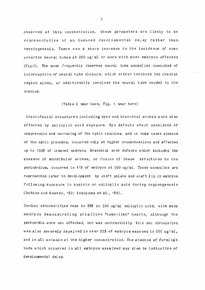
nI
observed at this concentration, these parameters are Iikely to be
representaLive of an induced developmental delay rather than
teratogenesis. There was a sharp increase in the incidence of open
anterior neural tubes at 200 ug/ mI or nore with rnosL enbryos affected
(Fig.1). The most frequently observed neural lube anomalies consisted of
interruption of neural tube closune, which either involved the cranial
region alone, or additionally involved the neural tube caudal lo the
cra ni um.
(lable 2 near here, Fig. 1 near here)
Craniofacial structures including eyes and branchial- arches were also
affected by salicyJ.ic acid exposure. Eye defects whích consÍsted of
compression and narrowing of the optic vesicles, and in some cases absence
of the optic pÌacodes, occurred only at higher concentrations and affected
up to 100t of treaLed embryos. Branchial arch defects which included the
absence of nandibular arches, or fusion of these structures to the
pericardiun, occurred in 411 of embryos at 100 uglnl. These anonalies are
represented later in developnent by cleft palate and cleft lip in embryos
foLlowÍng exposure to aspirín or saJ.icylic acid during organogenesis
(DePass and lleaver, t82; Yokoyana el aI., t84).
Cardiac abnornalities rose to 88f at 300 uglml salicylic acid, with many
enbryos demonsLrating primitive ntube-liken hearts, although the
perÍcardia were not affected, nor was contractility. YoLk sac circulafion
was also severely impaired inover 501 of embryos exposed to 200 ug/mI,
and in all animals al the higher concentration. The absence of forelinb
buds which occurred in all embryos exanined may also be indÍcative of
developnental delay.

I
SomeoftheteratologicatfeaturesobservedinwholeembryosinFigurel
were refÌlected in the histologicat sections (Fig' 2) of the cranial neural
tubes of salicylic acÍd-treated enbryos. TLre different appearance of the
sectÍons of cornparable Ievels in the craniun of control and salicylic
acid-treated animals in Figure 2, can be attrÍbuted to the compression of
the optic area and poor cephalic flexure in affected enbryos in t'he
forebrainandhindbrainareaSrespecbively.Inrepresentativeembryos
exposed to 300 ug/nl the neuraL folds had not fused and the neural tube
was severely distorted. The neuroepithelial walL was thin and there was a
l-argedegreeofcetlulardisorj.entation,withdark,irregulartissue
projecting into fhe ventricular Iumen of bhe neural tube' cells in the
neuroepitheliumandmesenchymer+erelessdenselypackedthanincontrol
enbryos, leading to targe extracelLular spaces' while large, dark
intracellular bodies !Iere also present within the neuroepithelíum'
Despite the severity of teratogenesis, nitotis did not appear to be
affected and niLotic figureswere visible at the lumenal nargin of the
neuroepithelium. The underlying mesenchyme contained ceIls which l¡ere
of ten closely apposed to the surface ectode¡'n whil-e most of the space in
this germ }ayer was occupied by large, abnormal, developing blood vessels.
(Fie.2 near here)
ultrastructural exanination of the anterior neural tube of affected
enbryos reveaÌed that the main affect of sal.icylic acid was on the
neuroepitheliun, and that the severity of necrosis was dose-dependent' At
100 ug/ml there was little evidence of dead or dying cells except in fhe
rostral part of the telencephalon which was consj'stent with normal
erobryonic development. Aü higher concentrations of salicylic acid (Fie' 3)
the thin neuroepithelial waI1 with its shrunken cells contained Iarge

9
extracellular spaces, and at the apical end condensed cyboplasmic blebs
pnojected into theventricufar Iumen. A comparison of this region in an
affecled embryo can be nade with a similar site in the control in Figune
3a.
The dark bodies observed previousJ-y at the cell-u-l-ar level appeared to be
membrane bound bodies or heterolysosomes containing a variety of dead cell
debris, within ot,her apparently healthy ceLls which appeared Lo have
phagocytosed these remnants. While the dead ceLl material contained
within these structures was often too condensed bo identify as individual
organelles, extracellular debris could be identified as degenerating
cytoplasmic organel-les including condensed or swollen mj.tochondria,
vesiculated rough endoplasmÍc retÍculum, and free ribososroes. Electron-
dense amcrphous structures which were frequently associated with sites of
necrosis may be primary lysosomes.
In regions of the neuroepithelium al the basaL end adjacent to bhe
mesenchyme, ceIl s w ere often sw oIIen, cell m embranes w ere severely
disrupted enabling t,he cytoplasrnic contents to leak from these cells into
extracellular spaces, and Ín sone cases into the nesenchyme through
disrupted regions of the neuroepithelial basal lamina.
DISCUSSION
This study demonstrates for the first Lime bhe uLtrastructural patholory
Índuced by salicylic acid, a metabolÍte of aspirin in the tissue of the
developÍng rat neural tube, and confirros and extends salicylic acid-
induced growth and norphological anonalies neported previously. The
malformations observed in embryos exposed to salicylic acfd 1n vitro

10
in this study demonstrated a dose-related dysmorphology, and were sinilar
Lo anonaLies reported in rats in vÍvo (Vlarkany and Takacs, r59; Kimmel et
aI., t71; McGarrity et al., t78) f olLowing aspirin administration. This
suggests thal salicylic acid exerts a teratogenic effect directly on the
devetoping embryo rather than through malernal mediation, and confirns
previous suggestions (Kinme1 eb aI. , '71i Koshakj i and Schu1ert, '72) that
this netabotite is a causative agent in aspirin teratogenesis. This
proposal is further strengthened by observations made by Yokoyana et al.,
lrgt¡) of differential effects of aspirin and its netabolite, whereby short
term exposure to salicyJ-ic acid 1ed to localised malforrnations such as
cleft Iip and curly or short tail, whereas aspirin appeared to induce
edenatous anonalies of the face, which was attributed by these workers fo
a decLine of systemic physiological- functions.
l¡hile initial doses used in rats in the present study were approximately
f ive times higher than human therapeutic level-s of aspi.rin' plasma or
serun salicyLic acid leveÌs at 6 and 12 hours post administration are
comparable in both species (Woodbury, '73), The doses that produced
malfornatÍons in vitro in Lhis report were lower than in some other
sLudies, which may be attributed to differences in experimental design
including the time, and route of drug adminislration, or differences in
the teratogenic activity of aspirin, its netabolites and othen salicylate
compounds used in the various studies.
The present observations also confirm previous report,s both in vivo
(l.Jarkany and Takacs, t59; Kimnel et al', r? 1; DePass and 1¡¡eaver, t62) and
in vÍtro (Greenaway et a).., t82; Yokoyama et âf., t84) that aspirin or its
derivatives led to gnowth retardatíon in a dose-dependent nanner. While
most sbudies have demonstrated this effect in rat fetuses at term, in

11
vitro studies performed during the cribical- period of organogenesis have
the advantage of demonstrafing bhe temporal relationship between aspirin
administration and growth reduction. These studies in rabs nay be of
particular reLevance and importance as sone hunan studies have implicated
aspirin as a causative agent for reduced birthweight (Turner and Collins,
'75).
I'lhiI e aspirin- induced teratogenesi s has been f irmly establ ished in
animals (!larkany and Takacs, t59i Goldnan and Yakovac, 163; Greenaway eb
aI., t82; Guy et a1., t86)' the resuLts from hurnan studies are not
consisLent. The variations in outcone in epideniological reponts may occur
because of differences in methodology and analyses between studies, as
well as confounding personal and envÍronnental factors. Despite these
inconsistencies, aspirin should not be underestimated as a possible
nervous system teratogen in hunans. Even if teratogenesÍs is not evident
when aspirin is administered alone, its effects nay be exacerbated if it
is ingested with conpounds which nodif! Íts metabolisn Agents such as the
comrnonly occurning food addibÍve, benzoic acid which increased the
concentration and persÍstence of salicylic acid in Lhe plasma of naternaL
rats, also increased the level of teratogenesis of the nervous system and
other ongans Ín term fetuses conpared with aspirin alone' when
administered during the critical period of neurogenesis (days 9-11)
(Kiromel et al., '71). The higher Ievels of dysroorphology reported Ín the
presenl study compared with those where aspirin or sodÍum salicylate were
used also indicate that salicylic acid is a nore pofent teratogen than
aspirin or ils sodluro salt.
The nost frequently observed neural tube ano¡nalles were related to the
fallure of the neural tube to fuse, either in the cranlal region aIone,

12
which has been reported previously foltowing cul-ture of enbryos in sodium
salicylabe (McGarrity et â1., t81; Greenaway et a1.r t82), or which
additionally involved the neuraL tube caudal to t'he cranium' a
precondition of craniorachischisis, observed in aspirin-treated aninals at
term (Warkany and Takacs, t5gi Kimmel et al' , \1)' It is likely that the
more severe neural- tube malfornatj.ons observed in the present sbudy may
not be compatible with viabÍlity at term. The absence of chorio-al-lantoÍc
fusion in many of the enbryos at higher concentratÍons also nakes survival
unlikely as the animal- in utero is dependenb on an active placenta for its
nutritional requirenents Iater in development (New, t78).
The cellul-ar necrosis described in the neural tube of these saLicylic
acid-cultured embryos did not appear to be assocj-ated wÍth progranmed cell
death or apoptosis, which is a normal consequence of differentiation
(schluter, t73i Geelen and Langman, 177), as necroLic cells induced by
saticylic acid were apparent throughout the entire newoepithelium instead
of being confined to the site of closure along the roid-dorsal aspect of
the anterior neural tube. It is likely that the severe ce]l shrinkage' and
Iarge condensed bl-ebs adjacent to the ventricular lurnen, together with the
extensive celL loss throughout the neuroepÍthelium, nay interfere with
fusion of the neural foIds. The severe neural tube dysnorphology
demonstrated in the present study was also sinilar to abnormalities
índuced by zinc and vitamin E deficiencies (Record et al', t85a; Harding
et aI., r8B) and vitamin A excesses (Joschko el aI., r89). Necrotic cel-ls
wer,e also apparent Ín the cranial neural tubes of embryos exposed to these
teratogens, and the neuroepitheliurn in salicylic acid-induced ernbryos in
the present study showed a sÍmilar patLern of cytotoxicÍty which could
aLso be rel-aled to the histologicat abnormalities observed by Greenaway et

13
aI. , ( '82) accomparving Na salicyi-ate-induced gross dysrnorphology.
Although cell death is not necessarily a prerequisite for a teratogenic
response, such events have been reported (Scobt, t77) to occur as a resull
of a wide range of Lreatnents. AspÍrin induced dysnorphology has been
linked previously to a cellul-ar abnornality in the adult gastric mucosa of
-several species (Hingson and lto, t73i Pf eiffer and Weibe] , t73i Rainsford
and Brune, tJB)r and also in embryonic hindlimb, as KIein eù al., (r81)
reported an unique pattern of cytotoxÍcity in tbe hindlimb bud where celI
death was localised to the preaxial nesoderm of bhis structure in rat
fetuses. The absence of severe cel1 death t,hroughout the open anterior
neural tube in some salicylic acid-treated exencephaJ-ic enbryos in the
present study suggests that severe neural fube necrosis, except in regÍons
adjacent to sites of neural Lube closure, may be an additional teratogenic
effect,, poSsÍbly unrelated to neural lube dysrnorpholory.
It has been reported in a recent epidemiological sbudy (PytkowÍcz-
Streissguth et al., t87) that maternal aspirin ingestion during the first
half of pregnancy was significantly related to low IQ scores, and
attention deficits in four year old children, while Butcher et al., (,72)
reported Ínpaired learning ability in rat offspring exposed to maternal
aspirin These observations led the authors to propose that the necrosis
observed at different sites in the anterion neural tube nay be
functionally different. The presence of severe cellular disruption and
blebbing along the dorsal nargins of the neural Lube al the site of neural
tube fusion may interfere with neuruLation, whÍIe necrosls and cell loss
throughout the renainÍng neuroepithelium nay be linked with intellectual
and behavioural abnormal ities.

14
Several possibilities exist to account for the nechanisns which underlie
neural tube dysmorpholory. As the yolk sac and vascular development w ere
inpaired Ín narry enbryos exposed to salicylic acid in cuJ.ture, similar to
the nicotine-induced effects seen previously (Joschko et, aI., t91), and
since the yolk sac is crÍtical for adequate nutrition and oxygen uptake at
this time in embryonic development (New, tf8)r some of the neural lube
pathology nay derive from an inability of the yolk sac to neet the demands
for'essential nutrients. However, it is unlikely that this is the primary
mode of saLicylic acid-induced teratogenesis in view of the dÍfferent
patterns of dysmorphology between nicotine and salicylic acid-induced
embryos. Alt,ernatively it is likely that salicylic acid exerts a direct
cytotoxic effect on cel-l- rnenbranes, which were observed to be seriously
disturbed at sone sites in the neuroepilheliun, particularly at sites
adjacent to the nesenchyne, where Lhe basal Ianina was also frequently
seen to be disrupled. It is at these sites that necrotic neuroepithelial
debris, which is not digested by heterolysosones located wiLhin apparently
healthy ceIIs, can be removed from the newoepithelium and extruded into
the nesenchyme. This method of removal of neuroepithelial debris has been
previously observed in nicotine-cultured embryos (Joschko et al., t91).
Prostaglandins have also been implicated Ín adequate fetal development
since they are known to have a large number of desirable effects some of
which occur at the celluIar level (Horrobin, t80).AspirÍn, like zinc
deficiency J.eads to an inhibÍtion of prostaglandin synthesis (Horrobin and
Cunnane, t80; Metz, t81), and since bot,h of these experimentally induced
conditions produces neural tube dysmorpholory such as exencephaly at tern
(Metz, t81 i Warkany and Takacs, t59), and open neural tubes accompanied by
severe cell death in the developing nervous system (Record et aI., t85;

15
Joschko et al., tB9), these observations could suggest a role for
prostaglandins in the adequate development and cl-osure of the neural tube.
The fÍndÍngs denonstrate that the developing rat embryo is at risk from
salicylic acid exposure, and that, when related to humans, maternal aspirin
ingestion nay lead to brain abnormalitÍes, impaired developnent and IQ
po sl na t,al Iy.
ACKNOITTLEDcEMENTS
The authors wish bo thank Dr. P. Baghursl of CSIRO., Division of Hunan
Nutribion for his st,atisbical advi.ce, Mr R. Murphy of AdelaÍde University
for photographic assistance, and Mr A. Partridge for typing this
manuscr ipt.

16
LITENATURE CTTED
Baker, R. J., and J. A. NeI der ( 1 97 8) The GLIM sy stem. ReI ease 3.
Generali sed LÍnear Interactive ModelL Íng. Num eri cal Al gorithm s Group'
Oxford.
Bostron, H., K. Berntsen and M.I\1.
properties of anti-inflammatory drugs"
Whitehouse ( 1 96 4) Bioch e¡o ical
metabol im Ín v ivo. Bioche¡0. PharnacoJ. . , 13 : 413-420
Charnock, J.S., L.J. 0pit, and B.S. Hetzel (1962) An evaluation of the
effect of salicylate on oxidative phosphorylatÍon in rat-Iiver
mitochondria. Biochem. J., 83: 602-606.
Co1).ins, E, and G. Turner (197ù Maternal effects of regular salicylate
ingestion in pregnancy. the Lancet., 2: 335-337
DePass, L.R, and E.V. l.leaver (1982) Conparison of teratogenic effecls of
aspirin and hydroxyurea in the FÍscher 344 and Wistar strains. J. Toxicol.
Envj.ron. Hea1th., 10: 297-305.
Goldman, 4.S., and W.C. Yakovac (1963) The
teratogenicity by rnaternal immobilization in
Ther. , 142: 351 -357 .
Greenaway, J.C., T.H. Shepard., A.G. FanteI, and
Sodiu¡n salicylate teratogenÍcity in vitro. Teratology.,
II. Some effects on sul phate-35s
enhancenent of salicylate
the rat. J. Pharnacol. exP.
M. R. Juchau ( 1 982)
167 -17 1 .26
Guy, J.F., and M.E. Sucheston (1986) TeratogenÍc effects on the cD-1
mouse embryo exposed t,o concurrenl doses of ethanol and aspirin.
Teratolory., l4: 249-261.

17
Horrobin, D.F. ( 1 9BO) A biochenical basis for aI coholism and aI cohol-
induced damage including the fetal alcohol- syndrome and cirrhosis:
rnterference with essentÍar fatty acid and prostag)'andin metabolism. Med'
Hypotheses., 6 : 929-942-
Horrobin, D.F., and s.c. cunnane (1980) Inüeractions between zinc'
essential fatty acids and prostaglandins. Med' tlypotheses', 6: 277-296'
Janakidevi, K., and M.J.H. Smith (1970) Effects of salicylate on RNA
polynerase activity and on the Íncorporabion of orotic acid and thymidine
into the nucLeic acids of rat foebuses invitro' J' Pharm' Pharnac" 22z
249-252.
Joschko, M.4., f. E. Dreosti, and R' S'
interactions and teratogenesis in rals:a
study. Nutr. Res. , 9z 205-216.
TuI si ( 1 989 ) zinc/ vitan in A
lieht and eleclron mÍcroscoPe
Joschko, M.4., r. E. Dreosti, and R. S. Tul si ( 1 991 ) The teratogenic
effects of nicotine in vitro in rats: a light and electron microscope
study. Neurotoxicol. Teratol., 13: 307-316.
Khera, K.S. (19?6) Teratogerricity studies with methotrexate, aroinopterint
and acetylsal.icylic acid in doroestic cats. Teratologl. I 14: 21-28'
Kimroel, C.4., J.G.!IiIson, and H.J. Schunacher (197 1) St'udies on
roetabolisn and identif ication of the causative agent Ín aspirin
teratogenesis in rats. Teratology., 4: 15-24.
Klebanoff, M.4., and H.Ï1. Berendes (1988) Aspirin exposure during the
firsb 20 weeks of gestationand IQ atfouryears of age. TeratoIoBY.r 37:
249-255.

18
Ko shakj i, R. P. ' and
salicylabe beratolory
A.R. Schulerb (1972) Biochemical" mechanj-sms of
in the rat. Biochen. Pharmacol., 222 407-416.
Larsson,K.S., and M. Eriksson (1966) Salicylate induced fefal death and
malforrnations in two mouse strains. Acta Paediatrica Scandinavica., 55:
569-576.
Levy, C. (1978) Clinical pharmacokinetics of aspirin Pediatnics (Suppl).,
62: 86T -872.
Levy, G., and J.R. Leonards (1966) Absorption, metabol-isn and excretion
of salicylates. In: The SaIicyIates. M.J.H. Smith and P.K. Smith' eds.
InterscÍence, New York, Pp. 5-48.
MeLz, SA- (1981) Anti-inflammatory agenLs as inhibitors of
syntbesis in man. Vred. CLin. Nth. Arnerica. , 65: 713-7Tl .
prostagl andi n
GI obus, and S. Robi nso n ( 1 96 5) Effect of som e
rat fetus. Toxicol. Appl. Pharmacol.,on the developing
McG arrity, C. '
(Abstract).
Sarnani, NJ, and Beck, F. (1978) J. AnaL.' 127: 646.
McGarrity, C., Samani, NJ., Beck, F. and Gulamhusein, A. (1981) The
effect of sodium salicylate on the rat embrao in culture: an invitro nodel
for t,he morphological assessmenb of teratogeniciby. J. Anat., 1332 257-
269.
M., and J.0. Forfar (197 1) Association between
McCo11, J.D., M.
therapeutic agents
7 z 409-417.
Nel so n, M.
a dmi nÍ stered
Br. Med. J.,
dr ugs
fetus.durÍng pregnancy
1: 523-527.
and congenital abnormalities of the

19
New, D.4.1. (1 978) whore-embryo cuLture and bhe study of namnalian
embryos during organogenesis. BioI. Rev., 532 81-122.
Pytkowicz-Streissguth, 4., R.P. Treder., H.M. Barr., T.H. Shepard., V,I.A.
BIeyer., P.D. Sampson, and D.C. MarLin ('1987) Aspirin and acetaminophen
use by pregnant women and subsequent child IQ and attention decrements.
Teratolory., 35: 211-219.
Record, I.R., R.S. Tulsi., f.E. Dreosti, and F.J. Fraser (1985a) In vitro
deveropnent of zinc-deficient and replete rat embryos. Aust. J. exp. BÍot.
Med. Sci., 6 3: 6 5-7 1,
Record, I. R., R. S. Tul si., f. E. Dreosti,
necrosis in zÍnc-deficient rat enbryos.
and F.J. Fraser (1985b) Cettul_ar
Terabology., 32: 397-40l-.
Richards, ï.D.G. ( 1 96 9) Congenital maI forn ations
soc. Med., 23:
and env ironmental
218-225.influences in pregnancy. Brit. J. prev.
(197il Associations between oral crefts and drugs t,aken during
Int. J. EpidemÍo1., 4: 3T-44.
Saxen, L.
pregnancy.
Shapiro, S., R.R. Monson., D.Vl. Kaufman.,
D. Slone (1976). Perinatal nortatity and
aspirin taken during pregnancy. Lancet., 1:
V. Siskind., 0.P. Heinonen and
birth-weight Ín relation to
137 5-137 6 .
sJ-one, D.v., 0. P. Heinonen., R.R. Monson., D.hr. Kaufman, and s. Shapiro
(1976) Aspirin and congenitar maLformalions. Lancel., 1: 1373-1375.
Snith' M.J.H. and P.K. Snith. The sal-icylates. J. tIlley and Sons, London
( 1966).

20
TrasIer, D.G. (1965) AspirÍn-induced cleft Iip and other nalformations in
mice. Lancet. , 1: 606-607.
Turner, G., and E. Collins (19?5) Fefal effects of negular salicylate
ingestion in pregnancy. Lancet, 2z 338-339.
vane, J.R (1g71) Inhibition of prostaglandin synthesis as a nechanisrn of
action for aspirin like drugs. Nature New BioI. ' 2312 232-235,
llarkany, J., and Takacs, E (1959) Experimental production of congenital
rnarf ormations in rats by saticyrate poisoning' Am' J' Pat'hol-' ' 352 315-
331 .
Woodbury, D.M. (1973) Analgesics
Pharmacological Basis of Therapeutics'
MacMillan, New York, PP 312'34U,
and antipyretics. In: The
L. S.Goodrnan and A. GiInan' eds.

21
TABLE 1
EFFECT OF IN VITRO S&TCYLTC ACID OI'I GRChITH OF RAT EMBRYOS ]N CULTIJRE.
TREATI"IENTlSalicyJ-ic Acid Concentration (uS/mI)
0 100 200 300
Total erobraos 15
Crown- rum p
leneth (nn)
No. of somÍtes
Yolk sacdiameter (rnn)
3.28t0.07
23.37t0.41
17
3.10r0.07
21 .66l¡0.51
15
2.60r0.08
17 .23¡1 .01
17
1 .45t0.08
7 .29t0.42
4.17ú.07 3.88¿0.10 3.56:10.13 3.02¿0.07
Values are means + SEM.

22
TABLE 2
EFFECT OF IN VITRO SALICYLIC ACID ON I'ORPHOLOGICAL DEVELOB'IENT IN RATS.
TREATMENT
SaIicy1ic acid concentration (ue/nI)o 100 200 300
SIGNIFICANCE OF
C0NCEXIITRATION
RH,ATED EFFECT (P)
Total enbryos 15 17 15 17
Ileuraldef ecf s
tube1 15( 100)
B( 53.3)
3( 20)
3Q0)
<0 .00 1
<0 .00 1
<<0 .00 1
<0 .00 1
Ele defecLs
Branchial archdef ect s
Heart defects
0( 0)
0( 0)
1(5.9)
0( 0)
0( 0)
0( 0)
0( 0)
0( 0)
17( 100)
1r(88.2)
7 (41 .2)
1 5( 88.2)
Absence off orel irnb buds
Incompletef1 exion
ï-mpairedfusion
Yolk saccircul ation
0(0) 1(5.9) r0(66.7) 17(100) <0.001
1(6.7) 4(23.Ð 15(100) 17(100),. <0.001
0(0) 4(23.5) 6(40) 17(1oo) <o.oo1
2(13.Ð 2(11.6) 8( 53.3) 17( 100) <0.001
lPercentages given in parentheses

23
FIG. 'I
FIG. 2
LEGENDS TO FIGURES
a. 11.5 day control enbryo. the neural foLds have fused along the
entire tength of the neural tube. b. 11.5 day exencephalic enbryo
exposed to 200 ug/mI salÍcylic acid. The cranial neural folds had
not fused except in the region between the forebrain vesicles
(arrow). The enbryo had not completed rotation, and only the
mandibu.l-ar arches ( 1) had developed. c. 11 .5 day embryos exposed to
300 uglml salicylic acid, with open neuraL tubes which extend fron
behind t,he forebrain vesicles (arrows) to the caudal end of the
enbryos. The neural tube was distorLed in the hindbrain region (ar
rowheads). Note thaü the allantois had not fused. d. YoIk sac of an
11.5 day control embryo with well developed blood vessels (arrows).
e. Abnormal 11.5 day yolk sac and embryo exposed to 200 uglml
salicylic acid with poorly developed blood vessels. Fb, forebrain;
Mb, midbrain; Hb, HÍndbrain; 0P, opbic placode; 0T, otic vesicle;
11213, branchial arches; H, heart; F, forelimb bud; YS, yolk sac.
Bars= 500um âr€i 1mm b,c,d.
a. Horizontal section through cranial neural tube of an 11.5 day
control embryo. b. Higher nagnification al a site near the arrolr Ín
a, showing mitotic figures (arrowheads) and closely packed cells in
the neuroepithelium. c. Horizontal seclion through the cranium of
an 11.5 day enbryo cultured in 300 ug/nJ. salicylic acid. The neural
folds have failed to fuse. The opbic vesicles are compressed and
the neuroepithelium is severely shrunken and thin Tt¡e cells in the
mesenchyme are confined Lo regions near the surface ectodern and
nost of the space is occupled by large, abnormal, developing blood

FIG.3
2\
vessers. d. Higher magnification of the neuroepitheLiun at a site
near the arnow Ín c, showing lange extracellurar spaces and
condensed, dead ceLl debris within the neuroepitherial walr and
projecting into the ventricular lumen (arrows). Numerous nitotic
figures are visible (arrowheads). Fb, forebraini Mb, midbrain; Hb,
hindbrain; N, neuroepitheliun; M, mesenchyne; E, surface ectoderm;
L, ventricular Iumen; NF, neural folds; 0, optic vesicle; bv, blood
vessel. Bars=50um.
a. Electron nicrograph of a section of the cranial portion of an
11.5 day controL embryo at the apicar end of the neuroepithelÍum,
with cytoplasmic projections (or blebs) (arrows) adjacent to the
ventricurar lunen. The ceLrs are in cLose proxinity and are
connected by junctional complexes at the apicar ends. b. A simirar
section of an 11.5 day ernbryo exposed to 300 ug/rn1 saricylic acid,
showing large irregular, condensed brebs (arrows) protruding into
the ventricular lunen, extensj.ve extracellular spaces and numerous
heteroJ.ysosomes (arrowheads). Note the nucreus has lost its normal
columnar orientation. c. A range of celt debris consisting of
cytoplasmic organelLes from the neuroepithelium of an 11.5 day
embtao cultured in 3oo uglmI salicylic acid. (The arrows indicate
sites where cell menbranes are disrupted). d. Neuroepitherial cell
debris consisting of dispersed cytoplasmic rÍbosornes and condensed
nitochondria, was extruded from cell-s at the basal end of the
neuroepithrium into the extracellurar natrix of the mesenchyne, at
sites where the basal Lamina has been disrupted (arrowhead). Arrows
indicate regions of severe celr menbrane disruption. N,
neuroepithelÍun; L, ventricular lumen; M, nesenchyne; BL, basal_

25
laminai ic, junctional complex; n' nucleusi m' miLochondria; vPr
vesiculated rough endoplasmic relÍculum; cr' cyboplasmic ribosones;
r, rough endopJ-asmic reticulum; Ly, lysosome' Bars= 4um' â' €i 1um'
b, c, d'

F i:i

I
l:ìq.z
f
t
I
a
I
Ì.¡
+
NF---
Fb
NF
t-_l
f'\. .a
;. 1
-¡
þv -.i:tr:a''
.ì
Li¿ I
ìji,tt\,.1I ¡rt.ì
c
lI
.- Itì
d
l'
Ii¡

I ix,z\L
\

Teratology 36, p151 , 1987 .
THE COMBINED EFFECTS OF ZINC DEFICIENCY AND HYPERVITAMINOSIS A ON THE
DEVELOPING CENTRAL NERVOUS SYSTEM IN RATS
M.A. Joschko *+ I.E. DreosEi,* and R.S. Tulsi+
'tCSIRO Division of Human NuEriEion, Adelaide, S'A' 5000*Department of Anatony and Histology, university of Adelaide' Adelaide 5000
Zinc an¿ vitamin A are known to play a role in cell membrane stabil-isation. Zinc is also required for the mobilísagion of vitamin A from Ehe
liver. Dietary manipulation of both nut.rients leads to a ÈeraEogenic effecEin -animals and subsequent impairrnenE of neural tube development' The
present Study invesEigated wheEher a teratogenic interaction occurs between
low maternal zinc "t"1n" and hypervttaminosis A during Pregnancy in raEs'
Electron microscopy was used t,o examine the individual and combined effectsof these t.".tt"r,is on norphology of the neural tube and its cellularmembrane conponenf.s.
Scanning electron nicroscopy of Il-day-old zinc-deficienE embryos ofEen
revealed extensively open neural tubes, as well aS anencephaly or micro-cephaly. Blebbing of the neuroepithelíurn r¡ras common. TreatmenE with excess
.rìlami" A led to õpen neural Eubes mainly confined to craníal regions. llanyembryos r¡Iere exencãphalic, wiEh neural folds severely evaginated and heavilybtebbed. When the treatments \¡Iere superimposed, teratology was enhanced'The neural tube was often extensively open and evaginated cranially.Exencephaly or anencephaly \./aS observed and special sensory organs \¡Iere
frequently affecEed.
Transrnission elect.ron micrographs of zinc deficient, 1l-day-old embryos
revealed severe neuroepichelial cell death, and disrupted cell- and míto-chondrial membranes in some living cells. The supporÈing mesenchynal cissuewas less affected. Ilypervitaninosis A exerted its teraEogenic assault nain-ly on Ehe nesenchyne, with a lesser insulE on neuroepithelium abutEing the
lumen. CeII death was common and severe swelling and breakdown of roiEochon-drial membranes vlas observed in many living cells. ConcurrenE EreaEmenf
with boEh Eeratogens showed extensive cell death in the neuroepiEhelium and
mesenchyme, and an enhanced degree of nembrane disrupEion.
This sEudy demonsEraEes E,he involvemenE of membrane damage and celldeath in the tera¡ogenesis associaEed with in uEero zínc deficiency and
hyperviEarninosis A. Superimposition of Ehe EreaEmenE.s leads Eo an additiverather than an inEeractive effecL on Èhe developing cent,ral nervous sysEem
making a common mechanism of action unlikely.

M.A. Joschko, I.E. Dreosti and R.S. Tulsi (1988) An in vitro study of the effects of
nicotine on the developing central nervous system in rats.
Journal of Anatomy, v. 161, pp. 259, 1988
NOTE: This publication is included in the print copy of the thesis
held in the University of Adelaide Library.

M.A. Joschko, I.E. Dreosti and R.S. Tulsi (1990) The teratogenic effects of nicotine in
the developing central nervous system in rats: an in vitro study, In: The Australian
Teratology Society, Inc. 7th Annual Scientific Meeting, December 6–8, 1989.
Teratology, v. 42 (3), pp. 329, September 1990
NOTE: This publication is included in the print copy of the thesis
held in the University of Adelaide Library.
It is also available online to authorised users at:
http://dx.doi.org/10.1002/tera.1420420317