Ontogenetic regulation of leukocyte recruitment in mouse yolk sac vessels
Upload
independentCategory
view
1download
0
Abstract Newly laid eggs of stick insects comprise aunique fluid ooplasm that is gradually partitioned into anumber of yolk granules by invasion of secondary vi-tellophages. This study aimed at establishing how yolkgranules become acidified in the course of embryonicdevelopment. Data show that acidified yolk granules arerather scarce and randomly distributed in vitellophagesof early embryos, while they tend to increase graduallyin number as development proceeds to completion. Yolkgranule acidification is progressively more inhibited inthe presence of increasing concentrations of chloroquine,monensin and bafilomycin. A pro-protease was identi-fied cytochemically and by immunoblotting in yolk ex-tracts of progressively more advanced embryos. A spe-cific monoclonal antibody raised against this pro-prote-ase helped to demonstrate that it is gradually processedto yield a lower molecular weight polypeptide as devel-opment proceeds to completion. This latter polypeptidewas identified as a protease using electrophoresis inpolyacrylamide gels containing yolk extracts. Simulta-neous administration of a fluorescent substrate for cys-teine protease and an acidotropic probe produced superi-mposable labelling patterns, suggesting that only acidi-fied yolk granules possess a proteolytic activity. On theother hand, yolk granules probed simultaneously for ac-idification and latent pro-protease yielded labelling pat-terns partially superimposed. Pro-protease labelling isgradually lost as yolk granules are progressively moreacidified during development. Distinct labelling patternswere also obtained in vitellophages processed for the si-multaneous detection of pro-protease and protease, sug-gesting that the two activities are expressed by different
yolk granule populations, and that one is gradually con-verted into the other as time goes by.
Keywords Vitellin · Yolk utilisation · Limited proteolysis ·Acidification · Carausius morosus (Insecta)
Introduction
In insect eggs, yolk provides a storage of raw material tosustain embryo development (McGregor and Loughton1977; Yamashita and Indrasith 1988). To accomplish thisgoal, oocytes are endowed with the capability of seques-tering a yolk precursor or vitellogenin (Vg) from the ma-ternal blood stream through a process of receptor-medi-ated endocytosis (Raikhel 1984; Telfer and Pan 1988;Raikhel and Dhadialla 1992; Schneider 1996). The se-questered Vg is deposited in yolk granules as vitellin(Vt) (Giorgi et al. 1993a; Snigirevskaya et al. 1997a,1997b), and eventually degraded by a process of limitedproteolysis to yield a number of lower molecular weightpolypeptides with the onset of development (Indrasith etal. 1987; Masetti and Giorgi 1989; Nordin et al. 1990).As this process occurs, yolk granules are structurallymodified and progressively reduced in size. Limited pro-teolysis of Vt polypeptides and the resulting structuralalterations of the yolk granules are both caused by acti-vation of specific proteolytic enzymes (Indrasith et al.1988; Kageyama and Takahashi 1990; Liu et al. 1997).Many, if not all, of these proteolytic enzymes are mater-nally derived gene products that are transferred endocy-tically to the oocyte during vitellogenesis (Fagotto1990a, 1990b; Cho et al. 1991; Takahashi et al. 1996;Giorgi et al. 1997; Cho et al. 1999). Given this possibili-ty, it then follows that yolk granules of insect oocytesshould be endowed with the capability of storing bothVt(s) and the enzymes apt to degrade them.
It is generally agreed that yolk utilisation in insects istriggered by acidification of the yolk granules duringembryogenesis (Fagotto 1991; Nordin et al. 1991).Through this process a latent inactive proteolytic pro-
A.M. Fausto · G. Gambellini · M. MazziniDepartment of Environmental Sciences, Tuscia University, Viterbo, Italy
A. Cecchettini · M. Masetti · F. Giorgi (✉ )Department of Human Morphology and Applied Biology, University of Pisa, Via A. Volta 4, 56126 Pisa, Italye-mail: [email protected].: +39-50-501078, Fax: +39-50-501085
Cell Tissue Res (2001) 305:433–443DOI 10.1007/s004410100392
R E G U L A R A R T I C L E
Anna Maria Fausto · Gabriella GambelliniMassimo Mazzini · Antonella CecchettiniMassimo Masetti · Franco Giorgi
Yolk granules are differentially acidified during embryo development in the stick insect Carausius morosus
Received: 30 June 2000 / Accepted: 5 March 2001 / Published online: 1 June 2001© Springer-Verlag 2001

enzyme is gradually activated and made available for Vt cleavage in the luminal space of the yolk granules(Takahashi et al. 1993, 1997). The evidence available todate suggests that yolk granules in insects become moreand more acidified as Vt polypeptides are gradually re-duced in molecular weight, suggesting a causal relation-ship between these two events (Nordin et al. 1991).However, since many of the investigations carried out sofar were actually performed on isolated yolk granules,determination of their intraluminal pH under these con-ditions leads inevitably to loss of spatial information. Wethus still need to know how acidified yolk granules aredistributed inside the vitellophages during developmentand, above all, how their acidification is regulated duringyolk utilisation.
We have studied yolk utilisation in embryos of thestick insect Carausius morosus and found that yolk gran-ules in this species are reconstituted anew with the be-ginning of embryonic development (Fausto et al. 1994).By this time, the fluid ooplasm is gradually transformedinto a collection of progressively smaller yolk granulesdue to invasion of secondary vitellophages from the eggperiphery (Fausto et al. 1997). On their way into the flu-id ooplasm these macrophagic cells engulf yolk by bulkendocytosis and partition it into newly formed yolk gran-ules. Thus, to establish how yolk granule acidification isspatially distributed in developing insect embryos onehas to clarify the role vitellophages play in relation togranule degradation and Vt polypeptide proteolysis. Thatacidification may play a key role in the control of intra-cellular proteolytic processing has been widely demon-strated in a variety of cell systems, including the islets ofLangerhans from mouse pancreas. In this tissue, proinsu-lin is converted to mature insulin as secretory vesiclesare gradually acidified on their way to the cell membranefor exocytosis (Orci et al. 1986). Acidification is also in-volved in controlling ligand degradation by activatingspecific hydrolases in rat liver lysosomes. Their intralu-minal pH is gradually acidified by activation of an ATP-driven proton pump inserted in the lysosomal membrane(Schneider 1982).
In the present study, we examined protease activityand yolk granule acidification in intact insect yolk sacsof progressively more advanced embryos. Data showthat the extent of acidification in vitellophagic yolk gran-ules is both temporally and spatially correlated with con-version of a latent pro-protease into an active proteolyticenzyme.
Materials and methods
Sample preparation
Specimens of the stick insect Carausius morosus (Br.) (Phasmat-odea, Lonchodinae) were reared on English ivy leaves and main-tained in aerated cages at constant temperature and humidity.Newly laid eggs were collected weekly, maintained under thesame conditions throughout development and staged according toFournier (1967) upon dissection.
Structural observations
For scanning electron-microscopic observations, yolk sacs weredissected in phosphate-buffered saline (PBS) and fixed in Karnov-sky’s fixative as previously reported (Fausto et al. 1994). Theywere then thoroughly rinsed in 0.1 M cacodylate buffer at pH 7.2,postfixed for 2 h in 1% osmium tetroxide in 0.1 M cacodylatebuffer at pH 7.2, dehydrated in an ascending series of alcohols andeventually critical point dried in a Balzer’s apparatus equippedwith a liquid CO2 inlet. Yolk sacs were subsequently mounted, either intact or manually fractured, over aluminium stubs and met-al shadowed in a sputtering unit (Balzer’s) equipped with an argoninlet. Samples were observed under a JEOL JSM 5024 scanningelectron microscope. For light-microscopic observations, dehy-drated yolk sacs were embedded in Epon-Araldite mixture andsemithin sections (1 µm thick) prepared by ultramicrotomy.
Image analysis
A number of light-microscopic sections from yolk sacs at variousdevelopmental stages were examined with a computer-assisted im-age analyser. Estimates of the yolk granule sizes were calculatedby averaging the number of granule counts for each developmen-tal stage. The mean and standard deviations of granule sizes weredetermined for each stage and plotted against developmental timeas a percentage of maximum granule size. Maximum granule sizewas defined as the equivalent area of unparted fluid ooplasm ex-amined by computer analysis in newly laid eggs.
Antibodies
The antibodies used in this study were obtained from the follow-ing sources: a mouse polyclonal anti-pro-protease serum was agenerous gift from Prof. J.H. Nordin, Department of Biochemistry,University of Massachusetts, USA. It was originally preparedagainst Blattella germanica and proved to be cross-reactive for thesame antigen in C. morosus. This antiserum was shown to be spe-cific for the peptide sequence trimmed from the pro-enzyme uponconversion to an active cysteine protease (Liu et al. 1997). Amonoclonal mouse antibody (P5) against the same antigen fromstick insect embryos was prepared by Dr. Antonella Cecchettini atthe Auburn University Hybridoma Facility (Auburn University,AL). In C. morosus, this antibody reacts both with the pro-prote-ase of newly laid eggs and a 40-kDa polypeptide of developmen-tally more advanced embryos.
Gel electrophoresis
Newly laid eggs or yolk sacs freshly dissected from embryos at var-ious stages of development were resolved by sodium dodecyl sul-phate polyacrylamide gel electrophoresis (SDS-PAGE). Haemo-lymph and developing ovarian follicles were also obtained fromadult females as previously described (Giorgi et al. 1993a). Sampleswere directly homogenised in 250 µl 50 mM TRIS-HCl buffer atpH 6.8 containing a mixture of proteolytic inhibitors (1 mM PMSF,1.4 µM pepstatin, 1.2 µM cystatin, 3 µM aprotinin and 10 mM iodo-acetic acid and 5 mM EDTA). After spinning at 10,000 rpm in a re-frigerated bench centrifuge, samples were diluted with an equal vol-ume of 50 mM TRIS-HCl buffer at pH 6.8 containing 5% β-mer-captoethanol, 5% SDS and boiled for 3 min. Protein concentrationwas determined spectrophotometrically using the procedure ofBradford (1976). Denaturing (SDS) gel electrophoresis was carriedout according to Laemmli (1970) using a 5–15% polyacrylamidegradient and run overnight in a Bio-rad protein II apparatus at 15°Cwith 0.1% SDS in the upper electrode reservoir. Polyacrylamidegels were stained for 1 h in 0.1% Coomassie brilliant blue R-250 in30% acetic acid–10% methanol, destained in the same solution andeventually dried under vacuum at 60°C.
Ovarian follicles and haemolymph were radioactively labelledfollowing 4 h exposure to [35S]-methionine (Radiochemical Centre,
434

Amersham, UK) and resolved by SDS-PAGE as specified above.Polypeptide B2 was electroeluted from polyacrylamide gels and di-gested for 30 min at 37°C with papain dissolved in 50 mM ammo-nium bicarbonate, pH 8.0, at the final concentration of 5 µg/100 µgsample. Reaction was stopped by addition of 25 µl SDS samplebuffer followed by boiling for 3 min. The reaction mixture wasthen loaded on a second polyacrylamide gel and re-run in parallelwith untreated Vt polypeptides, including the 40-kDa polypeptide.
Developing embryos of C. morosus were also resolved by elec-trophoresis in 7.5% polyacrylamide gels containing 250 µg newlylaid egg extracts as substrate for protease activity. After electro-phoresis, the gel was soaked in 2.5% Triton X-100 for 1 h, rinsedwith 50 mM TRIS-acetate buffer at pH 7.4 and incubated for up to72 h at 25°C in the same buffer containing 10 mM β-mercaptoeth-anol. Gels were eventually stained in Coomassie blue as specifiedabove. Proteolytic activity is associated with polypeptides remain-ing unstained in the gel.
Western blotting
Electrophoresed polyacrylamide gels were placed onto nitrocellu-lose sheets and trans-blotted for 2 h at 16°C following the proce-dure of Towbin et al. (1979). The nitrocellulose sheets were thenallowed to react with primary mouse antibodies for 3 h at roomtemperature and eventually treated for 1 h with goat anti-mousesecondary antibodies conjugated with alkaline phosphatase (Bio-rad). Reactivity was revealed by incubation with Bio-rad develop-ers and blocked by addition of 0.1 M HCl.
Confocal scanning laser microscopy
Yolk sacs were freshly dissected from developmentally differentembryos of C. morosus and placed for 30–60 min in 200 µl sterileGrace’s medium containing 100 nM of the acidotropic probe Lyso-tracker (Molecular Probes, OR). As a nuclear counterstain, Syto 16was added to the culture medium up to a final concentration of1 µM. By the end of incubation, samples were thoroughly rinsed inPBS and observed within 15 min. Specificity of probe incorporationinto the yolk granules was checked by incubating yolk sacs for thesame length of time in the presence of either monensin (at concen-trations of 8–80 µM), chloroquine (at concentrations of 10 µM to12.5 mM) or bafilomycin (at concentrations of 100 nM to 1.5 µM).
A few Lysotracker-labelled yolk sacs were fixed either in 4%formaldehyde or 1% glutaraldehyde in 0.1 M cacodylate buffer atpH 7.2. However, fixation greatly impaired reproducibility of obser-vations, formaldehyde causing the probe to leak out from the yolkgranules, and glutaraldehyde resulting in an enhanced autofluores-cent background. All data reported in this study are therefore restrict-ed to unfixed yolk sacs observed upon completion of incubation.
Freshly dissected yolk sacs were exposed for 30 min at 30°C toacridine orange at a concentration of 100 µM in 25 mM HEPES-NaOH at pH 7.00 containing 50 mM KCl and 200 mM NaCl aspreviously reported by Nordin et al. (1991). Samples were thenrinsed in the same buffer and immediately observed under the con-focal microscope. Calibrations for acridine orange accumulationin yolk granules were performed over dissected vitellophages fol-lowing the procedure of Fagotto and Maxfield (1994). Proteolyticactivity was revealed by incubating freshly dissected yolk sacs inequilibrating buffer (5 mM HEPES containing 0.15 N NaCl and2 mM EDTA at pH 7.35) at room temperature for 1 h. A cysteineprotease substrate (Molecular Probes, OR) was then added at thefinal concentration of 500 µM and allowed to react for an addi-tional hour. Samples were then rinsed in equilibrating buffer andimmediately observed under the confocal microscope. Specificityof the cysteine protease reactivity was checked by 10 min preincu-bation in the presence of E-64 at a final concentration of 10 µM.
Some yolk sacs were also investigated for the localisation of apro-protease. Freshly dissected yolk sacs were first permeabilised for30 min in 0.5% Triton X-100 in PBS and then incubated for 1 h in aprimary, anti-pro-protease, mouse antibody, diluted 1:100 in PBS.Yolk sacs were then thoroughly rinsed in PBS and incubated for
30 min at room temperature with either a rhodamine- or a fluores-cein-conjugated goat anti-mouse IgG antibody (Amersham, UK) di-luted 1:200 in PBS. A number of yolk sacs were also double labelledfor the simultaneous detection of: (1) protease and acidification, (2)pro-protease and acidification and (3) pro-protease and protease ac-tivities. In all of these instances, samples were incubated as specifiedabove with either Lysotracker or cysteine protease substrates andsecondary antibodies for the pro-protease added to the yolk sacs afterPBS rinsing. All samples exposed to the above probes were observedwithin 30 min of the last incubation under a Leica DM IRB light mi-croscope connected to a TCS 4D confocal scanning system equippedwith an Ar/Kr laser. Fluorescent signals were detected using488/568-nm and 520/590-nm emission wavelengths.
Results
Morphological observations
Newly laid eggs in stick insects comprise a unique fluidooplasm. During development, this fluid is gradually par-titioned into a number of granules due to invasion of sec-ondary vitellophages from the egg periphery (Fausto et al.1994). These behave as macrophagic-like cells engulfingportions of the fluid ooplasm by bulk endocytosis on theirway into the egg. Continuation of this process leads even-tually to confinement of the granulated ooplasm into a cy-lindrical yolk sac lying dorsally to the developing embryo(Fig. 1A). Upon reaching stage VII, embryos become dor-sally enclosed and sealed from the surrounding perivitel-line fluid by interposition of a thick parietal cuticle associ-ated with several muscle layers. Yolk sacs fractured priorto metal shadowing revealed the presence of numerous vi-tellophages closely adjoined to one another (Fig. 1B).When isolated from the yolk sac, these cells appear to hosta number of polygonally shaped yolk granules clusteredlike berries in a grape (Fig. 1C, D). Their cytoplasm is re-duced to an extremely thin fringe along the cell periphery,while the nucleus is either centrally compressed amidstthe yolk granules or peripherally displaced (Fig. 1E). Dur-ing development, yolk granules are gradually reduced insize. Figure 2 shows that size reduction is attained by pro-gressively diminishing the range of variability, suggestingthat the ooplasm shifts from an initial condition in whichyolk granules are highly heterogeneous (ranging in diame-ter from 40 to 4 µm) to a final one in which they are alluniformly small (with a mean diameter of about 4 µm). Inaddition, the way yolk granule size varies over develop-mental time does not entirely match the extent Vt is grad-ually depleted during development, protein concentrationin yolk sacs remaining almost unchanged until stage VIand diminishing abruptly only afterwards (Fig. 2).
Yolk granules are differentially acidified during development
In this study we aimed at establishing how yolk granulesbecome acidified in the course of embryonic develop-ment. To this end, yolk sacs were first exposed to the ac-idotropic probe Lysotracker. Figure 3A–D illustrates asequence of vitellophagic fields taken from embryos pro-
435

gressively more advanced in development. Early in de-velopment, acidified yolk granules are rather scarce andrandomly distributed within the yolk sac: even adjacentvitellophages may differ in the number of acidified yolkgranules (Fig. 3A, B). A higher number of acidified yolk
granules are observed in yolk sacs from embryos moredevelopmentally advanced (Fig. 3C). On approachingstage VII of embryonic development, almost all yolkgranules have become acidified (Fig. 3D). The initialstages of acidification are likely to include small vesiclesthat are either budding from the granule periphery (seeFig. 3B, inset) or embedded inside the granule matrix(see Fig. 3C, inset). When isolated from early yolk sacsand maintained in a culture medium for up to 4 h, vi-tellophages remained asymmetrically acidified (Fig. 4A).This suggests that no leakage of acidotropic probe islikely to occur in vitro, and that a selective proton per-meability is expressed autonomously by the yolk granulemembrane, regardless of the integrity of the yolk sac.
Even though repeatedly proven to accumulate intracel-lularly upon acidification (Griffiths et al. 1988; vonSteyern et al. 1996), the kinetics by which Lysotracker isincorporated in membrane-bound organelles do not corre-late linearly with their luminal pH. To verify whether yolkgranules are indeed differentially acidified, the above ex-periments were repeated in the presence of acridine or-ange. Figure 4B is an example of a yolk sac from a stageIV embryo exposed to acridine orange. Under these condi-tions, only a few yolk granules appear acidified, whileothers retain their greenish background of neutrality. Uponcalibration of the red/green ratio displayed by yolk sacsexposed to acridine orange, the lowest luminal pH at-tained by the yolk granules appears to be within the rangeof 5.5–5.8. To prove that granule acidification was indeeddue to activation of specific proton channels, Lysotrackeruptake was further checked in the presence of such specif-ic inhibitors as chloroquine (Fig. 5A–C), monensin(Fig. 5D–F) and bafilomycin (Fig. 5G–I). In all of theseinstances, increasing inhibitor concentrations reduced pro-gressively probe uptake. By the end of embryonic devel-
436
Fig. 1A–E Scanning- andlight-microscopic views of yolksacs dissected from early stageVII embryos. A An intact yolksac. B Part of a yolk sac frac-tured prior to metal shadowingto show the presence of severalclosely adjoined vitellophages.C A single vitellophage gentlyremoved from the yolk sac.D A 1-µm-thick epoxy sectionof a single vitellophage to showyolk granules inside. E A laserconfocal microscopic view ofan intact vitellophage exposedto Syto 16 to label specificallythe cell nucleus located exter-nally to the yolk granules. Bars 200 µm (A), 35 µm (B),15 µm (C), 12 µm (D, E)
Fig. 2 Yolk granule size variation in vitellophages of yolk sacsfrom embryos progressively more advanced in development. Sizevariations were determined on sections using a computer-assistedimage analyser and expressed as percentage of the maximumgranule size corresponding to an equivalent area of the fluid ooplasm in newly laid eggs. The protein content of yolk sacs forthe same developmental stages is also reported for comparison.The picture depicts an example of yolk granule heterogeneity in astage III3 yolk sac

437
opment, the acidotropic probe was no longer incorporatedby the yolk sac, suggesting that yolk granules may havebecome entirely fluidified by then (data not shown).
Acidified yolk granules express a proteolytic activity
Yolk granule acidification is thought to trigger limitedproteolysis of Vt polypeptides through specific activationof maternally derived proteases. To verify whether expres-
sion of this enzyme activity in stick insects is temporallyrelated to acidification of yolk granules, developmentallydifferent yolk sacs were exposed in vitro to a fluorescentsubstrate for cysteine protease and the resulting vitello-phages examined by confocal microscopy. Initially, only afew yolk granules appear to be fluorescently labelled(Fig. 6A, B). However, as development proceeds to com-pletion, more and more yolk granules become proteolytic-ally active (Fig. 6C). Yolk sacs cultured in the presence ofE-64, a specific inhibitor for cysteine protease (Warner et
Fig. 3A–D Yolk granule acid-ification in embryos at differentdevelopmental stages. Observa-tions were carried out follow-ing 60 min of in vitro exposureto 100 nM Lysotracker (Molec-ular Probes, OR). A Vitello-phages are from an early stageIV embryo. B Vitellophages arefrom a stage V embryo. C Vi-tellophages are from a stage VIembryo. D Vitellophages arefrom an initial stage VII em-bryo. Insets show two yolkgranules comprising vesicleseither budding from the granuleperiphery, as in B, or embeddedinside the granule matrix, as inC. Both pictures have been en-larged 4 times with respect tothe original size. Bars 18 µm(A–C), 10 µm (D)
Fig. 4A, B In vitro cultured yolksacs. A Vitellophages were cul-tured in vitro in Grace’s mediumfor up to 4 h and then tested fortheir ability to incorporate Lyso-tracker (100 nM) during the last30 min of incubation. Underthese conditions, vitellophagesretain a differential probe distri-bution, only a few yolk granulesexhibiting maximum labellingwith Lysotracker. B A yolk sacdissected from an early stage IVembryo exposed for 30 min toacridine orange at a final concen-tration of 100 µM. Only a fewyolk granules appear either or-ange or yellow, the remainingones retaining their neutral green-ish colour. Bars 20 µm (A, B)

438
Fig. 5A–I Lysotracker in thepresence of increasing concen-trations of inhibitors. Chloro-quine was supplied at the con-centration of 25 µM (A), 50 µM(B), and 500 µM (C). Monen-sin was supplied at the concen-tration of 8 µM (D), 20 µM (E),and 40 µM (F). Bafilomycinwas supplied at the concentra-tion of 15 nM (G), 150 nM(H), and 1.5 µM (I). Bars40 µm (A–I)
Fig. 6 Cysteine protease activ-ity in yolk granules. Yolk sacswere dissected from embryos atvarying developmental stagesand incubated for 1 h in equi-librating HBS buffer containinga cysteine protease substrate(Molecular Probes, OR) at theconcentration of 500 µM.A–C are stage V, VI and VIIyolk sacs, respectively. Thespecificity of cysteine proteasewas checked by 10 min prein-cubation with E-64 at a finalconcentration of 10 µM (D).Bars 18 µm

439
al. 1995), revealed little or no proteolytic activity(Fig. 6D). To verify the expectation that yolk granules ex-pressing a proteolytic activity are also acidified, develop-mentally advanced yolk sacs were cultured in the presenceof both Lysotracker and the fluorescent substrate for cys-
Fig. 7A–C Acidified yolk granules express a cysteine proteaseactivity. Developmentally advanced yolk sacs (stage VII) werecultured in Grace’s medium containing both Lysotracker at theconcentration of 100 nM and a fluorescent substrate for cysteine
protease at the concentration of 500 µM. The labelling pattern pro-vided by Lysotracker (A) appears perfectly superimposable (C) tothat of the cysteine substrate (B). Bars 20 µm (A–C)
Fig. 8 Upper panel Pro-protease is a maternally derived geneproduct. Ovarian follicles and developing yolk sacs were resolvedby gel electrophoresis under denaturing conditions (SDS-PAGE)and tested by immunoblotting with an antiserum raised againstcysteine pro-protease of Blattella germanica. Polypeptide B2 of57 kDa appears labelled both in the haemolymph (He) and earlyvitellogenic (EV) and late vitellogenic (LV) ovarian follicles. Poly-peptide B2 is also labelled in yolk sacs, though gradually decliningin staining intensity in stages progressively more advanced in de-
velopment. Molecular weights of vitellin polypeptides are report-ed as previously determined (Giorgi et al. 1993b). Lower panelDeveloping yolk sacs were resolved by SDS-PAGE and tested byimmunoblotting against mAb P5. Note that both polypeptide B2and the 40-kDa polypeptide are labelled. Polypeptide B2 was ex-tracted from [35S]-methionine-labelled haemolymph and treatedwith papain. After treatment two polypeptides corresponding to 57and 40 kDa can be resolved by gel electrophoresis. The extracted40-kDa polypeptide is electrophoresed for comparison
teine protease. Under these conditions, the labelling pat-terns provided by the two probes appeared perfectly su-perimposable (Fig. 7A–C), suggesting that acidificationand proteolysis of the yolk granules are spatially associat-ed and perhaps causally related phenomena.

440
Proteolytic activity results from conversion of a latent pro-protease
To verify the notion that Vt-processing proteases are ma-ternally derived gene products stored as inactive pro-enzymes, we made use of an antiserum specificallyraised against the cysteine pro-protease of Blattella ger-manica. When tested in stick insects, the antiserum ap-peared to cross-react with a single polypeptide of 57 kDacommon to both haemolymph and vitellogenic ovarianfollicles (Fig. 8), previously indicated as polypeptide B2(Giorgi et al. 1993b). The same polypeptide appeared tobe labelled with a declining intensity in developing yolksacs. This suggests the possibility that pro-protease maybe reduced in relative concentration as it is gradually
Fig. 9 Left panel Extracts from newly laid eggs were incubated at37°C for time intervals ranging from 0 to 72 h. The electrophoret-ic pattern shown here mimics the profile yielded by Vt polypep-tides during embryonic development (cf. lower panel of Fig. 8).Right panel Developing embryos were resolved by electrophore-sis in gels containing newly laid egg extracts as a substrate. Notethe presence of an unstained protein fraction corresponding to the40-kDa polypeptide
Fig. 10A, B Pro-protease and protease are expressed by differentyolk granule populations. A Vitellophages from yolk sacs of earlyembryos (stage V) provide staining patterns that are only partiallysuperimposable when tested simultaneously for Lysotracker andanti-pro-protease labelling. Many yolk granules retain their origi-nal green (for pro-protease) or red (for acidification) colours.B Vitellophages from yolk sacs of early embryos (stage V) yieldonly partially superimposable staining patterns when tested simul-taneously for cysteine protease activity (green colour) and anti-pro-protease labelling (red colour). Bars 10 µm (A, B)
converted to an active protease during development(Fig. 8). The antiserum raised against B. germanica pro-protease was previously shown to be specific for theleader sequence trimmed upon conversion to protease(Liu et al. 1997). This explains why the activated prote-ase is not specifically labelled by this antiserum both inB. germanica (Liu and Nordin 1998) and C. morosus(see Fig. 8). Thus, to verify whether polypeptide B2 instick insect yolk sacs may be processed to yield a lowermolecular weight protease, a monoclonal antibody wasspecifically raised against it and tested by immunoblot-ting on developmentally different embryos. Under theseconditions, polypeptide B2 was shown to hold a precur-sor-product relationship with a lower molecular weightderivative of 40 kDa (Fig. 8). Appearance of this latterpolypeptide could also be mimicked in vitro by partiallydigesting polypeptide B2 with papain (see Fig. 8), thusindicating that pro-protease conversion may be triggeredby an enzyme member of the papain superfamily. To fur-ther prove that pro-protease conversion in stick insectembryos is causally related to Vt polypeptide processing,two additional lines of evidence were provided. First,newly laid eggs were incubated at 37°C for time inter-vals ranging from 0 to 72 h and the resulting extracts re-solved by SDS-PAGE. Figure 9 shows that Vt polypep-tides processed under these conditions reproduce theelectrophoretic patterns yielded by yolk sacs during de-velopment, thus showing that newly laid eggs are enzy-matically autonomous, requiring no external supply tosustain Vt proteolysis. Second, a proteolytic activitycould be demonstrated by gel electrophoresis in the pres-ence of egg extracts and shown to be associated with the40-kDa polypeptide appearing de novo upon conversionof the acidified pro-protease (Fig. 9).
Having provided evidence that proteolytic activity re-sults from conversion of a latent pro-protease, we thenwished to verify how pro-protease itself is distributedwithin the yolk granule population during development.As already shown, vitellophages simultaneously testedfor Lysotracker uptake and cysteine protease providestaining perfectly superimposable patterns (see Fig. 7C).On the other hand, when developmentally earlier vitello-phages were exposed to Lysotracker and simultaneouslytested with anti-pro-protease antiserum, their stainingpatterns were only partially superimposable, most of the
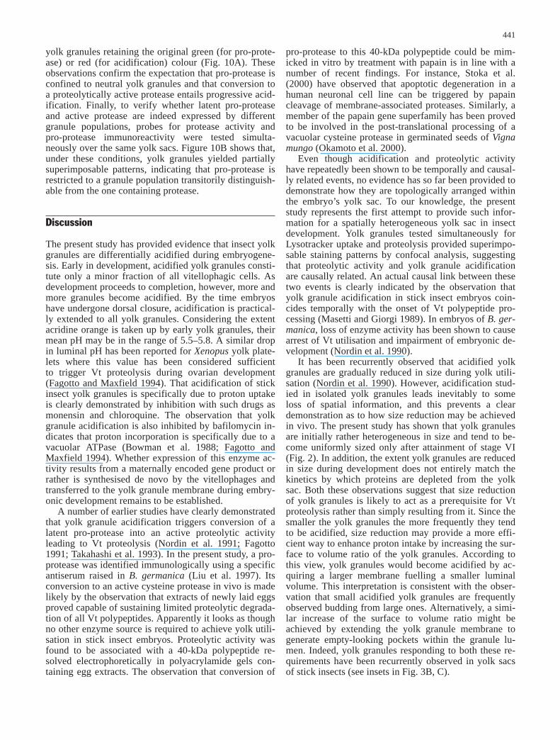
yolk granules retaining the original green (for pro-prote-ase) or red (for acidification) colour (Fig. 10A). Theseobservations confirm the expectation that pro-protease isconfined to neutral yolk granules and that conversion toa proteolytically active protease entails progressive acid-ification. Finally, to verify whether latent pro-proteaseand active protease are indeed expressed by differentgranule populations, probes for protease activity andpro-protease immunoreactivity were tested simulta-neously over the same yolk sacs. Figure 10B shows that,under these conditions, yolk granules yielded partiallysuperimposable patterns, indicating that pro-protease isrestricted to a granule population transitorily distinguish-able from the one containing protease.
Discussion
The present study has provided evidence that insect yolkgranules are differentially acidified during embryogene-sis. Early in development, acidified yolk granules consti-tute only a minor fraction of all vitellophagic cells. Asdevelopment proceeds to completion, however, more andmore granules become acidified. By the time embryoshave undergone dorsal closure, acidification is practical-ly extended to all yolk granules. Considering the extentacridine orange is taken up by early yolk granules, theirmean pH may be in the range of 5.5–5.8. A similar dropin luminal pH has been reported for Xenopus yolk plate-lets where this value has been considered sufficient to trigger Vt proteolysis during ovarian development (Fagotto and Maxfield 1994). That acidification of stickinsect yolk granules is specifically due to proton uptakeis clearly demonstrated by inhibition with such drugs asmonensin and chloroquine. The observation that yolkgranule acidification is also inhibited by bafilomycin in-dicates that proton incorporation is specifically due to avacuolar ATPase (Bowman et al. 1988; Fagotto andMaxfield 1994). Whether expression of this enzyme ac-tivity results from a maternally encoded gene product orrather is synthesised de novo by the vitellophages andtransferred to the yolk granule membrane during embry-onic development remains to be established.
A number of earlier studies have clearly demonstratedthat yolk granule acidification triggers conversion of alatent pro-protease into an active proteolytic activityleading to Vt proteolysis (Nordin et al. 1991; Fagotto1991; Takahashi et al. 1993). In the present study, a pro-protease was identified immunologically using a specificantiserum raised in B. germanica (Liu et al. 1997). Itsconversion to an active cysteine protease in vivo is madelikely by the observation that extracts of newly laid eggsproved capable of sustaining limited proteolytic degrada-tion of all Vt polypeptides. Apparently it looks as thoughno other enzyme source is required to achieve yolk utili-sation in stick insect embryos. Proteolytic activity wasfound to be associated with a 40-kDa polypeptide re-solved electrophoretically in polyacrylamide gels con-taining egg extracts. The observation that conversion of
pro-protease to this 40-kDa polypeptide could be mim-icked in vitro by treatment with papain is in line with anumber of recent findings. For instance, Stoka et al.(2000) have observed that apoptotic degeneration in ahuman neuronal cell line can be triggered by papaincleavage of membrane-associated proteases. Similarly, amember of the papain gene superfamily has been provedto be involved in the post-translational processing of avacuolar cysteine protease in germinated seeds of Vignamungo (Okamoto et al. 2000).
Even though acidification and proteolytic activityhave repeatedly been shown to be temporally and causal-ly related events, no evidence has so far been provided todemonstrate how they are topologically arranged withinthe embryo’s yolk sac. To our knowledge, the presentstudy represents the first attempt to provide such infor-mation for a spatially heterogeneous yolk sac in insectdevelopment. Yolk granules tested simultaneously forLysotracker uptake and proteolysis provided superimpo-sable staining patterns by confocal analysis, suggestingthat proteolytic activity and yolk granule acidificationare causally related. An actual causal link between thesetwo events is clearly indicated by the observation thatyolk granule acidification in stick insect embryos coin-cides temporally with the onset of Vt polypeptide pro-cessing (Masetti and Giorgi 1989). In embryos of B. ger-manica, loss of enzyme activity has been shown to causearrest of Vt utilisation and impairment of embryonic de-velopment (Nordin et al. 1990).
It has been recurrently observed that acidified yolkgranules are gradually reduced in size during yolk utili-sation (Nordin et al. 1990). However, acidification stud-ied in isolated yolk granules leads inevitably to someloss of spatial information, and this prevents a cleardemonstration as to how size reduction may be achievedin vivo. The present study has shown that yolk granulesare initially rather heterogeneous in size and tend to be-come uniformly sized only after attainment of stage VI(Fig. 2). In addition, the extent yolk granules are reducedin size during development does not entirely match thekinetics by which proteins are depleted from the yolksac. Both these observations suggest that size reductionof yolk granules is likely to act as a prerequisite for Vtproteolysis rather than simply resulting from it. Since thesmaller the yolk granules the more frequently they tendto be acidified, size reduction may provide a more effi-cient way to enhance proton intake by increasing the sur-face to volume ratio of the yolk granules. According tothis view, yolk granules would become acidified by ac-quiring a larger membrane fuelling a smaller luminalvolume. This interpretation is consistent with the obser-vation that small acidified yolk granules are frequentlyobserved budding from large ones. Alternatively, a simi-lar increase of the surface to volume ratio might beachieved by extending the yolk granule membrane togenerate empty-looking pockets within the granule lu-men. Indeed, yolk granules responding to both these re-quirements have been recurrently observed in yolk sacsof stick insects (see insets in Fig. 3B, C).
441

When yolk sacs were exposed to a pro-protease anti-body and simultaneously tested for acidification, yolkgranules appeared differentially stained. The fact that theresulting staining patterns were partially superimposed islikely to result from the antibody specificity that is re-stricted to the peptide sequence of the latent pro-proteasetrimmed upon conversion to active protease (Liu et al.1997). In line with our earlier findings in embryos of thecockroach B. germanica, it is possible that the leadingpeptide of the latent pro-protease may be transitorily re-tained within the yolk granules even after protease hasbeen fully activated (Giorgi et al. 1997).
In conclusion, the discovery that proteolytic enzymesoriginate from latent inactive pro-enzymes deposited inthe oocyte during ovarian development has entirely modi-fied our way of viewing yolk utilisation in insect embry-os. Rather than considering yolk granules as functionallyequivalent to endosome-like organelles, it would seemmore appropriate to consider them as a special type of ly-sosome or sleepy lysosomes (Fagotto 1995) endowedwith the capability to contain both the substrate to be de-graded and the enzymes capable of hydrolysing it. This isat variance from what is presently known about intracel-lular digestion in somatic cells where proteolysis followsfusion of endosomes with pre-existing lysosomes (Gruen-berg and Maxfield 1995; Mellman 1996). While proteoly-sis in most cells is triggered by lysosomal enzymes con-veying to the substrate within the endosome (Futter et al.1996), in insect embryos co-localisation of hydrolytic en-zymes and Vt within the same yolk granules points to ac-idification as a potential mechanism controlling the onsetof yolk degradation (Giorgi et al. 1999). Thus, evidencethat yolk granules are differentially acidified raises thefundamental question of how acidification is regulated invivo during embryo development. Since acidification ofmany eucaryotic cell organelles requires activation of avacuolar ATPase (Nelson 1992), a likely hypothesis isthat this activity may be transferred anew to the yolkgranules at the time secondary vitellophages invade thefluid yolk mass of newly laid eggs. Activation of ATPasecould in turn be controlled by the actual outcome of yolkgranule proteolysis. An interesting possibility arisingfrom this hypothesis is that differential acidification mayultimately lead to a spatially differentiated release of Vtpolypeptides from the yolk granules. Given this possibili-ty, an initially homogeneous distribution of Vt polypep-tides within the fluid ooplasm could eventually yield aheterogeneous distribution of yolk polypeptide deriva-tives along the embryo’s axis.
References
Bowman EJ, Siebers A, Altendorf K (1988) Bafilomycins: a classof inhibitors of membrane ATPases from microorganisms, ani-mal cells and plant cells. Proc Natl Acad Sci USA 85:7972–7976
Bradford MM (1976) A rapid and sensitive method for the quanti-tation of microgram quantities of proteins utilizing protein-dyebinding. Anal Biochem 72:248–254
Cho WL, Deitsch KW, Raikhel AS (1991) An extraovarian proteinaccumulated in mosquito oocytes is a carboxypeptidase acti-vated in embryos. Proc Natl Acad Sci U S A 88:10821–10824
Cho WL, Tsao SM, Hays AR, Walter R, Chen JS, SnigirevskayaES, Raikhel AS (1999) Mosquito cathepsin B-like protease in-volved in embryonic degradation of vitellin is produced as alatent extraovarian precursor. J Biol Chem 274:13311–13321
Fagotto F (1990a) Yolk degradation in tick eggs: I. Occurrence ofa cathepsin L-like acid proteinase in yolk spheres. Arch InsectBiochem Physiol 14:217–235
Fagotto F (1990b) Yolk degradation in tick eggs: II. Evidence thatcathepsin L-like proteinase is stored as a latent, acid activableproenzyme. Arch Insect Biochem Physiol 14:237–252
Fagotto F (1991) Yolk degradation in tick eggs: III. Developmen-tally regulated acidification of the yolk spheres. Dev GrowthDiffer 33:57–66
Fagotto F (1995) Regulation of yolk degradation, or how to makesleepy lysosomes. J Cell Sci 108:3645–3647
Fagotto F, Maxfield F (1994) Changes in yolk platelet pH duringXenopus laevis development correlate with yolk utilization. JCell Sci 107:3325–3337
Fausto AM, Carcupino M, Mazzini M, Giorgi F (1994) An ultra-structural investigation on vitellophage invasion of the yolkmass during and after germ band formation in embryos of thestick insect Carausius morosus (Br). Dev Growth Differ36:197–207
Fausto AM, Mazzini M, Cecchettini A, Giorgi F (1997) The yolksac in late embryonic development of the stick insect Cara-usius morosus (Br). Tissue Cell 29:257–266
Fournier B (1967) Echelle résumée des stades du developpementembryonnaire du phasme Carausius morosus Br. Acta SocLinn Bordeaux 104:1–30
Futter CE, Pearse A, Hewlett LJ, Hopkins CR (1996) Multivesicu-lar endosomes containing internalized EGF-EGF receptorcomplexes mature and then fuse directly with lysosomes. JCell Biol 132:1011–1023
Giorgi F, Cecchettini A, Lucchesi P, Mazzini M (1993a) Oocytegrowth, follicle cell differentiation and vitellin processing inthe stick insect Carausius morosus (Br.) (Phasmatodea). Int JInsect Embryol Morphol 22:271–294
Giorgi F, Masetti M, Ignacchiti V, Cecchettini A, Bradley JT(1993b) Postendocytic vitellin processing in ovarian folliclesof the stick insect Carausius morosus (Br.). Arch Insect Bio-chem Physiol 24:93–111
Giorgi F, Yin L, Cecchettini A, Nordin JH (1997) The vitellin-pro-cessing protease of Blattella germanica is derived from a pro-protease of maternal origin. Tissue Cell 29:293–303
Giorgi F, Bradley JT, Nordin JH (1999) Differential vitellin poly-peptide processing in insect embryos. Micron 30:579–596
Griffiths G, Hoflack B, Simons K, Mellman I, Kornfeld S (1988)The mannose 6-phosphate receptor and the biogenesis of lyso-somes. Cell 52:329–341
Gruenberg J, Maxfield FR (1995) Membrane transport in the en-docytic pathway. Curr Opin Cell Biol 7:552–563
Indrasith LS, Furusawa T, Shikata M, Yamashita O (1987) Limiteddegradation of vitellin and egg-specific protein in Bombyxmori. Insect Biochem 20:725–734
Indrasith LS, Sasaki T, Yamashita O (1988) A unique protease re-sponsible for selective degradation of yolk protein in Bombyxmori. Purification, characterization and cleavage profile. JBiol Chem 263:1045–1051
Kageyama T, Takahashi Y (1990) Purification and characteriza-tion of a cysteine proteinase from silkworm eggs. Eur J Bio-chem 193:203–210
Laemmli UK (1970) Cleavage of structural proteins during the as-sembly of the head of bacteriophage T4. Nature 227:680–682
Liu XD, Nordin JH (1998) Localization of the proenzyme fromthe vitellin-processing protease in Blattella germanica by af-finity purified antibodies. Arch Insect Biochem Physiol 38:109–118
Liu XD, McCarron RC, Nordin JH (1997) A cysteine protease thatprocesses insect vitellin. J Biol Chem 271:33344–33351
442

Masetti M, Giorgi F (1989) Vitellin degradation in developing em-bryos of the stick insect Carausius morosus. J Insect Physiol35:689–697
McGregor DA, Loughton BG (1977) Amino acid composition,degradation and utilization of locust vitellogenin during em-bryogenesis. Wilhelm Roux’s Arch 181:113–122
Mellman I (1996) Endocytosis and molecular sorting. Annu RevCell Dev Biol 12:575–625
Nelson N (1992) Organellar proton-ATPases. Curr Opin Cell Biol4:564–660
Nordin JH, Beaudoin EL, Liu XD (1990) Proteolytic processing ofBlattella germanica vitellin during early embryo development.Arch Insect Biochem Physiol 15:119–135
Nordin JH, Beaudoin EL, Liu XD (1991) Acidification of yolkgranules in Blattella germanica eggs is coincident with prote-olytic processing of vitellin. Arch Insect Biochem Physiol18:177–192
Okamoto T, Toyooka K, Minamikawa T (2000) Identification of amembrane-associated cysteine protease with possible dualroles in the endoplasmic reticulum and protein storage vacu-ole. J Biol Chem 276:742–751
Orci L, Ravazzola M, Amherdt M, Madsen O, Perrelet A, VassalliJD, Anderson RGW (1986) Conversion of proinsulin to insu-lin occurs coordinately with acidification of maturing secreto-ry vesicles. J Cell Biol 103:2273–2281
Raikhel AS (1984) The accumulative pathway of vitellogenin inthe mosquito oocytes; a high resolution immuno and cyto-chemical study. J Ultrastr Res 87:285–302
Raikhel AS, Dhadialla TS (1992) Accumulation of yolk proteinsin insect oocytes. Annu Rev Entomol 37:217–251
Schneider DL (1982) ATP-dependent acidification of membranevesicles isolated from purified rat liver lysosomes. J BiolChem 256:1833–1838
Schneider WJ (1996) Vitellogenin receptors: oocyte-specificmembers of low-density lipoprotein receptor supergene fami-ly. Int Rev Cytol 166:103–137
Snigirevskaya ES, Hays ES, Raikhel AS (1997a) Secretory and in-ternalization pathways of the mosquito yolk protein precur-sors. Cell Tissue Res 290:129–142
Snigirevskaya ES, Sappington TW, Raikhel AS (1997b) Internal-ization and recycling of vitellogenin receptor in the mosquitooocyte. Cell Tissue Res 290:175–183
Stoka V, Turk B, Schendel SL, Kim TH, Cirman T, Snipas SJ, Ellerby LM, Bredesen D, Freeze H, Abrahamson M, BrommeD, Krajewski S, Reed JC, Yin XM, Turk V, Salvesen GS(2001) Lysosomal protease pathways to apoptosis: cleavage ofbid, not procaspases, is the most likely route. J Biol Chem (inpress)
Takahashi SY, Yamamoto Y, Shionoya Y, Kageyama T (1993)Cysteine proteinase from the eggs of the silkmoth Bombyxmori: indication of a latent enzyme and characterization of ac-tivation and proteolytic processing in vivo and in vitro. J Bio-chem 114:267–272
Takahashi SY, Yamamoto Y, Watabe S, Kageyama T (1996) Bom-byx acid cysteine proteinase. Invert Reprod Dev 30:265–281
Takahashi SY, Yamamoto Y, Watabe S, Kageyama T (1997) Auto-lytic activation mechanism of Bombyx acid cysteine protease(BCP). Biochem Mol Biol Inter 42:591–600
Telfer WH, Pan M (1988) Adsorptive endocytosis of vitellogenin,lipophorin and microvitellogenin during yolk formation inHyalophora. Arch Insect Biochem Physiol 9:339–355
Towbin H, Staehelin T, Gordon J (1979) Electrophoretic transferof proteins from polyacrylamide gels to nitrocellulose sheets:procedure and some applications. Proc Natl Acad Sci USA76:4350–4354
von Steyern FV, Josefsson JO, Tagerud S (1996) Rhodamine B, afluorescent probe for acidic organelles in denervated skeletalmuscle. J Histochem Cytochem 44:267–274
Warner AH, Perz MJ, Osahan JK, Zielinski BS (1995) Potentialrole in development of the major cysteine protease in larvae ofthe brine shrimp Artemia franciscana. Cell Tissue Res 282:21–31
Yamashita O, Indrasith LS (1988) Metabolic fates of yolk proteinsduring embryogenesis in Arthropods. Dev Growth Differ30:337–346
443
Copyright © 2022 FDOKUMEN