extraembryonic coelom, yolk sac, fetal membranes - IS MUNI
35
Zápatí prezentace 1 GENERAL EMBRYOLOGY 2 • Folding of the embryo – 4th week of development • Development of extraembryonic structures – extraembryonic mesoderm, extraembryonic coelom, yolk sac, fetal membranes: amnion and chorion. • Development of the placenta. Anomalies of the placenta and umbilical cord. • Multiple pregnancy – arrangement of fetal membranes. • The length of pregnancy, calculation of delivery date. • Fetus position in the uterus – situs, positio, praesentatio, and habitus. The length and weight of fetus during i.u. development. The rule of Haase. • Mature and full-term fetus, marks of mature fetus.
-
Upload
khangminh22 -
Category
Documents
-
view
1 -
download
0
Transcript of extraembryonic coelom, yolk sac, fetal membranes - IS MUNI
Zápatí prezentace1
GENERAL EMBRYOLOGY 2
• Folding of the embryo – 4th week of development
• Development of extraembryonic structures – extraembryonic mesoderm,
extraembryonic coelom, yolk sac, fetal membranes: amnion and chorion.
• Development of the placenta. Anomalies of the placenta and umbilical cord.
• Multiple pregnancy – arrangement of fetal membranes.
• The length of pregnancy, calculation of delivery date.
• Fetus position in the uterus – situs, positio, praesentatio, and habitus. The length
and weight of fetus during i.u. development. The rule of Haase.
• Mature and full-term fetus, marks of mature fetus.

Zápatí prezentace2
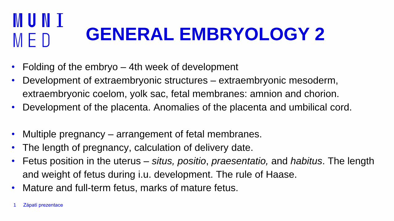
4th week – folding of the embryo (flexion)
Differentiation of intraembryonic mesoderm
3rd week
4th week
neural tubesomitesparaxial
intermediate
lateral
notochord
aortabody of embryo
primitive gut
aorta
sclerotome (base of vertebral
arch)
sclerotome (base of corpus
vertebrae)
dermatome, myotome
Mesoderm:
• paraxial mesoderm – thickened region of mesoderm along neural tube
• intermediate mesoderm (nephrotome) – in between paraxial and lateral mesoderm
• lateral mesoderm – keeps sheet-like structure
intermediate
lateral
paraxial
aorta dorsalisamniotic cavity
intercellular spaces
in lateral plate mesoderm
intermediate mesoderm
(nephrotome)
neural tube
intraembryonic coelom
somite
Mesoderm

4
Differentiation of intraembryonic mesoderm
– paraaxial (somites)
Somite
→ Dermatome,
Myotome, Sclerotome
Sefton EM, 2019
https://www.ncbi.nlm.nih.gov/pmc/articles/PMC6449175/
Zápatí prezentace5
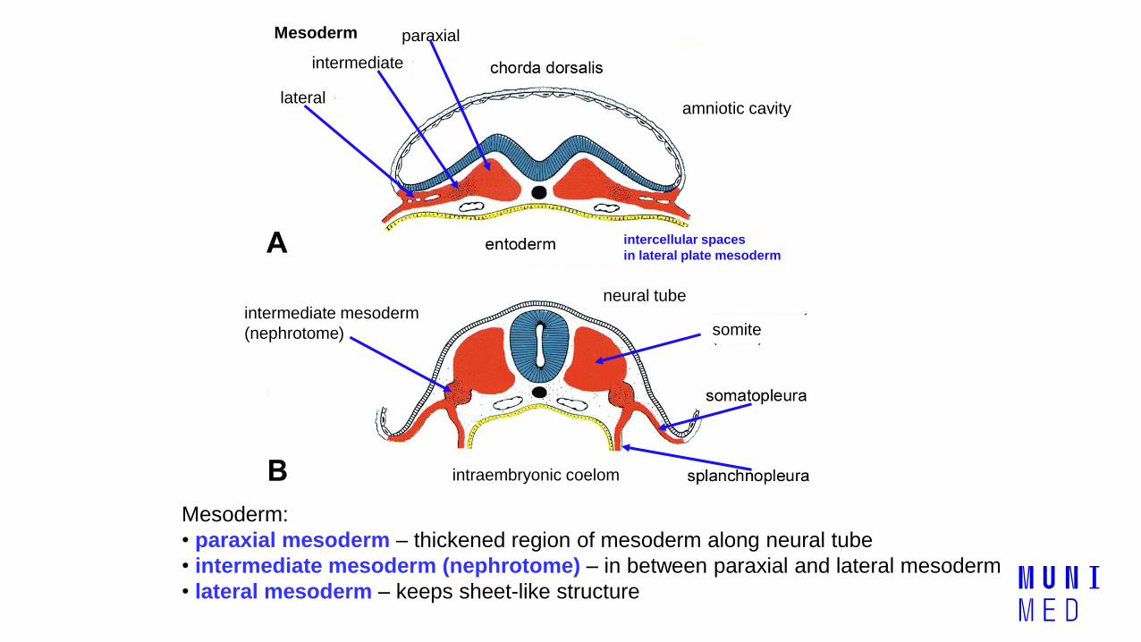
Differentiation of intraembryonic mesoderm
– intermediate and lateral
Intermediate mesoderm
→ urinary system (kidney, ureter),
genital system (gonads, ducts,
accessory glands)
Sefton EM, 2019
https://www.ncbi.nlm.nih.gov/pmc/articles/PMC6449175/
Lateral mesoderm (parietal and visceral layer)
→ dermis, hypodermis of ventral body parts,
connective tissue and muscle of viscera,
serous membranes, blood and lymphatic
vessels, spleen
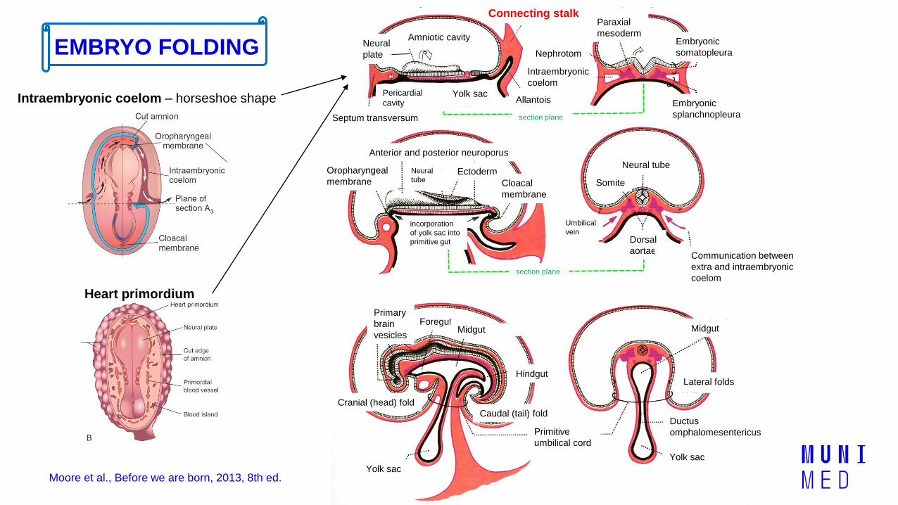
Amniotic cavityNeural
plate
Septum transversum
Connecting stalk
Allantois
Nephrotom
Intraembryonic
coelomPericardial
cavityYolk sac
Paraxial
mesodermEmbryonic
somatopleura
Embryonic
splanchnopleurasection plane
section plane
Umbilical
veinincorporation
of yolk sac into
primitive gut
Anterior and posterior neuroporus
Oropharyngeal
membrane
Neural
tubeEctoderm
Cloacal
membrane
Neural tube
Somite
Dorsal
aortaeCommunication between
extra and intraembryonic
coelom
Primitive
umbilical cord
Primary
brain
vesicles
ForegutMidgut
Hindgut
Cranial (head) fold
Caudal (tail) fold
Yolk sac
Midgut
Lateral folds
Ductus
omphalomesentericus
Yolk sac
EMBRYO FOLDING
Moore et al., Before we are born, 2013, 8th ed.
Intraembryonic coelom – horseshoe shape
Heart primordium
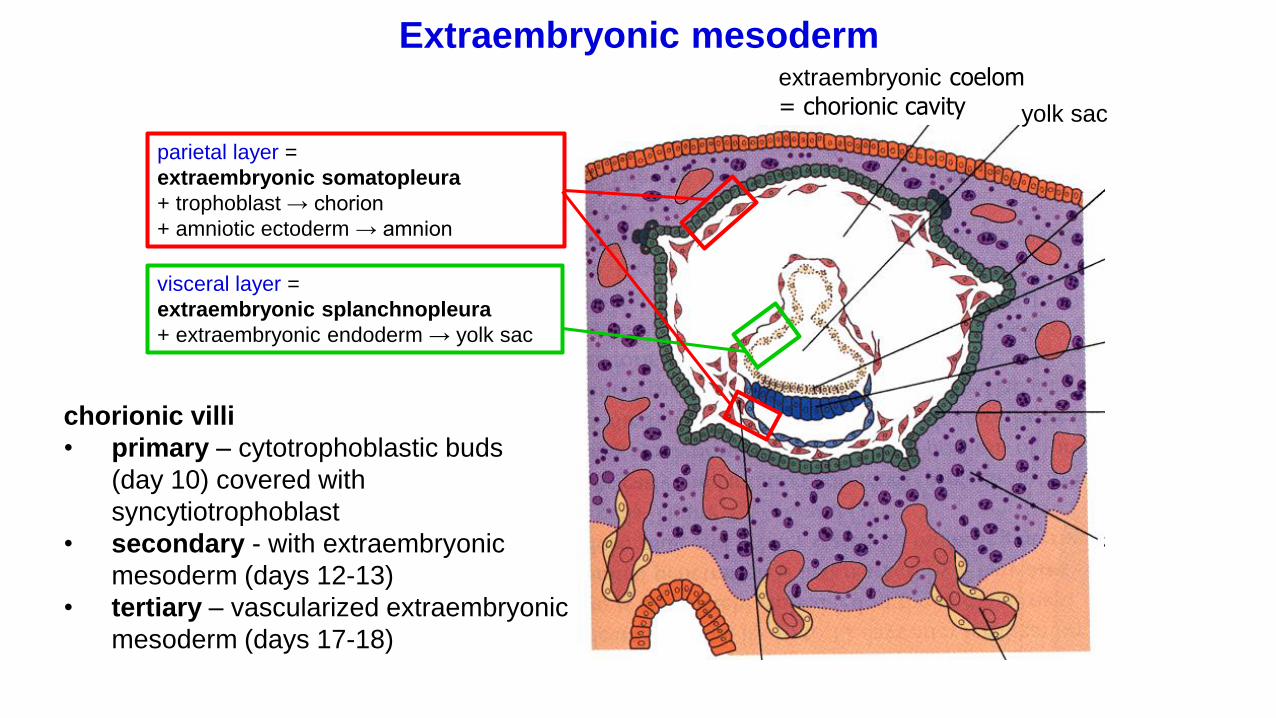
parietal layer =
extraembryonic somatopleura
+ trophoblast → chorion
+ amniotic ectoderm → amnion
visceral layer =
extraembryonic splanchnopleura
+ extraembryonic endoderm → yolk sac
extraembryonic coelom
= chorionic cavity
Extraembryonic mesoderm
chorionic villi
• primary – cytotrophoblastic buds
(day 10) covered with
syncytiotrophoblast
• secondary - with extraembryonic
mesoderm (days 12-13)
• tertiary – vascularized extraembryonic
mesoderm (days 17-18)
yolk sac

Yolk sac, amniotic sac, fetal membranes - amnion, chorion
chorion and
secondary chorionic villineural
tube
extraembryonic
splanchnopleura
connecting
stalk
extraembryonic
somatopleura
extraembryonic coelom
(chorionic cavity)
primary chorionic villi
umbilical
cord
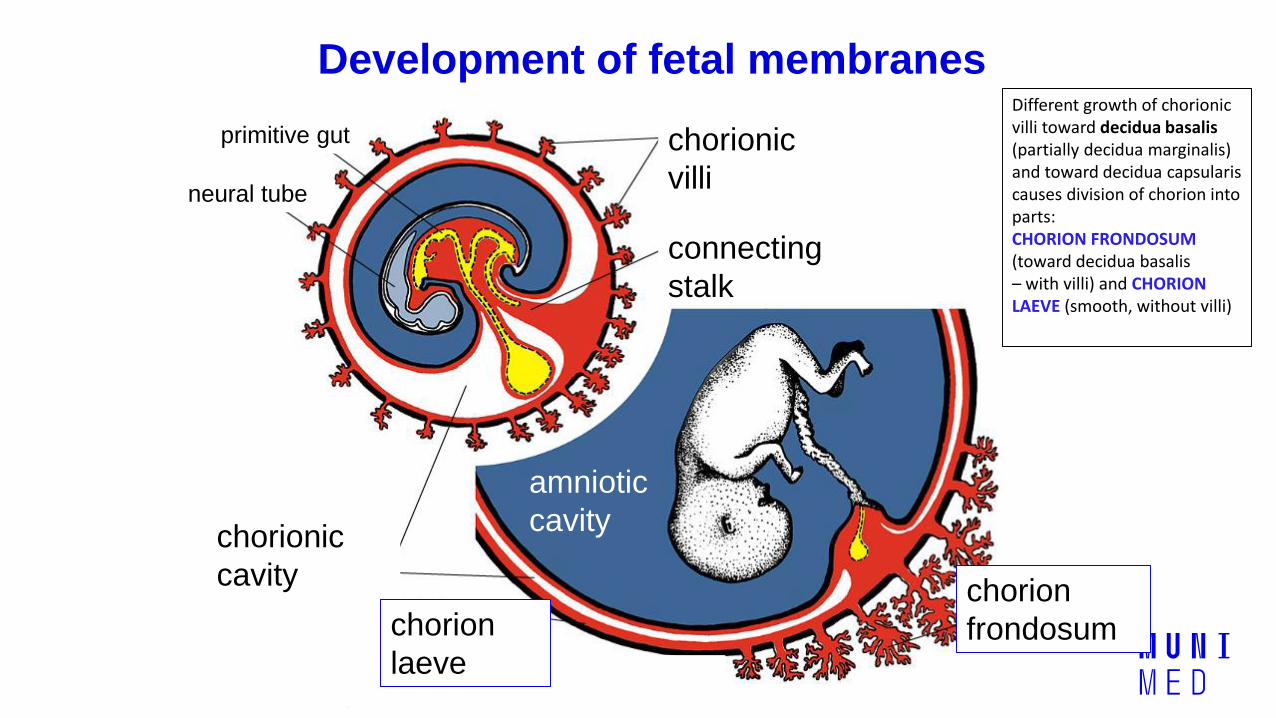
Development of fetal membranes
chorionic
villi
chorion
frondosum
primitive gut
neural tube
connecting
stalk
amniotic
cavitychorionic
cavity
chorion
laeve
Different growth of chorionicvilli toward decidua basalis (partially decidua marginalis) and toward decidua capsularis causes division of chorion into parts: CHORION FRONDOSUM (toward decidua basalis – with villi) and CHORION LAEVE (smooth, without villi)
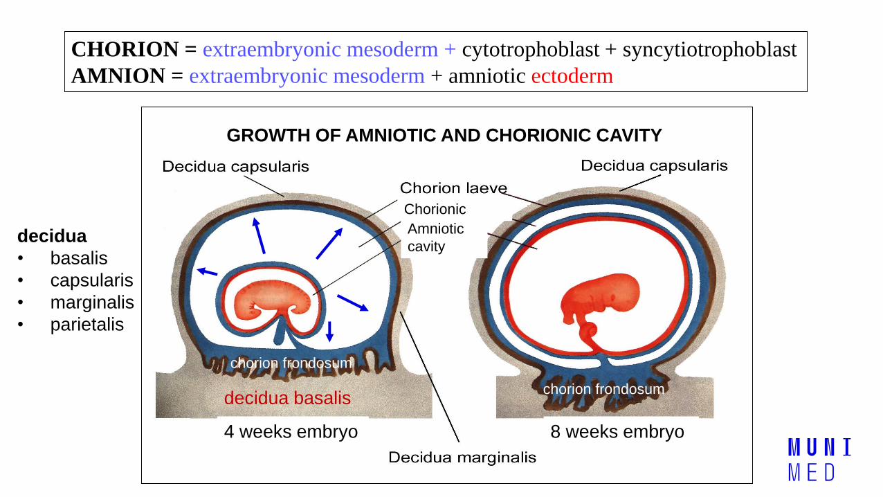
GROWTH OF AMNIOTIC AND CHORIONIC CAVITY
CHORION = extraembryonic mesoderm + cytotrophoblast + syncytiotrophoblast
AMNION = extraembryonic mesoderm + amniotic ectoderm
decidua basalis
4 weeks embryo 8 weeks embryo
Chorionic
cavityAmniotic
cavity
chorion frondosum
chorion frondosum
decidua
• basalis
• capsularis
• marginalis
• parietalis
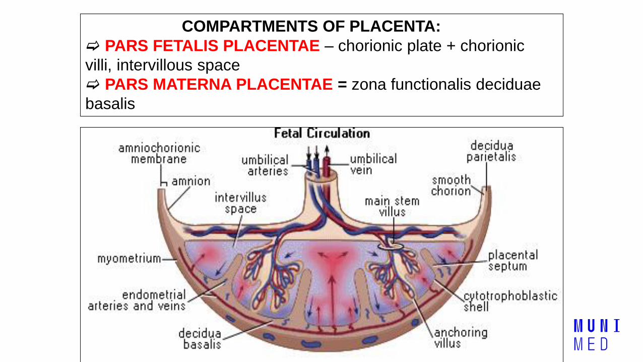
COMPARTMENTS OF PLACENTA:
PARS FETALIS PLACENTAE – chorionic plate + chorionic
villi, intervillous space
PARS MATERNA PLACENTAE = zona functionalis deciduae
basalis
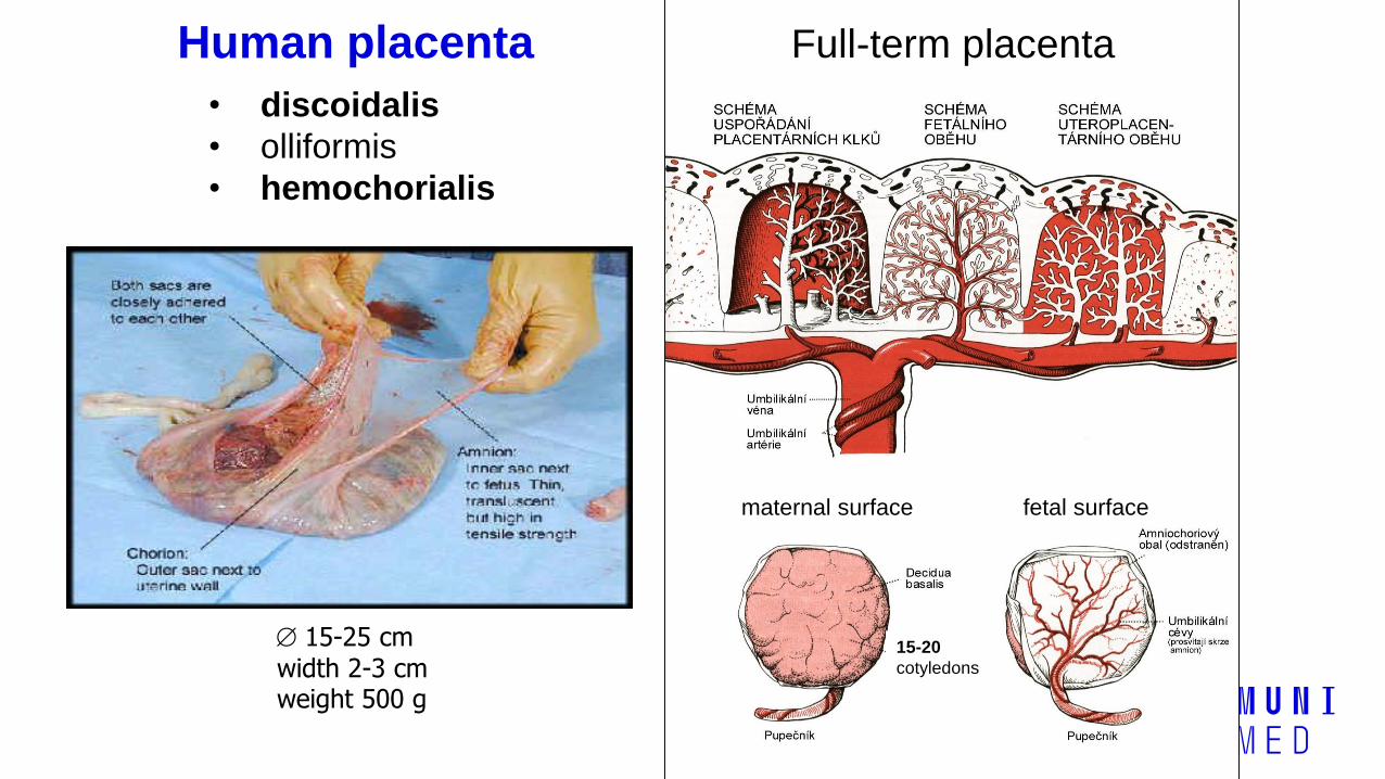
• discoidalis
• olliformis
• hemochorialis
Human placenta
maternal surface fetal surface
15-25 cm width 2-3 cmweight 500 g
15-20
cotyledons
Full-term placenta
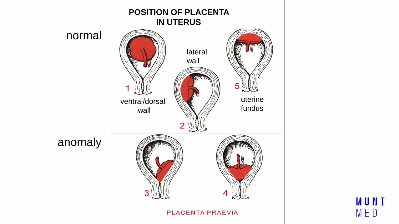
POSITION OF PLACENTA
IN UTERUS
lateral
wall
uterine
fundus ventral/dorsal
wall
normal
anomaly

Anomalies of chorionic villi (1 : 100 pregnancies) • mola hydatidosa
• chorionepitheliom
Anomalies in location: • placenta praevia (causes bleeding in week 28)
• absolute indication to CS
• placenta accreta (attached to myometrium)
• placenta increta (grown into myometrium)
• placenta percreta (grown through myometrium)
Anomalies of placenta

Anomalies of shape of placenta
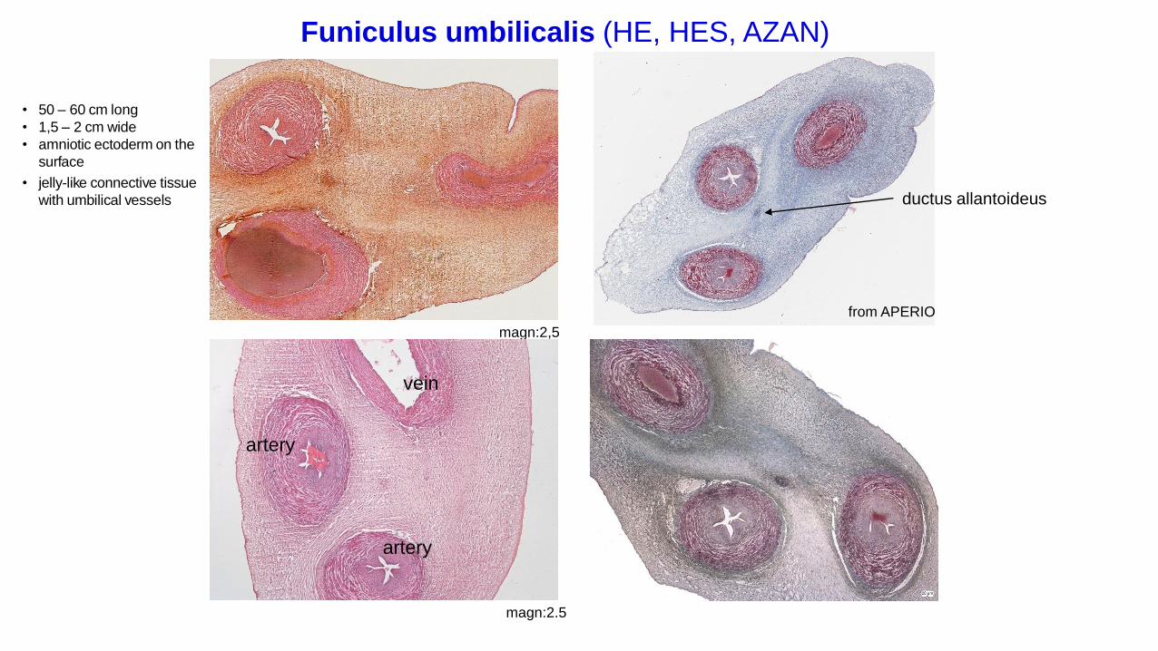
Funiculus umbilicalis (HE, HES, AZAN)
magn:2,5
magn:2.5
from APERIO
vein
artery
artery
• 50 – 60 cm long
• 1,5 – 2 cm wide
• amniotic ectoderm on the
surface
• jelly-like connective tissue
with umbilical vessels ductus allantoideus
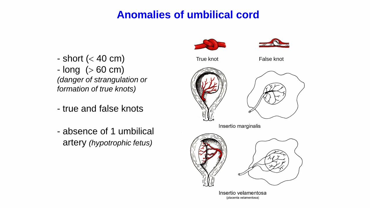
- short ( 40 cm)
- long ( 60 cm)(danger of strangulation or
formation of true knots)
- true and false knots
- absence of 1 umbilical
artery (hypotrophic fetus)
True knot False knot
Anomalies of umbilical cord

1 2
3
Umbilical cord – placenta insertion
1 – insertio centralis
2 – insertio marginalis
3 – insertio velamentosa
chorion
laeve
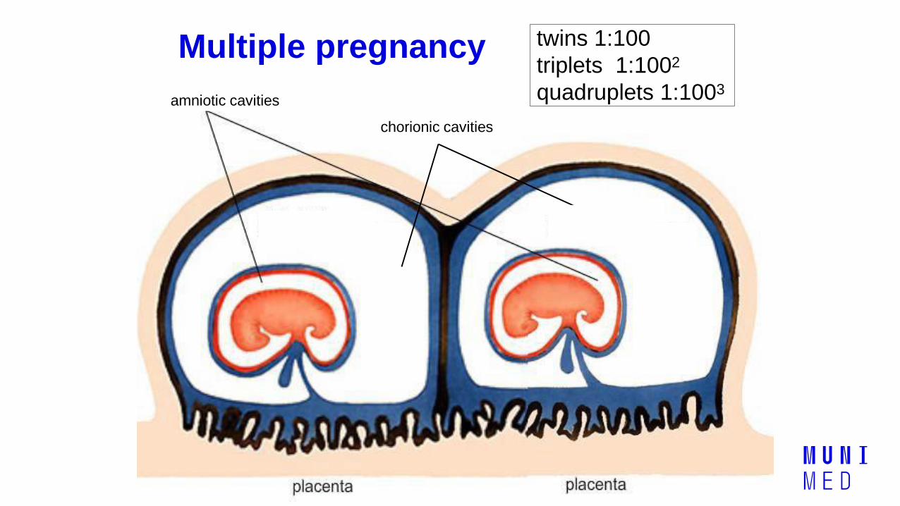
Multiple pregnancy twins 1:100
triplets 1:1002
quadruplets 1:1003amniotic cavities
chorionic cavities

DIZYGOTIC TWINS
• 2 spermatozoa fertilize2 oocytes
• each embryo developsseparately(has its own amnion,chorion, and placenta)
• twins can be ofdifferent sexes
• resemblance of twins asbetween siblings of differentage
Dizygotic
separate amnion,
chorion, placenta
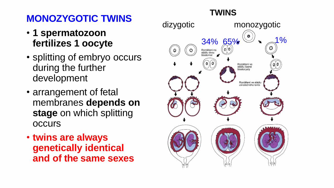
MONOZYGOTIC TWINS
• 1 spermatozoonfertilizes 1 oocyte
• splitting of embryo occursduring the furtherdevelopment
• arrangement of fetalmembranes depends onstage on which splittingoccurs
• twins are alwaysgenetically identicaland of the same sexes
34% 65% 1%
dizygotic monozygotic
TWINS
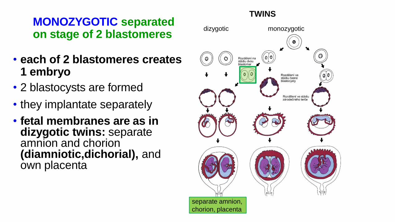
MONOZYGOTIC separatedon stage of 2 blastomeres
• each of 2 blastomeres creates1 embryo
• 2 blastocysts are formed
• they implantate separately
• fetal membranes are as indizygotic twins: separateamnion and chorion(diamniotic,dichorial), andown placenta
TWINS
dizygotic monozygotic
separate amnion,
chorion, placenta
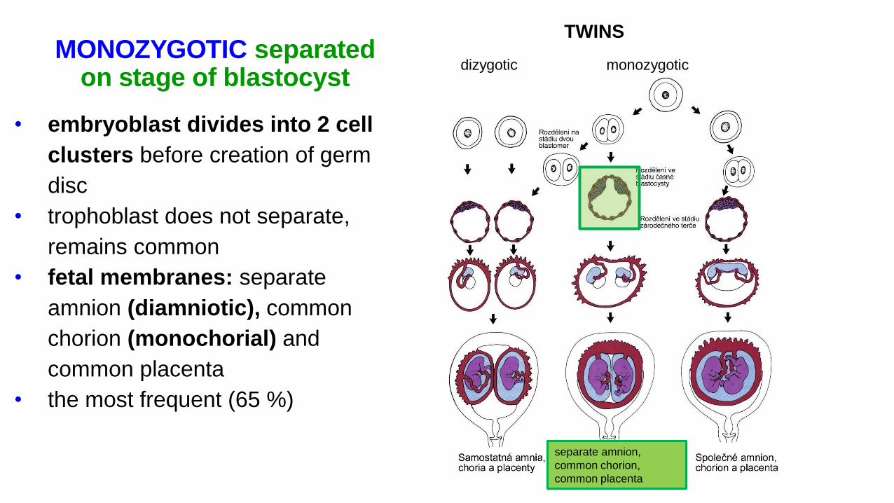
MONOZYGOTIC separatedon stage of blastocyst
• embryoblast divides into 2 cell
clusters before creation of germ
disc
• trophoblast does not separate,
remains common
• fetal membranes: separate
amnion (diamniotic), common
chorion (monochorial) and
common placenta
• the most frequent (65 %)
TWINS
dizygotic monozygotic
separate amnion,
common chorion,
common placenta
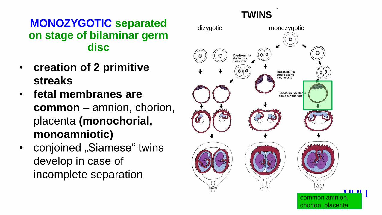
MONOZYGOTIC separatedon stage of bilaminar germ
disc
• creation of 2 primitive
streaks
• fetal membranes are
common – amnion, chorion,
placenta (monochorial,
monoamniotic)
• conjoined „Siamese“ twins
develop in case of
incomplete separation
TWINS
dizygotic monozygotic
common amnion,
chorion, placenta

38 týdnů = 266 dnů
Date of the 1st day of the last menstruation + 9 calendar months + 7 days
Length of pregnancy
preembryo embryo fetus
Calculation of the expected date of delivery:
Fertilization CONCEPTIONAL AGE 38 weeks = 266 days
week 0 3 8 38
0 40
1st day of last MENSTRUAL AGE 40 weeks = 280 days
menstruation = 10 lunar months
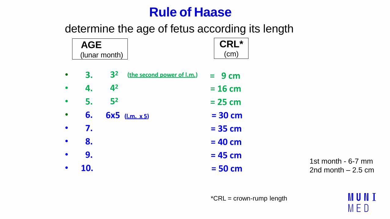
Rule of Haase
determine the age of fetus according its length
• 3.
• 4.
• 5.
• 6.
• 7.
• 8.
• 9.
• 10.
32
42
52
6x5 (l.m. x 5)
(the second power of l.m.) = 9 cm
= 16 cm
= 25 cm
= 30 cm
= 35 cm
= 40 cm
= 45 cm
= 50 cm
*CRL = crown-rump length
AGE(lunar month)
CRL*(cm)
1st month - 6-7 mm
2nd month – 2.5 cm
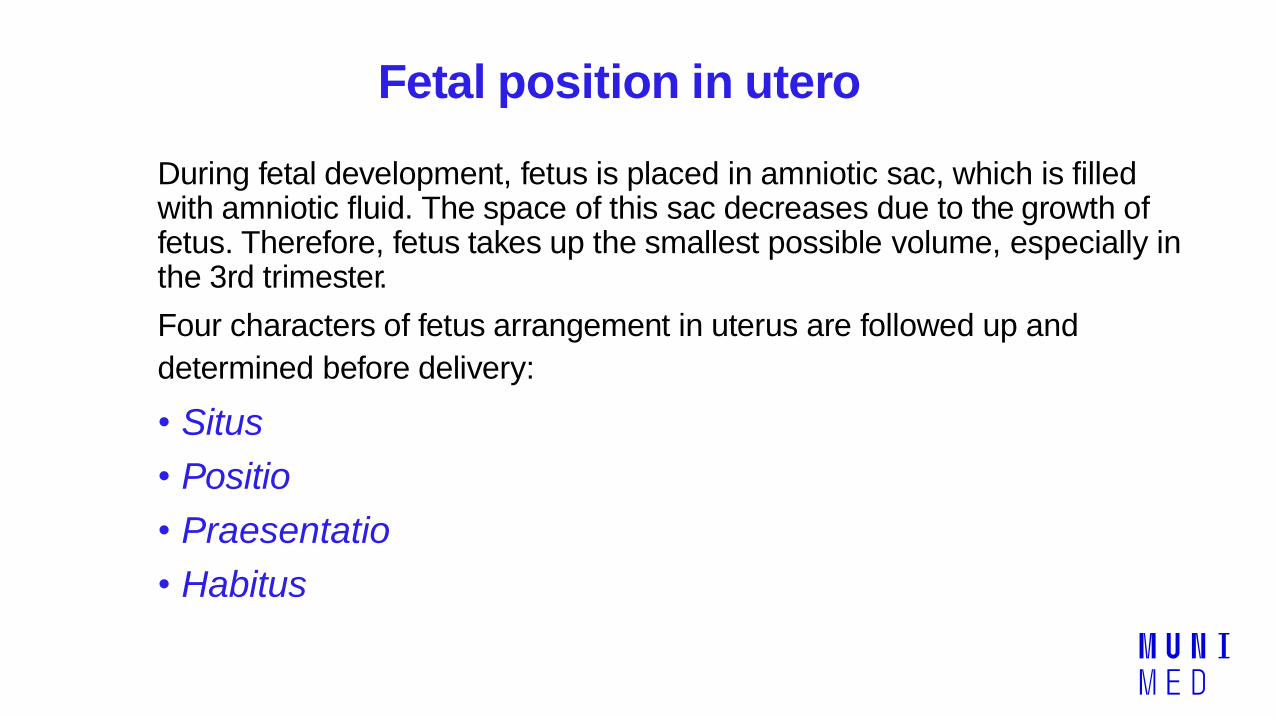
Fetal position in utero
During fetal development, fetus is placed in amniotic sac, which is filledwith amniotic fluid. The space of this sac decreases due to the growth offetus. Therefore, fetus takes up the smallest possible volume, especially inthe 3rd trimester.
Four characters of fetus arrangement in uterus are followed up and
determined before delivery:
• Situs
• Positio
• Praesentatio
• Habitus
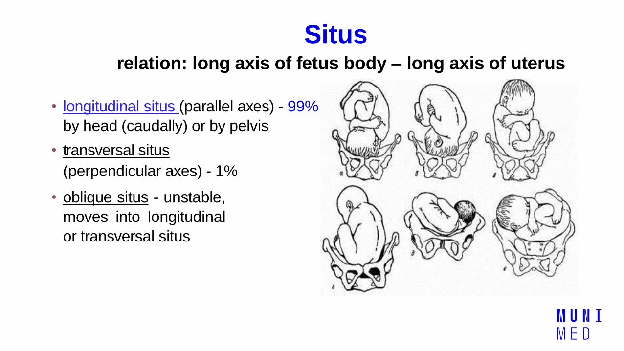
Situsrelation: long axis of fetus body – long axis of uterus
• longitudinal situs (parallel axes) - 99%
by head (caudally) or by pelvis
• transversal situs
(perpendicular axes) - 1%
• oblique situs - unstable,
moves into longitudinal
or transversal situs
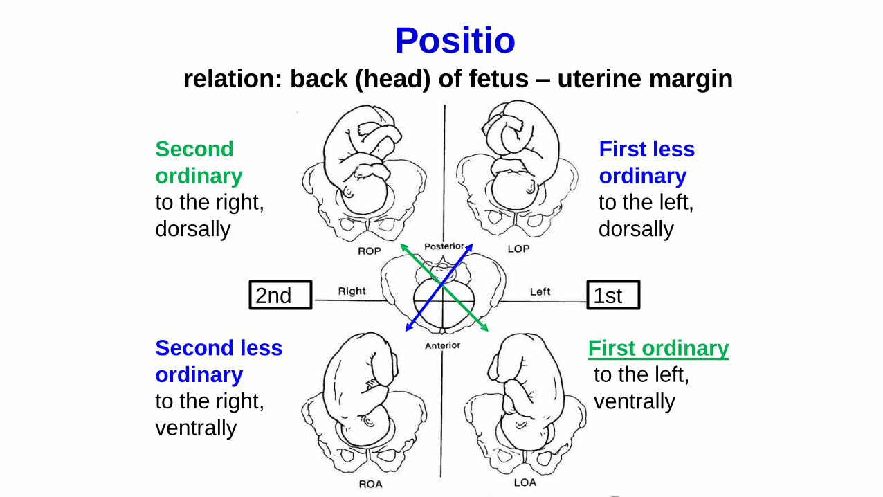
Positiorelation: back (head) of fetus – uterine margin
Second
ordinary
to the right,
dorsally
First less
ordinary
to the left,
dorsally
Second less
ordinary
to the right,
ventrally
First ordinary
to the left,
ventrally
1st2nd
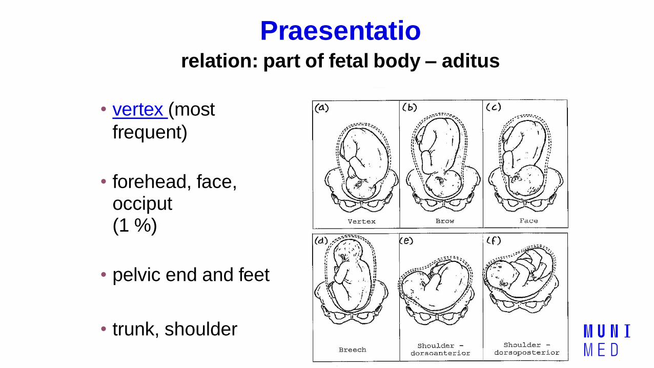
Praesentatiorelation: part of fetal body – aditus
pelvis
• vertex (most
frequent)
• forehead, face,occiput(1 %)
• pelvic end and feet
• trunk, shoulder
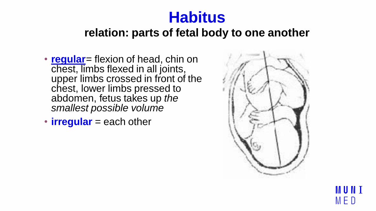
Habitusrelation: parts of fetal body to one another
• regular= flexion of head, chin on chest, limbs flexed in all joints,upper limbs crossed in front of thechest, lower limbs pressed toabdomen, fetus takes up thesmallest possible volume
• irregular = each other
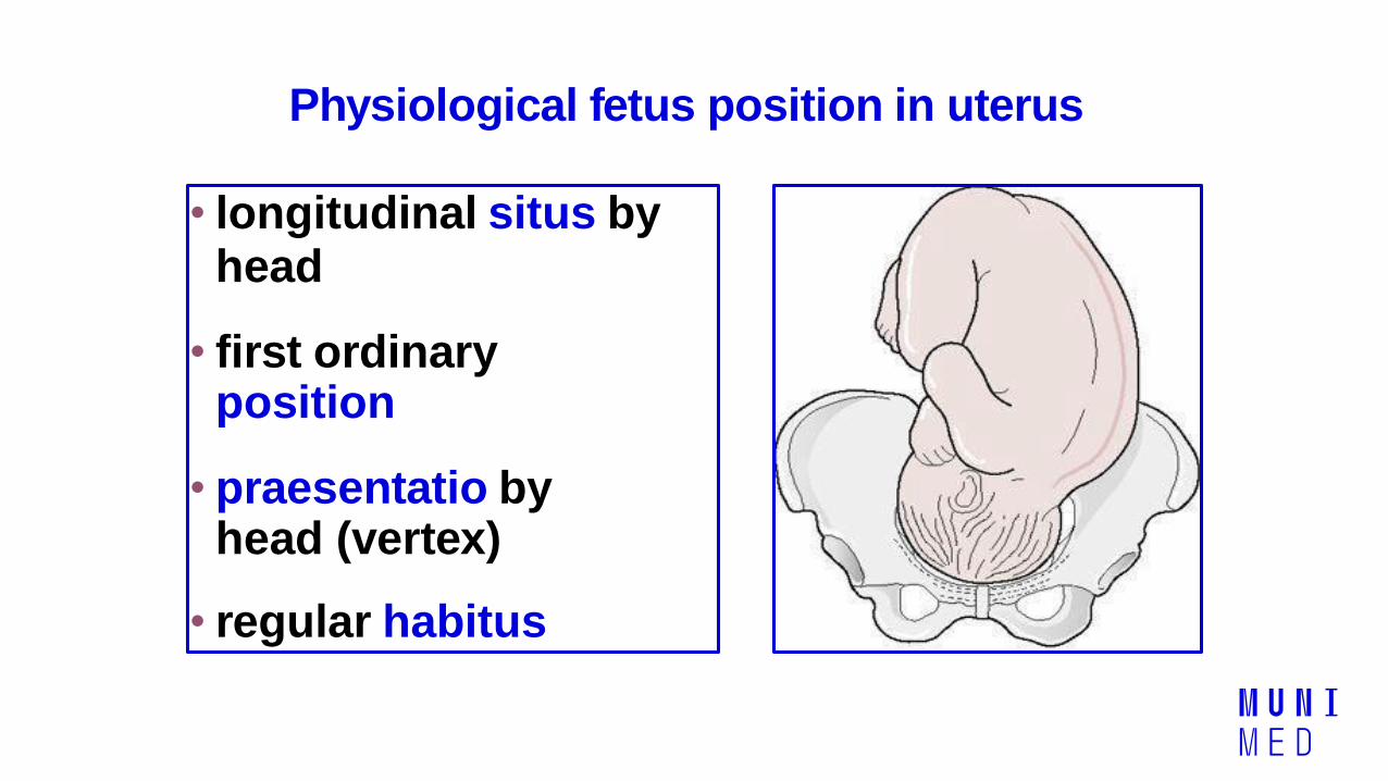
Physiological fetus position in uterus
• longitudinal situs byhead
• first ordinaryposition
• praesentatio byhead (vertex)
• regular habitus

Marks of full-term fetus
Main characters
• length (50-51 cm)
• weight (3,000-3,500 g)
• diameters of the head in norm
• ♂ testes are descended in scrotum
♀ labia majora cover labia minora
Auxiliary characters
• fetus is eutrophic, subcutaneous fat is well developed
• skin – rests of lanugo on shoulders and back only
• eyelashes, brow, hair (several cm) are developed,
nails overlap free end of fingers
• skull bones are hard, major and minor fontanellesare palpable and separated from each other
• newborn cries and moves

Mature and full-term fetus• Full-term fetus – relates to the length of pregnancy (menstrual
age):
- preterm (to 37th week)
- full-term (38 – 40 weeks)
- after term (more than 42 weeks)
• Mature fetus – relates to level of development:
- mature
- immature
• Level of nutrition• hypotrophic
• eutrophic (weight 3,000 – 3,500 g, length 50 - 51 cm)
• hypertrophic

Zápatí prezentace35
GENERAL EMBRYOLOGY 2
Set of embryological schemes II
Atlas of Cytology and Embryology – pages 76 – 81
Discussion
3 embryological schemes