Isolation of rhizobacteria from salt tolerant plant species and ...
Upload
khangminh22Category
view
0download
0
Chapter 4
Isolation of IgY from Yolk
CHRISTIAN STAAK, CHRISTINE SCHWARZKOPF, INGRID BEHN,
UNDINE HOMMEL, ANDREAS HLINAK, RUDIGER SCHADE,
and MICHAEL ERHARD
Introduetion
Constitution of the Yolk
The yolk in hens' eggs consists of dry matter (51,3 %) and water (48,7 %) in nearly equal proportions (Siewert and Bronch, Handbuch der Tierernăhrung, 1972; Romanoff und Romanoff, The Avian Egg, 1949). The dry matter is made up as shown in Table 1.
Regarding its constitution, yolk can be viewed as being an oil-in-water emulsion with a watery portion containing proteins and a dispersal portion of so-called yolk-granules and lipiddrops (Fischer, 1996; Fischer et al., 1996).
~ Christian Staak, Bundesinstitut fur gesundheitlichen Verbraucherschutz und Veterinărmedizin, Diedersdorfer Weg 1, Berlin, 12277, Germany (phone +49-30-8412-2053; fax +49-30-8412-2956 ; e-mail [email protected]) Christine Schwarzkopf, Zentrales Institut des Sanitătsdienstes der Bundeswehr Koblenz, Laborabt. II (Vet.Med), Generaloberst-Beck-Str. 17, Mainz, 55129, Germany Ingrid Behn, Universităt Leipzig, Fakultăt fur Biowissenschaften, Pharmazie und Psychologie, Talstr. 33, Leipzig, 04103, Germany Undine Hommel, Universităt Leipzig, Fakultăt fUr Biowissenschaften, Pharmazie und Psychologie, Talstr. 33, Leipzig, 04103, Germany Andreas Hlinak, Universitătsklinikum Charite der Humboldt-Universităt, Institut fUr Pharmakologie und Toxikologie, Dorotheenstr. 94, Berlin, 10117, Germany Rudiger Schade, Universitătsklinikum Charite der Humboldt-Universităt, Institut fUr Pharmakologie und Toxikologie, Dorotheenstr. 94, Berlin, 10 117, Germany Michael Erhard, Universităt Leipzig, Veterinărmedizinische Fakultăt, Veterinăr-Physiologisches Institut, An den Tierkliniken 7, Leipzig, 04103, Germany
PROTOCOL
R. Schade et al. (eds.), Chicken Egg Yolk Antibodies, Production and Application© Springer-Verlag Berlin Heidelberg 2001

66 CHRISTIAN STAAK et al.
Table 1. Constitution of Egg Y olk
Protein
Fats and lipoids
Carbohydrates
Anorganic matter
16.6%
32.6 %
1.0 %
1.1%
The proteins in the yolk can be divided roughly into four fractions:
1. The vitellin or lipovitellin fraction (47.5 %), a phosphoruscontaining nucleo-albumin (400 kDa) with subsidiary units having molecular weights between 35 and 140 kDa (Mehner and Hartfield, 1983).
2. The vitellelin fraction (38.6 %), with less phosphorus and more lipids than the vitellin fraction.
3. The phosvitin fraction (4.3 %) - very rich in phosphorus and without lipids - a protein with a Mw of 36 kDa (Mehner and Hartfield, 1983).
4. The livetin fraction (9.6 %) with soluble proteins like globu-lin.
Fraction 4 is very heterogeneous, containing about 10 proteins, about which various claims are made (Mehner and Hartfield, 1983). The original differentiation into alpha-, beta- and gamma-livetin (Martin et al., 1959) has been replaced as follows (Table 2):
The concentration and distribution of immunoglobulins in the serum and egg are as follows (Table 3, Rose et al., 1974):
Table 2. Composition of the Egg Yolk Livetin Fraction
alpha-livetin
beta-livetin
gamma-livetin
the same as plasma-albumins (54 kDa) (Williams, 1962)
alpha-2-glycoprotein (Hatta et al., 1990)
the same as serum gamma-globulins, together with transferrin (77 kDa, Williams, 1962) and IgY, which is sometimes identified with this fraction (Hatta et al., 1990)
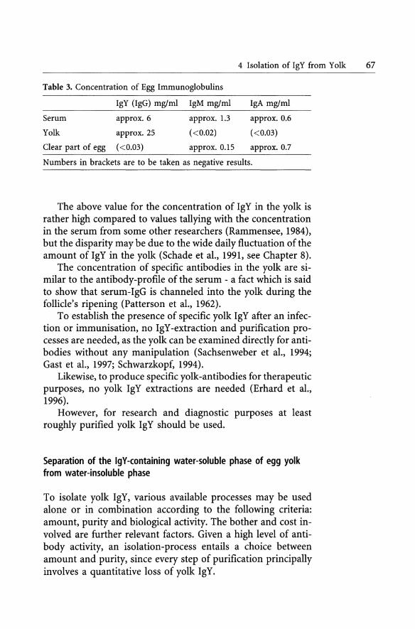
4 Isolation of IgY from Yolk 67
Table 3. Concentration of Egg Immunoglobulins
IgY (IgG) mglml IgM mglml IgA mglml
Serum approx.6 approx. 1.3 approx.0.6
Yolk approx.25 «0.02) «0.03)
Clear part of egg «0.03) approx.0.15 approx.O.7
Numbers in brackets are to be taken as negative results.
The above value for the concentration of IgY in the yolk is rather high compared to values tallying with the concentration in the serum from some other researchers (Rammensee, 1984), but the disparity may be due to the wide daily fluctuation of the amount of IgY in the yolk (Schade et al., 1991, see Chapter 8).
The concentration of specific antibodies in the yolk are similar to the antibody-profile of the serum - a fact which is said to show that serum-IgG is channeled into the yolk during the follicle's ripening (Patterson et al., 1962).
To establish the presence of specific yolk IgY after an infection or immunisation, no IgY -extraction and purification processes are needed, as the yolk can be examined directly for antibodies without any manipulation (Sachsenweber et al., 1994; Gast et al., 1997; Schwarzkopf, 1994).
Likewise, to produce specific yolk-antibodies for therapeutic purposes, no yolk IgY extractions are needed (Erhard et al., 1996).
However, for research and diagnostic purposes at least roughly purified yolk IgY should be used.
Separation of the IgY-containing water-soluble phase of egg yolk from water-insoluble phase
To isolate yolk IgY, various available processes may be used alone or in combination according to the following criteria: amount, purity and biological activity. The bother and cost involved are further relevant factors. Given a high level of antibody activity, an isolation-process entails a choice between amount and purity, since every step of purification principally involves a quantitative loss of yolk IgY.

68 CHRISTIAN STAAK et al.
All extraction processes begin by separating yolk and egg white. This should be done in such a way as to leave no contaminating traces of the egg white on the yolk. Various authors write of rinsing the yolk in flowing water or a buffer, and others roll yolks on fIlterpaper, to let the paper soak up the traces of the clear part. After removal of the vitellin membrane, the yolk is put into a measuring cylinder.
The following set of steps for IgY isolation is only a rough generalisation:
• Dilution of the yolk with distilled water or buffer or straightaway with an extraction medium, to bring the suspension into a precipitable or centrifugeable form; when the yolk is diluted with distilled water or buffer it is useful to keep the solution at -20 De for 72 h followed by thawing (slowly at 4 DC) before further use
• Separation of the lipophile from the watery phase of the yolk (organic solvents, precipitation)
• Obtaining of IgY from the watery phase (pecipitation, chromatographic separation); the fInal IgY -extract of a selected extraction procedure should be dialysed against an appropriate buffer (0,1 % sodiumazide may be added as preservative)
• Isolation of specifIc IgY
To classify the various isolation procedures, the fIrst step is usually defInitive. The consequent purifIcation processes vary widely in kind and combination and from institute to institute, so they are not suitable for classifying the various methods.
Subprotoeol 1 Extraction Using Organic Solvents
By now there are many non-wasteful extraction procedures, so the waste-disposal of organic solvents, which often tended to be used for isolating IgY, should early be taken into consideration in choosing a suitable extraction procedure. A procedure which in this respect is rather extravagant has been described by Bade and Stegemann (1984).

4 Isolation of IgY from Yolk 69
Outline
The IgY-extraction according to the chloroform-polyethylene glycol method (CH -PEG) is shown schematically in Figure 1.
Materials
- cool-centrifuge (Jouan MR 1822)
- tubes (Falcon, Blue Max™ 50 mI, Propylene conical tube)
- pH-meter
Fiii centrifuge tube to 1/3 with yolk homogenate (diluted 1:3 with PBS)
1 Supematant discard
V1e1d:
Add sarne volume of chloroform and shake
Centrifuge for 30 min (1000 x g .RD
wat8ry pI\ase
semi-soIid phase
orange ooIoured soIution
Decant the watelY phase, add puJverized PEG 6000 -(12 'III, wlv), stir well, incubate for 30 min (RD. centrifuge as above
1 Dissolve precipitate in PBS in a volume equivalent to 1/6 of original yolk
PEG-Method (Poisen el al., 1980): CH-PEG (Poison, 1990)
Protein Antibody titre
1 : 2.39 t : 2.57
Fig. 1. IgY-extraction by the chloroform-polyethylene glycol method (CHPEG).
Equipment
70 CHRISTIAN STAAK et al.
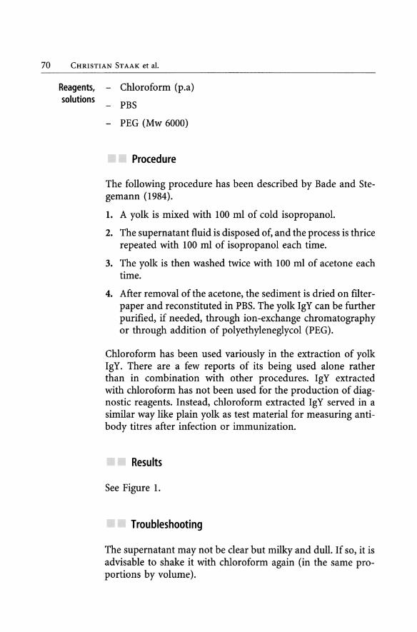
Reagents, - Chloroform (p.a) solutions - PBS
- PEG (Mw 6000)
Proeedure
The following procedure has been described by Bade and Stegemann (1984).
1. A yolk is mixed with 100 mI of cold isopropanol.
2. The supernatant fluid is disposed of, and the process is thrice repeated with 100 mI of isopropanol each time.
3. The yolk is then washed twice with 100 mI of acetone each time.
4. After removal of the acetone, the sediment is dried on fllterpaper and reconstituted in PBS. The yolk IgY can be further purified, if needed, through ion-exchange chromatography or through addition of polyethyleneglycol (PEG).
Chloroform has been used variously in the extraction of yolk IgY. There are a few reports of its being used alone rather than in combination with other procedures. IgY extracted with chloroform has not been used for the production of diagnostic reagents. Instead, chloroform extracted IgY served in a similar way like plain yolk as test material for measuring antibody titres after infection or immunization.
Results
See Figure 1.
Troubleshooting
The supernatant may not be clear but milky and duH. If so, it is advisable to shake it with chloroform again (in the same proportions by volume).
4 Isolation of IgY from Yolk 71
In investigating antibodies against Rous sarcoma virus, Aulisio and Shelokov (1967) used a chloroform extract. They mixed 1 volume of yolk with the same amount of physiological saline and 2 volumes of chloroform. The mixing and settling of the sediment followed at least four repeats of this procedure. After cooling overnight and centrifuging off at 2000 rpm, the clear watery supernatant was used for investigation in the indirect fluorescence-antibody test (IFAT). Mohammed et al. (1985) described a procedure which was similar but avoided the fourfold addition of chloroform during their investigations into Mycoplasma gallisepticum by ELISA.
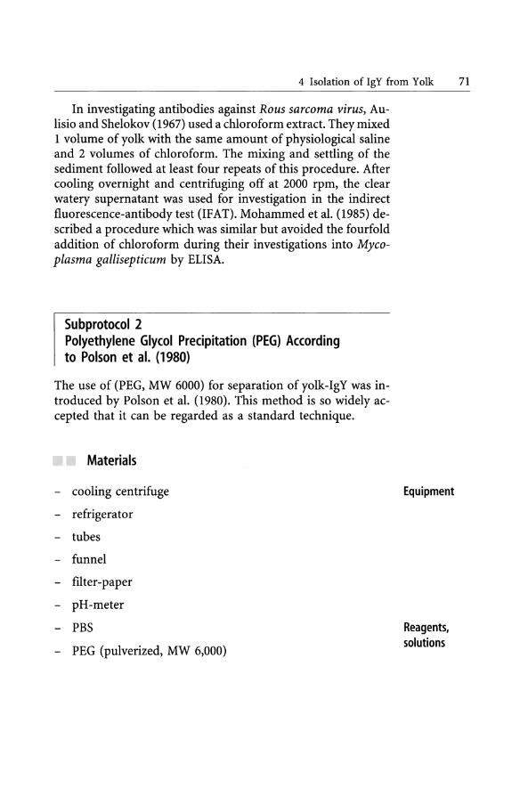
Subprotocol 2 Polyethylene Glycol Precipitation (PEG) According to Poison et al. (1980)
The use of (PEG, MW 6000) for separation of yolk-IgY was introduced by Polson et al. (1980). This method is so widely accepted that it can be regarded as a standard technique.
Materials
- cooling centrifuge
- refrigerator
- tubes
- funnel
- filter-paper
- pH-meter
- PBS
- PEG (pulverized, MW 6,000)
Equipment
Reagents, solutions
72 CHRISTlAN ST AAK et al.
Proeedure
The IgY -extraction according to the polyethylene glycol precipitation method is shown schematically in Figure 2.
1 part of egg yoIk + 2 parts of P8S (pH 7.5) + putverlzed
PEG 6000, 3.5 % (wlv) sUr weU.
Incubate for 20 min at RT,
cenlrifuga for 10 min (14 000 x g, Rn
1. yellow fatty layer
2 clear layer (lgY)
3 &eml solid layer
Supematenl discard
Supematant discard
IgY yield: 6 -12 mglml
decant carefully layer 1 and 2
V absortlent cotton U i (fatty layer)
B I
Add putverized PEG 10 a final ooncentratlon of 12 %
stirwell, Incubate for 20 min (RT)
Centrifuge as above
OissoIva lhe pt"ecipitale in PBS 10 lhe original yoIk YOIume,
precipitate Ihe IgY aga' wIth 12 '" PEGas above
l Centrifuge as above
Remove residual PEG supematant by suction,
centrifuge once agaln as above
Dissolve tha precipitate in half the original yolk volume
Fig. 2. IgY-extraction by polyethylene-glycol precipitation (PEG).

4 Isolation of IgY from Yolk 73
Results
See Figure 2 and Tables 6 and 7.
Troubleshooting
If the purity of the product is deemed inadequate, the addition of 2 M ammonium sulphate leads to precipitation of protein, while PEG is removed with the supernatant fluid. After completion of the IgY -extraction procedure, the final product should be dialysed against PBS or any other appropriate buffer. See Chapter 8 for a very detailed description of the procedure.
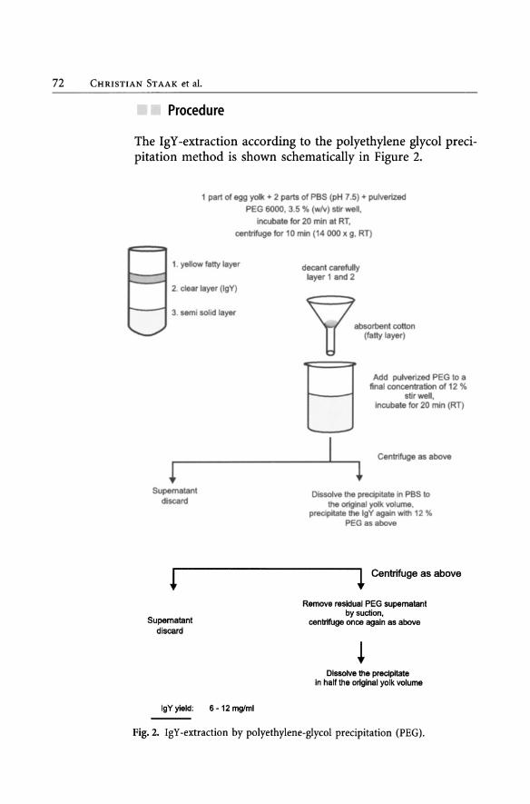
Subprotoeol 3 Polyethylene Glyeol/Ethanol Precipitation (PEG/ Ale) Aeeording to Poison et al. (1985)
Comparative investigations with other methods revealed that the IgY -yield of this original PEG-method is relatively low (Akita and Nakai, 1993; Schwarzkopf, 1994). Furthermore, residues of PEG may disturb some immunological assays (osmotic potency of PEG).
Consequently Poison et al. (1985) presented an improved method, in which a protein precipitation step with cold ethanol was added. In this way, residual PEG was completely removed. Since some yolk substances are solved better in organic solvent than in water, such contaminants are removed by this method.
Materials
See Subprotocol 2, additionally ethanol, refrigerator (-5 aCI -20 ac).
74 CHRISTIAN STAAK et al.
Proeedure
The IgY-extraction according to the polyethylene glycollethanol precipitation method is shown schematically in Figure 3.
Add 40 mi PBS (pH 7.6) ta 10 mi egg yolk, add pulverized PEG 6000 (3.5 %, w/v = 1.75 g), stir well,
incubate 20 min (RT),
Precipitate discard
! Supernatant
discard
! Supernatant
discard
Supernatant discard
centrifuge for 25 min (5,000 x g, RT)
I l
Filtrate the supernatant add pulverized PEG ta a final concentration
of 12 % (4.25 g), stir well and incubate for 10 min (RT),
centrifuge as above
l Dissolve the precipitate in 25 mi PBS,
add pulverized PEG (12 % = 3 g), stir well and incubate as above,
centrifuge as above
l Dissolve the precipitate in 2.5 mi PBS,
cool ta O°C, add equal volume of ethanol (50 %)
precooled ta -20°C,
centrifuge for 25 min (10,000 x g, _5°C)
l Dissolve the precipitate
IgY yield: 4.8 mg/ml egg yolk; Purity 89 %
in 2.5 mi PBS, dlalyse against PBS
(at 4°C)
Fig. 3. IgY -extraction by polyethylene-glycollethanol precipitation (PEG/alc).
4 Isolation of IgY from Yolk 75
Results
See Figure 3 and Tables 6 and 7.
Troubleshooting
A combination of chloroform extraction with the PEG-method, which Polson presented in 1990 produced a 2.6-fold greater activity and a 2.4-fold greater yield of protein. The steps of this procedure - known as the chloroform-PEG-method - are shown in Figure 1.
If further purification of the IgY -preparation from PEG is needed, the distribution-coefficient of PEG in water:chloroform of 1:20 can be used, which means that a further shaking of the preparation with chloroform may lead to a notable reduction of the PEG-concentration.
Subprotoeol 4 Dextransulphate Precipitation (DS) Aeeording to Jensenius et al. (1981)
The precipitation oflipoprotein from serum by dextransulphate (Burstein and Pavermann, quoted by Jensenius et al., 1981) was used by Jensenius et al. (1981) to separate yolk lipid from the watery phase of yolk to obtain yolk-IgY. This procedure takes place at room temperature.
Materials
- cooling-centrifuge
- tubes
- pH-meter
- TBS
- dextran sulfate
- CaCh
Equipment
Reagents, solutions

76 CHRISTIAN STAAK et al.
Proeedure
The IgY -extraction according to the dextransulphate precipitation method is shown schematically in Figure 4.
! Precipitate
discard
!
Oilute 10ml of egg yolk with 40 mi TBS (pH 7.4), centrifuge for 20 min (2,000 x g, RT)
I
Centrifuge as above
I
1 Supernatant
Add 3.0 mi OS (10 % OS [w/v] in TBS)
and 7.5 mi CaCI2 (1 M), mix and incubate 30 min (RT)
1 Supernatant A Precipitate
!
Add TBS to a volume equivalent to the original
volume of diluted egg yolk
Centrifuge 2,800 x g, 20 min, RT
I 1
Supematant B Precipitate (discard)
Combine both supernatants A and B.
Further purification of IgY by salting out using sodium sulfate or ammonium sulfate.
Fig. 4. The diagram shows IgY-extraction by means of dextran sulphate precipitation (DS).
4 Isolation of IgY from Yolk 77
Results
See Tables 6 and 7.
Troubleshooting
This method is flexible insofar as the first step of isolation can be repeated if there is any eloudiness in the first supernatant fluid due to a too meagre addition of DS and CaClz.
Yolk-IgY in the supernatant fluid can finally be precipitated by addition of either sodium sulphate or ammonium sulphate according to "salting out procedures" (see below). This procedure isolates about 70-80 % of the yolk-IgY.
Subprotoeol 5 Water-Dilution Method (WD)
With a neutral pH and a low concentration of ions, the yolk lipids aggregate. Low pH-values also lead to a separation of IgY (Jensenius et al., 1981; Akita and Nakai, 1992). This aggregation oflipids is used in the water-dilution method to separate IgY.
The method is used in so many forms that none can be viewed as standard. As an example, the water dilution procedures described by Akita and Nakai (1993) and by Schwarzkopf (1994) are given.
Materials
- cooling centrifuge
- refrigerator
- deep freezer
- tubes
- pH-meter
Equipment

78 CHRISTIAN STAAK et al.
Reagents, solutions
- distilled water
- sodium sulphate
- ammonium sulphate
Proeedure
The IgY -extraction according to the water-dilution precipitation method (Akita and Nakai 1993) is shown schematically in Figure 5.
Dilute egg yolk 1 : 9 with d.w.
(according to a previous publication of Akita and Nakaii (1992) it is of additional advantage to adjust the pH of d.w. to 5.0 - 5 .2).
1
Centrifuge for 25 min (10,000 x g, -4°C)
I 1
Precipitate Supematant discard Add sodium sulfate
(19 %, w/v), stir well
and incubate for 2 h (RT)
Centrifuge as above
1 I
1 Dissolve the precipitate Supematant
discard in PBS to original yolk volume
IgY in the precipitate is further purified by either salt precipitation (sodium sulfate 14 % w/v),
uHrafiHration or alcohol precipitation
IgY-yield after ultrafiltration: 9.8 mg/ml egg yolk purity : 94%
Fig. 5. IgY-extraction by the water dilution method (WD) according to Akita and Nakai (1993).
4 Isolation of IgY from Yolk 79
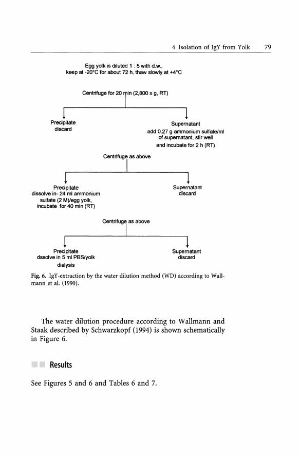
Egg yolk is diluted 1 : 5 with d.w., keep at -20°C for about 72 h, thaw slowly at +4°C
Centrifuge for 20 In (2,800 x g , RT)
! Precipitate
discard
! Precipitate
dissolve in- 24 mi ammonium sulfate (2 M)/egg yolk,
incubate for 40 min (RT)
! Precipitate
dssolve in 5 mi PBS/yolk
dialysis
1 Supematant
add 0.27 9 ammonium sulfate/ml of supematant, stir well
Centrifuge as above
I
centrifugl as above
and incubate for 2 h (RT)
1 Supematant
discard
1 Supematant
discard
Fig. 6. IgY -extraction by the water dilution method (WD) according to Wallmann et al. (1990).
The water dilution procedure according to Wallmann and Staak described by Schwarzkopf (1994) is shown schematically in Figure 6.
Results
See Figures 5 and 6 and Tabies 6 and 7.
80 CHRISTIAN STAAK et al.
Troubleshooting
Jensenius et al. (1981) used a ratio of yolk:water of 1:10, froze the mixture to -20 DC and thawed out, centrifuged off and worked with the supernatant fluid. Wallmann et al. (1990), as also Schwarzkopf (1994), used a ratio of yolk:distilled water of 1:5, likewise froze the mixture to -20 DC and especially emphasised that the mixture should be thawed slowly at 4 De.
Y okoyama et al. (1993) investigated the effects of varying the ratio of distilled water added to yolk from 1:4 to 1: 1 O and of incubating the mixtures at temperatures between 4 DC and 15 De. They also examined the influence of various amounts of hydroxypropylmethylcellulose phthalate (HPMCP) as a lipid-precipitating active substance.
The authors found the following parameters to be optimal in yielding IgY:
- dilution of yolk:water 1:8
- addition of HPMCP 0.25% (v/v)
- working temperature 10 DC
Subprotoeol 6 Further Proeedures for Separating Lipoproteins/Lipids from Water-Soluble Proteins
The following procedures for separating lipoproteins/lipids from water-soluble proteins are less widely adopted. Thus, these procedures are not described in detail.
Proeedure
Lipid-precipitating qualities of "natural gums"
Hatta et al. (1990) examined 12 various 'natural gums' as regards their lipid-precipitating qualities.
10 mI of yolk were diluted with 10 mI of distilled water, then 40 mI of distilled water were added together with 60 mI of the

4 Isolation ofIgY from Yolk 81
respective gum and incubated at room temperature for 30 minutes. Finally the mixture was centrifuged at 10,000 x g for 15 minutes. The watery phase of the supernatant fluid was examined for lipid contents. The lipids were measured by shaking the mixture with ch1oroform-methanol (3:1) and evaporating and weighing it (see above, Yokoyama et al., 1993). The natural gums with the greatest yield of IgY proved to be 3 various preparations of carrageenan with 0.4-1.1% and xanthan gum with 0.7% residuallipids.
Combined octane-acid/ammonium sulphate precipitation
The combined octane-acid/ammonium sulphate precipitation introduced by McKinney and Parkinson (1987) for the simple isolation of mammalian IgG from serum was used by Schwarzkopf (1994) to obtain yolk-IgY:
1. Dilute yolk 1:5 with 0.06 M acetate-buffer at pH 4.0.
2. Add 25 JlI of octane acid per mI of the mixture.
3. Incubate for 30 minutes at RT and centrifuge (10,000 x g, RT).
4. Add PBS to the supernatant (tenfold, v:v), final pH 7,2-7,4.
5. Add 10 volumes of 45% ammonium sulphate (AmS) to 9 volumes of the PBS/supernatant, incubate the mixture for 30 minutes at 4 DC and centrifuge (10.000 x g, 4 DC).
6. Add 5 ml PBS/yolk followed by dialysis against PBS.
7. The dialysate was heated to 50-55 DC for 20 minutes and finally purified by being centrifuged (5.000 x g) and ftltered (0.45 Jlm).
Separation of the IgV-fraction from the aqueous phase of egg yolk
The procedures described above are ways of separating the lipophile and watery phases of the yolk. Purification steps were mentioned insofar as they directly belong to a technique presented.
82 CHRISTIAN STAAK et al.
The result of the first step of the procedure is isolation of the watery phase with yolk-IgY but also other proteins, especially alpha- and beta-livetin. The second step involves procedures suited to isolate IgY from the aqueous phase of egg yolk.
Subprotoeol 7 Precipitation of Yolk-Protein With Ammonium Sulphate [(NH4hS04 - 'AmS'] or Sodium Sulphate [Na2S04 - 'NaS']
Proteins can be precipitated with AmS or with NaS* whatever procedures have been used tiU then. (* For historical reasons the concentration of AmS is given in percent saturation and of NaS in weight/volume.)
Steps taken for the salt-precipitation of proteins are in the following sequence: A slow addition of AmS or NaS in a measured amount as a substance or in a concentrated solution. Thorough mixing of the substances is important, to avoid having an uneven concentration of the salt. Sin ce the precipitation of certain proteins from a mÎXture by AmS or NaS depends on the concentration of the salt, this procedure can be used not only for isolating desired proteins but also for eliminating undesired ones (see below, Odans and Rose, 1972, as also Yamamoto et al., 1975). There follows a phase of incubation at ro om temperature under stirring. A precipitate is obtained through centrifuging, taken up in a certain amount of a desired buffer or distiUed water and finally dialysed.
Outline
This protocol gives information about a lot ofIgY-precipitation methods not described in detail. A procedure of IgY -precipitation by AmS is exemplary shown in Subprotocol 5.
Materials
See Subprotocol 5.

4 Isolation of IgY from Yolk 83
Proeedure
Orlans and Rose (1972), as also Yamamoto et al. (1975), used AmS precipitat ion for preliminary purification by homogenising yolk in 4 volumes of 16% saturated AmS and by isolating the watery supernatant fluid through incubation and centrifuging (see Table 1, Chapter 8). From this fluid, IgY was obtained as a precipitate in 50% saturated AmS and purified through a sequence of three further precipitations with 40% and twice 37% AmS-saturation. Pure IgY was obtained from the dialysed product by gel-filtration via sephadex G 200. Similar procedures were used by Kuroki et al. (1993), as also by Loeken and Roth (1983). They diluted the yolk with an equal volume of PBS and centrifuged at high velocity. IgY was obtained from the supernatant fluid by using AmS in three steps of precipitation, once with 50% AmS-saturation and twice with 33% saturation.
Several steps of precipitation are often described. Wallmann et al. (1990) used three steps of AmS-precipitation, with addition of solid AmS at each step to restore 33% saturation. Ntakarutimana et al. (1992) obtained their material through chloroform extraction. They used a gradual addition of 20% (w/v) NaS to yield IgY.
Hatta et al. (1990) used natural gum for the first step of isolation. To obtain IgY, NaS was added to the centrifuged supernatant fluid to a concentration of 0.2 M.
As a further purification after treatment with dextran sulphate, Jensenius et al. (1981) advised adding 20g NaS to 100 mI supernatant fluid and a final reprecipitation by adding 6.2 mI of 36% (w/v) NaS to 10 mI. Schmidt et al. (1989) took the advice but used AmS in a final saturation of 50%.
Poison et al. (1980) likewise saw a further possibility of purification of IgY after PEG-preparation in the precipitation of protein after addition of 2 M AmS.
A systematic investigation into the suitability of various AmS concentrations was presented by Svendsen et al. (1995). The initial material was supernatant fluid obtained by the method of water-dilution (yolk:distilled water 1:10) and freezing and thawing.
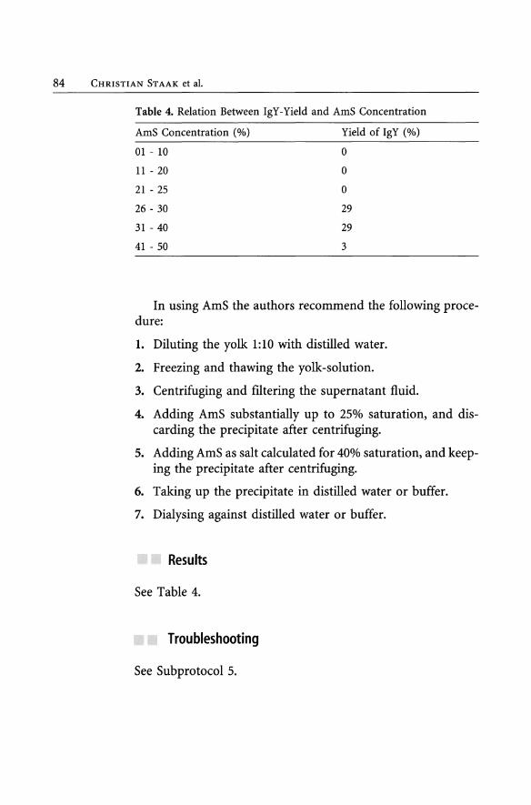
84 CHRISTIAN STAAK et aI.
Table 4. Relation Between IgY-Yield and AmS Concentration
AmS Concentration (%) Yield of IgY (%)
01 - 10 O
11 - 20 O
21 - 25 O
26 - 30 29
31 - 40 29
41 - 50 3
In using AmS the authors recommend the following procedure:
1. Diluting the yolk 1:10 with distilled water.
2. Freezing and thawing the yolk-solution.
3. Centrifuging and flltering the supernatant fluid.
4. Adding AmS substantially up to 25% saturation, and discarding the precipitate after centrifuging.
5. Adding AmS as salt calculated for 40% saturation, and keeping the precipitate after centrifuging.
6. Taking up the precipitate in distilled water or buffer.
7. Dialysing against distilled water or buffer.
Results
See Table 4.
Troubleshooting
See Subprotocol 5.

4 Isolation of IgY from Yolk 85
Subprotocol 8 Gelfiltration of an IgY-Extract According to the Subprotocols 1-7
IgY has a molecular weight of 165,000 (Gottstein and Hemmler, 1985). Other reports vary between 165 and 220 kDa (see Fischer, 1996; Fischer et al., 1996).
The separation of proteins according to their molecular weight through gelflltration is a further stage of purification of yolk IgY but rarely reported except in the desalting of preparations through sephadex G 25 instead of through a tedious dialysis.
Materials
- column (2 x 90 cm) and accessories: - valve - connecting piece - tubes - pump, like peristaltic pump PI, Pharmacia - UV-monitor - recording-unit (plotter or printer) - equipment for volume-reduction through vacuum dialy-
sis (collodium tubes, Sartorius) or through lyophilisation
- watery solution containing IgY
- buffer (e.g.PBS, containing 0,5 M NaCI to avoid non-specific binding)
- Sephacryl S-300 (Pharmacia)
- Blue-Dextran 2,000 (Pharmacia, MW 2,000 kDa)
If no specially made columns are available, one can proceed as follows (more detailed descriptions are given in the Pharmacia booklet 'Gel Chromatography').
Equipment
Reagents, solutions

86 CHRISTIAN STAAK et al.
Procedure
1. Wash and transfer Sephacryl S-300 to PBS, choosing a gelviscosity which enables air-bubbles to rise.
2. Put several mI of PBS into a column (close the lower end of the column).
3. Transfer the gel into the column carefully, the best way being to pour it down a glass rod.
4. Let the gel set, then connect the column to a buffer-container. It is advisable to equilibrate the columns with several column-volumes of buffer. Separation occurs under a pressure depending on the difference in level between the lower end of the column and the buffer-level in the container. It is simpIer to use a pump.
5. After equilibration of the column, apply the mixture of proteins to be separated (e.g.5 mI, depending on the column size). It is useful to connect a corresponding sample-container via a valve to the upper end of the column and to mark the sample with blue-dextransulphate, to see if the column separates properly.
6. Collect the fractions as soon as the blue fraction is eluted from the column, since blue-dextran 2000 has a higher MW than the proteins to be separated.
7. Examine the IgY-content ofthe samples collected with a suitable assay e.g. immuno- electrophoresis (see Figure 7) or ELISA (see Chapter 5).
8. Pool and concentrate fractions containing IgY, for instance by vacuum dialysis.
Results
For results see Figure 7. Hassl and Aspock (1987) produced yolk IgY -antibodies
against Toxoplasma gondii and used the PEG method according to Polson et al. (1980) for isolation. For purifying the yolk-IgY, the authors set a sephacryl S-300 column (1.6 x 60 cm), to which they applied 2 ml of the IgY fraction dissolved in PBS.

E c: o ce ~ c: o ~ o <J) .D «
I O
I 440
Elution (mI)
4 Isolation of IgY from Yolk 87
I 660
I 880
Fig. 7. The elution-profile of a yolk-extract (yolk:distilled water 1:10 frozen, thawed out and centrifuged) according to gel-filtration using Sephacryl S300. The continuous line marks the absorption at wavelength of 280 nm. For the development of immuno-electrophoresis (fused rocket-immunophoresis, see Chapter 5) shown in the upper part of the figure, a polyspecific antihen serum antibody was used. The profile in the lower part of the figure was developed with a monospecific anti-IgY antibody. From the gathered fractions, IgY can be isolated by means of a precipitation procedure.
In the eluate, two protein fractions were isolated, of which the lighter one showed antibody activity and, after reduction to the volume applied, yielded the initial titre of the sample used.
A comparison between the following methods - isopropanol (Bade and Stegemann, 1984), PEG (Poison et al., 1980) and PEG ethanol (Poison et al., 1985) - revealed three peaks in each ofthe first two cases and one peak with a small 'shoulder' in the third. This was taken as evidence of the superiority of the PEG-ethanol procedure over the two others in purifying IgY.
In a further investigation (Hassl and Aspock, 1988) the authors used a two step procedure (hydrophobic interaction chromatography in combination with gelfiltration) for purification of IgY.
88 CHRISTIAN ST AAK et al.
Troubleshooting
Mistakes may be made in pouring gel into the columns. If it is poured unevenly, eluated fractions may be not sufficiently uniform, and if the gel has not the appropriate consistence, air-bubbles will be caught. The pressure applied should depend on the gel's properties. Too much pressure on sensitive gels compresses them and changes their separation-properties. Likewise the elution's speed influences the efficacy of separation, since too high a speed leads to poor separation.
Subprotocol 9 Ion Exchange Chromatography of an IgY-Extract According to the Subprotocols 1-7
Ion-exchange chromatography is a widespread method for separating proteins from mixtures and is also used for isolating immunoglobulins. Proteins are bound electrostatically to an ion-exchange matrix with a reverse-charge. According to the strength ofbinding, the proteins are pushed away - Le. detached - by increasing concentrations of ions of the buffer.
The second method of detaching them reI ies on changing the pH till the iso-electrical point of the respective protein is reached, whereupon the protein becomes free of charge and no longer binds to the matrix.
Materials
Equipment - pH-meter
- shaker
- homogeniser (e.g. Ultra -Turrax)
- refrigerator (-20 0c)
- collodium-tubes (Sartorius)
- water-jet-pump (vacuum-dialysis)
- centrifuge

4 Isolation of IgY from Yolk 89
- Bakerbond wide-pore prepscale Abx, 40 flm (J.T.Baker)
- MES-buffer (25 mM, pH 5,6)
- phosphate buffer (HZP04, pH 7,0)
- ammoniumsulphate
Procedure
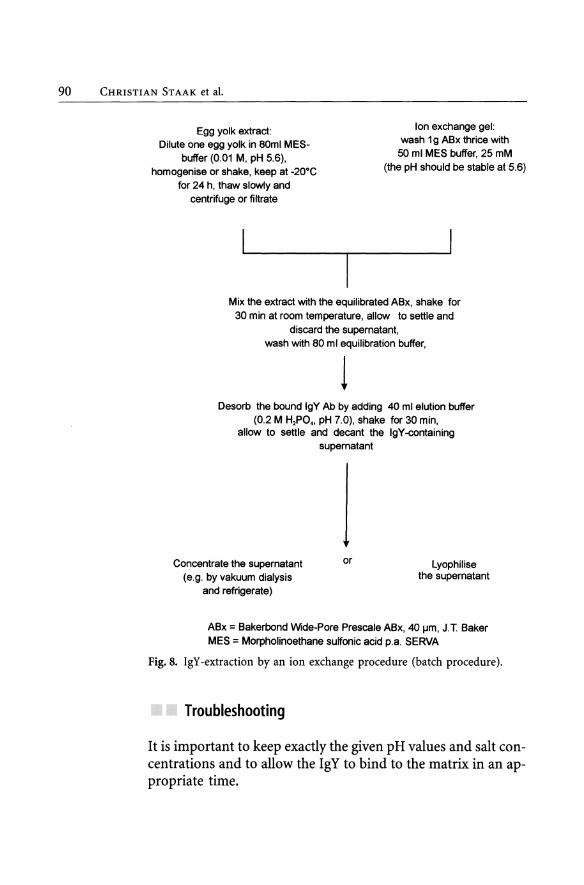
The course of an ion exchange procedure is shown schematically in Figure 8. In this case a so-called batch procedure is described.
Results
Under the chosen conditions, the yield is 70 - 80 % and the purity 80 - 90 %.
Hatta et al. (1990) used as their raw material for ion exchange chromatography the supernatant fluid obtained by lambda-carrageenan treatment, to which they added solid NaZP04 to a concentration of 0.02 M, using 3 N HeI to bring the pH to 8.0. The previous equilibration of the DEAE-sephacel-column, as also the washing (20 column-volumes), after production of the supernatal fluid, was performed with the same buffer. Bound protein was finally detached and eluated with a 0.2 M phosphate buffer.
Likewise Gassmann et al. (1990) used a rising ion-gradient. They began absorption with a 0.015 M potassium phosphate buffer (pH 8) and raised the ion concentration to 0.2 M at the same pH.
Schade et al. (1992) used a batch procedure with application of the DEAE ion exchanger (Pharmacia). Their starting material was 1:10 diluted yolk after freezing, thawing and filtering. The filtrate was dialysed against a 0.05 M phosphate buffer at pH 6.8 and finally mixed with the same buffer of swollen and equilibrated DEAE-A 50. The mixture was repeatedly washed. After the molarity of the buffer was rai sed, yolk -Ig Y was detached and obtained in the supernatant fluid or through filtration in the filtrate.
Reagents, solutions
90 CHRISTIAN STAAK et al.
Egg yolk extract: Dilute one egg yolk in 80ml MES
buffer (0.01 M. pH 5.6). homogenise or shake. keep at -20·C
for 24 h. thaw slowty and centrifuge or filtrate
Ion exchange gel: wash 19 ABx thrice with
50 mi MES buffer. 25 mM (the pH should be stable at 5.6)
Mix the extract with the equilibrated ABx. shake for 30 min at room temperature. allow to settle and
discard the supernatant. wash with 80 mi equilibration buffer.
1 Desorb the bound IgY Ab by adding 40 mi elution buffer
(0.2 M H,PO •• pH 7.0). shake for 30 min. allow to seltle and decant the IgY -containing
supernatant
Concentrate the supematant (e.g. by vakuum dialysis
and refrigerate)
or lyophilise the supernatant
ABx = Bakerbond Wide-Pore Prescale ABx. 40 IJm. J.T Baker MES = Morpholinoethane sulfonic acid p.a. SERVA
Fig. 8. IgY-extraction by an ion exchange procedure (batch procedure).
Troubleshooting
It is important to keep exactly the given pH values and salt concentrations and to allow the IgY to bind to the matrix in an appropriate time.
4 Isolation of IgY from Yolk 91
Subprotoeol 10 Affinity Chromatography of an IgY-Extract Aeeording to the Subprotoeols 1-7
The principle of affinity chromatography (AfC) consists in immobilising a reaction-partner (ligand) on a matrix. The specific second reaction partner in a mixture couples with the ligand, whereas the non-specific substances are rinsed away. The bound second reaction partner is finally detached through certain manipulations and isolated. In IgY -technology as with other fields, two principles of use are especially interesting:
• The obtaining of IgG-isotypes via AfC-columns with immobilised protein A (from Staphylococcus aureus) or protein G (from Streptococci of the C- or G-group), to which IgG-isotypes from various mammalian species bind differently. IgY from hens binds neither with protein A nor with protein G, in contrast to IgG from ducks, which couples with protein A (Higgins et al., 1995). Kronvall et al. (1970) report on the binding of IgG from rheiform birds' serum as a further exception. Therefore it is not possible to mark chicken IgY via enzyme-marked protein A or G for use in ELISA, or via FITC-marked protein A or G for use in fluorescence immuno histochemistry.
• The situation is quite different as regards immobilising a specific antigen on the matrix. The IgY antibodies to be obtained bind to the corresponding antigen and can be detached and eluted, as with IgG from mammals, by lowering the pH value to between 2.3 and 2.5.
The AfC procedure is of ten used. Since it is not essential to start with extremely purified IgY, a number of preparatory steps can be omitted. The final product is selectively obtained antibody, whose specificity depends on the composition of the immobilised antigen.
Thus AfC blunts the occasional demand for use of SPF animals. AfC is reversible, since in immobilising a specific antibody as a ligand on the matrix, the corresponding antigen is bound then isolated. Apart from it's universal applicability, this proce dure is practical in revealing contamination with penicil1in (De Leuw, 1996), in purifying parathyroid related protein (1 -

92 CHRISTIAN ST AAK et al.
36) (Rosoi et al., 1993) and in obtaining pure human alphazantiplasmin (Lee et al., 1997). As a matrix for serological work, the material most often used is sepharose 4B after activation with CNBr ('CNBr activated sepharose 4B').
CNBr activated sepharose 4B is available commercially, otherwise instructions for use may be taken from Tijssen (1985).
Materials
Equipment See also Subprotocol 8.
- column of appropriate size (small size)
- shaker
- refrigerator
Reagents, - CNBr-activated Sepharose solutions - HCI (ImM)
- NaHC03-buffer (0.1 M, pH 8.3, 0.5 M NaCI)
- ethanolamin or 0.2 M glycin (pH 8)
- acetate buffer (0.1 M, pH 4 with 0.15 M NaCI)
- Glycin/HCI (pH 2.4)
- PBS
- Tris (3M)
Proeedure
Coupling of Coupling of the ligand according to the second principle as de-the ligand scribed in the introduction (antigen as ligand to "fish" the IgY)
with CNBr activated sepharose B4 is performed in the following way (see also booklet 'Affinity Chromatography - principles and methods', Pharmacia, LKB, Biotechnology):
1. Weighing the calculated amount of lyophilised CNBr activated sepharose 4B (1 g lyophilisat produces about 3.5 mI of swollen gel).

4 Isolation of IgY from Yolk 93
2. Washing and swelling on a sintered glass fIlter (G3) with 1 mM HCI (need 200 ml/g sepharose).
3. Dissolving the protein for coupling in a coupling-buffer (NaHC03-buffer, 0.1 M, pH 8.3 with 0.5 M NaCI, ca. 5-10 mg protein/ml gel).
4. Mixing the protein/gel end over end (2 h RT or overnight at 4 ac, no magnetic stirrer).
5. Blocking remaining active groups by transferring the gel to a coupling buffer with a blocking substance (1 M ethanolamin or 0.2 M glycin, pH 8, 2 h at RT or 16 h at 4 aC).
6. Rinsing to remove superfluous absorbed protein (couplingbuffer followed by acetate buffer (0.1 M, pH 4 with 0.15 M NaCI), followed by coupling-buffer.
7. Rinsing to remove blocking substances (coupling-buffer). The protein-sepharose-conjugate is hereby usable in AfC.
Conservation at 4 - 8 ac after adding NaN3 in a final concentration of 0.1 %.
Isolation of specific IgY by binding to the corresponding antigen immobilised on CNBr-activated Sepharose:
1. Equilibration of the columns with PBS, application of the IgY solution, rinsing with PBS (low flow rate! Non-bound material must be wholly removed i.e. the absorption measured by the UV-monitor should be <0.1).
2. Elution (addition of the elution-buffer [glycin/HCI, pH 2.4] till no protein can be detected in the eluate).
3. Adjusting the pH value of the eluate with 3M Tris to 7.2.
4. Dialysis against a chosen buffer (24 h, 4 aC).
5. Centrifugation (2.800 X g, 15 min).
6. Concentration in the dialysis tube, e.g. by Carboxymethylcellulose (AquacideR).
Store the specific IgY in portions at -20 aC and the columns after long rinsing with PBS and addition of 0.1 % NaN3 at 4 aC. The columns can be used repeatedly.
Isolation of specific IgY

94 CHRISTIAN STAAK et al.
Results
An alternative matrix was presented by Staak et al. in 1996. The authors used pulverised micro-titre-plates to which the ligand was bound as in ELISA. Affinity chromatography followed the above method. The yield is notably less than in the case of chromatography with CNBr activated sepharose 4B, but the method is cheap and does not rely on imports. These affinity columns, too, can be used several times.
Troubleshooting
It is important to avoid overloading of the matrix with IgY (see Procedure).
Subprotocol 11 Amount of Specific Ab Extractable From Egg Yolk of Immunised Hens
In this protocol it is shown, that the amount of specific Ab obtained by affinity chromatography depends obviously on the antigen used for immunisation. In this respect, it is the same as with the immunisation of mammals.
Materials
As with Subprotocol 3, Chapter 3.
Procedure
As with Subprotocol 3, Chapter 3. After repeated immunisation with several antigens, antigen
specific IgY from the total fraction of yolk IgY is purified through affinity chromatography, then the percentage of specific antibodies is found (see Table 5).

4 Isolation of IgY from Yolk 9S
Results
The results are shown in Table 5.
Table 5. Percentage of Specific Antibodies in the Whole Egg Yolk of Chickens Immunised With Different Antigens
Antigen
Escherichia coli K88 fimbrial antigen
Recombinant bovine somatotropin
Human IgG
Low density lipoprotein
Bovine IgG
Specific IgY range from 1% to 20%
Troubleshooting
As with Subprotocol 1, Chapter 3.
Comments
Specific IgY (%)
0.9 - 1.8
2.7 - 8.0
7.4 - 18.9
2.1 - 5.6
12.4 - 13.5
Comparison of IgY-isolation proeedures as regards yield and purity
Three criteria serve as a basis for comparing various procedures for isolating IgY from the yolk of immunised hens:
• Yield of IgY
• Purity of IgY isolated
• Antibody activity Le. the amount of specific yolk-IgY.
Insofar as they are mentioned in the literature, the measurement procedures for the single criteria are given as follows. For implementing serological standard-techniques, reference is made to the relevant literature.
The yield of IgY without taking antibody-activity into account is expressed as mg/ml of the final product or as mgl yolk. These values are valid only if at the same time the purity of the yolk-IgY or its concentration as compared to the competing proteins is given.
96 CHRISTIAN STAAK et al.
For measuring proteins the following procedures were used: the Biuret method, the method according to Bradford (BioRad), Lowry's method, UV-absorption at 280 nm and the micro-Kjeldahl procedure (for instruction see Holtzhauer, 1997).
Immunological procedures for measuring the quantity and purity of the product included single radial immuno-diffusion (SRID, 'Mancini' -technique), used by researchers like Akita and Nakai, (1993), and the rocket -immuno electrophoresis or Laurell-technique, used by researchers like Schade et al., (1991); Svendson et al., (1995); Gross and Speck, (1996).
For testing the purity of isolated IgY, use was made of SDS polyacrylamide-gel-electrophoresis (PAGE) under non-reducing and reducing conditions.
Determination of antibody activities was generally performed with the test procedure for which the antibody was produced:
Enzyme-linked immunosorbent assay (ELISA) as a noncompetitive procedure (e.g. Schwarzkopf, 1994) and competitive (C-ELISA) procedure (Kierek-Jazcuk et al., 1997; Gutierrez and Gurriero, 1991). Of alI procedures, ELISA was the one most often mentioned.
The Agargel-immuno-double diffusion test (AGIDT, Ouchterlony) was used for instance byYamamoto et al. (1975) and by Poison et al. (1980, 1990).
Also mentioned are the virus neutralisation test (VNT) (Czerny and Eichhorn, 1989), the indirect haemagglutination test (IHA) (Hassl and Aspock, 1987), the radio-immuno-assay (RIA) (Rosoi et al., 1993) and immuno-fluorescence-technique (FAT, IFAT) (Schmidt et al., 1989; Gervelmeyer, 1997).
Moreover, affinity chromatografical investigations enable yolk-IgY to be expressed in mglml. (Gassmann et al., 1990; Schwarzkopf, 1994). For details see Chapter 5.
The incomplete listing of investigation procedures shows how widely yolk-IgY is used.
Comparison of various IgY-isolation techniques in regard to yield, purity and biological activity
Schwarzkopf (1994) compared various procedures for isolating IgY. The author used as starting material several yolks from the

4 Isolation of IgY from Yolk 97
Table 6. Comparison of Efficacy of Several IgY -Isolation Procedures Concerning Yield and Purity
Procedure Total protein Total IgY mg of specific Titre in ELISA content (TPC) content of the IgY anti-IgG after extraction of the extraction extraction (dog) before volume (= 6 mI) volume chromatography
1. WD 684 mgl6 mI 445 mg 9.7 1:128.000
llA % 65 % 2.2 %
2. DxS 282 mgl6 mI 278 mg 6.0 1: 8.000
4.7 % 98 % 2.2 %
3. PEG 42 mgl6 mI 32 mg 0.8 1: 2.000
0.7 % 77% 1.0 %
4. PEG/Ethanol 258 mgl6 mI 246 mg 504 1: 32.000
4.3 % 96 % 2.2 %
Measurement of the total protein content (TPC) with the Biuret method
Identification and quantification of IgY with cellulose-acetate-electrophoresis and the Biuret method (total IgY content)
Isolation of specific IgY (total specific IgY content) through affinity chromatography, determination of protein with the Bradford method (BioRad)
Determination of titre in ELISA (Ab-titre after extraction)
eggs of a hen immunised against dog-Ig, and for each of the isolation procedures listed in Table 6, she used 40 mI of the evenly mixed yolk. The isolates were adjusted to a final volume of 6 mI, so that protein content and antibody activity could be directly compared with each other.
Extraction procedures:
• Water dilution method (Schwarzkopf)
• Dextransulphate precipitation (Jensenius et al., 1981)
• PEG precipitation (Poison et al., 1980)
• PEG and ethanol (Polson et al., 1985)
In a further comparison between various yolk-IgY isolation procedures, Akita and Nakai (1993) used a yolk mixture of eggs with antibodies against enterotoxic Escherichia coli (ETEC), dividing it into 4 portions of 20 mI as starting material for 4 dif-

98 CHRISTIAN STAAK et al.
ferent extraction procedures. There was evaluation not only of the final but also of the intermediate product (supernatant-SN) after the first separation of the lipophile and hydrophile phases.
Water dilution method (WD):
• Dilution of yolk:d.w. 1:9, 6 hrs incubation at 4 aC, centrifu-ging off 10.000 X g for 25 min at 4 aC
• Supernatant fluid + 19% sodium sulphate (WD-SN)
• Dissolving of the precipitate
• Ultra-filtration (WD-UF)
polyethyleneglycol (PEG/ethanol method according to Polson et al., 1985): The supernatant fluid after the first PEG precipitation is called PEG-SN, and the final product is called PEG-Alc.
Dextransulphate precipitation (DS) according to Jensenius et al. (1981): The supernatant fluid after the first DS-precipitation as well as the 2nd supernatant fluid after elution of the precipitate is called DS-SN, and the final product after sodium-sulphate precipitation is called DS-As2.
Xanthan method (Xan) after Hatta et al. (1990): The supernatant fluid after xanthan precipitation is called XanSN, and the final product after the 2nd xanthan precipitation and the following sodium-sulphate one is called Xan-As2.
The SN-preparations as well as the final product are compared in Table 7.
Antibody activity was measured in the indirect ELISA with antigen from E.coli. Both the 4 SN fractions and the 4 final products were investigated after having been uniformly adjusted to 0.5 /-lg IgY ImI.
The levels of extinction of the SN-fractions of the WD, PEG and DS preparations were equally high (ca. 0.90), whereas both those of the xanthan method and those of the untreated yolk were lower (ca. 0.70). A similar profile was found in investigating the final product.
Though the conclusions drawn from the two investigations differ in detail, the authors agree that the water-dilution method is best.

4 Isolation of IgY from Yolk
Table 7. Comparison of Efficacy of Several IgY -Isolation Procedures Con-cerning Yield and Purity
Test material IgY Test material IgY
(supernatant Yield Purity (final Yield Purity % fluid) in % in % product) mglml yolk
Yolk 100
WD-SN 91 31 WD-UF 9.8 94
PEG-SN 47 25 PEG-Alc 4.9 89
DS-SN 71 x DS-As2 7.5 87
Xan-SN 72 32 Xan-As2 7.3 89
Protein determination by the Biuret method; quantification of IgY by RID (radial immune diffusion, Mancini-technique); check of purity with SDSPAGE
Special attention is to be given to the pH-value of the distilled water, sinee Akita and Nakai (1992) found that with a neutral pH the yield ofIgY lies between 55 and 60%, whereas with a pH of 5.0 - 5.2 it Iies between 92 and 96%.
Moreover the authors of both investigations agree on the simplicity of the water-dilution method in terms of time, materials and manipulations. As regards the produetion of proteetive antibodies, Akita and Nakai (1993) also emphasise the optimal opportunities for sealing up.
Conservation and stability of IgY ~xtracts I
Of importanee for the use of yoIk IgY is information about resistanee to heat, for instanee in spray-drying for obtaining therapeutic preparations (lkemori et aL, 1997), and about resistanee to acidic pH (see 'affinity ehromatography').
As regards resistanee to heat, Loseh et al. (1986) found no Ioss of antibodies in eggs eooked for 6 minutes.
On adding HeI in portions, Sehmidt et al. (1989) found a rapid falI in the amount of antibodies from pH 4 ono This inaetivation of antibody aetivity eould not be eonfirmed by using
99
100 CHRISTIAN STAAK et al.
glycin/HCl pH 2.3-2.5 to split bound antibodies in affinity chromatography (Schwarzkopf, 1994).
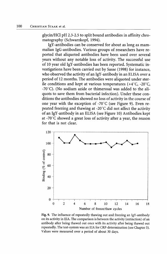
IgY-antibodies can be conserved for about as long as mammalian IgG-antibodies. Various groups of researchers have reported that aliquoted antibodies have been used over several years without any notable loss of activity. The successful use of 10 year old IgY-antibodies has been reported. Systematic investigations have been carried out by Sasse (1998) for instance, who observed the activity of an IgY -antibody in an ELISA over a period of 12 months. The antibodies were aliquoted under sterile conditions and kept at various temperatures (+4 aC, -20°C, -70°C). (No sodium azide or thimerosal was added to the aliquots to save them from bacterial infection). Under these conditions the antibodies showed no 10ss of activity in the course of one year with the exception of -70°C (see Figure 9). Even repeated freezing and thawing at -20°C did not affect the activity of an IgY -antibody in an ELISA (see Figure 10) Antibodies kept at -70°C showed agreat 10ss of activity after a year, the reason for that is not dear.
120
100
"O ... 80 15 o u
""' o
~ 60
0Il 1': :e 40 .5
!Xl
20
O O 2 4 6 8 10 12 14 16 18
Number of freeze/thaw cyeIes
Fig. 9. The influence of repeatedly thawing out and freezing an IgY -antibody on its activity in EIA. The comparison is between the activity (extinction) of an antibody after being thawed out once with its activity after being thawed out repeatedly. The test-system was an EIA for CRP-determination (see Chapter 5). Values were measured over a period of about 30 days.

100
75
c .:> .~ 50 o ~ ~
25
o 'O ~ O U
4 Isolation of IgY from Yolk 101
after 6 months after 12 months
oU U U - U U U g o o o o o '<:1" O O '<:t O O
+ N t- I: + N t-I I O I I
U
IgY obtained by watery extraction
100~~-=~~,-------~~----------'
-. -.t. '-"
C .:> '.jj o C'3 ~
~
75
50
25
1 ~ ~ ~ ] o + C"";I r;- § u u
u o '<:1" +
u o o N
I
IgY obtained by ammoniumsulphate precipitation
Fig. 10. The activity of an IgY-antibody kept at various temperatures. For the upper part of the figure use was made of a watery IgY -extract, whereas for the lower part use was made of an ammonium-sulphate preparation. The test system used was an EIA for detecting the lipopolysaccharide of E. coli 0111 (homologous antigen).
102 CHRISTIAN STAAK et al.
Systems eommercially available
U nlike the case several years ago, several firms are now offering extraction kits for preparing IgY from egg-yolk.
Table 8. Commercial IgY Extraction Kits
Firm
Promega
Pharmacia Biotech
Clontech
Biotez
Technogen
Kit
EGGstract© IgY Purification System
gammaYolk
Protein L agarose beads
IMSORB Anti IgY
Kaptiv-GY TM
The first two kits represent precIpItat ion procedures, whereas the third kit works like the affinity-chromatographical purification of IgG with protein G or A. Here too the material involved is a bacterial cell-wall protein from Peptostreptococcus magnus, protein L, which is able to bind the immunoglobulins of vertebrates. Unlike protein A/G, protein L does not interact with the Fc-part of the immunoglobulin molecule but with the light chain (Chateau et al. 1993. However, the authors of this paper themselves note that protein L does not bind chicken IgG. We have tested this system with no success, it may be that the system does not work properly). IMSORB anti IgY is also based on an affinity-chromatographic principle. IgY is simply purified by binding to an an ti IgY Ab.
References
Akita EM, Nakai S (1992) Immunoglobulin from egg yolk: Isolation and purification. J Food Sci 57:626-634
Akita EM, Nakai S (1993) Comparison of four purification methods for the production of immunoglobulins from eggs laid by hens immunized with enterotoxigenic E.coli strain. J Immunol Methods 160:207-214
Aulisio CG, Shelokov A (1967) Substitution of egg yolk for serum in indirect fluorescence assay for Roux Sarcoma virus antibody. Proc Soc Exp Biol Med 126:312-315
Bade H, Stegemann H (1984) Rapid method of extraction of antibodies from egg yolk. J Immunol Methods 72:421-426
4 Isolation of IgY from Yolk 103
Chateau, M de, Nilson, BHK, Erntell, M, Myhre, E, Magnusson, CGM, Akerstrom, B, Bjorck, L (1993). On the interaction between protein L and immunoglobulins ofvarious mammalian species. Scand. J. ImmunoI. 37, 399-405
Czerny CP, Eichhorn W (1989) Characterization of monoclonal and polyclonal antibodies to bovine enteric coronavirus: establishment of an efficient ELISA for antigen detection in feces. Vet Microbiol 20:111-122
De Leuw (1996) Gewinnung und Einsatz multispezifischer Antikorper als Hilfsmittel zur Probenvorbereitung in der Riickstandsanalytik von Penicillinen. Dr. Thesis, Bergische Universitat-Gesamthochschule, Wuppertal
Erhard MH, Bergmann J, Renner M, Hofmann A, Heinritzi K (1996) Prophylactic effect of specific egg yolk antibodies in diarrhea of weaned piglets caused by Escherichia coli K88 (F4). J Vet Med A 43:217-223
Fischer M (1996) Untersuchungen zu Standardpraparationen von Immunglobulinen aus dem Dotter von Eiern verschiedener Wirtschaftsgefliigelspezies. Dr. Thesis, Veterinarmedizin der FU Berlin, Journal Nr. 1988
Fischer M, Hlinak A, Montag T, Claros M, Schade R, Ebner D (1996) Ver-gleich von Standardmethoden zur Praparation von Dotterantikorpern. TierarztI.Praxis 24:411-418
Gassmann M, ThOmmes P, Weiser T, Hiibscher A (1990) Efficient production of chicken egg yolk antibodies against conserved mammalian protein. Faseb J 4:2528-2532
Gast RK, Porter RE, Hoit PS (1997) Assesing the sensitivity of egg yolk antibody testing for detecting Salmonella enteritidis infections in laying hens. Poultry Sci 76:798-801
Gervelmayer A (1995) Herstellung von virusspezifischen vitellinen Antikorpern und deren Einsatz im indirekten Fluoreszenztest zum Nachweis der Erreger der Gefliigelkrankheiten Newcastle Disease, Infektiose Bronchitis und Gumboro Disease. Dr. Thesis, FU Berlin, Journal Nr. 1835
Gottstein B, Hemmler E (1985) Egg yolk immunoglobulin Y as an alternative antibody in the serology of echinococcosis. Z Parasitenkd 71:273-276
Gross M, Speck J (1996) Aviare Dotter-Antikorper in Diagnostik und For-schung. Dtsch tierarztl Wschr 103:417-422
Grossfeld (1938) "Handbuch der Eierkunde" Verlag v. J. Springer, Berlin Guiterrez JA, Guirriero V (1991) J ImmunoI.Methods 143:81-888 Hassl A, Aspock H, Flamm H (1987) Comparative studies on the purity and
specificity of yolk immunoglobulin Y isolated from eggs laid by hens immunized with toxoplasma gondii antigen. Zbl Bakt Hyg A 267:247-253
Hassl A, Aspock H (1988) Purification of egg yolk immunoglobulins. J Immunol Meth 110:225-228
Hatta H, Kim M, Yamamoto T (1990) A novel isolation method for hen egg yolk antibody, "IgY". Agric Biol Chem 54:2531-2535
Higgins DA, Cromie RL, Liu SS, Magor KE, Warr GW (1995) Purification of duck immuno-globulins: an evaluation of protein A and protein G affinity chromatography. Vet Immunol Immun Path 44:169-180

lO4 CHRISTIAN STAAK et al.
Holtzhauer M (1997) Biochemische Labormethoden (Springer Labor Manual), Springer Berlin Heidelberg New York
Ikemori Y, Kuroki M, Peralta RC, Yokoyama H, Kodama Y (1992) Protection of neonatal calves against fatal enteric colibacillosis by administration of egg yolk powder from hens immunized with K99-pilliated enterotoxigenic E. coli. Am J Vet Res 53:2005-2008
Ikemori Y, Ohta M, Umeda K, !cado FC , Kuroki M, Yokoyama H, Kodama Y (1997) Passive protection of neonatal calves against bovine coronavirus-induced diarrhea by administration of egg yolk or colostrum antibody powder. Vet Microbiol 58:105-111
Jensenius JC, Andersen 1, Hau J, Crone M, Koch C (1981) Eggs: Conveniendy packaged antibodies, methods for purification of yolk IgG. J Immunol Meth 46:63-68
Kierek-Jaszczuk R, Marquardt R,. Abramson D (1997) Use of a heterologous solid-phase antigen in an indirect competitive antibody-capture anzyme-linked immunosorbent assay for T -2 Mycotoxin. J Food Protect 60:31-327
Kronvall G, Ulysses SS, Finstad J, Williams RC (1970) Phylogenetic insight into evolution of mammalian Fc fragment of gG globulin using staphylococcal protein A. J Immunol 104:140-147
Kuroki M, Ikemori Y, Yokoyama H, Peralta RC, Faustino CI, Kodama Y (1993) Passive protection against bovine rotavirus induced diarrhea in murine model by specific immunoglobulins from chicken egg yolk. Vet MicrobioI37:135-146
Lee SC, Lee KN, Schwartzott DG, Jackson KW, Tae EC, McKee PA (1997) Purification of human alphaTantiplasmin with chicken IgY specific to ist carboxy-terminal peptide. Prep Biochem Biotechnol 27:227-237
Loeken MR, Roth TF (1983) Analysis of maternal IgG subpopulations which are transported into the chicken oocyte. Immunology 49:21 - 28
LOsch U, Schranner 1, Wanke R, Jiirgens L (1986) The chicken egg, an antibody source. J Vet Med B 33:609-619
Martin WG, Turner NH, Cook WH (1959) Macromolecular properties of vitellin from egg yolk and its parent complex with lipids. Canad J Biochem Physiol 37:1197-1207
McKinney MM, Parkinson A (1987) A simple, non-chromatographic procedure to purify immuno-globulins from serum and ascites fluid. J Immunol Meth 96:271-278
Mehner A, Hartfield A (1983) Handbuch der Gefliigelphysiologie VoI 1, VEB Gustav Fischer Vedag Jena
Mohammed HO, Yamamoto R, Carpenter TE, Ortmayer HB (1985) A statistical model to optimize enzyme-linked immunosorbent assay parameters for detection of Mycoplasma gallisepticum and M. synoviae antibody in egg yolk. Avian Diseases 30:389-397
Ntakarutimana V, Demedts P, van Sande M, Scharpe S (1992) A simple and economical strategy for downstream processing of specific antibodies to human transferrin from egg yolk. J lmmunol Meth 153:133-140
Odans E, Rose ME (1972) An IgA-like immunoglobulin in the fowl. Immunochemistry 9:833-838

4 Isolation ofIgY from Yolk 105
Patterson R, Younger JS, Weigle WO, Dixon FJ (1962) Antibody production and transfer to egg yolk in chickens. J Gen Physio145:501-513
Pharmacia, LKB Biotechnology "Affinity Chromatography - principles & methods" Pharmacia LKB Biotechnology AB, Uppsala, Sweden
Poison A (1990) Isolation of IgY from the yolks of eggs by a chloroform polyethylene glycol procedure. Immun Invest 19:253-258
Polson A, von Wechmar MB, van Regenmortel MHV (1980) Isolation of viral IgY antibodies from yolks of immunized hens. Immunol Comm 9:475-493
Poison A, Coetzer T, Kruger J, v.Maltzahn E, van der Merwe KJ (1985) Improvements in the isolation of IgY from the yolks of eggs laid by immunized hens. Immun lnvest 14:323-327
Rammensee M (1984) Immunglobulinkonzentration in Serum, Sekret Ei und Embryo des Haushuhnes. Dr. Thesis, Ludwig-Maximilians Universităt Miinchen
Romanoff AL, Romanoff J (1949) "The Avian Egg", J Wiley & Sons INC., New York, Chapman & Hall, Limited, London
Rose ME, Orlan E, Buttress N (1974) Immunoglobulin classes in the hens egg: their segregation in yolk and white. Eur J Immunol 4:521-523
Rosoi TJ, Steinmeyer CL; McCauley LK, Merryman JI, Werkmeister JR, Grone A, Weckmann MT, Swayne DE, Capen CC (1993) Studies on chicken polyclonal anti-peptide antibodies specific for parathyroid hormone-related protein (1-36). Vet lmmunollmmunopath 35:321-337
Sachsenweber O, Lohr JE, Kosters J (1994) Beurteilung von drei kommerziellen ELISA-Testkits zum Nachweis von Antikorpern gegen Salmonella enteritidis. Tierărztl Praxis 22: 350-357
Sasse M (1998). Experimente zur Induktion, Prăparation und Charakterisierung aviărer vitelliner Antikorper gegen Endotoxine verschiedener gramnegativer Bakterien. Dr. Thesis, Veterinărmedizinische Fakultăt der Universităt Leipzig.
Schade R, Pfister C, Halatsch R, Henklein P (1991) Polyclonal IgY antibodies from chicken egg yolk - an alternative to the production of mammalian IgG type antibodies in rabbits. ATLA 19:403-419
Schade R, Schniering A, Hlinak A (1992) Die Gewinnung spezifischer polyklonaler Antikorper aus dem Dotter von Hiihnereiern als Alternative zur Immunisierung von Saugern - eine Ubersicht. ALTEX 9:43-56
Schwarzkopf C (1994) Gewinnung und immunologische Charakterisierung speziesspezifischer IgY, sowie deren Einsatz zur Bestimmung der Wirtstierart aus dem Abdominalblut hămatophager Insekten. Dr. Thesis, F.U. Berlin
Schmidt P, Wiedemann V, Kiihlmann R, Wanke R, Linckh E, Losch U (1989) Chicken egg antibodies for prophylaxis and therapy of infectious intestinal diseases. II: In vitro studies on gastric and enteric digestion of egg yolk antibodies specific against pathogenic E. coli strains. J Vet Med B 36:619-628
Siewert E, Bronch K (1972) In: Lenkeit, Breirem, Crasemann (eds) Handbuch der Tierernăhrung , Verlag Paul Parey, Hamburg und Berlin
106 CHRISTIAN STAAK et al.
Staak C, Salchow F, Clausen P.-H, Luge E. (1996) Polystyrene as an affinity chromatography matrix for the purification of antibodies. J Immunol Methods 194:141-146
Svendson, L, Crowley A, Ostergaard LH, Stodulski G, Hau J (1995) Development and comparison of purification strategies for chicken antibodies from egg yolk. Labor Anim Sci 45:89-93
Tjissen P (1985) "Laboratory Techniques in Biochemistry and Molecular Biology: Practice and Theory of Enzyme Immunoassays" Elsevier, Amsterdam, New York, Oxford.
UFAW (1987) The domestic fowl and turkey. In: Poole TB (ed) Handbook on the care and management oflaboratory animals. Longman Scientific & Technical. 6th Edition, p.645
Wallmann, J, Staak C, Luge E (1990) Einfache Methode zur Isolierung von Immunoglobulin (Y) aus Eiern immunisierter Hiihner. J Vet Med B 37:317-320
Williams J (1963) A comparison of conalbumin and transferrin in the domestic fowl. Biochem H 83:355-364
Yamamoto H, Watanabe H, Sato G (1975) Identification of immunoglobulins in chicken eggs and their antibody activity. Jap J Vet Res 23: 131-40
Yokoyama H, Peralta RC, Horikoshi T, Hiraoka J, Ikemori Y, Kuroki M, Kodama Y (1992) A two step procedure for purification ofhen egg yolk immunoglobulin G: utilization Hydroxypropyl-methykellulose phthalate and synthetic affinity ligand gel (Avid Al). Poult Sci 72:275-281
Suppliers
Promega Labeled anti-chicken Ab (as secondary Ab) are also available. Tel.: 0049-(0)621-8501-0 FAX: 0049-(0)621-8501-222 E-mail: [email protected] Internet: www.promega.com
Pharmacia Biotech Tel.: 0049-(0)761-4903-153 FAX: 0049-(0)761-4903-353 E-mail: [email protected] Internet: http://www.apbiotech.com
Biotez Tel.: 0049-(0)30-9489-3317 E-mail: [email protected] Internet: www.mdc-berlin.de/biotez/de
Technogen Tel.: +39-0823-612-208/228 Fax: +39-0823-612-222/230 E-mail: [email protected] Internet: http://www.technogen.it
4 Isolation of IgY from Yolk 107
Copyright © 2022 FDOKUMEN