Isolation and molecular characterization of Newcastle disease viruses from raptors
11
Isolation and Molecular Characterization of a Novel Picornavirus from Baitfish in the USA Nicholas B. D. Phelps 1,2 *, Sunil K. Mor 1 , Anibal G. Armien 1,2 , William Batts 3 , Andrew E. Goodwin 4 , Lacey Hopper 5 , Rebekah McCann 6 , Terry Fei Fan Ng 7,8 , Corey Puzach 6 , Thomas B. Waltzek 9 , Eric Delwart 7,8 , James Winton 3 , Sagar M. Goyal 1,2 1 Minnesota Veterinary Diagnostic Laboratory, St. Paul, Minnesota, United States of America, 2 University of Minnesota, Department of Veterinary Population Medicine, St. Paul, Minnesota, United States of America, 3 U.S. Geological Survey, Western Fisheries Research Center, Seattle, Washington, United States of America, 4 U.S. Fish and Wildlife Service, Portland, Oregon, United States of America, 5 U.S. Fish and Wildlife Service, Bozeman Fish Health Center, Bozeman, Montana, United States of America, 6 U.S. Fish and Wildlife Service, La Crosse Fish Health Center, Onalaska, Wisconsin, United States of America, 7 Blood Systems Research Institute, San Francisco, California, United States of America, 8 University of California, Department of Laboratory Medicine, San Francisco, California, United States of America, 9 University of Florida, College of Veterinary Medicine, Department of Infectious Diseases and Pathology, University of Florida, Gainesville, Florida, United States of America Abstract During both regulatory and routine surveillance sampling of baitfish from the states of Illinois, Minnesota, Montana, and Wisconsin, USA, isolates (n = 20) of a previously unknown picornavirus were obtained from kidney/spleen or entire viscera of fathead minnows (Pimephales promelas) and brassy minnows (Hybognathus hankinsoni). Following the appearance of a diffuse cytopathic effect, examination of cell culture supernatant by negative contrast electron microscopy revealed the presence of small, round virus particles (,30–32 nm), with picornavirus-like morphology. Amplification and sequence analysis of viral RNA identified the agent as a novel member of the Picornaviridae family, tentatively named fathead minnow picornavirus (FHMPV). The full FHMPV genome consisted of 7834 nucleotides. Phylogenetic analysis based on 491 amino acid residues of the 3D gene showed 98.6% to 100% identity among the 20 isolates of FHMPV compared in this study while only 49.5% identity with its nearest neighbor, the bluegill picornavirus (BGPV) isolated from bluegill (Lepomis macrochirus). Based on complete polyprotein analysis, the FHMPV shared 58% (P1), 33% (P2) and 43% (P3) amino acid identities with BGPV and shared less than 40% amino acid identity with all other picornaviruses. Hence, we propose the creation of a new genus (Piscevirus) within the Picornaviridae family. The impact of FHMPV on the health of fish populations is unknown at present. Citation: Phelps NBD, Mor SK, Armien AG, Batts W, Goodwin AE, et al. (2014) Isolation and Molecular Characterization of a Novel Picornavirus from Baitfish in the USA. PLoS ONE 9(2): e87593. doi:10.1371/journal.pone.0087593 Editor: Amit Kapoor, Columbia University, United States of America Received October 18, 2013; Accepted December 23, 2013; Published February 21, 2014 This is an open-access article, free of all copyright, and may be freely reproduced, distributed, transmitted, modified, built upon, or otherwise used by anyone for any lawful purpose. The work is made available under the Creative Commons CC0 public domain dedication. Funding: The authors have no support or funding to report. Competing Interests: The authors have declared that no competing interests exist. * E-mail: [email protected] Introduction Baitfish are economically and ecologically important through- out many regions of the world [1]. For example in the USA, 257 baitfish farms produced $38 million worth of baitfish in 2005, which ranks in the top five for aquaculture production [2]. In addition, a relatively undocumented, but significant sector of the baitfish industry relies upon fish harvested from the wild [3]. The most important baitfish species in the USA are the golden shiner (Notemigonus crysoleucas) and the fathead minnow (Pimephales promelas), although a variety of other wild and farmed fish species are also produced [4]. The baitfish industry in the USA ships more than 10 billion fish per year across state lines and nationwide. These fish are distributed through wholesale and retail networks to anglers who take the live baitfish to rivers and lakes where they may be consumed by predators or released into the wild [5]. In addition, large quantities of baitfish are sold as forage for the production of predatory species such as walleye (Sander vitreus) and muskellunge (Esox masquinongy). The movement of baitfish into new watersheds could potentially introduce viruses from an endemic area, where fish have co- evolved with a particular pathogen, to new bodies of water where fish hosts may have little or no natural resistance [5–8]. Regulatory oversight of these movements has become increasingly strict with the emergence of high profile pathogens, such as viral hemor- rhagic septicemia virus (VHSV) in the Great Lakes [9,10]. Currently, many state, national, and international regulations mandate the inspection of certain aquatic animal species prior to movement using standard methods [11,12]. Such methods often specify virus isolation in cell culture as the gold standard diagnostic test for the detection of fish viruses. The isolation of a virus in cell cultures is largely non-specific unless followed by virus identification and characterization. It is not surprising then, that due to the increased testing of baitfish and the use of cell culture-based diagnostic assays, reports of novel viral pathogens of fish are becoming more common. This was demonstrated during a 2009–2011 survey of Wisconsin baitfish dealers in which McCann [13] isolated one or more viruses in 36 of 82 lots inspected, resulting in a total of 39 viral isolates. The isolation of previously known aquareoviruses (51%) and nido- viruses (10%) was not surprising; however, 39% of the isolates could not be easily characterized and thus, cause for concern [13]. PLOS ONE | www.plosone.org 1 February 2014 | Volume 9 | Issue 2 | e87593
-
Upload
independent -
Category
Documents
-
view
1 -
download
0
Transcript of Isolation and molecular characterization of Newcastle disease viruses from raptors

Isolation and Molecular Characterization of a NovelPicornavirus from Baitfish in the USANicholas B. D. Phelps1,2*, Sunil K. Mor1, Anibal G. Armien1,2, William Batts3, Andrew E. Goodwin4,
Lacey Hopper5, Rebekah McCann6, Terry Fei Fan Ng7,8, Corey Puzach6, Thomas B. Waltzek9,
Eric Delwart7,8, James Winton3, Sagar M. Goyal1,2
1Minnesota Veterinary Diagnostic Laboratory, St. Paul, Minnesota, United States of America, 2University of Minnesota, Department of Veterinary Population Medicine, St.
Paul, Minnesota, United States of America, 3U.S. Geological Survey, Western Fisheries Research Center, Seattle, Washington, United States of America, 4U.S. Fish and
Wildlife Service, Portland, Oregon, United States of America, 5U.S. Fish and Wildlife Service, Bozeman Fish Health Center, Bozeman, Montana, United States of America,
6U.S. Fish and Wildlife Service, La Crosse Fish Health Center, Onalaska, Wisconsin, United States of America, 7 Blood Systems Research Institute, San Francisco, California,
United States of America, 8University of California, Department of Laboratory Medicine, San Francisco, California, United States of America, 9University of Florida, College
of Veterinary Medicine, Department of Infectious Diseases and Pathology, University of Florida, Gainesville, Florida, United States of America
Abstract
During both regulatory and routine surveillance sampling of baitfish from the states of Illinois, Minnesota, Montana, andWisconsin, USA, isolates (n = 20) of a previously unknown picornavirus were obtained from kidney/spleen or entire viscera offathead minnows (Pimephales promelas) and brassy minnows (Hybognathus hankinsoni). Following the appearance of adiffuse cytopathic effect, examination of cell culture supernatant by negative contrast electron microscopy revealed thepresence of small, round virus particles (,30–32 nm), with picornavirus-like morphology. Amplification and sequenceanalysis of viral RNA identified the agent as a novel member of the Picornaviridae family, tentatively named fathead minnowpicornavirus (FHMPV). The full FHMPV genome consisted of 7834 nucleotides. Phylogenetic analysis based on 491 aminoacid residues of the 3D gene showed 98.6% to 100% identity among the 20 isolates of FHMPV compared in this study whileonly 49.5% identity with its nearest neighbor, the bluegill picornavirus (BGPV) isolated from bluegill (Lepomis macrochirus).Based on complete polyprotein analysis, the FHMPV shared 58% (P1), 33% (P2) and 43% (P3) amino acid identities withBGPV and shared less than 40% amino acid identity with all other picornaviruses. Hence, we propose the creation of a newgenus (Piscevirus) within the Picornaviridae family. The impact of FHMPV on the health of fish populations is unknown atpresent.
Citation: Phelps NBD, Mor SK, Armien AG, Batts W, Goodwin AE, et al. (2014) Isolation and Molecular Characterization of a Novel Picornavirus from Baitfish in theUSA. PLoS ONE 9(2): e87593. doi:10.1371/journal.pone.0087593
Editor: Amit Kapoor, Columbia University, United States of America
Received October 18, 2013; Accepted December 23, 2013; Published February 21, 2014
This is an open-access article, free of all copyright, and may be freely reproduced, distributed, transmitted, modified, built upon, or otherwise used by anyone forany lawful purpose. The work is made available under the Creative Commons CC0 public domain dedication.
Funding: The authors have no support or funding to report.
Competing Interests: The authors have declared that no competing interests exist.
* E-mail: [email protected]
Introduction
Baitfish are economically and ecologically important through-
out many regions of the world [1]. For example in the USA, 257
baitfish farms produced $38 million worth of baitfish in 2005,
which ranks in the top five for aquaculture production [2]. In
addition, a relatively undocumented, but significant sector of the
baitfish industry relies upon fish harvested from the wild [3]. The
most important baitfish species in the USA are the golden shiner
(Notemigonus crysoleucas) and the fathead minnow (Pimephales
promelas), although a variety of other wild and farmed fish species
are also produced [4]. The baitfish industry in the USA ships more
than 10 billion fish per year across state lines and nationwide.
These fish are distributed through wholesale and retail networks to
anglers who take the live baitfish to rivers and lakes where they
may be consumed by predators or released into the wild [5]. In
addition, large quantities of baitfish are sold as forage for the
production of predatory species such as walleye (Sander vitreus) and
muskellunge (Esox masquinongy).
The movement of baitfish into new watersheds could potentially
introduce viruses from an endemic area, where fish have co-
evolved with a particular pathogen, to new bodies of water where
fish hosts may have little or no natural resistance [5–8]. Regulatory
oversight of these movements has become increasingly strict with
the emergence of high profile pathogens, such as viral hemor-
rhagic septicemia virus (VHSV) in the Great Lakes [9,10].
Currently, many state, national, and international regulations
mandate the inspection of certain aquatic animal species prior to
movement using standard methods [11,12]. Such methods often
specify virus isolation in cell culture as the gold standard diagnostic
test for the detection of fish viruses.
The isolation of a virus in cell cultures is largely non-specific
unless followed by virus identification and characterization. It is
not surprising then, that due to the increased testing of baitfish and
the use of cell culture-based diagnostic assays, reports of novel viral
pathogens of fish are becoming more common. This was
demonstrated during a 2009–2011 survey of Wisconsin baitfish
dealers in which McCann [13] isolated one or more viruses in 36
of 82 lots inspected, resulting in a total of 39 viral isolates. The
isolation of previously known aquareoviruses (51%) and nido-
viruses (10%) was not surprising; however, 39% of the isolates
could not be easily characterized and thus, cause for concern [13].
PLOS ONE | www.plosone.org 1 February 2014 | Volume 9 | Issue 2 | e87593
Simultaneously in other laboratories, previously unknown viruses
were isolated from fathead minnows from Illinois, Minnesota,
Montana, and Wisconsin, and brassy minnows (Hybognathus
hankinsoni) from Montana. Over the course of time it became
apparent that many of the aforementioned isolates were related to
each other and were putative members of the Picornaviridae family.
The Picornaviridae family is currently divided into 17 genera:
Aphthovirus, Aquamavirus, Avihepatovirus, Cardiovirus, Coso-
virus, Dicipivirus, Enterovirus, Erbovirus, Hepatovirus, Kobu-
virus, Megavirus, Parechovirus, Salivirus, Sapelovirus, Seneca-
virus, Teschovirus and Tremovirus [14,15], however the list is
rapidly expanding (www.picornaviridae.com). Picornaviruses are
small (,30–32 nm), icosahedral, non-enveloped single stranded
positive sense RNA viruses with genome size of approximately 7.2
to 9.0 kb [14]. The genome encodes a single polyprotein flanked
by 59 and 39 nontranslated regions (NTRs). The viral polyprotein
is post-translationally cleaved into three regions P1, P2 and P3.
These three regions are further processed into 10–12 small viral
proteins, such as viral capsid proteins (VP4, VP3, VP2, VP1),
which are encoded by P1 while P2 and P3 encode non-structural
proteins that facilitate protein processing (2Apro, 3Cpro and
3CDpro) and genome replication (2B, 2C, 3AB, 3B (VPg), 3CDpro,
3Dpol) [16]. In addition to these proteins, the picornaviruses in
some genera also contain a leader protein (L) upstream of the P1.
Picorna-like viruses have been reported sporadically in various
fish species [17–20], although some of these were later shown to be
members of other virus families [21,22]. From mortality events of
bluegill (Lepomis macrochirus) in Montana Lake in northeastern
Wisconsin, a picornavirus, tentatively known as the bluegill
picornavirus (BGPV), was recently isolated and molecularly
characterized [23]. Phylogenetic analysis of the BGPV genome
(GenBank NC_018506) revealed the virus to be the member of a
new genus in the family Picornaviridae [23].
Herein, we describe the isolation and characterization of a novel
picornavirus from baitfish and compare multiple isolates obtained
from various laboratories to determine the relationship of the
fathead minnow picornavirus (FHMPV) with previously reported
picornaviruses and discuss the implications of these findings.
Materials and Methods
Source of SamplesA total of 20, previously uncharacterized, viral isolates were
included in this study (Table 1). Eight of these isolates (FHMPV-01
thru FHMPV-08) were cultured at the La Crosse Fish Health
Center (U.S. Fish and Wildlife Service, Onalaska, WI) as a result
of mandatory inspections of four Wisconsin baitfish dealers [13].
The isolates originated from fathead minnows, collected from
licensed aquaculture facilities in Minnesota and Wisconsin.
However, these facilities commonly import baitfish from a variety
of sources in and out of state, combining them for later resale.
Consequently, the precise source populations are unknown.
Eleven viral isolates (FHMPV-09 thru FHMPV-19) cultured at
the Bozeman Fish Health Center (U.S. Fish and Wildlife Service,
Bozeman, MT) were also included in this study. The isolates
originated from fathead minnows and brassy minnows collected
from five different bodies of water in Montana as part of a routine
wild fish health survey.
One viral isolate (FHMPV-20) cultured at the University of
Arkansas at Pine Bluff, Fish Disease Laboratory (Pine Bluff, AR)
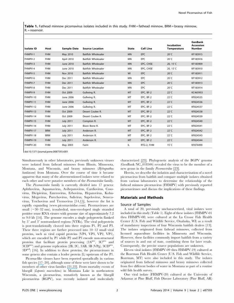
Table 1. Fathead minnow picornavirus isolates included in this study. FHM= fathead minnow, BRM=brassy minnow,R. = reservoir.
Isolate ID Host Sample Date Source Location State Cell LineIncubationTemperature
GenBankAccessionNumber
FHMPV-1 FHM May 2010 Baitfish Wholesaler MN EPC 20uC KF183915
FHMPV-2 FHM April 2010 Baitfish Wholesaler MN EPC 20uC KF183916
FHMPV-3 FHM June 2010 Baitfish Wholesaler MN EPC, CHSE 20, 15uC KF183909
FHMPV-4 FHM June 2010 Baitfish Wholesaler MN EPC, CHSE 20, 15uC KF183910
FHMPV-5 FHM Nov 2010 Baitfish Wholesaler WI EPC 20uC KF183911
FHMPV-6 FHM Dec 2011 Baitfish Wholesaler MN EPC 20uC KF183912
FHMPV-7 FHM Dec 2011 Baitfish Wholesaler MN EPC 20uC KF183913
FHMPV-8 FHM Dec 2011 Baitfish Wholesaler MN EPC 20uC KF183914
FHMPV-9 FHM Oct 2009 Gullwing R. MT EPC, BF-2 22uC KC465953
FHMPV-10 FHM June 2006 Gullwing R. MT EPC, BF-2 22uC KF824535
FHMPV-11 FHM June 2006 Gullwing R. MT EPC, BF-2 22uC KF824536
FHMPV-12 FHM June 2006 Gullwing R. MT EPC, BF-2 22uC KF824537
FHMPV-13 FHM Oct 2009 Desert Coulee R. MT EPC, BF-2 22uC KF824538
FHMPV-14 FHM Oct 2009 Desert Coulee R. MT EPC, BF-2 22uC KF824539
FHMPV-15 FHM July 2011 Compton R. MT EPC, BF-2 22uC KF824540
FHMPV-16 FHM July 2011 Bison Bone R. MT EPC, BF-2 22uC KF824541
FHMPV-17 BRM July 2011 Anderson R. MT EPC, BF-2 22uC KF824542
FHMPV-18 BRM July 2011 Anderson R. MT EPC, BF-2 22uC KF824543
FHMPV-19 FHM July 2011 Anderson R. MT EPC, BF-2 22uC KF824544
FHMPV-20 FHM May 2003 Farm IL RTG-2, FHM 15uC KF874490
doi:10.1371/journal.pone.0087593.t001
Novel Picornavirus of Fish
PLOS ONE | www.plosone.org 2 February 2014 | Volume 9 | Issue 2 | e87593

was also included in this study. The isolate originated from
apparently healthy fathead minnows during a routine farm
inspection. The source population was a fathead minnow farm
in Illinois.
Virus IsolationHomogenates of kidney/spleen or entire viscera were inoculat-
ed onto either the epithelioma papulosum cyprini (EPC), fathead
minnow (FHM), Chinook salmon embryo (CHSE-214), bluegill fry
(BF-2), or rainbow trout gonad (RTG-2) cell lines at 15–22uCusing standard methods [11]. Infected cell cultures exhibiting CPE
were subjected to additional procedures for virus identification and
characterization. Briefly, the cell culture suspension was centri-
fuged at 1,5006g for 15 min and the supernatant collected. The
RNA and DNA were extracted using the QIAamp viral RNA mini
kit or the DNeasy blood and tissue kit following the manufacturer’s
recommendation (Qiagen). Isolates were tested by polymerase
chain reaction (PCR) or reverse transcription-polymerase chain
reaction (RT-PCR) assays according to [11], unless otherwise
noted for common and/or reportable fish pathogens, such as
VHSV, spring viremia of carp virus (SVCV), infectious pancreatic
necrosis virus (IPNV), largemouth bass virus (LMBV), bluegill
Table 2. Related members of the Picornaviridae family used for phylogenetic analysis.
Sequence ID GenBank Accession Virus name Genus name
1. YP_003355055.1 Bovine rhinitis B virus Apthovirus
2. NP_937967.1 Foot-and-mouth disease virus - type Asia 1
3. NP_740383.1 Equine rhinitis A virus
4. NP_740571.1 Equine rhinitis B virus 2
5. NP_740368.1 Equine rhinitis B virus 1
6. YP_002956087.1 Human cosavirus E Cosavirus
7. YP_002956107.1 Human cosavirus A
8. YP_002268402.1 Seneca valley virus Senecavirus
9. NP_740434.1 Theilovirus Cardiovirus
10. YP_001950232.1 Human TMEV-like cardiovirus
11. YP_001816892.1 Saffold virus
12. NP_740359.1 Porcine teschovirus Teschovirus
13. ADN94255.1 Turkey hepatitis virus 2993D
14. ADI48258.1 Bat kobuvirus TM003k
15. YP_003853309.1 Turdivirus 2
16. YP_003853298.1 Turdivirus 1
17. AEA03667.1 Picornavirus chicken/CHK1/USA/2010
18. NP_740444.1 Aichi virus 1 Kobuvirus
19. YP_002473944.1 Porcine kobuvirus
20. NP_859028.1 Aichivirus B
21. YP_004564619.1 Pigeon picornavirus B
22. YP_164831.1 Avian sapelovirus
23. NP_740525.1 Human rhinovirus B14 Enterovirus
24. AAW83322.1 Human coxsackievirus B3
25. YP_001718564.1 Simian enterovirus 43
26. YP_004782555.1 Bat picornavirus 1
27. YP_005352655.1 Canine picornavirus
28. NP_937979.1 Simian sapelovirus 1 Sapelovirus
29. NP_740489.1 Porcine sapelovirus 1
30. NP_740559.1 Hepatitis A virus Hepatovirus
31. NP_705605.1 Avian encephalomyelitis virus
32. AGH06056.1 Turkey avisivirus
33. YP_001497184.1 Seal picornavirus type 1 Aquamavirus
34. AFH54140.1 Duck hepatitis A virus 1 Avihepatovirus
35. ABO09966.1 Duck hepatitis virus 2 strain 90D
36. NP_705884.1 Ljungan virus Parechovirus
37. ABS82469.1 Human parechovirus 1
38. YP_006628187.1 Bluegill picornavirus Piscevirus (proposed)
doi:10.1371/journal.pone.0087593.t002
Novel Picornavirus of Fish
PLOS ONE | www.plosone.org 3 February 2014 | Volume 9 | Issue 2 | e87593

picornavirus (BGPV) [23], golden shiner virus (GSV) [24], and
fathead minnow nidovirus (FHMNV) [25]. Cultures for which a
virus could not be identified were also subjected to negative
contrast electron microscopy (EM) for morphologic characteriza-
tion and to various PCR amplification strategies to obtain
authentic sequences for molecular analysis.
Electron MicroscopyThe culture supernatant from infected EPC cells (the FHMPV-
01 isolate) was centrifuged at 2,9006g for 10 min followed by
centrifugation at 30 PSI using an airfuge (Beckman Coulter) for
10 min. The supernatant from the final spin was discarded and the
pellet was re-constituted in 10 ml of double distilled water. The
suspension was placed on formvar-coated copper grids (Electron
Microscopy Science) and stained with 1% phosphotungstic acid
(Electron Microscopy Sciences) for 1 min. The grids were
observed under a JEOL 1200 EX II transmission electron
microscope (JEOL LTD). The images were obtained using a
Veleta 2K6 2K camera with iTEM software (Olympus SIS).
Partial Genome Sequencing: FHMPV-01 to FHMPV-08Genome sequences of FHMPV-01 to -08 were analyzed by the
Minnesota Veterinary Diagnostic Laboratory (St. Paul, MN). For
preliminary identification of the FHMPV-01, RNA was extracted
from infected and mock-inoculated cell culture supernatants by
using a viral RNA mini kit (Qiagen). cDNA was synthesized using
the superscript III RT kit (Invitrogen) and Oligo (dT)20 supplied
with the kit. The PCR reaction was carried out on amplified
cDNA by using universal primer 5-CCGACTCGA-
GINNNNNNTGTGG-3 [26]. The amplified products were run
on 1.2% agarose gel, purified using Qiagen PCR purification kit,
and then cloned with Zero blunt PCR cloning kit (Invitrogen).
Insertion was confirmed by running colony PCR with M13
primers (M13 Forward-59-GTAAAACGACGGCCAG-39 and
Figure 1. Fathead minnow picornavirus infected cell culture. A) Healthy epithelioma papulosum cyprini cells (EPC), B) EPC cells incubated at22uC, 6 days post-inoculation with fathead minnow picornavirus (FHMPV-09).doi:10.1371/journal.pone.0087593.g001
Novel Picornavirus of Fish
PLOS ONE | www.plosone.org 4 February 2014 | Volume 9 | Issue 2 | e87593

M13 Reverse-59-CAGGAAACAGCTATGAC-39) as per the
protocol given in the Zero blunt PCR cloning kit (Invitrogen).
The plasmid was purified from colonies with the insert using the
Qiagen miniprep kit followed by sequencing at the University of
Minnesota Genomic Center (UMGC) using both forward and
reverse M13 primers.
Following the preliminary classification of the novel virus as a
picornavirus, additional molecular analysis was performed. For
FHMPV-01 and -02, the nearly complete genome sequence was
obtained by primer walking. Sequences obtained from cloning
matched with different segments of picornaviruses (P1, P2, P3).
Primers were designed to fill the gap between sequences of
different regions. For the 59 NTR region, universal primers
targeting the 59 NTR of picornaviruses were used [27]. For the 39
NTR, Oligo (dT)20 was used as the reverse primer while a forward
primer was designed from sequence we obtained for the P3 region.
Further confirmation of the consensus sequence was achieved by
specific FHMPV primers designed to amplify the nearly complete
genome. The resulting sequence contained 500 nt of the 59 NTR
and complete polyprotein ORF and entire 39 NTR. For isolates
FHMPV-03 thru -08, the entire 3D gene was sequenced using
specific primers. Amplified PCR products were purified using
Qiagen PCR purification kit (Qiagen) and then submitted for
sequencing with both forward and reverse primers. Forward and
reverse sequences were aligned using Sequencher 5.1 software
(www.genecodes.com) followed by BLASTx analysis (www.ncbi.
nlm.nih.gov) to generate consensus sequences and to perform
phylogenetic analysis.
Partial Genome Sequencing: FHMPV-09 to FHMPV-19Viral RNA was extracted from isolates by TriReagent (Sigma)
with yeast tRNA carrier added for the RNA precipitation step
according to the manufacturer’s protocol. Two large genomic
regions of 11 FHMPV isolates (FHMPV-09 thru FHMPV-19)
were analyzed by the Western Fisheries Research Center (Seattle,
WA). The first region, designed to amplify the 39 end of the
polyprotein ORF including the conserved RNA polymerase
region, was amplified using primers 59-GAAAATCTCAC-
CAAAGGAGATTA-39 and 59-TTTGGGAAAACATTACAC-
TAAAC-39, resulting in 2047 nt of sequence. This portion, called
56–61, aligned with the genome sequence of BGPV beginning at
nt 6094 to include the end of the 3C proteinase, the entire 3D gene
(RdRp), and continuing to also include about 300 nt of the 39
NTR. The second region, designed to provide gene sequences for
several structural proteins, was amplified using 59-CGGATCAA-
GAGACCTGTTTTC-39 and 59-CAAACGTTGCACCCAC-
CAA-39, resulting in 1911 nt sequence. This portion called 73–
89 aligns with the BGPV at nt 2266–4284 and includes most of the
VP1, 2A1, 2A2, 2B, and some of the 2C regions. To amplify these
longer RT-PCR reactions, high fidelity Platinum Taq (Invitrogen)
was used. The 56–61 region was submitted to GenBank with
accession numbers KF824535– KF824544 and region 73–89 with
accession numbers KF824545– KF824555. The 56–61 region was
used for sequence comparison and phylogenetic analysis.
Partial Genome Sequencing: FHMPV-20FHMPV-20 was sequenced using a deep sequencing approach
by the Blood Systems Research Institute (San Francisco, CA) and
the University of Florida Wildlife and Aquatic Animal Veterinary
Disease Laboratory (Gainesville, Florida). Briefly, virus particles
were purified from culture supernatant by filtration and nuclease
treatment. Purified viral RNA was extracted and underwent
random PCR amplification as previously described [28,29]. The
randomly amplified library was quantified and sequenced using
the 454 GS FLX Titanium platform (Roche). The resulting 454
pyrosequences were trimmed for quality and primers, and
assembled de novo into contigs. Assembled contigs were compared
to the GenBank non-redundant protein database using BLASTx
Figure 2. Electron micrograph of fathead minnow picornavirus.Negative contrast electron microphotograph of FHMPV-03 showingaggregation of non-enveloped spherical (, 30–32 nm) virions consis-tent with virus of the Picornaviridae family. Bar = 200 nm.doi:10.1371/journal.pone.0087593.g002
Table 3. Prediction of different polyproteins in FHMPVgenome.
Nucleotidesequence Amino acid sequence
Gene Start End Size Start End SizePredictedcleavage site
59 NTR 1 557 557
VPO 558 1274 717 1 239 239
VP3 1275 1970 696 240 471 232 Q/G
VP1 1971 2768 798 472 737 266 Q/G
P1 558 2768 2211 1 737 737
2A1 2769 2867 99 738 770 33 Q/G
2A2 2868 3263 396 771 902 132 NPG/P
2A3 3264 3593 330 903 1012 110 NPG/P
2B 3594 3992 273 1013 1145 133 E/S
2C 3993 4991 1125 1146 1478 333 E/D
P2 2769 4991 2223 738 1478 741
3A 4992 5255 264 1479 1566 88 Q/A
3B 5256 5345 90 1567 1596 30 E/A
3C 5346 5948 603 1597 1797 201 Q/G
3D 5949 7487 1539 1798 2310 513 Q/G
P3 4992 7487a 2496 1479 2310 832
39 NTR 7491 7834 344
a7488–7490: Stop codon (TGA).doi:10.1371/journal.pone.0087593.t003
Novel Picornavirus of Fish
PLOS ONE | www.plosone.org 5 February 2014 | Volume 9 | Issue 2 | e87593

with an E-value cutoff of 1024. A near-complete genome of
FHMPV-20, including the entire polyprotein coding region, was
identified in the assembled data.
Full Genome Sequencing: FHMPV-09For full characterization, the entire genome of FHMPV-09
isolated from fathead minnows in Gulling Reservoir, Montana was
sequenced by the Western Fisheries Research Center. Because the
taxonomic affiliation of FHMPV was initially unknown, a
degenerate set of primers was designed based on the nucleotide
sequence of segment 10 of a fish aquareovirus. These aquareovirus
primers, 59-ATTCATCCMACTATYGCKACTCA-39 and 59-
GGCATGGCRTCWGTCTGRACGAT-39, amplified a 340 bp
amplicon from the RNA sample, which was suitable for
sequencing using Big Dye chemistry and a 3130 Genetic Analyzer
(Applied Biosystems). The sequence showed the highest identity to
that of duck hepatitis A virus 1 by BLASTx search (up to 34%
amino acid identity). Thus, new sets of PCR primers were made
using this authentic sequence and the full genome was obtained by
a combination of primer walking and 39 and 59 RACE. For the 39
and 59 RACE, specific primers were designed to bind genomic
RNA sequences of the virus according to the manufacturer9s
protocol (Life Technologies). Another primer 59-TTGAAAGA-
GAGTCCATACGG-39 was designed to bind to the 59 end of the
genome allowing confirmation of the 59 NTR. Additional repeat
sequence confirmations were obtained by either amplifying
genomic RNA by RT-PCR for small segments of the genome or
by using high fidelity Platinum Taq (Invitrogen) to amplify the
entire polyprotein open reading frame. Numerous primers were
used to obtain adequate sequence coverage of the genome in both
directions. Due to a relatively high level of sequence heterogeneity
that was characterized by the presence of both a major and a
minor peak in both directions of the sequence chromatogram at
specific nucleotide positions, additional PCR amplifications were
performed with subsequent sequence reactions to confirm the
consensus sequence.
Further predictions of structural and non-structural proteins
were done based on amino acid alignments with reference
picornavirus sequences from Genbank and the presence of
cleavage sites identified using the NetPicoRNA program [30].
Cleavage sites at the interdomain junctions were predicted based
on the preference of picornaviruses for glutamine (Q) and glutamic
acid (E) at the P1 position of the cleavage site (P3-P2-P1*P1’-P2’-
P3’, where cleavage is between P1 and P1’) and a small amino acid
residue (e.g., glycine (G), serine (S), arginine (R), methionine (M),
alanine (A) and asparagine (N)) at the P1’ position [30].
Sequence Comparisons and Phylogenetic AnalysisThe nucleotide sequences obtained were converted into amino
acid sequences and aligned with picornavirus sequences available
in Genbank (Table 2) by using ClustalW [31] in MEGA 5.2.1 [32].
The evolutionary distances were computed using the Maximum
Likelihood Method. The selection of protein evolution model was
done by using ProtTest [33] in Phylemon 2.0 [34] and found
JTT+G+I (JTT-Jones-Taylor-Thornton, G: Gamma, I: Invari-
able) as best fit on sorting models based on AIC score. A
phylogenetic tree of aligned amino acid sequences was constructed
using the best fit model JTT [35]. A discrete Gamma distribution
was used to model evolutionary rate differences among sites (5
categories, +G). A rate variation model allowed some sites to be
evolutionary invariable (+I). The 1000 bootstrap replicates were
used for statistical validation of the phylogenetic tree [36]. Amino
acid profile and identity figure of complete ORF comparison was
generated by using Geneious Pro [37]. Pairwise comparisons were
performed using Species Demarcation Tool [38].
Results
Virus IsolationTwenty samples from fathead minnows and brassy minnows
exhibited CPE five to six days after inoculation on the EPC, FHM,
CHSE-214, BF-2, or RTG-2 cell line at 15–22uC (Figure 1). The
CPE was characterized by rounding and aggregation of cells, with
eventual widespread epithelial cell sloughing. For FHMPV-01, a
total of four passages were made and the CPE was consistent and
present at each successive passage. No plaque assay was performed
and the virus was not titrated. No other viral agents were isolated
by cell culture. Six of the FHMPV isolates originated from fish
Figure 3. Fathead minnow picornavirus genome and diversity. Sequence divergence of FHMPV with other picornaviruses. A) Sequenceidentity plot comparing the complete polyprotein of FHMPV-09 and BGPV (YP_006628187.1), green represents amino acid identity between 30%–100% and red represents identity less than 30%. B) Pairwise comparisons of closely related picornaviruses in the P1, P2 and P3 regions. Each numberrepresents the pairwise amino acid identities between the corresponding species.doi:10.1371/journal.pone.0087593.g003
Novel Picornavirus of Fish
PLOS ONE | www.plosone.org 6 February 2014 | Volume 9 | Issue 2 | e87593

Novel Picornavirus of Fish
PLOS ONE | www.plosone.org 7 February 2014 | Volume 9 | Issue 2 | e87593

with ocular and dermal hemorrhage and the remaining 14 from
apparently healthy fish.
Morphologic CharacterizationNegative contrast electron microscopy of four of the isolates
revealed the presence of featureless, non-enveloped, spherical,
,30–32 nm virus particles (Figure 2) similar to those described for
members of the family Picornaviridae.
Molecular CharacterizationBy amplification and sequence analysis, FHMPV was observed
in all CPE positive cell cultures but not in mock-inoculated
negative control cells. BLASTx analysis of sequences obtained
from partial genome sequencing provided a match with members
of the Picornaviridae family.
The complete genome of the FHMPV-09 isolate was 7834
nucleotides in length and contained a single ORF encoding a
polyprotein of 2311 amino acids (Table 3). This positive sense
RNA virus genome began with a 59 NTR of 557 nt, followed by
the 6933 nt ORF (558–7490), a 39 NTR of 344 nt, and
concluding with a long poly (A) tail. The complete genome of
this isolate was submitted to GenBank as accession KC465953.
Polyprotein of FHMPV-09The P1 segment was 737 aa long. It encodes for capsid proteins
VP0, VP3, and VP1. VP0 was not cleaved into VP4 and VP2,
similar to human parechovirus (HPeV), Ljungan virus (LV) and
duck hepatitis virus (DHV). VP0 was predicted to be 239 amino
acids (557–1273 nt position) in size and gave maximum identity
(64%) with BGPV on BLASTx analysis followed by HPeV, LV
and DHV. No consensus sequence GXXX(S/T) responsible for
myristoylation was identified. In the VP3 protein of FHMPV-09, a
stretch of 22 aa (GRFAVFVLNPLTYTPACPSAVR) at the 426
to 447 amino acid position was identical with that found in BGPV.
In addition, the conserved amino acid sequence (VLNPLTYT)
was found in VP3, which is very similar to V(L/V)NRT(Y/V/F)N
sequence found in HPeV, LV and DHV. Among all proteins, VP3
of FHMPV shared the highest identity with BGPV (Figure 3). The
cleavage site Q471/G472 is predicted to be the start point of VP1
protein and is 266 aa in length. The integrin binding arginine-
glycine-aspartic acid (RGD) motif was absent in VP1 protein of
FHMPV-09.
The cleavage site Q/G at position 737/738 is predicted to be
the start of the P2 segment, which was 741 aa in length and
encoded for non-structural proteins 2A, 2B and 2C. The 2A
protein is further divided into 3 proteins (2A1, 2A2 and 2A3) of
which 2A1 is 33 aa long and has the conserved sequence
CGDVESNPGPD with G|P, which is considered the junction
of 2A1 and 2A2. 2A2 was 132 aa in size and had conserved motif
SGDVEQNPGPV. 2A3 begins at the second NPG|PV of the
previous protein and extends 110 aa in length. In the 2A protein
of FHMPV, the H-box and NC motif were not present. The 2B
protein is predicted to be 133 aa in length. Comparison with
BGPV, many insertion and deletions in the 2A and 2B region of
FHMPV-09 were found (Figure 3). The 2C protein begins at
position 1145 (cleavage site E/D) and is 333 aa in length. The 2C
sequence contained the conserved NTPase motif (G/
S)XXGXGK(S/T) [39].
Segment P3 contains non-structural proteins 3A, 3B, 3C and
3D. The 3A protein is 88 aa in length and did not match with
picornavirus sequences on BLASTx analysis. The 3B protein is the
smallest (30 aa) and has a tyrosine (Y) residue at the 3rd position.
In the 3C protein, the conserved sequence GDCGS was present,
similar to GXCG(G/S) in most picornaviruses. Based on amino
acid alignment with DHV, LV, and HPeV, a catalytic triad was
predicted: histidine (H), aspartic acid (D), and cysteine (C) at
positions 42, 78, and 154 of the 3C protein, respectively. The
protein 3D (513 aa) coding for RdRp contained KDELR, DYS,
PSG, YGDD and FLKR motifs, in which DYS, PSG, and YGDD
are the part of polymerase site of the 3D protein.
Sequence Comparisons and Phylogenetic AnalysisSequence comparisons based on the 491 amino acid sequence
of the 3D gene showed that all isolates of this study had 98.6% to
100% identity among themselves but only 49.5% identity to the
3D gene of BGPV. Comparison with picornaviruses from other
species indicated identity of 43.6% with LV and DHV-1, while
only 36.0% with human parechovirus (Figure 4). The amino acid
sequence analysis predicted the presence of highly conserved 3D
sequences KDELR, DYS, PSG, YGDD and FLKR in all isolates.
This novel picornavirus has been tentatively named the fathead
minnow picornavirus (FHMPV). Although all isolates analyzed in
this study were closely related, when analyzing single nucleotide
polymorphisms among the isolates, 95 synonymous and 2 non-
synonymous nucleotide changes were found. These changes
differentiate isolates FHMPV-01 thru -08 (from Minnesota and
Wisconsin) from FHMPV-09 thru -19 (from Montana) and
FHMPV-20 (from Illinois). In FHMPV isolates from Minnesota
and Wisconsin, the amino acids threonine and glutamic acid were
predicted at positions 95 and 394 of the 3D gene, compared to
serine and aspartic acid in the Montana and Illinois isolates. In
addition, there was an alanine residue in FHMPV-01 thru -19 at
position 149, while a valine residue was predicted at this site in
FHMPV-20. At position 476, a serine was predicted in FHMPV-
01 thru -08 and FHMPV-20 while asparagine was predicted in
FHMPV-09 thru -19.
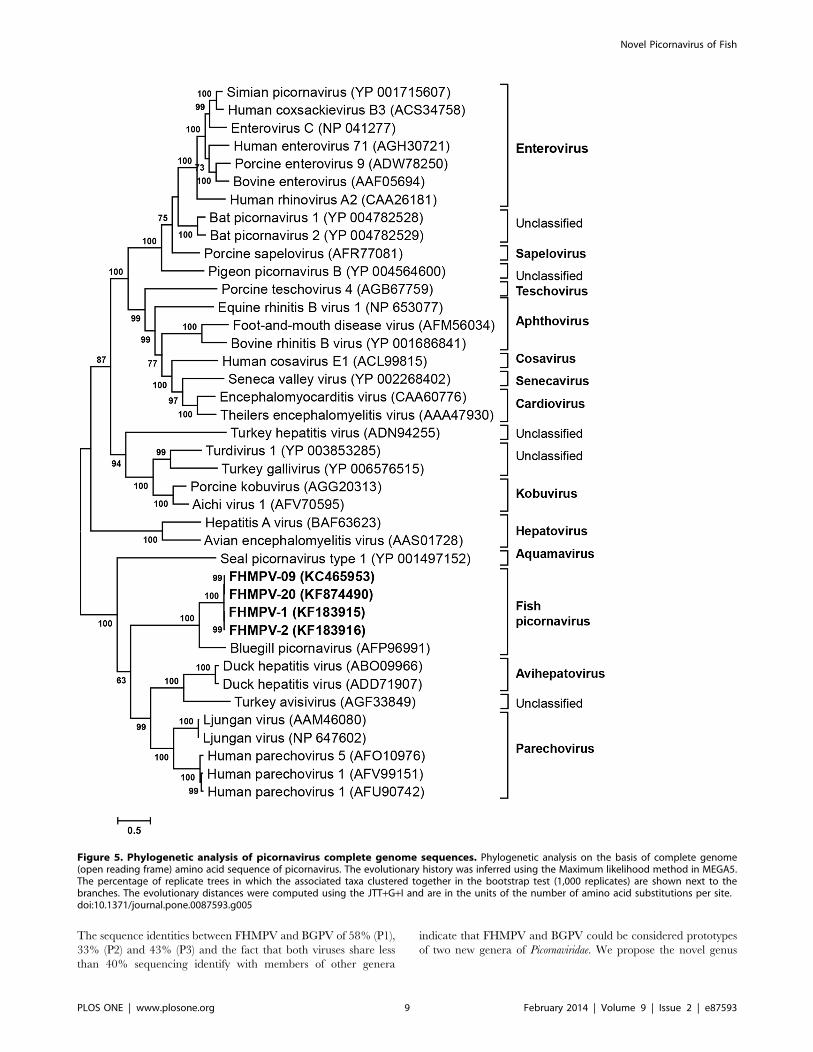
Pairwise comparison of the entire polyprotein of FHMPV-9 and
the BGPV (Genbank NC018506) resulted in 48.1% nucleotide
and 42.0% amino acid identity (Figure 5). FHMPV shared 58%
(P1), 33% (P2) and 43% (P3) amino acid identities with BGPV
(Figure 3). Both BGPV and FHMPV shared less than 40%
identities in all P1, P2, P3 regions with other picornavirus genomes
(Figure 3).
Discussion
In this study, we report the isolation, morphology, and
molecular characterization of FHMPV, a novel picornavirus of
baitfish. Highly conserved 3D sequences were found within the
FHMPV isolates from this study, which is a characteristic of the
Picornaviridae family [23,27]. FHMPV shared only a 49.5% amino
acid identity with BGPV, the nearest neighbor.
According to criteria proposed by the International Committee
on Taxonomy of Viruses (ICTV), members of the same
picornavirus genus should share greater than 40%, 40% and
50% amino acid identities for P1, P2, and P3 regions, respectively.
Figure 4. Phylogenetic analysis of picornavirus 3D gene sequences. Phylogenetic analysis on the basis of amino acid sequence of completepicornavirus 3D gene. The evolutionary history was inferred using the Maximum likelihood method in MEGA5. The percentage of replicate trees inwhich the associated taxa clustered together in the bootstrap test (1,000 replicates) are shown next to the branches. The evolutionary distances werecomputed using the JTT+G+I method.doi:10.1371/journal.pone.0087593.g004
Novel Picornavirus of Fish
PLOS ONE | www.plosone.org 8 February 2014 | Volume 9 | Issue 2 | e87593
The sequence identities between FHMPV and BGPV of 58% (P1),
33% (P2) and 43% (P3) and the fact that both viruses share less
than 40% sequencing identify with members of other genera
indicate that FHMPV and BGPV could be considered prototypes
of two new genera of Picornaviridae. We propose the novel genus
Figure 5. Phylogenetic analysis of picornavirus complete genome sequences. Phylogenetic analysis on the basis of complete genome(open reading frame) amino acid sequence of picornavirus. The evolutionary history was inferred using the Maximum likelihood method in MEGA5.The percentage of replicate trees in which the associated taxa clustered together in the bootstrap test (1,000 replicates) are shown next to thebranches. The evolutionary distances were computed using the JTT+G+I and are in the units of the number of amino acid substitutions per site.doi:10.1371/journal.pone.0087593.g005
Novel Picornavirus of Fish
PLOS ONE | www.plosone.org 9 February 2014 | Volume 9 | Issue 2 | e87593

name Piscevirus to reflect the host (Latin translation of fish is Pisces)
of FHMPV, a second picornavirus genus of fish.
The family Picornaviridae is very diverse having numerous genera
and serotypes. Hence, it is not surprising to detect more than a
single picornavirus among fish species. However, very little is
known about picornaviruses in fish. Together with the recent
discoveries of other fish picornaviruses [20,23], the picornavirus
host range is further expanded among ray-finned fishes (class
Actinopterygii). Although in the same class Actinopterygii, fathead
minnow, bluegill, and European eel belong to three different
orders, Cypriniformes, Perciformes, and Anguilliformes, respectively. The
broad characterization of picornaviruses in fish provides evidence
that picornavirus evolution may antedate the radiation of
vertebrate species including fish, reptile, amphibian and mammal,
perhaps in a ‘‘big bang’’ fashion similar to that of the Picornavirales
Superfamily [40]. Given that the known fish picornaviruses are
highly divergent, it suggests that a high diversity of picornavirus is
yet to be characterized in fish species.
The complete polyprotein of FHMPV divided into three
segments P1, P2 and P3 out of which P1 contains structural
proteins while P2 and P3 contains non-structural proteins. In
segment P1, the first protein, VP0, does not have the conserved
sequence GXXX(S/T) for myristoylation, which has been
observed mainly in avian encephalomyelitis virus, hepatitis A
virus, HPeVs and LV. Myristoylation involves the covalent linkage
of myristic acid to an N-terminal glycine residue in consensus
GXXX(S/T) motif, which plays an important role in virion
morphogenesis. However, the finding is in correlation with the
DHVs and BGPV, which also do not have this motif [23,41,42]. It
has been suggested that, for hepatoviruses, the blocked N-terminus
of VP0 possibly indicates an alternative modification that
substitutes for the myristate motif [43]. Thus, a similar hypothesis
could be proposed for the FHMPV. The VP3 protein was found to
be the most conserved among all proteins to BGPV, containing a
long stretch of 22 identical amino acids. The significance of this is
not known at this time. The absence of an integrin binding RGD
motif in the VP1 is similar to the absence of this receptor in DHV-
1, HPeV3 and LV. This motif is known to mediate many cell to
cell and microbe-host interaction, hence the absence of this motif
implies the existence of an alternative cell receptor in FHMPV,
which needs to be studied in the future. It is intriguing that
between FHMPV and BGPV, the P1 region that encodes for
structural proteins is more conserved than the P2 and P3 regions
that encode for non-structural proteins such as helicase and RdRp.
One possible explanation might be a greater degree of conserva-
tion among viral capsid sequences needed for the infection of fish.
Like DHVs, the 2A protein is likely divided into 3 proteins (2A1,
2A2, 2A3), with the presence of two conserved sequences
CGDVESNPG|PD and SGDVEQNPG|PV. The NPG|P seems
to be the junction of 2A1|2A2 and 2A2|2A3. The conserved
motif in 2A2 matched with human cosavirus and BGPV, with a
conserved motif RLXLLLSGDXEXNPGP and
SGDVEQNPGPX respectively. The proposed presence of 2A3
in FHMPV-09 genome was determined based on the presence of
amino acid sequence between second NPG|P and 2B. A similar
prediction was made by Barbknecht [23] for the BGPV genome.
The conserved H-box and NC motifs that are considered to
control cell proliferation [44] were absent in FHMPV. These
motifs are mainly present in DHVs and HPeVs, but also absent in
newly reported swine pasivirus 1 [42]. The 2A protein was found
to be divergent, containing insertion and deletions compared to
BGPV, and should be the focus of future study to better
understand the function of this protein. The 2C protein displayed
the NTPase motif (G/S)XXGXGK(S/T) that is present in all
picornaviruses. In the P3 segment, 3A and 3B proteins have less
identity with other picornaviruses. 3B have tyrosine residue which
is required for the attachment of VPg protein to the 59 NTR uracil
of RNA genome which act as an RNA replication primer as
suggested by Ambros and Baltimore [45]. In 3C, a conserved
motif GDCGS is present that correspond to GXCG(G/S) motif
present in all picornaviruses. The catalytic triad H42-D78-C154 is
very close to H41-D79-C154 catalytic triad of LV. In the 3D
protein, like all members of Picornaviridae family that have KDE(I/
L)R, DYS, (P/C)SG, YGDD and FLKR conserved motif at the
carboxyl end of the polyprotein, FHMPV also has the five
conserved motifs (KDELR, DYS, PSG, YGDD and FLKR).
The 18 isolates from fathead minnows and two from brassy
minnows were highly similar (98.6–100%), suggesting a rapid and
widespread dissemination. Many of these isolates were collected
from baitfish dealers, who were collecting fish from a variety of
sources and distributing them across a wide geographic range. For
example, it is possible for farm raised baitfish from South Dakota
to be mixed with wild harvested baitfish from Minnesota and farm
raised baitfish from Arkansas at a Wisconsin baitfish dealer, and
distributed to any other number of states. As the name suggests,
baitfish are often used for angling and have the potential to be
directly introduced into susceptible populations. While hemor-
rhagic lesions were observed in some fish from which the virus was
isolated, the two cannot be conclusively linked without a more
thorough study of viral pathogenesis.
While FHMPV may not induce mortality in fathead minnows,
susceptibility of other farmed and wild predatory species is
unknown. In the report on BGPV, there appears to be association
with hemorrhage on the skin, erythema, pale and swollen internal
organs, and mortality [23]. Diagnostic investigation, albeit limited,
of farmed and wild fish populations has not linked clinical lesions
in predatory species with FHMPV. Consequently, the long-term
impact to the health of fish populations is currently unknown.
However, the high level of FHMPV prevalence in the stocks we
tested demonstrate that the unregulated movement of ornamental
and baitfish species could serve as important pathways for
introduction of exotic pathogens, some of which may present
significant levels of risk to native aquatic species. Given the
importance of these host species to the economy and ecology of the
region, continued research is necessary.
Acknowledgments
We thank Wendy Wiese and Don Sanjiv Ariyakumar from the University
of Minnesota for technical assistance. The use of trade names does not
imply endorsement by the US Government.
Author Contributions
Conceived and designed the experiments: NP SM WB TN TW ED JW
SG. Performed the experiments: NP SM AA WB AG LH RM TN CP TW
JW. Analyzed the data: NP SM WB TN JW. Contributed reagents/
materials/analysis tools: NP AG TW ED JW SG. Wrote the paper: NP SM
AA WB AG LH RM TN CP TW ED JW SG.
References
1. Litvak MK, Mandrak NE (1993) Ecology of freshwater baitfish use in Canada
and the United States. Fisheries. 18: 6–13.
2. USDA (United States Department of Agriculture) (2006) Census of Aquaculture
(2005). 2002 Census of Agriculture: Vol. 3 Special Studies Part 2. AC-02-SP-2.
United States.
Novel Picornavirus of Fish
PLOS ONE | www.plosone.org 10 February 2014 | Volume 9 | Issue 2 | e87593

3. Gunderson JL, Tucker P (2000) A white paper on the status and needs of baitfish
aquaculture in the north central region. United States Department of
Agriculture, North Central Regional Aquaculture Center. Available: http://www.ncrac.org/content/status-and-needs-baitfish-aquaculture-north-central-
region Accessed 16 October 2013.
4. Stone N, Thomforde H (2001) Common farmed-raised baitfish. United States
Department of Agriculture, Southern Regional Aquaculture Center. PublicationNo. 120. Available: https://srac.tamu.edu/index.cfm/event/getFactSheet/
whichfactsheet/7 Accessed 16 October 2013.
5. Goodwin AE, Peterson JE, Meyers TR, Money DJ (2004) Transmission of exoticfish viruses: the relative risks of wild and cultured bait. Fisheries. 29: 19–23.
6. Hedrick RP. 1996. Movements of pathogens with the international trade of live
fish: problems and solutions. Rev Sci Tech Off Int Epizoot. 15: 523–531.
7. Harvell CD, Kim K, Burkholder JM, Colwell RR, Epstein PR, et al. (1999)
Emerging marine diseases – Climate links anthropogenic factors. Science. 285:
1505–1510.
8. Gaughan DJ (2002) Disease-translocation across geographic boundaries must berecognized as a risk even in the absence of disease identification: the case with
Australian Sardinops. Rev Fish Biol Fish. 11: 113–123.
9. USDA (United States Department of Agriculture) (2008) Federal Order toprevent the spread of VHSV. Available: http://www.aphis.usda.gov/animal_
health/animal_dis_spec/aquaculture/downloads/vhs_fed_order_amended.pdf
Accessed 16 October 2013.
10. Kim R, Faisal M (2011) Emergence and resurgence of the viral hemorrhagic
septicemia virus (Novirhabdovirus, Rhabdoviridae, Mononegavirales). J Adv Res. 2: 9–
23.
11. USFWS and AFS-FHS (U.S. Fish and Wildlife Service and American FisheriesSociety-Fish Health Section) (2010) Standard procedures for aquatic animal
health inspections. In: AFS-FHS, FHS blue book: suggested procedures for the
detection and identification of certain finfish and shellfish pathogens, ed.
Bethesda, Maryland: AFS-FHS.
12. OIE (World Organization for Animal Health) (2011) Manual of diagnostic tests
for aquatic animals, 7th ed. Paris, France: World Organization for Animal
Health.
13. McCann R (2012) Viral survey of fathead minnows, golden shiners, and white
suckers from baitfish dealers in Wisconsin. M.S. thesis. University of Wisconsin,
La Crosse, WI.
14. Knowles NJ, Hovi T, Hyypia T, King AMQ, Lindberg AM, et al. (2012)
Picornaviridae. In: King AMQ, Adams MJ, Carstens EB, Lefkowitz EJ, editors.
Virus Taxonomy: Classification and Nomenclature of Viruses, Ninth Report of
the International Committee on Taxonomy of Viruses. San Diego, CA: Elsevier.855–880.
15. Adams MJ, King AMQ, Carstens EB (2013) Ratification vote on taxonomic
proposals to the International Committee on Taxonomy of Viruses (2013). ArchVirol. doi:10.1007/s00705-013-1688-5.
16. Racaniello VR (2007) Picornaviridae: the viruses and their replication. In: Knipe
DM, Howley PM, Griffin DE, editors. Fields Virology, 5th edition. Philadelphia,
PA: Williams & Wilkins. 795–838.
17. Bloch B, Gravningen K, Larsen JL (1991) Encephalomyelitis among turbot
associated with a picornavirus-like agent. Dis Aquat Org. 10: 65–70.
18. Hedrick RP, Yun S, Wingfield WH (1991) A small RNA virus isolated fromsalmonid fishes in California, USA. Can J Fish Aquat Sci. 48: 99–104.
19. Iwanowicz LR, Goodwin AE, Heil N (2000) A small RNA virus isolated from
apparently healthy wild sandbar shiners, Notropis scepticus (Jordan & Gilbert). JFish Dis. 23: 349–352.
20. Fichtner D, Philipps A, Groth M, Schmidt-Posthaus H, Granzow H, et al. (2013)
Characterization of a novel picornavirus isolate from a diseased European eel
(Anguilla anguilla). J Virol. doi:10.1128/JVI.01094-13.
21. Mori KI, Nakai T, Muroga K, Arimato M, Mushiake K, et al. (1992) Properties
of a new virus belonging to nodaviridae found in larval striped jack (Pseudocarnax
dentex) with nervous necrosis. Vir. 187: 368–371.
22. Batts W, Yun S, Hedrick R, Winton J (2011) A novel member of the family
Hepeviridae from cutthroat trout (Oncorhynchus clarkii). Virus Res. 158: 116–123.
23. Barbknecht M (2009) Characterization of an unclassified virus and survey for its
presence in Wisconsin bluegill populations. M.S. thesis. University of Wisconsin,La Crosse, WI.
24. McEntire ME, Iwanowicz LR, Goodwin AE (2003) Molecular, physical, and
clinical evidence that golden shiner virus and grass carp reovirus are variants ofthe same virus. J Aquat Anim Health. 15: 257–263.
25. Batts WN, Goodwin AE, Winton JR (2012) Genetic analysis of a novel nidovirusfrom fathead minnows. J Gen Virol. 93: 1247–1252.
26. Nanda S, Jayan G, Voulgaropoulou F, Sierra-Honigmann AM, Uhlenhaut C, et
al. (2008) Universal virus detection by degenerate-oligonucleotide primedpolymerase chain reaction of purified viral nucleic acids. J Virol Methods.
152: 18–24.27. Kapoor A, Victoria J, Simmonds P, Wang C, Shafer RW, et al. (2008) A highly
divergent picornavirus in a marine mammal. J Virol. 82: 311–320.28. Victoria JG, Kapoor A, Li LL, Blinkova O, Slikas B, et al. (2009) Metagenomic
analyses of viruses in stool samples from children with acute flaccid paralysis. J
Virol. 83: 4642–4651.29. Ng TF, Marine R, Wang C, Simmonds P, Kapusinszk B, et al. (2012) High
variety of known and new RNA and DNA viruses of diverse origins in untreatedsewage. J Virol. 86: 12161–12175.
30. Blom N, Hanson J, Blaas D, Brunak S (1996) Cleavage site analysis in
picornaviral polyproteins: discovering cellular targets by neural networks.Protein Sci. 5: 2203–2216.
31. Thompson JD, Higgins DG, Gibson TJ (1994) CLUSTAL W: improving thesensitivity of progressive multiple sequence alignment through sequence
weighting, position specific gap penalties and weight matrix choice. NucleicAcids Res. 22: 4673–4680.
32. Tamura K, Peterson D, Peterson N, Stecher G, Nei M, et al. (2011) MEGA5:
Molecular Evolutionary Genetics Analysis using maximum likelihood, evolu-tionary distance, and maximum parsimony methods. Mol Biol Evol. 28: 2731–
2739.33. Abascal F, Zordoya R, Posada D (2005) ProtTest: selection of best-fit models of
protein evolution. Bioinformatics. 21: 2104–2105.
34. Sanchez R, Serra F, Tarranga J, Medina I, Carbonell J, et al. (2011) Phylemon2.0: a suite of web-tools for molecular evolution, phylogenetics, phylogenomics
and hypotheses testing. Nucleic Acids Res. 39: 1–5.35. Jones DT, Taylor WR, Thornton JM (1992) The rapid generation of mutation
data matrices from protein sequences. Comput Appl Biosci. 8: 275–282.36. Felsenstein J (1985) Confidence limits on phylogenies: an approach using
bootstrap. Evolution. 39: 783–791.
37. Drummond AJ, Ashton B, Buxton S, Cheung M, Cooper A, et al. (2011)Geneious v5.4. Available: http://www.geneious.com Accessed 16 October 2013.
38. Muhire B, Martin DP, Brown JK, Navas-Castillo J, Moriones E, et al. (2013) Agenome-wide pairwise-identity-based proposal for the classification of viruses in
the genus Mastrevirus (family Geminiviridae). Arch Virol. 158: 1411–1424.
39. Gorbalenya AE, Koonin EV, Donchenko AP, Blinov VM (1989) Two relatedsuperfamilies of putative helicases involved in replication, recombination, repair
and expression of DNA and RNA genomes. Nucleic Acids Res. 17: 4713–4730.Koonin EV, Wolf YI, Nagasaki K, Dolja VV (2008) The big bang of picorna-like
virus evolution antedates the radiation of eukaryotic supergroups. Nat RevMicro. 6: 925–939.
40. Tseng CH, Knowles NJ, Tsai HJ (2007) Molecular analysis of duck hepatitis
virus type 1 indicates that it should be assigned to a new genus. Virus Res. 123:190–203.
41. Sauvage V, Ar Gouilh M, Cheval J, Muth E, Pariente K, et al. (2012) A memberof a new Picornaviridae genus is shed in pig feces. J Virol. 86: 10036–10046.
42. Stanway G, Kalkkinen N, Roivainen M, Ghazi F, Khan M, et al. (1994)
Molecular and biological characteristics of echovirus 22, a representative of annew picornavirus group. J Virol. 68: 8232–8238.
43. Hughes PJ, Stanway G (2000) The 2A proteins of three diverse picornavirusesare related to each other and to the H-rev107 family of proteins involved in the
control of cell proliferation. J Gen Virol. 81: 201–207.
44. Ambros V, Baltimore D (1978) Protein is linked to the 59 end of poliovirus RNAby a phosphodiester linkage to tyrosine. J Biol Chem. 253: 5263–5266.
Novel Picornavirus of Fish
PLOS ONE | www.plosone.org 11 February 2014 | Volume 9 | Issue 2 | e87593