Avian mycobacteriosis in free-living raptors in Majorca Island, Spain
23
For Peer Review Only Avian mycobacteriosis in free-living raptors in Majorca Island, Spain Journal: Avian Pathology Manuscript ID: CAVP-2009-0104.R1 Manuscript Type: Original Research Paper Date Submitted by the Author: 14-Sep-2009 Complete List of Authors: Millán, Javier; Wildlife Health and Control, Govern Illes Balears Negre, Nieves Castellanos, Elena de Juan, Lucia Ana, Mateos Parpal, Lluis Aranaz, Alicia Keywords: Raptor, Mycobacterium avium, Tuberculosis , Majorca E-mail: [email protected] URL: http://mc.manuscriptcentral.com/cavp Avian Pathology
-
Upload
universidadiuem -
Category
Documents
-
view
2 -
download
0
Transcript of Avian mycobacteriosis in free-living raptors in Majorca Island, Spain

For Peer Review O
nly
Avian mycobacteriosis in free-living raptors in Majorca Island, Spain
Journal: Avian Pathology
Manuscript ID: CAVP-2009-0104.R1
Manuscript Type: Original Research Paper
Date Submitted by the Author:
14-Sep-2009
Complete List of Authors: Millán, Javier; Wildlife Health and Control, Govern Illes Balears Negre, Nieves Castellanos, Elena de Juan, Lucia Ana, Mateos Parpal, Lluis Aranaz, Alicia
Keywords: Raptor, Mycobacterium avium, Tuberculosis , Majorca
E-mail: [email protected] URL: http://mc.manuscriptcentral.com/cavp
Avian Pathology

For Peer Review O
nly
1
Cavp-2009-0104.R1
Avian mycobacteriosis in free-living raptors in Majorca Island, Spain
Javier Millána,
*, Nieves Negreb, Elena Castellanos
c, Lucía de Juan
c, Ana Mateos
c,
Lluis Parpald, and Alicia Aranaz
c
a Sanitat i Control de Fauna (Wildlife Health and Control), Conselleria de Medi Ambient,
Govern de les Illes Balears/Fundació Natura Parc, 07142-Santa Eugènia (Balearic
Islands), Spain b Fundació Natura Parc, 07142-Santa Eugènia (Balearic Islands), Spain
c Centro de Vigilancia Sanitaria Veterinaria, Universidad Complutense de Madrid, 28040-
Madrid, Spain d Consorci de Recuperació de la Fauna de les Illes Balears (COFIB),
Govern de les Illes Balears-Fundació Natura Parc, 07142-Santa Eugènia (Balearic
Islands), Spain
*Corresponding author:
Tel.: 0034 610241785; Fax: 0034 971144534; Email address:
Short title: Avian mycobacteriosis in raptors in Majorca.
Received 17 July 2009
Page 1 of 22
E-mail: [email protected] URL: http://mc.manuscriptcentral.com/cavp
Avian Pathology

For Peer Review O
nly
2
Abstract
Avian mycobacteriosis is a chronic, infectious disease caused by different species of
mycobacteria, usually belonging to the Mycobacterium avium complex (MAC). From 2004
to 2007, 589 raptors brought dead or sick to a wildlife rehabilitation centre in Majorca
(Balearic Islands, Spain) were necropsied. The birds belonged to 12 different species,
chiefly common kestrel (Falco tinnunculus) (n=297), scops owl (Otus scops) (n=109), barn
owl (Tyto alba) (n=75), long-eared owl (Asio otus) (n=58), peregrine falcon (Falco
peregrinus) (n=27), and booted eagle (Hieraaetus pennatus) (n=13). Gross lesions
compatible with mycobacteriosis were observed in 14 birds (2.4%) found in several
locations in Majorca. They were 12 kestrels (prevalence in this species=4.0%), one long-
eared owl (1.7%) and one scops owl (0.9%), all the birds presenting white-yellowish
nodules from pinpoint size to 1 cm in diameter in diverse organs, mainly in liver, spleen
and intestine. Affected organs were subjected to bacteriology and molecular identification
by PCR and, in all the cases, infection with Mycobacterium avium subsp. avium was
confirmed. The observed prevalences are similar to those previously observed in Holland,
though the actual prevalence detected in this study is likely to be higher than reported
because only birds with gross lesions were subjected to culture. Further molecular
characterization with a set of six MIRU-VNTR loci was used to sub-typify the isolates in
order to show the existence of possible epidemiological links. Six different genotypes were
found, which points to infection from multiple focuses. No temporal or geographical
aggregation of the cases was observed associated to the presence of positive birds or to the
different VNTR allelic profiles. The most feasible origin might be water or food sources,
though the reservoir of mycobacteria remains unknown.
Page 2 of 22
E-mail: [email protected] URL: http://mc.manuscriptcentral.com/cavp
Avian Pathology
For Peer Review O
nly
3
Introduction
Avian mycobacteriosis (AM) is a chronic, infectious disease caused by Mycobacterium
avium complex (MAC) organisms (Thoen, 1997; Thorel et al., 1997), although other
mycobacteria can produce mycobacteriosis in birds (Hoop et al., 1996; Tell et al., 2001).
All avian species are susceptible to infection by the MAC, although some bird orders, such
as Galliformes, Columbiformes or Gruiformes may be more susceptible to the disease
(Friend, 1999). AM is a relatively common disease in domestic flocks (González et al.,
2002; Bougiouklis et al., 2005), or in captive wild birds (Singbeil et al., 1993; Marco et al.,
2000; Dvorska et al., 2007). This is mainly due to the persistence of the organism once
established, its contagiousness and its ability to survive in the environment (Thorel et al.,
1997). Nevertheless, reports of AM in free-living wild birds are not uncommon (Smit et al.,
1987; Millán et al., 2004; Gerhold & Fischer, 2005).
Captive raptorial birds are not free of suffering from AM (van Nie et al., 1982).
However, in free-living raptors, cases are only sporadically reported. Kaliner and Cooper
(1973) detected a case of AM in an African fish eagle (Haliaeetus vocifer). In North
America, single cases of AM were reported in two red-tailed hawks (Buteo jamaicensis)
(Emerson et al., 1970; Sykes, 1982) and in two American bald eagles (Haliaeetus
leucocephalus) (Hoenerhoff et al., 2004; Heatley et al., 2007). In Europe, reports include
those of two common kestrels (Falco tinnunculus) (Wilson 1960; Bucke & Mawdesley-
Thomas 1974) and a barn owl (Tyto alba) (Wilson 1960). Smit et al. (1987) isolated M.
avium from multiple dead-found raptor species, both diurnal and nocturnal. The only
reported case in Spain was detected in a Eurasian goshawk (Accipiter gentilis) from
southern peninsular Spain (Aranaz et al., 1997). In the present article we report the
Page 3 of 22
E-mail: [email protected] URL: http://mc.manuscriptcentral.com/cavp
Avian Pathology

For Peer Review O
nly
4
prevalence of AM in free-living raptors that were brought to a wildlife rehabilitation centre
at Majorca Island, Spain.
Material and Methods
In the period 2004-2007, 589 free-living raptors were submitted for necropsy at the
“Cofib” rehabilitation centre at Majorca (Table 1). When gross lesions compatible with
mycobacteriosis (e.g. granuloma) were identified, tissue samples of the affected organs
were frozen at -20oC and analyzed for the detection and identification of mycobacterias.
Tissues from each animal were homogenized with sterile distilled water and
decontaminated with 0.35% hexadecylpyridinium chloride (Sigma Aldrich Chemie GmbH,
Buch, Switzerland) for 30 min (Corner and Trajstman, 1988), centrifuged at 1,068 x g for
30 min and cultured onto Colestos, 0.2% (wt/vol) pyruvate-enriched Löwenstein-Jensen
media (bioMèrieux España and Biomedics, Madrid, Spain) and Middlebrook 7H10
(Biocult Laboratorios, Madrid, Spain) at 37ºC. Isolates were identified by staining for acid-
alcohol fastness, a multiplex PCR amplification targeting the Mycobacterium specific 16S
rRNA fragment (Boddinghaus et al., 1990; Wilton and Cousins, 1992), and PCRs aimed at
specific insertion sequences IS901 and IS1245 (Kunze et al., 1992; Guerrero et al., 1995).
Recent studies have shown the usefulness of tandem repeats or repetitive units to
characterize members of MAC (Bull et al., 2003; Thibault et al., 2007; Mobius et al.,
2008). Included within these tandem repeats are the variable number tandem repeats
(VNTR), which are repetitive units dispersed throughout the genome with a length
between 10-100 bp (Supply et al., 2000); and also the mycobacterial interspersed repetitive
units (MIRU), small interspersed repetitive elements described by Supply et al. (1997) in
Page 4 of 22
E-mail: [email protected] URL: http://mc.manuscriptcentral.com/cavp
Avian Pathology

For Peer Review O
nly
5
the genome of M. tuberculosis. Isolates of M a. avium were sub-typified by MIRU-VNTR
with six specific loci previously published: MIRU-1, MIRU-2, MIRU-3, MIRU-4 (Bull et
al. 2003), VNTR-25 and VNTR-32 (Thibault et al. 2007; Mobius et al. 2008), using the
same specific primers described (Table 2).
PCR mix contained 5 µl of sample DNA, 1x standard reaction buffer with 1.5 mM
MgCl2, 200 µM deoxynucleoside triphosphate (Biotools, B&M Labs S.A, Madrid, Spain),
forward and reverse primers (Roche Diagnostics S.L., St. Cugat del Vallés, Spain) (0.4
µM), 10% of DMSO (Sigma Aldrich Chemie GmbH) and 2.5 U of Hot-Start-Taq-
Polymerase (Qiagen GmbH, Hilden, Germany), in a final volume of 50 µl. For MIRU-3,
primer concentrations were three times higher than for the rest of the loci (Mobius et al.
2008).
The amplification reactions for these MIRU-VNTR loci were: denaturation at 95°C
for 15 min, followed by 40 cycles of denaturation at 95°C for 30 sec, annealing at 58°C for
1 min, extension at 72°C for 2 min, with a final cycle of extension at 72°C for 10 min;
conditions reported by Frothingham and Meeker-O'Connell (1998) with slight
modifications. In the case of MIRU-3, MIRU-4 and VNTR-32 the same recommended
melting temperatures (Mobius et al. 2008; Thibault et al. 2007) were used.
Finally, PCR amplicons were separated in a 2.5% agarose gel, stained with SYBR
safe (Invitrogen, product no. 531478) and using 100 bp molecular ladder (Biotools B&M
Labs, Madrid, Spain) to detect the variation in the number of repeats for each of the locus.
Chi2 test was used to analyze differences in the observed prevalence among bird
species and years.
Results
Page 5 of 22
E-mail: [email protected] URL: http://mc.manuscriptcentral.com/cavp
Avian Pathology

For Peer Review O
nly
6
Overall, 2.4% of the necropsied raptors presented gross lesions compatible with AM.
Tissue samples from all the birds with lesions were cultured and then M. avium subsp.
avium was identified by PCR amplification of the insertion sequences IS901 and IS1245.
Lesions were observed in three species: 12 of 297 common kestrels (4.0%), one of 58
long-eared owls (Asio otus) (1.7%) and one of 109 scops owls (Otus scops) (0.9%). No
lesions were observed in the other species analyzed (Table 1). The kestrels were 9 adult
birds (5 males, 4 females) and 3 sub-adults between 1 and 2 years-old (2 males, 1 female);
the long-eared owl was an adult female; and the scops owl was a sub-adult female. The
affected birds arrived throughout the year, dead or sick, from multiple locations in Majorca
(Figure 1). Non-affected raptors also arrived from all around the island, including from the
locations were infected birds were found.
Infected birds included road-killed birds (n=5) as well as debilitated birds (n=9). Of
the latter, one bird suffered from gunshot wounds while the others presented no traumatic
lesions. The latter birds arrived as a rule emaciated but did not present other clinical signs.
With the exception of one kestrel that survived for eight months after arrival, the other
affected birds died within few days (usually 1-2 days, mean= 11.8 ±16.9 hours). Another
kestrel was euthanatized two months after arrival for humane reasons. On necropsy, all the
birds were cachectic and presented white-yellowish nodules from pinpoint size to 1 cm in
diameter in diverse organs, chiefly in liver, spleen and intestine (Table 3 and Figure 2).
Lesions did not markedly differ among species. No other signs were observed in the
infected birds, with the exception of one kestrel in which oral lesions of trichomoniasis
confirmed by microscopy were observed.
Regarding MIRU-VNTR typing, only MIRU-3 and MIRU-4 showed allelic
variation among the M. a. avium isolates tested with five and two allelic variants
Page 6 of 22
E-mail: [email protected] URL: http://mc.manuscriptcentral.com/cavp
Avian Pathology

For Peer Review O
nly
7
respectively. The combination of loci allowed the differentiation into six different
genotypes, where allelic profile number 1 (1-8-1-5-9-5) and 5 (1-8-1-5-7-5) represented
42.86% and 28.57% of the isolates tested respectively (Table 4). The two isolates from the
scops owl and the long-eared owl were both clustered into the most common allelic profile
no.1, which was shared with four isolates from kestrel.
No differences were observed in the prevalence of AM lesions among years.
Prevalence of AM lesions was higher in common kestrel than in barn owl, though
differences were only residually significant (Chi2=3.13, p=0.07).
Discussion
Other than the single goshawk reported by Aranaz et al. (1997), this is the first report of
widespread AM in raptorial birds from Spain. As mentioned above, AM in free-living
raptors has rarely been diagnosed. In the Netherlands, Smit et al. (1987) isolated M. avium
from, among other raptors, 17/450 (3.8%) of common kestrels, and in 3/313 (0.96%) of
long-eared owls. Those prevalences are similar to these reported in the present article. As
mentioned before, a case of AM was also reported in one common kestrel from UK
(Wilson, 1960). As far as we know, AM was not previously diagnosed in a scops owl.
However, it is worth to note that the prevalence of mycobacteriosis was likely to be higher
than reported here, because only tissue samples from birds with gross lesions were
subjected to culture.
Affected birds were mostly adults. This is in accordance with most of the previous
reports of AM in raptors (Sykes, 1982), or in other wild birds (Millán et al., 2004). Older
animals are more commonly affected by mycobacteriosis because of the long incubation
Page 7 of 22
E-mail: [email protected] URL: http://mc.manuscriptcentral.com/cavp
Avian Pathology

For Peer Review O
nly
8
period and accumulated risk of exposure (Thoen, 1997). According to Thorel et al. (1997),
affected birds die within two months or may survive for six months depending on the
extent of disease.
On necropsy, macroscopic lesions were seen in the liver, spleen and intestine of all
the affected raptors, which is the typical feature of AM in birds (Skyes, 1982; Thorel et al.,
1997; Millán et al., 2004; Saggese et al., 2008). According to the distribution of the lesions,
the affected birds probably contracted the infection by ingestion. However, due to the
presence of lesion in the lungs in one third of the cases, an inhalation route should not be
excluded (Kaliner & Cooper, 1973).
The source of the infection remains unknown. The hypothesis of a nosocomial
infection can be ruled out due to the affected birds arrived dead or survived for a short
period. Smit et al. (1987) proposed that kestrels and other aggressive raptors may contract
AM through injuries during fights, and even isolated M. avium from some local injuries.
However, in the present case no infected injuries were observed in any of these birds,
though subcutaneous granulomas were detected in seven birds that may be derived from
old injuries. In any case, the most feasible origin of the mycobacterias might be water or
food sources. Therefore, feeding ecology may explain the differences in prevalence among
species. Very little is known about the diet of the three affected species in Majorca. In the
Mediterranean part of the peninsular Spain, the common kestrel feeds mainly upon lizards
(Lacerta spp.), which are no present on Majorca, passerines, several species of insects and
micromammals (Valverde, 1967; Veiga, 1985). For the long-eared owl, the most important
prey species are Murinae, followed by Arvicolinae (not present in Balearic Islands) and
passerine birds (Corral et al.; 1979), although this species depends less on rodents in
Mediterranean regions (García & Cervera, 2001). However, the barn owl also feeds mainly
on rodents in Balearic Islands (Sommer et al., 2005), and no individual out of 75
Page 8 of 22
E-mail: [email protected] URL: http://mc.manuscriptcentral.com/cavp
Avian Pathology

For Peer Review O
nly
9
necropsied was apparently affected. If rodents were a major source of mycobacteria, this
would indicate a species specific difference in susceptibility, with barn owls being less
susceptible. However, several cases of AM have been reported in the species (Wilson,
1960; Bucke & Mawdesley-Thomas, 1974). Thus, differences may be related to prey
accessibility more than to susceptibility.
According to Friend (1999), AM is most commonly seen in scavenger species.
Thus, an alternative hypothesis of the infections is that the affected birds contracted AM
through the scavenging of carcasses of diseased red-legged partridges or pigeons (Columba
spp.), which consitute too large a prey for the affected raptors (Calderón, 1977). Due to the
intense, often illegal predator control carried out in Spanish hunting estates, sick animals
are not removed from the environment by carnivores, developing disseminated AM and
even dying by the disease, as has been sporadically observed in partridges (Millán et al.,
2004).
Regarding the molecular characterization by MIRU-VNTR, it discriminated the M.
a. avium isolates into six different genotypes. From the loci included for the analysis,
MIRU-3 yielded the highest number of allelic variants in comparison with the rest of the
loci. MIRU-3 is located between a two-component regulatory system SenX3-RegX3.
Former studies found the presence of a lower number of TRs at this locus in the M. bovis
(BCG) and M. avium subspecies paratuberculosis (316F) vaccine strains in comparison to
the rest of the strains they tested (Bull et al. 2003; Magdalena et al. 1998). Probably
differences in pathogenesis and progression of the disease have a relationship with the
variation of TR units at this particular locus. To our knowledge, this is the first analysis of
M. a. avium isolates from animals using MIRU-VNTR typing; therefore we cannot further
assess our results in comparison to other populations. However, available information from
literature shows that the genotypes isolated from raptors described in this report would be
Page 9 of 22
E-mail: [email protected] URL: http://mc.manuscriptcentral.com/cavp
Avian Pathology

For Peer Review O
nly
10
related to that described for the M. a. avium reference strain ATCC 25291 (1-7-1-5-5-5)
(Bull et al., 2003; Inagaki et al., 2009). Further studies concerning this issue would be
necessary.
In the light of the cases presented in this article, AM must be considered a factor in
the conservation of raptors in Majorca and the rest of the Balearic Islands. An undetected
source is acting as a reservoir of mycobacteria that is currently affecting raptors and
potentially may affect other protected species, livestock, or humans. As the infected raptors
were retrieved in several points of the Majorca Island, and six different genotypes were
found, there may be multiple infection focuses. However, though the affected species are
territorial, could have become infected in other locations different from where they were
found. In addition, some of the affected birds could be migrant individuals and may have
become infected in remote locations. Studies to detect the different reservoirs of this
infection in Majorca are currently being performed.
Acknowledgements
We wish to thank the team of “Protecció d’especies” (Conselleria de Medi Ambient,
Govern de les Illes Balears), Rangers of the Fish and Game Service (Govern de les Illes
Balears), members of the SEPRONA (Servicio de Protección de la Naturaleza, Guardia
Civil), and all the individual people who brought animals to the rescue centre.
We also thank Devin J. Morey for the careful revision of the manuscript.
Page 10 of 22
E-mail: [email protected] URL: http://mc.manuscriptcentral.com/cavp
Avian Pathology

For Peer Review O
nly
11
References
Aranaz, A., Liébana, E., Mateos, A. & Domínguez, L. (1997). Laboratory diagnosis of
avian mycobacteriosis. Seminars in Avian and Exotic Pet Medicine 6, 9-17.
Boddinghaus, B., Rogall, T., Flohr, T., Blocker, H. & Bottger, E.C. (1990). Detection and
identification of mycobacteria by amplification of rRNA. Journal of Clinical
Microbiology 288, 1751-1759.
Bougiouklis, P., Brellou, G., Fragkiadaki, E., Iordanidis, P., Vlemmas, I. & Georgopoulou,
I. (2005). Outbreak of avian mycobacteriosis in a flock of two-year-old domestic
pigeons (Columba livia f. domestica). Avian Diseases 49, 442-445.
Bucke, D. & Mawdesley-Thomas, L.E. (1974). Tuberculosis in a barn owl (Tyto alba).
Veterinary Record 95, 373.
Bull, T.J., Sidi-Boumedine, K., McMinn, E.J., Stevenson, K., Pickup, R. & Hermon-Taylor,
J. (2003). Mycobacterial interspersed repetitive units (MIRU) differentiate
Mycobacterium avium subspecies paratuberculosis from other species of the
Mycobacterium avium complex. Molecular Cell Probes 17, 157-164.
Calderón, J. (1977). El papel de la perdiz roja (Alectoris rufa) en la dieta de los predadores
ibéricos. Doñana Acta Vertebrata 4, 61–126.
Corner, L.A. & Trajstman, A.C. (1998). An evaluation of 1-hexadecylpyridinium chloride
as a decontaminant in the primary isolation of Mycobacterium bovis from bovine
lesions. Veterinary Microbiology 18, 127-134.
Corral F., J., Cortés, J.A. & Gil, J.M. (1979). Contribución al estudio de la alimentación de
Asio otus en sur de España. Doñana Acta Vertebrata 6, 179–190.
Dvorska, L., Matlova, L., Ayele, W.Y., Fischer, O.A., Amemori, T., Weston, R.T., Álvarez,
J., Beran, V., Moravkova, M. & Pavlik, I. (2007) Avian tuberculosis in naturally
Page 11 of 22
E-mail: [email protected] URL: http://mc.manuscriptcentral.com/cavp
Avian Pathology

For Peer Review O
nly
12
infected captive water birds of the Ardeideae and Threskiornithidae families
studied by serotyping, IS901 RFLP typing, and virulence for poultry. Veterinary
Microbiology 119, 36-74.
Emerson F.G., Brown, J.M. & Van Natta, B.S. (1970). Mycobacteriosis in a red-tailed
hawk. Journal of the American Veterinary Medical Association 157, 606.
Friend, M. (1999). Tuberculosis (avian). In: Friend, M. & Franson, J.C. (1999), Field
Manual of Wildlife Disease. General Field Procedures and Diseases of Birds (pp.
93-98). Madison, WI: USGS Biological Resources Division,
Frothingham, R. & Meeker-O'Connell, W.A. (1998). Genetic diversity in the
Mycobacterium tuberculosis complex based on variable numbers of tandem DNA
repeats. Microbiology 144, 1189-1196.
García, A.M. & Cervera, F. (2001). Notas sobre la variación estacional y geográfica de la
dieta del Búho Chico Asio otus. Ardeola 48, 75–80.
Gerhold, R.W. & Fischer, J.R., 2005. Avian tuberculosis in a wild turkey. Avian Diseases
49, 164-166.
González, M., Rodríguez-Bertos, A., Gimeno, I., Flores, J.M. & Pizarro, M. (2002).
Outbreak of avian tuberculosis in 48-week-old commercial layer hen flock. Avian
Diseases 46, 1055-1061.
Guerrero, C., Bernasconi, C., Burki, D., Bodmer, T. & Telenti, A. (1995). A novel
insertion element from Mycobacterium avium, IS1245, is a specific target for
analysis of strain relatedness. Journal of Clinical Microbiology 33, 304-307
Heatley, J.J., Mitchell, M.M., Roy, A., Cho, D.Y., Williams, D.L. & Tully, T.N. Jr. (2007).
Disseminated mycobacteriosis in a bald eagle (Haliaeetus leucocephalus). Journal
of Avian Medicine and Surgery 21, 201-209.
Page 12 of 22
E-mail: [email protected] URL: http://mc.manuscriptcentral.com/cavp
Avian Pathology

For Peer Review O
nly
13
Hoenerhoff, M., Kiupel, M., Sikarskie, J., Bolin, C., Simmons, H. & Fitzgerald, S., 2004.
Mycobacteriosis in an American bald eagle (Haliaeetus leucocephalus). Avian
Diseases 48, 437-441.
Hoop, R.K., Böttger, E.C. & Pfyffer, G.E. (1996). Etiological agents of mycobacterioses in
pet birds between 1986 and 1995. Journal of Clinical Microbiology 1996, 991-992.
Inagaki, T., Nishimori, K., Yagi, T., Ichikawa, K., Moriyama, M., Nakagawa, T.
Shibayama, T., Uchiya, K., Nikai, T. & Ogawa, K., (2009). Comparison of a
variable-number tandem-repeat (VNTR) method for typing Mycobacterium avium
with mycobacterial interspersed repetitive-unit-VNTR and IS1245 restriction
fragment length polymorphism typing. Journal of Clinical Microbiology 47, 2156-
2164.
Kaliner, G. & Cooper, J.E. (1973). Dual infection of an African fish eagle with acid-fast
bacilli and an Aspergillus sp. Journal of Wildlife Diseases 9, 51-55.
Kunze, Z.M., Portaels, F. & McFadden, J.J. (1992). Biologically distinct subtypes of
Mycobacterium avium differ in possession of insertion sequence IS901. Journal of
Clinical Microbiology 30, 2366-2372.
Magdalena, J., Supply, P. & Locht, C., (1998). Specific differentiation between
Mycobacterium bovis BCG and virulent strains of the Mycobacterium tuberculosis
complex. Journal of Clinical Microbiology 36, 2471-2476.
Marco, I., Domingo, M. & Lavín, S. (2000). Mycobacterium infection in a captive-reared
capercaillie (Tetrao urogallus). Avian Diseases 44, 227-230.
Millán, J., Gortázar, C. & Villafuerte, R. (2004). Mycobacterium avium infection in wild
red-legged partridges (Alectoris rufa L.). European Journal of Wildlife Research 50,
97-99.
Page 13 of 22
E-mail: [email protected] URL: http://mc.manuscriptcentral.com/cavp
Avian Pathology

For Peer Review O
nly
14
Mobius, P., Luyven, G., Hotzel, H. & Kohler, H. (2008). High genetic diversity among
Mycobacterium avium subsp. paratuberculosis strains from German cattle herds
shown by combination of IS900 restriction fragment length polymorphism analysis
and mycobacterial interspersed repetitive unit-variable-number tandem-repeat
typing. Journal of Clinical Microbiology 46, 972-981.
Saggese, M.D., Tizard, I. & Phalen, D.N. (2008). Mycobacteriosis in naturally infected
ring-neck doves (Streptopelia risoria): investigation of the association between
feather colour and susceptibility to infection, disease and lesions type. Avian
Pathology 37, 443-450.
Singbeil, B.A., Bickford, A.A. & Stoltz, J.H. (1993). Isolation of Mycobacterium avium
from ringneck pheasants (Phasianus colchicus). Avian Diseases 37:612-615.
Smit, T., Eger, A., Haagsma, J. & Bakhuizen, T. (1987). Avian tuberculosis in wild birds
in the Netherlands. Journal of Wildlife Diseases 23: 485-487.
Supply, P., Magdalena, J., Himpens, S. & Locht, C. (1997). Identification of novel
intergenic repetitive units in a mycobacterial two-component system operon.
Molecular Microbiology 26, 991-1003.
Supply, P., Mazars, E., Lesjean, S., Vincent, V., Gicquel, B. & Locht, C. (2000). Variable
human minisatellite-like regions in the Mycobacterium tuberculosis genome.
Molecular Microbiology 36, 762-771.
Sykes, G.P. (1982). Tuberculosis in a red-tailed hawk (Buteo jamaicensis). Journal of
Wildlife Diseases 18, 495-499.
Sommer, R., Zoller, H., Kock, D., Bohme, W.G. & Griesau, A. (2005). Feeding of the barn
owl, Tyto alba with first record of the European free-tailed bat, Tadarida teniotis
on the island of Ibiza (Spain, Balearics). Folia Zoologica 54, 364-370.
Page 14 of 22
E-mail: [email protected] URL: http://mc.manuscriptcentral.com/cavp
Avian Pathology

For Peer Review O
nly
15
Tell, L.A., Woods, L. & Cromie, R.L. (2001). Mycobacteriosis in birds. Revue Scientifique
et Technique de l'Office International des Épizooties 20, 180-203.
Thibault, V.C., Grayon, M., Boschiroli, M.L., Hubbans, C., Overduin, P., Stevenson, K.,
Gutierrez, M.C., Supply, P. & Biet, F. (2007). New variable-number tandem-repeat
markers for typing Mycobacterium avium subsp. paratuberculosis and M. avium
strains: comparison with IS900 and IS1245 restriction fragment length
polymorphism typing. Journal of Clinical Microbiology 45, 2404-2410.
Thoen, C.O., 1997. Tuberculosis. In: Calnek, B.W. (1997) Diseases of poultry, 10th edn.
(pp. 167–178). Ames: Iowa State University Press.
Thorel, M.F., Huchzermeyer, H., Weiss, R. & Fontaine, J.J. (1997). Mycobacterium avium
infections in animals. Literature review. Veterinary Research 28, 439-447.
Valverde, J.A. (1967). Estructura de una comunidad de vertebrados terrestres.
Monografías de la Estación Biológica de Doñana, 1. 218 pp. Sevilla: Estación
Biológica de Doñana.
van Nie, G.J., Lumeij, J.T., Dorrestein, G.M., Wolvekamp, W.T., Zwart, P. & Stam, J.W.,
(1982). Tuberculosis in raptorial birds. Clinical cases and differential diagnosis.
Tijdschrift voor Diergeneeskunde 107, 563-572.
Veiga, J.P. (1985). Ecología de las rapaces de un ecosistema mediterráneo de montaña.
Aproximación a su estructura comunitaria. PhD Dissertation. Universidad
Complutense, Madrid.
Wilson, J.E. (1960). Avian tuberculosis, an account of the disease in poultry, captive birds
and wild birds. British Veterinary Journal 116, 380-393.
Wilton, S. & Cousins, D. (1992). Detection and identification of multiple mycobacterial
pathogens by DNA amplification in a single tube. PCR Methods and Applications 1,
269-273.
Page 15 of 22
E-mail: [email protected] URL: http://mc.manuscriptcentral.com/cavp
Avian Pathology

For Peer Review O
nly
16
Figure Legends
Figure 1. Location of the cases and the distribution of VNTR allelic profiles of avian
tuberculosis in raptors in Majorca Island (Spain).
Figure 2. Avian tuberculosis granulomas in a scops owl (A) and two common kestrels (B,
C). A: in liver (box), lung (arrowhead), spleen (dotted arrow) and intestine (arrow); B: in
liver (arrow) and subcutaneous tissue (arrowhead); C: in lung.
Page 16 of 22
E-mail: [email protected] URL: http://mc.manuscriptcentral.com/cavp
Avian Pathology

For Peer Review Only
17
Table 1. Species of raptor necropsied per year and prevalence (in %) of avian mycobacteriosis in a wildlife rehabilitation centre in Majorca
Island (Spain).
2004 2005 2006 2007 Total
n (pos.) prev. n (pos.) prev. n (pos.) prev. n (pos.) prev. n (pos.) prev. (95% C.I.)
Accipitriformes
Hieraaetus pennatus 2 - 7 - 5 - 0 - 13 0 (0-19)
Accipiter nisus 0 - 1 - 1 - 2 - 4 0 (0-53)
Circus pygargus 0 - 0 - 0 - 1 - 1 0 (0-95)
Circus aeruginosus 0 - 1 - 1 - 0 - 2 0 (0-77)
Pandion haliaetus 0 - 1 - 0 - 0 - 1 0 (0-95)
Aegypius monachus 0 - 0 - 1 - 0 - 1 0 (0-95)
Falconiformes
Falco peregrinus 5 - 8 - 10 - 4 - 27 0 (0-14)
Falco eleonorae 0 - 1 - 0 - 0 - 1 0 (0-95)
Falco tinnunculus 47 (2) 4.2 89 (2) 2.2 87 (6) 6.9 74 (2) 2.7 297 (12) 4.0 (3-7)
Strigiformes
Tyto alba 15 - 19 - 21 - 20 - 75 0 (0-5)
Asio otus 10 - 12 - 12 (1) 8.3 24 - 58 (1) 1.7 (0-9)
Otus scops 29 - 40 - 26 - 14 (1) 7.1 109 (1) 0.9 (0-4)
Page 17 of 22
E-mail: [email protected] URL: http://mc.manuscriptcentral.com/cavp
Avian Pathology
For Peer Review Only
18
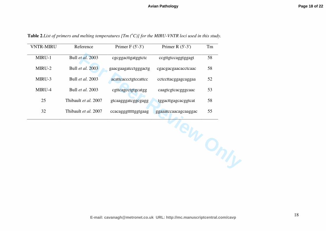
Table 2.List of primers and melting temperatures [Tm (oC)] for the MIRU-VNTR loci used in this study.
VNTR-MIRU Reference Primer F (5'-3') Primer R (5'-3') Tm
MIRU-1 Bull et al. 2003 cgcggacttgatggtctc ccgttgtccaggtggagt 58
MIRU-2 Bull et al. 2003 gaacgaagatcctgggactg cgacgacgaacacctcaac 58
MIRU-3 Bull et al. 2003 acattcaccctgtccattcc cctccttacggagcaggaa 52
MIRU-4 Bull et al. 2003 cgttcagcctgtgcatgg caagtcgtcacgggcaac 53
25 Thibault et al. 2007 gtcaagggatcggcgagg tggacttgagcacggtcat 58
32 Thibault et al. 2007 ccacagggtttttggtgaag ggaaatccaacagcaaggac 55
Page 18 of 22
E-mail: [email protected] URL: http://mc.manuscriptcentral.com/cavp
Avian Pathology

For Peer Review Only
19
Table 3. Locations of the granulomas due to avian mycobacteriosis observed in 14 raptors in Majorca Island (Spain). Some individuals
presented lesions in multiple organs.
Organ / Tissue n Locations
Liver 14
Spleen 14
Intestine 14
Subcutaneous tissue 7 Neck (7), mandible (1)
Gizzard 7
Lung 5
Kidney 4
Pancreas 1
Muscle 3 Stifle (2), intercostal (2), pectoral (1), neck (1)
Substernal serosa 1
Oral mucosa 1 Sublingual
Page 19 of 22
E-mail: [email protected] URL: http://mc.manuscriptcentral.com/cavp
Avian Pathology

For Peer Review Only
20
Table 4. Number or TRs and allelic variants observed in the Mycobacterium avium subspecies avium isolates with the combination of loci used.
MIRU-VNTR locus No. of M. a. avium
isolates Animal species
VNTR-25 VNTR-32 MIRU-1 MIRU-2 MIRU-3 MIRU-4 Allelic profile
n=6 Falco tinnunculus (n=4), Asio
otus (n=1), Otus scops (n=1) 1 8 1 5 9 5 1
n=1 Falco tinnunculus 1 8 1 5 5 5 2
n=1 Falco tinnunculus 1 8 1 5 3 5 3
n=1 Falco tinnunculus 1 8 1 5 5 3 4
n=4 Falco tinnunculus 1 8 1 5 7 5 5
n=1 Falco tinnunculus 1 8 1 5 12 5 6
Page 20 of 22
E-mail: [email protected] URL: http://mc.manuscriptcentral.com/cavp
Avian Pathology

For Peer Review O
nly
81x61mm (300 x 300 DPI)
Page 21 of 22
E-mail: [email protected] URL: http://mc.manuscriptcentral.com/cavp
Avian Pathology

For Peer Review O
nly
Avian tuberculosis granulomas in a scops owl (A) and two common kestrels (B, C). A: in liver (box),
lung (arrowhead), spleen (dotted arrow) and intestine (arrow); B: in liver (arrow) and subcutaneous
tissue (arrowhead); C: in lung.
82x53mm (300 x 300 DPI)
Page 22 of 22
E-mail: [email protected] URL: http://mc.manuscriptcentral.com/cavp
Avian Pathology