Avian Digestive System - MacFarlane Pheasants
15
1 Avian Digestive System Prepared for MacFarlane Pheasant Symposium March 2012 Rob Porter, DVM, PhD 1333 Gortner Avenue Minnesota Veterinary Diagnostic Laboratory St. Paul, MN 55108 612-624-7400 [email protected] The digestive system of birds has adaptations to facilitate flight. Many birds operate on a thin margin of metabolic safety and may require a constant source of feed to sustain activity. Birds cannot afford to store heavy food materials for long periods and digest their food rather quickly relative to mammals. In addition, the avian intestinal tract is generally shorter than that of mammals on a body weight basis. Birds lack teeth and heavy jaw muscles, which have been replaced by a light-weight beak. In gallinaceous birds, as well as others, food particles that are swallowed whole can be temporarily stored in the crop and later reduced in size by the muscular gizzard. This presentation will focus on the digestive tract of gallinaceous birds, which includes pheasants, partridges, quail, chickens and turkeys. What is the digestive system? This is defined as the apparatus that allows the bird to prehend and swallow feed, a muscular channel (esophagus, proventriculus, gizzard, intestine) that transports the feed through the body, the salivary glands and pancreas that contribute digestive enzymes, the liver that secretes bile for fat digestion. Anatomy of Digestive Tract: 1. Beak, mouth and pharynx 2. Esophagus and crop 3. Proventriculus and Gizzard 4. Small intestine 5. Cecum 6. Colon and cloaca 7. Pancreas 8. Liver
-
Upload
khangminh22 -
Category
Documents
-
view
1 -
download
0
Transcript of Avian Digestive System - MacFarlane Pheasants

1
Avian Digestive System
Prepared for MacFarlane Pheasant Symposium
March 2012
Rob Porter, DVM, PhD 1333 Gortner Avenue Minnesota Veterinary Diagnostic Laboratory St. Paul, MN 55108 612-624-7400 [email protected]
The digestive system of birds has adaptations to facilitate flight. Many birds operate on
a thin margin of metabolic safety and may require a constant source of feed to sustain activity.
Birds cannot afford to store heavy food materials for long periods and digest their food rather
quickly relative to mammals. In addition, the avian intestinal tract is generally shorter than that
of mammals on a body weight basis. Birds lack teeth and heavy jaw muscles, which have been
replaced by a light-weight beak. In gallinaceous birds, as well as others, food particles that are
swallowed whole can be temporarily stored in the crop and later reduced in size by the
muscular gizzard. This presentation will focus on the digestive tract of gallinaceous birds, which
includes pheasants, partridges, quail, chickens and turkeys.
What is the digestive system? This is defined as the apparatus that allows the bird to prehend
and swallow feed, a muscular channel (esophagus, proventriculus, gizzard, intestine) that
transports the feed through the body, the salivary glands and pancreas that contribute
digestive enzymes, the liver that secretes bile for fat digestion.
Anatomy of Digestive Tract:
1. Beak, mouth and pharynx
2. Esophagus and crop
3. Proventriculus and Gizzard
4. Small intestine
5. Cecum
6. Colon and cloaca
7. Pancreas
8. Liver
2
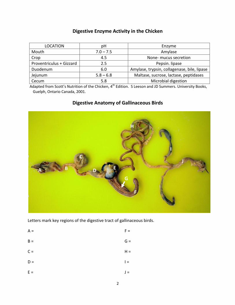
Digestive Enzyme Activity in the Chicken
LOCATION pH Enzyme
Mouth 7.0 – 7.5 Amylase
Crop 4.5 None- mucus secretion
Proventriculus + Gizzard 2.5 Pepsin. lipase
Duodenum 6.0 Amylase, trypsin, collagenase, bile, lipase
Jejunum 5.8 – 6.8 Maltase, sucrose, lactase, peptidases
Cecum 5.8 Microbial digestion Adapted from Scott’s Nutrition of the Chicken, 4th Edition. S Leeson and JD Summers. University Books,
Guelph, Ontario Canada, 2001.
Digestive Anatomy of Gallinaceous Birds
Letters mark key regions of the digestive tract of gallinaceous birds.
A =
B =
C =
D =
E =
F =
G =
H =
I =
J =
A B
C
B D
B
E
D
B
F
D
B
G
D
B
H
D
B
I
D
B J
D
B

3
1. Beak, tongue and pharynx: The beak is the primary organ to prehend feed and can crush,
tear or hold feed before swallowing. The upper beak (maxilla) is covered by hard keratin. Birds
do not have teeth, but the margins of the beak are often hard and might have ridges to help
with grasping feed. In addition, ridges (papillae) on the upper palate can help to prehend feed.
Taste buds are located on the mucosa of the upper beak and behind the tongue. Chickens are
reported to have less than 300 tastebuds, compared to 1700 for dogs and nearly 9000 for
humans. Perhaps taste plays a smaller role in eating in birds compared to mammals.
Gallinaceous birds generally do not chew, but swallow feed whole.
Tongues are covered with stiff papillae that help to used to manipulate and prehend feed. The
oral cavity also contains salivary glands that secrete saliva and variable amounts of amylase;
little is secreted in chickens and turkeys. Grain-eating birds have well developed salivary glands
while these glands are fewer in fish and meat eaters.
Upper beak of chicken: Note choanal slit, where nasal cavity communicates with oral cavity, and pointed papillae that help to prehend feed.
2. Esophagus and crop: The esophagus is a muscular tube that transports feed from the
pharynx to the proventriculus. Because feed is swallowed (birds rarely “chew”) whole the
Choanal slit Papillae Pharynx

4
esophagus must be quite distensible. The esophagus has a double-muscular wall (circular and
longitudinal smooth muscle) and mucus glands to lubricate the channel, easing the movement
of feed. Muscular contractions (peristalsis) propel the feed down the digestive tract.
Gallinaceous birds have a crop, a diverticulum of the esophagus, which holds food before
further digestion commences. This capacity enables the bird to take its food as “whole meals”
at single time while still allowing for continuous digestion. The crop has a smooth white to pink
lining (mucosa).
3. Proventriculus and Gizzard: The avian “stomach” consists of these two chambers. The proventriulus more closely resembles the mammalian stomach. The proventriculus is lined by compound glands that secrete mucus, pepsin and hydrochloric acid.
Liver
Tongue
Esophagus
Crop
Proventriculus
Gizzard

5
Gizzard unopened Gizzard opened- note the koilin cuticle lining
The gizzard consists mostly of two smooth muscle bands (thick and thin) that are oriented in
different directions to create a powerful crushing action when they contract. The interior of
the gizzard is lined with a thick protein cuticle (koilin) that protects the gizzard from 1) acid and
proteolytic enzymes and 2) from injury during grinding of hard food substances. The koilin
lining is often green from reflux of bile from duodenum into the gizzard. Grit (small stones)
consumed and stored within the gizzard can increase digestibility of hard feeds (grains and
seeds).
Gizzards of gamebirds with digestive tract disorders can distend with litter or particulate matter (abnormal feeding behavior from probable discomfort).

6
4. Small intestine: the intestine is lined by folds or villi with great absorptive capacity. Intestinal
length varies in birds, but tends to be shorter on a body weight basis compared to mammals.
This is where most of the food is enzymatically digested and absorbed.
a. Duodenum- This organ has a duodenal loop that surrounds the pancreas. Has less
digestive capacity than mammal duodenum because most digestive enzymes of birds
empty into the end (distal aspect) of the duodenum near the jejunum. Most
carbohydrate absorption occurs in the duodenum.
b. Jejunum- Generally ends where the yolk stalk (Meckel’s diverticulum) is located.
Major site of enzymatic digestion and absorption of amino acids, calcium and
phosphorus.
c. Ileum- shorter than jejunum and similar function, but on a slightly lower scale
5. Cecum: These are paired blind-ended sacs attached at the junction of the ileum and colon.
Cecal contents are distinct from those of the small intestine and colon, having a slightly dry to
tar-like consistency. The ceca are evacuated infrequently (several times a day) compared to the
colon. Cecum serves as a site of fluid absorption (along with lower segments of intestine) and
limited absorption of amino acids and carbohydrates.
Cecum
Colon
Ileum
Cecum
Cloaca

7
Three-week-old Chukar Partridge chick: Cecal coccidiosis- core of white exudate distends the cecum (arrows).
6. Colon and cloaca: The colon (rectum) is relatively short and links the ileum with the
coprodeal compartment of the cloaca. The cloaca and colon are important for reabsorption of
water from GI tract. The cloaca serves as a common end pathway for urinary tract (urates),
digestive tract (feces) and reproductive tract. The colon empties into the most cranial portion,
the coprodeum. The urinary tract and reproductive tracts empty into the middle and smallest
compartment, the urodeum. The final compartment, the proctodeum opens to the outside
(anus) and contains the bursa of Fabricius.
7. Pancreas: Reddish to tan tissue that lies within the loop of the duodenum. Releases insulin
(endocrine function) into bloodstream and secretes amylase, lipase, proteolytic enzymes and
sodium bicarbonate (exocrine function) into lumen of duodenum.

8
The pancreas (arrows) is situated within the loop of the duodenum, which Immediately follows the proventriculus and gizzard.
8. Liver: There are right and left lobes that are joined at the midline. The right lobe is largest in
gallinaceous birds. Liver produces bile that is transported to the lumen of the duodenum by
two ducts. Bile emulsifies lipid so that it can be more easily digested by lipase.

9
Liver of “normal” two-week pheasant chick Liver of one-week pheasant chick with “starve-out.” Note the enlarged gall bladder (arrows).
What does the liver do?
a. Carbohydrate metabolism: maintaining constant blood glucose concentration
b. Protein metabolism: synthesis of plasma proteins (such as albumin), coagulation factors
(e.g. fibrinogen, prothrombin, factors V, VII, IX and X)
c. Fat metabolism: hepatocellular up-take of surplus fatty acids bound to albumin (coming
either from ingestion (from enterocytes) or from adipocytes) → use for energy
production in hepatocytes plus synthesis of triglycerides
d. Synthesize and excrete bile. Bile = bile acids, cholesterol, phospholipids, bilirubin,
proteins plus mucoid secretions of gall bladder and bile duct glands, water and
electrolytes.
e. Bilirubin metabolism: end product of hemoglobin/myoglobin degradation during
erythrocyte turn over in mononuclear phagocyte system (e.g. spleen, bone marrow and
Kupffer cells of liver).
f. Immune function (Kupffer cells, etc.) = Innate immune response: severe liver disease
increases likelihood of endotoxemia and systemic infections.
g. Biotransformation, and usually inactivation, of toxins
Liver
Liver

10
Where does absorption of nutrients occur?
a. Carbohydrates: Absorption occurs more rapidly from the small intestine compared to
the cecum. Most glucose absorption occurs in the duodenum by active transport.
b. Amino acids and peptides: Most amino acid absorption occurs in the small intestine,
although minimal absorption might occur in crop, gizzard and proventiculus (minimal).
c. Fatty acids: Fatty acid and bile acid absorption occurs mostly in the jejunum and to a
lesser extent in the ileum.
d. Electrolytes: The upper jejunum is the major site of absorption of calcium, phosphorus
and Mg.
e. Water, sodium and chloride: Water is absorbed by the small and large intestines and
ceca, and usually follows the absorption glucose, sodium and amino acids. Substantial
absorption of sodium occurs in the jejunum and colon.
What are some of the common digestive tract diseases of gamebirds?
Candida albicans infection
Definition: Mycotic infection affecting a wide variety of birds, primarily in the upper digestive
tract. Yeast infection; fairly common, but generally not a major clinical problem
Synonym: Candidiasis = crop mold = thrush
Cause: Candida albicans; a yeast that forms pseudohyphae in tissues. Epidemiology/Transmission: Ubiquitous organism; overgrowth is usually controlled by normal
bacterial microflora in digestive tract. Often associated with other disease problems in the
house; digestive or respiratory tract disease. Long-term oral antibiotics, especially when
administered in drinking water, can alter microflora of upper digestive tract and promote yeast
growth. Not transmitted from bird to bird.
Clinical signs/Gross lesions:
Nonspecific; birds may appear unthrifty or have other more significant diseases being treated
with oral antibiotics
Lesions occur most often in the crop Fine, white pseudomembrane lining oral cavity or
esophagus. Membrane can be peeled off of mucosa- “Turkish towel” appearance to lining

11
Turkey poults with Candida infection. The photos above show moderate to severe crop infection (note the rough lining of the open crops) caused by Candida albicans.
Diagnosis: Gross and histopathological lesions are usually confirmatory, although fungal
culture can also be considered.
Ulcerative Enteritis
This is a common bacterial enteric disease of domestic bobwhite quail, but also affecting other
upland gamebirds, and young turkeys and white leghorn pullets. The lesions are characterized
by multifocal discoid ulcers in the small intestine and multifocal hepatic necrosis.
Cause: Clostridium colinum, a gram-positive, spore-forming bacterial rod. The organism is
hardy in the environment.
Transmission: The organism is spread in the feces of infected birds and can persist in the in the
soil or litter for several months. The agent spreads rapidly from bird to bird. The infection can
be spread in contaminated feces by flies. The disease is rare in birds raised on wire.
Clinical Signs: Quail are usually 6-10 weeks of age and sudden death associated with white,
watery diarrhea occur. Birds that do not die suddenly will be depressed with closed eyes and
ruffled feathers. Infected birds are thirsty and will huddle around the waterers. The course of
disease is about two weeks and can result in nearly 100% mortality in bobwhite quail.
Gross lesions: Lesions are prominent in most affected birds. Crops may be distended with
fluid. Affected birds have deep, punctate to discoid, tan to grey ulcers in the small intestine.
The ulcers often penetrate the entire wall of the intestine to result in peritonitis and adherence
of intestinal loops. The liver may or may not have pale foci of necrosis on the capsule and on
cut surfaces. Birds that live for more than ten days can become emaciated.

12
Four-week-old Bobwhite quail with ulcerative enteritis. Note the multifocal punctate ulcers in the small intestine and liver (arrows).
Salmonellosis
Definition: This is infection with the Salmonella bacterium that most often causes disease of the
digestive tract and yolk sac of gamebird chicks, but can also occur in adults with no clinical signs. In my
experience Salmonella infection in gamebirds is primarily a chick problem, although the adults
(breeders) can still be affected.
Cause: Salmonella pullorum-gallinarum as well as over 200 other serotypes (paratyphoid species) of
Salmonella can be involved. S. pullorum-gallinarum has been described in a variety of upland game
birds. S. pullorum-gallinarum is nonmotile and tends to be specific for birds, while the motile
paratyphoid serotypes can infect a wide variety of birds and mammals. These are gram-negative
bacterial rods.
Salmonella pullorum-gallinarum can be introduced into chicks by direct transmission from the ovary of
the hen into the egg. Chicks can appear normal at hatch, but experience heavy mortality during the
next three weeks- death in the early brooding stages.

13
The paratyphoid Salmonellae can enter the egg before hatch or infect the chick in the pen through a
variety of routes, including:
1. Soiled or manure-contaminated hatching eggs 2. Contaminated by-products in feed 3. Feces of other infected chicks 4. Hatchery contamination 5. Vectors: mice and wild birds 6. Dirty litter or poorly disinfected flooring, feeders, drinkers 7. Keeping young and old birds in close proximity
Clinical signs: Chicks will often appear as starve-outs- dead in pen or piling in corners.
Cecal cores in a pheasant chick with salmonellosis, which can closely resemble coccidiosis. Left- cecum intact (black arrows), right- cecum incised to show exudate (white arrows).
Diagnosis: Bacterial culture of sick birds (definitive) and blood testing (whole blood plate test) in adult
breeder birds to identify reactors. The same killed Salmonella antigen is used to detect antibodies to
both Salmonella pullorum and Salmonella gallinarum (fowl typhoid) in the whole blood plate test.
Prevention/Treatment: Purchase birds only from NPIP-approved hatcheries. Salmonella pullorum-
gallinarum is a reportable disease. Birds will be euthanized, not treated under supervision of
regulatory agency. Thoroughly disinfect the brooding pens prior to placing new chicks. Use clean,
mold-free litter.

14
Salmonella: Prevention
Solving the Puzzle
Pen
Feces
EggsBirds
Hen
Fluff FeedMice
The key to preventing or eliminating Salmonella infection is determining the source of the infection
Paratyphoid infections can be treated with antibiotics, but affected chicks will probably die because
they are usually not eating or drinking. (starve-outs). Antibiotics can help the birds in the pen that are
least affected and most susceptible to contracting the infection.
Small intestine of bobwhite quail with ulcerative enteritis. Note the multifocal punctuate
transmural ulcers that are observed in the intact (A) and open (B) intestine.
A B
15
Diagnosis: History and gross lesions are usually diagnostic. The organism has specific growth
requirements and bacterial isolated is generally not needed for a diagnosis.
Treatment: Bacitracin is generally the antibiotic of choice at 100-200 gm/ton of feed for 7-10
days, or in the water at 0.25-0.50 gm per gallon of water for 7-10 days. Penicillin and
tetracyclines can also be effective.
Prevention: Ulcerative enteritis rarely occurs in birds that are raised on wire. Rotation of pens
on a regular basis can reduce exposure to Clostridium spores.
References:
Aves Introduction, by AS King, In Sisson and Grossman’s The anatomy of Domestic Animals, R
Getty (ed.), Fifth Ed., W.B. Saunders Publishing, Philadelphia, PA, 1975.
Birds Their Structure and Function. AS King and J McClelland (eds.), Bailliere Tindall, London,
1977.
E R Dumont. Bone density and the lightweight skeletons of birds. Proc. Royal Soc. B , 277:
2193-2198, 2010.
Manual of Ornithology, Avian Structure and Function. NS Procor and PJ Lynch (eds.) Edwards
Brothers, Inc. Ann Arbor, MI, 1993
Ornithology in Laboratory and Field, Fourth Edition, O S Pettingill, Jr. (ed.), Burgess Publishing
Co., Minneapolis, MN, 1975.
Scott’s Nutrition of the Chicken, 4th Edition. S Leeson and JD Summers. University Books,
Guelph, Ontario Canada, 2001.
Sturkie’s Avian Physiology, Fifth Edition. GC Causey (ed.). Academic Press, San Diego, CA. 2000.
Sternum