APPLICATIONS OF THE HISTOMETRIC METHOD ASSISTED BY THE COMPUTER IN THE AVIAN DIGESTIVE TUBE...
12
¸˚˛˝˙ ˛ ˝˝ ˚˛˛˙ ¸˛˝ ¸˙ ˚˛˛˙ ˝ ˛˝ ˝˛˝ ˚¸ ˚ l ˛˘ ˝ ˛˝ § ÝÒÝÍ×Í Ý±¼ ìèÞõ ˚˛˛˙ ¸˛˝
-
Upload
independent -
Category
Documents
-
view
0 -
download
0
Transcript of APPLICATIONS OF THE HISTOMETRIC METHOD ASSISTED BY THE COMPUTER IN THE AVIAN DIGESTIVE TUBE...
ËÒ×ÊÛÎÍ×ÌÇÑÚ ßÙÎÑÒÑÓ×ÝßÔ ÍÝ×ÛÒÝÛÍ
ßÒÜ ÊÛÌÛÎ×ÒßÎÇÓÛÜ×Ý×ÒÛ ó ÞËÝØßÎÛÍÌ
ÚßÝËÔÌÇ ÑÚ ÊÛÌÛÎ×ÒßÎÇ ÓÛÜ×Ý×ÒÛ
ÍÝ×ÛÒÌ×Ú×Ý ÉÑÎÕÍ
Ý ÍÛÎ×ÛÍ
ÊÑÔËÓÛ ÔÊ øï÷
îððçÎÛÝÑÙÒ×ÆÛÜ ÍÝ×ÛÒÌ×Ú×Ý ÉÑÎÕÍ
¾§ÝÒÝÍ×Í � ݱ¼ ìèÞõ
ÊÛÌÛÎ×ÒßÎÇ ÓÛÜ×Ý×ÒÛ
ÞËÝØßÎÛÍÌ

49
Scientific works, C series LV(1), 2009ISSN 1222-5304
APPLICATIONS OF THE HISTOMETRIC METHOD ASSISTEDBY THE COMPUTER IN THE AVIAN DIGESTIVE TUBE
INVESTIGATION
E.O. Bucur1, N. Cornil 2, A. Popovici1,Ruth Messikommer3 ,P. Spring4, L. Pant 4, B. Frunz reanu1
Technical assistance: Lucre ia Androne1
1) The Pasteur Institute, Bucharest, Romania ([email protected])2) Faculty of Veterinary Medicine Bucharest, Romania
3) The Institute of Animal Nutrition, Zurich, Switzerland4) Alltech Biotechnology, SUA
Key words: histometry, small intestine, Bio-Mos, bird.
SUMMARY
The objective of this research consisted in the use of a histometric method by analysis ofcomputerized image on the epithelium of the small intestine mucosa in broiler chickens for thetesting of the prebiotical effect of mannanoligosaccharides (Bio-Mos, Alltech). There were madeassessments about the significance of the differences between the avian groups for fourparameters (height of villi, the width of villi, the number of goblets and the depth of the crypts)in fragments sampled from two areas of the ileum. The obtained values were statisticalprocessed. The results comparison of the two types of samples (A and B) revealed significantdifferences on the number of goblets (p < 0,001), the height of the villi (p < 0,01) and the widthof the villi (p < 0,05). The analysis of the obtained results between the animal groups was notconclusive because the comparison group was not known. The research conducted establishedthe morphometric diagnostic method on the intestinal mucosa in birds, being the first paper onhistometry application in veterinary medicine in Romania..
Morphometry (MM) uses methods derived from histology for theidentification of the effects of some toxins, drugs, stimulant preparationsand the establishment of some values of the cellular parameters inmalignant and benign tumors. The MM method analyzes from thequantitative point of view different tissues and cells.
Traditional, the histologists rely on a verbal description of the tissuestructure, sometimes approaching a mathematical description.
In recent years, at the international level, the measurement of thetissues microscopic images, known as histometry (HM) was usedincreasingly both in research and diagnostic. The histometry methodsrepresent a wide extension of those borrowed from geology andmetallurgy.
An important part of the HM technique is that it allows 3Dmeasurements which derived from the 2D microscopic images provided
50
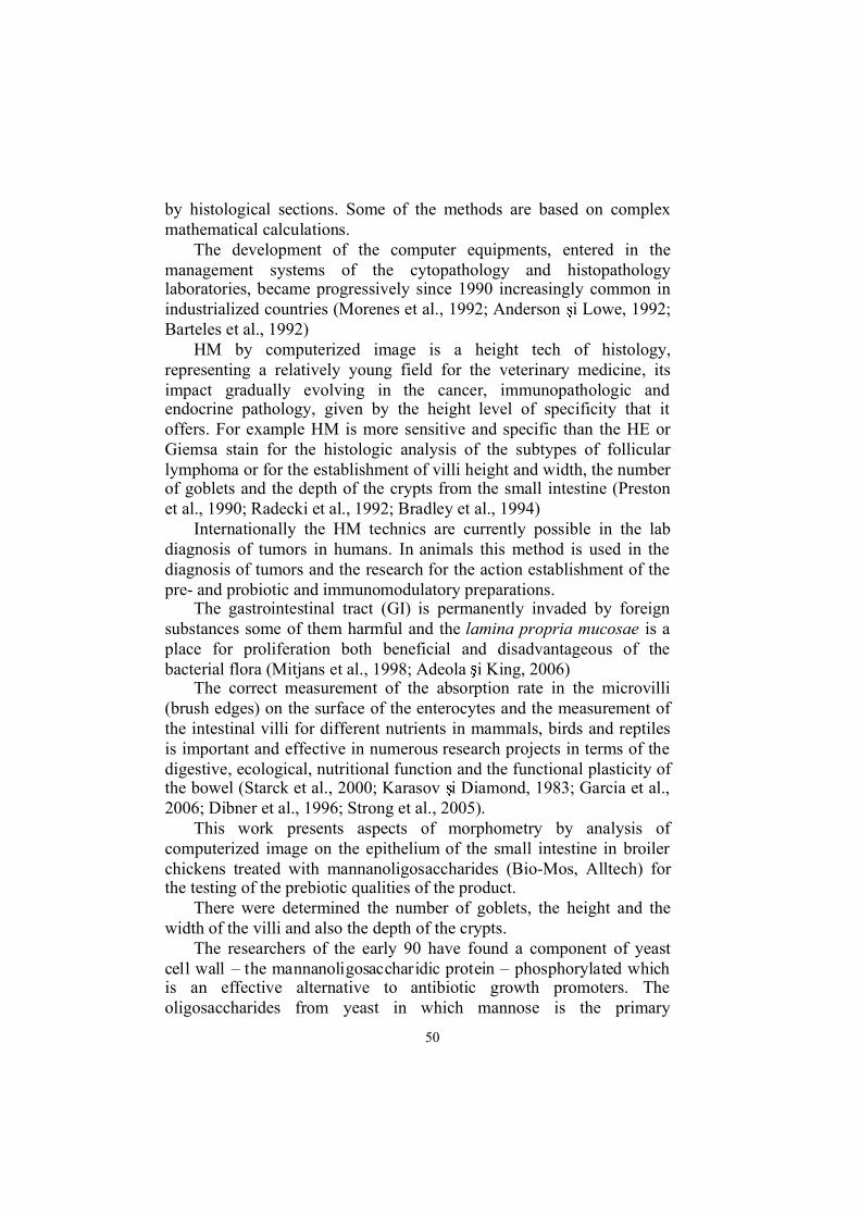
by histological sections. Some of the methods are based on complexmathematical calculations.
The development of the computer equipments, entered in themanagement systems of the cytopathology and histopathologylaboratories, became progressively since 1990 increasingly common inindustrialized countries (Morenes et al., 1992; Anderson i Lowe, 1992;Barteles et al., 1992)
HM by computerized image is a height tech of histology,representing a relatively young field for the veterinary medicine, itsimpact gradually evolving in the cancer, immunopathologic andendocrine pathology, given by the height level of specificity that itoffers. For example HM is more sensitive and specific than the HE orGiemsa stain for the histologic analysis of the subtypes of follicularlymphoma or for the establishment of villi height and width, the numberof goblets and the depth of the crypts from the small intestine (Prestonet al., 1990; Radecki et al., 1992; Bradley et al., 1994)
Internationally the HM technics are currently possible in the labdiagnosis of tumors in humans. In animals this method is used in thediagnosis of tumors and the research for the action establishment of thepre- and probiotic and immunomodulatory preparations.
The gastrointestinal tract (GI) is permanently invaded by foreignsubstances some of them harmful and the lamina propria mucosae is aplace for proliferation both beneficial and disadvantageous of thebacterial flora (Mitjans et al., 1998; Adeola i King, 2006)
The correct measurement of the absorption rate in the microvilli(brush edges) on the surface of the enterocytes and the measurement ofthe intestinal villi for different nutrients in mammals, birds and reptilesis important and effective in numerous research projects in terms of thedigestive, ecological, nutritional function and the functional plasticity ofthe bowel (Starck et al., 2000; Karasov i Diamond, 1983; Garcia et al.,2006; Dibner et al., 1996; Strong et al., 2005).
This work presents aspects of morphometry by analysis ofcomputerized image on the epithelium of the small intestine in broilerchickens treated with mannanoligosaccharides (Bio-Mos, Alltech) forthe testing of the prebiotic qualities of the product.
There were determined the number of goblets, the height and thewidth of the villi and also the depth of the crypts.
The researchers of the early 90 have found a component of yeastcell wall � the mannanoligosaccharidic protein � phosphorylated which is an effective alternative to antibiotic growth promoters. Theoligosaccharides from yeast in which mannose is the primary
51
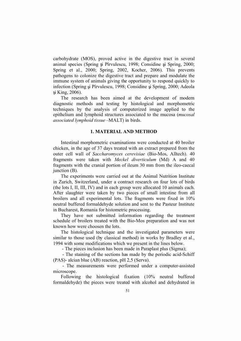
carbohydrate (MOS), proved active in the digestive tract in severalanimal species (Spring i Pîrvulescu, 1998; Considine i Spring, 2000;Spring et al., 2000; Spring, 2002, Kocher, 2006). This preventspathogens to colonize the digestive tract and prepare and modulate theimmune system of animals giving the opportunity to respond quickly toinfection (Spring i Pîrvulescu, 1998; Considine i Spring, 2000; Adeolai King, 2006).
The research has been aimed at the development of moderndiagnostic methods and testing by histological and morphometrictechniques by the analysis of computerized image applied to theepithelium and lymphoid structures associated to the mucosa (mucosalassociated lymphoid tissue �MALT) in birds.
1. MATERIAL AND METHOD
Intestinal morphometric examinations were conducted at 40 broilerchicken, in the age of 37 days treated with an extract prepared from theouter cell wall of Saccharomyces cerevisiae (Bio-Mos, Alltech). 40fragments were taken with Meckel diverticulum (Md) A and 40fragments with the cranial portion of ileum 30 mm from the ileo-caecaljunction (B).
The experiments were carried out at the Animal Nutrition Institutein Zurich, Switzerland, under a contract research on four lots of birds(the lots I, II, III, IV) and in each group were allocated 10 animals each.After slaughter were taken by two pieces of small intestine from allbroilers and all experimental lots. The fragments were fixed in 10%neutral buffered formaldehyde solution and sent to the Pasteur Institutein Bucharest, Romania for histometric processing.
They have not submitted information regarding the treatmentschedule of broilers treated with the Bio-Mos preparation and was notknown how were choosen the lots.
The histological technique and the investigated parameters weresimilar to those used (by classical method) in works by Bradley et al.,1994 with some modifications which we present in the lines below.
- The pieces inclusion has been made in Paraplast plus (Sigma);- The staining of the sections has made by the periodic acid-Schiff
(PAS)- alcian blue (AB) reaction, pH 2,5 (Serva).- The measurements were performed under a computer-assisted
microscope.Following the histological fixation (10% neutral buffered
formaldehyde) the pieces were treated with alcohol and dehydrated in

52
toluene in the Citadel device 1000 � Shandom (2000), and the inclusionhas been made in Histocentre 2 device � Shandon (2000).
The histological preparations were examined under the lightmicroscope Nikon (Labophot type) fitted with a Sony color videocamera (CCD-IRIS/RGB) which processed images and transmitted themto a computer equipped with software Lucia M 3.00b/2001 imageprocessing.
This program allows measurement with an accuracy of up to 1/1000m of the size of various cells by transforming pixels in m.
Before the start of the measurements the objects standardisation wasperformed. With the aid of a micrometer object it has been determinedthe number of pixels corresponding to a micrometer for the 4, 6, 10, 20,40 objects, and the computer was programmed to read directly in themicrometer size. After the mathematical calculations, the valuesobtained were converted from m in mm.
The establishment of the number of goblets, of the height and widthof the villi and also the depth of the crypts were determined on 9 villifrom each sample as shown in the Bradley i col., 1994) method, tomention that reading the samples was performed with a computer-assisted microscope. For the 80 samples of small intestine there wereperformed 2880 measurements.
The values obtained were statistically processed in the sense thatwere calculated for each parameter arithmetic average ( ), its standarderror(E.S. ) and the coefficient of variability (CV% /lot) Wardlaw, 1993.
There were made assessments about the significance of thedifferences between the avian groups for each parameter by the �t� test (student). It was also calculated the correlation coefficient (r) betweenthe height of villi and the number of goblets/mm.
The research conducted established the morphometric diagnosticmethod on the intestinal mucosa in birds, being the first paper on HMapplication in veterinary medicine in Romania.
2. RESULTS AND DISCUSSIONS
The data obtained were synthesized as both individual and groupmean values for the samples collected from the M. d. area (A) and forthose collected from the ileo-caecal junction (B).
53
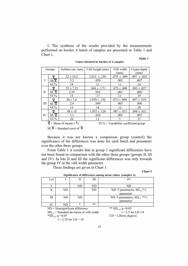
I. The synthesis of the results provided by the measurementsperformed on broiler A batch of samples are presented in Table 1 andChart 1.
Table 1Values obtained in batches of A samples
Groups Goblets (no./mm) Villi height (mm) Villi width(mm)
Crypta depth(mm)
52 ± 10.2 1.015 ± .159 .079 ± .009 .097 ± .024SE 3.2 .050 .003 .007IVC% 19 15 11 24
55 ± 7.25 .968 ± .173 .075 ± .009 .093 ± .027SE 2.29 .054 .003 .008IIVC% 13 .17 11 29
50 ± 7.6 1.070 ± .156 .073 ± .009 .097 ± .019SE 2.4 .049 .003 .006IIIVC% 15 14 12 20
48 ± 10 1.055 ± .120 .087 ± .015 .098 ± .021SE 3.1 .038 .005 .007IVVC% 20 11 5 21= Mean of means ( ) VC% = Variability coefficient/group
SE = Standard error of
Because it was not known a comparison group (control) thesignificance of the differences was done for each batch and parameterover the other three groups.
From Table 1 it results that in group 1 significant differences havenot been found in comparison with the other three groups (groups II, IIIand IV). In lots II and III the significant differences was only towardsthe group IV in the villi width parameter.
These findings are given in Chart 1.Chart 1
Significance of differences among mean values (samples A)
Lot I II III IV
I ND ND NDII ND ND ND /3 parameters; SDvw*/1
parameterIII ND ND ND /3 parameters; SDvw **/1
parameterIV ND * **
ND = Nonsignificant difference ** SDvw; p <0.05SDvw = Standard deviation of villi width t = 2.5 for LD=18*SDvw; p <0.05 LD = Liberty degrees
t = 2.18 for LD = 18

54
From the chart above results that the variability was the middleorder for three parameters (villi height = 11-17%, villi width = 5-12 %,goblet cells number = 13-20%); as for the cryptal depth, its variabilitywas high (20-29%).
We present some photo shoots obtained from the small intestinesamples prepared for histometric exams (photo 1,2,3)
As regards the correlation between the villi height and the gobletsnumber/mm the following were found:
- Groups I and II it is distinguish a weak positive correlation (r =0.276and r = 0.439 respectively)
- Groups III and IV present a weak negative correlation (r = - 0.240andr = 0.383 respectively).
The values recorded for samples A show that in the case of GroupsIII and IV the total cell number remains unchanged, while the villiheight is higher.
The evaluation may be that, with the Groups I and II, the totalnumber of goblet cells rises together with the villi height increase.
II. The Summary results of measurements obtained from the portionof small intestine from the ileo-caecal junction (probele B) are presentedin the Table 2 and Chart 2.
Table 2Values per groups in samples B
Groups Goblets(no./mm)
Villiheight
(mm)
Villiwidth
(mm)
Cryptadepth
(mm)
I 73 ± 13.3 .640 ±.062
.100 ±.009
.095 ±.021
Photo1. Ileum (M.D. area).Numerous villi, intestinalcrypts with submucosal
glands, muscular and seroustunics. PAS � alcian reaction
Photo2. Ileum (M.D. area).Intestinal villi with numerous
goblet cells.PAS � alcian reaction
(x250)
Photo3. Ileum (ileo-caecaljunction). Intestinal villi with
numerous goblet cellsPAS � alcian reaction
(x250)

55
SE 4.2 .019 .003 .006
VC% 18 9 9 2272 ± 15.9 .690 ±
.115.097 ±
.012.108 ±
.027SE 5.0 .036 .004 .008II
VC% 21 16 12 2467 ± 11.6 .638 ±
.111.094 ±
.010.095 ±
.010
SE 3.6 .035 .003 .003III
VC% 17 17 10 1070 ± 1.1 .648 ±
.137.102 ±
.013.102 ±
.021SE 3.5 .043 .004 .006IV
VC% 15 21 13 20
= Mean of means ( ) VC% = Variability coefficient/sample
SE = Standard error ofNo significant difference was noticed, with samples B, on
comparing each group with the other three groups.A weak negative linear correlation was found between the villi
height and goblet cells number in all the groups: Group I r = -0.375,Group II r = -0.569, Group III r = -0.592, Group IV r = -0,342.
We present some histological photo shoots obtained from the smallintestine (samples B) prepared for morphometric technic (photo 4 and5).
Photo 4. Ileum (ileo-caecal junction).Numerous villi, intestinal crypts with
submucosal glands, muscular and seroustunics. PAS � alcian reaction (x140)
Photo 5. Ileum (M.D. area)Intestinal crypts with submucosal glands.
PAS � alcian reaction (x250)

56
III. Samples A/B comparisonThe significance of the differences among the values of the test
parameters of groups A and B is presented in Chart 2.Chart 2
The significance of the difference among means
Groups Goblets(no./mm)
Villiheight (mm)
Villi width(mm)
Cryptadepth
(mm)I/IV ND ND ND NDII/IV ND ND ND NDIII/IV ND ND ND ND
A/B VSDt = 4.73 VSDt = 7.05 SDt= 2.35 NDp< 0.001 p< 0.001 p< 0.05
ND = Nonsignificant differenceVSD = Very significant differenceSD = Significant differenceOn comparatively analysis the results obtained the villi height was
found to be lower with samples B (Table 2) than with samples A (Table1).
With Group I, there were very significant differences (p < 0.001) forthe following parameters: number of goblets, villi height and width.
In the Group II, distinctly significant differences (p < 0.01) wererecorded with the goblet cell number, and very significant ones (p <0.001) for the height and width of the villi.
Group III presented very significant differences (p < 0.001) for thegoblets number, villi height and width.
In the Group IV very significant differences (p < 0.001) were foundas far as the goblets number and villi height are concerned. Also,significant differences (p < 0.05) were revealed in way of the villi width.
The safety of the methods for interpreting the results of intestinalepithelium measurements is based on the integrity of the mucosalepithelium and enterocytes and on the execution quality of thehistological preparations.
The ileum histological examination revealed in all the birdsexamined a normal aspect of villi with many goblet cells characterizedby the presence of large vesicles. The epithelium of the intestinalmucosa is delicate, with long intestinal villi. In the submucosa werefound many glands with normal glandular epithelium cells (photo 5).The simple prismatic epithelium covers the intestinal villi and goesdown in the Lieberkühn crypts in whose lumen is lining. It has beennoted many goblet cells PAS-positive or alcian positive irregularly

57
disseminated on the flanks of villi alternating with numerous intactepithelial absorption cells (photo 2).
In the histometrical examination no significant changes were foundin the studied parameters (the villi height and width, the number ofgoblet cells and the crypts depth) in the sections from the birds thatcame from the A samples. In the B samples were recorded lower valuesof the villi height parameter compared with the A samples.
However, the comparative analysis of the obtained results on theanalyzed parameters in A/B samples have highlighted significantdifferences (p<0.001; p<0.01; p<0.05), on the number of goblet cells,the villi height and width.
Numerous studies showed that the administration of Bio-Mos in thediet of broilers and piglets resulted in an increase in villi length and areduction in crypts depth. The overall size of the surface absorption inthe intestine and the reducing of the renewal rate of the epithelial cells inthe crypts improve the nutrient availability for absorbing (Adeola iKing, 2006; Kocher,2006)
In the work that we made, we were not aware of all the elements onthe experimental protocol in broilers and we could not conclude on theprebiotic action of the Bio-Mos preparation.
It results that the measurements performed on the ileum showed asignificant difference in the A samples over the B samples in threeparameters: goblet cells, the villi height and width
CONCLUSIONS
It were investigated by morphometric examinations fragments ofsmall intestine from the treated broilers with the Bio-Mos preparation,Alltech.
1. The histometric test results have shown statistically significantdifferences of the parameters: villi height, villi width, number of gobletcells and the crypts depth on the fragments collected from the samesubject with M.D. (A) compared with the obtained fragments from thecranial portion of ileum (B).
2. The registered values in the A samples demonstrates that thenumber of goblet cells remains the same (the Group III and IV) orincreases (the Groups I and II) while the villi height is bigger.
3. In the B samples were not found significant differences betweenthe groups, and between the villi height and the number of goblet cells ithas shown a weak linear negative increase in all the groups (the GroupsI, II, III, IV).

58
4. The comparison of the A samples results with the B samplesresults has shown significant differences about the number of gobletcells (p<0.001), the villi height (p<0.01) and the villi width (p<0.05).
5. The analysis of the obtained results in the groups of animalsstudied was not conclusive on the prebiotic action of the usedpreparation.
6. The studies have aimed the development of a diagnostic andtesting method by histometric technic by analysis of computerizedimage on the epithelium of the small intestine mucosa.
We thank to Mrs. Lucretia Androne for the excellent qualityof the histological preparations made
REFERENCES
1. Adeola O., D.E. King - Developmental Changes in Morphometry of the Small Intestine andJejunal Sucrase Activity During the First Nine Weeks of Postnatal Growth In Pigs.Journal of Animal Science, 84, pp112-118, 2006
2. Anderson J.M., J. Lowe - Histometry and Image Analysis. In: Theory and Practice ofHistological Techniques. Edited by Bancroft J.D., A. Stevens. Third Edition, ChurchillLivingstone, Edinburgh, London,Melbourne, New York, pp. 597-618, 1992
3. Bartels P.H., D. Thompson, J.E. Weber - Expert Systems in Histopathology. IV theManagement of Uncertainty. Analytical and Quantitative Cytology and Histology, 14(1),pp.1-13, 1992
4. Bradley G.L., T.F. Savage, K.I. Timm - The Effects of Supplementing Diets withSaccharomyces Cerevisiae var. Boulardii on Male poult Performance and IlealMorphology. Poultry Science, 73 (11):pp.1766-70,1994
5. Considine M., P. Spring -Yeast - derived Mannanoligosaccharides in Animal Nutrition: UnUpdate. The Food Revolution-Alltech`s 14th European, Middle Eastern, African LectureTour, pp.89-96, 2000
6. Dibner J.J., M.L. Kitchell, C.A. Atwell, F.J. Ivey - The Effect of Dietary Ingredients and Ageon the Microscopic Structure of the Gastrointestinal Tract in Poultry. Journal AppliedPoultry Research, 5, pp 70-77, 1996
7. Garcia I.J.R., F.R.O. Hernandez, J.P.N. Aguilar, J.J.U. Gomez - Intestine Morphometry ofthe Coturnix Conturnic Japonica in the Relation with Different Levels of Lysine in theFeed. Journal of Animal and Veterinary Advances, 5(12), pp1143-1145, 2006
8. Karasov W.H., J. M. Diamond - A simple method for measuring intestinal solute uptake vitro.Journal of Comparative Physiology. 152, pp 105-116, 1983
9. Kocher A. - Digestive Tract Health and Function. Future of Growth Promotion. Alltech`sEuropean Lecture Tour, pp 19-28, 2006
10. Mitjans M, G. Barniol , R. Ferrer. - Mucosal Surface Area in Chicken Small IntestineDuring Development. Cell Tissue Research, 290(1):pp. 71-78, 1997
11. Morens Annie, B. Krief, G. Brugal - The Home Microscope Workstation. A new Tool forCervical Cancer Screening. Analitical and Quantitative Cytology and Histology, 14(4),pp.289-294, 1992
12. Preston K.Jr, L.M. Firestone, B.N. Nathwani - Use of three-dimensional MathematicalMorphology in Analyzing Histologic Images, with Application to the Subtyping ofFollicular Lymphomas. Analytical and Quantitative Cytology and Histology 12(6), pp.399-416, 1990
59
13. Radecki S. V., P. K. Ku, M. R. Bennink, M. T. Yokoyama, E. R. Miller - Effect of DietaryCopper on Intestinal Mucosa Enzyme Activity, Morphology, and Turnover Rates inWeanling Pigs. Journal of Animal Science 70, pp. 1424-1431, 1992
14. Spring P.- Rolul Mannanoligozaharidelor Derivate din Peretele Celular de Drojdie înNutri ie i S n tate. Pia a Aditivilor Furajeri Naturali - de la Consumul Sporadic laConsumul Industrial. Alltech`s Ciclul de Conferin e pentru Europa, Orientul Mijlociu iAfrica, pp. 86-101, 2002
15. Spring P., M. Pîrvulescu - Mannanoligosaccharide: Its logical Role as a Natural FeedAdditive for Piglets. In: Biotechnology in the Feed Industry. Proceeding of the 13thAnnual Symposium (T.P.Lyons and K.A. Jacques, eds). Nottingham University Press,U.K., pp.553-62, 1998
16. Spring P.,C. Wenk, K.A. Dawson, K.E. Newman - Effect of Mannanoligosaccharide onDifferent Cecal Parameters and on Cecal Concentration on Enteric Bacteria inChallenged Broiler Chicks. Poultry Science 79, pp. 205-211, 2000
17. Strong T.R., P.R. Reimer, E.J. Braun .- Avian Cecal Microanatomy: a MorphometricComparison of Two Species. Journal of Experimental Zoology 252(53), pp. 10-20, 2005
18. Starck J. M., W. H. Karasov, D. Afik - Intestinal Nutrient Uptake Measurements and TissueDamage: Validating the Everted Sleeves Method. Physiological and BiochemicalZoology 73 (40), pp 454-460, 2000
19. Wardlaw A.C. - Practical Statistics for Experimental Biologists. Ed. Wiley, England,1993