structure and function of the digestive tract of the grasscarp ...
93
structure and function of the digestive tract of the grasscarp cover photograph: Pharyngeal teeth of the grasscarp. Usually, a well developed pharyngeal masticatory apparatus is present in sto- machless fish. The food is fragmented between the ventral teeth (photograph) and a dorsal masticatory plate. Scanningelectron.micrograph x 45. 0000
-
Upload
khangminh22 -
Category
Documents
-
view
0 -
download
0
Transcript of structure and function of the digestive tract of the grasscarp ...

structure and function of the digestive t rac t of the grasscarp
cover photograph: Pharyngeal teeth of the grasscarp. Usually, a well developed pharyngeal masticatory apparatus is present in sto-machless fish. The food is fragmented between the ventral teeth (photograph) and a dorsal masticatory plate. Scanning electron.micrograph x 45.
0000

Promotor: dr. L.P.M. Timmermans, hoogleraar in de algemene dierkunde
Co-Promotor: dr. J.W.M. Osse , hoogleraar in de algemene dierkunde

/uiuoYzo? k » i
HENRI W.J. STROBAND
STRUCTURE AND FUNCTION OF THE DIGESTIVE TRACT OF THE GRASSCARP
Proefschr i f t
ter verkr i jg ing van de graad van
doctor in de landbouwwetenschappen,
op gezag van de rector magnificus,
dr.H.C. van der Plas,
hoogleraar in de organische scheikunde,
in het openbaar te verdedigen
op woensdag 10 september 1980
des namiddags te half drie in de aula
van de Landbouwhogeschool te Wageningen

CONTENTS/INHOUD
VOORWOORD 6
GENERAL INTRODUCTION 9
A. Kaderstel l ing 9
B. Structure and function of the digestive t rac t of f i shes . . .11
C. Objectives of the experiments 24
GENERAL DISCUSSION AND SUMMARY 26
Concl usions 32
REFERENCES 34
SAMENVATTING (Zie ook bijlage) 38
PUBLICATIONS :
A. Stroband , H.W.J. Growth and d iet dependant s t ructura l
adaptations of the digestive t rac t in juveni le grass-
carp ( Ctenopharyngodon idella, Val.) J . Fish B io l . 11,
167-174 (1977) ^Ê
B. Stroband , H.W.J. & F.M.H. Debets. The u l t rast ructure and
renewal of the i n tes t ina l epithelium of the juveni le
grasscarp, Ctenopharyngodon idella (Val.) Cell T iss. Res.
187, 181-200 (1978) £
C. Stroband , H.W.J. , H. v .d . Meer & L.P.M. Timmermans.
Regional functional d i f fe ren t ia t ion in the gut of the
grasscarp, Ctenopharyngodon idella (Val.) Histochemistry
64, 235-249 (1979) f
D. Stroband , H.W.J. & F.H. van der Veen. The loca l iza t ion

STELLINGEN
1. Er z i j n geen argumenten voor de veronderstell ing dat b i j juveniele en adul
te maagloze vissen de e iwi tver ter ing in het darmlurnen minder e f fek t i e f zou z i jn
dan b i j maaghoudende soorten.
Dit proefschrift.
2. De opvatting van Trevisan, dat vooral het caudale deel van de graskarper-
darm betrokken is b i j de resorptie van nutr iënten, is on ju is t en komt voort u i t
het trekken van voorbarige konklusies u i t alleen morfologische informatie.
Trevisan, P. : Anat. Anz. 145, 237-248 (1979).
3. Het voorstel van Green & P h i l l i p s , graanmutanten te kweken met verminderde
feedbackinhibit ie van aspartaatkinase door lysine en threonine, om zo het ge
halte aan één of meer aminozuren u i t de aspartaat-famil ie te vergroten, verdient
s t e l l i g waardering maar l i j k t geen praktische bijdrage to t de oplossing van het
wereldvoedselvraagstuk te kunnen leveren.
Green, C E . & P h i l l i p s , R.L. : Crop Science 14, 827-829 (1974).
4. De circadische f luktuat ies in gevoeligheid van diverse weefsels voor bepaal
de hormonen suggereert dat hormoonreceptoren een korte levensduur hebben.
Meier, A.H. ; John, T.M. & Joseph, M.M. : Comp. Biochem. Phys io l . 40A, 459-465 (1971). Meier, A.H. ; Trobec, T.N. ; Joseph, M.M. & John, T.M. : P roc . Soc. Exp. Biol . and Med. 37, 408-415 (1971).
5. De i so la t ie van zowel één g lycoproteiner i jk- als één glycoproteinearm gona-
idotroop hormoon u i t hypofyses van beenvissen ondersteunt de opvatting dat "g lo
bula i re" en "vesiculai re" P.A.S.- posit ieve gonadotrope cellen t o t hetzelfde
(celtype behoren, maar s l u i t n ie t u i t dat er een tweede gonadotroop celtype, mo
ge l i j k n iet P.A.S.- pos i t i e f , aanwezig i s .
Ng, T.B. & I d l e r , D.R. : Gen. Comp. Endocrinol . 38, 410-420 (1979). Peute, J . ; Goos, H.J.Th. ; de Bruin, M.G.A. & van Oordt, P.G.W.J. : Ann. B io l . Anim. Bioch. Biophys. 13, 905-910 (1978). Ueda, H. : Bul l . Fac. F i sh . Hokkaido Univ. 31, 1-15 (1980).

6. Het zou van groot belang voor de wereldvrede kunnen z i j n , onderzoek te ver
r ichten naar de oorzaken van het verschijnsel dat achteruitgang van de levens
standaard veelal l e i d t t o t toename van onredel i jk vijand-denken en sti jgende wa-
penproduktie, en in d i t verband onder andere aandacht te schenken aan de moge
l i j k e rol van het " M i l i t a i r Industrieë'el Komplex" .
7. De boycot van de Olympische Spelen in Moskou, waarvoor de verschillende re
denen op z ichzelf terecht werden aangevoerd, doet vermoeden dat w i j , wester
l ingen, het restant van de boterberg die aan de Sovjetunie is verkocht op ons
hoofd hebben gesmeerd in plaats van ook onszelf boter op het brood te geven.
8. De "vertrossing" van de omroepen l e i d t er toe dat de a f le id ing die wordt
gebracht de bevolking a f l e i d t van de meest wezenlijke problemen van onze samen
leving.
9. Het f e i t , dat de vervoersorganisaties bezwaar maken tegen het gebruik van
"hun" belastinggelden ten behoeve van de aanleg van fietspaden betekent dat de
wegvervoerders te weinig oog hebben voor de fietsende medemens en onderstreept
derhalve de noodzaak van genoemde wielerpaden.
10.De "brede maatschappelijke discussie" over kernenergie z a l , wanneer het ka
binet zich n ie t onthoudt van voorbarige uitspraken, vergel i jkbaar b l i j ken met
een golfstroom : oeverloos en met voorspelbare bestemming.
Proefschrift van H.W.J. Stroband
Structure and function of the digestive tract of the grasscarp.
Wageningen, 10 september 1980.

of protein absorption during the transport o f food i n
the in test ine of the grasscarp, Ctenopharyngodon idella
(Val.) Submitted to J . Fish Biol
Stroband, H.W.J. & Annet G. Kroon. The development of
the stomach i n Clarias lazera and the absorption of
protein macromolecules. Submitted to Cell Tiss.Res
URRICULUM VITAE
IJLAGE :
Stroband , H.W.J., J.H.W.M. Rombout & J.H.M. Davina.
Maagloze vissen; Bouw en funct ie van het darmkanaal.
Natuur en Techniek 48, 38-55 (1980)

Vooo-u^ocyi-cL
De. ncduuuA- kvdt op mu <xl£u.oL e&n. astoti. cum
JttZM/ekAv'rL uoyn. Z&n. c m o W o « ^ in. eé-n. 7)uIusa^t<feJ&nÇ>cAc<.pp
sùjAe. Z/cÀtin^, yôle.clt ej&n. g&&ig&>n^rie-<.ol cnn. TTULT e*>',#A. diep,
to-n. t/Hl. AÀ&.CAZ. ec^ï. Q£JCagjesri^fte^.oL cnn rruW ç*iia«. cOep
«CHOJ« hvxjeu (e, *4h*- fw^- eé-n. AJbin. speojzx. uoyn. X«jf £ > t o / e
Geßcovun. ^cLout ote. TZocéuu^. sreMiq. lo . CJui mü. £etre/é Äe&ßt
<JUL ozJóislcL tot een. toe*tt*tuisnjoCe. t&L&'tAcd. Votyt. etedliusrit. ÏJCL.
uoort- W « ds, ScAefv/og. /'s , op t</e£»fe UJ'CLTJL <U&K£. ocrK éot
ITbjfa. %&ruuo7n cutting, mut ujere^r>cJicLPpeÂu^ on.J&vru>eÂ.
huât nvt kesÂ. sjt&i cù- ^te£cL.ti{/i^eM: vast. da?yruw>z£âke- kw
rus doen. tsi.vaA£*i. en. heoM- yniL Im. uzejP. opzJcÂtfrn. \MM£iae.uMyr--
pirn, tot ksi. TL)Vo uasn. zJesi. , nor&n., uoeJ&n., i.u,ike^rL. est- utA.bc
Z£sri-. ü e f o t t t n O UOUTL. U*AJüüonrLclsi/li«ri.cL. , SZA.bie.oi- 6*1. oùcunÂbcuxA.
r\£J<A- trtjuùm- op da. Oocrta^-orusL &n~hejt JÜfn. otCxjg.. a&ssoeJibn.b
UJcLeuL-cucun. \J( i/fL de. ee/xote. pXcœdb) uJrcUuJ&Âinq. u/il aju/esi. in. out
1/ocyiMJoeytoL. , cLouL [/o&ixui- e&ru oiovnJçuJoo^Uit- ia.
KJ^Lcuu-OL UJil ÙK OCUTL. C/JL. WÜ. rux. qjzJjooi&m. at&flAn. -
njüoL at&ruAÀk maAesn. om. igjjLtAJüe^n. Z&. / W o t o n ^ w i . a^e~, CÜA&C
ot- hmx/JsiiLcjt , e&n. zo-6. ÄJIBJ.6 çp^peii&t. SV'Ù. mijn. \/arvntÀsruL ,
-htU.ciUjL f o£ AsJt rot STconJC Jrvrnxsn- Uovn. otit UJtnJrie. . / a t u 77'u
aXlU. ^a^nftyi. Jçasn. IÂ 'un. out JstóteJ; voornam-. , mou**-. 'tA
hoop deut Z.U OÜJL. kei. cLosno<tjaL.t z-icÄ. a.Gvn.atàtrij&rçen. u/e.
U-n. .
O n . oCe. ee/idUL. p^Ccuodo Ârtsn. ïJL müm. ouu/t/vn ott&viJi
v&rs>chujŒ.<UjiLoL , l/ocn.c(sC ocr& omota-t ze. "ma Ucun. Lemgo a/^
cuwi. ez<ri- asuotiL. trT-ayfleJoi. uasn. o6t**Jt&n- &n. ncunoù^te^rt- eux.
Vts/i- , uJcLoiA-ctoo^~ man. sfçcvtv) op onZp<;ooi/na £>'&>%• u/eA.cL vtn
a/lx>ot . \Zk/LdU/u njL&Jc rLusn. OCÓT£JCJ'£\- are.unrL. un. u;o<ri*£ &n-
THicùù&vL. fnü. otm ^ c e t / e^ . ae?»/*//V-ee-^o£ 6^>- -*»%*>• &rucUe. n
<£xitrLci£.h 7n.ün. o n & ^ t o tx/LC UK nmCtn. &c4oort-OLLc/ksis>
cA-ovnJwsn- VtxrT- 06e. JOü^jm-oleA^. uj'ànje. U/a.a*.op *cji CL£JÜCL in&t-

bj>icma&rL &ÖM- cnn. ~mä ? cmaLcunÀb fud. JjuJen. in. QJvn. £si.q. W^fy-
xe/xJn., de. ojL^axjikbLoL Cfe. a eve / t . opéiiv)a.aJL a. -btuoùiAJim..
Van. oui. o-txjt. \yCui#ifU&n. 7ivi£ u/te. iJc vJcLctA.ua.i/u^_ tn,
yLcuuA-^cvmj.. fcrri-raJcf&n- kxJlr THoqgm. rieJlr^n- (jJiK. L& hm*. CAL.
JLa*mi&itL XjZ&JuisnJ>*s>-Qs 7>o4<*n£sn- cli*-, olocn- AcLavt. CLttsjTuxüfieJcL en.
pÜbCuaoLe.- iin. ort.œéTujiy£e£.oL OT.óle- \sn.\/-Coe.cL. op Wwn. orj-ujikht.-
'*\- *-ü"i- fooi. ynjL uctsn. cpioói JteJeJvbn.lo.
~T&n- CLam/ziin. L>um. kut. rtLzn- JirtocJvuu/e*L. omcù^u-
"sxJr AJ&rt- un un. ctz. te/uite. P£.CLCL£O cLasmJr ueyu>c/ujAcùa^-
xxvn. müm. P^crrfoTcTL. , Lu.cy Tùm*nin--y»cufts> , ß-n. co~ Pio-moro-T-,
"Itua. Os»se_ . Z ii rudl&tvL. e t . m&z. (ptou. in^La-t ubvx. g&.ü\/&u>L
e&rt- Ucuflç CfrMrtAD 02. yßfrVyrttAJlsn. C£LL- , TWLclJL. oLtxn- cZe. XsyxjyvMyi.
fa/n. vnJüb^otJr&ia op Ir^divioCu. , u/e&he£. es*- aiX v)/\so,ç&n.
'i'tLouaJS) ynoj^i. *-üii. LU<Z.CIA— oO'K ZBsn. on-caMu?*-* CL***> hxT cm. -
Wlnsfiau/ioJZ- XJn.t/o<- srCcvn— U/aruaCe^n- uÂ.i.aeA^o<jx-oC. . l/e*c&t_ oicurJv
 rutm^ UT&T- oU- cptote. Tnctte. ucen- w.ü.neJcL eile. mü. C.ot. is.
XILU aruÂA/iLoeJx a&J-cuUbn. uJt^.cL , e^n. uoan- <Ue_ zjuesi- A~cm^ -
bT-yi.ocJrtle.y/e- vutui. uicccuioP use. UCUTL. a&c/at.oAZii*t. /i^KrO^n-
J)e. snoKJxcpeL.'b vcuYL. de. uaJraftotj> Ç^pesu?rtiUru'e£a~
")'uiA^yncr^o\.ùaÀjt. &7L. GeJ2Joio£ccple. , cfae. oCoxn^ /LUW- sursit ri Z>CJU&.
lin. Zbn- Po4//le.\/e. ^>ü.ooi.a.a£- hjlJb&tsrt- QXJJU/ZA-GL eta^t- heJl
•m-clisvJLoeJç, u/iJ- Isfa n.ixyL- oJco Qkzo^p &e.oCa*idresi—. <£e^i_
WiAeMß- UJ'/j.on.ol&AJsriq. mouccJc LÀ i/ocrt- >«fn_ fKowoou-Û : u/ij.
Chu*. TA(jz£e. tri- Wee*?) uwn. o&t_ 7 % * ^ - »recé LA
dcA./'PP&u f G«A.-f-J^cum~ U&. G-T-GLaß-, (Zwner hieran- e^u &-jk
CLrL-
Vcvn.
'•>Ta.ae. - a/ncuCistèsn. oCU. i/oox. sn~cn.ze. ry^C oucon- n&-i emo«^_-"0e O
TjytÄ k&ß&fn- mjvexuMj&iJït. Ooo*-icpyno XJOU. £1*. e™. cuttele^.
7)'ULL TrtoojeJŒfftr azMjwïb t-üt- XJOTI-OUA- cue.. uTrtnheJßßäJft. u e t -
Z.(yLeLtnA. oiXe. amtuL. *&iotecÄvii/rus> 5u.rJtg- Lie^rLohxcc. </*- f10*/- ~
[/cur*- oU. s*r(&£.a<*J'S xVu-i*/erî_ cfe. Ucunrcpi*cp »Ut bÖLvUot--
fL&n- tdt keJL cm.d^unLoeJ\ uf'^C 'ußr rio&mem. olt nxAJun. 7. ocun.
CAL»X. éio€4K ( f-ys/o&aie. cfoi. Dinsw**- ) , otie. X-tf™- ïiarof**- -
sCa&oTa.ro-iiu*r*t- a<tAr\AUL. Uooz. oru) op€*isy&éu£. • S.A. HuAsivneUL
oüe. cu&> mibUejbuesisrct^. ua/n. où. Ch.a.<v*t)<>a,LL£. iiuu [/t^-b^he. -
luneL Ucuru oCt. sôiwrU*LV,i'Y>&iti de. -nvuvne. Q/las>itcLn.p&is> -&-
uKnjoül. IAJCUUICLWI. rit^t UJVuA is> ve^UcJuC- ; \ZcnA (C-A.6.0.)
oLCe., ti>e>n.a£o où AJUZSL. A.W.b.M. vcun. C^^-CUSLO (*>?/cne>6/o-
/ o * / ' e J FOL OTVXJL. asiole. oCcurun^cuuL neJe/- yO&uù.oL u/as> oùe.
LrwL CLwin<rJLu*iA—<xsr>aiJ!.y$&> uit te. \/o*WT- tsn. ù^vy^orr-e.
cù. AWL- C . l l RicJLr&t. f[/Cn^ee£.T e*u i /À^-ety J oùie. orv> CÙL
Cx<t/t-/<is> sZaLt^Mvle. ucrox. cmcùsi.z.crt*rc TtCLct*. où. cmTnJ'unJreJzünjL
(/CUn. où. mcLoLOr .
H&i iyeJU. uitAsk otaX Ujiiw \/a£e^i. i^uxicÀ.é'fe. t^n.
Jfe-noe-l/C. x/asn. où- Lcùuv-n.etJ'ùr) un. oLtT p^toe/oonyii^ U/CLOULCÙÉ*-
'uk bua.rn.oun.. De. JUZ&L W.6. C&A-i (T.F.O.L.) oLt*Jc ik uvcru
cù. JOuLcrrci^rig, Ucvn. AûJ. S. M. ^orontcut^-i'OUL£. .
JD asnJr Js&n. (sn crtrK u<AOcsnujÇ.&ùi<pc/- ouun. Ae^t. c&*.
Aa.t ueze. ^ypeu/e^Jr Uoon. hjuun. zeJa*u*i<L, TJcunuvn. ; m,£Hy
Ta.pi'A.cuna. , HcuM^y \AnJsu.cLZ , G toucla. 0-0*1. 7Pcuê»6~t- , 7h<~
UCVTL. CouppeJc^e. frru >rui.tsiou->*J P. IhjwzXAS) {TeJrsT o4smJ&nsn/\
Ûe. VTL&pcvn^ts*<L&>ri. Ucvn. <*e. riM/l. L.. ^£>o(mr.a.cut>iP
tin. JçonA&Jrtùe. U<vn. nvüsL. (sOfLoo^-) "esi.ae£o " AMX- 'un bjÀ-
Z<m.oUn. op fztf0 ppvÄcJcjvL .
TilOA-'à^rLe. , -nÓLÓLvt J^OUM/ zcrZ. cu&i -or&usn. e^i_ /o£</-*t_-
JLoLaX. ruiAr Jjt. où. sVCWl*ir>Utez&^t- , QL&paj*A.oC g.cut*n.t/e. -yruLC AAJ
i»q.
toc £>fa#>.o£ sfrovnjw- vcuru oùlt p^ofJ^cJuLiJt/ un. Aocpi. -nretÉe.
Jltfp&isnt cCocn. j^)u.ny s&4AßslcljLeJU- o^L e&wït. pcieut. v**ve*
Ucun. oU. \s<McJfa)'CZ&>rieU. m cvr)us>'TCA.ipr&rL. uÀt OL "rlJUresi.. J«.
VUL£L Tt'utX. ctccé LA J*. oCeteuiwocn. Oec/<w£, Tffocovi. wü ypvi-
ke>n. cJUreuvi- Trog w&£ -rrudest- .
(fL&ru*--^ ?

GENERAL INTRODUCTION
A : KADERSTELLING
Het in dit proefschrift beschreven onderzoek werd gestart in 1973 binnen
de toenmalige afdeling Dierkunde van de Landbouwhogeschool waarvan de lei
ding berustte bij prof. dr. J.W.M. Osse en mevr. dr. L.P.M. Timmermans. In
die periode werd een aantal jonge biologen aangetrokken in verband met de in
1970 gestarte studierichting Biologie aan de Landbouwhogeschool. Het
is dan ook niet verwonderlijk dat het onderzoek van meet af aan beïnvloed
werd door drie faktoren:
1. Het moest toegankelijk zijn voor doctoraal studenten, niet alleen in de
biologie maar ook in bijvoorbeeld voeding en (vee)teeltwetenschappen.
Daarom diende het voldoende breed te worden opgezet.
2. Het diende te passen binnen het kader van het onderzoek van de Landbouw
hogeschool, dat wil zeggen dat "landbouwkundige" aspekten o.i. gewenst
waren en het onderzoek daarom niet al te zeer zuiver wetenschappelijk van
karakter moest zijn.
3. De vele op te zetten pré doctorale onderwijs elementen eisten relatief
veel tijd op, in de eerste jaren zelfs praktisch 100%. zodat het onderzoek
een trage start ondervond.
De derde faktor leidde er toe dat er gedurende een lange periode kon wor
den nagedacht over de invulling van de onderzoekstaak van de afdeling, de
eerste twee faktoren bepaalden mede de richting waarin gedacht werd.
Bij enkele van de betrokken medewerkers bestond ervaring in het onderzoek
met vissen. De wetenschap dat vissen een belangrijke voedselbron (kunnen gaan)
vormen in vele landen naast het feit dat fundamenteel onderzoek aan deze lage
re gewervelde dieren aan de Landbouwhogeschool ontbrak hebben er toe geleid
dat van meet af aan vissen als proefdieren het meest in aanmerking leken te
komen.
Tenslotte werd gekozen voor o.a. onderzoek naar de voedselopname en -ver
werking bij vissen, waarbij morfologisch onderzoek t.a.v. de voedsel verwer
ving werd uitgevoerd binnen de sectie Functionele Morfologie terwijl binnen de
sectie Histologie/Ontwikkelingsbiologie onderzoek werd verricht naar bouw en
funktie van het darmkanaal. Dit laatste onderzoek bestond uit een tweetal

projecten, waarin respect ievel i jk vorm en funct ie van het darmepitheel en
aspecten van endocriene regu la t ie , met name structuur en funkt ie van hormoon
producerende cellen,de aandacht hadden (Rombout).
Van de diverse takken van onderzoek die zich l a te r (met name na de s p l i t
sing van de afdeling Dierkunde in een voorlopige vakgroep Experimentele Dier-
morfologie en Celbiologie en een voorlopige vakgroep Dierkunde) binnen de
vakgroep E.D.C, ontwikkeld hebben is er één zeer du ide l i j k aan het darmonder-
zoek gerelateerd, namelijk het onderzoek naar de ontwikkeling van het
immuun-apparaat b i j vissen (v. Muiswinkel). De r e la t i e met d i t binnen de
sectie Celbiologie bewerkte projekt l i g t vooral in de waarschi jn l i jk n ie t
onaanzienlijke rol die afweercellen in het darmepitheel spelen b i j het voor
komen van in fekt ies via het (maagloze) spijsverteringskanaal van de door
ons onderzochte proefdieren.
10

GENERAL INTRODUCTION
B : FORM AND FUNCTION OF THE DIGESTIVE TRACT IN TELEOSTS.
1. Morphology
a. Mouth cavity, pharynx and esophagus.
The digestive system of teleosts is not basical ly d i f fe rent from that of
other vertebrates including mammals. In a number of respects, however, i t s
structure is a s impl i f ied one. Digestive and absorptive functions seem to
be carr ied out by a lesser d i vers i f ied morphological system.
Mouth cavity and pharynx in aquatic vertebrates are fo r continuous res
pi ratory and discontinuous feeding funct ions, more so than in t e r res t r i a l
vertebrates. G i l l s and associated structures occupy nearly the whole pharynx
( f i g . 3) . Taste buds in the pharyngeal epithelium are numerous in many f i sh
species (Curry, 1939; Mc Vay & Kaan, 1940; G i rg is , 1952; Kapoor et a l . ,
1975a; Sinha, 1976a; Sinha & Moitra, 1975a; f i g . 1, 2 ) . No sal ivary glands
have been found (Fahrenholz, 1937). The posit ion of the mouth, the presence
or absence of teeth on the jaws, vomer, palatines and pharyngeal bones, the
morphology of the g i l l rakers, and other character ist ics are closely related
to the feeding habits. Reviews on th is subject have been presented by
Suyehiro (1942) and Kapoor et a l . (1975b).
The esophagus is usually l ined with a squamous epithel ium, j us t as the
pharynx ( f i g . 2). I t s surface shows concentric microridges, j us t as e p i t h e l i
al ce l ls in the skin of teleosts (Merri lees, 1974; Reutter et a l . , 1974).
In the epithel ium, mucous ce l ls are abundant and tastebuds may be present
(Chitray, 1965; Sinha, 1976b; Sinha & Moitra, 1975a; Verigina, 1976; Moitra
& Ray, 1977). Esophageal mu l t i ce l lu la r glands are not common (Kapoor, 1975 ).
b. Stomach.
In about 85% of all teleost species the esophagus leads into the stomach.
The other 15% of the bony fishes do not have a stomach (Jacobshagen, 1937);
the esophagus enters the intestine directly. The same applies to the larval
stages of most species of fish (Balon, 1975), when they take exogenous food
although their stomach has not yet developed.
11

'̂•̂ ,"•''.'.
12

The walls of the stomach and the in test ine consist o f s imi lar layers of
t issue as found in higher vertebrates ,but the i n tes t i na l mucosa lacks a
muscularis mucosae (C iu l lo , 1975; Korovina, 1976).
The stomach usually shows two d i s t i nc t sections: a corpus part with a
l i n i ng of mucous-producing ce l ls with underlying gastr ic glands, and a
py lor ic part without gastr ic glands (Mohsin, 1962; Kapoor et a l . , 1975 ;
Moitra & Ray, 1977). The glands are formed by only one type of c e l l ; no
d is t inc t ion can be made between pepsin producing ce l ls and oxyntic ce l l s .
I t has been shown that corpus gland cel ls produce pepsin as well as hydro
chlor ic acid in bony fishes (Blake, 1936; Barrington, 1957; Bucke, 1970;
Verma & Tyagi, 1974; Moitra & Ray, 1977; Noail lac Depeyre & Gas, 1978). The
same applies to a l l nonmammalian Vertebrates (Smit, 1968).
e. Pyloric aaeaae
Pylor ic caecae are found in many teleosts with a stomach. The histology
is not d i f fe rent from that of the in test ine and th is suggests that t he i r
primary role is to enlarge the intestinal area (Moitra & Ray, 1977).
i. Intestine.
In stomachless f i sh and in f i sh larvae, the f i r s t part of the in test ine
is a widened tube, cal led i n tes t ina l bulb. I t is assumed to have a storage
function (Babkin & Bowie, 1928; Mc Vay & Kaan, 1940; Berry & Low, 1970;
/er ig ina, 1978 ). In the bigmouth bu f fa lo , Verigina (1976) found an i n t e s t i -
lal bulb with a very thick par t l y s t r ia ted muscularis. This may be related to
;he mechanical processing of food, and might be seen as an adaptation to the
loorly developed pharyngeal teeth and the absence of a pharyngeal plate in
;his species.
The b i l e and pancreatic ducts, generally located closely together, jo in
he in tes t ina l bulb at a short distance from the entrance of the esophagus
Rogick, 1931; Curry, 1939; G i rg is , 1952; Noaillac-Depeyre & Gas, 1976;
e r ig ina , 1978b).
ig. ]. Scanning electron micrograph of the rostral part of the buccal cavity of a 6-months old grasscarp ( x 105). L = dorsal lip; V = valve prohibiting outflow of water during the expiratory phase of respiration. R = roof of the buccal cavity with concentrations of taste buds (arrows), also present in the area directly behind the lip.
13

fig. 2. Scanning electron micrograph of part of the roof of the pharynx. Note the presence of concentric microridges on epithelial cells, possibly facilitating gas exchange of the cells and/or holding mucous at the cell surface(Reutter et al., 1974). In the left lower corner, a taste bud ( x 5250) .
14

Morphologically, no clear regional differentiation was made in the intes
tine of teleosts with a stomach. Some species have a rectum, set apart from
the rest of gut by some kind of valve or folds (Jacobshagen, 1937; Ciullo,
1975).
e. Histology of the intestinal mucosa
The mucosa of the teleost intestine shows a more or less complex pattern
of folds (fig. 4). Villi have never been observed, multicellular glands and
crypts of Lieberkühn, present within the wall of parts of the mammalian
intestine, are absent in teleosts, except in the family Gadidae (Jacobshagen,
1937; Klust, 1940; Bishop & Odense, 1966). The epithelium is a simple
columnar one and contains three epithelial cell types. Absorptive entero-
cytes bear microvilli (fig. 6) and cilia have been found in a few species
(Barrington, 1957; Iwai, 1967a' b; Bucke, 1970; Verigina, 1978a). Furthermore
the epithelium contains mucous goblet cells (fig. 3, 4) and enteroendocrine
cells.
Between the three epithelial cell types, migrating cells may be present
(Rogick, 1931; Girgis, 1952; Bullock, 1963; Hale, 1965; Smit, 1968; Bucke,
1970; Krementz & Chapman, 1974; Weinberg, 1975; Davina et al. 1980), These
are probably lymphocytes, macrophages and granular leucocytes. The
presence of socalled "pear-shaped cells" or "rodlet cells" in the epithelium
las been reported for many teleost species. Some authors are of the opinion
that these cells are in fact protozoan parasites (Rhabdospora thelohani) bannister, 1966; Iwai, 1968). Al Hussaini (1949b, 1964) mentioned the
lossibility of rodlet cells being developing mucous cells, but this is not
:orrect (Hirji & Courtney, 1979). Catton (1957) suggested that the rodlet
:ells are granulocytic leucocytes, but concentrations in bloodcell-
iroducing organs were never found. Many recent authors consider the rodlet
:ells as unicellular glands (Leino, 1974; Grünberg & Hager, 1978; Mattey et
1., 1979).
Cytology of absorptive cells.
The general morphology of the absorptive enterocytes of bony fishes
hows a striking resemblance with that of higher vertebrates. Similar orga-
elles are found. Minor differences are the lack of complex interdigitations
15

fig. 3. Lateral part of the pharynx floor, with two branchial arches, gi filaments (F) and gill lamellae (L). On the branchial arches the gill rakers (R) (x 100). Inset: Taste buds (arrows) on the gill rakers ( x 700) .
16

of the la tera l plasma membranes of adjacent c e l l s , and the presence
of lamellar infoldings of the plasma membrane in the basal part o f the
ce l ls in teleosts (Yamamoto, 1966). These infoldings appear to be s imi lar
to those described fo r the "basal labyr in th" of proximal tubule ce l ls o f the
kidney in mammals. A function in osmoregulation has been suggested (Yamamoto,
1966; Noaillac-Depeyre & Gas, 1973b).
In stomachless f i sh three segments can be d ist inguished, on the strength
of the morphology of the absorptive c e l l s , and thi s morphology is d i rec t l y
related to the absorption of food. Therefore, some information about the
digestive enzymes and absorption in teleosts should be discussed before
dealing with the regional d i f f e ren t i a t i on .
2. Physiology
a) Digestive enzymes
The enzymes in the intestinal lumen of bony fishes are essentially
similar to those found in mammals. In theory they are produced in the pancreas,
the gastric mucosa or the intestinal mucosa (including pyloric caecae).
Production by the intestine is doubtful (Kenyon, 1925; Jany, 1976), although
Kapoor et al., (1975 ) are of the opinion that the main protein-, carbo-
lydrate- and fat digesting enzymes are also produced by the pyloric caecae
înd intestinal mucosa. It is more likely, however, that enzyme molecules,
derived from the pancreas, have the tendency to accumulate in the glycocalix
)f the enterocytes (Fänge & Grove, 1979). This may also explain the presence
)f proteinases, lipase and amylase in extracts from carp intestine (Al
ïussaini, 1949 ). Only an enterokinase and probably an amino peptidase are
)roduced by the mucosa of the fish gut (Creach, 1963; Ishida, 1936; Bondi
1 Spandorf, 1954).
In fish with a stomach the corpus glands produce hydrochloric acid and
>epsinogen. When activated the enzyme shows a optimum pH of about 2.5,
/hich is common in vertebrates. Since more than one pH optimum has often been
ound in acid protease activity (Alliot et al., 1974; Creach, 1963), a second
iroteolytic enzyme is likely to be present, probably cathepsin with an
iptimum pH of 3-3.5. This has the same quantitative proteolytic activity as
lepsin in pike and trout (Buchs, 1954).
There are no important differences in the production of enzymes by the
17

18

pancreas for f i sh with or without a stomach. In both cases the pH in the
in test ine is neutral to s l i g h t l y a lkal ine (Shcherbina & Kazlauskene, 1971;
Creach, 1963; A l l i o t et a l . , 1974). Consequently there is no peptic a c t i v i t y
in the digestive t rac t of stomachless f i sh (Kenyon, 1925; Babkin & Bowie, 1928;
Smit, 1968; Ishida, 1936; Kawai & Ikeda, 1972; Jany, 1976). The p ro teo ly t ic
enzymes produced by the pancreas are t r yps in , chymotrypsin and carboxy pep
tidases (Creach, 1963; A l l i o t et a l . , 1974; Fänge & Grove, 1979). Among the
other enzymes in the in tes t ina l lumen of f i sh are the carbohydrases. Amylase,
maltase, glycogenase, sucrase and saccharase a c t i v i t i e s have been found in
some stomachless teleosts by Ishida (1936), Sarbahi (1951), and Kawai &
Ikeda (1971), invertase by Ishida (1936) and Dhaliwal (1975). Cellulase could
not be detected in the te leost in test ine ( Ish ida, 1936; Migita & Hashimoto,
1949). The pancreatic ju ice and the in tes t ina l lumen of most of the studied
teleosts contain also l ipase (Babkin & Bowie, 1928; Agrawal et a l . , 1975;
Goei, 1974; Sastry, 1974a 'b ; Kapoor et a l . , 1975b; Falge & Shpannkhof,1976).
Patton et a l .1975, suggested the presence of another fat-hydrolysing enzyme
in f i sh that may compete e f fec t i ve ly with l ipase as a major f a t digesting
enzyme.
Apart from endogenous enzymes, exogenous substances might be of importance
for the digestion of food. The possible role of microorganisms in digestion
has been studied fo r carp, grasscarp and tench by Jankevicius and colleagues
(Syvokiene et a l . , 1974; Syvokiene & Jankevicius, 1976, 1977; Lubyanskiene
et a l . , 1977). Other studies were made by Paris et a l . (1977) and Sacquet et
a l . (1979)for carp, grasscarp and t rou t . The results show that microorganisms
nay play a role in the fermentation of carbohydrates and the digestion of
Droteins, but only the l a t t e r may be physiological ly relevant in stomachless
f i sh . According to Dabrowski & Glogowski (1977), au to ly t i c enzymes in food
night play an important role in the digestion of proteins in f i sh larvae.
Other enzymes, probably produced by in tes t ina l c e l l s , and located in the
nembranes of the microvi l lous border of the absorptive enterocytes might be of
Eig. 4. Scanning electron micrograph of some mucosal folds in the intestinal hulb. Note the many mucous goblet cells (arrows) (x 150).
:ig. 5. As fig. 4, x 1800. B = bacteria.
fig. 6. As fig. 4, x 8750. Note the presence of many microvilli on the epithelial cells.
19

significance at least in mammals (Ugolev, 1971; Gossrau, 1975). Only a few
studies on this subject have been made for t e leosts . Of in terest are the
findings of Fänge & Grove (1979) in white grunt. A dipeptidase act ivi ty was
noticed, especially in the epithelium of the anterior in tes t ine . In mammals
proteins are l ikely to be broken down to oligopeptides, and these are
absorbed (Smyth, 1971; Crampton, 1972).
The presence of a dipeptidase, especially in the anterior gut musoca
is in accordance with Babkin & Bowie (1928), Hickling (1966), Alliot e t a l . ,
(1974) and Cockson & Bourn (1973), who found maximum proteolytic act ivi ty in
the anterior intestine of the studied fish species. Similar results were
obtained for lipase and amylase ac t iv i t ies (Hickling, 1966; Al Hussaini, 1949 )
but Cockson & Bourn (1973) found similar amylase activity in anterior and
posterior intestine of Barbus paludinosus.
b. Absorption of nutrients.
The expectation that the localization of most digestive enzyme ac t iv i t ies
in the anterior part of the intestine might lead to a proximal to distal
gradient in absorption of nutrients has not yet been confirmed. There are a
number of s tudies, however, that suggest this to be true for many fish.
Alkaline phosphatase act ivi ty is higher in the anterior than in the posterior
part of the gut of several teleosts (Al Hussaini, 1949 ; Arvey, 1960; Sastry,
1975; Srivastava, 1966). Khalilov (1969) found an increase in the size of the
Golgi apparatus and in alkaline phosphatase act ivi ty in the anterior intestine
of tench after a fatty meal. The same was found after feeding starch. Broussy
& Serfaty (1958), Sivadas (1964), Iwai (1968, 1969), Tanaka (1972), Gauthier &
Landis (1972), and Noaillac-Depeyre & Gas (1974, 1976) by applying morphologi
cal techniques found that most of the l ipid was absorbed in the anterior in
testine of carp, goldfish, Tilapia, and in a number of fish larvae. This was
confirmed by physiological experiments (Shcherbina, 1973, concerning l ip ids ,
Shcherbina & Sorvatchev, 1969 and Shcherbina e t a l . , 1976, for proteins;
Farmanfarmaian et a l . , 1972, Sastry en Garg, 1976 and Shcherbina e t a l . , 1977
for absorption of sugar). Sastry en Garg (1976),however, found absorption of
l ipids all over the intest ine of Ophiocephalus and Heteropneustus by the
application of histochemical techniques.
20

3. Regional differentiation of the intestine.
In stomachless teleosts and in f ish larvae two in tes t ina l segments can
be often dist inguished: an anter ior segment with enterocytes loaded with
l i p i d par t ic les a f ter a f a t t y d i e t , and a posterior segment with many pino-
cy to t ic vesicles in the apical part of the ce l ls (Yamamoto, 1966; Iwai ,
1969; Gauthier & Landis, 1972; Tanaka, 1971; Noail lac Depeyre & Gas, 1973a,
1974, 1976). In some cases a t h i r d segment (rectum) has been described.
The caudal segment contains many lamellar i n fo ld ings, which according to
Noaillac-Depeyre & Gas (1973 ) might have a specialized function in osmo
regulat ion.
Oral ly administered horseradish peroxidase was absorbed by ce l ls of the
second segment in adult stomachless f i sh (Gauthier & Landis, 1972; Noai l lac-
Depeyre & Gas, 1973 ). This capacity to absorb protein macromolecules is
also known fo r suckling mammals, in which the digestive system is s t i l l not
f u l l y developed (Clark, 1959; Leissring et a l . , 1962; Kraehenbuhl & Campiche
1969; Staley et a l . , 1972). Some authors suggested that the presence of a
"second segment" i s to be correlated to the lack of a stomach and peptic
digestion in order to account for the i ne f f i c i ën t protein digestion in the
gut lumen (Yamamoto, 1966; Gauthier & Landis, 1972; Noaillac-Depeyre & Gas,
1976). I t is of in terest that Shcherbina & Sorvatchev (1969) found a protein
absorption of 78 % in carp. In f i sh with a stomach, even higher percentages
j f ingested protein may be absorbed (Kapoor et a l . , 1975 ). I t should be
real ized, however, that protein d i g e s t i b i l i t y can be correlated to the
;ame extend with the composition of the d iet (Inaba et a l . , 1962, 1963).
(itamikado & Morishita (1965) found a protein d i g e s t i b i l i t y of 10% i f t rout
vere fed with soybeans. Aoe et a l . (1974) noticed that for carp hydrolysates
) f casein (amino acids and peptides) are far i n f e r i o r in nu t r i t i ve value to
intact protein. Some native proteins seem d i f f i c u l t to be d igest id by carp
[Jany, 1976).
K Relations between food preference and digestive tract
Many authors t r i ed to correlate the morphology and physiological character-
st ies of the digestive t rac t to the feeding habits of certain species,
teviews were presented by Suyehiro (1942) and Kapoor et a l . (1975 ). The
lossible relat ionships seem to be very complex. This applies in par t icu lar
21

to the lack of a stomach in a number of te leosts : stomach-
less f i sh are supposed to be descendant from f i sh with a stomach and many
of them are believed to have adapted t he i r d ie ts , as a resul t of which there
are herbivorous, omnivorous and carnivorous stomachless f ishes (Rogick, 1931;
Klust, 1940; Gi rg is , 1952; Kapoor et a l . , 1975b; Kafuku, 1977).
Many ecological studies indicate an apparent preference fo r one or several
types of food, but others point to the opportunist ic feeding behaviour of
f i sh in periods of food scarc i ty . Carnivorous, herbivorous, e tc. only
indicate general tendencies in feeding habi ts, and not character is t ic
habi ts.
Only the length of the in test ine seems to be c lear ly associated with the
feeding habits of a par t icu lar species. Some herbivorous and microphagous
stomachless f i sh have a r e la t i ve ly long gut: 7 x to 24 x standard body
length (Rogick, 1931; G i rg is , 1952; Das & Nath, 1965; Sinha & Moitra, 1975
' ; Kafuku, 1977). The gut length of omnivorous species l i ke Cyprinus
cavpio and Barbus aonahonius i s 2-3 times the body length (Curry, 1939; Das
& Nath, 1965). Carnivorous species have the shortest gut lengths, approxi
mately 0,5 - 0,8 times the body length (Klust , 1940; Das & Nath, 1965).
Khanna & Mehrotra(1971) found a r e la t i ve ly short gut in 5 species of
carnivorous te leosts. A large v a r i a b i l i t y may be found wi th in a given
species, fo r example in the cypr inid Carasaius auratus (Vickers, 1962).
This seems to be related to the kind of food administered during the ontogeny.
The same is known from some cyprinodonts (Hykes & Moravek, 1933).
Therefore, the v a r i a b i l i t y is evidently less pronounced in stenophags
(Kapoor e t a l . , 1975 ). An addit ional problem is that seasonal factors
(quantity of food?) may a f fect the gut length (Ciborowska, unpublished
resu l ts ) .
In some studies a re la t ion is described between the d ie t and the a c t i v i t y
of several digestive enzymes. According to Sarbahi (1951) and Kitamikado &
Tachino (1961), carbohydrases are more abundant in the stomachless goldf ish
than in the largemouth bass. Carbohydrases show the highest a c t i v i t i e s in
herbivorous f i sh (Vonk, 1927; Al Hussaini, 1949 ; Agrawal e t a l . , 1975).
Turpayev (1941) found t r yp t i c a c t i v i t y to be dominant in a carnivorous
cyprinid (Asp-ius) , and in the herbivorous Soavdinus the amylase a c t i v i t y
was evident. Agrawal et a l . (1974) noticed a higher peptidase a c t i v i t y in
omnivorous than in herbivorous species. Kawai & Ikeda (1972) found a re la t ion
22

of the a c t i v i t y of amylase and protease in carp to the composition of the
food. According to Sinha (1978), protease and l ipase are the major digestive
enzymes in Cirrhinus mrigala larvae, which are zooplankton feeders. In juve
ni les (omnivorous) and adults (herbivorous) strong amylase and weak protease
ac t i v i t i es have been observed.
Of special in terest is the development of the digestive t rac t and i t s
functions during ontogeny, as most f i sh larvae are f i r s t carnivorous, feeding
on zooplankton. The food regime of omnivorous and herbivorous species changes
during ontogeny. This may be correlated with changes in the morphology and
physiology of the digestive apparatus.
23

GENERAL INTRODUCTION
C : Objectives of our investigations.
From the start of this study we have realized that some fish change their
feeding habits during development. Grasscarp larvae were supposed to be
zooplankton feeders, just as most fish larvae, whereas the adults were
called herbivorous (Gidumal, 1958; Aliyev & Bessmertnaya, 1968; Fisher,
1968). The grasscarp might be of special importance to agriculture because it
feeds on and thus controls water weeds (Cross, 1969; Stott &. Robson, 1970;
Opuszynsky, 1972; Michewicz et al., 1972). The species might be also valuable
for consumption, as it has a high growth rate (Hickling, 1960; Anderson, 1970).
The selection of grasscarp for the present study was not based primarily on the
absence of a stomach.
The first objective was to find morphological evidence for the adaptation of
the intestine to the changing feeding habits during ontogeny. For this purpose
the experiment described in paper A has been carried out. The results were not
encouraging, but our attention was attracted to the distinct regional differen
tiation of the intestine in this stomachless species.
Because, just as in mammals, the intestinal epithelium in cyprinids is regu
larly renewed (Vickers, 1962; Hyodo, 1965; Gas & Noaillac-Depeyre, 1974), the
species might be used for studying the differentiation of enterocytes within th
successive segments. Paper B deals mainly with the renewal of the gut epitheliu
and the morphology of absorptive and differentiating cells. Many interesting
results have been obtained, but the unexpected discovery of functional cells in
the proliferative area indicated that the intestinal epithelium is not the most
useful tissue for studying the differentiation of absorptive cells.
It appeared that just as in other cyprinids a segment with the ability of
pinocytosis is present in the caudal part of the gut, and this might be related
to the lack of a stomach. First it had to be ascertained whether active absorp
tion takes place in the rostral intestinal segment and absorption of macromole-
cules by pinocytosis may be restricted to the second part of the gut. In additi
the absorptive activity along the mucosal folds had to be investigated. Therefc
the experiments described in paper C have been carried out. To test whether the
ability to absorp protein macromolecules is related to the lack of a stomach, t
location of protein absorption in the grasscarp intestine had to be determined
(paper D).
24

The development of the in test ine in a te leost (Clarias lazera) in which
a stomach is developed at the end of the larval stage is described in
paper E. I t was investigated whether the development of the stomach might
be related to the disappearance of pinocytosis in the second gut segment.
25

GENERAL DISCUSSION AND SUMMARY
General morphology of the digestive tract
Structurally and functionally the digestive tract of fishes has much in
common with that of other vertebrates, but in fishes the system is simplified.
Salivary glands are absent, just as esophageal glands. Approximately 15 % of the
species does not have a stomach. The intestine is less complex due to the absen
ce of folds of Kerkring and real villi. Furthermore, most fishes lack the crypts
of Lieberkühn, the glands of Brunner and a muscularis mucosae, there is no
differentiation in small and large intestine, and in many species a rectum
cannot be recognized.
For these reasons fishes appear to offer several advantages for studying mor
phological and physiological aspects, of the digestive system.
Food, preference and the need for animal protein
Another advantage is that many species change their food preference during
their life. An example is the chinese grasscarp, Ctenopharyngodon idella, the
animal used for our experiments. Paper A deals mainly with changes in the mor
phology of the intestine in relation to growth and changing diet of the young
grasscarp.
Animal food was found to stimulate rapid growth, also after the animals becom
capable of ingesting plant material. The composition of the food protein, espe
cially as to the essential aminoacids may be the main factor determining the
effectiveness of the food. Many plant materials are known to contain only small
amounts of methionine and lysine and much vegetable material in the food might
cause an improper amino acid balance (Shcherbina et al. 1964, quoted by Phillips
1969) and consequently a low growth rate. Grasscarp, kept in ponds for weed
control, may form a serious threat to other species as a predator as well as a
competitor for food.
Length and regional differentiation of the gut
The digestion and absorption of plant food in grasscarp is mainly facilitated
by an increase of the relative length of the intestine from + 0,7 to about twic
the bodylength. The latter was found in 6 months old specimens and also in adult
(paper D ) , and is low for a presumably herbivorous species.
26

In many other fish species also a change in feeding habits takes place during
evelopment. This is always related to an increase in gut length from + 0,5 x
odylength in the carnivorous larvae to much higher values in omnivorous and
erbivorous juveniles and adults. In fig.7 the gut lengths of several Cyprinids
re given at a number of stages of development. In the herbivorous Catla oatla
he most rapid increase in relative gut length was found, while the grasscarp
hows a slower increase in gut length than the mirror-carp, Cyprinus oarpio,
presumably omnivorous species. 5-,
^
X I -
LU
z
> —I UJ
* / AO
10
BODY LENGTH (cm)
7. Graph showing the relative length of the gut of some Cyprinid larvae at several ages. From Stroband, H.W.J. & Dabrowski, K.R., in: "Nutrition des Poissons" C.N.E.R.N.A. Paris (in press).
O- common carp,»- grass carp (Ctenopharyngodon idella) ,A- silver carp (Hypophthalmichthys molitrix) ,A- big head (Aristiohthys nob-ilia)(after Ciborowska, unpublished data),*- common carp (after Klust, 1940),
•- grass carp (after Stroband, 1977)jjCatla aatla (after Kafuku, 1977).
27

The morphology of the intestinal epithelium of the stomachless grasscarp is
described in paper A and, in more detail, in paper B. The gut shows a similar
regional differentiation as in other cyprinids (Yamamoto, 1966; Gauthier &
Landis, 1972; Noaillac-Depeyre & Gas, 1974, 1976). The anterior segment shows
the characteristics of lipid absorption; in the"second segmenfenterocytes con
tain many pinocytotic vesicles and one or more supranuclear vacuoles probably
representing large secundary lysosomes. A third caudal segment contains entere
cytes with the characteristics of water or ion transport.
The length of the intestine is also affected by the diet during early life.
Vegetable food causes a slight increase in gut length, which was also found ir
other species. Our results indicate that the anterior segment is involved in t
growth, and this might well be related to the main absorptive function of this
part of the intestine (paper D ) .
The intestinal epithelium as a cell renewal system
The regional differentiation and the relatively less complex structure of 1
mucosa seem to make the fish intestine a useful model for studying the diffère
tiation of epithelial cells. In paper B the cell renewal system of the gut epi
thelium of the grasscarp is described with light-microscopic radioautography,
3
using H thymidine. The system appeared to be comparable to that in the mamma
lian small intestine; proliferation takes place in the basal parts of the mucc
sal folds in fishes, and in the crypts of Lieberkühn in the small intestine o1
mammals. The renewal time in the grasscarp is relatively long: 10-15 days at
20 °C. In paper B the ultrastructure of the intestinal epithelium is describee
for starved and fed specimens.
Functional absorptive cells proved to be present in the proliferative area
whereas undifferentiated cells could not be identified. This is a major diffe
rence in respect to mammals, in which undifferentiated proliferative cells in
the crypts only become functional after migration towards the intestinal villi
(Rijke, 1977).
A comparison of radioautoqraphs and electronmicrographs for larvae of Barbus
aonchonius (Rombout et al., 1980) and Clavias lazera (paper E) shows that funi
tional absorptive cells are capable of proliferation. Similar results were foi
in the larvae of the amphibian Xenopus laevis (Marshall & Dixon, 1978). The
difference of the cell renewal system in mammals and fishes is also reflected
the presence of alkaline phosphatase activity in the microvillous border of
enterocytes in the proliferative area of the grasscarp (paper C ) , which also
28

ndicates the cells to be capable of absorption.
Intracellular factors, related to the state of differentiation, and extra-
ellular factors (chalones, hormones, stimuli from the food) are possibly in-
olved in determining the rate of proliferation of vertebrate cells. Inhibition
f proliferation by intracellular factors possibly develops only when the cells
ave reached a relatively high state of differentiation in fishes. In this
onnection it is interesting that in the colon of mammals functional cells and
ndifferentiated proliferative cells are intermingled (Lipkin, 1973). This im-
lies a major effect of the intracellular factors in blocking cell division in
ne differentiated cells.
he digest-ion and absorption of protein and the function of the second gut moment
An important factor is the regional differentiation of the grasscarp intestine,
id especially the presence of a "second segment" with pinocytotic activity.This
is also found after the ingestion of exogenous food in other stomachless fishes
id in fish larvae, which usually are initially stomachless (Tanaka, 1969;
»per E ) .
Paper C deals with the activity of alkaline phosphatase and the uptake of
'ally administered horse radish peroxidase in the intestine of grasscarp larvae
id juveniles. A proximo-distal gradient in alkaline phosphatase activity can
; demonstrated. This suggests that the main active absorption takes place in
ie anterior gut segment. Peroxidase was demonstrated histochemically in entero-
'tes of only the second segment. This implies that the enzyme must have been
»sorbed by pinocytosis. Similar results have been found in other Cyprinids
loaillac-Depeyre & Gas, 1973) and in Clarias lazera larvae (paper E).
The enterocytes in the second segment of the grasscarp have also the ability
» pinocytosis of large ferritin particles (paper D ) . Since pinocytosis of macro-
»lecules takes place also in the intestine of suckling mammals before the sto
len functions are fully developed, it is widely believed that the presence of
"second segment" in stomachless fishes is related to the lack of peptic di-
istion. Recently, however, a "second segment" was also found in the intestine r the perch, a fish with a stomach (Noaillac-Depeyre & Gas, 1979): this segment,
wever, is relatively small (+ 11 % of the gut length and + 20 % in fish larvae
d stomachless fishes).
Shcherbina and Sorvatchev (1969) and Shcherbina et al. (1976), who used the
ert marker technique, discovered that most protein is absorbed in the anterior
29

60 % of the carp intestine, i.e. in the anterior gut segment. In their experi
ments Shcherbina et al. pooled the gut contents of 6 specimens. Therefore, the
location of protein absorption in the grasscarp intestine has been determined
also in individual test animals. The results are described in paper D. It will
be proved that proteins are almost completely absorbed within the anterior
40-50 % of the gut i.e. in the rostral two third of the first segment. This
implies adequate digestion of protein in the gut lumen, since no noticeable
pinocytosis of macromolecules was found in the anterior segment. Our results
also indicate that essential amino acids are preferentially absorbed in the
grasscarp, just as in mammals (Orten, 1963; Ben-Ghedalia et al., 1974). The
rapid disappearance of lysine and arginine from the chyme suggests a tryptic
breakdown of proteins.
In their paper on the perch, Noaillac-Depeyre & Gas (1979) suggested that
protein digestion in the gut lumen of fish might be less efficient than in
mammals. They suggest that completion of hydrolysis takes place by intra
cellular enzymes in the second segment. Our results prove this to be incorrect
proteins are digested and absorbed adequately within the anterior gut segment,
but an exception should possibly be made for fish larvae, in which food reache
the distal part of the intestine very shortly after feeding (Fänge & Grove,
1979).
Under physiological circumstances pinocytosis of a few macromolecules undoub
ly takes place. This may be relevant in a qualitative sense. It may be compara
to the situation in adult mammals, in which macromolecules are absorbed in sma
quantities and may be antigenic or biologically active (Walker & Isselbacher,
1974; Warshaw et al., 1974).
Similarity between the anterior gut segment in fish and the small intestine in mammals
As to its absorptive functions, the anterior intestinal segment of fishes Y much in common with the small intestine of mammals. Mammals also show a proxin
distal gradient in alkaline phosphatase activity, and in both classes of verte
brates sugars, lipids and protein are merely absorbed in the anterior part of
the gut (Bell et al., 1972; Crampton, 1972; Farmanfarmai an et al., 1972; Shche
bina et al., 1973; 1976; 1977; Ben Ghedalia et al., 1974; Noaillac-Depeyre &
Gas, 1976; paper B ) . The morphological characteristics of lipid absorption are
similar in mammals and fishes, and in both the transport of sugars and amino-
acids is sodium-dependent (Cartier et al., 1979).
30

The development of the stomach in Glorias lazera
The digestive system of fish larvae resembles that of stomachless teleosts
in many respects: a "second gut segment" may be recognized immediately after the
beginning of exogenous feeding. This is also true for fish species that develop
a stomach at the end of the larval period.
In paper E the results are described of another attempt to establish a possi
ble relation between the presence of a "second segment" and the lack of a sto
mach. The development of the stomach in clarias lazera is described, and its
ultrastructure is compared to that of adult specimens. It is of interest that
the stomach corpus glands contain only one cell type, which apparently is in
volved in producing hydrochloric acid and pepsinogen. From the ultrastructure 3
of the glands, the H-thymidine labeling index, and the pH indicator tests, it
is concluded that a functional stomach is present from approximately 12 days
after fertilization. However, in larvae as well as in juvenile Cl. lazera a
"second segment" of approximately 20 % of gut length shows the ability to pino-
cytosis of horseradish peroxidase. The same may apply to adult specimens,since
they also possess a "second segment" of 20 % of gut length, as proved with light-
and electron-microscopic studies. Therefore, the conclusion seems justified,
that the ability to pinocytosis of macromolecules is not positively correlated
to the lack of a functional stomach. Our results also indicate that a regional
differentiation of the intestine, including a gut segment with pinocytosis, is
a general feature of the digestive tract in teleosts.
Function of the stomach in vertebrates
Since proteins are digested adequately in the intestine of stomachless fishes
- although food passes through the gut in not more than about 7 h in the grass-
carp (Hickling, 1966) - the question arises whether peptic digestion in an acid
environment, as found in other vertebrates, is in fact of minor importance.
According to Bertin (1958), most stomachless teleosts possess some kind of masti
catory apparatus, like the pharyngeal teeth in Cyprinids. Fishes without this
apparatus are supposed to need a stomach with a low pH to fractionate the food.
Both are therefore believed to facilitate feeding with relatively large organisms.
It is widely believed that the stomach evolved together with the jaws but pri
marily as a storage organ. Acid production may have followed, and acted in frac-
tioning large food elements and possibly in protecting the stored food against
bacterial digestion. The production of pepsin for the digestion of proteins in
31

the acid environment might have been the last important step in the evolution
of the stomach (Barrington, 1957; Romer, 1962; Waterman et al., 1971). From
phylogenetic studies it is generally believed that the absence of a stomach in
teleosts is a secundary feature (Barrington, 1942).
CONCLUSIONS
1. The grasscarp is a stomachless teleost. The intestine does not contain multi
cellular glands.
2. The relative length of the grasscarp intestine increases from 0,7 x body
length in young larvae to 2 x body length in adults. The gut length is the
only morphological characteristic to change when the grasscarp turns from
carnivorous to herbivorous feeding.
3. After feeding the grasscarp with vegetable food, the increase in gut length
is higher than in fishes fed with animal food.
4. Animal food stimulates rapid growth in the grasscarp, also after they are
able to ingest plant material. Grasscarp may represent a serious threat
to other species as a predator as well as a competitor for food.
5. The intestine of the grasscarp shows a similar regional differentiation as
found in other Cyprinids,with a proximo-distal gradient in alkaline phospha
tase activity.
6. The anterior gut segment is involved in the absorption of lipids and proteins
(and probably also of sugars), which are absorbed in enterocytes after hydro
lysis in the lumen. Also in regard to the morphological characteristics of
lipid absorption, the epithelium shows resemblance with the epithelium in
the small intestine of mammals.
7. A"second gut segment",running from + 60-85 % of gut length, is characterized
by enterocytes with a high pinocytotic activity. These cells are capable of
absorbing protein macromolecules like horseradish peroxidase and ferritin.
8. Protein digestion is efficient despite the absence of a stomach. Quantita
tively relevant amounts of protein are not absorbed by the"second segment".
9. The "second segment" is not related to the lack of peptic activity in sto
machless fishes, since it is also present in clavias lazera after the stomach
has developed and has become functionally active.
10. The stomach of Clavias lazera contains only one type of corpus glandular
cells with the morphological characteristics of chief- as well as parietal
cells.
32

1. The intestinal epithelium of the grasscarp represents a cell renewal system
and is completely renewed within 10-15 days.
2. In contrast to mammals the proliferative pool of cells consists of function
al cells in grasscarp larvae and juveniles and in Clavias lazera larvae.
33

REFERENCES
Agrawal, V.P., Sastry, K.V. & Kaushab, S.K.S. (1975). Acta Physiol. Acad. Sei. Hung 46, 93-98.
Al Hussaini, A.H. (1949). Quart. J. Micr. Sei. 90, 323-354. Al Hussaini, H.A. (1964). Bull. Inst. Egypt. 40, 23-32 Aliyev, D.S. & Bessmertnaya, R. Ye. (1968).
Amer. Fish. Soc. 8, 319-321. Alliot, E. Febvre, A. & Métailler, R. (1974). Ann. Biol. anim.
Biophys. 14, 229-237. Anderson, E.N.Jr. (1970). J. Trop. Geograph. 30, 11-16. Aoe, H., Ikeda, K. & Saito, T. (1974) Bull. Jap. Soc. Sei. Fish.
40, 375-379. Arvy, L. (1960). C R . Soc. Biol. (Paris) 154, 313-315. Babkin, B.P. & Bowie, D.J. (1928). Biol. Bull. 54, 254-277. Balon, E.K. (1975). J. Fish Res. Bd. Can. 32, 1663-1670. Bannister, L.H. (1966). Parasitology, 56, 633-688. Barrington, E.J.W. (1942). Biol. Revs.17 , 1-27. Barrington, E.J.W. (1957). In: "The Physiology of fishes" (M.E. Brown, ed.).
Vol. I, 109-161. Acad. Press. New York. Bell, G.H., Davidson, J.N., Emslie-Smith, D. (1972):"Textbook of physiology
and biochemistry".Churchill Livingstone, Edinburgh, London. Ben-Ghedalia, D., Tagaris H., Bondi, H., Tadmor, A. (1974). Br. J. Nutr. 32,
125-142 Berry, P.Y. & Low, M.P. (1970). Copeia, 1970, 708-726. Bertin, L. (1958). In: "Traité de Zoologie" (Grasse, P.P. ed.) vol. XIII 1248.
Masson, Paris. Bishop, C. & Odense, P.H. (1966). J. Fish.Res. Bd. Can. 23, 1607-1617. Blake, I.H. (1936). J. Morphol. 60, 77-102. Bondi, A. & Spandorf, A. (1954). Brit. J. Nutr. 8, 240-246. Broussy, J. & Serfaty, A. (1958). Bull. Soc. Hist.Nat. Toulouse 93,
81-85. Buchs, S. (1954). Z. Vergl. Physiol. 36, 165-175. Bucke, D. (1970). J. Fish. Biol. 3, 421-423. Bullock, W.L. (1963). J. Morphol. 112, 23-44. Cartier, M., Buclon, M., Robinson, J.W.L. (1979). Comp. Biochem. Physiol.
62A, 363-370. Catton, W.T. (1957). Blood 6, 39-60. Chitray, B.B. (1965). Ichthyologica IV (i), 53-62. Ciullo, R.H. (1975). In: : The pathology of fishes" (W.E. Ribelin and G.
Migaki eds.). Univ. Wisconsin Press, Wisconsin. Clark, S.L. (1959) J. Biophys. Biochem. Cytol. 5, 41-50. Cockson, A. & Bourn, D. (1973). Hydrobiologia 43, 357-363. Crampton, R.F. (1972). In: "Peptide transport in bacteria and mammalian gut".
Ciba Foundation Symposium 11 nov. 1971. Ass. Publ., Amsterdam. Creach, P.V. (1963). Ann. Nutr. Alim. 17, 375-471. Cross, D.G. (1969). J. Fish. Biol. 1, 27-30. Curry, E. (1939). J. Morphol. 65, 53-78. Dabrowski, K. & Glogowski, J. (1977). Hydrobiologia 52, 171-174. Das, S.M. & Nath, S. (1965). Ichthyologia 1-2, 63-79. Davina, J.H.M., Rijkers, G.T., Rombout, J.H.W.M., Timmermans, L.P.M.,
Muiswinkel van, W.B. (1980). In: "Development and Differentiation of Vertebrate lymphocytes", J.D. Horton (ed.). Elsevier/North Holland Biomedical Press.
Dhaliwal, R. (1975). Sei. Cult. 41, 523-524.
34

Fahrenholz, C. (1937). In: "Handbuch der vergleichenden Anatomie der Wirbeltiere" (L. Bolk, E. Goppert, E. Kallius & Lubosch eds.) Vol.III. Urban und Schwärzenberg, Berlin und Wien.
Falge, R. & Shpannkhof, L. (1976). J. Ichthyol. 16, 672-677. Fänge, R. & Grove, D. (1979). In: "Fish Physiology" (W.S. Hoar, D.J.
Randall and J.R. Brett eds.) Vol. VIII, 161-260. Acad. Press,New York. Farmanfarmaian, A., Ross, A. & Mazal, D. (1972). Biol. Bull. 742,427-445. Fisher, Z. (1968). Pol. Arch. Hydrobiol. 15, 1-8. Gas, N. & Noaillac Depeyre, J. (1974). C R . Acad. Sei. 279, 1085-1088. Gauthier, G.F. & Landis, S.C. (1972). Anat. Ree. 172, 675-701. Gidumal, J.C. (1958). Hong Kong Univ. Fish. J. 2, 1-6. Girgis, S. (1952). J. Morphol. 90, 317-362. Goel, K.A. (1974). Acta Histochem. 51, 13-17. Gossrau.R. (1975). Acta Histochem. Cytochem. 8, 153-163. Grünberg, W. & Hager, G. (1978). Anat. knz.143 , 277-290. Haie, P.A. (1965). J. Zool. 146, 132-149. Hickling, C F . (1960). Malay Agric. J. 42, 49-53. Hickling, C F . (1966). J. Zool. 140, 408-419. Hirji, K.N. & Courtney, W.A.M. (1979). J. Fish. Biol. 15, 469-472. Hykes,0.V.& Moravek, Fr.(1933) . C R . Soc. Biol. Paris 113, 1239-1241. Hyodo, Y. (1965). Rad. Res. 26, 383-394. Inaba, D. , Ogino, C , Takamatsu, C , Sugano, S. & Hâta, H. (1962).
Bull. Jap. Soc. Sei. Fish.20 , 367-371. Inaba, D., Ogino, C , Takamatsu, C , Ueda, T. & Kurokawa, K. (1963).
Bull. Jap. Soc. Sei. Fish. 29, 242-244. Ishida, J. (1936). Annot. Zool. Jap. 15, 263-284. Iwai, T. (1967*). Bull. Jap. Soc. Sei. Fish. 33, 1116-1119. Iwai, T. (1967 ) . Bull. Jap. Soc. Sei. Fish. 33, 489-496. Iwai, T. (1968). Bull. Jap. Soc. Sei. Fish. 34, 973-978. Iwai, T. (1969). Arch. Histol. Jap. 30, 183-189. Jacobshagen, E. (1937). In: "Handbuch der Vergleichende Anatomie der Wirbel
tiere" (L. Bolk., E. Göppert, E. Kallius and W. Lubosch. eds.). Vol. III, 563-724. Urban und Schwarzenberg, Berlin und Wien.
Jany, K.D. (1976). Comp. Biochem. Physiol. 53 B, 31-38. Kafuku, T. (1977). Bull. Freshwater Fish. Res. Lab. (Tokyo) 27, 1-20. Kapoor, B.G., Evans, H.E., & Peuzner, R.A. (1975 ) . In: "Advances in Marine
Biology Vol. 13 (F.S. Russell and M. Yonge, eds.). pp. 53-108 Acad. Press., London.
Kapoor, B.G., Smit, H. & Verigina, I.A. (1975 ). In: "Advances in Marine Biology Vol. 13 (F.S. Russell and M. Yonge,eds.)pp. 109-239. Acad.Press.London.
Kenyon, W.A. (1925). Bull. U.S. Bur. Fish., Washington, 4J, 181-200. Kawai, S. & Ikeda, S. (1971). Bull. Jap. Soc. Sei. Fish. 37, 333-337. Kawai, S. & Ikeda, S. (1972). Bull. Jap. Soc. Sei. Fish. 38, 265-270. Khalilov, F. Kh. (1969). "Materials on morphology and Histochemistry of the
digestive system in teleosts"- Maktop Alma Ata (In Russian). Quoted by Kapoor et al., 1975 ) .
Khanna, S.S. & Mehrotra, B.K. (1971). Anat. Anz. 129, 1-18. Kitamikado, M. & Tachino, S. (1961). Chem. abstr. 55, 5789 a, b, c. Kitamikado, M. & Morishita T. (1965). Chem. abstr. 62, 15129 d, e. Klust, G. (1940). Int. Rev. Hydrobiol. Hydrograph. 40, 88-173. Korovina, V.M. (1976). J. Ichthyol. 16, 617-624. Kraehenbuhl, J.P. (1969). J. Cell Biol. 42, 345-365. Krementz, A.B. & Chapman, G.B. (1974). J. Morphol. 145, 441-482. Leino, R.L. (1974). Cell Tiss. Res. 155, 367-381. Leissring, J.C, Anderson, J.W. & Smith, D.W. (1962). Amer. J. dis. Children
103, 160-165
35

Lipkin, M. (1973). Physiol. Rev. 53, 891-915. Lubyanskiene, V., Jankevicius, K., Trepsiene, 0. & Zableckis, J. (1977).
Liet. TSR. Mokslu Akad. Darb. Ser. C. Biol. Mokslai 1 (77), 87-92. Marshall, J.A., Dixon, K.E. (1978) J. Exp. Zool. 203, 31-40 Mattey, D.L., Morgan, M. & Wright, D.E. (1979). J. Fish. Biol. 15, 363-370 Mc Vay, J.A. & Kaan, H.W. (1940). Biol. Bull. 78, 53-67. Merrillees, M.J. (1974). J. Ultrastr. Res. 47, 272-283. Michewicz, J.E., Sutton, D.L. & Blackburn,R.D. (1972). Weed Science 20, 106-
110. Migita, M. & Hashimoto, Y. (1949). Bull. Jap. Soc. Sei. Fish. 15, 259-261. Mohsin, S.M. (1962). Acta Zool. (Stockholm). 43, 79-133. Moitra, S.K. & Ray, A.K. (1977). Anat. Anz. 141, 37-58. Noaillac-Depeyre, J. & Gas, N. (1973^ . Z. Zeilforsch 146, 525-541. Noaillac-Depeyre, J. & Gas, N. (1973 ) . C R . Acad. Sei. (Paris) 276, 773-776. Noaillac-Depeyre, J. & Gas, N. (1974). Cell. Tiss. Res. 153, 353-365. Noaillac-Depeyre, J. & Gas, N. (1976). Tissue & Cell.S, 511-530. Noaillac-Depeyre, J. & Gas, N. (1978). Tissue & Cell. 10, 23-37. Noaillac-Depeyre, J. & Gas, N. (1979). Anat. Rec. 195,
621-640. Opuszynski, K. (1972). Aquaculture 1, 61-74. Orten, A.U. (1963). Fed. Proc. 22, 1103-1109. Paris, H. Murat, J.C. & Castilla, C. (1977). C R . Acad. Sei. (Paris) 171,
1297-1301. Patton, J.S. (1975). Lipids, 10, 575-583. Philips, A.M. Jr. (1969). In: "Fish Physiology". (Hoar, W.S. & Randall, D.J.,
eds.) vol.1, 391-432. Academic Press, New York and London. Reutter, K., Breipohl, W., Bijvank, G.J. (1974). Cell Tiss. Res. 153, 151-165. Rogick, M.D. (1931). J. Morphol. 52, 1-25. Rombout, J.H.W.M., Stroband, H.W.J., Verstijnen, C.P.H.J.: Proliferation and
differentiation of intestinal epithelial cells during development of Barbus oonohonius {Teleostei, Cyprinidae). Submitted to J. Exp. Zool.
June 1980. Romer, A.S. (1962). The Vertebrate body. Saunders, Philadelphia, London. Rijke, R.P.C. (1977). Control mechanisms of cell proliferation in intestinal
epithelium. Thesis, Erasmus University, Rotterdam. Sacquet, E., Lesel, R., Mejean, C Riottot, M. & Leprince, C (1979). Ann.
Biol. anim. Biochem. Biophys. 19, 285-391. Sarbahi, D.S. (1951). Biol. Bull. 100, 244-257. Sastry, K.V. (1974H. Acta Histochem. 48, 320-325. Sastry, K.V. (1974 ) . Acta Histochem. 51, 18-23. Sastry, K.V. (1975). Anat. Anz. 137, 159-165. Sastry, K.V. & Garg, V.K. (1976). Z. Mikrosk. Anat. Forsch. Leipzig 90,
1032-1040. Shcherbina, M.A. (1973). J. Ichthyol. 13, 104-111. Shcherbina, M.A. & Kazlauskene,0. P.( 1971) . Vopr. Ichtiol. 11, 103-108. Shcherbina, M.A. & Sorvatchev, K. (1969). Vopr. Prud. Rubov. 16, 315-322. Shcherbina, M.A., Trofimova, L.N. & Kazlauskene, 0. (1976). J. Ichthyol.
16, 632-636. Shcherhina, M.A., Shcherbina, T.V., Kazlauskene, O.P. (1977). J. Ichthyol.
17, 327-331. Sinha, C M . (1976*). Z. Mikrosk. Anat. Forsch. Leipzig 89, 294-304. Sinha, C M . (1976 ) . Anat. Anz. 139, 348-362. Sinha, C M . (1978). Zool. Beitr. 24, 349-358.
36

Sinha, G.M. & Moitra, S.K. (1975,). Anat. Anz. 138, 222-239. Sinha, G.M. & Moitra, S.K. (1975 ) . Anat. Anz. 137, 395-407. Sivadas, P. (1964). J. Cell, and Comp. Physiol. 65, 249-254. Smit, H. (1968). In: "Handbook of Physiology sect. 6 Alimentary canal"
(F.C. Code, ed.). Vol. V., 2791-2805. Am. Physiol. Soc. Washington D.C. Smyth, D.H. (1971). In: "Peptide transport in bacteria and mammalian gut"
Ciba Foundation symposium 11 nov. 1971, ass. Sei. Publ. Amsterdam. Srivastava, A.K. (1966). Curr. Sei. 35, 154-155. Staley, T.E., Corley, L.D. Bush, L.J. & Jones, E.W. (1972). Anat. Rec. 172,
559-579. Stott, B. & Robson, T.O. (1970). Nature, 226, 870. Suyehiro, Y. (1942). Jap. J. Zool. 10, 1-303. Syvokiene, J. Jankevicius, K., Lesauskiene, L. & Antanyniene, A. (1974).
Liet. TSR. Mokslu Akad. Darb. Ser. C. Biol. Mokslai 1 (65) 143-150. Syvokiene, J. & Jankevicius, K. (1976). Liet. TSR. Mokslu Akad. Darb. Ser.
C. Biol. Mokslai 2(74), 143-150. Syvokiene, J. & Jankevicius, K. (1977).
C. Biol. Mokslai 2 ( ) 79-86. Tanaka, M. (1969) Jap. J. Ichthyol. 16, 141-149. Tanaka, M. (1971). Jap. J. Ichthyol. 18, 164-174. Tanaka, M. (1972). Jap. J. Ichthyol. 19, 15-25. Turpayev, T.M. (1941). Bull. Exp. Biol. Med. Moscow 12
quoted by Kapoor et al., 1975 ) . Ugolev, A.M. (1971). In: "Peptide transport in bacteria and mammalian gut"
Ciba Foundation symposium 11 nov. 1971, Ass. Sei. Publ. Amsterdam.
Liet. TSR. Mokslu Acad. Darb. Ser.
46-48.(in Russian)
-95.
Jahrb. 120, 244-253. 103, 93.
Verigina, I.A. (1976). J. Ichthyol. 16, Verigina, I.A. (1978). J. Ichthyol. 17, 424-431. Verigina, I.A. (1978 ). J. Ichthyol. 17, 964-968. Verma, S.R. & Tyagi, M.P. (1974). Morph. Vickers, T. (1962). Quart. J. Micr. Sei. Vonk, H.J. (1927). Physiologie 5, 445-546. Walker, W.A., Isselbacher, K.J. (1974). Gastroenterol. 67, 531-550. Warshaw, A.I., Walker, W.A., Isselbacher, K.J. (1974). Gastroenterology 66,
987-992. Waterman, A.J., Frye, B.E., Johanssen, K., Kluge, A.G., Moss, M.L., Noback,
C R . , Olsen, I.D., Zug, G.R. (1971) "Chordate Structure and Function". Mac Millan Company, New York.
Weinberg, S. (1975). Bijdr. Dierk. 45, 195-204. Weisel, G.F. (1973). J. Morphol. 140, 243-256. Yamamoto, T. (1966). Z. Zeilforsch. 72, 66-87.
The
37

SAMENVATTING
De bouw van het spijsverteringskanaal van vissen is in het algemeen
minder complex dan b i j hogere vertebraten het geval i s . Zo ontbreken speek
selk l ieren en gewoonlijk ook slokdarmklieren. Ongeveer 15% der v issoorten,
waaronder de karperachtigen, is maagloos. Wat de darm bet re f t va l t vooral het
ontbreken van plooien van Kerkring, v i l l i met crypten en meercellige k l ieren
in de darmwand op. Bovendien kan er geen onderscheid worden gemaakt i n dunne-
en dikke darm. Eén en ander heeft er toe geleid dat vissen een steeds belang
r i j k e r plaats innemen i n het fundamenteel onderzoek naar vorm en funkt ie van
elementen van het sp i j sver te r ingss te lse l .
De graskarper is als larve carnivoor en voedt zich met zoöplankton.
Tijdens z i j n ontwikkel ing, waarschi jn l i jk al gedurende de eerste maanden,
verandert het dieet en aangenomen wordt dat de volwassen graskarper herbivoor
i s . Deze verandering l i j k t deze v issoort bijzonder geschikt te maken voor
onderzoek naar de r e l a t i e tussen de struktuur van de darm en het d ieet .
U i t a r t i ke l A b l i j k t echter dat graskarpers, ook lang nadat ze voor het
eerst in staat z i j n plantaardig voedsel te nut t igen, het snelst groeien
wanneer ze met d i e r l i j k materiaal worden gevoed. De enige morfologische
verandering die werd waargenomen gedurende de periode waarin het dieet zich
zou wi jz igen was een toename van de re lat ieve darmlengte van 0,7 t o t 2 maal
de lichaamslengte. Dit is tameli jk kort voor een vis die verondersteld wordt
herbivoor te z i j n , en w i j s t als zodanig meer op een omnivore levenswijze.
Di t kan betekenen dat graskarpers, wanneer ze massaal worden uitgezet ter be
s t r i j d i n g van waterplanten, een bedreiging worden voor andere soorten, zowel
als predator alsook als konkurrent wat het voedsel be t re f t .
In publ ikat ie A, en meer gedetai l leerd i n a r t i ke l B, wordt de morfologie
van het darm epitheel beschreven. Er z i j n 3 darmsegmenten te onderscheiden.
Het meest rostral e segment, dat + 60% van de darm beslaat, bezi t resorberende
cel len die dezelfde morfologische kenmerken van vetopname vertonen die ook i n
enterocyten in de dunne darm van zoogdieren worden aangetroffen. In het mid
delste of tweede segment (+ 25% van de darmlengte) komen resorberende cel len
voor die veel pinocytotische a k t i v i t e i t vertonen. Het caudale of 3e segment
(+ 15%) speelt waarschi jn l i jk een rol b i j de osmoregulatie.
38
Wegens deze vormverschillen, die ook een regionale d i f f e ren t i a t i e t . a . v .
•esorptie van bepaalde voedingsstoffen l i j k en i n te houden (zo kunnen e iw i t -
:en in het tweede segment worden gepinocyteerd), werd de graskarper een ge-
;chikt object geacht om de d i f f e ren t i a t i e van verschillende typen resorptie
:ellen te bestuderen.
In a r t i ke l B wordt de vernieuwing van het darm epitheel beschreven. Deze
il i j k t r e l a t i e f traag te verlopen: 10 à 15 dagen b i j 20°C tegenover 2 à 3
lagen b i j de meeste zoogdieren. In de dunne darm van zoogdieren is de pro-
ifererende "pool" van cellen gelokaliseerd in de crypten van Lieberkühn.
e bestaat u i t ongedifferentieerde ce l len , die zich d i f ferent ieren t i jdens
e migratie naar de v i l l u s , alwaar zich funktionele cel len bevinden. De
esultaten b i j de graskarper wijken in belangri jke mate af van d i t beeld.
aar crypten ontbreken, bevinden zich de prol i fererende cel len aan de basis 3
an de darmplooien, zoals b l i j k t u i t de H-thymidine label ing van epitheel -e l l en , die al leen in d i t gebied optreedt. Er worden h ier echter a l leen
unktionele cellen aangetroffen. In publ ikat ie E wordt aangetoond dat ook
i j Clarias lazera larven de p ro l i f e ra t i ve cel "pool" i n het darm epitheel
i t funktionele cellen bestaat. Bovendien b l i j ken b i j deze vissoort funkt io-
ele cellen u i t het maag epitheel in staat te z i j n zich te delen. Terwij l
oogdiercellen al vroeg in het d i f f e ren t i a t i e proces, lang vóórdat ze
unktioneel worden, hun delingsvermogen ver l iezen, l i j k t de blokkering
an de mogelijkheid t o t p ro l i f e ra t i e b i j vissen pas i n een veel l a te r s ta-
ium van de d i f f e ren t i a t i e op te treden. Eén en ander houdt in dat de gras-
arper zich minder goed leent voor het bestuderen van morfologische aspecten
an het d i f f e ren t i a t i e proces van darm epitheel ce l len .
In a r t i ke l C wordt aangetoond dat min of meer komplete protéine macro-
Dleculen (mierikswortel peroxidase) in het tweede darmsegment kunnen worden
=pinocyteerd. Terwi j l i n het eerste segment de m i c rov i l l i der resorpt ie
ï l l en een hoge alkal ische fosfatase a k t i v i t e i t vertonen, ontbreekt deze
- i jwel geheel in het tweede segment. Di t w i j s t er op dat ak t ie f t ransport
)or de apical e cel membraan vooral p laatsvindt in het eerste segment.
Omdat ook pasgeboren zoogdieren de mogelijkheid t o t pinocytose van macro-
)leculen bezitten - vóórdat a l le maagfunkties to t ontwikkeling gekomen z i j n
wordt wel aangenomen dat deze mogelijkheid in verband staat met het ont-
eken van een maag en vooral van peptische e iw i t ver ter ing. Deze hypothese
lakte het wenselijk de e iw i tver ter ing en opname nader te onderzoeken.
ertoe werden de experimenten zoals beschreven in publ ikat ie D uitgevoerd.
39

Bi j de graskarper b l i j k t , dat de 75% van het voedsel e iw i t die worden op
genomen vr i jwel vol ledig door de rostra le 40% van de darm worden geresor-
beerd. Een kwant i tat ief belangri jke rol kan dus, wat de e iw i t opname bet re f t ,
n ie t aan het 2e segment worden toegeschreven. Ui t de experimenten b l i j k t
tevens dat er een preferentie bestaat voor essentiële aminozuren, zoals dat
ook b i j zoogdieren het geval i s . De r e l a t i e f snelle resorptie van arginine
en lysine w i j s t op het belang van vooral t rypt ische verter ing b i j de gras
karper.
Een tweede manier om na te gaan of de aanwezigheid van een tweede segment
met pinocytotische a k t i v i t e i t i n r e l a t i e staat t o t het ontbreken van een
maag is het bestuderen van de ontwikkeling van het spijsverteringskanaal
van een maaghoudende v i s . B i j v islarven is de maag nog n iet aangelegd, en
de darm vertoont dezelfde regionale d i f f e ren t i a t i e als die van maagloze
vissen. In a r t i ke l E wordt de vorming van de maag beschreven b i j de tropische
meerval Clarias lazeva.
Een bijzonderheid ten aanzien van de struktuur van de maag van vissen i s ,
dat - i n tegenstel l ing t o t zoogdieren - slechts één type k l ie rce l voorkomt
waarin zowel pepsinogeen als HCl wordt geproduceerd. Het tweede segment
b l i j k t na de vorming en het funktioneel worden van de maag te b l i j ven be
staan. B i j larven én b i j adulte specimen van clarias lazeva beslaat het
+ 23% van de darmlengte. Er bestaat dus geen re la t ie tussen de mogelijkheid
macromoleculen op te nemen en het ontbreken van een maag. Ui t a r t i ke l D
b l i j k t bovendien dat de afwezigheid van een maag n ie t of nauwelijks van
invloed is op de e f f i c i ë n t i e van de e iw i t ver ter ing.
De funkt ie van het 2e segment b l i j f t dan ook onduidel i jk . B i j v is larven,
waarbij het voedsel zéér snel het achterste deel van de darm bere ik t , kan
het een kwant i ta t ie f belangri jke rol spelen b i j de eiwitopname, maar b i j
oudere vissen heeft de pinocytose van macromoleculen vermoedelijk al leen
kwal i tat ieve betekenis. Ook b i j zoogdieren worden kleine hoeveelheden e iw i t
als macromoleculen opgenomen. Deze hebben geen betekenis voor de voeding,
maar z i j n we l l i ch t op één of andere wi jze biologisch ak t ie f (bi jvoorbeeld
als anti geen).
U i t het f e i t dat b i j de graskarper ondanks het ontbreken van een maag
en een v r i j snelle passage van het voedsel door de darm (+ 7 uur)toch een
zeer e f f i c iën te e iwi tver ter ing plaatsvindt va l t af te leiden dat de maag
met betrekking t o t de e iw i tver ter ing slechts van secundaire betekenis i s ,
of althans dat de productie van pepsine en/of zuur geen voorwaarde vormt
voor een adequate verter ing van voedseleiwitten.
40

Reprinted from J. Fish Biol. (1977) 11,167-174
Growth and diet dependant structural adaptations of the digestive tract in juvenile grass carp {Ctenopharyngodon
idella, Val.)
H. W. J. STROBAND
Agricultural University, Department of Experimental Animal Morphology and Cell Biology, Hollandseweg 13, Wageningen
The Netherlands
{Accepted 8 June 1976)
The morphology of the intestinal epithelium is described and compared with published data. In the gut of this stomachless fish, three zones can be distinguished based on light and electron microscopic observations of absorptive cells: a rostral zone with numerous fat droplets, a second zone with large P.A.S.-positive supranuclear vacuoles and pino-cytotic vesicles, and a third zone without these characteristics. In these zones deep invaginations of the cell membrane is a feature of the basal part of the cells. The presence of pinocytotic vesicles in the second zone is considered evidence of the uptake of undigested protein-like substances.
Juvenile grass carp from 15 to 335 days, fed on either animal or vegetable food, were measured. The growth rates indicate that animal food stimulates rapid growth. The data also show a vegetable diet causes a slight increase in relative gut length, mainly in the length of the first zone.
I. INTRODUCTION The adult White Amur or grass carp (Ctenopharyngodon idella Val.) eats large quantities of aquatic plants and as a result is effective in weed control (Lin, 1935; Penzes & Tolg, 1966; Fischer, 1968; Cross, 1969; Stott & Robson, 1970). The fish also has a high growth rate (Hickling, 1960; Anderson, 1970) which increases its economic significance, and consequently the grass carp has been the subject of many investigations.
Several authors have described the histology of the alimentary tract in Cyprinids (Rogick, 1931 ; Curry, 1939; Klust, 1940; McVay & Kaan, 1940; Al Hussaini, 1949a, b; Girgis, 1952). It appears to be difficult to relate histological and morphological adaptations to the types of food. According to Nikolsky (1956, cited by Hickling, 1966) the young grass carp is carnivorous until reaching a length of 2-5 cm; when its feeding habits change rapidly, and it reaches the herbivorous stage at a length of 3 cm.
In several Cyprinids, fat and protein appear to be absorbed in different parts of the gut (Yamamoto, 1966; Iwai, 1968,1969; Gauthier & Landis, 1972; Noaillac Depeyre & Gas, 1973). The location, relative length and histological features of these parts were the subject of our investigation.
II. MATERIALS AND METHODS A number of grass carp fry (15 days after hatching) were provided by the ' Organisatie ter
Verbetering van de Binnenvisserij ', Holland in February 1975. On arrival the fishes were 167

168 H. W. J. STROBAND
divided into three groups, all were fed on Tubificidae (their own bodyweight a day). Group 2 received additional plant material (young darnel). From the 28th day after hatching (when the darnel was consumed for the first time) the fish in group 2 received tubifex and an excessive amount of darnel and the third group was fed an excessive amount of darnel only. After 50 days the darnel was replaced by grass pellets and the daily amounts of tubifex were reduced to 100 g a day per 30 animals. Each group of 30 animals was kept in a 2001 tank at 23 °C±1° C, with light for 14 h a day. Standard length and weight were regularly measured. For histological examination, the animals were sacrificed at 15,27,48 days, 3 and 7 months, each time 2-4 h after feeding. The whole fish (up to 2 months old; the ventral body wall removed) or small pieces of tissue (fish 2 months or older) were fixed in Flemmings osmium tetroxide fixative (Romeis, 1968). The tissues were embedded in paraffin wax and cut at 1-4 urn.
A zonal differentiation of the gut was made histologically. To determine the length of the subdivisions, the number of histological sections of each zone was counted. In fish larger than 50 mm, the length of the total alimentary tract was measured before fixation, and parts of the gut were examined histologically to locate the transition of each zone.
For electron microscopical examination, the tissues were prefixed in a mixture of 1 % osmium-tetroxide and 2% glutaraldehyde buffered with 0 1 M sodium cacolylate buffer pH 7-2 for 15 min, and postfixed for 45 min in the same mixture to which l % potassium bichromate was added. Fixation was carried out at 0° C. The tissues were embedded in epoxy resin. Ultrathin sections were cut on a LKB Huxley or LKB III Ultramicrotome and photographs were made on a Philips E.M. 300 electron microscope.
TABLE I. Growth rate of grass carp fed on animal or vegetable food
Age (days)
15 28 48 57 70
110 206 335
r
Group 1 (animal food)
— —
43 ±4-7 (8)* 49 ±7 (3) 61 ±5-6 (2) 95 ±7 (2)
113±9-4(10) 120±8 (10)
Standard length (mm)
Group 2 (control animals)
16-3 ±1-4 (6)* 25-7±2-8(9) 42-2±3-9(5)
— 55 ±7 (2) 88 ±5 (2)
— 110±13(10)
Group 3 (plant food)
— 31-9±3-2(8)*
45 ±4 (3) 56±5-4(4)
66-7±5-2(4) 76±8-l (10) 75 ±6 (10)
* The number of fish examined is indicated between brackets.
III. GROWTH RATE
A difference in growth rate was observed between grass carp fed on tubifex (group 1) and those fed on grass (group 3) (see Table I). Between the groups 1 (animal food) and 2 (animal+plant food) no significant differences were found. Tubifex-fed fish grew about 0-8 mm/day to an age of 110 days ; then the growth diminished. A reduced growth rate for plant fed animals was noticed from the start of the experiment. The reduced growth of tubifex fed fish between 110 and 335 days may be partly due to crowding.
The mean weight of specimens from the two groups is also different: at 30 days this was 370 mg, at 7 months the mean weight of plant fed animals was 7-3±2-6 g, and that of tubifex fed specimens was 24-7±7 g (for ten animals per group).

DIGESTIVE TRACT OF GRASS CARP 169
IV. MORPHOLOGY AND REGIONAL DIFFERENTIATION OF THE GUT
In Cyprinids, the stomach is absent. The oesophagus opens directly into the intestine a few mm anterior to the entrance of the ductus choledochus. The first limb or intestinal bulb (intestinal swelling) is wide. Its rostral part has complex branching mucosal folds; at the end of the intestinal swelling, these folds are at right angles to the long axis of the gut. From this area the diameter of the intestine decreases towards the anus.
15 Days
cp^ t o
27 Days
48 Days
I- 5mm
FIG. 1. A schematic drawing of the digestive tract of grass carp at three stages of development. Ventral view: cp, caudal pharynx; t, position of pharyngeal teeth; o, oesophagus; ib, intestinal bulb; ip, intestine proper; a, anus. A and B, boundaries of the second zone of the intestine.
Based on histological features the intestine can be divided into three zones (Fig. 1). Table II shows the relative length of the total alimentary canal, the intestine and its three zones in specimens of different ages and fed on different kinds of food. The relative length of the gut increases from approximately 1 x standard body length in 15 days to 2 x body length in 210 days. This increase was mainly due to growth of the first zone. The relative length of the gut is larger in grass fed animals than in those fed on tubifex. The difference can be attributed to growth of the first zone of the intestine.

170 H. W. J. STROBAND
TABLE II. Ratio of standard body length to length of the gut and its subdivisions
Age (days)
15 27 48(1)* 48(3)
210(1) 210(1) 210(1) 210(1)
210(3) 210(3) 210(3) 210(3) 210(3)
Total canal
10 11 1-4 1-4
1-8 1-8 1-7 1-9
2-4 2-2 20 1-9 20
r Total
07 0-8 1-1 11
1-5 1-5 1-5 1-6
20 1-9 1-7 1-6 1-7
Intestine
Zonel
0-4 0-4 0-6 0-6
0-9 0-9 0-9 0-9
1-3 1-2 10 0-9 0-9
A
Zone 2
0-3 0-3 0-3 0-3
0-3 0-4 0-3 0-4
0-4 0-4
0-4 0-4
Zone 3
005 01 0-2 0-2
0-3 0-2 0-3 0-3
0-3 0-3
0-7 0-3 0-3
* (1) = group 1 fed on animal food. (3) = group 3 fed on plant food.
V. HISTOLOGY HISTOLOGY OF THE INTESTINAL EPITHELIUM
The gut does not possess villiform projections, but the surface contains mucosal folds which gradually decrease in height from the anterior to the posterior parts of the intestine. The mucosa is lined with a columnar epithelium consisting of two types of cells: absorptive cells with microvilli and, less frequently, glandular cells of the goblet type with P.A.S. positive contents. The latter cells are more abundant in the caudal part of the gut. Other cell types found between the epithelial cells include: lymphocytes (Plate I), cells with rod-like inclusions [probably Rhabdospora thelohani (Laguesse)], and a few acidophilic granular cells. At the base of the folds, mitotic stages can be observed (Plate I). Special attention was paid to the appearance of the absorptive cells throughout the intestinal canal. These cells are high columnar with nuclei situated in the lower third, the nuclei have clear nucleoli, the cytoplasm is granular and has a subapical basophilic band and a basal basophilic area.
The intestine was divided into three distinct zones on the basis of the appearance of the absorptive cells. In the first zone (including the intestinal bulb and the rostral part of the intestine proper), many cells in the apical region of the mucosal folds showed a number of clear vacuoles between nucleus and the subapical basophilic cytoplasm (Plate I). The vacuoles after Bouins fixation appeared empty but showed an osmiophilic substance after fixation in Flemmings fluid (Plate II). The nuclei in this zone become larger and more rounded towards the apical region of the mucosal folds. The cells contained small P.A.S. positive granules in the perinuclear cytoplasm.
The second zone of the gut is characterized by the presence of large ' supra-nuclear bodies ' and vacuoles with basophilic and P.A.S. positive contents (Plate III). Cells with vacuoles were observed all over the mucosal folds. The volume of the vacuoles

PLATE I. Cross section through mucosal fold in the first zone of intestine of an animal of 48 days, fed on tubifex and plant material. Note the presence of clear vacuoles in absorptive cells at the apex of the fold. Mitotic figures can be seen at the base of the fold. A few goblet cells are present (arrows). Bouin fixative, Azan stain.

DIGESTIVE TRACT OF GRASS CARP 171
increased from rostral to caudal in the second zone. The apical basophilic area of the cells is distinct in this zone (Plate III). Finally, a third zone is found near the anus. In this zone the absorptive cells do not contain vacuoles.
This description is based on control and tubifex fed animals, between which no difference could be detected. The epithelium of plant fed fish is nearly similar to that of animal fed specimens, the absorptive cells of the first zone of the gut do not, however, contain vacuoles, and the cells in all three zones are shorter.
ULTRASTRUCTURAL FEATURES OF ABSORPTIVE CELLS* OF CONTROL ANIMALS
The absorptive cells in the first zone of the intestine are typically columnar and contain well-developed microvilli (Plate IV). In the terminal web area beneath the microvillous border, cellular organelles are absent and the cytoplasmic matrix consists of a dense fibrillar meshwork. Between the terminal web and the nucleus (which is situated at one third of cell height) the cytoplasm is packed with vesicles varying in diameter. They are less numerous in the cytoplasm between nucleus and basal membrane. The vesicles contain small particles with a diameter of approximately 70 nm (Plate IV). Similar particles were found in the numerous intercellular spaces. Mitochondria and polyribosomes are present throughout the cell, the former are more numerous in the basal region (Plates IV, V). In some cells strands of rough endoplasmatic reticulum (RER) have been detected. In the basal part of the cells and occasionally in apical positions lamellar structures are present, generally running parallel to the long axis of the cells (Plate V). Only cells at the apices of the mucosal folds contain large vacuoles with electron lucent contents (Plate IV).
In the second zone of the gut the absorptive cells show invaginations of the apical cell membrane into the terminal web area (Plate VI). One or more large supranuclear vacuoles, mostly surrounded by mitochondria, contain an electron lucent mass and numerous fibrillar structures (Plate VI). A distinct Golgi apparatus, a number of mitochondria and scattered strands of RER are present between these vacuoles and the nucleus. Many mitochondria, RER and lamellar structures are present in the basal cytoplasm.
The cells of the third zone of the intestine are characterized by the irregular arrangement of the microvilli, which are short and few in number (Plate VII). The cells contain many mitochondria, especially in apical and basal position. A perinuclear Golgi apparatus is always present. Many vesicles and tubules of smooth endoplasmatic reticulum can be observed in the cytoplasm. Lamellar structures similar to those described for first zone cells are also present near the basal membrane.
VI. DISCUSSION The observation that the grass carp juveniles grow faster on animal than vegetable
food suggests the fishes are not herbivorous in the first 7 months after birth. This is in accordance with Fischer (1972) who stressed the need for animal proteins for normal growth of Ctenopharyngodon idella. The length of the intestinal canal of 7 month old specimens does not indicate a herbivorous way of life since other presumed herbivorous Cyprinids have an intestine of six times the body length (Campostoma sp. Rogick,
* Details of ultrastructure are given as far as they underline the characteristics of the absorptive cells in the three zones, and to compare our results with experimental ultrastructural data in the literature. A detailed description of the intestinal ultrastructure will be published elsewhere.

172 H. W. J. STROBAND
1931) or even 15 times the body length (Labeo horie, Girgis, 1952). Our results do not allow conclusions as to the time interval in which the change from a carnivorous to a strictly herbivorous way of life takes place. Older juveniles and even adult grass carp have the same relative gut length as our 7 months old fish (Inaba & Nomura, 1956; Berry & Low, 1970); consequently the suggestion of Inaba & Nomura (1956) that grass carp gradually acquires omnivorous habits after the first month and actually never becomes real herbivorous seems plausible. Hickling (1960) reported very rapid growth of grass carp on Napier-grass. However the fish were kept in large ponds and the stomach contents were not analysed, it cannot be excluded therefore that small amounts of animal food were swallowed with the grass.
McVay & Kaan (1940) were the first authors to describe a regional differentiation of the gut of a Cyprinid. According to these authors, the anterior half of the goldfish Carassius aura tus, intestine is characterized by the presence of absorptive cells without a single perinuclear vacuole, which was invariably found in cells of the posterior intestine. This is supported by our results in plant fed animals. In tubifex fed grass carp, however, osmiophilic droplets were found in cells situated in the apical part of the mucosal folds in the first zone of the intestine. This suggests that absorption of fat takes place in these cells. Gauthier & Landis (1972) found absorbed lipids appearing first in the apical cells and later in more lateral regions of folds in the anterior gut of the goldfish. Our ultrastructural examination indicates the presence of lipid droplets, possibly chylomicrons, in vesicles of different sizes and in intercellular spaces of only the first zone of the intestine of the grass carp. The large vacuoles found at the apex of the mucosal folds possibly represent stored lipids, since absorption seems most active in this area. The absence of pinocytotic invaginations in anterior intestine absorptive cells was also observed by Yamamoto (1966) and Gauthier & Landis (1972) in goldfish and by Iwai (1969) in juvenile carp. Therefore, fat is probably hydrolysed in the gut lumen and absorbed and resynthesized in cells of the first zone of the gut. This is in accordance with Al Hussaini (1949è), Sarbahi (1951) and Hickling (1966), who stated that lypolytic activity is located mainly in the anterior intestine of stomachless teleosts. This mechanism of fat absorption closely resembles that of the intestinal triglyceride absorption in the rat (Cardell et al., 1967).
In the grass carp, the large supranuclear vacuoles in the second zone of the gut contain a P.A.S.-positive substance. This is consistent with the results of Gauthier & Landis (1972) and Noaillac-Depeyre & Gas (1973a), obtained in goldfish and carp respectively. The latter authors proved that digestion by amylase did not affect this affinity for P.A.S. On the basis of this observation and of the staining reaction of the substance in the vacuoles with toluidine blue, they concluded the substance is a neutral or slightly anionic glycoprotein. The same authors showed that proteins are absorbed as intact molecules in the epithelial cells of the posterior intestine of goldfish and carp; in these cells they found indications of pinocytotic activity. Yamamoto (1966) concluded that pinocytosis takes place in absorptive cells of the posterior half of the gut in the goldfish. Our results indicate that protein absorption in the grass carp takes place by pinocytosis in the second zone of the intestine, as in other cyprinids. Studies of the activities of several digestive enzymes made it clear that peptic digestion is absent in these stomachless teleosts (Sarbahi, 1951 ; Ishida, 1936). Tryptic activity, however, was found in the posterior half of the intestine in grass carp (Hickling, 1965), goldfish (Sarbahi, 1951), carp, roach Rutilus rutilus and gudgeon Gobio gobio (Al Hussaini, 19496). The incomplete digestion of proteins by the absence of a

DIGESTIVE TRACT OF GRASS CARP 173
stomach is probably compensated by pinocytosis. A regional differentiation as in the gut of adult Cyprinids was observed in some teleost larvae (Iwai, 1968a, b, 1969 ; Iwai & Tanaka, 1968a, b).
Most authors do not distinguish different zones in what they call the posterior intestine of fishes. In the grass carp a caudal third zone can be clearly distinguished histologically from the second zone. The presence of relatively few short microvilli suggests that absorption is not as important as in other zones of the gut. The presence of many lamellar structures in the basal part of the absorptive cells is in accordance with the results of Noaillac-Depeyre & Gas (1973è) for the ' rectum ' of the goldfish. These authors suggested that this part of the gut is specialized in ion transport. Yamamoto (1966) did not pay special attention to the most posterior part of the intestine of the goldfish, but described lamellar structures in the absorptive cells of the whole intestine. If these structures are related to ion transport, our results indicate that in the grass carp the first and the third zone of the gut are best adapted to this function.
Our results indicate the more rapid increase in relative length of the intestine of plant fed grass carp compared to that in tubifex fed specimens is merely a relative increase in length of the lipid absorbing zone of the gut. This zone may be also responsible for carbohydrate absorption, as a P.A.S. positive granulation was found in the absorptive cells, and amylase is found chiefly in the anterior intestine of a number of Cyprinids (Al Hussaini, 1949Ô, Sarbahi, 1951 ; Hickling, 1966). The cause of the enlargement of the gut and its physiological significance needs further investigation.
The distribution of absorptive cells in the intestine and mucosal folds differs from that in mammals. The distinct regional differentiation in the carp intestine is considered useful for investigating the differentiation of epithelial cells. The cyprinid is a useful model for examining fat and protein absorption pathways, the kinetics of the epithelial cells in different zones of the gut, and the influence of different kinds of food on the latter.
The author wishes to thank Professor Dr J. W. M. Osse and Dr Lucy P. M. Timmermans for their stimulating interest, Mr W. Valen for preparing the illustrations and Mr Eleri for his technical assistance in electron microscopy.
References Al-Hussaini, A. H. (1949a). On the functional morphology of the alimentary tract of some
fish in relation to differences in their feeding habits. I : Anatomy and Histology. Q. J. microsc. Sei. 90,109-139.
Al Hussaini, A. H. (1949A). On the functional morphology of the alimentary tract of some fish in relation to differences in their feeding habits. II: Cytology and Physiology. Q. J. microsc. Sei. 90,323-354.
Anderson, E. N. Jr. (1970). Traditional aqua culture in Hong Kong. J. Trop. Geogr. 30, 11-16.
Berry, P. Y. & Low, M. P. (1970). Comparative studies on some aspects of the morphology and histology of Ctenopharyngodon idellus, Aristichthys nobilis and their hybrid (Cyprinidae). Copeia 1970,4,708-726.
Cardell, R. R. Jr., Badenhausen, S. & Porter, K. R. (1967). Intestinal triglyceride absorption in the rat. / . Cell Biol. 34,123-155.
Cross, D. G. (1969). Aquatic weed control using grasscarp. J. Fish. Biol. 1, 27-30. Curry, E. (1939). The histology of the digestive tube of the carp (Cyprinus carpio communis).
J. Morph. 65,53-78.

174 H. W. J. STROBAND
Fischer, Z. (1968). Food selection in grasscarp (Ctenopharyngodon idella Val) under experimental conditions. Polskie Archwm. Hydrobiol. 15,1-8.
Fischer, Z. (1972). The elements of energy balance in grasscarp {Ctenopharyngodon idella Val). Part. Ill, Assimilability of proteins, carbohydrates and lipids by fish fed with plant and animal food. Polskie Archwm. Hydrobiol. 19,83-95.
Gauthier, G. F. & Landis, S. C. (1972). The relationship of ultrastructural and cytochemical features to absorptive activity in the goldfish intestine. Anat. Rec. 172, 675-701.
Girgis, S. (1952). On the anatomy and histology of the alimentary tract of an herbivorous bottom-feeding cyprinoid fish, Labeo horie (Cuvier). J. Morph. 90, 317-362.
Hickling, C. F. (1960). Observations on the growthrate of the chinese grasscarp, Ctenopharyngodon idellus (Cuv. et Val). Malay Agric. J. 43,49-53.
Hickling, C. F. (1966). On the feeding process in the white Amur, Ctenopharyngodon idella. J. Zool. 140,408-419.
Inaba, D. & Nomura, M. (1956). On the digestive system and feeding habits of young chinese carp collected in the River Tone. J. Tokyo Univ. Fish. 42,17-25.
Ishida, J. (1936). Distribution of the digestive enzymes in the digestive system of stomachless fish. Annotnes Zool. Jap. 15,263-284.
Iwai, T. (1968a). Fine structure and absorption patterns of intestinal epithelial cells in rainbow trout alevins. Z. Zellforsch. Mikrosk. Anat. 91,366-379.
Iwai, T. (19686). The comparative study of the digestive tract of teleost larvae V. Fat absorption in the gut epithelium of goldfish larvae. Jap. Soc. Sei. Fish. 34, 973-978.
Iwai, T. (1969). Fine structure of gut epithelial cells of larval and juvenile carp during absorption of fat and protein. Arch, histol. Jap. 30,183-189.
Iwai, T. & Tanaka, M. (1968a). The comparative study of the digestive tract of teleost larvae. III. Epithelial cells in the posterior gut of half beak larvae. Jap. Soc. Sei. Fish. 34,44-48.
Iwai, T. & Tanaka, M. (1968b). The comparative study of the digestive tract of teleost larvae. IV. Absorption of fat by the gut of half beak larvae. Jap. Soc. Sei. Fish. 34,871-875.
Klust, G. (1940). Über Entwicklung, Bau und Funktion des Darmes beim Karpfen. (Cyp-rinus carpio L.). Int. Revue ges. Hydrobiol. Hydrogr. 40,88-173.
Lin, S. Y. (1935) Life history of Waan Ue, Ctenopharyngodon idellus (Cuv. & Val.). Lignan Sei. J. 14,129-135.
McVay, J. A. & Kaan, H. W. (1940). The digestive tract of Carassius auratus. Biol. Bull. Mar. biol. Lab. Woods Hole. 78, 53-67.
Noaillac-Depeyre, J. & Gas, N. (1973a). Absorption of protein macromolecules by the enterocytes of the carp. (Cyprinus carpio L.). Z. Zellforsch. Mikrosk. Anat. 146, 525-541.
Noaillac-Depeyre, J. & Gas, N. (1973b). Mise en évidence d'une zone adaptée au transport des ions dans l'intestine de carpe commune (Cyprinus carpio L.). C.r. Hebd. Séanc. Acad. Sei. (Paris) 276, 773-776.
Penzes, B. & Tolg, I. (1966). Etude de la croissance et de l'alimentation de la ' grasscarp ' (Ctenopharyngodon idella) en Hongrie. Bull. fr. Piscic 39, 70-76.
Rogick, M. D. (1931). Studies on the comparative histology of the digestive tube of certain teleost fishes. II. A minnow (Campostoma anomalum). J. Morph. 52,1-25.
Romeis, B. (1968). Mikroskopische Technik. München: Oldenbourg Verlag. Wien. 16 Auflage.
Sarbahi, D. S. (1951). Studies of the digestive tracts and the digestive enzymes of the goldfish, Carassius auratus (1.) and the largemouth black bass, Micropterus salmoides (Lacépède). Biol. Bull. mar. biol. lab. Woods Hole, 100,244-257.
Stott, B. & Robson, T. O. (1970). Efficiency of grasscarp (Ctenopharyngodon idella Val.) in controlling submerged water weeds. Nature, Lond. 226,870.
Yamamoto, T. (1966). An electron microscope study of the columnar epithelial cell in the intestine of fresh water teleosts: goldfish (Carassius auratus) and rainbow trout (Salmo irideus). Z. Zellforsch. Mikrosk. Anat. 72, 66-87.
Printed in Great Britain by Henry Ling Ltd., at the Dorset Press, Dorchester, Dorset

Cell and Tissue Cell Tiss. Res. 187, 181-200 (1978) Research
© by Springer-Verlag 1978
The Ultrastructure and Renewal of the Intestinal Epithelium of the Juvenile Grasscarp, Ctenopharyngodon idella (Val.)
H.W.J. Stroband and F.M.H. Debets* Agricultural University, Department of Experimental Animal Morphology and Cell Biology, Zodiac, Wageningen, The Netherlands
Summary. The intestinal absorptive epithelium of starved and fed fish has been studied electron microscopically. After feeding, cells of the proximal segment of the intestine show morphological characteristics of lipid absorption. Absorptive cells in the middle segment contain many pinocytotic vesicles in both fasted and fed specimens. Absorption of protein macromolecules is supposed to be one of the main functions of this part of the gut. In the most caudal part of the intestine, absorptive cells carry relatively few and short microvilli. The proximal and distal segments show structural indications of a function in osmoregulation.
The renewal of the epithelium has been studied with light microscopic autoradiography, using tritiated thymidine. The intestinal mucosal fold epithelium represents a cell renewal system. The cells proliferate at the base of the fold and migrate towards the apex in 10-15 days at 20° C. The functional absorptive cells proved to be generally present in the intestinal epithelium, including the proliferative area. Undifferentiated cells have not been identified.
The results will be compared with data on absorption of lipid and protein macromolecules in teleostean and mammalian intestines and with descriptions of the cell renewal system in the mammalian intestine.
Key words: Intestine - Teleost - Epithelium - Renewal - Ultrastructure.
Introduction
A differentiation of the intestinal epithelium was found in a number of Cyprinids (which do not have a stomach) (Yamamoto, 1966 and Gauthier and Landis, 1972 in the goldfish; Iwai, 1969 in the carp; Noaillac-Depeyre and Gas, 1976 in the tench). The rostral part of the gut, including the intestinal bulb, was shown to absorb lipids,
Send offprint requests to: Dr. H.WJ. Stroband, Agricultural University, Department of Experimental Animal Morphology and Cell Biology, Zodiac, Prinses Marijkeweg 40, Wageningen, Niederlande
* The authors wish to thank Prof. Dr. J.W.M. Osse and Dr. Lucy P.M. Timmermans for their stimulating interest, Mr. H.G. Eleri (T.F.D.L.) for his technical assistance in electron microscopy, and Mr. W.J.A. Valen for making the drawings and light microscopic photographs
0302-766X/78/0187/0181/ $04.00

182 H.W.J. Stroband and F.M.H. Debets
and the posterior part of the intestine absorbs protein by pinocytosis. Noaillac-Depeyre and Gas (1973 b) demonstrated that ion transport takes place in the most caudal part of the gut of the goldfish. In a recent morphological study, the grasscarp intestine was shown to consist of three segments (Stroband, 1977).
In the goldfish (Vickers, 1962; Hyodo, 1965) and the carp (Gas and Noaillac-Depeyre, 1974), the intestinal epithelium represents a cell renewal system. Cell proliferation takes place in the basal part of the mucosal folds, and the cells migrate to the top of the folds where they are probably discarded.
The present study deals mainly with the renewal of the intestinal epithelium of the grasscarp. Autoradiography was applied after injecting 3H-thymidine, to locate the supposedly present proliferative pool of cells and to trace the renewal of the epithelium. The ultrastructure of absorptive cells has been studied in three areas of the mucosal fold: the base, the central part and the apex. The purpose was to obtain general information on the ultrastructure of absorptive cells and on the morphology of possibly differentiating cells, migrating from the proliferative pool to the apex of the folds. To identify functional absorptive cells, the differences have been studied between these cells in fed fish and in starved specimens.
Materials and Methods
Autoradiographic studies were made on 21 one year old juvenile grasscarp (Ctenopharyngodon idella, Val.) provided by O.V.B. Lelystad, Holland. These were injected intraperitoneally with methyl-3H-thymidine (1 uCi/gram body-weight; spec, activity 18 Ci/mmol; Radiochemical Centre, Amersham, England). Before and during the experiment the fish were fed with Tubificidae. The water temperature was 20°C±1°C. Three animals were killed with ±0.3U/Oo MS222 (Sandoz, Basel, Switzerland) after 3 h, 24 h, 2 days, 5, 10, 15 and 21 days. Tissues were fixed in Bouin's fixative and embedded in paraffin. Sections of 3 um were stained with haemalum and eosin. Radioautograms were made with slides prepared in Kodak NT B 2 emulsion (Eastman-Kodak, Rochester, New York), diluted 1 :1 with distilled water. The slides were exposed for three weeks.
For electron microscopic purposes, 7-9 months old grasscarp were starved for 15 days (2 animals) or 40 days (4 animals). In addition, two specimens were regularly fed. Three of the starvelings and the two regularly fed fishes were given tubifex approximately 3 h before being killed. All these animals were anaesthetized in 0.1°/00 MS 222, and small fragments of intestinal tissue were excised from the locations shown in Fig. 1. For electron microscopical examination, tissues were prefixed for 15 min in a mixture of 1 % osmium tetroxide and 2% glutaraldehyde buffered with 0.05 M Na-cacodylate (pH 7.2) at 0" C. Postfixation was made in a similar mixture to which 1 % potassium bichromate was added. The tissues were sectioned on a LKB III ultramicrotome. The sections were contrasted with uranyl acetate, followed by lead citrate according to Reynolds (1963), and photographs were made on a Philips 300 electron microscope.
Results
1. The Renewal of the Intestinal Epithelium
3 h after injection of 3H-thymidine, labelled epithelial cells can be noticed in the three segments of the gut. Most of these cells are found in the central segment (Table 1), and they are concentrated in the basal third of the folds (Figs. 2,4; Table 1).

Grasscarp Intestinal Epithelium 183
Table 1. Percentage of labelled cells in a given area of the mucosal folds in two specimens (I and II), 3 h after 3H-thymidine
Area (in % of total fold length) 0% = base of the fold
0-10 10-20 20-30 30-40 40-50 50-60
injection
Percentage of labelled cells
proximal segment
I
3.3±0.5 1.1±0.4 1.3±0.7 ---
II
6.5±1.0 2.9±1.6 ----
in a given area measured
middle segment
I
8.2±1.8 4.6±1.1 2.5±0.8 2.0±1.0 0.9±0.4 0.7±0.4
II
10.2±2.6 2.3±1.7
----
in 5-8 folds/segment
distal segment
I
3.3±0.9 3.7±1.1 0.7±0.5 1.7±1.3 --
II
3.9±1.1 1.5±0.4 0.6±0.3 ---
Figure 4 shows that labelled cells migrate to the apex of the mucosal folds. The result after 10-15 days are shown in Figure 3. The height in the folds reached by labelled cells 48 h after 3H-thymidine injection and the large standard deviation in this column suggest a considerable individual variation in the proliferative activity of the intestinal epithelium.
Three hours after injection labelling in the intestinal epithelium may be quite different from place to place; this indicated that proliferation varies in the folds with time. The well-labelled areas have been used for quantification.
2. The Ultrastructure of the Epithelium of the Proximal Segment
a) In Fed Fish. Most of the cells of the (columnar) epithelium consist of absorptive cells. Goblet cells are present too, and also some endocrine cells (Fig. 18 ; Rombout, 1977) and migrating cells (lymphocytes, granulocytes and macrophages).
The apical part of the absorptive cell bears numerous microvilli (Fig. 5) with an asymmetric cell membrane (Fig. 8). The part of the cell adjacent to the lumen is covered with a thin fuzzy coating or glycocalyx. The cytoplasm of the microvilli contains a number of longitudinal fibrillar structures, penetrating the terminal web. The tips of the microvilli have a dense cap (Fig. 5), named capitulum by Krementz and Chapman (1974).
The lateral plasma membranes of adjacent cells run closely together from the microvillous border to the basement membrane without complex interdigitations. Desmosomes are frequently encountered, most of them lying in the terminal web area where a junctional complex is present (Fig. 9). In the basal half of the cells, the lateral plasma membranes form long lamellar infoldings parallel to the long axis of the cells. These infoldings can be recognized by the regular distance between the membranes ( ± 250 Â) and by the presence of lumen material with a relatively high electron density. The membranes are relatively thick and asymmetrical, just as in the microvilli (Fig. 6). The same type of infoldings originates in the basal plasma membrane (Fig. 7). Similar structures were described by Noaillac-Depeyre and Gas

184 H.W.J. Stroband and F.M.H. Debets
anus
yt.
\t,-' • - . ' i
... «, • * > $ , < .
« ^
. è #•' . i
j> *r- - I
' l :'• ;4A>.*V •tv>- •'t1-."'*••
%i;'~ '
«
* % % . ' • - • •
i551t .. .•• i • v > ; f ... V _ _
HA» • >/ t - *
À
2 a 2 b
iV- * . t ••'•.••• *•<;-'% ** . ** ^ t, '?-w
< • - ' . # . ' < • / , ; - ' , . « - . » . v . . _
. • ' ^ >. * * - • j, ••••* • -
? " .'
• •ƒ••
t' % %'..
$': >••"
• . -..''*
1?
, * #*
\ '
A-I *-
m A l ;
**.. ..', '•
t ••
X. !

Grasscarp Intestinal Epithelium 185
à rb
"T"l
im 10096
3h > < 24h < 5d < lOd > < I5d
Fig. 4. Areas of the mucosal folds containing labelled cells. Each value represents the mean of 3 animals; 5-8 folds have been measured per fish. Ordinate: % of fold height. Abscissa: time after injection
(1973 b) in cells of the "rectum" of the carp, and it was suggested that they may play a role in ion transport.
Between the microvilli, pinocytotic vesicles may be present (Fig. 9), which do not contain fat droplets, most membranes of which have a coated appearance.
A terminal web is present below the microvillous border (Fig. 5). In this area cytoplasmic organelles are scarce, and ribosomes and some vesicles may be present also. Mitochondria, located in the basal parts of the cells (Fig. 13), are generally closely connected with the lamellar structures.
The nucleus of the absorptive cell is oval and located in the basal part of the cell at about one third of the height. A few ribosomes may be attached to the outer membrane of the nuclear envelope, which is generally smooth (Fig. 10).
Rough-(RER) and smooth-(SER) endoplasmic reticulum are overall present, except in the terminal web area. In the upper part of the cells, the SER is predominant. Many small fat particles, approximately 700 Â in diameter, are present in the SER, RER, Golgi apparatus (which is located in the perinuclear cytoplasm) and intercellular spaces (Figs. 9,10,11,12). Large droplets of fat are present too (Fig. 10), especially in the cytoplasm above the nucleus, and these are surrounded by a membrane.
b) In Fasted Fish. The most striking change in the absorptive cells after fasting is the absence of fat particles and fat droplets in the cells and in the intercellular spaces.
Fig. 1. Scheme of the position of the grasscarp intestine. The oesophagus (O) opens directly into the gut. In the wide first limb, or intestinal bulb (IB), the ductus choledochus (DC) opens a few mm from the oesophagus (O). From the end of the first limb the intestine proper (IP) narrows and leads to the anus. The arrows indicate the locations of the tissues used in this study. A and ZJ represent the boundaries of the middle segment. GB gallbladder
Fig. 2 a and b. Radioautograph of the middle segment of the intestine 3 h after injection with 3H-thymidine. The DNA precursor is present in nuclei in cells of the basal part of the mucosal folds, x 125. b As Fig. 2 a. x250
Fig. 3. Radioautograph of the middle segment 10 days after injection. Labelled cells have reached the apex of the mucosal folds, x 125

Figs. 5-8. Absorptive cells in the proximal segment

Grasscarp Intestinal Epithelium 187
Mitochondria were not only found in large numbers in the basal part of the cells, as in fed fish, but they are concentrated in the upper third of the absorptive cells also. This suggests that several of the mitochondria have migrated to more apical parts of the cells during the fasting period. This type of apical concentration of mitochondria is not noticed three hours after feeding, following a long period of fasting. The matrix of mitochondria of fasted specimens (Fig. 5) contains dense granules, which disappear after feeding.
c) Cells at Different Locations of the Mucosal Folds. All cell organelles appear to be well developed, even in the most basal part of the folds. A distinct indication of cells at the base of the folds being less differentiated is their narrow shape compared to the shape in other areas of the fold (the average number of microvilli per cell is 15 in sections at the base of the folds and 30 in sections at the top of the mucosal folds in the proximal segment) and the great number of free polyribosomes (Fig. 15). One type of columnar cell is restricted to the base of the folds. This cell contains many large mitochondria with electron dense granules intermingled with ER and free ribosomes (Fig. 17). This type may be found in groups of two or three cells. Lymphocytes are present in the basal part of the mucosal folds, generally in large numbers, most of which are located near the basement membrane.
After feeding, the cells in the basal part of the folds are similar to cells at other locations. The ER (Fig. 16) and Golgi vesicles are filled with lipid particles, and most mitochondria are located in the basal part of the cells, but this is less distinct than at other locations of the fold. After fasting neither lipid particles nor lipid droplets have been observed.
After feeding the cells at the top of the folds are different from those at other locations. Next to structures mentioned above, several large lipid droplets (up to 4u) are present, most of them in the supranuclear cytoplasm (Fig. 14). The extracellular spaces are very large in this area and the different location of the mitochondria in fed and fasted fish is most conspicuous in these parts.
Fig. 5. The apical part of some cells in the middle area of a mucosal fold in a fasted fish. Many vesicles of SER without lipid particles below the terminal web (TfV). Note the abundant presence of mitochondria (M) in this part of the cells, containing dense granules x 16,600
Fig. 6. The lateral plasma membranes (LM) of adjacent cells in the nuclear (NU) area. The membranes form infoldings (I) containing an electron dense material. The membranes are asymmetric and thicker than the lateral plasma membrane, x 81,500
Fig. 7. Basal portion of a cell with infoldings of the basal plasma membrane (arrows), closely connected with mitochondria (M). Between the basal cell membrane and the basement membrane (BM) a narrow space can be noticed, x 41,000
Fig. 8. Transverse section through microvilli. Many fibrils form the core of the microvilli. The unit membrane is asymmetric. The microvilli are covered with a thin fuzzy coating, x 104,000
1
« • «•» w n om. .£vi^y >A ^«*<fe^W^iASBrî
200 H.W.J. Stroband and F.M.H. Debets
Noaillac-Depeyre, J., Gas, N.: Absorption of protein macromolecules by the enterocytes of the carp (Cyprinus carpio L.). Z. Zellforsch. Mikrosk. Anat. 146, 525-541 (1973a)
Noaillac-Depeyre, J., Gas, N. : Mise en évidence d'une zone adaptée au transport des ions dans l'intestine de carp commune (Cyprinus carpio L.). C.R. Hebd. Séance. Acad. Sei. (Paris), 276, 773-776 (1973b)
Noaillac-Depeyre, J., Gas, N.: Fat absorption by the enterocytes of the carp (Cyprinus carpio L.). Cell Tiss. Res. 155, 353-365 (1974)
Noaillac-Depeyre, J., Gas, N. : Electron microscopic study on gut epithelium of the tench (Tinea tinea L.) with respect to its absorptive functions. Tissue & Cell 8, 511-530 (1976)
Palay, S.L., Karlin, L.J.: An electron microscopic study of the intestinal villus II. The pathway of fat absorption. J. biophys. biochem. Cytol. 5, 373-383 (1959)
Reynolds, E.S.: The use of lead at high pH as an electron-opaque stain in electron microscopy. J. Cell Biol. 17, 208-212 (1963)
Rombout, J.H.W.M.: Enteroendocrine cells in the digestive tract of Barbus conchonius (Teleostei, Cyprinidae). Cell. Tiss. Res. 185, 435^150 (1977)
Sarbahi, D.S. : Studies of the digestive tracts and the digestive enzymes of the goldfish, Carassius auratus (L) and the largemouth black bass, Micropterus salmoides (Lacépède) Biol. Bull. mar. biol. lab. Woods Hole, 100, 244-257 (1951)
Strauss, E.W.: Electron microscopic study of intestinal fat absorption in vitro from mixed micelles containing linolic acid, monolein and bile salt. J. Lipid Res. 7, 307-323 (1966)
Stroband, H.W.J.: Growth and diet dependant structural adaptations of the digestive tract in juvenile grasscarp (Ctenopharyngodon idella, Val.). J. Fish. Biol. 11, 167-174 (1977)
Toner, P.G.: Cytology of intestinal epithelial cells. Int. Rev. Cytol. 24, 233-243 (1968) Trier, J.S., Rubin, CE . : Electron microscopy of the small intestine. A review. Gastroenterology 49,574-
603 (1965) Vickers, T. : A study of the intestinal epithelium of the goldfish Carassius auratus: its normal structure,
the dynamics of cell replacement, and the changes induced by salts of cobalt and manganese. Quart. J. Micr. Sei. 103,93-110(1962)
Yamamoto, T. : An electron microscope study of the columnar epithelial cell in the intestine of fresh water teleosts: goldfish (Carassius auratus) and rainbow trout (Salmo irideus). Z. Zellforsch. 72,66-87 (1966)
Accepted October 28, 1977


"»••' r'-fi-WJfm-'1,'-' " ̂ f>F"ISJJ** "
ï ' • ï - < , »? ' IA- * \ > «.« »'S
3 t *• • (
* .~ , i
i.*li4f
.;>*
±LL

Hlstochemlstry 64, 235-249 (1979) Histochemistry © by Springer-Verlag 1979
Regional Functional Differentiation in the Gut of the Grasscarp, Ctenopharyngodon idella (Val.)
H.W.J. Stroband, H. v.d. Meer, and Lucy P.M. Timmermans Agricultural University, Department Experimental Animal Morphology and Cell Biology, Zodiac, Marijkeweg 40, Wageningen, The Netherlands
Summary. A regional differentiation - reflecting structural differences - of the intestine of larval and juvenile grasscarps can be illustrated by studying the activity of alkaline phosphatase and the uptake of orally administered horseradish peroxidase.
Pinocytosis of peroxidase takes place in a welldefined area of about 23% of the length of the gut (segment II). Neither the rostral ±68% (segment I) nor the caudal ±9% (segment III) shows absorption of the enzyme.
Alkaline phosphatase activity, mainly localized at the microvilli of the enterocytes is high in the first segment of the gut and low in the second segment. In larvae, the activity decreases sharply at the transition from segment I to segment II. The activity is weak or absent in the caudal third segment. Quantitative histochemical data are confirmed by biochemical analyses.
Alkaline phosphatase activity is found all over the mucosal folds of the first segment, with relatively weak activity at the base and at the tip of the folds. This may be related to a renewal of the epithelium.
Our results suggest that active absorption of digested food takes place mainly in the rostral first segment, while the uptake of macromolecules by pinocytosis is a function of the second segment. Comparison of the results with information available in literature leads to a rejection of the hypothesis that the uptake of protein macromolecules in Cyprinids is to be attributed to the absence of a stomach and therefore to an inefficient digestion of proteins.
Introduction
An understanding of absorption processes and related morphological and physiological characteristics in larval, juvenile and adult stomachless teleosts is valuable for fish culture. This is especially true because most teleosts pass through a larval period (Balon, 1975) in which exogenous food is used when a stomach
0301-5564/79/0064/0235/$03.00


LOCALIZATION OF PROTEIN ABSORPTION DURING TRANSPORT OF FOOD IN THE INTESTINE OF THE GRASSCARP, CTENOPHARYNGODON IDELLA (VAL.)
H.W.J. Stroband and F.H. van der Veen,
Agricultural University, Department of Experimental Animal Morphology and
Cell Biology, Zodiac, Marijkeweg 40, Wageningen, The Netherlands
SUMMARY
The uptake of protein and aminoacids in the intestine of young (18 months old) and adult grasscarp has been studied with the inert marker (Cr„0~) method. The absorption of protein takes place in the anterior 40-50% of the gut.
The second intestinal segment (+ 25% of gut length) has the ability to absorb macromolecules of protein. Ferritin (MW 300.000) was found in pinocytotic vesicles in enterocytes of this second segment. On the basis of morphological and histochemical evidence, it has previously been suggested that the pinocytotic uptake of whole protein molecules might be related to the lack of peptic digestion in stomachless fishes. A major quantitative function in protein absorption however cannot be attributed to the second gut segment, and proteins appear to be digested adequately in these stomachless animals. The function of this part of the gut and the role of the stomach in protein digestion in other vertebrates remain to be explained.
The absorption of individual aminoacids in the intestine of the grasscarp suggests that essential aminoacids are preferentially absorbed. The rapid disappearance of lysine and arginine from the chyme points to a tryptic breakdown of proteins.
I

REFERENCES
Austreng, E. (1978). Digestibility determination in fish using chromic oxide marking and analysis of contents from different segments of the gastro intestinal tract. Aquaculture J3^ 265-272.
Barrington, E.J.W. (1957). The alimentary canal and digestion. In: "The Physiology of Fishes" (M.E. Brown, ed.), Vol I 109-161. Academic Press, New York,
Bell, G.H., Davidson, J.N. & Emslie4smith, D. (1972). "Textbook of physiology and biochemistry" Churchill Livingstone, Edinburgh, London.
Ben-Ghedalia, D., Tagari, H., Bondi, H. & Tadmor, A. (1974). Protein digestion in the intestine of sheep. Br. J. Nutr. 3J_> 125-142.
Bergot, P. (1976). Demonstration with ruthenium red of deep invaginations in the apical plasma membrane of enterocytes in rainbow trout intestine. Ann. Biol. Anim. Biochem. Biophys. 16, 37-43.
Cowey, C.B. & Sargent, J.R. (1979). Nutrition. In: "Fish Physiology", Hoar W.S., Randall, D.J. & Brett, J.R. (ed|s). Vol. VIII, 1-69. Acad. Press, New York, London.
Crampton, R.F. (1972). Nutritional and metabolic aspects of peptide transport. In: "Peptide transport in bacteria and mammalian gut", Ciba Foundation symposium 11 nov. 1971. Ass. Sei. publ. Amsterdam.
Creach, P.V. (1963). Les enzymes protéolytiques des poissons. Ann. Nutr. Alim. J2. 375-471.
Davenport , H.W. (1977). "Physiology of the digestive tract". Year book medical publishers, Inc., Chicago.
Dijke, J.M. van & Sutton, D.L. (1977). Digestion of duckweed (Lemma spp) by the grasscarp (Ctenopharyngodon idella) J. Fish. Biol. JJ_, 273-278.
Farmanfarmaian, A., Ross, A. & Mazal, D. (1972). In vivo intestinal absorption of sugar in the toadfish (Marine teleost, Opsanus tau). Biol. bull. 142, 427-455.
Furakawa, A. & Tsukahara, H. (1966). On the acid digestion method for the determination of chromic oxide as an index substance in the study of digestibility of fish feed. Bull. Jap. Soc. Sei. fish. 32, 502-506.
Gauthier, G.F. & Landis, S.C. (1972). The relationship of ultrastructural and cytochemical features to absorptive activity in the goldfish intestine. Anat. Rec. 172, 675-701.
Hickling, C F . (1966). On the feeding process in the white Amur, Ctenopharyngodon idella. J. Zool. 140, 408-419.
Hsu, Y.L. & Wu, J.L. (1979). The relationship between feeding habits and digestive proteases of some freshwater fishes. Bull. Inst. Zool. Acad. Sin. 18, 45-53.
Iwai, T. (1968). The comparative study of the digestive tract of teleost larvae V fat absorption in the gut epithelium of goldfish larvae. Bull. Jap. Soc. Sei. Fish 34, 973-978.
Iwai, T. (1969). Fine structure of gut epithelial cells of larval and juvenile carp during absorption of fat and protein. Arch, histol. jap. 30_, 183-189.
Jacobshagen, E. (1937). Darmsystem IV Mittel und Enddarm. Rumpfdarm. In: "Handbuch der Vergleichenden Anatomie der Wirbeltiere" (L. Bolk, E. Göppert E. Kallius and W. Lubosch, eds). Vol. Ill pp. 563-724. Urban and Schwärzenberg, Berlin und Wien.
Jany, K.D. (1976). Studies on the digestive enzymes of the stomachless bonefish Carassius auratus Gibelio (Bloch): Endoptidases. Comp. Biochem. Physiol. 53B, 31-38.
Kawai, S. & Ikeda, S. (1973). Studies on digestive enzymes of fishes-IV development of the digestive enzymes of carp and blacksea bream after hatching. Bull. Jap. Soc. Sei. Fish. 39, 877-881.
14

Krause, W. J. , Cutts, J.H. & Leeson, CR. (1977). The postnatal development of the alimentary canal in the opossum: III small intestine and colon. J. Anat. 123, 21-46.
Krementz, A.B. & Chapman, G.B. (1974). Ultrastructure of the posterior half of the intestine of the channal catfish, Iotalurus punotatus. J. Morph. _U5, 441-482.
tfepham, T.B. & Smith, M.W. (1966). Aminoacid transport in the goldfish intestine. J. Physiol. 184, 673-684.
Noaillac-Depeyre, J. & Gas, N. (1973) • Absorption of protein macromolecules by the enterocytes of the carp {Cyprinus aarpio L.). Z. Zellforsch. 146, 525-541.
Noaillac-Depeyre, J. & Gas, N. (1974). Fat absorption by the enterocytes of the carp {Cyprinus aarpio L.) Cell Tiss. Res. 155, 353-365.
foaillac-Depeyre, J. & Gas, N. (1976). Electron microscopic study on gut epithelium of the tench {Tinaa tinea L) with respect to its absorptive functions. Tissue & Cell 8, 511-530.
Joaillac-Depeyre, J. & Gas, N. (1979). Structure and function of the intestinal epithelial cells in the perch {Peraa fluviatilis L) . Anat. Rec. 195, 621-640.
)nishi, T., Murayama, S. & Takeuchi, M. (1976). Changes in digestive enzyme levels in carp after feeding: III response of protease and amylase to twice a day feeding. Bull. Jap. Soc. Sei. Fish 42, 921-929.
)rten, A.U. (1963). Intestinal phase of amino acid nutrition. Fed. Proc. 22, 1103-1109.
ihcherbina, M.A. (1973). A study of digestive processes in the carp {Cyprinus aarpio) Communication I absorption of crude fat in the intestine from synthetic diets. Vopr. Ikhtiol. J_3, 119-127.
ihcherbina, M. & Sorvatchev, K. (1969). A few data concerning the absorption of amino acids in the digestive tract of two years old common carp. Vopr. Prud. Rubov. \6, 315-322.
Ihcherbina, M.A. et al. (1976). Trofimova, L.N. & Kazlauskene, O.P. The activity of protease and the resorption intensity of protein with the introduction of different quantities of fat into the food of the carp Cyprinus aarpio J. Ichthyol. J6^ 632-636.
ihcherbina, M.A., Shcherbina, T.V. & Kazlauskene, O.P. (1977). Amylase activity and rate of carbohydrate resorption with the introduction of various amounts of fat into the diet of the carp, Cyprinus aarpio. J. Ichthyol. 17, 327-331.
mith, M.W. (1966). Sodium-glucose interaction in the goldfish intestine J. Physiol. 182, 559-573.
taley, T.E., Corley, L.D., Bush, L.J. & Jones, E.W. (1972). The ultrastructure of neonatal calf intestine and absorption of heterologous protein. Anat. Rec. 172, 559-579.
troband, H.W.J. (1977). Growth and diet dependant structural adaptations of the digestive tract in juvenile grasscarp {Ctenopharyngodon idella Val.) J. Fish. Biol. H.» 167-174.
troband, H.W.J. & Debets, F.M.H. (1978). The ultrastructure and renewal of the intestinal epithelium of the juvenile grasscarp Ctenopharyngodon idella (Val.). Cell. tiss. Res. 187, 181-200.
troband, H.W.J., Meer, H. v.d. & Timmermans, L.P.M. (1979). Regional function differentation in the gut of the grasscarp Ctenopharyngodon idella (Val.). Histochemistry 64, 235-249.
troband, H.W.J. & Dabrowski, K.R. Morphological and physiological aspects of the digestive system and feeding in freshwater fish larvae. In: "La Nutrition des Poissons" CNERNA (Paris), in press.
15

Stroband, H.W.J. & Annet G. Kroon. The development of the stomach in Clarias lazera and the intestinal absorption of protein macromolecules. Submitted to Cell. tiss. Res. June 1980.
Tanaka, M. (1969). Studies on the structure and function of the digestive syste in teleost larvae II. Characteristics of the digestive system in larvae at the stage of first feeding. Jpn. J. Ichthyol. J_6, 141-149.
Tanaka, M. (1970). Studies on the structure and function of the digestive syste in teleost larvae III. Development of the digestive system during postlarv stage. Jap. J. of Ichthyology J_8, 164-174.
Walter, W.A. & Isselbacker, K.J. (1974). Uptake and transport of macromolecules by the intestine. Possible role in clinical disorders. Gastro Enterol. 67, 531-550.
Yamamoto, T. (1966). An electron microscope study of the columnar epithelial ce in the intestine of freshwater teleosts: goldfish (Cavassius auratus) and rainbow trout (Salmo irideus). Z. Zeilforsch. 72, 66-78.
16

THE DEVELOPMENT OF THE STOMACH IN CLARIAS LAZERA AND THE INTESTINAL ABSORPTION OF PROTEIN MACROMOLECULES
H.W.J. Stroband and Annet G. Kroon
Agriculture University, Department of Experimental Animal Morphology and Cell
Biology, Wageningen, The Netherlands.
SUMMARY
The development of the stomach of the teleost Clarias lazera is described for the early posthatching period. A comparison is made to the stomach of adult Clarices.
The stomach develops in two distinct parts: the corpus, which is the earliest differentiated part, and the pylorus. The corpus contains a mucous surface epithelium, arranged in folds, and a tubular gland system containing only one type of gland cell, to which the secretion of pepsinogen and HCl is attributed. The pyloric region does not contain tubular glands.
From the ultrastructure of the gland cells, the H thymidine labeling index, and the onset of acid production (as determined with pH indicators) it is concluded that a functional stomach is present in juveniles with a standard length of +_ 1 1 mm. (approximately 12 days after fertilization at 23-24 C) .
In addition to the stomach, the ultrastructure of the intestinal epithelium has been studied. The gut shows three segments, similar as described for stomach-less teleosts and a number of fish larvae. In larvae as well as in juveniles, the enterocytes of the second segment show pinocytosis of horse radish peroxidase, although in the juveniles the stomach has already developed. This second segment has the same relative length in all studied larvae and juveniles and is also present in adult Clarias•
Therefore it must be concluded that the ability of absorbing protein macro-molecules is not definitely related to the absence of a functional stomach in this teleost species.
Key words: Stomach - Intestine - Epithelium - Teleost - larvae.
INTRODUCTION
In stomachless teleosts the intestine shows a regional differentiation. The
anterior segment has the morphological characteristics of lipid absorption, while
the shorter second segment has the ability to absorb protein macromolecules by

pinocytosis (Yamamoto, 1966; Iwai, 1969; Gauthier & Landis, 1972; Noaillac-De-
peyre & Gas, 1973a, 1976; Stroband, 1977; Stroband & Debets, 1978; Stroband et
al., 1979). The third segment is probably involved in ion and water transport
(Noaillac-Depeyre & Gas, 1973b; Stroband & Debets, 1978).
It was postulated by the above mentioned authors that the absorption of
macromolecules might be related to the lack of a stomach and to an inadequate
protein digestion. This is also known in suckling mammals, in which pinocytosis
of protein macromolecules takes place in the small intestine before the stomach
is fully functional (Cornell & Padykula, 1965; Kraehenbühl & Campiche, 1969;
Krause et al., 1977). In our view there are two ways for testing the mentioned
hypothesis. If no appreciable quantities of protein are absorbed by the second
gut segment in stomachless fish, the hypothesis would be without any foundation.
Experiments are in progress and the first results will be published elsewhere
(Stroband & Van der Veen, in preparation). The second way is to study the post
hatching development of the digestive tract of a teleost with a stomach. Usuall)
teleost larvae lack a functional stomach until several weeks after the start of
exogenous feeding (Tanaka, 1972). During this period the intestine shows similai
morphological characteristics as found in the stomachless fishes. Consequently,
the second segment of the intestine of teleost larvae is likely to absorb prote:
macromolecules also.
The principal objective of the present study is to ascertain whether change
occur in the form and function of the intestine, especially in the second gut
segment, when the stomach becomes functional. Therefore, the development of the
digestive tract has been studied with light- and electron microscopical methods. 3
The period of development of the stomach was also determined with H thymi
dine as a marker of cell proliferation. Moreover it was expected that the label
ing index decreases when cells become functionally active.
pH Indicators have been used to find out when acidity reaches pH 5 - 4, as
cathepsin as well as pepsin has an optimum pH below 5. The uptake of horse radi:
peroxidase served as a marker for macromolecule absorption.
MATERIAL AND METHODS
Two batches of new-hatched Clarias lazera (Cuv. and Val.) larvae and 6 adu:
specimen were kindly provided by the Department of Fish Culture and Inland
Fisheries of the Agricultural University. Although the larvae were reared in
exactly the same way, the two batches showed somewhat different growthrates
(fig. 1).

g. 1. Growth rates of the two batches of Clavias larvae, (a) and (b).
E z 10.
The larvae were reared at 23-24 C. At 5, 6, 8, 9, 12 and 15 days after fer-
.lization a number of fishes of bath (a) were injected with + 30-40 nl. of a
I thymidine solution (1 Ci/L, spec. act. 24 Ci/mmol, Radiochemical Centre,
ïersham, England). After 3-4 h incubation some specimens were fixed in Bouin's
jcative and subsequently embedded in paraffin. 4 p Sections were stained with
emalum and eosin or with Alcian Blue and Schiffs reagens (Rcmeis, 1968).
dioautographs were made with Kodak NTB-2 liquid emulsion (Eastman-Kodak,
ehester, New York), diluted 1 : 1 . The slides were exposed for two weeks. For
e experiments described below, the second batch (b) of Clarias larvae was used,
ecimens of 4, 7, 9, 10, 11, 12 and 15 days old were prepared for electron
croscopy: they were fixed in a mixture containing 2% osmium tetroxide, 2% glu-
raldehyde and M potassium bichromate, buffered with 0,05 M Na - cacodylate
H 7.2) at 0 C. 1 \i Sections were prepared for radioautography. Ultrathin sec-
ons were made on a Reichert Ultratome IV microtome, contrasted with uranyl
etate and lead citrate according to Reynolds (1963), and photographed with a
ilips 300 electron microscope.
Furthermore, specimens of 4 to 21 days were fed with Artemia satina in a
horse radish peroxidase solution (Sigma, St. Louis; type II, mol. Wt. +
.000). They swallowed the enzyme together with the food. After 2 h the fishes
re fixed for 2 h in 3% glutaraldehyde buffered with 0,05 M Na-cacodylate (pH
2), rinsed with water for 3 h, frozen in liquid nitrogen and freeze dried for
h. After embedding in paraffin, 5 y sections were prepared and incubated at
Dm temperature for 10-30 minutes in a medium after van Duyn (Pearse, 1972).

1 2 3 4 5
Fig. 2. a. schematic drawings of the digestive tract of Clarias (batch a) at t stages of development. Dorsal view: o = oesophagus; s = stomach; i = intestin« a = anus. A and B represent the boundaries of the second gut segment
= wall of the corpus of the stomach. = wall of the pyloric part of the stomach.
b. ordinate: Mean percentages of H thymidine labeled epithelial cells three fishes at the same 6 stages of development as fig. 2 a. abscissa: 1, 2, 4 and 5 represent the caudal oesophagus, the most rostral, mid and caudal (pyloric) part of the stomach, and the intestine respectively. For the stomacl 2 slides have been counted per location in each fish. In the intestine the pel centage of labelled cells was counted at 7 locations over the entire length.

During the experiments, every day some specimens of bath (b) were anaesthe-
Lzed with 0,1 /oo MS 222 immediately after feeding. A small dose of methyl-red
r congo-red was injected into the stomach through a glass capillary.
Parts of the digestive tracts of adult Clarias lazera were fixed and pre-
ired for light and electron microscopy as described above.
5SULTS
, The morphology and regional differentation of the digestive tract.
Fig. 2a shows the morphology of the digestive tract as reconstructed from
Lght microscopic preparations. Before day 5, an "Anlage" of the future stomach
)uld not be detected. The intestine showed the three segments as described for
:omachless fish. The esophagus is directly followed by the first segment, and
ie bile duct enters this first segment very close to the esophagus. As from day
, the most caudal part of the "esophagus" gradually develops into the stomach,
lieh is easily recognized histologically by the glandular tissue in the thickened
.11 (figs. 3, 4, 5). Between day 9 and day 15 the pyloric part of the stomach is
veloped.
After this stage no important changes seem to take place in the morphology
the digestive tract, apart from a proportional growth of the various parts,
e relative length of the gut (as compared to body length) and the relative
ngths of the intestinal segments remain the same (table 1). In adults the third
gment is relatively short, but the second segment has the same length as in
rvae.


5
6
8
9
12
15
adult
8,0*
8,4 + 0,5
10,4*
11,9 + 0,5
14,3 + 0,6
15,2 + 0,8
257,8 + 15,7
Table 1. Ratio of the length of the intestine to the standard bodylength, and the relative length of the three gut segments as determined for 3 specimens of each age (batch a).
age standard length length intestine relative length of gut segments (%) (days) (mm) body length I II III
0,36 68,3+1,6 22,0+1,6 9,8+2,4
0,44 73,0 + 4,0 16,7 +_ 3,8 10,3 + 1,5
0,34 67,7+3,5 20,0+2,6 12,3+1,5
0,29 69,7+2,1 22,0+1,0 8,3+1,5
0,35 64,3+5,5 20,7+4,0 12,0+1,0
0,37 66,0+1,0 23,7+0,6 10,1+1,5
0,45 70,8+3,1 23,9+2,4 5,2+0,5
* : extrapolations from fig. 1.
2. The differentiation of the digestive tract epithelium as determined by its 3 H-thymidine labeling index.
3
The percentage of H-thymidine labeled cells in several parts of the diges
tive tract and at different stages of development are presented in fig. 2b. In
larvae of 5 days, 21 to 231- of the epithelial cells were labeled over the entire
length of the intestine, but large variations were noticed in each specimen.
Labeled cells were found all over the epithelium, but most are present in the
lower parts of the mucosal folds (fig. 6). During development the percentage of
labeled cells in the gut gradually dimished to + 31 on day 15, when labeled cells
3 Fig. 3-6: Incorporation of H thymidine in the gut of Clarias lazera.
Fig. 3. Larva, 4 days old. Light microscopic radioautograph of a 1 y epon section stained with toluidine blue. Note the high labeling index of the tissues. S = surface epithelium; Gl = undifferentiated glandular cells, (x 500)
Fig. 4. 1 u epon section through the corpus (C) pylorus (P) and rostral intestine (Int) of a 7 days old Clarias. Note the relatively high labeling index in the pyloric area and the very low percentages of labeled cells in the gut (x 250)
Fig. 5. Developing stomach of a 12 days old larva. A few labeled cells in the anterior corpus of the stomach (C) and a high labeling index in the posterior pyloric area (P). x 250
Fig. 6. Mucosal fold of the second intestinal segment of a 4 days old larva. Many supranuclear vacuoles in the enterocytes and most labeled cells at the base of the folds, x 500


ire restricted to the basal part of the mucosal folds. In the area of the develo-
)ing stomach, the labeling index diminishes later, first in the anterior part and
ater more caudally (figs. 2-5). As from day 9 the pyloric area can be recognized.
ts labeling index of + 30°s decreases between day 12 and day 15 to similar values
.s found in other areas of the digestive tract. As in the intestinal epithelium,
iroliferative surface mucus cells in corpus and pylorus are merely found in the
asal part of the folds after the drop of the labeling index. Glandular cells
emain labeled at random in all stages.
For the proliferation of the epithelium of the successive parts of the di-
estive tract, the following is of interest. A comparison of radioautographs of
p sections with ultrathin sections did not show any distinct differences between 3
he cytology of H-thymidine labeled cells and other cells, neither in the surface
pithelium (fig. 21) and glandular epithelium of the stomach nor in the epithelium
f the intestine of the studied fishes (upto 10 days old). Functional cells appa-
ently have the facility to synthesize DNA; consequently they are able to divide.
. Ultrastructure and regional differentiation of the intestine.
In larvae of 4 days the esophagus, mainly lined with mucous cells, enters
irectly into the first segment. This first segment is involved in lipid absorp-
ion. Absorptive enterocytes, recognized by the apical border of microvilli, con-
ain chylomicrons in their endoplasmatic reticulum (ER) and Golgi apparatus,
irge lipid droplets are also present (fig. 7). Chylomicrons are also found in
ie intercellular spaces between the enterocytes. Pinocytotic vesicles are scarce
r absent. Mitochondria and strands of ER are present throughout the cytoplasm
Lgs. 7-9: Electron micrographs of gut absorptive cells in Clarias lazera.
Lg. 7. Apical part of an absorptive cell in the caudal part of the first itestinal segment of a 4 days old larva. MV = microvilli; TW = terminal web; L = mitochondria; L = lipid droplet; Ch = chylomicrons; G = Golgi apparatus Lth chylomicrons, x 17.000
Lg. 8. Some absorptive cells in the second intestinal segment. Many pinocytotic :sicles (arrows) are present beneath the microvillous border (MV), Supranuclear icuoles (SV), probably originating from fusing pinocytotic vesicles and lyso->mes. 4 Days old larva, x 26.000
.g. 9. Enterocytes in the third segment of the gut of a 7 days old larva. Many imellar infoldings of the plasma membrane in stacks (arrows) and a few small .crovilli (MV). x 13.000

10

except in that of the terminal web. Lamellar infoldings of the basal and lateral
plasma membranes occur, just as in enterocytes of other teleosts.
Absorptive enterocytes of the second segment show heavy pinocytosis beneath
their microvillous border (fig. 8). Moreover many pinocytotic vesicles and one 01
more large supranuclear vacuoles may be present (figs. 6, 8). Chylomicrons have
not been found in the ER, in the Golgi apparatus, and in the intercellular spaces
but some fat droplets may be present in the cytoplasm.
The most caudal segment does not have the characteristics of the first and
second segments; it lacks lipid droplets, chylomicrons, pinocytotic vesicles, and
supranuclear vacuoles in the enterocytes. The microvilli are relatively short
and few in number. As in other teleosts, food absorption does not seem to be an
important function of these cells. Lamellar infoldings, however, are found in
large numbers, generally concentrated in stacks (fig. 9).
During development no important changes can be noticed in the morphology of
the absorptive enterocytes. It is noteworthy that the second segment remains of
the same length in all stages. In adult Clarias this segment has the same charac
teristics as in larvae (table 1).
4. Ultrastrueture of the developing stomach.
The first "Anlage" of the stomach can be recognized on day 4 with the elec
tron microscope by the presence of a few lobules of future glandular cells, forme
by invaginations of the surface epithelium of what seems to be the most caudal
part of the esophagus.
Epithelial cells (fig. 10) are elongate, and the nucleus (with large nucleo
lus) is situated in the basal half of the cells. Neighbouring cells have complex
interdigitations of the lateral cell membranes. A distinct Golgi apparatus is
present and many strands of RER are found throughout the cells. Mucoid granules
may be found in the cytoplasm, but are accumulated in the apex of the cell. The
round to ovoid granules are membrane bound (fig. 10). They proved to be P.A.S.
positive after Alcian-blue-P.A.S. reaction on paraffin material; therefore these
granules probably contain neutral mucopolysaccharides. The cytology of the epithe
lium lining of the lumen does not change much in later stages of development, but
Fig. 10. Epithelial cells from the stomach of an 11 days old larva. NU = nucleus; Int. = complex interdigitations of the lateral plasma membranes; G = Golgi apparatus; M = Mucoid granules; Mi = mitochondria; RER = rough endoplas-matic reticulum, x 28.000
11


more granules and strands of endoplasmic reticulum and fewer free ribosomes are
found in older specimens. A few very short microvilli may be present on the sur
face of the epithelium lining. In adult specimens the secretion granules are
flatter and more numerous (fig. 19); they are more electron dense than in larvae.
The transition from surface epithelial layer to tubular gland epithelium is
very abrupt, without a transition or "neck" zone.
The glandular cells can be easily distinguished from the epithelium lining
cells by the lack of mucous granules and the presence of a number of smooth
vesicles in the apical area (figs. 11-16). The cells are pyramidal and show com
plex interdigitations of the lateral cell membranes. The nuclei contain prominent
nucleoli. Many mitochondria are present in the cytoplasm. A welldeveloped Golgi
apparatus is found near the nucleus. At 4 days only a few strands of RER have
been noticed. At that age the cytoplasm is filled with free ribosomes (fig. 11).
Apart from the lining epithelium only one type of stomach glandular cell
was found in the various stages. The apical tubular system, in early stages con
sisting of a number of vesicles, develops rapidly (fig. 13-14) and fills up the
greater part of the cells in specimens of 11 days. Since the membranes of the
vesicles strongly resemble the apical plasma membrane (fig. 14), these membranes
may have the same origin. Important changes have been noticed in the morphology
of the tubular system during later ontogeny. In larvae from 11 days old the
vesicles were more elongated, possibly because of mutual fusion. In adult speci
mens the more elongate tubules are merely extending in longitudinal direction
(fig. 16).
As from the age of 7 days small secretion granules appear in the cytoplasm
of the glandular cells (fig. 12). In the following days large zymogen granules
are formed (figs. 4, 13) and on day 11 the first indications of exocytosis can
be observed (fig. 15).
In adult specimens the RER fills up the basal part of the glandular cells
(fig. 17). A difference in the morphology of glandular cells of young animals
and adults is the presence of many microfilaments in adults, running from the
junctional complex into the cytoplasm (fig. 18). Glandular cells are absent
beneath the epithelium lining in the pyloric area of the stomach, which is also
Fig. 11. Corpus gland cells of a 4 days old larva. T = apical tubular system; Nu = nucleus; Mi = mitochondria; G = Golgi apparatus x 22.000
Fig. 12. Corpus gland cells in a 7 days old larva. Some small secretion granules (arrows). Legends as fig. 11. x 22.000
13

14

'ecognized by a relatively well developed muscular wall. The cells of the epi-
helial lining are similar to those in the corpus region.
. pH indicator test.
The pH indicator methyl-red (colour change from yellow to red between pH
nd 4,4) only shows a red colour when injected into the stomach of 13 days an<
lder larvae. However, after injecting a large dose of indicator this colour
hange was not noticed at 13 days. In larvae of 14 days and older the colour
hange was distinctly visible, even with larger quantities of indicator. Conge
ed (pH 5,2 to 3,3) changes to blue in larvae of 15 days. During the next few
ays the reaction was even more distinct. This shows that the acidity of the Ï
ach contents after feeding is higher than pH 5 in young larvae of up to 11,5
tandard length. During the next few days the pH drops to values of 5,2 - 3,3.
A number of older larvae survived the treatment with indicator. The day £
he experiment the dyes were found in the gut lumen. Their colours were yellov
nd red (methyl-red, resp. Congo-red), which indicates that the pH in the gut
jmen is higher than 5.
. Uptake of horse radish peroxidase by enterocytes.
After the ingestion of horse radish peroxidase, enzyme activity is notice
i enterocytes of the second segment only (fig. 20). A positive reaction was i
i all larvae and juveniles we studied (4 to 15 days). The localization of the
îroxidase activity suggests that the enzyme is present in the supranuclear pa
E the absorptive cells, evidently within the pinocytotic vesicles and the lar
ipranuclear vacuoles. The length of the segment with peroxidase absorption an
s location correspond to the second segment as determined histologically.
g. 13. Corpus glandular cells in a 12 days old larva. Many large zymogen gra îles (Zy) especially in the basal part of the cells. The apical part is fille th tubules. Legends as fig. 11. x 15.000
g. 14. The apical portion of a corpus gland cell of a 12 days old larva. The ructure of the apical plasma membrane is similar to that of the membranes of e many apical tubules, since both are asymmetrical, x 52.000
g. 15. Apical part of a corpus gland cell of a 12 days old larva, showing ocytosis of a secretion granule, x 26.000
15

16
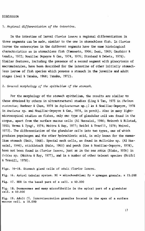
DISCUSSION
1. Regional differentiation of the intestine.
In the intestine of larval Clarias lazera a regional differentiation in
three segments can be made, similar to the one in stomachless fish. In Clarias
larvae the enterocytes in the different segments have the same histological
characteristics as in stomachless fish (Yamamoto, 1966; Iwai, 1969; Gauthier &
Landis, 1972; Noaillac Depeyre & Gas, 1974, 1976; Stroband & Debets, 1978).
Similar features, including the presence of a second segment with pinocytosis of
macromolecules, have been described for the intestine of other initially stomach
less larvae of fish species which possess a stomach in the juvenile and adult
stages (Iwai & Tanaka, 1968; Tanaka, 1972).
2. General morphology of the epitheliwn of the stomach.
For the morphology of the stomach epithelium, the results are similar to
those obtained by others in ultrastructural studies (Ling & Tan, 197S in Chelmon
rostratus; Huebner & Chee, 1978 in Hoplosternum sp.;j äs & Noaillac-Depeyre, 1978
in Ameiurus sp. and Noaillac-Depeyre & Gas, 1978, in perch). Also in many light-
microscopical studies on fishes, only one type of glandular cell was found in the
corpus, apart from the surface mucous cells (Al Hussaini, 1946; Weinreb & Bilstad,
1955; Verma & Tyagi, 1974; Moitra & Ray, 1977; Reifel & Travill, 1978; Weisel,
1973). The differentiation of the glandular cells into two types, one of which
produces pepsinogen and the other hydrochloric acid, is only known for the mamma
lian stomach (Smit, 1968). Special neck cells, as found in Mulloides sp. (Al Hus
saini, 1946), stickleback (Hale, 1965) and perch (Gas & Noaillac-Depeyre, 1978),
have not been found in Clarias lazera, just as in the sea robin (Blake, 1936) in
Colisa sp. (Moitra & Ray, 1977), and in a number of other teleost species (Reifel
& Travill, 1978).
Figs. 16-18. Stomach gland cells of adult Clarias lazera.
Fig. 16. Apical tubular system. Mi = mitochondrium; Zy = zymogen granule, x 23.000
Fig. 17. RER in the basal part of a cell, x 40.000
Fig. 18. Desmosomes and many microfibrills in the apical part of a glandular cell, x 40.000
Fig. 19. Adult Cl. lazera',secretion granules located in the apex of a surface mucous cell, x 18.000
17

Fig. 20. Light micrograph of part of the gut after horse radish peroxidase ingestion. Black deposits of reaction product indicate the presence of enzyme activity. 1 = segment 1; 3 = segment 3; A = anus.
Fig. 21. Functional cell of the epithelial lining of the stomach, x 10.000 The inset shows a light microscopical radioautogram. The arrow points to the H-thymidine labeled nucleus of the cell in the electron micrograph, x 400
18

The cells of the epithelial lining have the same appearance in all studied
sh species. Usually these cells show PAS positivity in their apices after
cian Blue-PAS staining, and this suggests that they contain neutral mucopoly-
ccharides. In pike, however, Reifel & Travill (1978) found acid carbohydrates
these cells.
Development of stomach functions.
It is difficult to determine when the stomach becomes actually "functional",
e volume increases rapidly as from day 8, and one of the important stomach
nctions, the storage of food may begin as of that day. The digestive function
the stomach clearly develops later, between the formation of secretion granules
the corpus gland cells and the production of as much hydrochloric acid as re
ared for reaching low pH values in the stomach lumen.
From the morphological evidence it can be concluded that in Clarias lazera
e corpus of the stomach is formed between day 4 when the first glandular cells
e found, and day 12 when exocytosis of secretion granules is first observed.
An acid environment in the stomach as from day 13 allows catheptic or peptic
gestion. From day 12 the apical vesicular system shows a tendency to develop
bules, which are distinct in adults. These vesicles and tubules possess mem-
anes with a coating at the inner side. This coating, also present on the mem-
anes of the microvilli, consist of glycoproteins in Ameiurus nebulosus (Gas &
aillac-Depeyre, 1978) and in Perca fluviatilis (Noaillac-Depeyre & Gas, 1978).
similar tubulo-vesicular system is also present in stomach gland cells of
lier non-mammalian vertebrates and apparently plays a certain role in hydrochlo-
c-acid production (Sedar, 1961). In mammals the volume of the tubulo-vesicular
stem, as only found in parietal cells, is in inverse ratio to the volume of
3 socalled intracellular canaliculi, which are maximally developed when much
irochloric acid is produced (Padykula, 1977). Intracellular canaliculi, however,
re neither been found in Clarias lazera, nor in A. nebulosus, P. fluviatilis,
i Chelmon rostratus (Ling & Tan, 1975).
After day 13 the acidity increases. The pyloric sphincter is present as
3m day 12. These results are in general accordance with the radioautographic
suits; the labeling index decreased sharply in the stomach corpus glands between
f 9 and day 12 and in the pyloric area between day 12 and 15. Differentiation
the epithelium must take place first in the intestine, then in the rostral
rt of the stomach, and finally in the pylorus.
19

Grizzle & Curd (1978) found a larval period of approximately 35 days in
logperch (Percina eaprodes), reared at 22 C. The first gastric glands were foi
on day 27, whereas the pyloric sphincter developed some days later. This is o:
interest, as Tanaka (1971) concluded for a large number of teleost species th;
gastric glands develop after about 3/4 of the total larval stage. The stomach
develops very early in Clarias lazera and the larval stage is evidently very
short when the fish are reared at 23-24 C. The latter is beginning when, at +
30 h after fertilization, exogenous feeding starts. The completion of the stoi
is usually considered to coincide with the end of the larval stage (Tanaka, 1!
4. Physiological relevance of the second gut segment.
The morphology of the intestine does not change after the time when the :
mach becomes functional. As the second segment with the facility for absorbin;
protein macromolecules remains to be present beyond this period, this facilit;
cannot be related to the lack of a stomach in Clarias lazera. This is underlii
by the fact that the length of the segment remains the same and also by the p
sence of pinocytotic enterocytes in the posterior intestine of adult Clarias.
last argument is in according with the results of Krementz & Chapman (1974),
Bergot (1976) and Noaillac-Depeyre & Gas (1979), who found pinocytosis in the
posterior part of the gut of adult catfish {Ictalurus punctatus), in 15 cm la
trout, and in adult perch, respectively. However, supranuclear vacuoles have :
been found in adult Ictalurus . This might be attributed to the long time inte
between feeding and fixing of the tissues; starvation causes the disappearanc
supranuclear vacuoles but not of pinocytosis, as was found in Coregonus larva
(Stroband & Dabrowski, in press). In the adult perch the second segment seeme<
relatively short (+ 111 of gut length, but _+ 20% in Clarias lazera larvae, ju
niles and adults). Tanaka (1972) studied 15 fish species. In 5 of these he no
ced at the transitional stage from larva to juvenile, when the gastric glands
become functional, the gradual disappearance of acidophylic granules (i.e. su
nuclear vacuoles) in the posterior intestine. This was not observed in the ot'
10 species. In his experiments some fixations were possibly carried out long
after feeding, and ultrastructural studies might show pinocytosis in the seca
segment of the gut.
The facility of absorbing macromolecules is probably a general feature o
the teleostean intestine, but it might be more common in larvae and stomachle
species than in specimens with a functional stomach. The physiological role o
20

"second segment" in fish larvae might be different from that in adults. In
irvae the food reaches the caudal intestine in a very short time, and absorption
: macromolecules might be of quantitative importance (Iwai, 1968). In adult spe-
.mens, including stomachless fish, many hours go by before the food reaches the
»sterior gut, and digestion of protein then may be in an advanced stage. That
(sorption of undigested food in the second gut segment plays a role in fish lar
ie is also indicated by the absence of supranuclear vacuoles in starved Coregonus
irvae (O.C.). The observation of Iwai (1969) that after feeding the number of
'sosomes in carp larvae increases together with the supranuclear vacuoles points
i intracellular digestion of absorbed material.
In adults, even in stomachless fish, absorption of protein takes place main-
' in the anterior part of the intestine (Shcherbina & Sorvatchev, 1969; Stroband
jVan der Veen, in press; studying carp and grasscarp respectively). Therefore it
ems likely that adult stomachless fish are able to digest proteins efficiently
though a pepsin-hydrochloricacid system is absent (Creach, 1963; Jany, 1976).
fferences have not yet been found in the part of food protein absorbed by fishes
th or without a stomach. The question is why other species developed a stomach
d a pepsin-HCl system. Other stomach functions, e.g. storage of food, parti-
oning of food items, or defence against microorganisms may have to be considered
this respect.
Renewal of the epithelium.
3 The random distribution of H-thymidine labeled cells in the digestive tract
ithelium in the early stages of development is in accordance with the results
Rombout et al. (in press) obtained in Barbus aonohonius larvae, in which the
Deling index sharply declined before the first exogenous feeding. In Clarias
zera the decrease was noticed two days later. In both species, the location of
jeled cells during this decrease changes towards the basal parts of the intes-
lal mucosal folds. The presence of thymidine label in nuclei of functional
Lis (glandular and surface stomach cells as well as absorptive cells in
xrias lazera larvae) was observed also by Rombout et al. and by Stroband &
jets (1978) in the intestinal absorptive enterocytes of Barbus larvae and
senile grasscarp, respectively. The ability to proliferate was also found in
ictional small-intestinal cells in Xenopus laevis larvae by Marshall & Dixon
)78).
21

It is interesting that notwithstanding the decreasing labeling index, wh<
the tissues have differentiated and become functionally active, only function;
proliferating cells appear to be present. This in contrast to the mammalian si
intestine, where only undifferentiated crypt cells are able to proliferate
(Lipkip, 1973). The results indicate that fish enterocytes and stomach cells
loose their proliferative activity in a relatively late stage of differentiat:
The authors wish to thank Prof.Dr. Lucy P.M. Timmermans and Prof.Dr. J.W.M. 0:
for their constructive comments, Miss J.J. Thiele for technical assistance in
electron microscopy, and Messrs. W. Valen en H.G. Eleri (T.F.D.L.) for prepar:
the illustrations. Special thanks are due Dr. L. Boomgaart for correcting
linguistic errors.
22

REFERENCES
Al-Hussaini, A.H. The anatomy and histology of the alimentary tract of the bottomfeeder Mulloides auriflamma (Forsk.). J. Morph. 78.» 121-153 (1946).
Bergot, P. Demonstration with ruthenium red of deep invaginations in the apical plasma membrane of enterocytes in rainbow trout intestine. Ann. Biol. Anim. Biochem. Biophys. J_6> 37-43 (1976).
Blake, I.H. Studies on the comparative histology of the digestive tube of certain teleost fishes. J. Morph. 60, 77-102 (1936).
Creach, P.V. Les enzymes protéolytiques des poissons. Ann. Nutr. Alim. 17, 375-471 (1963).
Cornell, R. & Padykula, H.A. A cytochemical study of intestinal absorption in the postnatal Rat. Anat. Rec. L5_i> 139 (1965).
Gas, N. & Noaillac-Depeyre, J. Types cellulaires de l'estomac, assurant la production du suc gastrique chez Ameiurus nebulosus L. Biologie Cellulaire 31, 181-190 (1978).
Gauthier, G.F. & Landis, S.C. The relationship of ultrastructural and cytochemical features to absorptive activity in the Goldfish intestine. Anat. Rec. 172, 675-701 (1972).
Grizzle, J.M. & Curd, M.R. Posthatching histological development of the digestive system and swimbladder of logperch, Peraina oaprodes . Copeia 3^ 448-455 (1978).
Hale, P.A. The morphology and histology of the digestive systems of two freshwater teleosts, Vo&ciVia reticulata and Gasterosteus aauleatus. J. Zool. J_46, 132-149 (1965).
Huebner, E. & Chee, G. Histological and ultrastructural specialization of the digestive tract of the intestinal air breather Hoplosternum thoraoatum (Teleost). J. Morph. 157, 301-328 (1978).
Iwai, T. Fine structure and absorption patterns of intestinal epithelial cells in rainbow trout alevins. Z. Zellforsch. 9J_, 366-379 (1968).
Iwai, T. Fine structure of gut epithelial cells of larval and juvenile carp durinj absorption of fat and protein. Arch. Histol. jap. 30, 183-189 (1969).
Iwai, T. & Tanaka, M. The comparative study of the digestive tract of teleost larvae III. Epithelial cells in the posterior gut of halfbeak larvae. Jap. Soc. Sei. Fish. 34, 44-48 (1968).
Jany, K.D. Studies on the digestive enzymes of the stomachless bonefish Cavassius auvatus Gibelio (Bloch): Endopeptidases. Comp. Biochem. Physiol. 53B, 31-38 (1976).
Krause, W.J. & Cutts, J.H. The postnatal development of the alimentary canal in the opossum: III small intestine and colon. J. Anat. 123, 21-46 (1977).
Kraehenbuhl, J.P. & Campiche, M.A. Early stages of intestinal absorption of specific antibodies in the newborn. J. Cell Biol. 42_, 345-365 (1969).
Krementz, A.B. & Chapman, G.B. Ultrastructure of the posterior half of the intestine of the channal catfish, Iatalurus punatatus. J. Morph. 145, 441-482 (1974).
Lin, E.A. & Tan, C.K. Fine structure of the gastric epithelium of the Coral fish, Chelmon rostratus Cuvier. Okajmas Fol. Anat. Jap. 5J_, 285-310 (1975).
Lipkin, M. Proliferation and differentiation of gastro intestinal cells. Physiol. Rev. 53, 891-915 (1973).
Marshall, J.A. & Dixon, K.E. Cell proliferation in the intestinal epithelium of Xenopus laevis tadpoles. J. Exp. Zool. 203, 31-40 (1978).
Mepham, T.B. & Smith, 11.W. Aminoacid transport in the golsfish intestine. J. Physiol. J_84, 673-684 (1966).
23

Moitra, S.K. & Ray, A.K. Morphohistology of the alimentary canal of an indian freshwater perch Cotisa faoiata (Bloch) in relation to food and feeding habits. Anat. Anz. 141, 37-58 (1977).
Noaillac-Depeyre, J. & Gas, N. Absorption of protein macromolecules by the en-terocytes of the carp {Cyprinus oarpio L). Z. Zeilforsch, 146, 525-541 (1973a).
Noaillac-Depeyre, J. & Gas, N. Mise en évidence d'une zone adaptée au transport des ions dans l'intestine de carpe commune {Cyprinus oarpio L) . CR. Acad. Sei. (Paris) 276, 773-776 (1973b).
Noaillac-Depeyre, J. & Gas, N. Fat absorption by the enterocytes of the carp {Cyprinus oarpio L) . Cell. Tiss. Res. J_55, 353-365 (1974).
Noaillac-Depeyre, J. & Gas, N. Electron microscopic study on gut epithelium of the tench {Tinoa tinea L) with respect to its absorptive functions. Tissue & Cell, 8, 511-530 (1976).
Noaillac-Depeyre, J. & Gas, N. Ultrastructural and cytochemical study of the gastric epithelium in a freshwater teleostean fish {Peroa fluviatilis). Tissue & Cell K), 23-37 (1978).
Noaillac-Depeyre, J. & Gas, N. Structure and function of the intestinal epitheli. cells in the perch {Peroa fluviatilis L). Anat. Rec. 195, 621-640 (1979).
Padykula, H.A. The digestive tract. In: "Histology" (L. Weiss & R.O. Greep, eds.] New York: McGraw-Hill (1977).
Pearse, A.G.E. "Histochemistry, theoretical and applied". London: J. & A. Churchill Ltd. (1972).
Rombout, J.H.W.M., Stroband, H.W.J. & Verstijnen, C.P.H.J. Proliferation and differentiation of intestinal epithelial cells during development of Barbus oonohonius {Teleostei, Cyprinidae). Submitted to J. Exp. Zool. (June, 1980)
Romeis, B. "Mikroskopische technik" München: Oldenbourg Verlag (1968). Reifel, C.W. & Travill, A.A. Structure and carbohydrate histochemistry of the
stomach in eight species of teleosts. J. Morph. 158, 155-168 (1978). Sedar, A.W. Electron microscopy of the oxyntic cell in the gastric glands of the
bullfrog, Rana eatesbiana. j. Biophys. Biochem. Cytol. JJ), 47-57 (1961). Shcherbina, M. & Sorvatchev, K. A few data concerning the absorption of amino
acids in the digestive tract of two years old common carp. Vopr. Prud. Rubo1
j_6, 315-322 (1969). Smit, H. Gastric secretion in lower vertebrates and birds. In: "Handbook of
Physiology" sect. 6. "Alimentary Canal" (CF. Code, ed) Vol. V 2791-2805. Washington, D.C.: Am. Physiol. Soc. (1968).
Stroband, H.W.J. Growth and diet dependant structural adaptations of the digestive tract in juvenile grasscarp {Ctenopharyngodon idella Val.). J. Fish. Biol. _1_U 167-174 (1977).
Stroband, H.W.J. & Debets, F.M.H. The ultrastructure and renewal of the intestin; epithelium of the juvenile grasscarp, Ctenopharyngodon idella (Val.) Cell. Tiss. Res. 187, 181-200 (1978).
Stroband, H.J.W., v.d. Meer, H. & Timmermans, L.P.M. Regional functional differei tiation in the gut of the grasscarp, Ctenopharyngodon idella (val.). Histochemistry 64_, 235-249 (1979).
Stroband, H.W.J. & Dabrowski, K.R. Morphological and physiological aspects of th< digestive system and feeding in freshwater fish larvae. In: "La Nutrition des Poissons" CNERNA (Paris) in press.
Stroband, H.W.J. & v.d. Veen, F.H. The localization of protein absorption during the transport of food along the intestine of the grasscarp, Ctenopharyngodon idella (Val.), submitted to J. Fish. Biol. July 1980.
Tanaka, M. Studies on the structure and function of the digestive system in teleost larvae III. Development of the digestive system during postlarval stage. Jap. J. of Ichthyol. J_8, 164-174 (1971).
24

'anaka, M. Studies on the structure and function of the digestive system in teleost larvae V. Epithelial changes in the posterior gut and protein ingestion. Jap. J. Ichthyol. \9_, 172-180 (1972).
'erma, S.R. & Tyagi, M.P. A comparative histological study of the stomach of a few teleost fishes. Morph. Jahrb. J_20, 244-253 (1974).
(einreb, E.L. & Bilstad, N.M. Histology of the digestive tract and adjacent structures of the rainbow trout, Salmo gairdneri irideus. Copeia 3̂ 194-204 (1955).
'eisel,G.F. Anatomy and histology of the digestive system of the paddlefish (Polyodon spathula). J. Morph. 140, 243-256 (1973).
amamoto, T. An electron microscope study of the columnar epithelial cell in the intestine of freshwater teleosts: Goldfish (Carassius auratus) and rainbow trout {Salmo irideus). Z. Zeilforsch. 72, 66-87 (1966).
25

CURRICULUM VITAE
Het levenslicht werd door mij aanschouwd in Zeist op 31 december 1946. Hoewel naar ik meen nooit door mijn ouders opgejaagd, voelde ik, als ieder kind, al snel dat ik geboren was in een samenleving waarin "het presteren" van grote betekenis wordt geacht. Groot was dan ook mijn frustratie toen ik na een vliegende start (ik werd pas eind februari 1947 verwacht) al op de openbare kleuterschool "Griffensteijn" in Zeist mijn eerste doubleren te verwerken kreeg. Achteraf heeft 3 jaar fröbelen stellig in positieve zin aan mijn vorming bijdragen.
Later kon ik aan de maatschappelijke verwachtingen voldoen door het slagen voor het toelatingsexamen, na daarop uitstekend voorbereid te zijn op de jongensschool der Evangelische Broedergemeente.
Het voorbereidend wetenschappelijk onderwijs werd aangevangen in 1959 aan het Ie Christelijk Lyceum te Zeist. Een poging tot het volgen van het Gymnasium was tot mislukken gedoemd, maar in 1965 werd het einddiploma H.B.S.-B verworven.
Na een korte periode van arbeid bij Philips telecommunicatie industrie te Hilversum volgde een oproep voor militaire dienst. Deze werd vervuld van januari 1966 tot juli 1967, grotendeels bij een infanterie bataljon te Ermelo. Hoewel strepen voor mij niet waren weggelegd, behaalde ik het diploma "militaire lichamelijke vaardigheid" in 1967, iets waarop ik vanzelfsprekend nóg trots ben.
In september 1967 ben ik, na enig "voorwerk" in diensttijd (aula-pockets e.d.) gestart met de studie Biologie aan de Rijksuniversiteit te Utrecht. Het kandidaatsexamen werd, tot niet alleen mijn genoegen, behaald in mei 1970 en in december 1972 studeerde ik "met lof" af met als hoofdvak Vergelijkende Endocrinologie (prof. v. Oordt) en als bijvakken Planten-fysiologie (Prof. v. Die) en Didaktiek der Biologie (Drs. Saaltink).
Vanaf januari 1973 ben ik werkzaam bij de afdeling Dierkunde, later vakgroep Experimentele Diermorfologie en Celbiologie aan de Landbouwhogeschool te Wageningen. Naast vele andere aktiviteiten zoals onderwijs aan pre- en postkandidaten van verschillende studierichtingen en het behalen van het diploma "C-deskundige" aan het ITAL te Wageningen in 1978, bleef er tijd beschikbaar voor het doen van het onderzoek waarvan de resultaten in dit proefschrift zijn neergelegd.