
Isolation of Shewanella sp. from Algeria and characterization ...
290
FRERES MENTOURI CONSTANTINE 1 UNIVERSITY Faculty of Natural Sciences and Life Molecular and Cellular Biology laboratory And AIX-MARSEILLE UNIVERSITY Faculty of Life and Health Sciences National Center for Scientific Research Bioenergetics and Protein Engineering laboratory A Thesis Submitted in fulfillment of the requirements for the degree of Doctor in Microbiology Presented and publicly defended by Hiba BAAZIZ 10 July 2018 Isolation of Shewanella sp. from Algeria and characterization of a system involved in detoxification of chromate Jury Ilham MIHOUBI Rabah ARHAB Véronique BROUSSOLLE Patricia BONIN Radia ALATOU Michel FONS Professor, UFMC1, Constantine Professor, Larbi Ben M’Hidi University Research Director, INRA, Avignon University Research Director, MIO, Marseille Assistant Professor, UFMC1, Constantine Professor, Aix Marseille University President Reviewer Reviewer Examiner Thesis Advisor Thesis Co-advisor
-
Upload
khangminh22 -
Category
Documents
-
view
0 -
download
0
Transcript of Isolation of Shewanella sp. from Algeria and characterization ...

FRERES MENTOURI CONSTANTINE 1 UNIVERSITY
Faculty of Natural Sciences and Life
Molecular and Cellular Biology laboratory And
AIX-MARSEILLE UNIVERSITY
Faculty of Life and Health Sciences
National Center for Scientific Research
Bioenergetics and Protein Engineering laboratory
A Thesis
Submitted in fulfillment of the requirements for the degree of
Doctor in Microbiology
Presented and publicly defended by
Hiba BAAZIZ
10 July 2018
Isolation of Shewanella sp. from Algeria and characterization
of a system involved in detoxification of chromate
Jury
Ilham MIHOUBI
Rabah ARHAB
Véronique BROUSSOLLE
Patricia BONIN
Radia ALATOU
Michel FONS
Professor, UFMC1, Constantine
Professor, Larbi Ben M’Hidi University
Research Director, INRA, Avignon University
Research Director, MIO, Marseille
Assistant Professor, UFMC1, Constantine
Professor, Aix Marseille University
President
Reviewer
Reviewer
Examiner
Thesis Advisor
Thesis Co-advisor


Preface and acknowledgement
The work presented in this thesis was conducted at the the laboratory of Molecular
and cellular biology of University of Freres Mentouri Constantine 1 and the laboratory of
Bioenergetics and Protein Engineering (BIP3), National Center for Scientific Research Aix
Marseille University. Fréres Mentouri Constantine 1 University and PROFAS B+ doctoral
scholarship are gratefully acknowledged for their partial financial support of the present
thesis.
I submit my heartiest gratitude to the members of my dissertation committee: ARHAB
Rabah, BOURSOLLE Véronique, MIHOUBI Ilham and BONIN Patricia for granting me honor
of reading and evaluating my work.

A Doctoral Journey
« Ladies and gentlemen, this is your captain speaking, welcome to Marseille-Provence Airport. Local time is
11:55 for your safety and comfort, please remain seated with your seat belt fastened, we will be landing
momentarily»
I could see Marseille, a foreign city, ghosts of Constantine cross my mind and I already feel homesick.
I left the airport to find Michel, my advisor, I expected a chubby old man with a strict face only to find a
tall good looking one wearing a welcoming smirk on his face, after a few minutes I found myself greeting foreign
lab mates.
Three years later…
I just finished my thesis and I have acquired knowledge and gained a new family. I feel sincerely grateful to
have an advisor like Michel, who continuously supported my Ph.D. study, who had the patience, and always
radiated me with motivation and immense knowledge, whose special sense of humor made me laugh all the time.
Radia, whom I am deeply indebted to; without her support I would not join the BIP3 lab and acquire this
amazing experience.
Vincent, the head of BIP3, who provided me the opportunity to join their team and steered me in the right
direction through my PhD, his warm hospitality and constant humoring vibe made me always thankful to have
him by my side; Cécile, who was a mother figure in the lab, the door to her office was always open whenever I
ran into a trouble spot or had questions about my research or writing, which I am forever grateful for; Chantal,
Olive, and Sandra, whom I will treasure their stimulating discussions and invaluable help; Amine and Ahmed,
who were a scent from home and continuously helped me during my thesis.
My fellow lab mates more like my family! Whose presence, scientific help and the unforgettable funny
moments I am forever grateful for. The brilliant Zitoun, who was my personal Google Scholar I am grateful for
his advices and for dealing kindly with my endless bothering questions; Baptist, who I cannot imagine going to
USA without; Natou, Sophie and Flora whom am forever grateful to their kindness and immense support; Cyril
and Anne, my siblings, the little shrimp who was a pain in the neck but I am deeply thankful to him for being
the first to welcome me in the team; Annouchka, who had a great impact on both myself and my research and
who took it upon herself to hear my constant nagging.
Aside from my lab, I have never felt this lucky to have people back home who stood by me and cared for
my wellbeing like the Algerian lab team in Constantine, who supported me throughout my first year and
showered me with love when I was leaving to France. My parents, whom I revere the patronage and moral
support extended with love, whose financial support and passionate encouragement made it possible for me to
complete this PhD thesis. As my joy knows no bounds in expressing how grateful and lucky I am to have crazy
sisters that supported me in every step in my journey.
This may be the end to this journey, but certainly, the beginning of another unexpected one is waiting
around the corner…

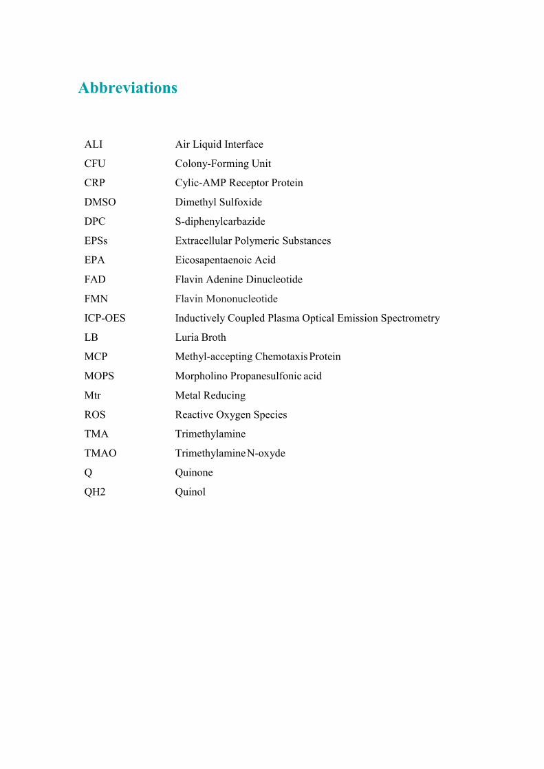
Abbreviations
ALI Air Liquid Interface
CFU Colony-Forming Unit
CRP Cylic-AMP Receptor Protein
DMSO Dimethyl Sulfoxide
DPC S-diphenylcarbazide
EPSs Extracellular Polymeric Substances
EPA Eicosapentaenoic Acid
FAD Flavin Adenine Dinucleotide
FMN Flavin Mononucleotide
ICP-OES Inductively Coupled Plasma Optical Emission Spectrometry
LB Luria Broth
MCP Methyl-accepting Chemotaxis Protein
MOPS Morpholino Propanesulfonic acid
Mtr Metal Reducing
ROS Reactive Oxygen Species
TMA Trimethylamine
TMAO Trimethylamine N-oxyde
Q Quinone
QH2 Quinol

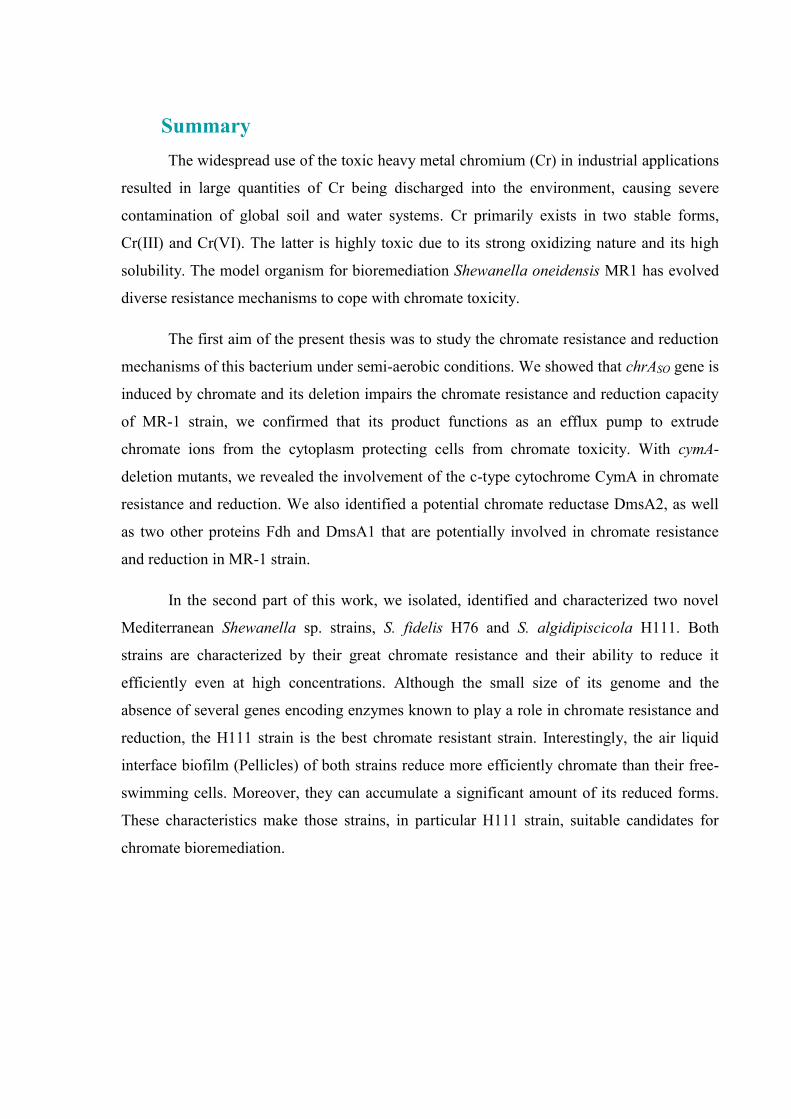
Summary
The widespread use of the toxic heavy metal chromium (Cr) in industrial applications
resulted in large quantities of Cr being discharged into the environment, causing severe
contamination of global soil and water systems. Cr primarily exists in two stable forms,
Cr(III) and Cr(VI). The latter is highly toxic due to its strong oxidizing nature and its high
solubility. The model organism for bioremediation Shewanella oneidensis MR1 has evolved
diverse resistance mechanisms to cope with chromate toxicity.
The first aim of the present thesis was to study the chromate resistance and reduction
mechanisms of this bacterium under semi-aerobic conditions. We showed that chrASO gene is
induced by chromate and its deletion impairs the chromate resistance and reduction capacity
of MR-1 strain, we confirmed that its product functions as an efflux pump to extrude
chromate ions from the cytoplasm protecting cells from chromate toxicity. With cymA-
deletion mutants, we revealed the involvement of the c-type cytochrome CymA in chromate
resistance and reduction. We also identified a potential chromate reductase DmsA2, as well
as two other proteins Fdh and DmsA1 that are potentially involved in chromate resistance
and reduction in MR-1 strain.
In the second part of this work, we isolated, identified and characterized two novel
Mediterranean Shewanella sp. strains, S. fidelis H76 and S. algidipiscicola H111. Both
strains are characterized by their great chromate resistance and their ability to reduce it
efficiently even at high concentrations. Although the small size of its genome and the
absence of several genes encoding enzymes known to play a role in chromate resistance and
reduction, the H111 strain is the best chromate resistant strain. Interestingly, the air liquid
interface biofilm (Pellicles) of both strains reduce more efficiently chromate than their free-
swimming cells. Moreover, they can accumulate a significant amount of its reduced forms.
These characteristics make those strains, in particular H111 strain, suitable candidates for
chromate bioremediation.


Résumé
L'utilisation répandue du chrome (Cr) dans les applications industrielles a entraîné le
rejet de grandes quantités de ce métal lourd toxique Cr dans l'environnement, causant une
sévère contamination des sols et des systèmes hydrologiques globaux. Le Cr est
principalement rencontré sous deux formes stables, Cr(III) et Cr(VI). Ce dernier est
hautement toxique en raison de sa forte nature oxydante et de sa grande solubilité.
L'organisme modèle pour la bioremédiation, Shewanella oneidensis MR1, a développé divers
mécanismes de résistance pour faire face à la toxicité du chromate.
Le premier objectif de cette thèse était d'étudier les mécanismes de résistance au
chromate et de sa réduction chez cette bactérie dans des conditions semi-aérobies. Nous
avons a montré que le gène chrASO est induit par le chromate et que sa délétion altère la
capacité de la souche MR-1 de résister au chromate et de le réduire. Nous avons confirmé
que ChrASO fonctionne comme une pompe d'efflux pour extruder les ions chromate du
cytoplasme. En délétant le gène cymA, nous avons révélé son implication dans la résistance
au chromate ainsi que dans sa réduction. Nous avons également identifié une potentielle
chromate réductase DmsA2, ainsi que deux autres protéines Fdh et DmsA1 potentiellement
impliquées dans la résistance au chromate et dans sa réduction chez la souche MR-1.
Dans la deuxième partie de ce travail, nous vous on a isolé, identifié et caractérisé
deux nouvelles souches de Shewanella, S. fidelis H76 et S. algidipiscicola H111. Les deux
souches se caractérisent par leur grande résistance au chromate et leur capacité à le réduire
efficacement même à des concentrations élevées. En dépit de la petite taille de son génome et
l'absence de plusieurs gènes codant des enzymes connues pour jouer un rôle dans la
résistance au chromate ainsi que dans sa réduction, la souche H111 est la plus efficace. De
façon intéressante, les biofilms de l'interface liquide-air (Pellicules) des deux souches
réduisent plus efficacement le chromate que leurs cellules planctoniques. De plus, les
pellicules des souches H76 and H111 peuvent accumuler une quantité importante de formes
réduites du chromate. Ces caractéristiques montrent que ces souches, en particulier la souche
H111, sont des candidats appropriés pour la bioremédiation du chromate.


الملخصعي ميو اس ل لس قيل ل ع سع ل ي (Cr)لو ي ك يغ ك ل تف عي لص يق ل في
حيط، م ي ل ه في ي لم و ش ي نألت ي بس ل ئيلي ج .ل و تين Crي س ع صو ين س ب ه ث
Cr(III) Cr (VI) لي أخي ع يع حيث أ ب ي بس لس ي أكس ج ل نلقوي و ب . لق ي ل
Shewanella oneidensis MR1 ي مل مع س ع ف ل م م ، آلي مق يولوجي ل لج ع ئن حي ل و ل ، كأن
. م ل
ل ل س فيأ من ه جزءت م آلي ل ل م لس مق ز يو تحت ع ه هخ فل
ئي-نصف س ، حيثهو ل لجين أ م تحفيز SOchrAأ م ي ل م هسح أ ب يل مق ل تع ي
م ق له ل ز ل خ تج ، MR-1سال ع ل تين ل جي عنك تم تأكي أ فق خ ل ك لجين يع ه
م م ل يون ايل ل ي ل ح م م ي وبا لسي ك ن . تم مش م ل م ب س ل من
cytochrome CymA و ل ل cمن ز مح خ نزيم ف ع ع ل ؛ ك تم م ل ز خ م في مق
م ين ه DmsA2ل ين أخ تي لك ع ب ك في Fdh DmsA1ك ل مش ين يح ز مقل خ م
م ع .MR-1لسال ل
س تين من ج ي ين ج ييز لسال ل ف ع ل لعز ، تم س ل ني من ل لجزء . Shewanellaفي
وسط ه ل أبيض ح ل ل من ين S. fidelis H76 S. algidipiscicola H111معز لسال تين يز ه . ت
ي ل ل م ق لسال ب . لي لع كيز ل لي ح ع له بفع ز ت اخ ق أحسن H111م لسال هي
ه في ف ب شف إنزي مع ل لجي ي من لع غي وم غم من صغ حجم جي ل م ب ل م في مق
. م ل ز خ م ي مق ل يو فيلاه من ل ن م، لي مق م بفع ل ز ين ي لسال ا في ل ل
ين لسال يز ل . تجعل ه م زل ل ل لصو ي مع من كم ك ه أ ي ؛ ك ي لح بح لس اي ل ب
لسال أخص ي H111ب ين مائ شح لج نم ع .ل م يولوجي ل ل

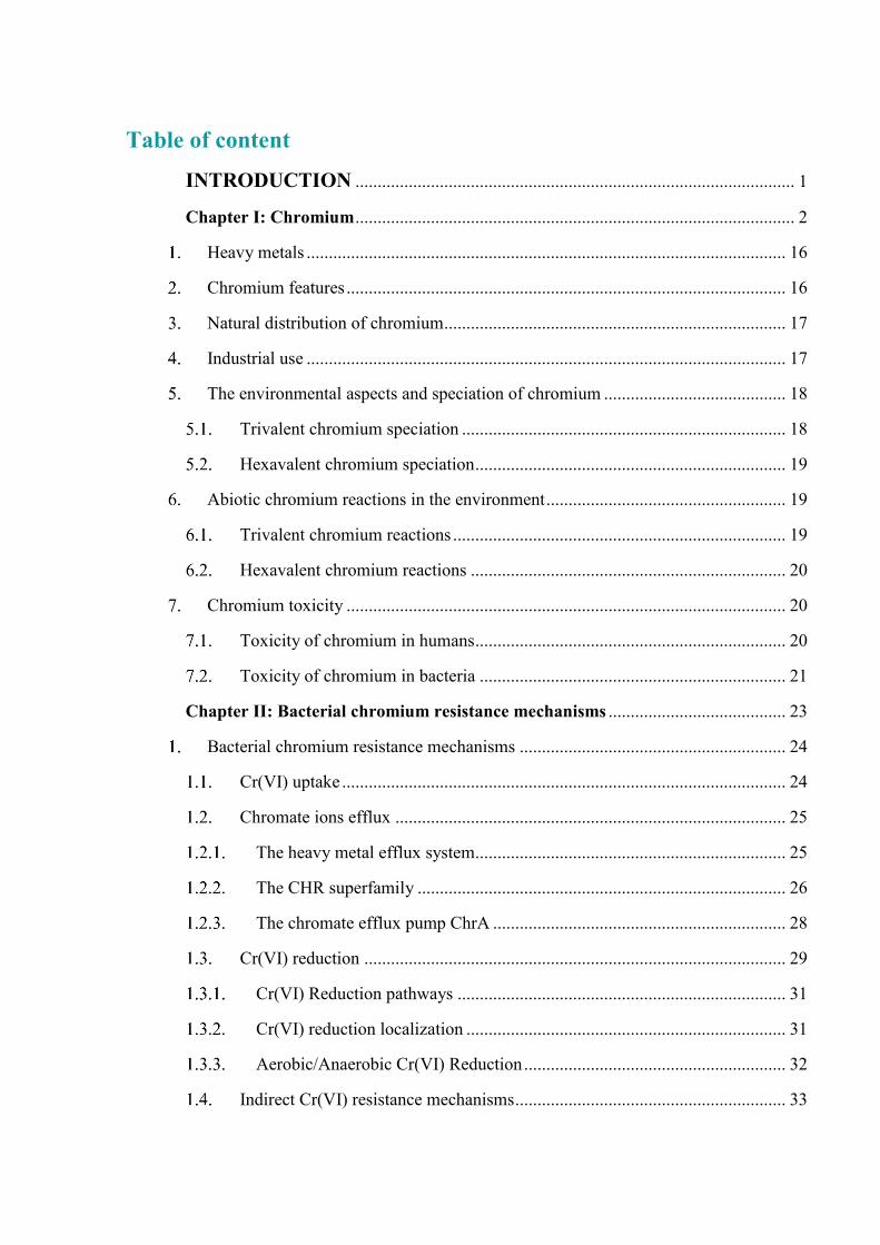
Table of content
INTRODUCTION ................................................................................................... 1
Chapter I: Chromium ................................................................................................... 2
Heavy metals ............................................................................................................ 16
Chromium features ................................................................................................... 16
Natural distribution of chromium ............................................................................. 17
Industrial use ............................................................................................................ 17
The environmental aspects and speciation of chromium ......................................... 18
Trivalent chromium speciation ......................................................................... 18
Hexavalent chromium speciation ...................................................................... 19
Abiotic chromium reactions in the environment ...................................................... 19
Trivalent chromium reactions ........................................................................... 19
Hexavalent chromium reactions ....................................................................... 20
Chromium toxicity ................................................................................................... 20
Toxicity of chromium in humans ...................................................................... 20
Toxicity of chromium in bacteria ..................................................................... 21
Chapter II: Bacterial chromium resistance mechanisms ........................................ 23
Bacterial chromium resistance mechanisms ............................................................ 24
Cr(VI) uptake .................................................................................................... 24
Chromate ions efflux ........................................................................................ 25
The heavy metal efflux system...................................................................... 25
The CHR superfamily ................................................................................... 26
The chromate efflux pump ChrA .................................................................. 28
Cr(VI) reduction ............................................................................................... 29
Cr(VI) Reduction pathways .......................................................................... 31
Cr(VI) reduction localization ........................................................................ 31
Aerobic/Anaerobic Cr(VI) Reduction ........................................................... 32
Indirect Cr(VI) resistance mechanisms ............................................................. 33


Chapter III: Shewanella oneidensis MR-1 ................................................................ 35
Shewanella genus ..................................................................................................... 36
Shewanella genus characteristics ............................................................................. 37
Phenotypic characteristics ................................................................................. 37
Pathogenicity .................................................................................................... 38
Energy metabolism ........................................................................................... 38
Shewanella oneidensis MR-1 ................................................................................... 39
Chemotaxis ....................................................................................................... 40
Biofilms ............................................................................................................ 42
Surface biofilm in S. oneidensis .................................................................... 42
Pellicle development in S. oneidensis ........................................................... 43
S. oneidensis MR-1 respiratory capacities ........................................................ 44
Electron transfer in S. oneidensis MR-1........................................................ 46
Direct electron transfer .................................................................................. 47
Mediated electron transfer ............................................................................. 48
S. oneidensis MR-1 and chromate .................................................................... 49
Chromate reduction in S. oneidensis MR-1 .................................................. 49
S. oneidensis MR-1 response to chromate .................................................... 50
Results ........................................................................................................................ 52
Part I: S. oneidensis MR-1 chromate resistance and reduction .............................. 53
S. oneidensis MR-1 chromate resistance and reduction ........................................... 54
Chromate efflux pump of S. oneidensis MR-1 ................................................. 55
The involvement of chromate efflux pump in chromate survival and reduction of S. oneidensis MR-1 ............................................................................................ 56
Additional results .............................................................................................. 73
The involvement of a formate dehydrogenase in chromate reduction in S.
oneidensis MR-1 ..................................................................................................................... 74
The involvement of DMSO reductases DmsA1 and DmsA2 in chromate reduction of S. oneidensis MR-1 ............................................................................................ 74

The involvement of cytochrome-c CymA in chromate reduction of S.
oneidensis MR-1 ..................................................................................................................... 75
Part II: Isolation and characterization of new Shewanella sp. ............................... 77
Isolation and identification of new Shewanella species ........................................... 78
Growth characteristics .............................................................................................. 79
Chemotaxis behavior of S. fidelis H76 and S. algidipiscicola H111 ....................... 81
Chromate resistance of S. fidelis H76 and S. algidipiscicola H111 ......................... 81
Genome analysis of S. algidipiscicola H111 and S. fidelis H76 strains................... 83
General features of S. fidelis H76 genome ....................................................... 83
chrA- and fdh-like genes ............................................................................... 83
chrR-like genes .............................................................................................. 84
nfsA-like genes .............................................................................................. 84
dmsA1-like and dmsA2-like genes................................................................. 84
General features of S. algidipiscicola H111 genome........................................ 84
chrA- and fdhA-like genes ............................................................................. 85
chrR-like and nfsA-like enzymes................................................................... 85
dmsA1-like and dmsA2-like enzymes ............................................................ 85
Part III: Chromate reduction by pellicles ................................................................. 92
Pellicle formation ability of S. fidelis H76 and S. algidipiscicola H111 ................. 93
Chromate reduction capacity of H76 and H111 pellicles ........................................ 93
Chromium accumulation capacity of pellicles ......................................................... 95
Discussion and Perspectives ............................................................................... 97
Conclusion ............................................................................................................... 109
Material and Methods ........................................................................................ 110
1. Bacterial strains, plasmids and culture media ........................................................ 111
2. Molecular biology techniques ................................................................................ 112
Isolation, purification and hydrolysis of DNA ............................................... 112
DNA amplification by PCR ............................................................................ 112
Plasmid construction ....................................................................................... 113


Construction of deletion mutants .................................................................... 113
3. Biochemical techniques.......................................................................................... 116
Measurement of -galactosidase activities ..................................................... 116
Measurement of chromate reduction .............................................................. 116
In vivo and In vitro Cr(VI)-reduction assay ................................................ 116
Cr(VI) reduction measurement of Shewanella pellicles ............................. 117
Total chromium measurement assay ........................................................... 117
4. Assessment of strains chromate resistance............................................................. 118
Chromate resistance assays in Shewanella strains .......................................... 118
Chromate resistance assay in E. coli ............................................................... 118
5. Divers assays .......................................................................................................... 119
Pellicle assay ................................................................................................... 119
Chemotaxis assay ( plugs microscopy technique) ......................................... 119
6. Isolation of new Shewanella species method ......................................................... 120
Samples collection .......................................................................................... 120
Isolation .......................................................................................................... 120
7. Genome sequencing and bioinformatic analysis .................................................... 120
Annexes .................................................................................................................... 122
References ............................................................................................................... 123


INTRODUCTION

CHAPTER Iμ
CHROMIUM


Introduction: Chapter I
16
Heavy metals
Heavy metals can be defined by various criteria including density, atomic weight, atomic
number and chemical properties. However density is the main aspect to be considered as the
defining feature (Prabhakaran et al, 2016). The "heavy metals" term refers from a purely
physical point of view to an intrinsic property of elements: the density. According to Alloway
& Ayres, 1997 designating natural metallic elements, metals and in some cases metalloid
elements (about 65 elements) are characterized by a high density greater than 5 to 6 and an
atomic number above 20.
From a biological point of view, two types of heavy metals can be distinguished according
to their physiological and/or toxic effects: essential metals and toxic metals. The former are
crucial elements in a trace state for many cellular processes and are found in a very low amount
in biological tissues (Singh et al, 2011). They become toxic when their concentration exceeds a
certain threshold. This is the case of copper (Cu), nickel (Ni), zinc (Zn) and iron (Fe). However,
toxic metals have a polluting feature with toxic effects to living organisms even at low
concentration and they have no known beneficial effects for the cell. This is the case for lead
(Pb), mercury (Hg), cadmium (Cd) and chromium (Cr) (Figure 1) (Summers, 2009).
Unlike most organic contaminants, heavy metals are natural constituents in rocks and
mineral deposits. Thus, these elements are normally present at low concentrations (in a trace
state, less than 0.1%) in soils, sediments, surface waters and living organisms (Alloway &
Ayres, 1997; Callender, 2003). Accumulation of heavy metals above the threshold level is
mainly due to anthropogenic activities including mining, chemical manufacturing, agriculture,
hospital wastewater and electronic waste. Heavy metals can pose cytotoxic, carcinogenic and
mutagenic effects (Tóth et al, 2016).
Chromium features
The name “chromium” is derived from the Greek word “chroma” meaning “color” which
refers to the multiple colored compounds that contain chromium such as Na Cr O (bright
orange), Cr O (green) and the Zn and Pb salts of CrO −(bright yellow) (Lear, 2016). It was

Figure 1. Periodic table of metals and toxic metalloids: Elements Figuring in
blue are toxic at high concentrations and the yellow colored elements are toxic and
have no biological role in most organisms. The transition metals are shown in the
red frame and the elements framed by the green are the metalloids. Figure adapted
from (Summers, 2009).
Figure 2. Chromium traces in gemstones: A. Rubies found in marble deposits
(https://www.gia.edu/ruby-description) B. Emerald crystals in mica schist
(http://geology.com/gemstones/emerald/).

Introduction: Chapter I
17
named by the French pharmacist and chemist Louis Nicolas Vauquelin who published in 1809
his discovery of the yellow pigment of chromium (Aharchaou, 2017).
The chromium (Cr) element belongs to the group of transition metals and it is the 24th
element of the Mendeleev periodic table, located between vanadium and manganese. It has an
atomic weight of 51.996, atomic number 24, a density of 7.14, a melting point of 1900°C, a
moderate thermal expansion and a stable cubic crystal structure. In terms of abundance,
chromium is the seventh element on Earth and the 21st in the Earth's crust, with an average
concentration of 125 mg.kg-1 (Cervantes et al, 2001) (Figure 1).
Natural distribution of chromium
Chromium is a naturally occurring element that is ubiquitous in the environment. It is found
in all compartments of the environment, in water, air, soil and also in living organisms. It is
widely present in igneous rocks where it can substitute for Fe3+ and for Al3+ in other minerals
such as tourmalines, micas and garnets (Guertin et al, 2016). Chromium traces are often
responsible for the color of these minerals such as the red of the ruby and the green of the
emerald (Bunn et al, 2013) (Figure 2). Rock alteration and erosion is an important source of
chromium release to the environment (Nriagu, 1989).
Industrial use
Because of its hardness, gloss, high melting point and anti-corrosive properties, chromium
is used in various industrial activities. It is extracted as chromite ore ( FeCr O ). From total
chrome ore production, 90% is used in metallurgical industries for steel, alloy and nonferrous
alloy production, 5% in refractory (cement, glass, ceramics and machinery) and the remaining
5% in chemical industries such as leather tanning, electroplating, wood preservation, pigment
production and it can be used as an oxidizing agent (Dhal et al, 2013). As consequence of its
extensive anthropogenic use, chromium is present in effluents originated from the different
activities and represents a serious pollutant of sediments, soil, water and air (Focardi et al,
2013).


Introduction: Chapter I
18
The environmental aspects and speciation of chromium
In the environment, chromium exists in different oxidation states ranging from -2 to +6.
The stable forms, commonly occur in the pH and redox potential values found in the
environment, are the trivalent Cr(III) which naturally predominates in the environment, and the
hexavalent Cr(VI) which is rarely naturally occurring and is introduced in the environment
mainly by anthropogenic activities (Focardi et al, 2013). On the other hand, Cr(IV) and Cr(V)
are unstable intermediate forms of reactions between oxidizing and reducing agents of Cr(III)
and Cr(VI) (Aharchaou, 2017).
The speciation of Cr(III) and (VI) depends on several parameters such as pH and their
concentration in the environment. In natural media, major part of Cr(III) is included in
hydroxides or in complexes with organic ligands while Cr(VI) occurs mainly in the form CrO −(Hossain et al, 2005).
Trivalent chromium speciation
Cr(III) is the most stable form of chromium, it is considered to be relatively immobile,
sparsely soluble and of limited ecotoxicological interest (Gonzalez et al, 2003). However, at
high concentration it becomes toxic, carcinogenic and teratogenic (Ahmad et al, 2009).
Cr(III) has little affinity for oxygen but has a greater affinity toward organic and inorganic
ligands (Zayed & Terry, 2003). It forms insoluble complexes that precipitate as oxides,
hydroxides or sulfates (Focardi et al, 2013). Under normal environmental conditions, Cr(III) is
found in aqueous solution in the form of Cr +, Cr OH +, Cr OH − and Cr OH 0. The latter
form presents the most frequently encountered solid form. It is known to have a very low
solubility at natural pH, which makes Cr(III) less toxic (Ramírez-Díaz et al, 2008). Indeed, the
internalization mechanisms of Cr(III) remain poorly known. Some studies consider that it is
unable to pass through cell membranes which would explain its relatively poor toxicity
(Francisco R. et al, 2002).

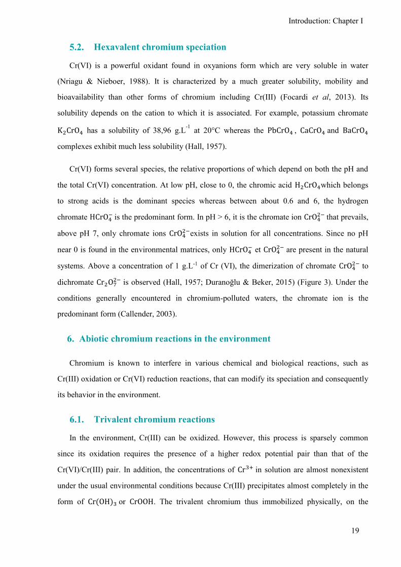
Figure 3. Cr(VI) Speciation diagram depending on pH. The dominant Cr(VI)
species at pH lower than 0 is chromic acid (H2CrO4). Predominant Cr(VI) species at
pH between 1.0 and 6.5 is hydrogen chromate ion (HCrO4−) while CrO42−ions are
dominant above pH 6.5. Dichromate anions (Cr2O72−) exist only at high concentrations
about more than 1 g/L (Duranoğlu & Beker, 2015).
Introduction: Chapter I
19
Hexavalent chromium speciation
Cr(VI) is a powerful oxidant found in oxyanions form which are very soluble in water
(Nriagu & Nieboer, 1988). It is characterized by a much greater solubility, mobility and
bioavailability than other forms of chromium including Cr(III) (Focardi et al, 2013). Its
solubility depends on the cation to which it is associated. For example, potassium chromate K CrO has a solubility of 38,96 g.L-1 at 20°C whereas the PbCrO , CaCrO and BaCrO
complexes exhibit much less solubility (Hall, 1957).
Cr(VI) forms several species, the relative proportions of which depend on both the pH and
the total Cr(VI) concentration. At low pH, close to 0, the chromic acid H CrO which belongs
to strong acids is the dominant species whereas between about 0.6 and 6, the hydrogen
chromate HCrO− is the predominant form. In pH > 6, it is the chromate ion CrO − that prevails,
above pH 7, only chromate ions CrO −exists in solution for all concentrations. Since no pH
near 0 is found in the environmental matrices, only HCrO− et CrO − are present in the natural
systems. Above a concentration of 1 g.L-1 of Cr (VI), the dimerization of chromate CrO − to
dichromate Cr O7− is observed (Hall, 1λ57; Duranoğlu & Beker, β015) (Figure 3). Under the
conditions generally encountered in chromium-polluted waters, the chromate ion is the
predominant form (Callender, 2003).
Abiotic chromium reactions in the environment
Chromium is known to interfere in various chemical and biological reactions, such as
Cr(III) oxidation or Cr(VI) reduction reactions, that can modify its speciation and consequently
its behavior in the environment.
Trivalent chromium reactions
In the environment, Cr(III) can be oxidized. However, this process is sparsely common
since its oxidation requires the presence of a higher redox potential pair than that of the
Cr(VI)/Cr(III) pair. In addition, the concentrations of Cr + in solution are almost nonexistent
under the usual environmental conditions because Cr(III) precipitates almost completely in the
form of Cr OH or CrOOH. The trivalent chromium thus immobilized physically, on the


Introduction: Chapter I
20
matrix of the soil or sedimented in a liquid media, is then protected from oxidation (Ahmad et
al, 2009). It have been shown that only oxides of manganese (MnOOH) and molecular oxygen
are capable of oxidizing Cr(III) to Cr(VI) (Fendorf, 1995).
Hexavalent chromium reactions
Cr(VI) is a powerful oxidant and can be readily reduced in the presence of several reducing
agents. Fe(II) appears to be the most important possible reductant of Cr(VI) in the environment.
Studies on the reduction of Cr(VI) by Fe(II) in solution have shown that, in addition to the
respective concentrations of both species, temperature and pH influence the speed of reaction.
The latter increases by the increasing of the temperature. The fastest reaction kinetics are
observed in pHs between 6 and 8. Sulphides are also potential candidates for Cr(VI) reduction.
They may be combined with Fe(II) in the form of ferrous sulfide, or with other divalent cation
metals such as Mg2+, Pb2+, Cu2+, Cd2+, Ni2+ and Mn2+, or may be alone in the form of H2S
(Pettine et al, 1998; Sedlak & Chan, 1997). Natural organic matter, humic acids or fulvic acids,
in soils or waters is also likely to reduce Cr(VI) (Bartlett & James, 1979; Alloway & Ayres,
1997).
Cr(VI) can also be reduced photochemically. Studies on photo-reduction of Cr(VI) in
natural media have shown that the mechanism is indirect and needs the Fe(II)/Fe(III) pair to
transfer electrons from the organic ligands to Cr(VI). The organic Fe(III)-ligand complexes
absorb light and produce Fe(II). The latter in its turn reduces Cr(VI) to Cr(V) then to Cr(IV)
and finally to Cr(III). At each step, Fe(II) is reoxidized to Fe(III) which can again be
complexed with organic ligands and thus resume the cycle (Gaberell et al, 2003).
Chromium toxicity
Toxicity of chromium in humans
Cr(III) has long been considered to be an essential micronutrient in animal and human
alimentation, since it seems to participate in the metabolism of glucose and lipids (Anderson,
1997). Nevertheless, this role as an essential element has been recently debated, some authors
do not recognize Cr(III) as a nutrient for human health and reported that exposure to high levels


Introduction: Chapter I
21
via inhalation, ingestion or dermal contact may causes some adverse health effects (Di Bona et
al, 2011).
It is well known that Cr(VI) is carcinogenic at high doses and presents a greatest health risk
(Keegan et al, 2008). Cr(VI) is generally considered more toxic than Cr(III) and its high
toxicity comes from its great facility of crossing biological membranes and its properties of
powerful oxidant (Katz S & Salem H, 1993). Cr(VI) enters the body by three routes of
exposure: inhalation, absorption through the skin or ingestion. For occupational exposure, the
airways and skin are the primary routes of uptake (De Flora, 2000). Breathing high levels of
Cr(VI) can cause irritation to the nasal cavity, breathing difficulty (asthma and cough), severe
ulcers and perforations of the nasal septum and it may also cause respiratory tract cancer. Skin
contact with certain Cr(VI) compounds can cause skin allergies and skin ulcers. Its ingestion
can lead to disturbances in the storage organs causing damage to the DNA which leads to
mutations and possible carcinogenicity. Concentrations about 100 mg.kg-1 of body mass are
lethal to humans (Jomova & Valko, 2011).
Toxicity of chromium in bacteria
Cr(VI) is highly toxic for bacteria because of its rapid entry to the cytoplasm where it may
exert its toxic effects. Its toxicity is mainly related to the process of its reduction to lower
oxidation states. During the stepwise Cr(VI) reduction, a whole spectrum of reactive oxygen
species (ROS) are formed, which exert deleterious effects on cells (Cabiscol Català et al, 2000;
Cheung & Gu, 2007; Thatoi et al, 2014).
Once inside the bacterial cell, Cr(VI) can be primarily reduced to highly cytotoxic Cr(V) by
certain reductants such as ascorbic acid, glutathione (GSH), cysteine, hydrogen peroxide
(H O ,) and flavoenzymes such as glutathione reductase (GR). During this process, molecular
oxygen is reduced to O−•,which generates H O , via dismutation. The resultant reactive
intermediate Cr(V) species are quickly reoxidized to Cr(VI) by reacting with H O , which
generates OH • radical via Fenton reaction (reaction 1). The reactive intermediates Cr(IV) and
the final product Cr(III) resulting from Cr(VI) reduction, can also generate harmful ROS
effectively through Fenton reaction during their re-oxidation processes (reactions 2 and 3,
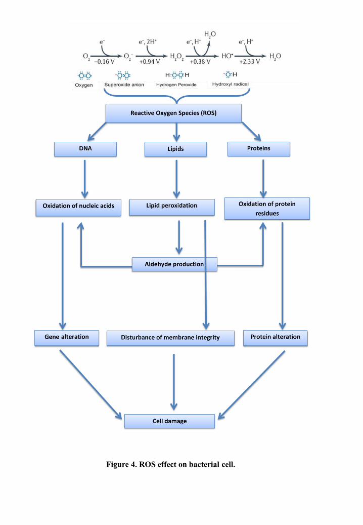
Reactive Oxygen Species (ROS)
Figure 4. ROS effect on bacterial cell.
Introduction: Chapter I
22
respectively). The reaction products of partial Cr(VI) reduction include H O , superoxide anion
(O−•) and hydroxyl radical (OH •), which are primarily responsible for the cyto- and
genotoxicity of Cr(VI) (Poljsak et al, 2010).
Cr V + H O → Cr VI + OH− + OH • (Reaction 1)
Cr IV + H O → Cr V + OH− + OH • (Reaction 2)
Cr III + H O → Cr IV + OH− + OH • (Reaction 3)
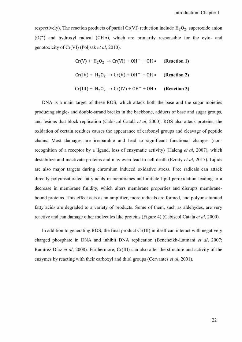
DNA is a main target of these ROS, which attack both the base and the sugar moieties
producing single- and double-strand breaks in the backbone, adducts of base and sugar groups,
and lesions that block replication (Cabiscol Català et al, 2000). ROS also attack proteins; the
oxidation of certain residues causes the appearance of carbonyl groups and cleavage of peptide
chains. Most damages are irreparable and lead to significant functional changes (non-
recognition of a receptor by a ligand, loss of enzymatic activity) (Haleng et al, 2007), which
destabilize and inactivate proteins and may even lead to cell death (Ezraty et al, 2017). Lipids
are also major targets during chromium induced oxidative stress. Free radicals can attack
directly polyunsaturated fatty acids in membranes and initiate lipid peroxidation leading to a
decrease in membrane fluidity, which alters membrane properties and disrupts membrane-
bound proteins. This effect acts as an amplifier, more radicals are formed, and polyunsaturated
fatty acids are degraded to a variety of products. Some of them, such as aldehydes, are very
reactive and can damage other molecules like proteins (Figure 4) (Cabiscol Català et al, 2000).
In addition to generating ROS, the final product Cr(III) in itself can interact with negatively
charged phosphate in DNA and inhibit DNA replication (Bencheikh-Latmani et al, 2007;
Ramírez-Díaz et al, 2008). Furthermore, Cr(III) can also alter the structure and activity of the
enzymes by reacting with their carboxyl and thiol groups (Cervantes et al, 2001).


CHAPTER IIμ
BACTERIAL CHROMIUM
RESISTANCE MECHANISMS
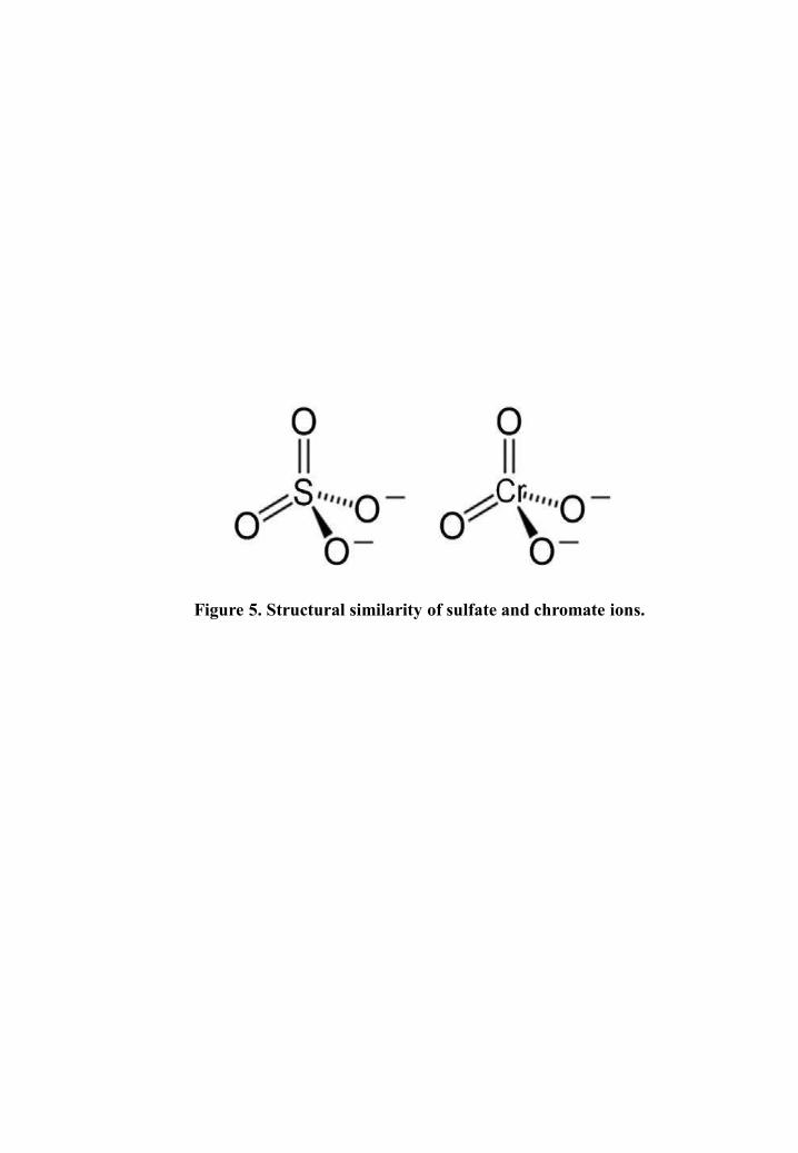
Figure 5. Structural similarity of sulfate and chromate ions.
Introduction: Chapter II
24
Bacterial chromium resistance mechanisms
The majority of microbial species are sensitive to Cr(VI), but some species are resistant
and can tolerate high levels of it. According to Gadd G.M., 1992, the resistance is “the ability
of a microorganism to survive toxic effects of metal exposure by means of a detoxification
mechanism produced in direct response to the metal species concerned”. Bacteria have
selected different resistance mechanisms to overcome Cr(VI)-stress. They primarily respond
by using mechanisms that directly target Cr(VI) (Brown et al, 2006), which comprise:
- The Cr(VI) regulation uptake.
- The efflux of chromate ions from the cell cytoplasm and the reduction of Cr(VI) into
Cr(III).
In addition, bacteria can undertake simultaneously other resistance strategies to prevent
cell components from Cr(VI) toxicity and to repair the damages induced by Cr(VI)-stress
(Brown et al, 2006; Ramírez-Díaz et al, 2008). The main mechanisms include:
- The activation of ROS scavenging enzymes for offsetting Cr(VI)-induced oxidative
stress.
- Iron homeostasis to prevent the production of hydroxyl radicals through the Fenton
reaction.
- The induction of the SOS response enzymes to counter DNA damage.
Cr(VI) uptake
One of common bacterial mechanisms to deal with Cr(VI) is the regulation of sulfate
uptake shuttle system that is involved in initial Cr(VI) cellular accumulation (Brown et al,
2006). As mentioned previously, chromium exists mainly as the tetrahedral chromate ions CrO −, this form is chemically analogous to biologically important inorganic anions such as SO −. The structural similarity of chromate to sulfate (Figure 5) most likely constitutes the
basis for its active transport across cell membranes via the sulfate ABC transporters which

Figure 6. Major transporter families taking part inheavy metal resistance.
P-type ATPase transporter, CBA transporter and the cation diffusion facilitator
(CDF) (Prabhakaran et al, 2016).
Introduction: Chapter II
25
belong to the sulfate/tungstate uptake transporter (SulT) family of the ABC superfamily of
transporters (Cervantes et al, 2001; Aguilar-Barajas et al, 2011).
Some bacteria species such as Caulobacter crescentus down-regulate the sulfate ABC
transporter in Cr(VI) exposure to reduce the entry of chromate into the cells and thus its
intracellular accumulation (Hu et al, 2005). In contrast, it has been shown that Pseudomonas
putida F1, Cupriavidus metallidurans CH34 and Arthrobacter sp. FB24 respond differently to
chromate stress by up-regulating the sulfate ABC transporter coding genes as well as genes
involved in sulfur metabolism (Brown et al, 2006; Thompson et al, 2007; Henne et al, 2009;
Monsieurs et al, 2011). This overexpression suggests that chromate induces sulfur starvation
in cells resulting whether from competition between sulfate and chromate for the transport
which reduces the bacterial capability to uptake sulfate, or from the oxidative stress induced
by Cr(VI) which decreases sulfur availability in cells (Brown et al, 2006).
Chromate ions efflux
The heavy metal efflux system
Active transport, also called efflux system, is the most important category of metal
resistance systems. Bacteria use active transport mechanisms to export toxic metals from the
cytoplasm to the extracellular medium in order to reduce their intracellular accumulation
(Nies & Silver, 1995). This mechanism is mainly provided by primary transporters P-type
ATPases which span the inner membrane and use ATP energy to pump metal ions from the
cytoplasm to the periplasm, their substrates are inorganic cations such as H+, Na+, K+, Mg2+,
Ca2+, Cu+, Ag+, Zn2+, Cd2+, Co2+ and Pb2+. And it is also provided by secondary active
transporters where the passage through the membrane occurs by utilizing an electrochemical
gradient (Nies, 2003; Prabhakaran et al, 2016). This type of transporters comprises the cation
diffusion facilitator (CDF) family transporters, which act as chemiosmotic ion-proton
exchangers driven by a proton motive force and they export metal ions such as Fe2+, Co2+,
Ni2+, Zn2+ and Cd2+ (Grass et al, 2001), and CBA (resistance-nodulation-cell division proteins
family) transporters which are three-component transenvelope pumps that act as
chemiosmotic antiporters. They expel ions such as Cd2+, Zn2+ and Co2+ from cyto- and
periplasm to outside using a chemiosmotic gradient (Nies & Silver, 1995). CBA transporters

Figure 7. Taxonomy of the CHR superfamily. CHR proteins two families and 10
subfamilies. The group(s) of organism(s) in which these proteins were found are
indicated on the right (Díaz-Pérez et al, 2007).
Introduction: Chapter II
26
further remove periplasmic ions transported by ATPases or CDF transporters (Scherer Judith
& Nies Dietrich H., 2009) (Figure 6).
None of the abovementioned transporters seem to be implicated in chromate ion efflux.
However, a distinct transporter family called CHR superfamily have been reported. It
encloses chromate efflux specific transporters.
The CHR superfamily
Chromate resistance determinants have been identified in Archaea, Bacteria and Eukarya
(Nies et al, 1998; Flores-Alvarez et al, 2012). They consist of genes belonging to the
chromate ion transport (CHR) superfamily (Ramírez-Díaz et al, 2008; Viti et al, 2014); which
is classified as TC # 2.A.51 and includes CHR proteins encoded in chromosomes and in
plasmids (Cervantes & Campos-Garcia, 2007). Their members belong to two protein families
(LCHR and SCHR) and 10 different subfamilies. These proteins possess differences in
membrane topology orientation as well as in their genomic context, probably evolved diverse
physiological functions in addition to chromate transport (Diaz-Pérez et al, 2007).
Based on the CHR proteins sizes, they have been classified into two families (Figure 7):
SCHR family: It comprises monodomain proteins with a primary structure length of
123–234 amino acids, called short-chain CHR (SCHR) where bacterial SCHR protein pairs
were clustered into three subfamilies (SCHR1–SCHR3) (Cervantes & Campos-Garcia, 2007;
Diaz-Pérez et al, 2007).
LCHR family: It comprises bidomain proteins (homologous duplicated domains) with
a primary structure length of 345–495 amino acids, called long-chain CHR (LCHR), where
the proteins are clustered into six main subfamilies (LCHR1–LCHR6) (Nies et al, 1998;
Cervantes & Campos-Garcia, 2007; Díaz-Pérez et al, 2007).
There are striking differences in the distribution of SCHR and LCHR subfamilies into
bacterial taxa. LCHR1 subfamily is considered to possess the widest distribution inside
bacterial taxa. Different subfamilies of CHR can be found in a single bacterial taxon. For
example, -proteobacteria possess CHR proteins from five different subfamilies (SCHR1,

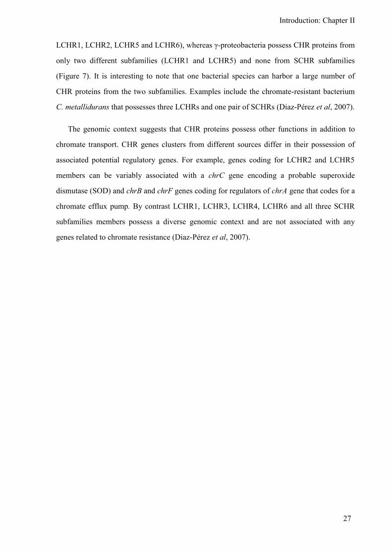
Introduction: Chapter II
27
LCHR1, LCHRβ, LCHR5 and LCHR6), whereas -proteobacteria possess CHR proteins from
only two different subfamilies (LCHR1 and LCHR5) and none from SCHR subfamilies
(Figure 7). It is interesting to note that one bacterial species can harbor a large number of
CHR proteins from the two subfamilies. Examples include the chromate-resistant bacterium
C. metallidurans that possesses three LCHRs and one pair of SCHRs (Diaz-Pérez et al, 2007).
The genomic context suggests that CHR proteins possess other functions in addition to
chromate transport. CHR genes clusters from different sources differ in their possession of
associated potential regulatory genes. For example, genes coding for LCHR2 and LCHR5
members can be variably associated with a chrC gene encoding a probable superoxide
dismutase (SOD) and chrB and chrF genes coding for regulators of chrA gene that codes for a
chromate efflux pump. By contrast LCHR1, LCHR3, LCHR4, LCHR6 and all three SCHR
subfamilies members possess a diverse genomic context and are not associated with any
genes related to chromate resistance (Diaz-Pérez et al, 2007).

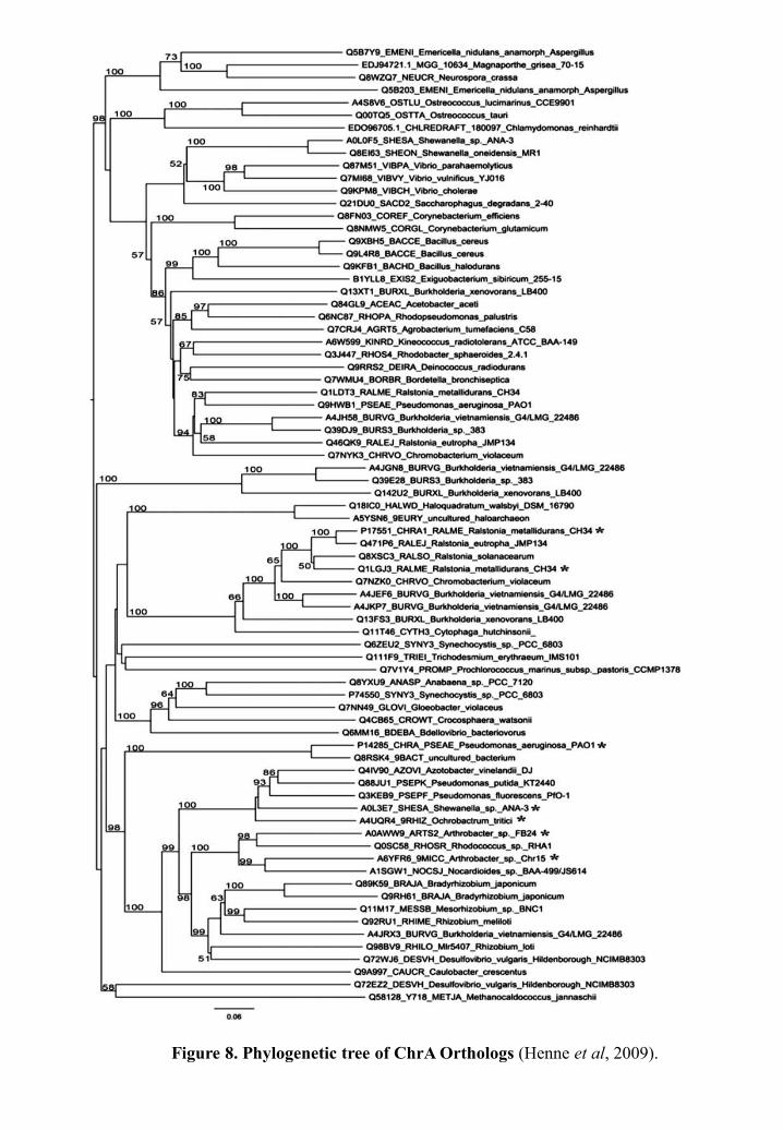
Figure 8. Phylogenetic tree of ChrA Orthologs (Henne et al, 2009).

Introduction: Chapter II
28
The chromate efflux pump ChrA
The efflux of chromate is a resistance mechanism in bacteria generally conferred by
chromate resistance determinants which include the chrA gene that encodes a hydrophobic
inner membrane chromate efflux protein (Pimentel et al, 2002). ChrA functions as a
chemiosmotic pump that extrudes chromate from the cytoplasm using the proton motive force
(Ramírez-Díaz et al, 2008; Viti et al, 2014). Chromate efflux pumps are believed to be
responsible for Cr(VI) resistance in bacteria (Diaz-Pérez et al, 2007; Ramírez-Díaz et al,
2008).
A ChrA protein has been characterized in detail in C. metallidurans CH34 (Nies et al,
1990) and Pseudomonas aeruginosa (Cervantes et al, 1990) and numerous putative ChrA
homologs have been identified after the sequencing of bacterial genomes (Saier, 2003; Henne
et al, 2009) (Figure 8).
In bacteria, the chrA genes can be located on plasmid and/or chromosomal DNA and they
generally belong to operons with other chr genes (Juhnke et al, 2002; Viti et al, 2014).
C. metallidurans CH34 (previously Alcaligenes eutrophus and Ralstonia metallidurans)
(Cervantes et al, 1990; Cervantes & Campos-Garcia, 2007) harbors two chromate resistance
determinants, the chr(1) cluster (chrIB1A1CEF1) present on the megaplasmid pMOL28
(Figure 9.A) and chr(2) cluster (chrB2A2F2) present on the chromosome (Figure 9.B)
(Branco et al, 2008). ChrI, ChrB and ChrF are proposed to play regulatory roles for the
expression of chrA with, respectively, ChrB as an activator and ChrF and ChrI as repressors.
The ChrE protein is thought to be involved in the cleavage of chrome-gluthation complex
(Juhnke et al, 2002; Cervantes & Campos-Garcia, 2007). The chromate-resistance
determinant of P. aeruginosa belongs to the LCHR5 family. It is located on the pUM505
plasmid and organized in a putative chrBAC operon (Figure 9.C) (Ramírez-Díaz et al, 2011).
ChrB plays a regulatory role for expression of chrA and chrC gene encoding a truncated
protein which is probably not functional (Tauch et al, 2003).
The two ChrA homologs, encoded by genes carried on plasmids pMOL28 from
C. metallidurans and pUM505 of P. aeruginosa are 29% identical and they are reported to
display a different topology, 10 transmembrane segments for the C. metallidurans protein
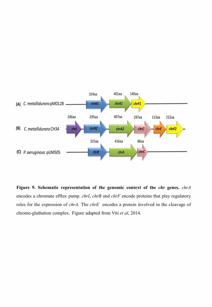
Figure 9. Schematic representation of the genomic context of the chr genes. chrA
encodes a chromate efflux pump. chrI, chrB and chrF encode proteins that play regulatory
roles for the expression of chrA. The chrE encodes a protein involved in the cleavage of
chrome-gluthation complex. Figure adapted from Viti et al, 2014.

Introduction: Chapter II
29
(Nies et al, 1998) and 13 transmembrane segments for that of P. aeruginosa (Jiménez-Meji-a
et al, 2006).
Cr(VI) reduction
Bacterial reduction of Cr(VI) to Cr(III) is one of the main chromate resistance
mechanisms. Since the first report of anaerobic Cr(VI) reduction by Romanenko VI,
Koren’kov VN, 1λ77 in uncharacterized Pseudomonas sp., worldwide researchers have
isolated both aerobic and anaerobic Cr(VI)-reducing bacteria belonging to a wide range of
genera from diverse environments. Cr(VI) reduction can be achieved either non-
enzymatically or enzymatically under aerobic and/or anaerobic conditions depending on the
bacterial species (Ahemad, 2014). On one hand, non-enzymatic-reduction may take place by
chemical reactions associated with intra/extracellular compounds produced during microbial
metabolism, including amino acids, nucleotides, sugars, vitamins, organic acids, glutathione,
sulfite and thiosulfates (Cervantes et al, 2001; Donati et al, 2003; Dhal et al, 2013; Joutey et
al, 2015; Gutiérrez-Corona et al, 2016). Moreover, Cr(VI) can be reduced by Fe(II) and H2S,
the anaerobic metabolic end products of iron and sulphate-reducing bacteria (Somasundaram
et al, 2009).
On the other hand, numerous bacterial genera, including Pseudomonas, Bacillus and
Arthrobacter have been widely reported to reduce Cr(VI) using an enzymatic process (Thatoi
et al, 2014; Viti et al, 2014). The ability to reduce Cr(VI) can be a secondary function for
Cr(VI) reducing enzymes, which have a different primary role other than Cr(VI) reduction.
For example, the nitroreductases NfsA/NfsB from Vibrio harveyi possess a nitrofurazone
nitroreductase as primary activity and a Cr(VI) reductase activity as a secondary function
(Kwak et al, 2003). Similarly, ferric reductase FerB from Paracoccus denitrificans uses both
Fe(III)-nitrilotriacetate and Cr(VI) as substrates (Mazoch Jiří et al, 2004). These secondary
functions may be related to bacterial enzymatic adaptation as a result of the relatively increase
of Cr(VI) content in the environment owing to anthropogenic activities (Silver & Phung,
1996).
The enzymatic reduction can be partial, where Cr(VI) gets reduced to the highly unstable
Cr(V) intermediate, which can get oxidized back to Cr(VI) in a redox cycle, giving its

Figure 10. Enzymatic Cr(VI) reduction.
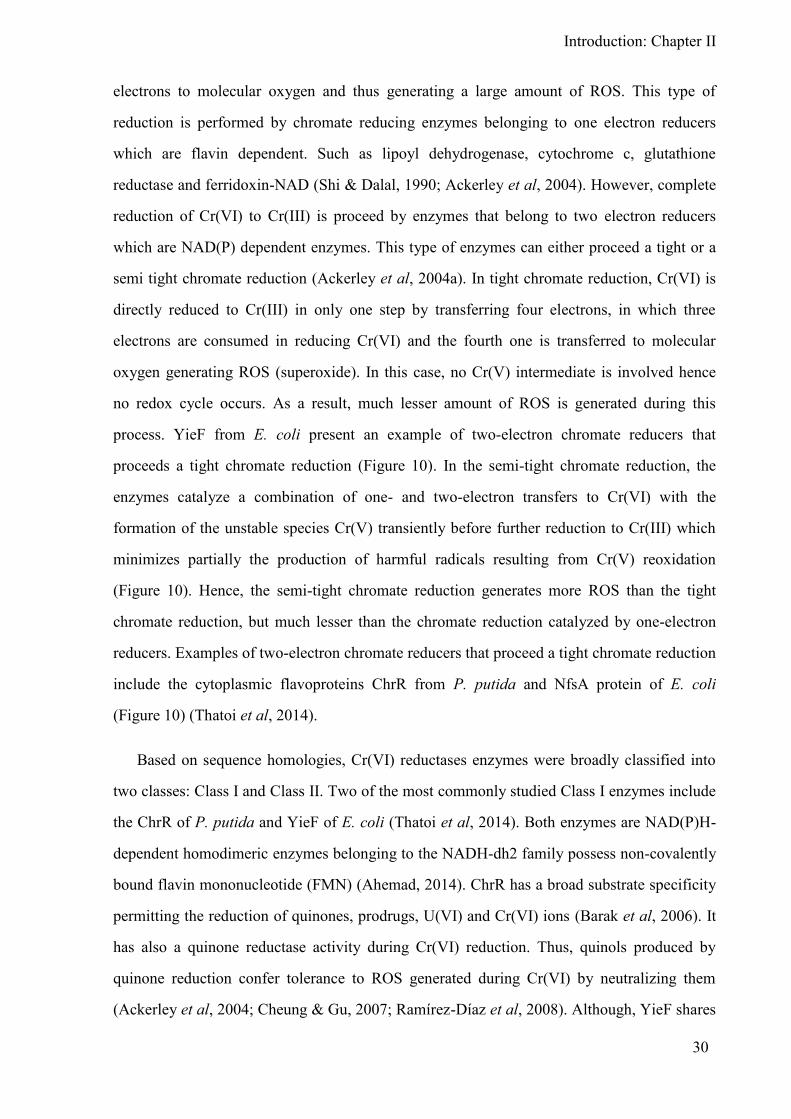
Introduction: Chapter II
30
electrons to molecular oxygen and thus generating a large amount of ROS. This type of
reduction is performed by chromate reducing enzymes belonging to one electron reducers
which are flavin dependent. Such as lipoyl dehydrogenase, cytochrome c, glutathione
reductase and ferridoxin-NAD (Shi & Dalal, 1990; Ackerley et al, 2004). However, complete
reduction of Cr(VI) to Cr(III) is proceed by enzymes that belong to two electron reducers
which are NAD(P) dependent enzymes. This type of enzymes can either proceed a tight or a
semi tight chromate reduction (Ackerley et al, 2004a). In tight chromate reduction, Cr(VI) is
directly reduced to Cr(III) in only one step by transferring four electrons, in which three
electrons are consumed in reducing Cr(VI) and the fourth one is transferred to molecular
oxygen generating ROS (superoxide). In this case, no Cr(V) intermediate is involved hence
no redox cycle occurs. As a result, much lesser amount of ROS is generated during this
process. YieF from E. coli present an example of two-electron chromate reducers that
proceeds a tight chromate reduction (Figure 10). In the semi-tight chromate reduction, the
enzymes catalyze a combination of one- and two-electron transfers to Cr(VI) with the
formation of the unstable species Cr(V) transiently before further reduction to Cr(III) which
minimizes partially the production of harmful radicals resulting from Cr(V) reoxidation
(Figure 10). Hence, the semi-tight chromate reduction generates more ROS than the tight
chromate reduction, but much lesser than the chromate reduction catalyzed by one-electron
reducers. Examples of two-electron chromate reducers that proceed a tight chromate reduction
include the cytoplasmic flavoproteins ChrR from P. putida and NfsA protein of E. coli
(Figure 10) (Thatoi et al, 2014).
Based on sequence homologies, Cr(VI) reductases enzymes were broadly classified into
two classes: Class I and Class II. Two of the most commonly studied Class I enzymes include
the ChrR of P. putida and YieF of E. coli (Thatoi et al, 2014). Both enzymes are NAD(P)H-
dependent homodimeric enzymes belonging to the NADH-dh2 family possess non-covalently
bound flavin mononucleotide (FMN) (Ahemad, 2014). ChrR has a broad substrate specificity
permitting the reduction of quinones, prodrugs, U(VI) and Cr(VI) ions (Barak et al, 2006). It
has also a quinone reductase activity during Cr(VI) reduction. Thus, quinols produced by
quinone reduction confer tolerance to ROS generated during Cr(VI) by neutralizing them
(Ackerley et al, 2004; Cheung & Gu, 2007; Ramírez-Díaz et al, 2008). Although, YieF shares


Introduction: Chapter II
31
sequence homology with ChrR (Ackerley et al, 2004), it reduces differently the chromate as
explain above, and contrary to ChrR, YieF does not show semiquinone flavoprotein
generation during chromate reduction (Viti et al, 2014; Thatoi et al, 2014).
Class II chromate reductases that bear no homology to the class I enzymes, possess
nitroreductase activity. They reduce quinones and nitrocompounds effectively and vary in
their ability to transform chromate. There are two members of the class II family, namely
NfsA protein of E. coli and the ChfN protein of B. subtilis that possess also a chromate
reductase activity (Park et al, 2000).
Cr(VI) Reduction pathways
Cr(VI) reduction can occur inside and/or outside the cell under aerobic and/or anaerobic
conditions through Cr(VI) reductases either localized in the membrane fraction (Wang et al,
1991; Cheung & Gu, 2007) or in the cytosolic fractions (Suzuki et al, 1992; Park et al, 2000;
Bae et al, 2005) of the Cr(VI) reducing bacteria.
Cr(VI) reduction localization
In intracellular processes, Cr(VI) is reduced in the cytosol using cytoplasmic soluble
reductase enzymes. These enzymes play an intermediate role between associated biological
electron donors involved in this process such as NADH and NADPH. This reduction process
is not energy consuming but will directly affect the cell, since most of intracellular proteins
catalyze a one-electron reduction from Cr(VI) to Cr(V) (Joutey et al, 2015). Many bacteria
are known to participate in the intracellular reduction of Cr(VI) like the Gram negative
bacteria P. aeruginosa, E. coli ATCC 33456 and Enterobacter. In addition, B. subtilis was
also reported to carry out intracellular reduction of Cr(VI) (Thatoi et al, 2014).
In contrast, extracellular Cr(VI) reduction is beneficial to the cell since it does not require
transport mechanisms to carry the chromate into the cell, and to later expel the Cr(III). Hence,
extracellular reduction of Cr(VI) protects the cell from ROS effects resulting from Cr(VI)
reduction. This process can be meditated by membrane bound reductases that can reduce
Cr(VI) extracellularly as an electron acceptor by using electron shuttling compounds coupled
to membrane reduction (Joutey et al, 2015). In some cases, soluble Cr(VI) reducing enzymes

Introduction: Chapter II
32
such as flavin reductases, nitrate reductases, flavin proteins and ferrireductases, produced in
the cytoplasm are exported into the media to extracellularly reduce Cr(VI), as reported in
P. putida (Cheung & Gu, 2007).
Aerobic/Anaerobic Cr(VI) Reduction
Bacterial Cr(VI) reduction in the presence of oxygen occurs as a two or three step process,
with Cr(VI) initially reduced to the short-lived intermediates Cr(V) and/or Cr(IV) before
being further reduced to Cr(III). NADH, NADPH and electrons from the endogenous reserve
are implicated as electron donors in the Cr(VI) reduction process. Aerobic Cr(VI) reduction is
generally associated with soluble proteins which are localized as cytosolic proteins. Which is
the case in aerobes like P. putida (Ishibashi et al, 1990). As exceptions, P. maltophilia O-2
and Bacillus megaterium TKW3 were found to utilize membrane-associated reductases for
Cr(VI) reduction, in spite of being aerobes (Cheung & Gu, 2007).
In anoxic conditions, Cr(VI) can serve as a terminal electron acceptor in the respiratory
chain for a large array of electron donors, including carbohydrates, proteins, fats, hydrogen,
NAD(P)H and endogenous electron reserves. The Cr(VI)-reducing activities of anaerobes are
associated with their electron transfer systems catalyzing the electron shuttle along the
respiratory chain. Furthermore, the cytochrome family is frequently involved in enzymatic
anaerobic Cr(VI) reduction (Mary Mangaiyarkarasi et al, 2011). Both membrane-associated
and soluble enzymes mediate the process of Cr(VI) reduction under anaerobic conditions
(Cheung & Gu, 2007). Reduction involving membrane-associated reductase has been reported
in some chromate-reducing bacteria which utilized H2 as electron donor and Cr(VI) as an
electron acceptor (QuiIntana et al, 2001). In addition, some soluble enzymes have been also
found to mediate this process, such as the soluble cytochrome c3 in Desulfovibrio vulgaris
(Barrera-Díaz et al, 2012).
Some bacteria are capable of reducing chromate under both aerobic and anaerobic
conditions. Examples include, P. fluorescens LB300 (Bopp & Ehrlich, 1988), Achromobacter
sp. (Ma et al, 2007), E. coli, P. ambigua (McLean & Beveridge, 2001), P. putida (Barak et al,
2006) and Bacillus sp. (Liu et al, 2006), although, their rate of reduction can vary widely
between the two conditions. For example, faster reduction rate of Cr(VI) under aerobic


Introduction: Chapter II
33
conditions than anaerobic conditions has been reported in the case of E. coli ATCC 33456
(Shen & Wang, 1993).
Indirect Cr(VI) resistance mechanisms
The generation of ROS during partial Cr(VI) reduction causes oxidative stress in bacteria.
The participation of bacterial proteins in the defense against stress induced by Cr(VI)
represents one of the main mechanisms of Cr(VI) resistance that does not deal directly with
Cr(VI) ions but rather protects cells from their damaging effects (Ramírez-Díaz et al, 2008).
As a strategy for scavenging ROS, bacteria commonly modulate gene expression by inducing
genes encoding antioxidant enzymes and proteins that can directly decompose the oxidant
such as superoxide dismutase and catalase (Ackerley et al, 2004a), alkylhydroperoxide
reductase (Ahp) and various peroxidases (Shi et al, 2015). And also induces genes encoding
non-enzymatic antioxidant such as NADPH and NADH pools, glutathione/glutaredoxin,
thioredoxins and ascorbic acid to restrain intracellular concentrations of ROS in order to
prevent the damages beyond unmanageable. These antioxidants are not only known to diffuse
free radicals and limit the risk of oxidative stress but they can also chelate the metal ions
responsible for generating ROS (Mishra & Imlay, 2012).
Expression of these genes is typically regulated by OxyR, the primary regulator of
oxidative stress response. It directly controls over 20 genes, including genes involved in ROS
detoxification and genes with other roles in oxidative stress defense such as genes
maintaining iron homeostasis (Li et al, 2014). Since free iron in its reduced form Fe + can
generate ROS converting the less reactive hydrogen peroxide to the more reactive oxygen
species, hydroxyl radical (Reaction 4), intracellular levels of iron must be carefully controlled
to meet the metabolic needs of the cell while limiting cellular damage due to iron overload
(Yang et al, 2009).
Fe + + H O → Fe + + OH− + OH • (Reaction. 4)
To cope with all protein and DNA damages caused by Cr(VI), bacteria possess a repair
system acting as another defensive shield against Cr(VI)-induced oxidative stress. They
primarily remove the damaged biomolecules before they accumulate and result in altered cell


Introduction: Chapter II
34
metabolism or viability. For example, oxidized proteins are removed by proteolytic systems.
Furthermore, they repair oxidatively damaged nucleic acids by specific enzymes belonging to
SOS repair system (Poljsak et al, 2010).


CHAPTER III:
Shewanella oneidensis
MR-1


Introduction: Chapter III
36
Shewanella genus
Shewanella is a genus of Gamma proteobacteria (Dikow, 2011) belonging to the
Alteromonadales Order and the Family of Alteromonadacea (Vogel et al, 2005). Like many
contemporary genera, the Shewanella have experienced a rocky road to their present status, as
evidenced by the species S. putrefaciens. Originally isolated as an active agent in food
spoilage (Derby & Hammer, 1931), this organism was first called “Achromobacter
putrefaciens”, then Pseudomonas putrefaciens (Shewan et al, 1960), Alteromonas
putrefaciens (Lee et al, 2016) and finally, based on 5S rRNA sequences, the species was
renamed Shewanella putrefaciens (MacDonell & Colwell, 1985). The genus Shewanella,
named after James Shewan for his work in fisheries microbiology (Vogel et al, 2005), has
only been recognized with its present name since 1985 and no further reclassifications at the
level of the genus have been made to date (Dikow, 2011).
In general, members of Shewanella genus compose a diverse group of facultative
anaerobic bacteria that are gram-negative rods 2–γ m in length, 0.4–0.7 m in diameter and
motile due to a single polar flagellum. Most can easily grow on usual laboratory culture media
after enrichment from environmental samples. The hallmark of many shewanellae is the
ability to utilize a diverse array of final electron acceptors in the absence of oxygen, and many
have capitalized on this ability that allows their wide distribution in nature by surviving in
diverse habitats (Hau & Gralnick, 2007).
Today, this genus comprises 66 recognized species (http://www.bacterio.cict.net). Most of
them, have been isolated from marine environments including seawater, sediment, tidal flats,
marine invertebrates and fish, Antarctic sea ice, clinical samples and some species are found
in activated sludge (Lee et al, 2016). Forty-one Shewanella species were proposed from 2002
to 2010 with a peak number of species described in 2006 (Janda & Abbott, 2014). Among the
species proposed during this time period, two Shewanella species were described, Shewanella
fidelis KMM 3582 T in 2003 and Shewanella algidipiscicola in 2007, isolated respectively
from sediments of the South China Sea and from marine fish (cod and plaice) caught in the
Baltic Sea (Ivanova, 2003; Satomi et al, 2007).
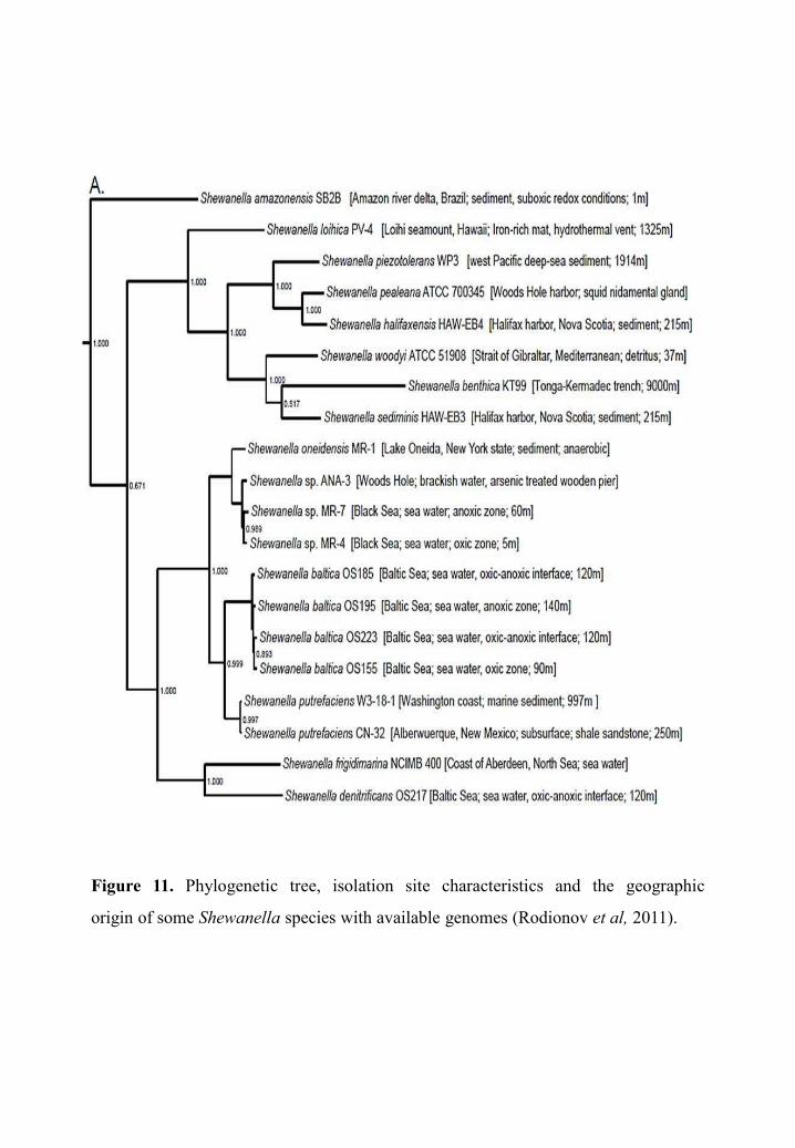
Figure 11. Phylogenetic tree, isolation site characteristics and the geographic
origin of some Shewanella species with available genomes (Rodionov et al, 2011).
Introduction: Chapter III
37
Shewanella genus species are of great interest for environmental clean-up due to their
ability to convert heavy metals and toxic substances (e.g. cadmium, uranium) into less toxic
products by using them as electron acceptors in certain respiratory situations(Dikow, 2011).
In order to understand the underlying mechanisms of this ability of biotechnical interest, the
genome of more than 32 Shewanella species have been fully sequenced and deposited in
GenBank. Twenty of them are represented in a phylogenetic tree in Figure 11 (Rodionov et
al, 2011).
A 16S rRNA gene-based phylogenetic reconstruction has revealed two major groups in
the Shewanella genus. Usually, group 1 includes species that are piezotolerant and
psychrotolerant such as Shewanella violacea, which has been isolated from the deep sea.
Group 2 species are usually pressure sensitive and mesophilic and include
Shewanella oneidensis, Shewanella baltica and Shewanella putreficans which have been
isolated from a variety of environments, including fresh water lakes and spoiled meat
products (Kato & Nogi, 2001). Overall, Shewanella is phylogenetically most closely related
to the genera Pseudoalteromonas, Alteromonas, Moritella, Ferrimonas and Colwellia, all of
which are members of the family Alteromonadaceae (Figure 12) (Bowman, 2015; Dufault-
Thompson et al, 2017)
Shewanella genus characteristics
Phenotypic characteristics
Shewanella colonies on complex nutrient media typically have a pale tan to pink-orange
or salmon color, which is due to strong accumulation of cytochrome proteins. However, this
tendency is less pronounced in some species such as S. hanedai and S. benthica, S.
colwelliana and S. hanedai (Bowman, 2015).
Unlike some Shewanella species that require Na+ or seawater media for growth, S. algae,
S. frigidimarina, S. oneidensis, S. fidelis and S. algidipiscicola do not require Na+ for growth.
However, they are capable of growing on certain NaCl concentrations. Some species like S.
colwelliana, S. hanedai and S. benthica require amino acids and/or vitamins for growth. In

Figure 12. Phylogenetic reconstruction of the Shewanella genus showing the two
groups: Group 1 containing piezotolerant and psychrotolerant species and group 2
enclosing pressure sensitive and mesophilic species. It shows also five outgroup
species to which Shewanella is phylogenetically most closely related (Dufault-
Thompson et al, 2017).
Introduction: Chapter III
38
defined media, such as basal medium agar, these requirements can be met by adding yeast
extract at concentrations of 0.05–0.1% (w/v) (Bowman, 2015).
Regarding the fatty acid profile, Shewanella species are rich in branched and odd-chain-
length fatty acids. Psychrotolerant and psychrophilic Shewanella species have the unusual
ability to synthesize eicosapentaenoic acid (EPA) which is essential for their survival at low
temperatures, the levels of which range from 2–22%. Nevertheless, mesophilic species do not
produce EPA (Bowman, 2015).
Pathogenicity
Usually, bacteria of the genus Shewanella are not pathogenic. However, Shewanella
putrefaciens, has been associated with serious health disorders in freshwater fish. Therefore, it
has been described as a new aetiological agent of a disease, named shewanellosis which
causes skin disorders and haemorrhages in internal organs. It should be noted that S.
putrefaciens could also be associated with different infections in humans, such as skin and
tissue infections, bacteraemia and otitis. Enzymatic activity, cytotoxin secretion, adhesion
ability, lipopolysaccharide and the presence of siderophores are potential virulence factors of
S. putrefaciens (Paździor, β016). Recent studies suggest that S. algae may in fact be more
pathogenic than S. putrefaciens (Janda, 2014) and consider it as an emerging pathogen of skin
and soft tissue infections mainly in patients with chronic ulcers and at times be multidrug
resistant (Jampala, 2015). Its hemolysin production is believed to be an important factor of its
pathogenicity (Khashe & Janda, 1998).
Energy metabolism
Shewanella species are chemoheterotrophic facultative anaerobes, with anaerobic growth
typically of a respiratory nature. However, some species can also grow fermentatively; these
include S. frigidimarina and S. benthica, both of which can ferment D-glucose, as well as S.
gelidimarina which can ferment N-acetylglucosamine and chitin, but not D-glucose
(MacDonell & Colwell, 1985; Bowman, 2015). In anaerobiosis, Shewanella species can use
nitrate as an electron acceptor for growth. Moreover, Trimethylamine-N-oxide (TMAO) is
also a common terminal electron acceptor among Shewanella species; its reduction to


Introduction: Chapter III
39
trimethylamine (TMA) is usually responsible for the odors associated with Shewanella food
spoilage (Shewan et al, 1972). They can also grow anaerobically by reduction of various
sulfur compounds to H2S including thiosulfate and sulfite or via reduction of fumarate to
succinate, coupled to the oxidation of format (Bowman, 2015).
The majority of Shewanella species have the ability to facultatively reduce ferric iron,
manganese and other metals, examples include S. oneidensis MR-1. They can grow
anaerobically by coupling the oxidation of carbon compounds or H2 to the reduction of Fe3+
to Fe2+ or of Mn4+ to insoluble Mn3+. Their dissimilatory metal reduction is believed to be
important in terms of metal cycling and mobilization in the environment (Bowman, 2015).
Shewanella oneidensis MR-1
Shewanella oneidensis MR-1 is an aquatic bacterium formerly known as Alteromonas
putrefaciens MR1. Based on both 16SDNA and gyrB nucleotide sequences it was reclassified
as a member of the genus Shewanella. It was isolated in 1988 from sediments of Oneida Lake
at New York, whence its name. Like all Shewanella group 2 species, S. oneidensis MR-1 is
able to grow at mesophilic temperatures (optimal growth was observed at 30°C) and it shows
weak growth at NaCl concentrations above 3%. This bacterium exhibits cytochrome oxidase,
catalase and gelatinase activities (Venkateswaran et al, 1999). Furthermore, it is capable of
dissimilatory metabolism of manganese and iron oxides (MR-1 for Mn-Reducing bacterium)
(Myers & Nealson, 1988; Venkateswaran et al, 1999).
In 2002, S. oneidensis MR-1 was the first of Shewanella spp whose genome have been
fully sequenced and thus serves as the model organism for studying the functional repertoire
of the Shewanella genus (Yang et al, 2015). It has a 5.13Mb genome containing 4590 genes,
including 184 on a mega-plasmid (Heidelberg et al, 2002). S. oneidensis MR-1 possesses
genes encoding the synthetic pathways of all amino acids and phenotypic analyzes have
shown it to be prototrophic (Serres & Riley, 2006). It can therefore develop in a minimum
medium with a single element as a source of carbon and electrons. It uses N-acetyl-
glucosamine (chitin monomer), inosine as well as amino acids as a carbon source (Ringo et
al, 1984; Yang et al, 2006; Driscoll et al, 2007). However, S. oneidensis MR-1 is unable to


Introduction: Chapter III
40
use glucose naturally, but a simple pre-exposure of 24 hours to glucose allows the appearance
of mutants able to use this substrate as a sole carbon source (Howard et al, 2011). This means
that all genetic elements necessary for glucose metabolism are present but cannot be used by
the wild-type strain.
S. oneidensis MR-1 harbors also genes coding for cytochromes and hydrogenases, which
are integral members of the electron transport system and the reason of its great respiratory
flexibility (see Respiratory capacities). Moreover, its genome contains many genes dedicated
to mobility and chemotaxis, including 70 genes encoding components of the flagellum, as
well as genes encoding two motor systems and three clusters of genes encoding components
of the chemosensing (Heidelberg et al, 2002).
Chemotaxis
Bacterial survival depends on the ability to respond and adapt to changing environmental
conditions, among the many challenges faced are scarcity of nutrients and the accumulation of
toxic substances (Porter et al, 2011). Bacteria such as S. oneidensis MR-1 respond to these
changes by altering their motile behavior in response to signal molecules. This behavior,
called chemotaxis. It allows bacteria migration under the influence of a chemical gradient
(Pandey & Jain, 2002) in order to find better environments by moving, through changes in the
rotary behavior of the flagellum, toward areas where the concentration of favorable chemicals
(attractants) is high. Or away from an area when they perceive an increase of unfavorable
chemicals (repellents) to find more suitable area where the repellent is in lower concentration
(Porter et al, 2011). Flagellar rotation is controlled by a chemotaxis pathway, where signals
are transduced from chemoreceptor proteins, called methyl accepting chemotaxis proteins
(MCPs), through a histidine protein kinase (CheA) and a response regulator (CheY) to the
flagellar motor (Porter et al, 2011).
The chemotaxis ability of S. oneidensis MR-1 as well as all Shewanella species may
provide a competitive advantage, allowing cells to move into more favorable environments
which increases their chances of survival and growth. This ability, coupled with their wide
range of potential electron acceptors, may help explain their nearly ubiquitous presence in
widely disparate environmental niches (Li et al, 2010).

Introduction: Chapter III
41
S. oneidensis MR-1 was shown to be chemotactic only toward some compounds, mainly
corresponding to electron acceptors including nitrate, nitrite, TMAO, DMSO, fumarate,
thiosulphate, Mn(III) and Fe(III) (Bencharit & Ward, 2005; Baraquet et al, 2009). Recently,
other signals generating a chemotactic response in S. oneidensis MR-1 were identified. These
includes the L-malate that was identified as attractants, as well as four new metals: Nickel and
cobalt which were identified as repellents, whereas copper and chromate were identified as
attractants (Armitano et al, 2011). Recent studies on S. oneidensis MR-1, confirmed that its
taxis towards many, if not all, exogenous electron acceptors occurs via an energy taxis
mechanism (Bencharit & Ward, 2005; Baraquet et al, 2009), where the signal is not the
electron acceptor itself but it originates from its metabolism (Alexandre Gladys et al, 2004;
Schweinitzer & Josenhans, 2010). Therefore, cells move towards an environment that is
optimal for metabolic activity rather than one with the maximal attractant concentration
(Porter et al, 2011).
S. oneidensis MR-1 is predicted to have a complex chemotaxis system that includes 27
MCPs genes and 3 clusters of chemosensing genes (che) (Fredrickson et al, 2008). Among the
11 MCPs containing a known detection domain in S. oneidensis MR-1, 5 MCPs (SO2240,
SO3282, SO3642, SO3890 and SO4454) were shown to be involved in energy mechanism
(Baraquet et al, 2009). Further, Armitano showed that MCP SO0987 is one of the MCPs
involved in chromate detection (Armitano et al, 2011). Among the 3 clusters of chemosensing
genes, the Che3 cluster that contains genes encoding CheA3, CheY3, CheW3, CheZ, CheR3,
CheD3 and CheB3, is the essential one for chemotaxis (Li Jun et al, 2007; Armitano et al,
2013). So far, CheA3 the chemotaxis histidine protein kinase is the only one of three putative
CheA that has been shown to be necessary for behavioral responses to anaerobic electron
acceptors (Fredrickson et al, 2008). Additionally, the deletion of its response regulator CheY3
gene has also been shown to abolish the chemotactic capacities of S. oneidensis MR-1
(Armitano et al, 2013).

Figure 13. Surface biofilm development stages in Pseudomonas.
Figure adapted from Foster & Slonczewski, 2017.
1. Initial
attachement
2. Irreversible
adhesion
3. EPS
production
4. Final
maturation
5. Biofilm
dispersal

Introduction: Chapter III
42
Biofilms
The theory of the lifestyle of bacteria not free but adhered to a surface was advanced by
Henrici in 1933. He showed that the bacteria were predominantly sessile rather than
planktonic. The term "biofilm" was introduced for the first time in 1978 by Costerton. Until
1987, the biofilm was perceived as a "slab" of matrix in which the sessile bacteria were
integrated. Further, the development of optical then confocal microscopy, showed that
biofilms have complex structures organized in three dimensions composed of aggregates of
microorganisms, separated by free spaces called channels where the structure of the matrix is
less dense allowing the supply of bacteria with nutrients and oxygen, the diffusion of
communications signals and the elimination of their degradation products.
Today, we consider that the main lifestyle of bacteria is not planktonic but in the form of a
multicellular community called biofilm. Ninety to 99.9% of the bacteria live in a biofilm
(Pantaleon, 2015). The definition of a biofilm, according to (Hall-Stoodley et al, 2004) is a
bacterial community adhered to an interface (air-liquid or solid-liquid) and living in a matrix
composed essentially of water (up to 97%), polysaccharidic polymers secreted by
microorganisms, degradation products and substances from the external environment and
other components such as DNA, RNA and lipids. The biofilm matrix protects bacteria from
many stresses, such as UV, free radicals, pH variations, detergents or dehydration and from
the penetration of certain antibiotics. It also helps to concentrate nutrients, metals or useful
ions close to cells (Davey & O’toole, β000). Biofilm would be a strategic lifestyle for micro-
organisms since it represents a protected environment for the growth in a hostile environment.
Surface biofilm in S. oneidensis
The ability of S. oneidensis to form surface biofilms has been extensively studied. It is
capable of forming biofilms on mineral surfaces but also on aerobic glass (Lower et al, 2001;
Thormann et al, 2004). Confocal microscopy studies have shown that the formation of
biofilm by S. oneidensis is similar to that seen for Pseudomonas aeruginosa (Tolker-Nielsen
et al, 2000; Foster & Slonczewski, 2017) (Figure 13). It begins with a reversible attachment of
individual cells to a solid surface which will then multiply to form microcolonies, thus the
adhesion becomes irreversible. The biofilm can then thicken by forming three-dimensional

Figure 14. Pellicle formation in S. oneidensis MR-1 over time (Armitano et al, 2013).

Introduction: Chapter III
43
structures (Thormann et al, 2004), through production of extracellular polysaccharides,
nucleic acids, proteins and additional factors, which together compose the matrix of the
mature biofilm (O’Toole & Ha, β015). Studies showed that the flagellum in S. oneidensis
plays an important role in the transition from a flat to a structured biofilm (Thormann et al,
2004; Armitano et al, 2013). Some bacterial cells may detach from the mature biofilm and
enter the dissemination phase, which permits the colonization of new surfaces. Biofilm
dispersal can be the result of several cues, such as alterations in nutrient availability, oxygen
fluctuations and increase of toxic products or other stress-inducing conditions (Figure 13)
(O’Toole & Ha, β015).
Pellicle development in S. oneidensis
When grown in a liquid under aerobic and static conditions, some bacteria such as
S. oneidensis MR1, Bacillus subtilis, E. coli, some Pseudomonas, Salmonella and Vibrio
species exploit the air liquid interface (ALI) to build a floating biofilm, this structure is called
pellicle, ‘floating’ biofilm or ALI biofilm (Armitano et al, 2014). The ALI is a favorable
ecological niche because it provides access to high concentrations of oxygen originating from
the air in addition to the nutrients present in the liquid. This interface is defined by opposing
gradients: a downward-diffusing O2 and upward-diffusing nutrients.
As previously said, S. oneidensis MR1 is capable of forming pellicles in a static condition
and in the presence of oxygen (Liang et al, 2010), which is required to initiate pellicle
formation and also for pellicle maturation steps. Pellicle cannot be formed under anaerobic
conditions, even in the presence of alternative electron acceptors. Armitano (2013) have
proposed that aerotaxis, which is the mechanism allowing the active localization of cells at
the ALI, is involved in the pellicle formation in S. oneidensis MR-1. Pellicle initiates in the
absence of growth and it occurs in three steps: rapid formation of a thin pellicle followed by
the random aggregation of cells in the nascent pellicle and finally, the establishment of a thick
mature and homogeneous pellicle (Figure 14). S. oneidensis MR1 pellicle formation requires
the presence of a functional flagellum since it allows an active localization of the cells at the
ALI. As already mentioned above, the flagellum rotation is under the control of the
chemotaxis system (Che3) which is also essential for pellicle formation. Armitano showed


Introduction: Chapter III
44
that the deletion of the histidine kinase CheA3 or its response regulator CheY3 gene leads to a
heterogeneous pellicle or to the absence of pellicle, respectively, which means that the
detection of chemotactic signals is important for pellicle development in S. oneidensis MR1
(Armitano et al, 2013).
The matrix of the S. oneidensis MR1 pellicle is mainly composed of exopolysaccharides,
probably rich in glucose, but unlike surface biofilm, DNA does not appear to be a major
component (Liang et al, 2010). Transcriptomic analyzes and analyzes of deletion mutants
have shown that iron import mechanisms and cytochromes are important for pellicle
development in S. oneidensis MR1 (Liang et al, 2012). It has also been shown that certain
cations (Ca2 +, Mn2+, Cu2 + or Zn2 +) are required for the formation of pellicle (Liang et al,
2010).
S. oneidensis MR-1 respiratory capacities
S. oneidensis MR-1 has remarkably diverse respiratory capacities as mentioned above. It
uses oxygen as the terminal electron acceptor during aerobic respiration. However, under
anaerobic conditions, it undertakes respiration by reducing more than 10 alternative terminal
electron acceptors such as oxidized metals (including Mn(III) and (IV), Fe(III), U(VI)),
fumarate, nitrate, TMAO, dimethyl sulfoxide (DMSO), sulfite, thiosulfate and elemental
sulfur. Whereas organic molecules are taken into the cell for oxidation within the cytoplasm,
the reduction of terminal electron acceptors occurs outside the cell or within the periplasm
(Venkateswaran et al, 1999; Heidelberg et al, 2002; Fredrickson et al, 2008; Breuer et al,
2015). Such plasticity in alternative electron acceptors has not been observed in any other
organism and that is what makes S. oneidensis MR-1 the most metabolically diverse species
of its genus, which allows its competitiveness in complex aquatic and sedimentary systems
and underpins its biotechnological contributions (Cordova et al, 2011). We can cite as an
example for that, the large amounts of sulfide produced by S. oneidensis MR-1 from
thiosulfate and sulfur that allow toxic metals to be immobilized as insoluble metal sulfides, as
well as its ability to reduce soluble U(VI) and Cr(VI) to insoluble precipitates, which helps to
remove soluble metal pollutants from the environment (Breuer et al, 2015).

Introduction: Chapter III
45
Utilization of a wide range of electron acceptors is mediated by a complex respiratory
network. The latter extends electron transfer from the cytoplasmic membrane to periplasm
and outer membrane, and includes 42 putative c-type cytochromes reduced by quinols
(Cordova et al, 2011; Breuer et al, 2015). S. oneidensis MR-1 depends on c-type cytochromes
for the maintenance of respiratory versatility, as mutants lacking functional cytochrome c
maturation genes are completely compromised in their ability to respire anaerobically
(Fredrickson et al, 2008).
Regardless of the nature and location of electron acceptors, the core of anaerobic electron
transport chain in S. oneidensis MR-1 is the redox cycling of quinone (Q) and its reduced
form quinol (QH2) (Marritt et al, 2012a, 2012b). Electrons from the cytoplasmic oxidation of
organic electron donors drive the reduction of Q to QH2 at the inner membrane (IM) level, by
a membrane-associated NADH dehydrogenase, after which the resulting electrons are
directed to periplasmic redox partners through the action of quinol dehydrogenases such as
TorC and CymA. Which are penta-haem and tetra-haem c-type cytochromes, respectively,
anchored to the IM by a single α-helix and contain periplasmic domains that catalyze QH2
oxidation (Cordova et al, 2011; Marritt et al, 2012a; Breuer et al, 2015). It should be noted
that TorC has a single redox partner, the TMAO reductase TorA, in contrast, CymA is able to
transfer electrons to several partners (explained in more details below). This difference in the
biochemistry of TorC and CymA is reflected at the genome level. TorC and TorA are
produced from the same operon. Whereas, CymA is produced from an orphan gene that is
expressed constitutively which allows its synthesis to be regulated independently from that of
its redox partners (Breuer et al, 2015). Moreover, recent study showed that its expression can
increase during anaerobiosis (Marritt et al, 2012b, 2012a).
CymA belongs to the NapC/NirT family of quinol dehydrogenases. It is considered a
central element of many electron acceptors reduction pathways (Marritt et al, 2012a), since it
acts as a common branching point in the electron transport chain by delivering electrons from
the quinone pool to multiple terminal reductases as described above (Schwalb et al, 2003).
Although, CymA possesses the common structural features of its family, which are an N-
terminal transmembrane α-helical anchor and a globular tetraheme periplasmic domain, it

Figure 15. Electron transfer in S. oneidensis MR-1.
Introduction: Chapter III
46
lacks specificity contrary to other family members (Schwalb et al, 2003). This allows its
involvement in electron transfer to multiple terminal reductases comprising the CymA-
dependent reductases that include the nitrate, nitrite, DMSO, Fe(III) and fumarate reductases,
that appear to be located in the periplasm or outer membrane of S. oneidensis MR-1.
Nevertheless, CymA is not involved in electron transfer to inner membrane anchored
enzymes such as the TMAO, thiosulfate and sulfite reductases (Breuer et al, 2015; Kouzuma
et al, 2015) (Figure 15).
Electron transfer in S. oneidensis MR-1
Among the various electron acceptors that S. oneidensis MR-1 reduces, sulfite, nitrite,
nitrate and fumarate are able to cross the outer membrane for reduction in the periplasm. They
are reduced respectively by SirA octahaem cytochrome, NrfA penta-haem cytochrome,
NapAB nitrate reductase with a molybdenum catalytic cofactor and the fumarate reductases,
FccA and IfcA, that cofactor is flavin adenine dinucleotide (FAD) (Figure 15) (Breuer et al,
2015).
Some electron acceptors as metal oxides need an extracellular electron transfer across the
outer membrane to be reduced outside the cell. Three main mechanisms have been proposed
to account for the respiratory reduction of extracellular electron acceptors by S. oneidensis
MR-1. The first one is the direct electron transfer following physical contact of the
extracellular substrate and outer membrane redox-active cytochromes. The two left
mechanisms represent a mediated extracellular electrons transfer between the extracellular
substrates and outer membrane cytochromes, either through flavins, also called electron
shuttles (Figure 16), or along extracellular conductive appendages “nanopods” that can span
distances greater than 100 Å to establish contact with solid substrates such as electrodes
(Breuer et al, 2015).
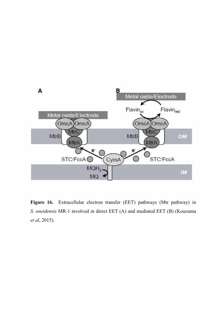
Figure 16. Extracellular electron transfer (EET) pathways (Mtr pathway) in
S. oneidensis MR-1 involved in direct EET (A) and mediated EET (B) (Kouzuma
et al, 2015).

Introduction: Chapter III
47
Direct electron transfer
The extracellular reduction of insoluble electron acceptors such as solid metals and
electrodes by S. oneidensis MR-1 takes place via the metal reducing (Mtr) pathway which
involves five proteins (Figures 15 and 16). These include CymA as well as the MtrABC
complex and finally OmcA (Coursolle et al, 2010). The core of metal reduction components
consists of the cytochrome Mtr complex which is located at the outer membrane and
composed of three subunits, MtrA, MtrB and MtrC. These proteins are encoded by the
mtrCAB operon that is expressed under microaerobic and anaerobic conditions, even in the
absence of metal electron acceptors. Expression of this operon as well as omcA, that have role
in metal reduction as described in more detail below, is controlled by the cAMP receptor
protein CRP which regulates the expression of many anaerobic reductase genes in S.
oneidensis MR-1 (Figures 15 and 16) (Saffarini, 2015).
MtrA is a 35 kDa decaheme c-type cytochrome which is associated with MtrCB with a
1:1:1 stoichiometry. It is regarded as a key protein for electron transfer to outer membrane c-
type cytochromes (Kouzuma et al, 2015). It interacts with CymA which leads to MtrA
reduction by electron transfer. Certain authors believe that the gap between CymA and MtrA
is too wide to allow direct electron transfer between the two proteins. The possibility of an
intermediate electron carrier that mediates electron transfer between the two proteins was then
proposed. Thereby, two c-type cytochromes were suggested, FccA which is a
flavocytochrome c with confirmed fumarate reductase activity, and STC also called CctA that
is a small tetraheme c-type cytochrome whose function has not been determined yet
(Kouzuma et al, 2015; Saffarini, 2015).
MtrA is embedded within the -barrel protein MtrB, that forms a pore of 30–40 Å in
diameter that can easily fit MtrA, forming a “porin cytochrome” electron transfer module
(Richardson David J. et al, 2012). Some authors suggest that MtrB is required in its turn for
the proper localization and insertion of the decaheme OmcA and MtrC in the outer membrane
(Myers & Myers, 2000) by forming a stable complex with these outer membrane c-type
cytochromes and supporting electron exchange between them (Kouzuma et al, 2015).
Biochemical data indicate that OmcA and MtrC form a complex with a stoichiometry of

Figure 17. Microscopic image of nanopods synthesized by S. oneidensis. Left
column: overlay of the fluorescence Light Microscopy (fLM) and Transmission
Electron Microscopy (TEM) images. Middle and Right columns: Enlarged fLM and
TEM views of nanopods. White arrows indicate the nanopods (Subramanian et al,
2017).

Introduction: Chapter III
48
approximately 2:1 (Shi et al, 2006) and serve as the terminal reductases for extracellular
electron acceptors in the Mtr pathway (Myers C.R. & Myers J.M., 2003, 2004).
In addition, to OmcA and MtrC, the S. oneidensis MR-1 genome encodes another
decaheme c-type cytochrome designated MtrF which is an MtrC homologue whose genes is
located upstream of the mtrCAB operon. MtrF is predicted be a component of the MtrDEF
complex that is similar to MtrCAB but is postulated to have a function distinct from other
outer membrane c-type cytochromes (Paquete et al, 2014). It is suggested that it plays a role
in detoxification or reduction of radionuclides under aerobic conditions (McLean J. S. et al,
2008). Recent study has suggested the involvement of MtrF in metal reduction in S.
oneidensis MR-1 (Coursolle & Gralnick, 2012).
Mediated electron transfer
Electron shuttles are soluble redox-active molecules that can mediate electron transfer
between the cell surface and solid metal oxides or electrodes. In S. oneidensis MR-1
riboflavin (RF) (vitamin B2) and flavin mononucleotide (FMN) have been identified as the
electron shuttling molecules (Marsili et al, 2008; Von Canstein et al, 2008). These allow
greater access to electron acceptors and can accelerate electron transfer to solid metals oxides
through interaction with the outer membrane c-type cytochromes (Okamoto et al, 2013).
Flavins bind specifically to outer membrane c-type cytochromes, FMN binds to MtrC while
riboflavin is shown to associate with OmcA (Figure 16) (Okamoto et al, 2013, 2014).
In addition to flavins, the third mechanism proposed to support electron transfer from S.
oneidensis MR-1 to solid surfaces involves nanopods to transfer electrons to metal oxides and
electrodes (Barchinger et al, 2016). Nanopods, were detected in S. oneidensis MR-1 cells
grown under limited oxygen conditions. They are not pilus-based structures, but rather,
extensions of the outer membrane and periplasm containing outer membrane c-type
cytochromes that are capable of extracellular electron transfer (Pirbadian et al, 2014). This
type of mediated extracellular electron transfer mechanism requires the presence of outer
membrane c-type cytochromes, as mutants lacking theses cytochromes are not only deficient
in metal reduction, they also produced non-conductive nanowires (Figure 17) (Gorby et al,
2006; El-Naggar et al, 2010; Subramanian et al, 2017).


Introduction: Chapter III
49
S. oneidensis MR-1 and chromate
Chromate reduction in S. oneidensis MR-1
S. oneidensis MR-1 have evolved diverse resistance mechanisms to cope with chromate
toxicity. The main and the most studied Cr(VI)-stress response, that deals directly with
chromate ions, in S. oneidensis MR-1 is the reduction of Cr(VI) to Cr(III).
It was shown that S. oneidensis MR-1 reduces Cr(VI) to Cr(III) as a terminal electron
acceptor under anoxic conditions with outer membrane c-type cytochromes serving as
terminal reductases for extracellular reduction (Myers C. R. et al, 2001; Belchik et al, 2011;
Wang et al, 2013). Mutations of mtrC, or of the related genes coding MtrA, MtrB, CymA,
alone or in combination, yield a partial decrease in the reduction of Cr(VI), but not complete
inhibition, suggesting the existence of multiple Cr(VI) reduction pathways (Bencheikh-
Latmani et al, 2005). Which is consistent with the existence in the S. oneidensis MR-1
genome of MtrABC paralog (Coursolle & Gralnick, 2012). Besides the direct electron transfer
from c-type cytochromes to Cr(VI), the electron shuttle-mediated Cr(VI) reduction was also
reported in S. oneidensis MR-1 (Han et al, 2016).
Recent study showed that the highest Cr(VI) reduction rates of S. oneidensis MR-1 in
anaerobiosis are obtained at pH 7.0 and 30°C, with sodium lactate serving as an electron
donor. However, the accumulation of reduction product, Cr(III), can substantially inhibit
Cr(VI) reduction as well as cell growth. Moreover, the presence of O2 strongly inhibits Cr(VI)
reduction, suggesting that it might compete with Cr(VI) as an electron acceptor in cells (Han
et al, 2016).
Although the extensive respiratory versatility of S. oneidensis MR-1 and its capacity to
use Cr(VI) as terminal electron acceptor, to this day no study proved that this bacterium can
use the Cr(VI) as respiratory substrate in anaerobic conditions.


Introduction: Chapter III
50
S. oneidensis MR-1 response to chromate
Several transcriptome and proteome studies have analyzed the differential gene and
protein expression profiles of S. oneidensis MR-1 in response to chromate stress (Brown et al,
2006; Thompson et al, 2007).The results reveal that some genes including those responsible
for chemotaxis and motility are largely repressed under Cr(VI) exposure. However the main
principal molecular response was the induction of various genes whose products are involved
in general stress protection, in addition to genes with annotated functions in DNA
metabolism, cell division and electron transport (eg., NADH dehydrogenase gene cluster and
cytochrome c) (Chourey et al, 2006; Thompson, 2007).
Like Pseudomonas putida F1, Cupriavidus metallidurans CH34 and Arthrobacter sp.
FB24, S. oneidensis MR-1, in response to Cr(VI), induces the sulfate ABC transporter genes
(sbp, cysW-2 and cysA) (Thompson et al, 2007) as well as genes coding for proteins involved
in sulfur metabolism (cysC, cysDN and cysIJ) (Brown et al, 2006; Thompson et al, 2007;
Henne et al, 2009; Monsieurs et al, 2011). The overexpression of the sulfate transporter under
Cr(VI) exposure suggests that chromate induces sulfur starvation in cells. Two main factors
have been evoked to explain the reason why chromate induces sulfur limitation. The first is
competition between sulfate and chromate for the transport which reduces the bacterial
capability to uptake sulfate, and the second possible reason is that oxidative stress induced by
Cr(VI) decreases sulfur availability in cells (Figure 7) (Brown et al, 2006).
In addition, transcriptome and proteome analysis revealed also the up-regulation of the
gene SO3585 annotated as a putative azoreductase, as well as two downstream genes SO3586
(glyoxalase family protein) and SO3587 (hypothetical protein) in response to Cr(VI) exposure
(Brown et al, 2006; Thompson et al, 2007). Further studies showed that SO3585 gene is not
critical for cell survival in the presence of chromate and does not participate in its enzymatic
reduction. Thus, it was suggested that the product of SO3585, in conjunction with the
glyoxalase family protein (SO3586) and hypothetical protein (SO3587), functions in cellular
defense against natural thiol-reactive electrophiles as methylglyoxal, which is a by-product of
glycolysis (Mugerfeld et al, 2009).

Introduction: Chapter III
51
In contrast to Chr plasmidic and chromosomal clusters of C. metallidurans, that are up-
regulated in the presence of chromate (Monsieurs et al, 2011; Viti et al, 2014), it was reported
that the chromosomal gene SO0986, annotated as putative chromate transporter, was not
upregulated in Cr(VI)-reducing conditions (Bencheikh-Latmani et al, 2005; Chourey et al,
2006; Viti et al, 2014). In fact, there is no expression evidence to indicate that active efflux of
Cr(VI) is used as a mechanism involved in chromate toxicity response in S. oneidensis MR-1.
At the end of this introduction part, we can conclude that the interaction between
Shewanella oneidensis MR-1 and chromate is extremely complicated, thus little is known
about the underlying chromate resistance mechanisms of this bacterium. The objective of the
first part of this thesis is to improve understanding about these mechanisms by deciphering
the role of certain genes (SO0986, fdh, dmsA1, dmsA2 and cymA) in S. oneidensis MR-1
resistance toward chromate. The aim of the second part of thesis is to search for new
Shewanella sp. strains able to efficiently resist to and reduce various chromate concentrations
under planktonic and pellicle conditions, and to carry out a comparative genomic analysis
between the new isolated strains and the model strain, S. oneidensis MR-1, in pursuance of
emerging common or specific genetic characteristics related to chromate resistance.


RESULTS

PART I:
S. oneidensis MR-1
CHROMATE RESISTANCE AND
REDUCTION
Results: Part I
58
Article 1:
ChrASO, the chromate efflux pump of
Shewanella oneidensis, improves chromate survival and
reduction
Hiba Baaziz, Cyril Gambari, Anne Boyeldieu, Amine Ali Chaouche, Radia
Alatou, Vincent Méjean, Cécile Jourlin-Castelli and Michel Fons
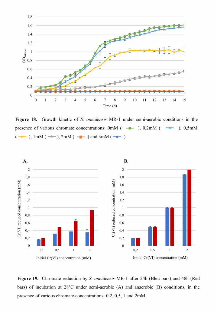
0
0,2
0,4
0,6
0,8
1
1,2
1,4
1,6
1,8
0 1 2 3 4 5 6 7 8 9 10 11 12 13 14 15
OD
600n
m
Time (h)
Figure 18. Growth kinetic of S. oneidensis MR-1 under semi-aerobic conditions in the
presence of various chromate concentrations: 0mM ( ), 0,2mM ( ), 0,5mM
( ), 1mM ( ), 2mM ( ) and 3mM ( ).
Figure 19. Chromate reduction by S. oneidensis MR-1 after 24h (Bleu bars) and 48h (Red
bars) of incubation at 28°C under semi-aerobic (A) and anaerobic (B) conditions, in the
presence of various chromate concentrations: 0.2, 0.5, 1 and 2mM.
0
0,2
0,4
0,6
0,8
1
1,2
1,4
1,6
1,8
2
0,2 0,5 1 2
Cr(
VI)
red
uced
con
cent
rati
on (
mM
)
Initial Cr(VI) concentration (mM)
0
0,2
0,4
0,6
0,8
1
1,2
1,4
1,6
1,8
2
0,2 0,5 1 2
Cr(
VI)
red
uced
con
cent
rati
on (
mM
)
Initial Cr(VI) concentration (mM)
A. B.
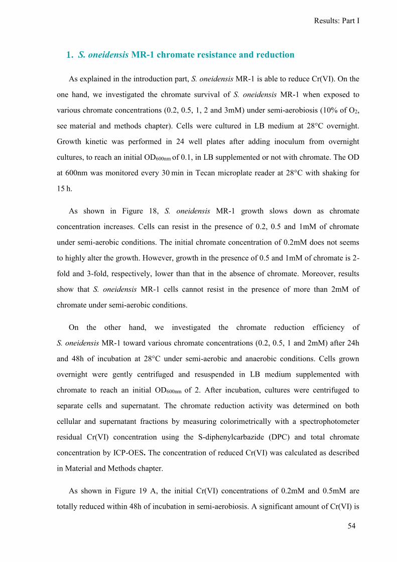
Results: Part I
54
S. oneidensis MR-1 chromate resistance and reduction
As explained in the introduction part, S. oneidensis MR-1 is able to reduce Cr(VI). On the
one hand, we investigated the chromate survival of S. oneidensis MR-1 when exposed to
various chromate concentrations (0.2, 0.5, 1, 2 and 3mM) under semi-aerobiosis (10% of O2,
see material and methods chapter). Cells were cultured in LB medium at 28°C overnight.
Growth kinetic was performed in 24 well plates after adding inoculum from overnight
cultures, to reach an initial OD600nm of 0.1, in LB supplemented or not with chromate. The OD
at 600nm was monitored every 30 min in Tecan microplate reader at 28°C with shaking for
15 h.
As shown in Figure 18, S. oneidensis MR-1 growth slows down as chromate
concentration increases. Cells can resist in the presence of 0.2, 0.5 and 1mM of chromate
under semi-aerobic conditions. The initial chromate concentration of 0.2mM does not seems
to highly alter the growth. However, growth in the presence of 0.5 and 1mM of chromate is 2-
fold and 3-fold, respectively, lower than that in the absence of chromate. Moreover, results
show that S. oneidensis MR-1 cells cannot resist in the presence of more than 2mM of
chromate under semi-aerobic conditions.
On the other hand, we investigated the chromate reduction efficiency of
S. oneidensis MR-1 toward various chromate concentrations (0.2, 0.5, 1 and 2mM) after 24h
and 48h of incubation at 28°C under semi-aerobic and anaerobic conditions. Cells grown
overnight were gently centrifuged and resuspended in LB medium supplemented with
chromate to reach an initial OD600nm of 2. After incubation, cultures were centrifuged to
separate cells and supernatant. The chromate reduction activity was determined on both
cellular and supernatant fractions by measuring colorimetrically with a spectrophotometer
residual Cr(VI) concentration using the S-diphenylcarbazide (DPC) and total chromate
concentration by ICP-OES. The concentration of reduced Cr(VI) was calculated as described
in Material and Methods chapter.
As shown in Figure 19 A, the initial Cr(VI) concentrations of 0.2mM and 0.5mM are
totally reduced within 48h of incubation in semi-aerobiosis. A significant amount of Cr(VI) is
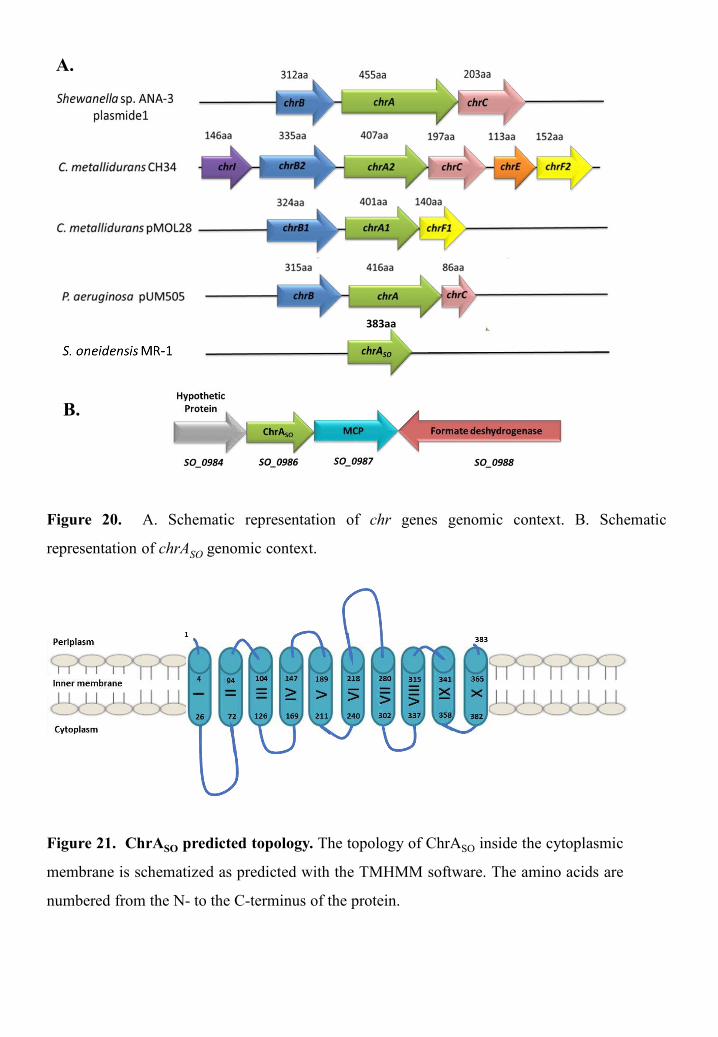
Figure 20. A. Schematic representation of chr genes genomic context. B. Schematic
representation of chrASO genomic context.
A.
B.
Figure 21. ChrASO predicted topology. The topology of ChrASO inside the cytoplasmic
membrane is schematized as predicted with the TMHMM software. The amino acids are
numbered from the N- to the C-terminus of the protein.
383aa

Results: Part I
55
reduced at high initial chromate concentrations 1mM and 2mM. MR-1 shows the ability to
reduce totally the initial chromate concentration of 0.2, 0.5 and 1mM within 24h of incubation
in anaerobic conditions (Figure 19 B). The initial chromate concentration of 2mM is reduced
at 91% within 24h of incubation, before being completely reduced after 48h of incubation in
anoxic conditions (Figure 19 B).
Overall, these results show that in both semi-aerobic and anaerobic conditions the amount
of reduced Cr(VI) increases with increasing initial chromate concentration, and the chromate
reduction efficiency of MR-1 in anaerobic condition is higher than in semi-aerobiosis.
Chromate efflux pump of S. oneidensis MR-1
As previously mentioned, chromate efflux pumps (ChrA) can be responsible for chromate
resistance (Díaz-Pérez et al, 2007; Ramírez-Díaz et al, 2011). Although, the numerous studies
show S. oneidensis MR-1 ability to reduce Cr(VI) as a direct defense mechanism to cope with
chromate toxicity, the involvement of a chromate efflux pump in Cr(VI) resistance behavior
has never been reported.
We performed a Blastp search on S. oneidensis MR-1 using the well-known chromate ion
transporters, such as the ChrA proteins of Shewanella sp. ANA3, Cupriavidus metallidurans
and Pseudomonas aeruginosa. We only identified the SO0986 protein, encoded by a
chromosomal gene, as a ChrA homolog. In contrast to the species cited above, S. oneidensis
MR-1 does not contain homologs of the chrB and chrF genes that sometimes co-occur with
chrA (Figure 20 A). The corresponding gene of SO0986 protein, that we decided to hereafter
refer to as ChrASO (SO for Shewanella oneidensis), has a different genomic context. chrASO is
located upstream to SO0987 gene coding for an MCP, which has been previously shown to be
implicated in chromate detection in S. oneidensis MR-1 (Armitano et al, 2011), and to
SO0988 gene coding for a putative formate dehydrogenase (Figure 20 B).
ChrASO contains 383 amino acids with an apparent molecular mass of 40.9 kDa. It
includes two chromate transporter regions (PF02417) and therefore belongs to the LCHR
subfamily of the chromate ion transporter (CHR) superfamily. The TMHMM program
predicts that ChrASO contains 10 transmembrane helices, which is usual for the proteins of the

Figure 22. Phylogenetic tree of a subset of ChrA proteins. Sequence and species
names are indicated. The names are colored according to their origin: α-Proteobacteria
in green, -Proteobacteria in orange, ε-Proteobacteria in black, -Proteobacteria in blue
and Terrabacteria in brown. ChrASO is indicated by a red arrow. The asterisks indicate
plasmid-encoded ChrA proteins.

Results: Part I
56
LCHR family. The ChrASO protein is most likely inserted in the cytoplasmic membrane with
its N-terminus oriented towards the periplasm, as schematized in Figure 21.
Furthermore, we performed a Blastp search using ChrASO as query. Several hundreds of
homologous putative proteins were identified in bacteria belonging for the vast majority of
them to the Proteobacteria group, but also for some of them to the Terrabacteria group. The
highest percentages of identities were found between ChrASO and proteins from other
Shewanella species. ChrASO is also highly homologous to proteins belonging in particular to
Vibrio and Pseudoalteromonas species. We constructed then a phylogenetic tree using a
subset of proteins taken from species representative of the different taxonomic families
(Figure 22). As expected, ChrASO clusters with ChrA proteins of the -proteobacterial class
but more particularly with those of the Aeromonadales, Alteromonadales and Vibrionales
orders. Strinkigly, ChrASO shows the highest percentage of homology with the chromosome-
encoded ChrA proteins from Shewanella sp. ANA-3, C. metallidurans and P. aeruginosa
compared to their plasmid-encoded ChrA proteins, that were functionally characterized
(Juhnke et al, 2002; Pimentel et al, 2002; Aguilar-Barajas et al, 2008). As an example,
ChrASO displays 89% sequence identity with the chromosome-encoded ChrA and only 26%
with the plasmid-encoded ChrA proteins of Shewanella ANA-3.
The involvement of chromate efflux pump in chromate
survival and reduction of S. oneidensis MR-1
We investigated the involvement of ChrASO in chromate resistance and evaluated its
impact on of S. oneidensis MR-1 ability to reduce chromate. Therefore, we constructed a
strain deleted of chrASO and compared its capability to survive and reduce chromate to that of
the wild-type. Our results as reported in detail in figure 2 and 3 of article 1, show that the
chrASO deleted strain exhibits a chromate sensitive phenotype compared to the wild-type. The
chrASO absence affects strongly the Cr(VI) reduction ability of S. oneidensis MR-1, where the
amount of Cr(VI) reduced by the chrASO mutant is about one third of that reduced by the
wild-type strain. Furthermore, we measured the in vitro chromate reductase activity on crude
extracts from the wild-type and the chrASO mutant strains, to decipher whether ChrASO
possesses a chromate reductase activity. The results show that the specific activity of

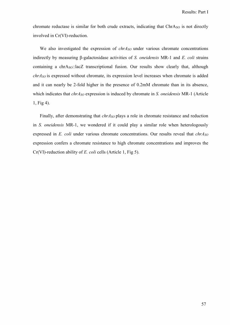
Results: Part I
57
chromate reductase is similar for both crude extracts, indicating that ChrASO is not directly
involved in Cr(VI)-reduction.
We also investigated the expression of chrASO under various chromate concentrations
indirectly by measuring -galactosidase activities of S. oneidensis MR-1 and E. coli strains
containing a chrASO::lacZ transcriptional fusion. Our results show clearly that, although
chrASO is expressed without chromate, its expression level increases when chromate is added
and it can nearly be 2-fold higher in the presence of 0.2mM chromate than in its absence,
which indicates that chrASO expression is induced by chromate in S. oneidensis MR-1 (Article
1, Fig 4).
Finally, after demonstrating that chrASO plays a role in chromate resistance and reduction
in S. oneidensis MR-1, we wondered if it could play a similar role when heterologously
expressed in E. coli under various chromate concentrations. Our results reveal that chrASO
expression confers a chromate resistance to high chromate concentrations and improves the
Cr(VI)-reduction ability of E. coli cells (Article 1, Fig 5).


Results: Part I
58
Article 1:
ChrASO, the chromate efflux pump of
Shewanella oneidensis, improves chromate survival and
reduction
Hiba Baaziz, Cyril Gambari, Anne Boyeldieu, Amine Ali Chaouche, Radia
Alatou, Vincent Méjean, Cécile Jourlin-Castelli and Michel Fons

RESEARCH ARTICLE
ChrASO, the chromate efflux pump ofShewanella oneidensis, improves chromatesurvival and reduction
Hiba Baaziz1,2, Cyril Gambari1, Anne Boyeldieu1, Amine Ali Chaouche1, Radia Alatou2,
Vincent Mejean1, Cecile Jourlin-Castelli1*, Michel Fons1*
1 Unite de Bioenergetique et Ingenierie des Proteines, Institut de Microbiologie de la Mediterranee, Aix-Marseille Universite, Centre National de la Recherche Scientifique, Marseille, France, 2 Laboratoire de
Biologie Moleculaire et Cellulaire, Universite des Frères Mentouri Constantine 1, Constantine, Algeria
* [email protected] (CJC); [email protected] (MF)
Abstract
The chromate efflux pump encoding gene chrASOwas identified on the chromosome of She-
wanella oneidensisMR1. Although chrASO is expressed without chromate, its expression
level increases when Cr(VI) is added. When deleted, the resulting mutant ΔchrASO exhibits
a chromate sensitive phenotype compared to that of the wild-type strain. Interestingly, heter-
ologous expression of chrASO in E. coli confers resistance to high chromate concentration.
Moreover, expression of chrASO in S. oneidensis and E. coli significantly improves Cr(VI)
reduction. This effect could result either from extracytoplasmic chromate reduction or from a
better cell survival leading to enhanced Cr(VI) reduction.
Introduction
The transition metal chromium has different oxidation states, but only two of them are stable,
namely the trivalent Cr(III) and the hexavalent Cr(VI) forms. The Cr(III) form which is rela-
tively insoluble is considered to be poorly toxic since it can hardly penetrate the cells. The
Cr(VI) form (chromate and dichromate) is soluble and can be conveyed through cell mem-
branes by different transporters, making it a very toxic compound for both eukaryotic and
prokaryotic cells [1].
Chromate, which is chemically and structurally similar to sulfate, is thought to enter
the bacterial cells mainly via sulfate ABC transporters. Once inside the cells, Cr(VI) can be
reduced, either enzymatically or not, giving rise to intermediates which are responsible for var-
ious damages to DNA and cellular components. Some of these alterations are due to the gener-
ation of reactive oxygen species (ROS). When present inside the cells, Cr(III), the final product
of Cr(VI) reduction, causes toxic effects on both DNA and proteins by binding respectively to
phosphate, and to carboxyl and thiol groups [2].
Bacteria have developed different strategies to resist chromate [2,3]. One strategy is to
limit its cell entry by reducing Cr(VI) to Cr(III) extracellularly. Reduction can be achieved
anaerobically by membrane bound reductases like flavin reductases, cytochromes and
PLOSONE | https://doi.org/10.1371/journal.pone.0188516 November 22, 2017 1 / 15
a1111111111a1111111111a1111111111a1111111111a1111111111
OPENACCESS
Citation: Baaziz H, Gambari C, Boyeldieu A, Ali
Chaouche A, Alatou R, Mejean V, et al. (2017)
ChrASO, the chromate efflux pump of Shewanella
oneidensis, improves chromate survival and
reduction. PLoS ONE 12(11): e0188516. https://
doi.org/10.1371/journal.pone.0188516
Editor: Eric Cascales, Centre National de la
Recherche Scientifique, Aix-Marseille Universite,
FRANCE
Received: September 22, 2017
Accepted:November 8, 2017
Published: November 22, 2017
Copyright:© 2017 Baaziz et al. This is an open
access article distributed under the terms of the
Creative Commons Attribution License, which
permits unrestricted use, distribution, and
reproduction in any medium, provided the original
author and source are credited.
Data Availability Statement: All relevant data are
within the paper and its Supporting Information
files.
Funding: The authors acknowledge funding by the
Centre National de la Recherche Scientifique (www.
cnrs.fr), Aix-Marseille Universite (www.univ-amu.
fr) and HTS-BIO (www.htsbio.com, Grant N˚152
660). H.B. was supported by Universite de
Constantine 1. A.B. was supported by a MESR
fellowship. C.G. was supported by the Conseil

hydrogenases that are part of electron transport systems using chromate as electron acceptor.
In sulfate-reducing bacteria for example, extracellular Cr(VI) reduction can be carried out
both enzymatically and chemically by oxidation of the H2S generated during anaerobic respi-
ration [4,5].
A second strategy is to reduce aerobically the intracellular Cr(VI) [6]. The best known cyto-
solic proteins involved in this reduction are ChrR of Pseudomonas putida and YieF of Escheri-
chia coli [7,8]. Both enzymes share sequence homologies and belong to the class I chromate
reductases. Nevertheless their mechanisms of action are different. YieF directly reduces Cr(VI)
to Cr(III) through a four-electron transfer reaction while ChrR proceeds by combining one-
and two-electron transfer steps [7]. As a result, ChrR generates more ROS than YieF during
chromate reduction. The class II chromate reductases could require NAD(P)H as an electron
donor and generally possess a nitroreductase activity. One of them, NfsA from E. coli, was
characterized and shown to increase chromate tolerance when overproduced [9]. Intracellular
chromate reduction is often associated with mechanisms to detoxify the cell from the ROS and
to repair the lesions, caused in particular to DNA [10,11].
A third strategy to deal with chromate toxicity is to rapidly extrude it from the cell cyto-
plasm by the mean of chromate efflux pump. The ChrA proteins of Pseudomonas aeruginosa
and Cupriavidus metallidurans were the first proteins proved to be involved in this process
[12,13]. Thereafter, several ChrA homologues were identified based on genome sequence anal-
ysis and grouped into a large family, called the Chromate ion transporter (CHR) family [14].
This latter was further divided into two subfamilies on the basis of the protein length. One
subfamily contains proteins with a sequence length of about 180 amino acids, named short-
chain CHR (SCHR), and bearing only one chromate transporter domain. The proteins of
the second subfamily, containing two chromate transporter domains, are larger (around 400
amino acids) and called long-chain CHR (LCHR). The ChrA proteins possess several trans-
membrane regions and are encoded by genes that can be found either on chromosome or on
plasmid. Some bacteria possess several chrA genes associated or not with other genes involved
in chromate resistance or regulation (like chrB, chrC or chrF, found for example in C.metalli-
durans, Ochrobactrum tritici and Shewanella sp. strain ANA-3) [14]. These examples are not
exhaustive and different strategies can co-exist in a same bacterium.
Shewanella oneidensis is an aquatic bacterium that belongs to the ɣ-Proteobacteria class. Itcan utilize a wide range of terminal electron acceptors including fumarate, nitrate, TMAO, as
well as oxidized metals including Fe(III) and Mn(IV). This respiratory versatility makes it a
key player for bioremediation [15]. The capability of S. oneidensis MR-1 to resist to and/or
reduce Cr(VI) has been explored in various conditions, pointing out the involvement of differ-
ent mechanisms for these functions [16–18]. A transcriptomic analysis revealed that more
than 80 genes were upregulated when strain MR-1 was under Cr(VI)-reduction condition
[19]. Moreover, the response regulator SO2426 was shown to control the expression of genes
involved in the relationship between Fe homeostasis and Cr(VI)-induced stress tolerance [20].
Concerning reduction, MtrC and OmcA, two extracellular decaheme cytochromes located in
the outer membrane of the cells and previously known to be involved in Fe(III) reduction,
were characterized as terminal Cr(VI) reductases in vitro as well as in vivo [18,21]. A protein
homologous to ChrR of P. putida was also identified in S. oneidensis (SO3585), but its role in
chromate resistance seems not to be crucial since only the initial rate of chromate disappear-
ance is affected in the so3585-deleted strain [22].
The aim of this work was to confirm that resistance to chromate in S. oneidensis MR-1
involves a chromate efflux pump and to evaluate the impact of this efflux pump on the capabil-
ity of this bacterium to reduce Cr(VI) to the less toxic form Cr(III).
Chromate resistance in S. oneidensis
PLOSONE | https://doi.org/10.1371/journal.pone.0188516 November 22, 2017 2 / 15
General PACA (www.regionpaca.fr). The funders
had no role in study design, data collection and
analysis, decision to publish, or preparation of the
manuscript.
Competing interests: The authors have declared
that no competing interests exist.


Materials andmethods
Strains, medium and growth conditions
The E. coli strains CC118 λpir and 1047/pRK2013 used for conjugation, and MC1061 deriva-
tives used for chromate resistance and reduction assays were routinely grown in Lysogeny
Broth (LB) medium at 37˚C, or 30˚C when specified [23,24].
All S. oneidensis strains used in this study are derivatives of strain MR1-R (referred as wild-
type and used instead of MR-1 to allow counter-selection by rifampicin in the conjugation
experiments) and were routinely grown at 28˚C in LB under aerobic (agitation) or anaerobic
(static) conditions. In the latter case, LB medium was supplemented with 20 mM trimethyla-
mine oxide (TMAO) as electron acceptor. When growth was performed in the presence of
chromate, disposable tubes were used and incubated either statically or under agitation. The
dissolved oxygen (DO) of the latter condition measured by a Clark electrode was 10%. This
condition was then referred as semi-aerobic. Chromate challenge was carried out by supple-
menting LB medium at the required final concentration with a filter-sterilized stock solution
of potassium chromate (K2CrO4, Sigma-Aldrich).
If required, media were solidified by adding 17 g.L-1 agar. When needed, chloramphenicol
(Cm) was used at 25 μg.mL–1. Growth was determined spectrophotometrically by monitor-
ing changes in optical density at 600 nm compared to the same medium without bacterium
(OD600nm).
Chromate resistance assays in S. oneidensis
S. oneidensis pre-cultures grown overnight on LB plates were suspended in LB medium and
used to inoculate fresh LB medium to an initial OD600nm = 0.2. Cells were then grown until an
OD600nm = 0.5 and submitted to two different assays.
For the spot assay, 10-fold serial dilutions of cell cultures were spotted on LB plates supple-
mented or not with 0.5 mM of chromate and incubated at 28˚C. Plates were scanned after 4
days of incubation.
For the viability assay, chromate was added to the cell cultures (OD600nm = 0.5) to a final
concentration of 0.2 mM. After 5 hours, cells were appropriately diluted in LB and spread onto
LB agar, and incubated at 28˚C. The total number of viable cells was estimated based on the
number of colony-forming unit (CFU). Results were expressed as the percentage of viable
counts measured in these conditions compared to that of the same culture grown in the
absence of chromate.
Cr(VI)-reduction assays in S. oneidensis
In vivo Cr(VI) measurements were carried out on both semi-aerobic and anaerobic cultures.
When fresh cultures reached an OD600nm = 0.5, chromate was added to a final concentration
of 1 mM. After 2h of incubation at 28˚C, cell cultures were centrifuged at 8,000 g for 5 min
and residual Cr(VI) concentration present in the supernatant was determined using the S-
diphenylcarbazide (DPC) method [25] slightly modified. Briefly, 10, 20, and 50 μL supernatant
samples were added in a 2 mL tube containing 1 mL of H3PO4 0.5% and the volume of water
necessary to obtain a 1960 μL final volume. Forty μL of DPC reagent (5 mg.mL-1 in 95% ace-
tone and stored in dark at room temperature) were added, gently mixed and kept at room tem-
perature for 5–10 min. Absorbance was measured at 540 nm. Cr(VI) concentration in the
samples was calculated from a standard curve. These experiments were repeated three times.
In vitro measurements of chromate reductase activity were performed on crude extracts as
described previously [26] with slight modifications. Briefly, wild-type and chrASO deleted
Chromate resistance in S. oneidensis
PLOSONE | https://doi.org/10.1371/journal.pone.0188516 November 22, 2017 3 / 15


mutant strains were grown overnight in LB medium at 28˚C under agitation. The cells were
then harvested, washed with sodium phosphate buffer (pH7) and resuspended in the same
buffer before disruption by French press. The cell lysates were then centrifuged and the super-
natants (crude extracts) recovered. Protein concentrations were measured using Protein Assay
Dye Reagent (Bio-Rad), and bovine serum albumin as standard. Reaction mixtures (1 mL)
containing 100 μM of K2CrO4 [Cr(VI)] and 1.5 mg of proteins (crude extracts) were incubated
at 30˚C for 30 min. The residual Cr(VI) concentration in the reaction mixture was estimated
using the DPC method as described above.
Construction of plasmid pchrASO
To evaluate the impact of ChrASO on chromate resistance and reduction in E. coli, the chrASO
open reading frame (so0986) was PCR-amplified fromMR1-R chromosomal DNA, from
upstream of the ATG start codon (with an optimized Shine Dalgarno) to the TAA stop codon,
and cloned between the SacI and the XbaI restriction sites of the pBAD33 vector, under the
control of the inducible Para promoter [27]. The resulting plasmid, called pchrASO, was intro-
duced into strain MC1061 and controlled by DNA-sequencing (S1 Table).
Chromate resistance and reduction assays in E. coli
Pre-cultures of MC1061 derivatives containing either the plasmid pchrASO or the pBAD33 vec-
tor were used to inoculate fresh LB medium to an initial OD600nm = 0.05. Cells were grown
until an OD600nm = 0.2 prior to the addition of chromate at various final concentrations (0,
0.2, 0.4, 0.8 and 1.2 mM), then the cells were incubated at 30˚C. The cultures were regularly
sampled to measure the OD600nm and to quantify the chromate using the DPC method.
In vitro chromate reductase activities were measured on crude extracts from E. coli cells
containing either the pBAD33 vector or the plasmid pchrASO as described above for the mea-
surements on S. oneidensis crude extracts.As ChrASO is most probably inserted inside the cyto-
plasmic membrane, its overexpression is toxic for the host. Preliminary growth assays of the E.
coli strain harboring pchrASO were carried out at different temperatures (30˚C or 37˚C) in the
presence of different concentrations of arabinose (0, 0.005 or 0.01%). We observed that a tem-
perature of 30˚C and the absence of inducer allowed a better growth of the strain, thus these
parameters were used in further experiments.
Construction of a deletion mutant
The chrASO-deleted strain was constructed as described previously [28]. Briefly, upstream and
downstream regions flanking the gene were amplified by PCR from aMR1-R strain (by using
primers SO_0986 D2 to D5, S1 Table) and cloned into the suicide vector pKNG101 at the
restriction sites BamHI and SpeI. The ligation product was transformed into the E. coli CC118
λpir strain. The resulting plasmid was introduced into MR1-R strain by conjugation using the
E. coli helper strain 1047/pRK2013 [29]. The plasmid was integrated in the MR1-R chromo-
some by a first recombination event and removed by a second recombination event in the
presence of 6% sucrose. Deletion of chrASO gene was confirmed by PCR.
Reporter gene assay
A putative promoter region was searched upstream of the start codon of chrASO using the
BPROM Softberry online software (http://www.softberry.com/berry.phtml?topic=
bprom&group=programs&subgroup=gfindb). A DNA fragment corresponding to the 400 bp
upstream from the ATG was amplified by PCR using S. oneidensis MR1-R chromosomal DNA,
Chromate resistance in S. oneidensis
PLOSONE | https://doi.org/10.1371/journal.pone.0188516 November 22, 2017 4 / 15

digested with XbaI and SpeI, and ligated upstream of the lacZ gene of vector pACYC184-lacZ,
which was previously constructed by cloning the β-galactosidase-encoding gene lacZ from
pGE593 into the vector pACYC184 (S1 Table) [30,31]. The ligation product was transformed
into E. coli strain MC1061. The resulting plasmid called pchrASO::lacZwas introduced into S.
oneidensis MR1-R by conjugation. Plasmid construction was checked by DNA sequencing. As a
control, another fusion, named pmxd450::lacZ, was similarly constructed using a DNA fragment
corresponding to the 450 bp upstream from the ATG of mxdA [32]. S. oneidensis MR1-R strain
containing pchrASO::lacZ or pmxd450::lacZwas grown overnight on LB-agar plate. The over-
night culture was diluted to an OD600nm = 0.1 in fresh liquid LB medium containing various
concentrations of chromate (0, 0.05, 0.1 and 0.2 mM) and incubated in semi-aerobiosis at 28˚C
for 16 hours prior to β-galactosidase activity quantification. The β-galactosidase activity wasdetermined by a Miller assay adapted for use with plate reader [33]. Briefly, cells were trans-
ferred into a microtiter plate and OD600nm was measured using a Tecan Spark 10Mmicroplate
reader. Cells were then lysed using lysozyme and PopCulture reagent (Sigma-Aldrich) prior to
incubation with Z buffer (62 mMNa2HPO4, 45 mMNaH2PO4, 10 mMKCl, 1 mMMgSO4, 50
mM β-mercaptoethanol). O-nitrophenyl-ß-D-galactopyranoside (ONPG) was added to the mix
before kinetic quantifications of OD420nm. Slope of the obtained curves were calculated and β-galactosidase activity (arbitrary units) was determined by the following equation:
ð1000� slopeÞ=ðOD600nm � volumeof reactionðmLÞÞ:
Bioinformatics analysis
The amino acid sequence of ChrASO was retrieved from the MicroScope Microbial Genome
Annotation and Analysis Platform (https://www.genoscope.cns.fr/agc/microscope/home/).
Prediction of transmembrane helices was carried out using the TMHMM online software
(http://www.cbs.dtu.dk/services/TMHMM-2.0/).
Proteins sharing homologies with ChrASO were searched in the Bacteria kingdom using the
BlastP software on the NCBI server (https://blast.ncbi.nlm.nih.gov). Representative sequences
of different phyla were subsequently selected, as well as the sequences of four plasmid-encoded
ChrA proteins that were previously characterized. For the phylogenetic analysis, we used the
“Phylogeny.fr” software in the “one-click” mode, i.e. with the default parameters optimized by
the authors (http://www.phylogeny.fr/). For the tree rendering step, we used the software “Fig-
Tree” version 1.4.3 (http://tree.bio.ed.ac.uk/software/figtree/) in which we entered the result
in Netwick format obtained with “Phylogeny”. The tree rooting was performed with the mid-
point option.
Results
S. oneidensis chromosome contains a chromate efflux pump encodinggene
The S. oneidensis MR-1 genome contains only one gene (so0986) whose product is annotated
as a putative chromate efflux pump. This gene is not located on the megaplasmid but on the
chromosome. Accordingly, a Blastp search performed on S. oneidensis using well-known chro-
mate ion transporters, such as the ChrA proteins of P. aeruginosa and C.metallidurans, only
identified the SO0986 protein as a ChrA homolog. In contrast, S. oneidensis does not contain
homologs of the chrB and chrF genes which sometimes co-occur with chrA.
For clarity, we will refer to the SO0986 protein as ChrASO. This protein of 383 amino acids
with a molecular mass of 40.9 kDa contains two chromate transporter regions (PF02417) and
Chromate resistance in S. oneidensis
PLOSONE | https://doi.org/10.1371/journal.pone.0188516 November 22, 2017 5 / 15

therefore belongs to the LCHR subfamily of the chromate ion transporter (CHR) superfamily.
The TMHMM program predicted that ChrASO contains 10 transmembrane helices, which is
usual for the proteins of the LCHR family. The ChrASO protein is most likely inserted in the
cytoplasmic membrane with its N-terminus oriented towards the periplasm, as schematized in
Fig 1A.
A Blastp search was performed using ChrASO as query. Several hundreds of homologous
proteins were identified in bacteria belonging for the vast majority of them to the Proteobac-
teria group, but also for some of them to the Terrabacteria group. As expected, the highest per-
centages of identities were found between ChrASO and proteins from other Shewanella species.
ChrASO is also highly homologous to proteins belonging in particular to Vibrio and Pseudoal-
teromonas species. A phylogenetic tree was constructed using a subset of proteins taken from
species representative of the different taxonomic families (Fig 1B). Not surprisingly, ChrASO
clusters with ChrA proteins of the γ-proteobacterial class but more particularly with those of
the Aeromonadales, Alteromonadales and Vibrionales orders. Strinkigly, ChrASO shows the
highest percentage of homology with the chromosome-encoded ChrA proteins from Shewa-
nella sp. ANA-3, P. aeruginosa and C.metallidurans compared with their plasmid-encoded
ChrA proteins, that were functionally characterized [12,13,34]. As an example, ChrASO dis-
plays 89% sequence identity with the chromosome-encoded ChrA and only 26% with the plas-
mid-encoded ChrA proteins of Shewanella ANA-3. Moreover S. oneidensis megaplasmid does
not harbor chrA gene.
ChrASO is involved in chromate resistance in S. oneidensis
Since ChrASO clearly belongs to the chromate ion transporter family, we wondered if it could
be involved in chromate resistance in S. oneidensis. We therefore constructed a strain deleted
of the chrASO gene and compared its capability to survive in the presence of chromate to that
of the wild-type strain.
Wild-type and mutant cells were first allowed to grow at 28˚C until an OD600 of 0.5. These
pre-cultures were then submitted to two chromate resistance tests. In the first one, cells were
allowed to grow for 5 hours after addition of 0.2 mM chromate in the pre-cultures. Afterwards,
bacterial cells were diluted and plated on LB-agar to determine the percentage of survival after
“colony forming unit” counting. The same experiment was carried out in the absence of chro-
mate as a control. As observed on Fig 2A, the survival percentage is about 40% in the wild-type
strain while it is only about 25% in the chrASO deleted strain. In the second test, serial 10-fold
dilutions of the bacterial pre-cultures were performed and then spotted on plates containing
either no chromate or 0.5 mM chromate. The plates were incubated at 28˚C to allow bacterial
growth. In the absence of chromate, no growth difference is observed between the wild-type
and the chrASO mutant strains (Fig 2B). The presence of chromate impairs the growth of both
strains, but the effect is clearly more pronounced for the chrASO mutant strain (Fig 2B). These
results strongly suggest that ChrASO is involved in chromate resistance in S. oneidensis.
Chromate reduction is impaired in a chrASO deleted strain
The impact of ChrASO on the capability of S. oneidensis to reduce Cr(VI) was evaluated by
comparing the percentage of Cr(VI)-reduction obtained when the wild-type, the chrASO
mutant and the chrASO mutant containing the plasmid pchrASO were exposed to 1 mM chro-
mate either in semi-aerobic (as defined in the Materials and methods section) or anaerobic
conditions during 2 hours. The concentration of Cr(VI) was evaluated by the DPC method. As
shown in Fig 3, in both conditions (semi-aerobiosis and anaerobiosis), the deletion of chrASO
Chromate resistance in S. oneidensis
PLOSONE | https://doi.org/10.1371/journal.pone.0188516 November 22, 2017 6 / 15

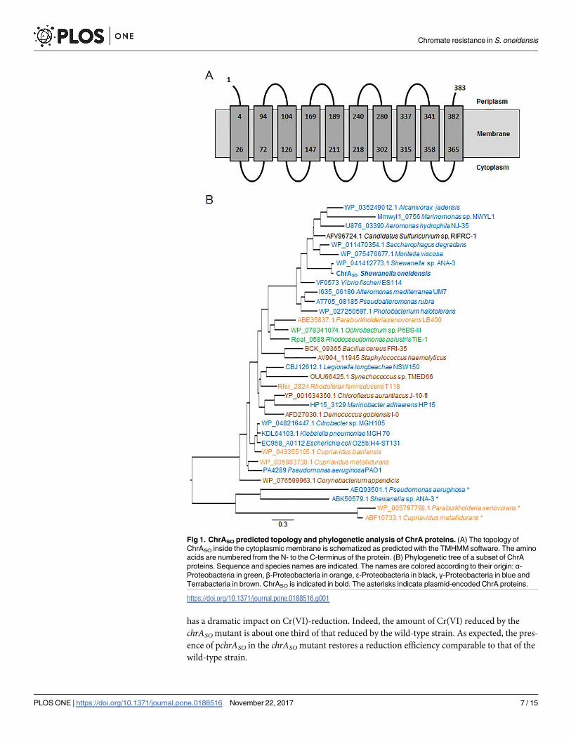
has a dramatic impact on Cr(VI)-reduction. Indeed, the amount of Cr(VI) reduced by the
chrASO mutant is about one third of that reduced by the wild-type strain. As expected, the pres-
ence of pchrASO in the chrASO mutant restores a reduction efficiency comparable to that of the
wild-type strain.
Fig 1. ChrASO predicted topology and phylogenetic analysis of ChrA proteins. (A) The topology ofChrASO inside the cytoplasmic membrane is schematized as predicted with the TMHMM software. The aminoacids are numbered from the N- to the C-terminus of the protein. (B) Phylogenetic tree of a subset of ChrAproteins. Sequence and species names are indicated. The names are colored according to their origin: α-Proteobacteria in green, β-Proteobacteria in orange, ε-Proteobacteria in black, γ-Proteobacteria in blue andTerrabacteria in brown. ChrASO is indicated in bold. The asterisks indicate plasmid-encoded ChrA proteins.
https://doi.org/10.1371/journal.pone.0188516.g001
Chromate resistance in S. oneidensis
PLOSONE | https://doi.org/10.1371/journal.pone.0188516 November 22, 2017 7 / 15


Fig 2. Effect of chrASO on chromate resistance in S. oneidensis strains. Two different assays wereconducted to evaluate the impact of chrASO on chromate resistance. (A) In the first assay, [CFU + Cr(VI)]/[CFU—Cr(VI)] (%) indicates the percentage of viable counts of the wild-type (WT) and the chrASO deletedmutant (ΔchrASO) after 5 hours of growth in semi-aerobiosis conditions and in the presence of 0.2 mMchromate, compared to that of the same strains grown in similar conditions without chromate. Values aremeans ± standard deviations (error bars) from at least two experiments. The mean absolute values of thenumber of CFU counted in the absence of chromate are 2.49 x 10⁹.mL-1 and 2.41 x 10⁹.mL-1 for the wild-typeand the chrASO deleted mutant strains, respectively. (B) In the second assay, 10-fold serial dilutions of thewild-type (WT) and the chrASO deleted mutant (ΔchrASO) cultures were spotted on LB agar supplemented ornot with 0.5 mM chromate before incubation at 28˚C during 4 days.
https://doi.org/10.1371/journal.pone.0188516.g002
Chromate resistance in S. oneidensis
PLOSONE | https://doi.org/10.1371/journal.pone.0188516 November 22, 2017 8 / 15


To decipher whether ChrASO possesses a chromate reductase activity, we performed in
vitro chromate reductase activity on crude extracts from the wild-type and the chrASO mutant
strains and the results show that the specific activity of chromate reductase is similar for both
crude extracts (0.78 ± 0.1 and 0.88 ± 0.05 nmol reduced-Cr(VI).min-1.mg-1, respectively), indi-
cating that ChrASO is not directly involved in Cr(VI)-reduction.
Fig 3. Impact of chrASO on chromate reduction in S. oneidensis strains. TheWT/pBAD33, ΔchrASO/pBAD33 and ΔchrASO/pchrASO strains were challenged with 1 mM chromate before quantification of Cr(VI).Results were expressed as the percentage of chromate reduced after 2 hours of incubation in semi-aerobic(A) or anaerobic conditions (B). Values are means ± standard deviations (error bars) from at least threeexperiments.
https://doi.org/10.1371/journal.pone.0188516.g003
Chromate resistance in S. oneidensis
PLOSONE | https://doi.org/10.1371/journal.pone.0188516 November 22, 2017 9 / 15

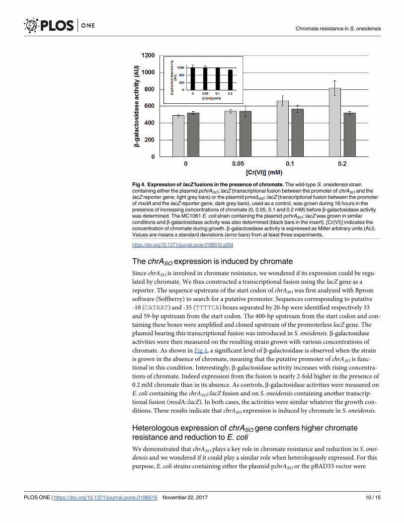
The chrASO expression is induced by chromate
Since chrASO is involved in chromate resistance, we wondered if its expression could be regu-
lated by chromate. We thus constructed a transcriptional fusion using the lacZ gene as a
reporter. The sequence upstream of the start codon of chrASO was first analyzed with Bprom
software (Softberry) to search for a putative promoter. Sequences corresponding to putative
-10 (CATAAT) and -35 (TTTTCA) boxes separated by 20-bp were identified respectively 33
and 59-bp upstream from the start codon. The 400-bp upstream from the start codon and con-
taining these boxes were amplified and cloned upstream of the promoterless lacZ gene. The
plasmid bearing this transcriptional fusion was introduced in S. oneidensis. β-galactosidaseactivities were then measured on the resulting strain grown with various concentrations of
chromate. As shown in Fig 4, a significant level of β-galactosidase is observed when the strain
is grown in the absence of chromate, meaning that the putative promoter of chrASO is func-
tional in this condition. Interestingly, β-galactosidase activity increases with rising concentra-
tions of chromate. Indeed expression from the fusion is nearly 2-fold higher in the presence of
0.2 mM chromate than in its absence. As controls, β-galactosidase activities were measured on
E. coli containing the chrASO::lacZ fusion and on S. oneidensis containing another transcrip-
tional fusion (mxdA::lacZ). In both cases, the activities were similar whatever the growth con-
ditions. These results indicate that chrASO expression is induced by chromate in S. oneidensis.
Heterologous expression of chrASO gene confers higher chromateresistance and reduction to E. coli
We demonstrated that chrASO plays a key role in chromate resistance and reduction in S. onei-
densis and we wondered if it could play a similar role when heterologously expressed. For this
purpose, E. coli strains containing either the plasmid pchrASO or the pBAD33 vector were
Fig 4. Expression of lacZ fusions in the presence of chromate. The wild-type S. oneidensis straincontaining either the plasmid pchrASO::lacZ (transcriptional fusion between the promoter of chrASO and thelacZ reporter gene; light grey bars) or the plasmid pmxd450::lacZ (transcriptional fusion between the promoterofmxdA and the lacZ reporter gene; dark grey bars), used as a control, was grown during 16 hours in thepresence of increasing concentrations of chromate (0, 0.05, 0.1 and 0.2 mM) before β-galactosidase activitywas determined. The MC1061 E. coli strain containing the plasmid pchrASO::lacZwas grown in similarconditions and β-galactosidase activity was also determined (black bars in the insert). [Cr(VI)] indicates theconcentration of chromate during growth. β-galactosidase activity is expressed as Miller arbitrary units (AU).Values are means ± standard deviations (error bars) from at least three experiments.
https://doi.org/10.1371/journal.pone.0188516.g004
Chromate resistance in S. oneidensis
PLOSONE | https://doi.org/10.1371/journal.pone.0188516 November 22, 2017 10 / 15

grown in the presence of various concentrations of chromate (0, 0.2, 0.4, 0.8 and 1.2 mM) and
the time course of growth was followed by measuring the OD600 of the different cultures. To
avoid toxicity due to ChrASO overproduction, cultures were performed at 30˚C without the
inducer arabinose (for details see the Materials and methods section). As shown in Fig 5A,
when the initial concentration of chromate is 0.2 mM, growth of both strains is similar during
the first 8 hours. Then, cells containing the pchrASO continue to grow whereas cells containing
only the vector stop growing. With higher initial concentrations of chromate, the growth
remains possible when the pchrASO is present, although it slows down as concentration in
chromate increases. In contrast, higher concentrations of chromate have a dramatic impact on
cells harboring only the vector. The residual concentration of Cr(VI) was evaluated after 2
and 7 hours of growth. As shown on Fig 5B, the percentage of Cr(VI)-reduction is quickly
improved when the cells contain the pchrASO compared to the ones with the vector. Reduction
seems to be inefficient after 7 hours of incubation when the vector is present, whereas it is
clearly improved in cells harboring the pchrASO. These results suggest that ChrASO has also a
positive effect on the reduction efficiency of Cr(VI) in E. coli.
We performed in vitro chromate reductase activity assays on crude extracts from E. coli
strains containing either the pBAD33 vector or the pchrASO plasmid and the results show that
the specific activity of chromate reductase is similar for both crude extracts (1.1 ± 0.12 and
1.05 ± 0.06 nmol reduced-Cr(VI).min-1.mg-1, respectively), confirming that ChrASO is not
directly involved in Cr(VI)-reduction.
Discussion
A bioinformatics analysis revealed that ChrASO (SO0986) belongs to the large family of chro-
mate ion transporters. In agreement with this prediction, a S. oneidensis strain deleted of the
chrASO gene is less resistant to chromate than the wild-type strain. Strikingly, expression of
chrASO in E. coli makes the strain capable of growing in the presence of high chromate concen-
trations, as also observed for the plasmid-encoded ChrA of Shewanella ANA-3 and for several
ChrA of Burkholderia xenovorans LB400 [34,35]. Therefore ChrASOmost probably functions
as a chromate efflux pump and expulses chromate ions from the cytoplasm, as demonstrated
for some members of the CHR family [13,34].
It is noteworthy that, in the presence of ChrASO, the percentage of Cr(VI) reduction is
higher than in its absence in both S. oneidensis and E. coli. A direct role of ChrASO in Cr(VI)
reduction was ruled out, since the specific activity of chromate reductase measured in vitro on
crude extracts from both S. oneidensis and E. coli strains is similar in the absence and the pres-
ence of ChrASO. One hypothesis to explain this phenomenon is that ChrASO promotes Cr(VI)
reduction by expulsing chromate outside the cells where it can then be reduced. In S. oneiden-
sis, this scenario is worth considering as two outer membrane decaheme cytochromes, MtrC
and OmcA, are known to be involved in extracellular chromate reduction [21]. Another
hypothesis could be that the presence of ChrASO improves cell survival by lowering intracellu-
lar Cr(VI) concentration and consequently Cr-induced damages inside the cells, which allows
the cells to grow and reduce Cr(VI) over an extended period of time. This could be true for
both S. oneidensis and E. coli.
This study also shows that, although chrASO is expressed without chromate, the level of
expression is higher in its presence. Two previous studies performed on S. oneidensis using
transcriptomic approaches missed this regulation, probably because the applied cut-offs were
quite stringent [19,36]. Indeed, these studies considered only the genes showing at least a 2-fold
or a 3-fold change in expression, respectively. Moreover the experimental conditions used in
both studies were different from ours, which can also explain this discrepancy. The fact that
Chromate resistance in S. oneidensis
PLOSONE | https://doi.org/10.1371/journal.pone.0188516 November 22, 2017 11 / 15

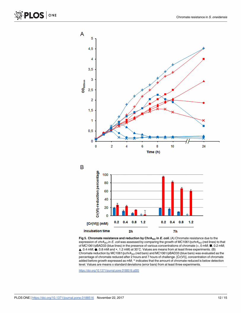
Fig 5. Chromate resistance and reduction by ChrASO in E. coli. (A) Chromate resistance due to theexpression of chrASO in E. coliwas assessed by comparing the growth of MC1061/pchrASO (red lines) to thatof MC1061/pBAD33 (blue lines) in the presence of various concentrations of chromate (+, 0 mM;■, 0.2 mM;▲, 0.4 mM;●, 0.8 mM and ×, 1.2 mM) at 30˚C. Values are means from at least three experiments. (B)Chromate reduction by MC1061/pchrASO (red bars) and MC1061/pBAD33 (blue bars) was evaluated as thepercentage of chromate reduced after 2 hours and 7 hours of challenge. [Cr(VI)], concentration of chromateadded before growth expressed as mM; * indicates that the amount of chromate reduced is below detectionlevel. Values are means ± standard deviations (error bars) from at least three experiments.
https://doi.org/10.1371/journal.pone.0188516.g005
Chromate resistance in S. oneidensis
PLOSONE | https://doi.org/10.1371/journal.pone.0188516 November 22, 2017 12 / 15


chrASO expression is chromate-induced is reminiscent of what was observed for the expression
of several chr genes in other bacteria. For example, in C.metallidurans, transcription of the
plasmidic chrA1 gene was shown to be up-regulated by chromate [12]. In B. xenovorans LB400,
which possesses six genes encoding chromate ion transporters, only the chrA2 gene present on
a megaplasmid is subject to chromate-induced expression [35]. Interestingly, it was recently
demonstrated that ChrA2 is the major determinant of chromate resistance in this organism
[37]. The chrBACF operon of Ochrobactrum tritici 5bvl1, which is located in a chromosomally
integrated transposon, proved also to be chromate-induced [38]. It was subsequently shown
that the ChrB protein is a transcriptional regulator that binds to the chrBACF promoter region
to regulate its expression [39]. As mentioned in the result section, there is no ChrB homolog in
S. oneidensis, meaning that the regulation of chrASO expression probably depends on a regula-
tory protein belonging to a family different from that of ChrB. It will be interesting to identify
this protein, since no chromate-sensing regulator except from ChrB was previously described.
In conclusion, although ChrASO plays a key role in chromate resistance in S. oneidensis,
other transporters could be involved in this process. Of interest, genes encoding the three com-
ponents of a putative heavy metal efflux pump (SO0518 to SO0520, CzcCBA) were observed
to be up-regulated by chromate [19]. This suggests a role in chromate resistance, although this
pump does not belong to the CHR family.
Supporting information
S1 Table. Plasmids and primers used in this study.
(TIF)
Acknowledgments
We are very grateful to Carole Baffert for her help with dissolved oxygen measurements. We
would like to thank Ahmed Zellat for technical assistance and all the members of the Mejean
group, in particular O. Lemaire and S. Bouillet, for fruitful discussions.
Author Contributions
Conceptualization:Hiba Baaziz, Cyril Gambari, Anne Boyeldieu, Radia Alatou, Vincent
Mejean, Cecile Jourlin-Castelli, Michel Fons.
Data curation: Vincent Mejean, Cecile Jourlin-Castelli, Michel Fons.
Formal analysis: Vincent Mejean, Cecile Jourlin-Castelli, Michel Fons.
Funding acquisition: Radia Alatou, Vincent Mejean.
Investigation:Hiba Baaziz, Cyril Gambari, Anne Boyeldieu, Vincent Mejean, Cecile Jourlin-
Castelli, Michel Fons.
Methodology:Hiba Baaziz, Cyril Gambari, Anne Boyeldieu, Amine Ali Chaouche, Vincent
Mejean, Cecile Jourlin-Castelli, Michel Fons.
Project administration: Vincent Mejean.
Supervision: Vincent Mejean, Cecile Jourlin-Castelli, Michel Fons.
Validation: Radia Alatou, Vincent Mejean, Cecile Jourlin-Castelli, Michel Fons.
Writing – original draft:Hiba Baaziz, Cyril Gambari, Anne Boyeldieu, Vincent Mejean,
Cecile Jourlin-Castelli, Michel Fons.
Chromate resistance in S. oneidensis
PLOSONE | https://doi.org/10.1371/journal.pone.0188516 November 22, 2017 13 / 15


Writing – review & editing: Vincent Mejean, Cecile Jourlin-Castelli, Michel Fons.
References1. Cervantes C, Campos-Garcıa J, Devars S, Gutierrez-Corona F, Loza-Tavera H, Torres-Guzman JC,
et al. Interactions of chromiumwith microorganisms and plants. FEMSMicrobiol Rev. 2001 May; 25(3):335–47. PMID: 11348688
2. Ramırez-Dıaz MI, Dıaz-Perez C, Vargas E, Riveros-Rosas H, Campos-Garcıa J, Cervantes C. Mecha-nisms of bacterial resistance to chromium compounds. Biometals Int J Role Met Ions Biol BiochemMed. 2008 Jun; 21(3):321–32.
3. Viti C, Marchi E, Decorosi F, Giovannetti L. Molecular mechanisms of Cr(VI) resistance in bacteria andfungi. FEMSMicrobiol Rev. 2014 Jul; 38(4):633–59. https://doi.org/10.1111/1574-6976.12051 PMID:24188101
4. Michel C, BrugnaM, Aubert C, Bernadac A, Bruschi M. Enzymatic reduction of chromate: comparativestudies using sulfate-reducing bacteria. Key role of polyheme cytochromes c and hydrogenases. ApplMicrobiol Biotechnol. 2001 Jan; 55(1):95–100. PMID: 11234966
5. Chardin B, Giudici-Orticoni M-T, De Luca G, Guigliarelli B, Bruschi M. Hydrogenases in sulfate-reducingbacteria function as chromium reductase. Appl Microbiol Biotechnol. 2003 Dec; 63(3):315–21. https://doi.org/10.1007/s00253-003-1390-8 PMID: 12861426
6. Thatoi H, Das S, Mishra J, Rath BP, DasN. Bacterial chromate reductase, a potential enzyme for bioreme-diation of hexavalent chromium: A review. J EnvironManage. 2014 Dec 15; 146(Supplement C):383–99.
7. Ackerley DF, Gonzalez CF, Park CH, Blake R, Keyhan M, Matin A. Chromate-reducing properties ofsoluble flavoproteins from Pseudomonas putida and Escherichia coli. Appl Environ Microbiol. 2004Feb; 70(2):873–82. https://doi.org/10.1128/AEM.70.2.873-882.2004 PMID: 14766567
8. Park CH, KeyhanM, Wielinga B, Fendorf S, Matin A. Purification to homogeneity and characterizationof a novel Pseudomonas putida chromate reductase. Appl Environ Microbiol. 2000 May; 66(5):1788–95. PMID: 10788340
9. Ackerley DF, Gonzalez CF, KeyhanM, Blake R, Matin A. Mechanism of chromate reduction by theEscherichia coli protein, NfsA, and the role of different chromate reductases in minimizing oxidativestress during chromate reduction. Environ Microbiol. 2004 Aug; 6(8):851–60. https://doi.org/10.1111/j.1462-2920.2004.00639.x PMID: 15250887
10. Miranda AT, Gonzalez MV, Gonzalez G, Vargas E, Campos-Garcıa J, Cervantes C. Involvement ofDNA helicases in chromate resistance by Pseudomonas aeruginosa PAO1. Mutat Res. 2005 Oct 15;578(1–2):202–9. https://doi.org/10.1016/j.mrfmmm.2005.05.018 PMID: 16105671
11. Llagostera M, Garrido S, Guerrero R, Barbe J. Induction of SOS genes of Escherichia coli by chromiumcompounds. Environ Mutagen. 1986; 8(4):571–7. PMID: 3525136
12. Juhnke S, Peitzsch N, Hubener N, Grosse C, Nies DH. New genes involved in chromate resistance inRalstonia metallidurans strain CH34. Arch Microbiol. 2002 Dec; 179(1):15–25. https://doi.org/10.1007/s00203-002-0492-5 PMID: 12471500
13. Pimentel BE, Moreno-Sanchez R, Cervantes C. Efflux of chromate by Pseudomonas aeruginosa cellsexpressing the ChrA protein. FEMSMicrobiol Lett. 2002 Jul 2; 212(2):249–54. PMID: 12113942
14. Dıaz-Perez C, Cervantes C, Campos-Garcıa J, Julian-Sanchez A, Riveros-Rosas H. Phylogenetic anal-ysis of the chromate ion transporter (CHR) superfamily. FEBS J. 2007 Dec; 274(23):6215–27. https://doi.org/10.1111/j.1742-4658.2007.06141.x PMID: 17986256
15. Hau HH, Gralnick JA. Ecology and biotechnology of the genus Shewanella. Annu Rev Microbiol. 2007;61:237–58. https://doi.org/10.1146/annurev.micro.61.080706.093257 PMID: 18035608
16. Viamajala S, Peyton BM, Apel WA, Petersen JN. Chromate/nitrite interactions in Shewanella oneiden-sisMR-1: evidence for multiple hexavalent chromium [Cr(VI)] reduction mechanisms dependent onphysiological growth conditions. Biotechnol Bioeng. 2002 Jun 30; 78(7):770–8. https://doi.org/10.1002/bit.10261 PMID: 12001169
17. Myers CR, Carstens BP, AntholineWE, Myers JM. Chromium(VI) reductase activity is associated withthe cytoplasmic membrane of anaerobically grown Shewanella putrefaciensMR-1. J Appl Microbiol.2000 Jan; 88(1):98–106. PMID: 10735248
18. Han R, Li F, Liu T, Li X, Wu Y, Wang Y, et al. Effects of Incubation Conditions on Cr(VI) Reduction by c-type Cytochromes in Intact Shewanella oneidensisMR-1 Cells. Front Microbiol. 2016; 7:746. https://doi.org/10.3389/fmicb.2016.00746 PMID: 27242759
19. Bencheikh-Latmani R, Williams SM, Haucke L, Criddle CS,Wu L, Zhou J, et al. Global transcriptionalprofiling of Shewanella oneidensisMR-1 during Cr(VI) and U(VI) reduction. Appl Environ Microbiol.2005 Nov; 71(11):7453–60. https://doi.org/10.1128/AEM.71.11.7453-7460.2005 PMID: 16269787
Chromate resistance in S. oneidensis
PLOSONE | https://doi.org/10.1371/journal.pone.0188516 November 22, 2017 14 / 15

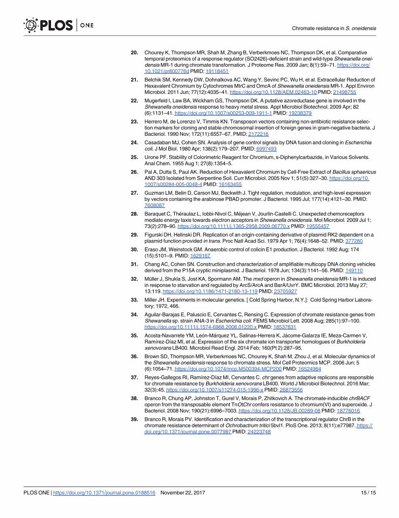
20. Chourey K, ThompsonMR, Shah M, Zhang B, Verberkmoes NC, Thompson DK, et al. Comparativetemporal proteomics of a response regulator (SO2426)-deficient strain and wild-type Shewanella onei-densisMR-1 during chromate transformation. J Proteome Res. 2009 Jan; 8(1):59–71. https://doi.org/10.1021/pr800776d PMID: 19118451
21. Belchik SM, Kennedy DW, Dohnalkova AC,Wang Y, Sevinc PC,Wu H, et al. Extracellular Reduction ofHexavalent Chromium by CytochromesMtrC and OmcA of Shewanella oneidensisMR-1. Appl EnvironMicrobiol. 2011 Jun; 77(12):4035–41. https://doi.org/10.1128/AEM.02463-10 PMID: 21498755
22. Mugerfeld I, Law BA,WickhamGS, Thompson DK. A putative azoreductase gene is involved in theShewanella oneidensis response to heavy metal stress. Appl Microbiol Biotechnol. 2009 Apr; 82(6):1131–41. https://doi.org/10.1007/s00253-009-1911-1 PMID: 19238379
23. Herrero M, de Lorenzo V, Timmis KN. Transposon vectors containing non-antibiotic resistance selec-tion markers for cloning and stable chromosomal insertion of foreign genes in gram-negative bacteria. JBacteriol. 1990 Nov; 172(11):6557–67. PMID: 2172216
24. CasadabanMJ, Cohen SN. Analysis of gene control signals by DNA fusion and cloning in Escherichiacoli. J Mol Biol. 1980 Apr; 138(2):179–207. PMID: 6997493
25. Urone PF. Stability of Colorimetric Reagent for Chromium, s-Diphenylcarbazide, in Various Solvents.Anal Chem. 1955 Aug 1; 27(8):1354–5.
26. Pal A, Dutta S, Paul AK. Reduction of Hexavalent Chromium by Cell-Free Extract of Bacillus sphaericusAND 303 Isolated from Serpentine Soil. Curr Microbiol. 2005 Nov 1; 51(5):327–30. https://doi.org/10.1007/s00284-005-0048-4 PMID: 16163455
27. Guzman LM, Belin D, Carson MJ, Beckwith J. Tight regulation, modulation, and high-level expressionby vectors containing the arabinose PBAD promoter. J Bacteriol. 1995 Jul; 177(14):4121–30. PMID:7608087
28. Baraquet C, Theraulaz L, Iobbi-Nivol C, Mejean V, Jourlin-Castelli C. Unexpected chemoreceptorsmediate energy taxis towards electron acceptors in Shewanella oneidensis. Mol Microbiol. 2009 Jul 1;73(2):278–90. https://doi.org/10.1111/j.1365-2958.2009.06770.x PMID: 19555457
29. Figurski DH, Helinski DR. Replication of an origin-containing derivative of plasmid RK2 dependent on aplasmid function provided in trans. Proc Natl Acad Sci. 1979 Apr 1; 76(4):1648–52. PMID: 377280
30. Eraso JM,Weinstock GM. Anaerobic control of colicin E1 production. J Bacteriol. 1992 Aug; 174(15):5101–9. PMID: 1629167
31. Chang AC, Cohen SN. Construction and characterization of amplifiable multicopy DNA cloning vehiclesderived from the P15A cryptic miniplasmid. J Bacteriol. 1978 Jun; 134(3):1141–56. PMID: 149110
32. Muller J, Shukla S, Jost KA, Spormann AM. Themxd operon in Shewanella oneidensisMR-1 is inducedin response to starvation and regulated by ArcS/ArcA and BarA/UvrY. BMCMicrobiol. 2013 May 27;13:119. https://doi.org/10.1186/1471-2180-13-119 PMID: 23705927
33. Miller JH. Experiments in molecular genetics. [ Cold Spring Harbor, N.Y.]: Cold Spring Harbor Labora-tory; 1972, 466.
34. Aguilar-Barajas E, Paluscio E, Cervantes C, Rensing C. Expression of chromate resistance genes fromShewanella sp. strain ANA-3 in Escherichia coli. FEMSMicrobiol Lett. 2008 Aug; 285(1):97–100.https://doi.org/10.1111/j.1574-6968.2008.01220.x PMID: 18537831
35. Acosta-Navarrete YM, Leon-Marquez YL, Salinas-Herrera K, Jacome-Galarza IE, Meza-Carmen V,Ramırez-Dıaz MI, et al. Expression of the six chromate ion transporter homologues of Burkholderiaxenovorans LB400. Microbiol Read Engl. 2014 Feb; 160(Pt 2):287–95.
36. Brown SD, ThompsonMR, Verberkmoes NC, Chourey K, Shah M, Zhou J, et al. Molecular dynamics ofthe Shewanella oneidensis response to chromate stress. Mol Cell Proteomics MCP. 2006 Jun; 5(6):1054–71. https://doi.org/10.1074/mcp.M500394-MCP200 PMID: 16524964
37. Reyes-Gallegos RI, Ramırez-Dıaz MI, Cervantes C. chr genes from adaptive replicons are responsiblefor chromate resistance by Burkholderia xenovorans LB400. World J Microbiol Biotechnol. 2016 Mar;32(3):45. https://doi.org/10.1007/s11274-015-1996-x PMID: 26873556
38. Branco R, Chung AP, Johnston T, Gurel V, Morais P, Zhitkovich A. The chromate-inducible chrBACFoperon from the transposable element TnOtChr confers resistance to chromium(VI) and superoxide. JBacteriol. 2008 Nov; 190(21):6996–7003. https://doi.org/10.1128/JB.00289-08 PMID: 18776016
39. Branco R, Morais PV. Identification and characterization of the transcriptional regulator ChrB in thechromate resistance determinant ofOchrobactrum tritici 5bvl1. PloS One. 2013; 8(11):e77987. https://doi.org/10.1371/journal.pone.0077987 PMID: 24223748
Chromate resistance in S. oneidensis
PLOSONE | https://doi.org/10.1371/journal.pone.0188516 November 22, 2017 15 / 15
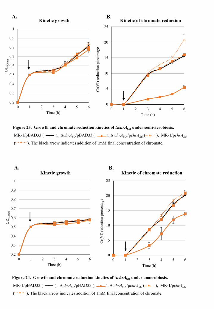
0
5
10
15
20
25
0 1 2 3 4 5 6
Cr(
VI)
red
ucti
on p
erce
ntag
e
Time (h)
0,2
0,3
0,4
0,5
0,6
0,7
0,8
0,9
1
0 1 2 3 4 5 6
OD
600n
m
Time (h)
0,2
0,3
0,4
0,5
0,6
0,7
0,8
0,9
1
0 1 2 3 4 5 6
OD
600n
m
Time (h)
0
5
10
15
20
25
0 1 2 3 4 5 6
Cr(
VI)
red
ucti
on p
erce
ntag
e
Time (h)
Kinetic growth Kinetic of chromate reduction
Kinetic growth Kinetic of chromate reduction
A. B.
A. B.
Figure 23. Growth and chromate reduction kinetics of ΔchrASO under semi-aerobiosis.
MR-1/pBAD33 ( ), ΔchrASO/pBAD33 ( ), Δ chrASO /pchrASO ( ), MR-1/pchrASO
( ). The black arrow indicates addition of 1mM final concentration of chromate.
Figure 24. Growth and chromate reduction kinetics of ΔchrASO under anaerobiosis.
MR-1/pBAD33 ( ), ΔchrASO/pBAD33 ( ), Δ chrASO /pchrASO ( ), MR-1/pchrASO
( ). The black arrow indicates addition of 1mM final concentration of chromate.

Results: Part I
73
Additional results
In addition to the results reported in article 1, we investigated the impact of chrASO
deletion on the ability of S. oneidensis MR-1 to resist and reduce chromate over time, by
using a similar method to that described in detail in Article 1. The wild-type strain
(S. oneidensis MR-1/pBAD33), chrASO deleted strain (ΔchrASO /pBAD33) and chrASO mutant
harboring pchrASO (ΔchrASO/pchrASO) were incubated in LB medium supplemented with
1mM of chromate under semi-aerobic and anaerobic conditions for 5 hours at 28°C. The time
course of growth was followed by measuring the OD600nm of cultures to investigate strains
survival under chromate, while the chromate reduction ability was evaluated by measuring the
residual Cr(VI) concentration each hour starting from two until five hours of incubation in the
presence of chromate. The latter was added when cells reached an OD600nm of 0.5.
During the first hours of incubation, the absence of chrASO does not seem to alter the
growth in the presence of chromate under semi aerobic conditions. In contrast, the growth of
both wild-type and mutant seems to be affected in the presence of chromate under anaerobic
conditions (Figure 23A and 24A). Moreover, the ability of the chrASO deleted strain to reduce
chromate remains dramatically impaired within 5h of incubation in both semi-aerobic and
anaerobic conditions (Figure 23B and 24B). We noticed that the impact of the chrASO deletion
is more pronounced in semi-aerobiosis, where the chromate reduction of chrASO deleted strain
is about 5-fold lower than that of the wild-type (Figure 23B). However, in anaerobic
conditions chromate reduction of the mutant strain is only 2-fold lower than that of the control
strain (Figure 24B), which indicates that the chromate efflux pump ChrASO plays a major and
indirect role in chromate reduction response of S. oneidensis MR-1 especially under semi-
aerobic conditions.
As expected, the complemented mutant ΔchrASO/pchrASO is able to reduce efficiently
chromate as well as the wild type strain under both incubation conditions. Moreover, the
overexpression of chrASO in the wild-type strain seems to improve slightly the reduction
efficiency of S. oneidensis MR-1 under both incubation conditions (Figure 23B and 24B).

0
5
10
15
20
25
0 1 2 3 4 5 6
Cr(
VI)
red
ucti
on p
erce
ntag
e
Time (h)
0,2
0,3
0,4
0,5
0,6
0,7
0,8
0,9
1
0 1 2 3 4 5 6
OD
600n
m
Time (h)
Kinetic growth Kinetic of chromate reduction
Figure 25. Growth and chromate reduction kinetics of Δfdh under semi aerobiosis.
MR-1/pBAD33 ( ), Δfdh/pBADγγ( ), Δfdh/pfdh ( ). The black arrow indicates
addition of 1mM final concentration of chromate.
A. B.
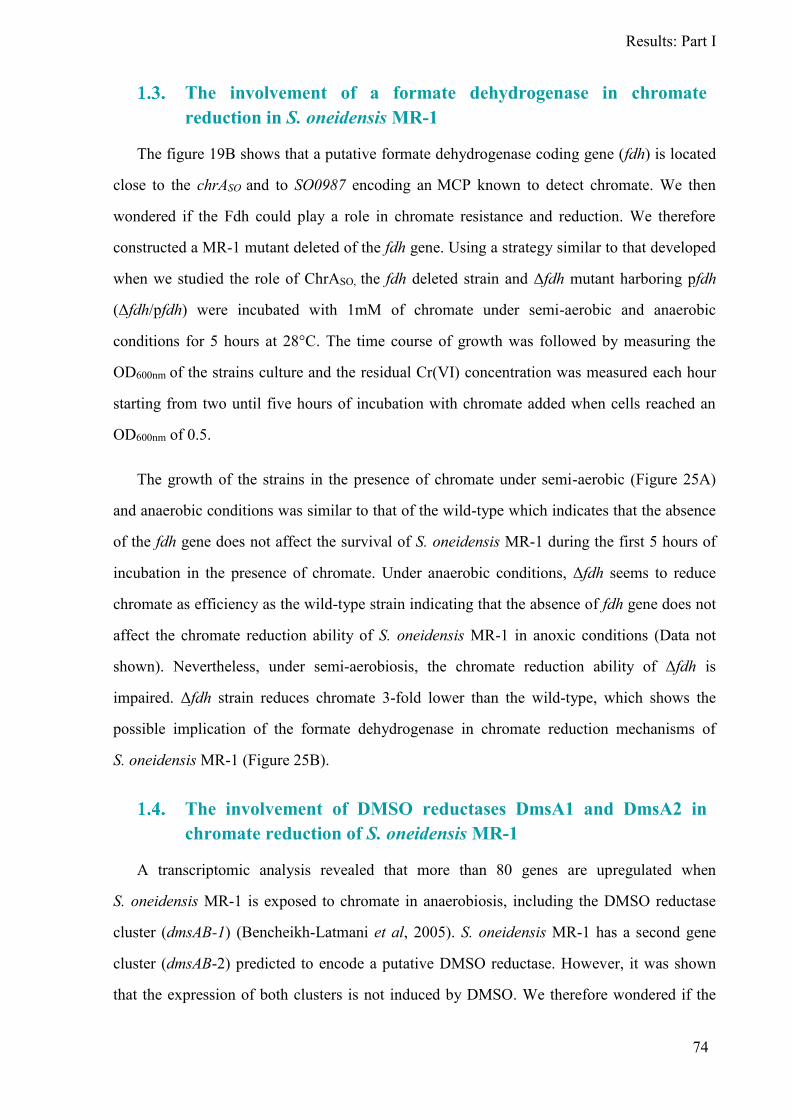
Results: Part I
74
The involvement of a formate dehydrogenase in chromate
reduction in S. oneidensis MR-1
The figure 19B shows that a putative formate dehydrogenase coding gene (fdh) is located
close to the chrASO and to SO0987 encoding an MCP known to detect chromate. We then
wondered if the Fdh could play a role in chromate resistance and reduction. We therefore
constructed a MR-1 mutant deleted of the fdh gene. Using a strategy similar to that developed
when we studied the role of ChrASO, the fdh deleted strain and Δfdh mutant harboring pfdh
(Δfdh/pfdh) were incubated with 1mM of chromate under semi-aerobic and anaerobic
conditions for 5 hours at 28°C. The time course of growth was followed by measuring the
OD600nm of the strains culture and the residual Cr(VI) concentration was measured each hour
starting from two until five hours of incubation with chromate added when cells reached an
OD600nm of 0.5.
The growth of the strains in the presence of chromate under semi-aerobic (Figure 25A)
and anaerobic conditions was similar to that of the wild-type which indicates that the absence
of the fdh gene does not affect the survival of S. oneidensis MR-1 during the first 5 hours of
incubation in the presence of chromate. Under anaerobic conditions, Δfdh seems to reduce
chromate as efficiency as the wild-type strain indicating that the absence of fdh gene does not
affect the chromate reduction ability of S. oneidensis MR-1 in anoxic conditions (Data not
shown). Nevertheless, under semi-aerobiosis, the chromate reduction ability of Δfdh is
impaired. Δfdh strain reduces chromate 3-fold lower than the wild-type, which shows the
possible implication of the formate dehydrogenase in chromate reduction mechanisms of
S. oneidensis MR-1 (Figure 25B).
The involvement of DMSO reductases DmsA1 and DmsA2 in
chromate reduction of S. oneidensis MR-1
A transcriptomic analysis revealed that more than 80 genes are upregulated when
S. oneidensis MR-1 is exposed to chromate in anaerobiosis, including the DMSO reductase
cluster (dmsAB-1) (Bencheikh-Latmani et al, 2005). S. oneidensis MR-1 has a second gene
cluster (dmsAB-2) predicted to encode a putative DMSO reductase. However, it was shown
that the expression of both clusters is not induced by DMSO. We therefore wondered if the
0,2
0,3
0,4
0,5
0,6
0,7
0,8
0,9
1
0 1 2 3 4 5 6
OD
600n
m
Time (h)
0
5
10
15
20
25
0 1 2 3 4 5 6
Cr(
VI)
red
ucti
on p
erce
ntag
e
Time (h)
0
5
10
15
20
25
0 1 2 3 4 5 6
Cr(
VI)
red
ucti
on p
erce
ntag
e
Time (h)
0,2
0,3
0,4
0,5
0,6
0,7
0,8
0,9
1
0 1 2 3 4 5 6
OD
600n
m
Time (h)
Kinetic growth Kinetic of chromate reduction
Kinetic growth Kinetic of chromate reduction
Figure 27. Growth and chromate reduction kinetics of ΔdmsA2 under semi aerobiosis.
MR-1/pBAD33 ( ), ΔdmsA2/pBAD33 ( ), ΔdmsA2/pdmsA2 ( ). The black arrow
indicates addition of 1mM final concentration of chromate.
A. B.
Figure 26. Growth and chromate reduction kinetics of ΔdmsA1 under semi aerobiosis.
MR-1/pBAD33 ( ), ΔdmsA1/pBAD33 ( ), ΔdmsA1/pdmsA1 ( ( ). The black arrow
indicates addition of 1mM final concentration of chromate.
A. B.
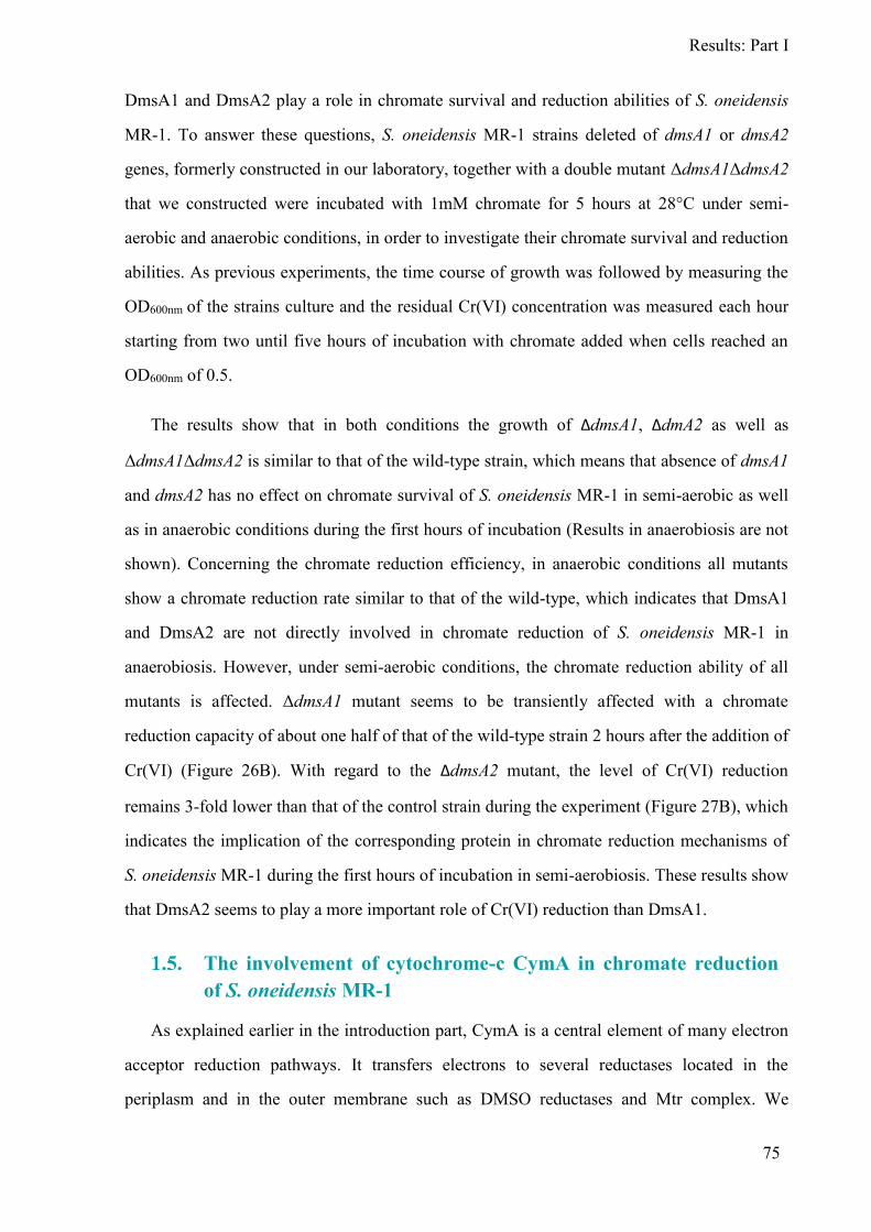
Results: Part I
75
DmsA1 and DmsA2 play a role in chromate survival and reduction abilities of S. oneidensis
MR-1. To answer these questions, S. oneidensis MR-1 strains deleted of dmsA1 or dmsA2
genes, formerly constructed in our laboratory, together with a double mutant ΔdmsA1ΔdmsA2
that we constructed were incubated with 1mM chromate for 5 hours at 28°C under semi-
aerobic and anaerobic conditions, in order to investigate their chromate survival and reduction
abilities. As previous experiments, the time course of growth was followed by measuring the
OD600nm of the strains culture and the residual Cr(VI) concentration was measured each hour
starting from two until five hours of incubation with chromate added when cells reached an
OD600nm of 0.5.
The results show that in both conditions the growth of ΔdmsA1, ΔdmA2 as well as
ΔdmsA1ΔdmsA2 is similar to that of the wild-type strain, which means that absence of dmsA1
and dmsA2 has no effect on chromate survival of S. oneidensis MR-1 in semi-aerobic as well
as in anaerobic conditions during the first hours of incubation (Results in anaerobiosis are not
shown). Concerning the chromate reduction efficiency, in anaerobic conditions all mutants
show a chromate reduction rate similar to that of the wild-type, which indicates that DmsA1
and DmsA2 are not directly involved in chromate reduction of S. oneidensis MR-1 in
anaerobiosis. However, under semi-aerobic conditions, the chromate reduction ability of all
mutants is affected. ΔdmsA1 mutant seems to be transiently affected with a chromate
reduction capacity of about one half of that of the wild-type strain 2 hours after the addition of
Cr(VI) (Figure 26B). With regard to the ΔdmsA2 mutant, the level of Cr(VI) reduction
remains 3-fold lower than that of the control strain during the experiment (Figure 27B), which
indicates the implication of the corresponding protein in chromate reduction mechanisms of
S. oneidensis MR-1 during the first hours of incubation in semi-aerobiosis. These results show
that DmsA2 seems to play a more important role of Cr(VI) reduction than DmsA1.
The involvement of cytochrome-c CymA in chromate reduction
of S. oneidensis MR-1
As explained earlier in the introduction part, CymA is a central element of many electron
acceptor reduction pathways. It transfers electrons to several reductases located in the
periplasm and in the outer membrane such as DMSO reductases and Mtr complex. We
0
5
10
15
20
25
0 1 2 3 4 5 6
Cr(
VI)
red
ucti
on p
erce
ntag
e
Time (h)
0,2
0,3
0,4
0,5
0,6
0,7
0,8
0,9
1
0 1 2 3 4 5 6
OD
600n
m
Time (h)
Kinetic growth Kinetic of chromate reduction
Figure 28. Growth and chromate reduction kinetics under semi aerobiosis.
MR-1/pBAD33 ( ), ΔcymA/pBADγγ ( ), ΔcymA/pcymA ( ), MR-1/pcymA ( ).
The black arrow indicates addition of 1mM final concentration of chromate.
A. B.
Results: Part I
76
investigated the effect of CymA absence on chromate survival and reduction of S. oneidensis
MR-1. Therefore S. oneidensis MR-1 strain deleted of cymA previously constructed in our
laboratory together with the mutant complemented strain that we constructed (ΔcymA/pcymA)
were incubated under semi-aerobiosis and anaerobiosis with 1mM chromate added when cells
reached OD600nm of 0.5. Growth and chromate reduction kinetics were performed using the
same techniques used previously.
Results show that in anaerobic conditions the growth of mutant and the complemented
mutant strains is similar to that of the wild-type strain, which mean that the absence of cymA
does not affect the chromate survival of S. oneidensis MR-1 under anoxic conditions (data not
shown). However, we noticed that under semi-aerobiosis, the mutant ΔcymA shows a slight
growth defect in the presence of chromate compared to the wild-type strain. This slight
growth defect is restored to the wild-type growth levels when the mutant is complemented
(ΔcymA/pcymA) (Figure 28A). This result indicates that CymA may play a partial role in
chromate resistance of S. oneidensis MR-1 under semi-aerobic conditions during the first
hours of incubation.
Within 5 hours of incubation with chromate, ΔcymA shows chromate reduction rates
similar to that of the wild-type strain in anaerobiosis which indicates that the absence of cymA
has no effect on chromate reduction ability of S. oneidensis MR-1 in anaerobiosis.
Nevertheless, under semi-aerobic conditions, the chromate reduction ability of ΔcymA is
strongly affected. ΔcymA reduces chromate 4-fold lower than the wild-type which reveals that
cymA plays an important role in chromate reduction of S. oneidensis MR-1 in semi-aerobic
conditions (Figure 28B). As expected, the complemented mutant is able to reduce chromate as
efficiently as the wild-type strain. Moreover, the overexpression of cymA in the wild-type
strain does not seem to highly improve the reduction efficiency of S. oneidensis MR-1. CymA
may be probably involved in electron transfer to different terminal reductases playing a partial
role in Cr(VI) reduction.

PART II:
ISOLATION AND
CHARACTERIZATION OF NEW
Shewanella STRAINS

Figure 29. Maps showing the location of Stora harbor ( 36°54'06.9"N 6°52'45.4"E) . It is situated in the
northeast of Algeria in the Sekikda province. Pictures are taken from Google earth (2018).
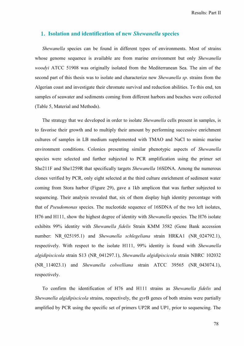
Results: Part II
78
Isolation and identification of new Shewanella species
Shewanella species can be found in different types of environments. Most of strains
whose genome sequence is available are from marine environment but only Shewanella
woodyi ATCC 51908 was originally isolated from the Mediterranean Sea. The aim of the
second part of this thesis was to isolate and characterize new Shewanella sp. strains from the
Algerian coast and investigate their chromate survival and reduction abilities. To this end, ten
samples of seawater and sediments coming from different harbors and beaches were collected
(Table 5, Material and Methods).
The strategy that we developed in order to isolate Shewanella cells present in samples, is
to favorise their growth and to multiply their amount by performing successive enrichment
cultures of samples in LB medium supplemented with TMAO and NaCl to mimic marine
environment conditions. Colonies presenting similar phenotypic aspects of Shewanella
species were selected and further subjected to PCR amplification using the primer set
She211F and She1259R that specifically targets Shewanella 16SDNA. Among the numerous
clones verified by PCR, only eight selected at the third culture enrichment of sediment water
coming from Stora harbor (Figure 29), gave a 1kb amplicon that was further subjected to
sequencing. Their analysis revealed that, six of them display high identity percentage with
that of Pseudomonas species. The nucleotide sequence of 16SDNA of the two left isolates,
H76 and H111, show the highest degree of identity with Shewanella species. The H76 isolate
exhibits 99% identity with Shewanella fidelis Strain KMM 3582 (Gene Bank accession
number: NR_025195.1) and Shewanella schlegeliana strain HRKA1 (NR_024792.1),
respectively. With respect to the isolate H111, 99% identity is found with Shewanella
algidipiscicola strain S13 (NR_041297.1), Shewanella algidipiscicola strain NBRC 102032
(NR_114023.1) and Shewanella colwelliana strain ATCC 39565 (NR_043074.1),
respectively.
To confirm the identification of H76 and H111 strains as Shewanella fidelis and
Shewanella algidipiscicola strains, respectively, the gyrB genes of both strains were partially
amplified by PCR using the specific set of primers UP2R and UP1, prior to sequencing. The
Figure 30. 16SDNA phylogenetic tree showing the position of S. fidelis H76, S. algidipiscicola H111
and closely related Shewanella species including S. oneidesnsis MR-1. The group of S. sediminis
includes HAW EB3 and DSM 17055 strains. The group of S. woodyi includes NR_074846, ATCC
51908 and MS32 strains. The group one and two represent the main Shewanella groups. The scale bar
indicates a genetic distance of 0.005.
Gro
up
1G
rou
p 2
Results: Part II
79
sequences obtained were compared with the NCBI gene bank database using the BlastP
search program. The sequence homology search shows that, GyrB of H76 strain display the
highest degree of homology (99%) with that of Shewanella fidelis Strain KMM 3582
(NR_025195.1) that was firstly isolated from sediments of the South China Sea in 2003 (Lee
et al, 2016). The GyrB primary structure of H111 strain is identical to that of Shewanella
algidipiscicola strain NBRC 102032 (NR_114023.1) previously isolated from marine fish
(cod and plaice) caught in the Baltic Sea of Denmark in 2007 (Ivanova, 2003). The strains
H76 and H111 were then identified as Shewanella fidelis and Shewanella algidipiscicola,
respectively. These results were confirmed later when we could access to the draft genome of
both strains.
The 16SDNA sequences of some of the nearest neighbours of H76 and H111 together
with the reference strain MR-1 were used for the construction of the phylogenetic tree by
MOLE BLAST (http://blast.ncbi.nlm.nih.gov/moleblast/moleblast.cgi) to evaluate their
evolutionary origin and to show the genetic relatedness between H76 and H111. Unlike the
reference strain MR-1 that is a member of the second group enclosing pressure sensitive and
mesophilic species, both H76 and H111 strains belong to the first group of Shewanella that
usually contains piezotolerant and psychrotolerant species. Furthermore, the two strains are
present in different sub-groups, despite the fact that they were isolated from the same sample
(Figure 30).
Growth characteristics
During the isolation procedure of the novel strains S. fidelis H76 and S. algidipiscicola
H111, we noticed that both are not able to grow in regular LB medium. They require a final
concentration of NaCl higher than 5g/l that is usually found in LB medium to grow. In order
to determine the favorable growth conditions for S. fidelis H76 and S. algidipiscicola H111,
cells of both strains were cultivated aerobically at 28°C in LB medium supplemented with
different concentrations of NaCl (15, 20, 25 and 30g/l). The time course of growth was
followed by measuring the OD600nm of cultures.
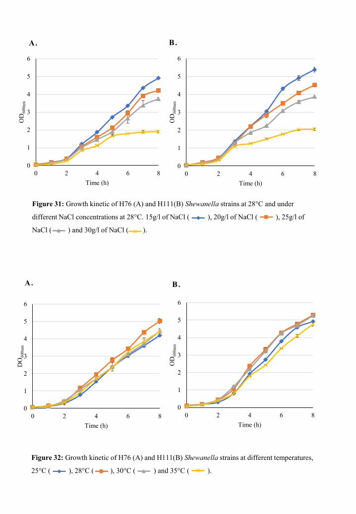
0 2 4 6 80
1
2
3
4
5
6
Time (h)
OD
600n
m
B.
0 2 4 6 80
1
2
3
4
5
6
Time (h)
OD
600n
m
A.
0 2 4 6 80
1
2
3
4
5
6
Time (h)
DO
600n
m
A.
0 2 4 6 80
1
2
3
4
5
6
Time (h)
OD
600n
m
B.
Figure 32: Growth kinetic of H76 (A) and H111(B) Shewanella strains at different temperatures,
25°C ( ), 28°C ( ), 30°C ( ) and 35°C ( ).
Figure 31: Growth kinetic of H76 (A) and H111(B) Shewanella strains at 28°C and under
different NaCl concentrations at 28°C. 15g/l of NaCl ( ), 20g/l of NaCl ( ), 25g/l of
NaCl ( ) and 30g/l of NaCl ( ).

Results: Part II
80
The kinetic of growth shows that both strains are able to grow in the presence of 20g/l,
25g/l and 30g/l of NaCl in culture medium with an optimum growth at 15g/l of NaCl.
However, results reveal impairment in growth of both strains in the presence of 30g/l of NaCl
(Figure 31).
We investigated also the temperature range of growth of S. fidelis H76 and S.
algidipiscicola H111. Therefore, strains were cultured aerobically in liquid LB medium
supplemented with NaCl (15g/l) at different temperatures 25°C, 28°C, 30°C and 35°C. The
time course of growth was followed by measuring the OD600nm of the strains culture. Results
Show that S. fidelis H76 and S. algidipiscicola H111 are capable of growing in temperatures
ranging from 25°C to 35°C (Figure 32).

Figure 33: Chemotactic behavior of S. oneidensis MR-1, S. fidelis H76 and S. algidipiscicola H111
toward nitrate, chromate, nickel and cobalt.
Results: Part II
81
Chemotaxis behavior of S. fidelis H76 and S. algidipiscicola H111
It is known that S. oneidensis MR-1 is chemotactic towards many compounds. We sought
the chemotactic response of the two-novel isolated Shewanella strains S. fidelis H76 and
S. algidipiscicola H111 towards nitrate, chromate, nickel and cobalt using the technique of
microplug (µPlug) assay described in Material and Methods.
Results show that S. fidelis H76 and S. algidipiscicola H111 have a chemotactic behavior
similar to that of S. oneidensis MR-1. For all strains, cells accumulate and appear as a white
halo around the plugs containing nitrate and chromate indicating that S. fidelis H76 and S.
algidipiscicola H111 as well as S. oneidensnsis MR-1 are attracted to nitrate and chromate.
However, the halos around the plugs containing nitrate are slightly more intense than those
observed around plugs containing chromate. In contrast, a repulsion zone surrounded by a
faint accumulation of cells is observed around plugs containing nickel and cobalt indicating
that both strains are repulsed by theses metals (Figure 33).
Chromate resistance of S. fidelis H76 and S. algidipiscicola H111
The main reason for isolating new Shewanella strains was to investigate their chromate
resistance and reduction capacities. As a preliminary test to determine a range of Cr(VI)
concentrations in which growth of S. fidelis H76 and S. algidipiscicola H111 could occur
under semi-aerobic conditions, cells were cultured in LB medium supplemented with different
chromate concentrations (0.2, 0.5, 1, 2 and 3mM).
As shown in Figure 34, S. fidelis H76 and S. algidipiscicola H111 growth that occurs at
0.2mM of chromate is similar to that of the chromate-free culture. Moreover, contrary to MR-
1 that shows a 40% decrease in cells survival rate in the presence of 0.5mM of chromate, the
growth and of H76 and H111 cells does not seem to be highly altered under semi-aerobiosis
(Figure 34 and 35). These results indicate that H76 and H111 strains strongly resist to
chromate concentrations lower than 0.5mM, better than MR-1 strain. MR-1, H76 and H111
strain in the presence of 1mM of chromate show a cell survival rates of 30%, 40% and 60%,
respectively, which indicates that S. algidipiscicola H111 present a better growth in the
presence of 1mM of chromate compared to S. fidelis H76 and to S. oneidensis MR-1 (Figure

0
0,2
0,4
0,6
0,8
1
1,2
1,4
1,6
1,8
0 5 1 0 1 5
OD
600n
m
Time (h)
0
0,2
0,4
0,6
0,8
1
1,2
1,4
1,6
1,8
0 5 1 0 1 5
OD
600n
m
Time (h)
Figure 34. Growth kinetic of S. fidelis H76 (A), S. algidipiscicola H111 (B) under semi-
aerobic conditions in the presence of increasing concentrations of chromate: 0mM ( ),
0,2mM ( ), 0,5mM ( ), 1mM ( ), 2mM ( ) and 3mM ( ).
S. algidipiscicola H111 S. fidelis H76 A. B.
0
10
20
30
40
50
60
70
80
90
100
0 5 10 15
Cel
lsu
rviv
al
rate
(%
)
Time (h)
0
10
20
30
40
50
60
70
80
90
100
0 5 10 15
Cel
lsu
rviv
al
rate
(%
)
Time (h)
1mM Cr(VI)0,5mM Cr(VI)A. B.
Figure 35. Survival rate of MR-1 ( ), H76 ( ) and H111 ( ) in the presence of
0,5mM (A) and 1mM (B) of chromate under semi-aerobic conditions. The percentage of cell
survival represents the optical density of chromate free culture compared to that of culture in
the presence of chromate: (OD600nm- Cr(VI)/ OD600nm+ Cr(VI))*100.
Results: Part II
82
35). Nevertheless, chromate concentrations higher than 2mM seem to highly alter the growth
of H76 and H111 strains as well as MR-1 under semi-aerobic conditions (Figure 34).
Subsequently, we investigated the effect of chromate on cell survival of H76 and H111
strains as well as MR-1, in the presence of chromate. To this end, cells with an initial OD600nm
of 0.5 were incubated in the presence of various chromate concentrations (0.2, 0.5, 1, 1.5, 2
and 3mM) for 5 hours at 28°C under semi-aerobic conditions. Chromate free cultures of
strains served as a negative control. Afterwards, 10-fold serial dilution of each culture was
spotted on LB plates. The latter were then incubated 24h at 28°C to allow bacterial growth.
As shown in Figure 36, the viability of the reference strain MR-1 as well as H76 decreases
with increasing chromate concentrations. Despite the fact that high chromate concentrations
starting from 1mM seem to strongly alter the viability of MR-1 and H76 cells, the latter show
a better chromate resistance compared to that of MR-1. Surprisingly, the viability rate in
H111 cells is significantly greater than that of MR-1 and H76 cells. The high chromate
concentrations starting from 1.5mM does not seem alter the viability of H111 cells, indicating
that S. algidipiscicola H111 can resist and survive even at high chromate concentrations
which strongly confirms our previous results.

Figure 36. Chromate survival of S. oneidensis MR-1, S. fidelis H76, and S. algidipiscicola
H111 after 5h of incubation at 28°C under semi-aerobic conditions, in the presence of various
chromate concentrations: 0,2, 0,5, 1, 1.5, 2 and 3mM.
S. oneidensis MR-1
S. algidipiscicola H111 S. fidelis H76
Results: Part II
83
Genome analysis of S. algidipiscicola H111 and S. fidelis H76 strains
The uncommon capabilities of strains H76 and H111 to resist and reduce chromate,
prompted us to investigate their genetic characteristics compared to the model strain MR-1.
To this end, the whole genome of H111 and H76 strains was sequenced at the “Molecular
Research LP (MR DNA) Laboratory” (USA).
As mentioned previously in the introduction, in aerobic conditions, resistance to chromate
is mainly due to chromate efflux pumps (ChrA-like), and to type I (ChrR- and YieF-like) or
type II (NfsA-like) chromate reductases. S. oneidensis strain MR-1 can express a ChrR-like
protein corresponding to an azoreductase which does not seem to be crucial as only the initial
rate of chromate disappearance is affected in the ΔchrR mutant (Mugerfeld et al, 2009a). We
demonstrated in the first part of this thesis, that chrASO encodes a chromate efflux pump. We
also showed that two other genes fdhA and dmsA2 are involved in the chromate resistance
phenotype of the strain MR1 when grown in semi-aerobic conditions. In contrary to the
limited role of DmsA1.
We focused throughout the genome analysis on the presence of homologous genes
(chrAso, chrR, NfsA, fdh, dmsA1 and dmsA2) that could code for homologous functions
previously identified in strain MR-1 to be involved in chromate resistance and/or reduction.
The analysis was carried out using the BlastP and the synteny programs of the Mage platform
(http://www.genoscope.cns.fr/agc/microscope/home/index.php).
General features of S. fidelis H76 genome
Genome sequencing reveals that H76 genome consists of 4873,941 bases and contains
4,717 predicted genes with a mean G+C% value of 45.70. Among them, 4,552 are potential
protein coding genes (CDS) without artefacts, 10 rRNAs and 118 tRNAs.
chrA- and fdh-like genes
Genomic analysis shows the presence of chrA-like (SFH7-v1-11572) and fdh-like (SFH7-
v1-11571) genes on the chromosome. Their putative products share 61.46% and 69.18% of
identity, respectively with the corresponding proteins of MR-1. As in MR-1, the chrA-and

Figure 37. Schematic comparison of the organization of genes coding for chrA-like and fdh-
like proteins in MR1 and H76. ChrA-like and Fdh-like proteins of H76 share 61.46 % and
69.18 % identity, respectively with the corresponding proteins of MR-1.
S. oneidensis MR-1
S. fidelis H76
Results: Part II
84
fdh-like genes are convergent. However, it is noticeable that the gene coding for the chromate
sensing MCP is lacking (Figure 37).
chrR-like genes
The putative products of two genes, SFH7_v1_11418 and SFH7_v1_11419, share
significant identity of 69.48% and 59.82% with the N-terminal and the C-terminal parts of the
ChrR-like azoreductase of the strain MR-1, respectively. This suggests that the genes
SFH7_v1_11418 and SFH7_v1_11419 probably result from a fission event of an initially
functional gene or from a sequence artifact. Synteny analysis shows that a part of genes
surrounding SO3585 are also conserved in H76.
nfsA-like genes
Despite the absence of the chromate reductase NfsA in the reference strain MR-1, we
carried out a BlastP search using the sequence of the chromate reductase NfsA of E. coli. The
results reveal the presence of the SFH7_v1_11027 gene coding for a putative protein that
presents 57.92% identity with NfsA from E. coli. The blastp search using the sequence of
SFH7_v1_11027, show that the corresponding protein shares high levels of identity with
potential gene products of S. halifaxensis (91.25%), S. peleana (83.33%) and S. piezotolerans
(72.5%), 3 species phylogenetically close to S. fidelis (Figure 30).
dmsA1-like and dmsA2-like genes
The presence of gene SFH7_v1_20363 encoding a DmsA1-Like protein that shares
63.46% of identity with that from MR-1 was revealed. Moreover, the synteny analysis shows
that the entire region surrounding the SFH7_v1_20363 gene is highly conserved.
Subsequently, a BlastP analysis was carried out with the dmsA2 gene product as query.
The same SFH7_v1_20363 gene product was identified exhibiting only a low identity
percentage of 32.93%. These results indicate that H76 genome lacks dmsA2-like genes.
General features of S. algidipiscicola H111 genome
The genome sequence reveals that H111 genome consists of 4.14 Mb and contains
3,678 potential protein coding genes (CDSs) without artifacts, 13 rRNAs and 90 tRNAs. The

Figure 38. Schematic comparison of the organization of genes encoding Fdh-like proteins in
strains MR1 and H111. The MCP and Fdh-like of H111 share 79.8 % and 66.01 % identity
respectively with the corresponding proteins of MR1.
Proteins ChrA Fdh DmsA1 DmsA2 ChrR ( YieF)
Strains H76 H111 H76 H111 H76 H111 H76 H111 H76 H111
MR-1 61% NI 69% 66% 63% 64% 33% 34%
60%+
69%68%
H76/ / /
71%/
79% / / /
68% +
74%
Table 1. Identity percentage of proteins present in MR-1, H76 and H111. NI: Not
identified.
S. oneidensis MR-1
S. algidipiscicola H111
Results: Part II
85
mean G+C% value is 46.63. S. algidipiscicola H111 genome is one of the smallest so far
sequenced in the Shewanella genus. Strikingly, the minimal gene set defined using the MaGe
comprises the same 205 genes as S. oneidensis MR-1 (4.97 Mb) and H76 (4.874 Mb)
suggesting that genome sequencing is nearly if not complete.
chrA- and fdhA-like genes
Proteins sharing homologies with the ChrAso and Fdh of MR-1 were searched in H111
genome using the BlastP. No chrASO-like gene could be detected on the chromosome.
However, the gene SAH1-v1-20276 coding a protein that displays 66 % of identity with Fdh
of MR-1 strain is present. It is noticeable that the fdh-like gene of strain H111 is located close
to a gene coding a putative MCP (SAH1-v1-20275) harboring 79.8% identity with the
chromate-sensing MCP of MR-1 (SO0987) (Figure 38).
chrR-like and nfsA-like enzymes
We investigated the presence of homologous of ChrR-like azoreductase (SO3585) of
MR-1 in H111 strain. Results of the BalstP search show the presence of a putative product of
SAH1_v1_10414 that shares 66.01% of identity with ChrR-like azoreductase of MR1.
Moreover, a Synteny analysis shows that part of the genes surrounding SO3585 is also
conserved in H111.
No gene coding for NfsA-like, type II chromate reductase, is detected on the chromosome
of strain H111.
dmsA1-like and dmsA2-like enzymes
We searched also the presence of proteins homologs to DmsA1 and DmsA2 of MR-1
using a BlastP search. Results demonstrate the presence of SAH1_v1_50121 gene which
product shares 64.41% of identity with DmsA1 from the strain MR-1. Moreover, the synteny
analysis reveals that the entire region surrounding the SAH1_v1_50121 gene is highly
conserved. However, the research of DmsA2 homologs show only 33.66% of identity of
SAH1_v1_10414 gene product with DmsA2 from the strain MR-1, and there is no other
putative protein of strain H111 presenting significant identity with it.

Results: Part II
86
When blasted, the proteins Fdh-like, DmsA1-like and ChrR-like present in H76 show a
high identity with their homologs in H111 strain. All results concerning this comparative
analysis between strains MR-1, H76 and H111 are summarized in Table 1.
From the phenotypic study and the global genome analysis of both H76 and H111, we can
notice the originality of S. algidipiscicola H111 strain. Despite the small size of the genome
and the absence of several genes encoding enzymes known to play a role in chromate
resistance and reduction, H111 strain is characterized by its strong ability to efficiently resist
and reduce chromate at high concentrations. Therefore, we decided to publish its whole
genome sequence (Article 2). To simplify, the S. algidipiscicola H111 strain is named in
Article 2 as H1 strain.

Results: Part II
87
Article 2.
Draft genome sequence of Shewanella algidipiscicola H1,
a highly chromate-resistant strain isolated from
Mediterranean marine sediments.
Hiba BAAZIZ, Olivier LEMAIRE, Cécile JOURLIN-CASTELLI, Chantal
IOBBI-NIVOL, Vincent MEJEAN, Radia ALATOU, Michel FONS.

Results: Part II
88
Draft genome sequence of Shewanella algidipiscicola H1, a highly chromate-resistant
strain isolated from Mediterranean marine sediments.
Running title (54 caractères + espaces / 54 max)
Draft genome sequence of Shewanella algidipiscicola H1
Hiba BAAZIZab, Olivier LEMAIREa, Cécile JOURLIN-CASTELLIa, Chantal
IOBBI-NIVOLa, Vincent MEJEANa, Radia ALATOUb, Michel FONSa#.
aUnité de Bioénergétique et Ingénierie des Protéines, Institut de Microbiologie de la
Méditerranée, Aix-Marseille Université, Centre National de la Recherche Scientifique,
Marseille, France.
bLaboratoire de Biologie Moléculaire et Cellulaire, Université des Frères Mentouri
Constantine 1, Constantine, Algeria.
#Corresponding author
Footnote
Address correspondence to Michel Fons, [email protected].
Abstract (49 mots / 50 max)
The ability of different Shewanella sp. to convert heavy metals and toxic substances
into less toxic products by using them as electron acceptors has led to their use in
environmental clean-up strategies. We present here the draft genome of Shewanella
algidipiscicola H1, a strain resistant to high chromate concentration.
Text (480 mots / 500 max)
Most of Shewanella sp. were isolated from marine environments. The hallmark of many
strains is their ability to utilize an extended array of final electron acceptors in addition to


Results: Part II
89
dioxygen. This property, coupled to other adaptive features, allows their survival in various
habitats and consequently their wide distribution in nature (1, 2, 3).
Chromate (Cr(VI)), the hexavalent form of chromium, is a powerful oxidant found in
oxyanions form which are highly soluble in water. For bacteria, it is highly toxic because of
its rapid entry into the cytoplasm where it can generate a whole spectrum of reactive
oxygen species (ROS), which exert deleterious effects on cells (4).
Bacteria can repair the damages induced by chromate. To overcome Cr(VI)-stress, they
have also developed different resistance mechanisms that directly target Cr(VI). These
mechanisms comprise regulation of chromate ions uptake, their efflux from the cell
cytoplasm and the reduction of Cr(VI) into Cr(III) (5, 6).
In aerobic conditions, Cr(VI) reduction is usually achieved in the cytoplasm by type I
(like ChrR of Pseudomonas putida or YieF of Escherichia coli) or type II (NfsA from E.
coli) chromate reductases (7, 8).
The S. algidipiscicola strain H1 was isolated from the muddy sediment of the Stora
harbor (in the city of Skikda, 36°54'06.9"N 6°52'45.4"E, on the Mediterranean Algerian
coast) sampled from the uppermost 3 centimeters. The genome sequencing of S.
algidipiscicola H1 was carried out at the “Molecular Research LP (MR DNA) Laboratory”
(USA). The library was prepared using Nextera DNA Sample Preparation Kit (Illumina)
following the manufacturer's user guide and sequenced using the HiSeq 2500 system
(Illumina). The draft genome consists of 4.14 Mb and contains 3,857 predicted genes.
Among them 3,678 are potential protein coding genes (CDSs) without artifacts, 13 rRNAs
and 90 tRNAs. Until now, only one Shewanella sp. genome from the Mediterranean
environment was available, that of S. woodyi ATCC51908. Besides S. putrefasciens
HRCR-6 (3.631350 Mb)
(https://img.jgi.doe.gov/cgibin/pub/main.cgi?section=TaxonDetail&page=taxonDetail&tax
on_oid=2540341238#statistics), S. algidipiscicola H1 genome is one of the smallest so far


Results: Part II
90
sequenced in the Shewanella genus. Strikingly, the minimal gene set defined using the
MaGe (Magnifying Genomes, http://www.genoscope.cns.fr) web-based interface comprises
the same 205 genes as S. oneidensis MR1 (4.97 Mb).
Preliminary results show that strain H1 is able to grow in the presence of 3 mM
chromate under aerobic conditions, whereas the model strain, S. oneidensis MR1, stops
growing when Cr(VI) concentration is above 0.5 mM.
Unexpectedly, the analysis of the strain H1 genome revealed neither a nfsA-like gene
nor a chrASO-like gene that was recently shown to significantly improve chromate
resistance and reduction in S. oneidensis MR1 (9). However, a ChrR-like protein could be
produced by strain H1, but the role of this azoreductase in chromate resistance is probably
not crucial as only the initial rate of chromate disappearance is affected in the ΔchrR
(so3585) S. oneidensis MR1 mutant (10).
The draft genome sequence of S. algidipiscicola H1 could thus provide with novel
information concerning chromate resistance and reduction systems.
Accession number(s). The results obtained from this whole-genome shotgun project
have been deposited at the European Nucleotide Archive (ENA) database under accession
number XXXXXX.
Acknowledgments
The authors acknowledge funding by the Centre National de la Recherche Scientifique
(www.cnrs.fr), Aix-Marseille Université (www.univ-amu.fr) and HTS-BIO
(www.htsbio.com, Grant # 152 660). H.B. was supported by Université de Constantine 1.
(The funders had no role in study design, data collection and analysis, decision to publish,
or preparation of the manuscript). We are grateful to Dr Scot E. Dowd (Molecular research
LP MR DNA Laboratory) for sequencing and assembly of our genome. The LABGeM

Results: Part II
91
(CEA/IG/Genoscope & CNRS UMR8030) and the France Génomique National
infrastructure (funded as part of Investissement d'avenir program managed by Agence
Nationale pour la Recherche, contract ANR-10-INBS-09) are acknowledged for support
within the MicroScope annotation platform.
References
1. Hau HH, Gralnick JA. 2007. Ecology and biotechnology of the genus Shewanella. Annu Rev Microbiol 61:237–258.
2. Honoré FA, Méjean V, Genest O. 2017. Hsp90 Is Essential under Heat Stress in the Bacterium Shewanella oneidensis. Cell Rep 19:680–687.
3. Bouillet S, Genest O, Jourlin-Castelli C, Fons M, Méjean V, Iobbi-Nivol C. 2016. The General Stress Response σS Is Regulated by a Partner Switch in the Gram-negative Bacterium Shewanella oneidensis. J Biol Chem 291:26151–26163.
4. Thatoi H, Das S, Mishra J, Rath BP, Das N. 2014. Bacterial chromate reductase, a potential enzyme for bioremediation of hexavalent chromium: a review. J Environ Manage 146:383–399.
5. Brown SD, Thompson MR, Verberkmoes NC, Chourey K, Shah M, Zhou J, Hettich RL, Thompson DK. 2006. Molecular dynamics of the Shewanella oneidensis response to chromate stress. Mol Cell Proteomics 5:1054–1071.
6. Ramírez-Díaz MI, Díaz-Pérez C, Vargas E, Riveros-Rosas H, Campos-García J, Cervantes C. 2008. Mechanisms of bacterial resistance to chromium compounds. Biometals Int J Role Met Ions Biol Biochem Med 21:321–332.
7. Ackerley DF, Gonzalez CF, Park CH, Blake R, Keyhan M, Matin A. 2004. Chromate-reducing properties of soluble flavoproteins from Pseudomonas putida and Escherichia coli. Appl Environ Microbiol 70:873–882.
8. Park CH, Keyhan M, Wielinga B, Fendorf S, Matin A. 2000. Purification to homogeneity and characterization of a novel Pseudomonas putida chromate reductase. Appl Environ Microbiol 66:1788–1795.
9. Baaziz H, Gambari C, Boyeldieu A, Ali Chaouche A, Alatou R, Méjean V, Jourlin-Castelli C, Fons M. 2017. ChrASO, the chromate efflux pump of Shewanella oneidensis, improves chromate survival and reduction. PloS One 12:e0188516.
10. Mugerfeld I, Law BA, Wickham GS, Thompson DK. 2009. A putative azoreductase gene is involved in the Shewanella oneidensis response to heavy metal stress. Appl Microbiol Biotechnol 82:1131–1141.


PART III:
CHROMATE REDUCTION BY
PELLICLES
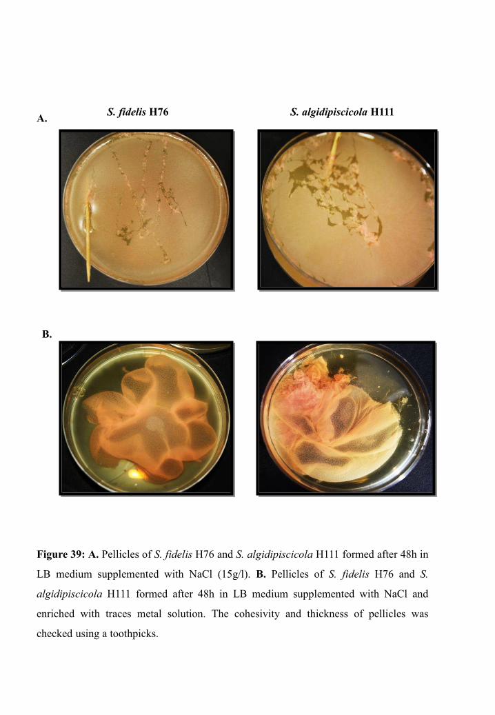
S. fidelis H76 S. algidipiscicola H111A.
B.
Figure 39: A. Pellicles of S. fidelis H76 and S. algidipiscicola H111 formed after 48h in
LB medium supplemented with NaCl (15g/l). B. Pellicles of S. fidelis H76 and S.
algidipiscicola H111 formed after 48h in LB medium supplemented with NaCl and
enriched with traces metal solution. The cohesivity and thickness of pellicles was
checked using a toothpicks.
Results: Part III
93
Pellicle formation ability of S. fidelis H76 and S. algidipiscicola H111
The hallmark of bacterial biofilms is their remarkable resistance and tolerance to a wide
range of toxic compounds. The capacity of S. fidelis H76 and S. algidipiscicola H111 to form
biofilm at the air-liquid interface, named pellicle, was investigated under aerobic and static
conditions. We searched culture parameters allowing S. fidelis H76 and S. algidipiscicola
H111 to form a thick and cohesive pellicle. As a preliminary assay, we sought their ability to
form pellicle under similar conditions needed for pellicle formation in S. oneidensis MR-1,
with addition of NaCl (15g/l) to LB medium. After 48h of following pellicle formation, we
observed a formation of a thin non-cohesive layer of pellicle for both strains using the
toothpick test (Figure 39A).
As known, certain cations such as Mn2 +, Cu2 + or Zn2 + are required for the formation and
maturation of pellicle in S. oneidensis MR-1 (Liang et al, 2010). We wondered if the
enrichment of the culture medium would improve the cohesiveness of S. fidelis H76 and
S. algidipiscicola H111 pellicles. Therefore, prior to incubation, we added traces metal
solution, containing iron, molybdenum, cobalt, copper, manganese and zinc, to a culture
suspension at an OD600nm of 0.2. After 48h of incubation, we observed that S. fidelis H76 and
S. algidipiscicola H111 form very thick and cohesive pellicles (Figure 39B).
Trace metals were also added separately to test the effect of each metal on pellicle
formation capacity of S. fidelis H76 and S. algidipiscicola H111. After 48h of incubation, we
noticed that the addition of each metal separately does not improve the cohesiveness of both
strains pellicles.
Chromate reduction capacity of S. fidelis H76 and S. algidipiscicola
H111 pellicles
Knowing that, usually, bacteria living in biofilm are more resistant to toxic compounds,
including heavy metals, compared to planktonic bacteria cells when they are isolated as
planktonic forms (free-swimming cells) studied before. We wondered whether the chromate
reduction ability of Shewanella strains, MR-1, H76 and H111, could be improved when cells
are in biofilm life form.

0
0,2
0,4
0,6
0,8
1
1,2
1,4
1,6
1,8
2
2,2
0,2 0,5 1 2
Cr(
VI)
red
uce
d c
on
cen
tra
tio
n (
mM
)
Initial chromate concentration (mM)
0
0,2
0,4
0,6
0,8
1
1,2
1,4
1,6
1,8
2
2,2
0,2 0,5 1 2
Cr(
VI)
red
uce
dco
nce
ntr
ati
on
(m
M)
Initial chromate concentration (mM)
0
0,2
0,4
0,6
0,8
1
1,2
1,4
1,6
1,8
2
2,2
0,2 0,5 1 2
Cr(
VI)
red
uce
d c
on
cen
tra
tio
n (
mM
)Initial chromate concentration (mM)
24h 48h
Figure 40. Chromate reduction by S. oneidensis MR-1 (Bleu bars), S. fidelis H76 (Green
bars) and S. algidipiscicola H111 (Red bars) planktonic cells, after 24h and 48h of
incubation at 28°C under semi-aerobic conditions, in the presence of various chromate
concentrations: 0.2, 0.5, 1 and 2mM.
Figure 41. Chromate reduction by S. oneidensis MR-1 (Bleu bars), S. fidelis H76 (Green
bars) and S. algidipiscicola H111 (Red bars) pellicles after 24h of incubation at 28°C under
semi-aerobic conditions, in the presence of various chromate concentrations: 0.2, 0.5, 1 and
2mM.
Results: Part III
94
In order to answer this question, we investigated the chromate reduction efficiency of
pellicles and planktonic. To this end, mature pellicles formed by MR-1, H76 and H111 were
collected and then incubated in fresh LB medium supplemented with various chromate
concentrations (0.2, 0.5, 1 and 2mM) under static conditions (see Material and Methods) to
maintain the three-dimensional architecture of pellicles. Simultaneously, strains cells grown
overnight were gently centrifuged and resuspended in LB medium supplemented with
chromate (0.2, 0.5, 1 and 2mM) to reach an initial OD600nm of 2. The latter OD600nm was
chosen to approach pellicle assay conditions, where the final optical density of cells forming
pellicle in culture medium was 2. After incubation of 24h and 48h in semi-aerobic and
anaerobic conditions, the chromate reduction activity in both conditions, pellicles and
planktonic cells, was determined by measuring residual Cr(VI) concentration using the DPC
colorimetric assay and total chromate concentration using ICP-OES.
As shown in Figure 40, planktonic cells of H111 strains are able to reduce completely the
initial chromate concentration of 1mM after 48h of incubation in semi-aerobiosis, while only
77.9 % and 65.7% are reduced by H76 and MR-1 cells, respectively. Furthermore, H111 is
able to reduce completely the initial chromate concentration of 2mM within 48h of incubation
under static conditions, contrary to H76 and MR-1 cells that are able to reduce only 59.7%
and 47% of it, respectively. These results indicate that, in planktonic life form, the chromate
reduction efficiency of H111 strain is higher than H76 and MR-1 under semi-aerobic
conditions.
With respect to the chromate reduction capacity of pellicles, results shown in figure 41
indicate that in semi-aerobic conditions the initial chromate concentrations 0.2, 0.5 and 1mM
are totally reduced within 24h. Moreover, the highest chromate concentration 2mM is totally
reduced by H111 within 24h of incubation while it is totally reduced by H76 and MR-1 after
48h of incubation.
Overall, the chromate reduction rates of pellicles and planktonic cells of H76, H111 and
MR-1 indicate that chromate reduction ability of strains in pellicle life form is more efficient
than that of their planktonic cells.

S. oneidensis MR-1
S. fidelis H76
S. algidipiscicola H111
Initial pellicules
grown in the absence
of chromate
New pelliculesformed in
the presenceof chromate
After 48h of incubation in the presence of 1mM chromate
Figure 42. Formation of pellicles of S. oneidensis MR-1, S. fidelis H76 and S.
algidipiscicola H111 after 48h of incubation at 28°C under semi-aerobic conditions, in the
presence of 1mM of chromate.

Results: Part III
95
Under anoxic conditions, MR-1, H76 and H111, in both planktonic and pellicle life forms,
show the ability to totally reduce all initial chromate concentrations tested (0.2, 0.5, 1 and
2mM) within only 24h of incubation (Results not shown), indicating that the chromate
reduction ability of strains is more efficient in anaerobic conditions.
During our experiment, we noticed the capacity of Shewanella strains to reform a new
pellicle when the initial pellicle is incubated 48h in the presence of chromate concentrations
lower than 1mM. The pellicles formed after 48h of chromate incubation are slightly thinner
than the initial ones. However, those formed by H76 and H111 are more cohesive that those
of MR-1 (Figure 42).
Chromium accumulation capacity of pellicles
It is known that heavy metal ions in both soluble and insoluble forms can potentially be
accumulated by bacterial cells (live or dead) and their byproducts such as the extracellular
polymeric substances (EPSs) (Gupta & Diwan, 2017). We investigated the capacity of H76,
H111 and MR-1 planktonic cells and pellicles to uptake and accumulate different chromate
concentrations (0.2, 0.5, 1 and 2mM). To this end, cells grown overnight were gently
centrifuged and then resuspended in LB medium supplemented with chromate to reach an
initial OD600nm of 2, while mature strains pellicles were gently collected and then moved into
fresh LB medium supplemented with chromate. Afterward, planktonic cells and pellicles were
incubated at 28°C in the presence of chromate for 24h and 48h under semi-aerobic conditions.
After incubation, cultures were centrifuged to separate cells from supernatant. The total
chromium concentration present in both cellular and supernatant fractions was measured
using ICP-OES.
Results show that pellicles of all strains accumulate much better chromium compared to
their planktonic cells. As an example, when exposed to initial chromate concentrations of
2mM for 24h, pellicles of MR-1, H76 and H111 show a chromium accumulation capacity 10-
fold greater than that of their planktonic cells (Figure 43). Furthermore, H76 and H111
strains, in their pellicle life state, accumulate more efficiently chromium than the reference
strain MR-1. As shown in figure 43, after 48h of incubation in the presence of the initial

0,0
0,1
0,2
0,3
0,4
0,5
0,6
0,2 0,5 1 2
To
tal
chro
miu
mco
nce
ntr
ati
on
(mM
)
Initial chromate concentration (mM)
Figure 43. Total chromium accumulation by planktonic cells (bleu bars) and pellicles
(Red bars) of S. oneidensis MR-1 (A), S. fidelis H76 (B) and S. algidipiscicola H111 (C)
after 24h and 48h of incubation at 28°C under semi-aerobic conditions, in the presence of
various chromate concentrations: 0,2, 0,5, 1 and 2mM.
A.
24h 48h
B.
C.
0,0
0,1
0,2
0,3
0,4
0,5
0,6
0,2 0,5 1 2
To
tal
chro
miu
m c
on
cen
tra
tio
n
(mM
)
Initial chromate concentration (mM)
0,0
0,1
0,2
0,3
0,4
0,5
0,6
0,2 0,5 1 2
To
tal
chro
miu
m c
on
cen
tra
tio
n
(mM
)
Initial chromate concentration (mM)
0,0
0,1
0,2
0,3
0,4
0,5
0,6
0,2 0,5 1 2
To
tal
chro
miu
m c
on
cen
tra
tio
n
(mM
)
Initial chromate concentration (mM)
0,0
0,1
0,2
0,3
0,4
0,5
0,6
0,2 0,5 1 2
To
tal
chro
miu
m c
on
cen
tra
tio
n
(mM
)
Initial chromate concentration (mM)
0,0
0,1
0,2
0,3
0,4
0,5
0,6
0,2 0,5 1 2
To
tal
chro
miu
m c
on
cen
tra
tio
n
(mM
)
Initial chromate concentration (mM)
24h 48h
24h 48h

Results: Part III
96
chromate concentration 2mM, 19.68% and 25.23% of chromium are accumulated in H76 and
H111 pellicles, respectively, while only 15.96% is accumulated by MR-1 strain in its pellicle
life state. These results also show that the amount of total chromium accumulated in both
planktonic cells and pellicles increases with increasing initial chromate concentrations.
To investigate the nature of chromium accumulated in planktonic cells and pellicles, we
performed the specific DPC colorimetric assay in order to detect the presence of the
hexavalent chromate Cr(VI) in cellular fractions of strains cultures. Results show clearly the
absence of Cr(VI) in cellular fractions indicating that the chromium accumulated in
planktonic cells and pellicles are the reduced forms of Cr(VI), most probably the stable form
Cr(III) known to be less soluble than Cr(VI) (Data not shown).


DISCUSSION
AND PERSPECTIVES


Discussion and perspectives
98
Between the two stable chromium forms Cr(III) and Cr(VI), the latter is the highest
toxic for bacterial cells due to its great solubility and rapid entry into the cytoplasm where
it may exert its toxic effects. Among the various strategies that bacteria developed to resist
chromate, efflux of chromate ions from the cytoplasm and reduction of Cr(VI) into Cr(III)
are considered to be the main ones (Ramírez-Díaz et al, 2008). Although numerous studies
have been conducted, the molecular mechanisms developed by Shewanella sp. to overcome
chromate remain largely unknown. Therefore, the main aim of the present dissertation was
to improve understanding about chromate resistance and reduction mechanisms of
Shewanella sp. We studied first these mechanisms in the model strain S. oneidensis MR-1.
The strain MR-1 is considered as a great candidate for chromate bioremediation
investigations due to its metabolic diversity, available sequenced genome and easy growth
in laboratory conditions.
We first confirmed the ability of S. oneidensis MR-1 to grow in the presence of
chromate concentrations up to 0,5mM under semi-aerobiosis, which indicates that this
bacterium is powered by mechanisms that allow its resistance to such high chromate
concentration. We showed that chromate concentrations higher than 1mM alter cell growth
while concentrations above 2mM inhibit the growth of MR-1 cells. These findings are in
good agreement with previously published results which showed that the minimum
inhibitory chromate concentration (MIC) for aerobically grown S. oneidensis MR-1 is 2mM
(Brown et al, 2006). The cease of growth in the presence of high chromate concentrations
results probably from the inhibition of some metabolic processes by chromate and mainly
from the accumulation of Cr(III) precipitates in the cytoplasm, as physiological studies on
the toxicity of chromate in S. oneidensis MR-1 previously reported (Viamajala et al, 2008;
Parker et al, 2011).
Among the numerous studies on the chromate reduction by S. oneidensis MR-1, most
were conducted under anaerobic conditions. Hence, we focused our investigation on
chromate resistance/reduction under semi-aerobic conditions. We firstly confirmed the
ability of S. oneidensis MR-1 to reduce, if not totally, a significant amount of chromate,
even at high initial concentrations under semi-aerobic conditions. It is noteworthy to

Discussion and perspectives
99
mention that the strain MR-1 shows a higher chromate reduction efficiency in anoxic
conditions, which may be explained by the absence of competition between oxygen and
chromate as electron acceptors that usually results in a lower chromate reduction efficiency
(Han et al, 2016) or by less ROS production. Aerobic and anaerobic chromate reduction by
S. oneidensis MR-1 was previously reported (Lowe et al. 2003). However, there is no
evidence until to now that MR-1 strain respires chromate by using it as a sole electron
acceptor (Bencheikh-Latmani et al, 2005). S. oneidensis MR-1 may develop this capacity
not to gain energy by using chromate as electron acceptor but to simply protect itself from
toxicity under different specific conditions. This capacity could provide S. oneidensis MR-1
with a selective advantage over other sensitive bacteria, allowing its colonization in
challenging environments (Trevors et al, 1985).
Knowing that chromate efflux pumps belonging to the superfamily of CHR can be
involved in chromate resistance in other bacteria like C. metallidurans CH34 and
Pseudomonas aeruginosa, we searched for the presence of this type of pump in S.
oneidensis MR-1. Our bioinformatic analysis showed that chromosomal gene SO0986
likely encodes a chromate efflux pump ChrASO that belongs to long chain CHR subfamily.
We confirmed that ChrASO is a chromate efflux pump, as demonstrated for some
members of the CHR family (Pimentel et al, 2002; Aguilar-Barajas et al, 2008), by
demonstrating, on the one hand, that its absence affects significantly the chromate
reduction and resistance of S. oneidensis under semi-aerobic and anaerobic conditions. It is
noticeable that the deletion of chrASO does not dramatically affect chromate reduction under
anoxic conditions compared to semi-aerobiosis. This suggests that other transporters might
be involved in this process under anaerobic conditions. Of interest, genes encoding the
three components of a putative heavy metal efflux pump (SO0518 to SO0520 and CzcCBA)
were observed to be up-regulated by chromate in anaerobic conditions (Bencheikh-Latmani
et al, 2005). This suggests a role in chromate resistance, although this pump does not
belong to the CHR family. It would be interesting to delete genes coding for this pump in
order to investigate its involvement in chromate resistance.

Cr(VI) Cr(III)
Cr(V)
Interacellular chromate reduction
Extracellular chromate reduction
Cell damages
Figure 44. The role of ChrASO. Chromate enters cells via sulphate transporters, the role
of ChrASO is to decrease the rate of chromate inside the cells by extrude it from the
cytoplasm to be later reduced extracellularly by outer membrane cytochromes MtrA and
OmcA. Limiting the presence of chromate in the cytoplasm, protect the cell from
damages resulting from its intracellular presence and reduction. Hence, cells grow better
and therefore can detoxify theirs environement from chromate by reducing it,
extracellularly, more efficiently.

Discussion and perspectives
100
In order to reaffirm the role of ChrASO, we demonstrated that the heterologous
expression of chrASO in E. coli confers resistance to high chromate concentrations, which is
also observed for the plasmid-encoded ChrA of Shewanella ANA-3 and for several ChrA
of Burkholderia xenovorans LB400 (Aguilar-Barajas et al, 2008; Acosta-Navarrete et al,
2014). This heterologous expression strikingly improves the chromate reduction ability of
E. coli cells. A direct role of ChrASO in chromate reduction was ruled out since the specific
activity of chromate reductase measured in vitro on crude extracts from both S. oneidensis
MR-1 and E. coli strains is similar in the absence or in the presence of ChrASO. Our
hypothesis to explain the improvement of chromate reduction efficiency is that ChrASO
promotes chromate reduction by expulsing chromate outside the cells where it can then be
reduced extracellularly by membrane bound reductases. In S. oneidensis MR-1, this
scenario is worth considering as two outer membrane decaheme cytochromes, MtrC and
OmcA, are known to be involved in extracellular chromate reduction (Belchik et al, 2011)
(Figure 44). An additional effect could be that ChrASO improves cell survival by lowering
intracellular chromate concentration and consequently Cr-induced damages inside the cells
(Joutey et al, 2015), which protects and allows the cells to grow and reduce chromate over
an extended period of time (Figure 44). This could be true for both S. oneidensis MR-1 and
E. coli.
We revealed that although chrASO is expressed in the absence of chromate, its level of
expression is higher in the presence of chromate. Two previous studies performed on
S. oneidensis using transcriptomic approaches missed this regulation, probably because the
applied cut-offs were quite stringent (Bencheikh-Latmani et al, 2005; Brown et al, 2006).
Indeed, these studies considered only the genes showing at least a 2-fold or a 3-fold change
in expression, respectively. Moreover, the experimental conditions used in both studies
were different from ours, which can also explain this discrepancy. The fact that chrASO
expression is induced by chromate is reminiscent of what was previously observed for the
expression of several chr genes in other bacteria. As an example, the transcription of the
chrA1 and chrA2 genes of C. metallidurans and B. xenovorans LB400 were shown to be
up-regulated by chromate (Juhnke et al, 2002; Acosta-Navarrete et al, 2014). The
chromosomally located chrBACF operon of Ochrobactrum tritici 5bvl1 presents another

Discussion and perspectives
101
example since its expression is also induced by chromate (Branco et al, 2008). It was
subsequently shown that the ChrB protein is a transcriptional regulator that binds to the
chrBACF promoter region to regulate its expression (Branco & Morais, 2013). The fact that
there is no ChrB homolog in S. oneidensis MR-1, suggests that the regulation of chrASO
expression probably depends on a regulatory protein belonging to a family different from
that of ChrB. It would be interesting to identify this protein, since no chromate-sensing
regulator except from ChrB was previously described.
Besides extruding chromate outside the cytoplasm, the expression of chromate-
reductases is an efficient strategy to resist to Cr(VI) under aerobic conditions. During our
study, 3 loci of the strain MR-1 genome caught our attention. The first one, SO0988, codes
for a putative formate dehydrogenase α subunit. It is located close to chrASO and to SO0987
encoding an MCP known to detect chromate. The second one is dmsAB-1 which codes for a
DMSO reductase. Several studies demonstrated DmsAB-1 involvement in the respiration of
DMSO despite the fact that dmsAB-1 is not induced by DMSO (Bencheikh-Latmani et al,
2005). It have been shown that dmsAB-1 cluster is upregulated under thiosulfate-reducing
conditions as well as in the presence of chromate (Beliaev et al. 2005,Bencheikh-Latmani
et al. 2005), which may be indicative of a broad substrate specificity of corresponding
complex in the MR-1 strain. The third one is dmsAB2 which is predicted to code for a
second putative DMSO reductase. However, there is no evidence until now about the
involvement of the second cluster dmsAB-2 products in DMSO respiration. Previous
studies revealed that the expression of dmsAB-2 cluster is not significantly induced by
DMSO (Beliaev et al, 2005). However, it is interesting to note that the genomic context of
dmsA2 contains SO4359 and SO4360 genes whose products are not only redundant to
cytochroms MtrB and MtrA known to be implicated in chromate reduction (Belchik et al,
2011), but they function as a terminal reductase localized at the cell surface (Schicklberger
et al, 2013).
These data prompted us to wonder whether the 3 loci could play a role in chromate
resistance and reduction in S. oneidensis MR-1, we therefore deleted SO0988, dmsA1 and
dmsA2 separately in order to investigate their involvement under semi-aerobic conditions.
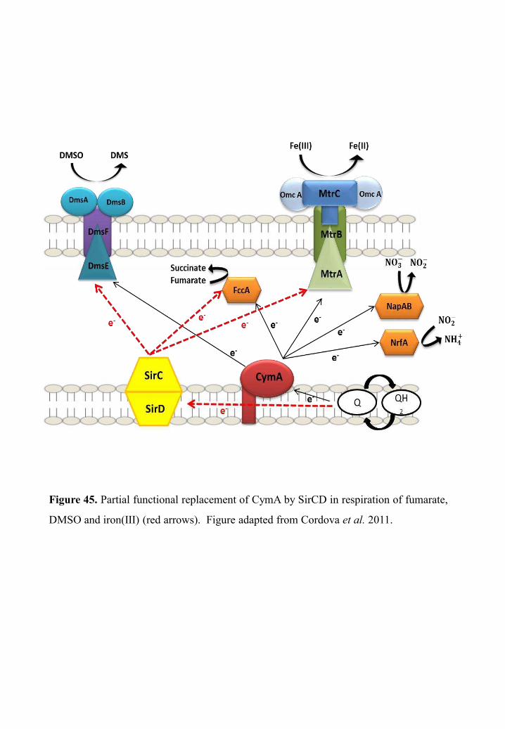
Figure 45. Partial functional replacement of CymA by SirCD in respiration of fumarate,
DMSO and iron(III) (red arrows). Figure adapted from Cordova et al. 2011.

Discussion and perspectives
102
While the deletion of dmsA1 affected slightly and transitionally the chromate reduction
capacity of the strain in the first hours of incubation, the deletion of SO0988 and dmsA2
showed a constant drastic effect on the chromate reduction capacity of strains which
reduced chromate 3-fold lower than the wild-type. These results indicate that the proteins
encoded by these genes are involved either directly or indirectly in chromate reduction in
semi-aerobiosis. The high impairment of chromate reduction in the absence of SO0988
gene and dmsA2 highlights the fact that efficient chromate reduction in semi-aerobiosis
requires the presence of these important genes. Although, SO0988 gene is annotated as a
putative formate dehydrogenase α subunit, some authors predicted that the SO0988 product
is cytoplasmic because the gene does not encode a twin-arginine translocation pathway
signal sequence contrary to other Fdh periplasmic α subunits known in S. oneidensis MR-1
(Kane et al. 2016). Moreover, the involvement of the SO0988 product in formate oxidation
has never been reported. Until now the function of SO0988 product is unknown, therefore,
further studies are needed to clarify the precise role of this cytoplasmic protein and its
direct or indirect relation with chromate reduction under semi-aerobiosis. The collected
data and the results obtained about DmsA2 strongly suggest that it plays a role as a
chromate reductase. It would be interesting to investigate the chromate reductase activity in
vitro of both DMSO reductases, especially that of DmsA2. The expression of both genes in
the presence of different chromate concentrations under semi-aerobic and anaerobic
conditions would be also interesting to investigate in order to complete our understanding
about the expression regulation of these genes and likely the broad substrate specificity of
their products.
Finally, we checked the involvement of the membrane bound c-type cytochrome in the
efficiency of MR-1 strain to reduce Cr(VI). CymA is considered as a central element of
multiple electron acceptor reduction pathways. It delivers electrons from the quinone pool
to a number of terminal reductases located in the periplasm and in the outer membrane
including the Mtr complex (Figure 45), whose coding genes (mtrABC and omcA) have been
shown to be upregulated in the presence of chromate (Beliaev et al, 2005). Furthermore, it
was reported that MtrC and OmcA complex are used by S. oneidensis MR-1 for
extracellular chromate reduction (Belchik et al, 2011). Therefore, we sought the effect of

Discussion and perspectives
103
CymA deletion on chromate resistance and reduction in S. oneidensis MR-1 under both
semi-aerobic and anaerobic conditions. On the one hand, we showed that CymA does not
appear to play a key role in chromate resistance and reduction under anaerobic conditions,
in agreement with results of previous studies (Bencheikh-Latmani et al, 2005). The first
hypothesis to explain this phenomenon is that the Mtr complex could be supplied with
electrons via other donors under anaerobic conditions. This scenario is worth considering as
previous studies revealed that CymA could be partially substituted by the SirCD complex,
which primarily function as quinol dehydrogenases in respiration of several electrons
acceptors, such as fumarate, DMSO and iron(III) but has no effect on nitrate and nitrite
reduction (Cordova et al, 2011) (Figure 45). A second hypothesis would be the presence of
unknown chromate reductases CymA-independent, since even when the whole MtrBAC
was deleted, the chromate reduction was not completely abolished (Bencheikh-Latmani et
al, 2005). It would be interesting to investigate if the complex SirCD could fulfill the role
of CymA in chromate reduction in anaerobiosis since it can transfer electrons to the Mtr
complex to reduce Fe(III).
On the other hand, we demonstrated that CymA may play a partial role in chromate
survival of S. oneidensis MR-1 under semi-aerobic conditions. Moreover, we showed that
CymA is most likely involved in chromate reduction mechanisms of S. oneidensis MR-1
under semi-aerobiosis, since the chromate reduction ability of the cymA-mutant strain is
strongly, but not completely, impaired. This impairment may explain the slight decrease of
growth seen when cymA-mutant is challenged with chromate, as it is unable to reduce
chromate effeciently and, consequently, chromate-induced damages result in a slight
decline of cells viability. Overall, we demonstrated that under semi-aerobiosis, CymA may
transfer electrons to terminal reductases that partially reduce chromate. The shortcoming of
CymA as branching position for many pathways, is that bacteria can lose their ability to use
various electron acceptors simultaneously, resulting in sever physiological damages, when
CymA is mutated. Therefore, S. oneidensis MR1 evolved a strategy to circumvent the risk
by developing functional substituent proteins for CymA. This may explain the residual
chromate reduction activity of cymA-mutant under semi-aerobiosis. We suggest the
presence of a protein that partially substitutes CymA under these conditions, which is not


Discussion and perspectives
104
surprising given that it has been reported that under nitrate/nitrite reduction and aerobic
conditions the role of CymA could be partially fulfilled by the bc1 complex that functions
as quinol dehydrogenases to transport electrons to the cytochrome cbb3 oxidase, the
enzyme complex predominantly responsible for oxygen respiration and which expression is
favored in aerobic conditions (Cordova et al, 2011; Fu et al, 2014). It will be interesting to
investigate the possibility if cb1 complex could substitute CymA under semi-aerobiosis by
transferring electrons to chromate reductases.
Taken together, our findings highlight the fact that S. oneidensis MR-1 uses different
strategies to resist and reduce chromate depending on the concentration of oxygen. The
chromate reduction of this bacterium seems to overlap with its metabolic complex network.
Therefore, further studies need to be pursued to decipher the enzymes implicated or related
to chromate resistance and reduction in S. oneidensis MR-1 under different conditions.
Shewanella species are frequently isolated from marine and freshwater environments.
More than 32 Shewanella genomes have been fully sequenced to date
(https://www.ncbi.nlm.nih.gov/genome/browse/#!/overview/shewanella) due to their
known energy and bioremediation capabilities, phylogenetic relatedness, and inhabitance of
environments (Fredrickson et al, 2008). Among the marine species whose genome is
available, only Shewanella woodyi ATCC 51908 was originally isolated from the
Mediterranean Sea. During this thesis, we isolated and identified two novel Mediterranean
Shewanella strains from sediment water of Stora harbor, located in the north east of Algeria
and previously reported to be polluted by heavy metal (Gueddah & Djebar, 2014). Based
on their 16SDNA and GyrB sequences, the strains H76 and H111 were identified as
Shewanella fidelis and Shewanella algidipiscicola, respectively. The later were firstly
isolated from the South China Sea in 2003 and the Baltic Sea of Denmark, respectively
(Ivanova, 2003; Lee et al, 2016). The phylogenetic tree that we constructed using 16SDNA
sequences of some of the nearest neighbors of H76 and H111, surprisingly showed that
strains are present in different sub-groups of the first group of Shewanella that enclose
piezotolerant and psychrotolerant species, contrary to MR-1 which belongs to the second
group that contains pressure sensitive and mesophilic species. Phenotypic characterization


Discussion and perspectives
105
showed that the two strains can grow at temperatures ranging from 25°C to 35°C indicating
that although both strain belong to the first group of Shewanella they cannot be
psychrotolerants this may be due to the adaptation of species to Mediterranean
environments conditions. It would be interesting to test the pressure sensitivity of our
strains since no S. fidelis and S. algidipiscicola previously isolated strains have been
reported as a piezotolerants.
Unlike MR-1, both strains require a relatively high salt concentration for optimal
growth; consequently, they are able to grow over a range of NaCl concentrations from 15 to
30g/l, with an optimum growth at 15g/l of NaCl. These results are not surprising since H76
and H111 are originating from a marine environment whereas MR-1 was first isolated form
freshwater. We demonstrated also the chemotaxis capacity of H76 and H111 towards few
metals. The strains showed chemotactic behavior similar to that of MR-1 as they were
repulsed from nickel and cobalt and were attracted to nitrate and chromate (Armitano et al,
2011). Two hypotheses can be suggested to explain the attraction toward chromate, either
this attraction allows cells to localize favorable environments containing metals that can be
used as electron acceptors (Baraquet et al, 2009) or confers selective advantage to cells by
allowing their colonization in environments that are hostile for many other chromate-
sensitive bacteria.
During this study, we revealed the capacity of both H76 and H111 strains to resist and
reduce chromate more efficiently than the model strain MR-1 under semi-aerobiosis. This
may indicate that both strains use an efficient strategy to cope with chromate toxicity. It is
interesting to note that among the three studied strains, H111 showed striking chromate
resistance and reduction. We demonstrated that its minimum inhibitory chromate (MIC)
concentration would be above 3mM while the MIC of the reference strain MR-1 is below
2mM (Viti et al, 2014). These promising results intrigued and prompted us to set out a whole
genome-sequencing project to analyze and decipher the genetic background of these
particular chromate resistance and reduction efficiencies of both H76 and H111 strains.
Our genetic analysis was based on the investigation of the presence of chrASO-, chrR- and
nfsA-like genes whose products were previously described to play a key role in Cr(VI)


Discussion and perspectives
106
resistance and reduction, as well as fdh-, dmsA1- and dmsA2-like genes for which we
demonstrated earlier the involvement of their products in Cr(VI) reduction. Genome
sequencing revealed that S. fidelis H76 genome size of 4.874 Mb is considered similar to that
of the model strain MR1 (4.97 Mb), whereas S. algidipiscicola H111 genome is the one of the
smallest so far sequenced in the Shewanella genus with 4.14 Mb. It contains the same 205
essential genes set as S. oneidensis MR1 and H76 suggesting that genome sequencing is
almost complete.
We showed that the H76 strain can probably express ChrA-, Fdh-, ChrR- and DmsA1-like
putative proteins that share a high homology with those present in the MR-1 strain.
Interestingly, the strain H76 genome harbors a gene that code for a putative chromate
reductase NfsA-like that presents a significant homology with that of E. coli (Ackerley et al,
2004). Despite the absence of a putative DmsA2 homologous to that of the MR-1 strain the
chromate resistance and reduction of the H76 strain is more efficient than that of MR-1. One
can ask whether the high chromate reduction observed in the H76 strain is due to the presence
of nfsA-like gene. It would be interesting to confirm the role of these homologous proteins
present in the H76 strain and to evaluate their involvement in chromate resistance and
reduction mechanisms.
We showed that the H111 strain harbors Fdh-, ChrR- and DmsA1-like putative proteins
sharing a significant homology with those present in the MR-1 strain. Unexpectedly, no
putative proteins of the H111 strain presenting homology with the chromate reductase NfsA
nor with the chromate efflux pump ChrASO and DmsA2 were identified. In spite of the small
size of the genome and the absence of these putative proteins known to play a key role in
chromate resistance mechanisms in bacteria, H111 strain shows strong capacity to resist to
high chromate concentrations. Unquestionably, these promising results reveal that H111 strain
have original chromate resistance/reduction mechanisms. To decipher theses mechanisms, a
new project was set up in our laboratory. A genomic library of S. algidipiscicola H111 using
E. coli as surrogate is under construction. This library will help us targeting and identifying
potential genes involved in chromate resistance by selecting clones presenting a chromate-
resistance phenotype (CrR).

Discussion and perspectives
107
S. oneidensis MR-1 is able to form pellicle, and the machinery involved in the setting of
this particular floating biofilm was previously described in detail (Armitano et al, 2013). By
supplementing the culture medium with traces metals, we showed that the strains H76 and
H111 are able to form a very cohesive and thick pellicle. This was not surprising as previous
studies revealed that the traces metals that we add such as Mn2 +, Cu2 + or Zn2 + are required
for the formation and maturation of pellicle in S. oneidensis MR-1 (Liang et al, 2010). We
demonstrated afterwards that the chromate resistance and reduction efficiency of H76 and
H111 was much higher when cells were in pellicle life form. For instance, within 24h, the
pellicles of both strains were able to reduce entirely 1mM of chromate. Whereas, only 40 and
90% were reduced by H76 and H111 planktonic cells, respectively. This increase of chromate
reduction might result from a better resistance of biofilm cells compared to free swimming
cells, hence cell’s metabolic functions, including those responsible for detoxifying chromate,
are more protected resulting in an efficient chromate reduction. The differential between
planktonic and surface biofilm resistance to heavy metal have been previously demonstrated
in Pseudomonas (Teitzel & Parsek, 2003). A potential explanation for this, is that the
extracellular polymeric substances (EPSs) encasing the pellicle cells may be responsible for
protecting them from chromium toxicity by binding chromate ions and retarding their
diffusion within the biofilm (Kaplan & Christaen, 1987; Nocelli et al, 2016; Gupta & Diwan,
2017), which allows bacteria to survive and reduce gradually chromate present in their
environment. The contribution of EPSs of biofilm in immobilization of metals was previously
shown in Shewanella sp.HRCR-1 (Cao et al, 2011).
We showed that the pellicles of H76, H111 as well as MR-1 strains are able to accumulate
reduced-chromate forms better than their planktonic cells. This may be due to the
accumulation of the reduced chromate as a precipitate not only in the cytoplasm but also
outside the cells related to the large amount of matrix embedding pellicle cells (Belchik et al.
2011, Gupta and Diwan 2017). It is worth noting that H76 and H111 strains showed a better
reduced-chromate accumulation than MR-1. This can be the result of their high chromate
reduction capacity, hence their high generation of reduced-chromate forms. The nature of H76
strain and H111 pellicles may also play a role in this high accumulation, as both strain’s
pellicles were more cohesive than that of MR-1, which probably means that they can produce

Discussion and perspectives
108
more EPSs which can subsequently accumulate more chromium molecules. It would be
interesting to quantify the contribution of EPSs, embedding pellicle cells, in immobilization
of chromate and it reduced-forms.
Some Shewanella sp. were found to be potentially applicable for use in the remediation of
toxic chromate and other metal contaminants (Aguilar-Barajas et al, 2008). The fact that both
strains H76 and H111 are able to form cohesive pellicles, to tolerate high chromate
concentrations, to reduce rapidly and efficiently chromate and to accumulate reduced
chromate which facilitate its further physical removal, make them really interesting
candidates for chromate bioremediation of polluted aquatic environments. The major
advantages of the use of theses strains in bioremediation over conventional physicochemical
methods include low cost, good efficiency, minimization of chemicals, and the possibility of
recovering pollutant metals. Thus, further studies on chromate resistance and reduction of
these strains are needed to better understand and control their capacities to cope with
chromate for an optimal use in bioremediation.


Discussion and perspectives
109
Conclusion
During the present thesis, we studied the chromate resistance and reduction
mechanisms of Shewanella oneidensis MR-1 under semi-aerobiosis. We showed that MR-1
is able to overcome chromate toxicity by using two main direct strategies. The first one is
extruding chromate ions from the cytoplasm using the chromate efflux pump ChrASO
whose function has been confirmed during this thesis. The second strategy is to reduce
chromate into a less toxic form. We demonstrated that the MR-1 strain has a complex
network of proteins involved in chromate reduction and can use different pathway
depending on the oxygen concentration present in its environment. We focused on the
potential functions involved in chromate reduction under semi-aerobiosis. This
investigation allowed us to reveal that, in contrast to anaerobic conditions CymA is crucial
in chromate reduction under semi-aerobiosis as well as DmsA2 that we suggest to play the
role of a chromate reductase. We also found that DmsA1 and Fdh are involved in chromate
reduction under semi-aerobic conditions. We studied also the chromate resistance and
reduction capacity of two Shewanella sp. strains, S. fidelis H76 and S. algidipiscicola H111
that we isolated from the Mediterranean Sea. We showed that, under planktonic and pellicle
life forms, both strains resist and reduce higher chromate concentrations than MR-1, H111
strain shines with its exceptional chromate resistance and reduction capacities in spite of its
small genome size and the absence of many putative proteins known to play a key role in
chromate resistance mechanisms. Therefore, the powerful chromate resistance and
reduction mechanisms of S. algidipiscicola H111 deserve further studies, which could lead
to novel new possibilities in chromate bioremediation.


MATERIAL AND METHODS
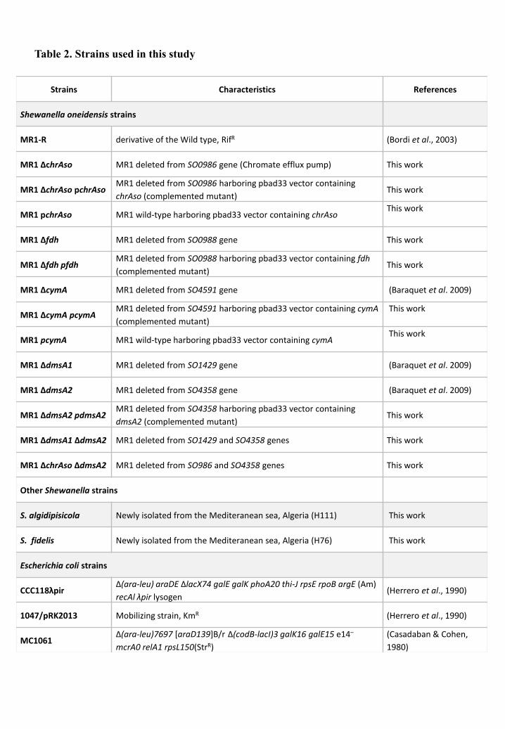
Strains Characteristics References
Shewanella oneidensis strains
MR1-R derivative of the Wild type, RifR (Bordi et al., 2003)
MR1 ΔchrAso MR1 deleted from SO0986 gene (Chromate efflux pump) This work
MR1 ΔchrAso pchrAsoMR1 deleted from SO0986 harboring pbad33 vector containing
chrAso (complemented mutant)This work
MR1 pchrAso MR1 wild-type harboring pbad33 vector containing chrAsoThis work
MR1 Δfdh MR1 deleted from SO0988 gene This work
MR1 Δfdh pfdhMR1 deleted from SO0988 harboring pbad33 vector containing fdh
(complemented mutant)This work
MR1 ΔcymA MR1 deleted from SO4591 gene (Baraquet et al. 2009)
MR1 ΔcymA pcymAMR1 deleted from SO4591 harboring pbad33 vector containing cymA
(complemented mutant)
This work
MR1 pcymA MR1 wild-type harboring pbad33 vector containing cymAThis work
MR1 ΔdmsA1 MR1 deleted from SO1429 gene (Baraquet et al. 2009)
MR1 ΔdmsA2 MR1 deleted from SO4358 gene (Baraquet et al. 2009)
MR1 ΔdmsA2 pdmsA2MR1 deleted from SO4358 harboring pbad33 vector containing
dmsA2 (complemented mutant)This work
MR1 ΔdmsA1 ΔdmsA2 MR1 deleted from SO1429 and SO4358 genes This work
MR1 ΔchrAso ΔdmsA2 MR1 deleted from SO986 and SO4358 genes This work
Other Shewanella strains
S. algidipisicola Newly isolated from the Mediteranean sea, Algeria (H111) This work
S. fidelis Newly isolated from the Mediteranean sea, Algeria (H76) This work
Escherichia coli strains
CCC118λpirΔ(ara-leu) araDE ∆lacX74 galE galK phoA20 thi-J rpsE rpoB argE (Am)
recAl λpir lysogen(Herrero et al., 1990)
1047/pRK2013 Mobilizing strain, KmR (Herrero et al., 1990)
MC1061Δ(ara-leu)7697 [araD139]B/r Δ(codB-lacI)3 galK16 galE15 e14–
mcrA0 relA1 rpsL150(StrR)
(Casadaban & Cohen,
1980)
Table 2. Strains used in this study

Material and Methods
111
1. Bacterial strains, plasmids and culture media
The bacterial strains and plasmids used in this study are described in Tables 2 and 3.The
E. coli strains CC118 pir and 1047/pRKβ01γ (Herrero et al, 1990) used for conjugation
and MC1061 (Casadaban & Cohen, 1980) derivatives used for chromate resistance and
reduction assays were routinely grown aerobically at 37°C or 30°C when specified, in
Lysogeny Broth (LB) medium containing yeast extract (5g/L), bacto-tryptone (10g/L) and
NaCl (5g/L) (Miller, 1972).
All Shewanella strains including S. fidelis H76 and S. algidipiscicola H111 were
routinely grown at 28°C in classic LB medium for S. oneidensis MR-1 (Bordi et al, 2003)
strains. However, addition of NaCl to a final concentration of 15g/L to LB medium was
necessary for growth of H76 and H111 strains. Strains were either incubated under
anaerobic (static) or aerobic (gentle agitation) conditions. In Anaerobic conditions, LB
medium was supplemented with 20mM trimethylamine oxide (TMAO) as electron acceptor
and buffered with 40mM of MOPS. When growth was performed in the presence of
chromate, disposable tubes were used and incubated either statically or under agitation. The
dissolved oxygen (DO) of the latter condition measured by a Clark electrode was 10%. This
condition was then referred as semi-aerobic. Chromate challenge was carried out by
supplementing LB medium at the required final concentration with a filter-sterilized stock
solution of potassium chromate (K2CrO4, Sigma-Aldrich). If required, media were
solidified by adding 17 g/L agar. When needed, antibiotics are added at the following
concentrationμ chloramphenicol β5 g/mL, ampicillin 50 g/mL, kanamycin 10 g/mL and
streptomycin 100 g/mL. Arabinose is added to induce the expression of a gene under the
control of a pBAD promoter carried on a dedicated plasmid (pBAD33, Annexes, Figure
S1). Growth was determined spectrophotometrically by monitoring changes in optical
density at 600 nm compared to the same medium without bacterium (OD600nm). The
bacterial strains are kept in their culture medium supplemented with glycerol (20% final),
then frozen at -80°C.

Plasmids Characteristics References
pBAD33Vector containing pBAD promoter with a p15 origin of
replication, CmR (Guzman et al., 1995)
pchrAso chrAso sequence cloned into pBAD33 This work
pdmsA2 dmsA2 sequence cloned into pBAD33 (Baraquet et al. 2009)
pfdh fdh sequence cloned into pBAD33 This work
pGE593 lacZ transcriptional reporter fusion cloning vector, AmpR(Eraso and Weinstock,
1992)
pACYC185 Cloning vector containing the p15A origin of replication, CmR(Chang and Cohen,
1978)
pACYC184-lacZ pACYC184 vector cotaining lacZ sequence from pGE593 This work
pmxd450::lacZ Promoter region upstream of mxdA cloned into pACYC184-lacZ This work
pchrAso::lacZPromotor region upstream of chrAso cloned into pACYC184-
lacZThis work
pRK2013 Broad-host-range helper vector; Tra+,KnR(Figurski and Helinski,
1979)
pKNG101 Marker exchange suicide vector; sacB, mobRK2, oriR6K, SmR(Figurski and Helinski,
1979)
Table 3. Plasmids used in this study

Material and Methods
112
2. Molecular biology techniques
Isolation, purification and hydrolysis of DNA
The plasmid DNA was prepared using the «GenEluteTM Plasmid Miniprep» (Sigma
Aldrich) and their concentration was estimated after agarose gel electrophoresis.
Chromosomal DNA was obtained using the kit «GenEluteTM Bacterial Genomic DNA kit»
(Sigma Aldrich). Restriction enzymes used come from New Biolabs. Conventional plasmid
preparation techniques, restriction enzyme digestion and ligation digestion were performed
under the conditions stipulated by the suppliers.
DNA amplification by PCR
In general, for a 50 L reaction, a mixture containing the template DNA (10 to β0 ng of
chromosomal DNA or approximately γ00 g of plasmid DNA), a pair of oligonucleotide
primers (100 to β00 ng each primer), the four dXTPs (100 M) and a unit of Taq DNA
polymerase « GoTaq® DNA Polymerase » (Promega) as well as 1X of its specific buffer, is
subjected to a succession of cycles: 1-Initial denaturation: 2 min at 95°C; 2-Denaturation:
30 sec to 95°C; 3-Hybridization: 30 sec at 53°C; 4-Elongation: 1 min per kilobase at 72°C;
5-Final elongation: 3 min to 72°C.
Steps 2 to 4 are repeated between 20 and 30 times depending on the amount of DNA
desired. The duration of step 4 varied according to the size of the DNA to be amplified,
given that on average the elongation rate of the polymerase is greater than 1 kb per min.
The temperature of step 3 may also vary depending on the Tm (melting temperature) of the
primers used. If the enzyme used for PCR is the high-fidelity DNA polymerase « Q5 hot
start High-Fidelity », with its specific buffer «Q5 Reaction Buffer» (Biolabs), we used the
following program: 30sec of initial denaturation at 98°C followed by 24 amplification
cycles including 10sec of denaturation at 98°C, 30sec of hybridization at 55°C and of 72°C
for elongation, and finally 2 min at 72°C for the final elongation step.
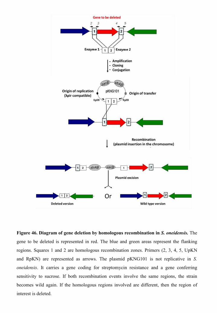
Figure 46. Diagram of gene deletion by homologous recombination in S. oneidensis. The
gene to be deleted is represented in red. The blue and green areas represent the flanking
regions. Squares 1 and 2 are homologous recombination zones. Primers (2, 3, 4, 5, UpKN
and RpKN) are represented as arrows. The plasmid pKNG101 is not replicative in S.
oneidensis. It carries a gene coding for streptomycin resistance and a gene conferring
sensitivity to sucrose. If both recombination events involve the same regions, the strain
becomes wild again. If the homologous regions involved are different, then the region of
interest is deleted.

Material and Methods
113
Plasmid construction
The cloning vector pBAD33 (Guzman et al, 1995) carrying inducible promoter was
used during this work (Annexes, Figure S1). In general, the genes to be cloned were
amplified by PCR without their promoter, from chromosomal DNA of S. oneidensis
MR1-R using « Q5 hot start High-Fidelity ». The primers allowed the generation of
restriction sites compatible with those present in the multiple cloning sites of the expression
vector pBAD33. The PCR products were then purified using « EZ‐10 Spin Column PCR
Products » kit (Bio Basic Inc) and digested and then cloned into the expression vector
hydrolyzed by the same enzymes as the PCR product. Hybrid plasmids thus obtained were
verified by DNA sequence.
The pACYC184-lacZ vector was constructed by cloning the -galactosidase-encoding
gene lacZ from pGE593 (Eraso & Weinstock, 1992) (Annexes, Figure S2) into the vector
pACYC184 (Chang & Cohen, 1978) (Table 2, Annexes, Figure S3). The ligation product
was transformed into E. coli strain MC1061. The resulting plasmid called pchrASO::lacZ
was introduced into S. oneidensis MR1-R by conjugation. Plasmid construction was also
checked by DNA sequencing. Another fusion used as control, named pmxd450::lacZ, was
similarly constructed using a DNA fragment corresponding to the 450 bp upstream from
the ATG of mxdA.
Construction of deletion mutants
The construction of deletion mutants in S. oneidensis was carried out by a four-step
homologous recombination mutagenesis technique (Figure 46). The first step was to
amplify the two 500-base pair (bp) fragments flanking the gene to be deleted by PCR using
the high-fidelity DNA polymerase enzyme « Q5 hot start High-Fidelity » (Biolabs) and two
pairs of primers that should be previously purified using the « GenEluteTM Bacterial
genomic DNA » kit (Sigma) following the manufacturer's protocol. The first (primers 2 and
3) and the second pair (primers 4 and 5) used as template the chromosomal DNA of the
wild strain of S. oneidensis. The PCR products obtained were then purified with the « EZ-
10 Spin Column PCR Products » kit (Bio Basic Inc). Primers 3 and 4 share a

Material and Methods
114
complementary sequence which allowed the realization of a second PCR with primers 2
and 5 using the products of the first PCR as DNA template. This second PCR resulted in
the fusion of the two fragments flanking the gene of interest, thus obtaining a fragment of
approximately 1kb. After purification, PCR products and the plasmid pKNG101 (Annexes,
figure S2) were digested with the same restriction enzymes (Biolabs) for 1h30 at 37°C.
After purification, the products resulting from the digestion were incubated for 16h at 16°C
with DNA ligase of T4 phage (Biolabs) in order to fuse the gene of interest together with
the plasmid.
The second step is the introduction of the obtained plasmid in E. coli CC118 pir strain
by transformation. To this end, the E. coli strain CC118 pir should be treated by CaCl2 to
become competent. From an overnight preculture of E. coli CC118 pir strain, 50 ml of LB
culture medium were inoculated with 1:100 of starter culture and then incubated at 37°C in
a shaking incubator. Growth was stopped when OD600nm reached values between 0.4 and
0.6, to be centrifuged at 4000 rpm for 10 min at 4°C. The pellets were then resuspended in
25 ml of 50mM CaCl2 buffer (50mM CaCl2 and 15% of glycerol) previously cooled to 4°C.
After 20 minutes of incubation at 4°C, a second centrifugation was then carried out. The
pellet was resuspended in 4 mL of the same buffer and left for another 20 min at 4°C before
being divided into several aliquots and then stored at -80°C.
For the transformation, 100 L of competent cells were added to 7 L of ligation mixture
for 20 min at 4°C. The cells were then subjected to a thermal shock for 2 min at 42°C, and
immediately thereafter incubated for 2 min at 4°C. LB medium was then added, and the
suspension was incubated at 37°C for 1h to allow expression of the antibiotic resistance
genes. The vector pKNG101 (Figurski & Helinski, 1979) contains an pir-dependent
replication origin (R6K), a sacB cassette and a streptomycin resistance cassette (stpR)
(Annexes, Figure S4). The later allows the selection of colonies harboring the plasmid.
Transformants were therefore selected on solid agar LB medium supplemented with
Streptomycin at 37°C. The presence of the insert in the various transformants was
confirmed by PCR using two primers, RpKN and UpKN, which hybridize on the plasmid
on either side of the multiple cloning sites (Figure 46).


Material and Methods
115
The third step was the introduction of the plasmid into S. oneidensis by conjugation.
The mobilizing strain E. coli 1047 which harbors a pRK2013 plasmid (the helper) (Figurski
& Helinski, 1979) possessing the genes encoding the proteins necessary for the synthesis of
the conjugative bridge and the donor strain CC118 pir containing a non-replicative
plasmid, were placed in contact on LB plates at 37°C for 2 h. This contact time allows the
transfer of the plasmid "helper" into the donor strain CC118 pir. After that, the receptor
strain S. oneidensis was added and the plates were then incubated at 28°C for 6h. This
second contact time allows the transfer of the recombinant plasmid pKNG101 through the
conjugation bridge to the receptor strain. Afterwards, the set of cells was resuspended in 1
ml of LB containing 4 l of colicin A, in order to eliminate the E. coli cells (Cascales et al,
2007), and incubated for 1h at 28°C. Since the plasmid pKNG101 is non-replicative in S.
oneidensis, it must be inserted in the chromosome to be conserved and to confer resistance
to streptomycin. The strain of S. oneidensis used in this study is naturally resistant to
rifampicin, unlike E. coli. Thus, conjugants were selected on solid agar medium (LB agar)
containing streptomycin (100 g/mL) and rifampicin (10 g/ml) at β8°C.
The fourth step consists of the excision of the plasmid from the chromosome using
another recombination step. For this purpose, the streptomycin-resistant cells were plated
on LB containing 6% sucrose, to select the second recombination event, and incubated for 2
days at 16°C, and then for 1 day at 28°C. In fact, the plasmid pKNG101 possesses the sacB
cassette which makes the cells sensitive to sucrose. To survive, the cell must eliminate the
plasmid sequence from its chromosome. Two possibilities of recombination were possible,
either the strain becomes wild again or it loses the gene of interest. The colonies were then
tested by PCR using the primers 1 and 5 which hybridize upstream and downstream of the
gene of interest, to determine whether the strains obtained are wild or lacking the gene of
interest.

Material and Methods
116
3. Biochemical techniques
Measurement of β-galactosidase activities
The -galactosidase activity was measured on the total extracts according to Miller's
assay adapted for use with plate reader. The S. oneidensis MR1-R strain containing
pchrASO::lacZ or pmxd450::lacZ was grown overnight on LB-agar plate. The overnight
culture was diluted to an OD600nm = 0.1 in fresh liquid LB medium containing various
concentrations of chromate (0, 0.05, 0.1 and 0.2 mM) and incubated in semi-aerobiosis at
β8°C for 16 hours prior to -galactosidase activity quantification. Thereafter, cells were
transferred into a microtiter plate and OD600nm was measured using a Tecan Spark 10M
microplate reader. Cells were then lysed using 1µl of lysozyme at 10 mg/mL and 9µl of
« PopCulture ReagentR » (NovagenR) prior to incubation with 100µl of Z buffer (62 mM
Na2HPO4, 45 mM NaH2PO4, 10 mM KCl, 1 mM MgSO4, 50 mM -mercaptoethanol) in
each well. Forty microliters of O-nitrophenyl-ß-D-galactopyranoside (ONPG) at 4 mg/ml
were added to the mix in each well, before kinetic quantifications of OD420nm every second
for 15 minutes. Slope of the obtained curves were calculated and -galactosidase activity
(arbitrary units) determined by the following equation:
(1000×slope)/(OD600nm×volume of reaction ( L)).
Measurement of chromate reduction
In vivo and In vitro Cr(VI)-reduction assay
In vivo Cr(VI) measurements were carried out on the supernatant of strain cultures
exposed to several chromate concentrations during different time of incubation under
anaerobic and semi-aerobic conditions. Cell cultures were centrifuged at 10 000 rpm for
5 min and residual Cr(VI) concentration present in the supernatant and cell pellets was
determined using the S-diphenylcarbazide (DPC) method (Urone, 1955) slightly modified.
Briefly, 10, β0, and 50 L supernatant samples were added in a β mL tube containing 1 mL
of H3PO4 0.5% and the volume of water necessary to obtain a 1λ60 L final volume. Forty
microliters of DPC reagent (5 mg/mL in 95% acetone and stored in dark at room
temperature) were added, gently mixed and kept at room temperature for 5–10 min.

Figure 47. Schematic representation of the technique used prior to the measurement of
Cr(VI) reduction of Shewanella strains pellicles.
Incubation of the recovred pellicles in fresh LB medium
supplemeted with chromate

Material and Methods
117
Absorbance was measured at 540nm. Cr(VI) concentration in the samples was calculated
from a standard curve.
In vitro measurements of chromate reductase activity of S. oneidensis strains (WT and
the chrASO deleted mutant) and E. coli strains (containing either the pBAD33 vector or the
plasmid pchrASO) were performed on crude extracts as described previously with slight
modifications. Cells were grown aerobically overnight in LB medium at 28°C and 30°C
respectively, and then harvested, washed with sodium phosphate buffer (pH7) and
resuspended in the same buffer before disruption by French press. The cell lysates were
then centrifuged and the supernatants (crude extracts) recovered. Protein concentrations
were measured using Protein Assay Dye Reagent (Bio-Rad), and bovine serum albumin as
standard. Reaction mixtures (1 mL) containing 100 M of K2CrO4 [Cr(VI)] and 1.5 mg of
proteins (crude extracts) were incubated at 30°C for 30 min. The residual Cr(VI)
concentration in the reaction mixture was estimated using the DPC method as described
above.
Cr(VI) reduction measurement of Shewanella pellicles
In order to measure pellicles chromate reduction rates, Shewanella mature pellicles
were collected using a sterile syringe while avoiding the recovery of the LB medium that is
underneath. Pellicles were then placed into an empty Petri dish. Thereafter LB medium
supplemented with different chromate concentrations was poured gently on the recovered
pellicles, which were then incubated for 24h and 48h at 28°C under static conditions. After
incubation time, pellicles and LB medium were recovered and centrifuged at 10 000 rpm
for 10-15 min prior to measurement of total chromium (As described below) and residual
Cr(VI) in the cellular and supernatant fractions (Figure 47).
Total chromium measurement assay
Total chromium in both the resuspended pellet and the supernatant, resulting from
centrifugation of cultures previously exposed to chromate, were measured by inductively
coupled plasma optical emission spectrometry (ICP-OES). Prior to this measurement,
71.4µL samples (cellular or supernatant fractions) were resuspended in 428.6µL of LB.


Material and Methods
118
Thereafter, 500µL of pure nitric acid (HNO3) was added. The mixture of sample and HNO3
was then incubated in 95°C water bath for 30 min, and 4 ml of 20% HNO3 solution was
then added to the treated samples.
4. Assessment of strains chromate resistance
Chromate resistance assays in Shewanella strains
Shewanella strains pre-cultures grown overnight on LB plates were suspended in LB
medium and used to inoculate fresh LB medium to an initial OD600nm of 0.2. Cells were
then grown until an OD600nm of 0.5 and submitted to two different assays. For the spot
assay, 10-fold serial dilutions of cell cultures were spotted on LB plates supplemented or
not with 0.5mM of chromate and incubated at 28°C. Plates were scanned after 4 days of
incubation. For the viability assay, chromate was added to the cell cultures (at OD600nm of
0.5) to a final concentration of 0.2mM. After 5 hours, cells were appropriately diluted in
LB and spread onto LB agar, and incubated at 28°C. The total number of viable cells was
estimated based on the number of colony-forming unit (CFU). Results were expressed as
the percentage of viable counts measured in these conditions compared to that of the same
culture grown in the absence of chromate.
Chromate resistance assay in E. coli
Pre-cultures of MC1061 derivatives containing either the plasmid pchrASO or the
pBAD33 vector were used to inoculate fresh LB medium to an initial OD600nm of 0.05.
Cells were grown until an OD600nm of 0.2 prior to the addition of chromate at various final
concentrations (0, 0.2, 0.4, 0.8 and 1.2mM), then the cells were incubated at 30°C. The
cultures were regularly sampled to measure the OD600nm and to quantify the chromate using
the DPC method (As described above).
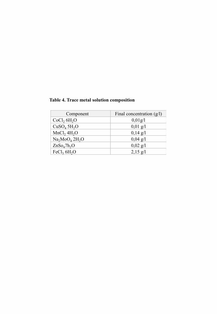
Component Final concentration (g/l)CoCl2 6H2O 0,01g/lCuSO4 5H2O 0,01 g/lMnCl2 4H2O 0,14 g/lNa2MoO4 2H2O 0,04 g/lZnSo47h2O 0,02 g/lFeCl3 6H2O 2,15 g/l
Table 4. Trace metal solution composition
Material and Methods
119
5. Divers assays
Pellicle assay
Cells were grown overnight and then diluted in 50 ml of fresh LB medium,
supplemented with NaCl in the case of S. fidelis H76 and S. algidipiscicola H111, to reach
an OD600nm of 0.2. This suspension was then transferred into a Petri dish and incubated at
28°C without being shaken for 24h for S. oneidensis MR-1 and 48h for H76 and H111
strains (Figure 47). For the two latter strains the LB medium was supplemented with
different concentrations of trace elements either separately or combined in a solution a trace
metal solution containing CoCl2 6H2O, CuSO4 5H2O, MnCl2 4H2O, Na2MoO4 2H2O,
ZnSo4 7H2O and FeCl3 6H2O. The final concentration added of each component is
indicated in Table 4.
Chemotaxis assay (μplugs microscopy technique)
The bacteria were grown aerobically in LB medium, supplemented with NaCl in the
case of S. fidelis H76 and S. algidipiscicola H111 at 28°C to an optical density at 600nm of
about 0.1-0.2. Afterwards strains cultures were centrifuged (10 min at 3500 rpm) and
subsequently resuspended in buffer (LM supplemented with 15mM lactate, 0, 1% Tween
β0 and 10 g/ml tetracycline) in order to obtain an optical density at 600nm of 0.5. A
bridge was constructed by placing two square coverslips (22mmx22mm) on each side of a
plastic slide (76mmx26mm) (Figure 48). For this technique, since S. oneidensis MR-1 cells
adhere to the glass, it is necessary to use plastic slides that made from PETG and manually
cut from a plate Vivak (Bayer). A 5 L agarose drop prepared from β% low-melting agarose
containing either the test compound (nitrate, chromate, nickel and cobalt) or buffer was
then placed on the PETG slide. The final concentrations of compounds tested were 50mM
for nitrate and 20mM for chromate, nickel and cobalt. The whole was then covered with
glass rectangular coverslip (24mmx60mm). This structure was held in place by
cyanoacrylate glue. After 30 minutes, for the agarose and the adhesive to solidify,
approximately 150 L of cells were introduced into the space between the slide and the
coverslip. The microscopic images were taken after 30 minutes at the edges of the drops
using a 10x objective (Figure 48).

Glass coverslips
PETG slide
The agarose drop
containing the substrate
Bacterial cells
injection
Microscopic observation
The agarose drop
containing an attractant The agarose drop
containing a repellent
Figure 48. Representation of the μplugs microscopy technique The substrate to be
tested is premixed with agarose and deposited on the PETG slide. The cells are
placed between PETG slide and the coverslip.
Material and Methods
120
6. Isolation of new Shewanella species method
Samples collection
To isolate new marine Shewanella sp. strains, more than ten samples of seawater and
sediments coming from different Algerian harbors, beaches were collected. Aseptically using
plastic bottles, seawater samples were taken from the uppermost 60 centimeters and sea-
sediments samples were taken from about 10 to 15 meters deep. All samples were stored at
room temperature until processed. The collected samples as well as the geographical
coordinates of the sampling sites were summarized in Table 5
Isolation
To isolate Shewanella cells present in the samples, the latter were cultured in LB
medium supplemented with NaCl (30g/l), TMAO (20mM) and MOPS (40mM) in aerobic
conditions at 28°C to mimic marine environment conditions. After 24h of incubation, 10-
fold serial dilutions were spread on LB plates containing TMAO and NaCl.
Simultaneously, 10µl of the first culture was used to inoculate 15ml of fresh LB medium.
Plates and liquid cultures were then incubated at 28°C. This procedure was repeated
successively two more times in order to enhance the amount of Shewanella cells which
facilitate their isolation. Colonies presenting similar phenotypic aspects of Shewanella
species were selected and further subjected to PCR amplification using the primer set
She211F and She1259R that specifically targets Shewanella 16SDNA.
7. Genome sequencing and bioinformatic analysis
The genome sequencing of H76 and H111 strains was carried out at the “Molecular
Research LP (MR DNA) Laboratory” (USA). The library was prepared using Nextera DNA
Sample Preparation Kit (Illumina) following the manufacturer's user guide and sequenced
using the HiSeq 2500 system (Illumina).
The genome analysis was carried out using the programs of the Mage platform
(http://www.genoscope.cns.fr/agc/microscope/home/index.php). The phylogenic tree
representing the nearest neighbours of H76 and H111, based on 16S ribosomal DNA
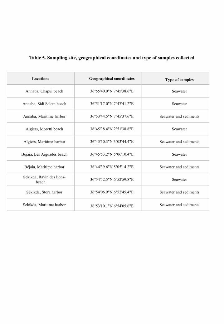
Locations Geographical coordinates Type of samples
Annaba, Chapui beach 36°55'40.0"N 7°45'38.6"E Seawater
Annaba, Sidi Salem beach 36°51'17.0"N 7°47'41.2"E Seawater
Annaba, Maritime harbor 36°53'44.5"N 7°45'37.6"E Seawater and sediments
Algiers, Moretti beach 36°45'38.4"N 2°51'38.8"E Seawater
Algiers, Maritime harbor 36°45'50.3"N 3°03'44.4"E Seawater and sediments
Béjaia, Les Aiguades beach 36°45'53.2"N 5°06'10.4"E Seawater
Béjaia, Maritime harbor 36°44'39.6"N 5°05'14.2"E Seawater and sediments
Sekikda, Ravin des lions-beach
36°54'52.5"N 6°52'59.8"E Seawater
Sekikda, Stora harbor 36°54'06.9"N 6°52'45.4"E Seawater and sediments
Sekikda, Maritime harbor 36°53'10.1"N 6°54'05.6"E Seawater and sediments
Table 5. Sampling site, geographical coordinates and type of samples collected
Material and Methods
121
sequences, was constructed using MOLE BLAST
(http://blast.ncbi.nlm.nih.gov/moleblast/moleblast.cgi)
Proteins sharing homologies with ChrASO were searched in the Bacteria kingdom using
the BlastP software on the NCBI server (https://blast.ncbi.nlm.nih.gov). For the
phylogenetic analysis, we used the “Phylogeny.fr” software in the “one-click” mode, i.e.
with the default parameters optimized by the authors (http://www.phylogeny.fr/). To obtain
the phylogenetic tree of a subset of ChrA proteins, we used the software “FigTree” version
1.4.3 (http://tree.bio.ed.ac.uk/software/figtree/) in which we entered the result in Netwick
format obtained with “Phylogeny”. The tree rooting was performed with the midpoint
option. Prediction of transmembrane helices of ChrASO was carried out using the TMHMM
online software (http://www.cbs.dtu.dk/services/TMHMM-2.0/).


ANNEXES


Figure S1. pBad33 vector map


Figure S2. Operon fusion vector pGE593 map

Figure S3. pACYC184 vector map


Figure S4. pKNG101 vector map used for gene deletion in S. oneidensis


REFERENCES


References
127
Ackerley DF, Gonzalez CF, Keyhan M, Blake R & Matin A (2004a) Mechanism of chromate reduction by the Escherichia coli protein, NfsA, and the role of different chromate reductases in minimizing oxidative stress during chromate reduction. Environ. Microbiol. 6: 851–860
Ackerley DF, Gonzalez CF, Park CH, Blake R, Keyhan M & Matin A (2004b) Chromate-reducing properties of soluble flavoproteins from Pseudomonas putida and Escherichia coli. Appl. Environ. Microbiol. 70: 873–882
Acosta-Navarrete YM, Leon-Marquez YL, Salinas-Herrera K, Jacome-Galarza IE, Meza-Carmen V, Ramirez-Diaz MI & Cervantes C (2014) Expression of the six chromate ion transporter homologues of Burkholderia xenovorans LB400. Microbiology 160: 287–295
Aguilar-Barajas E, Diaz-Pérez C, Ramirez-Diaz MI, Riveros-Rosas H & Cervantes C (2011) Bacterial transport of sulfate, molybdate, and related oxyanions. BioMetals 24: 687–707
Aguilar-Barajas E, Paluscio E, Cervantes C & Rensing C (2008) Expression of chromate resistance genes from Shewanella sp. strain ANA-3 in Escherichia coli. FEMS
Microbiol. Lett. 285: 97–100
Aharchaou I (2017) Écotoxicité et bioaccumulation du Cr (III) et du Cr (VI): spéciation, biodisponibilité et effets biologiques. Available at: https://tel.archives-ouvertes.fr/tel-01549080/ [Accessed July 26, 2017]
Ahemad M (2014) Bacterial mechanisms for Cr(VI) resistance and reduction: an overview and recent advances. Folia Microbiol. (Praha) 59: 321–332
Ahmad WA, Shahir S & Zakaria ZA (2009) Mechanisms of bacterial detoxification of Cr (VI) from industrial wastewater in the presence of industrial effluent as potential energy source. Univ. Teknol. Malays. [Accessed June 22, 2017]
Alexandre Gladys, Greer‐ Phillips Suzanne & Zhulin Igor B (2004) Ecological role of energy taxis in microorganisms. FEMS Microbiol. Rev. 28: 113–126
Alloway BJ & Ayres DC (1997) Chemical Principles of Environmental Pollution Dordrecht: Springer Netherlands Available at: http://link.springer.com/10.1007/978-94-011-2148-4 [Accessed April 1, 2018]
Anderson RA (1997) Chromium as an Essential Nutrient for Humans. Regul. Toxicol.
Pharmacol. 26: S35–S41
Armitano J, Baraquet C, Michotey V, Méjean V & Jourlin-Castelli C (2011) The chemical-in- wellμ a high-throughput technique for identifying solutes eliciting a chemotactic response in motile bacteria. Res. Microbiol. 162: 934–938
Armitano J, Méjean V & Jourlin-Castelli C (2014) Gram-negative bacteria can also form pellicles: Floating biofilm in Gram-negative bacteria. Environ. Microbiol. Rep. 6: 534–544

128
Baaziz H, Gambari C, Boyeldieu A, Ali Chaouche A, Alatou R, Méjean V, Jourlin-Castelli C & Fons M (2017) ChrASO, the chromate efflux pump of Shewanella oneidensis, improves chromate survival and reduction. PloS One 12: e0188516
Bae W-C, Lee H-K, Choe Y-C, Jahng D-J, Lee S-H, Kim S-J, Lee J-H & Jeong B-C (2005) Purification and Characterization of NADPH-Dependent Cr(VI) Reductase from Escherichia coli ATCC 33456. J Microbiol 43 1: 21–7
Barak Y, Ackerley DF, Dodge CJ, Banwari L, Alex C, Francis AJ & Matin A (2006) Analysis of Novel Soluble Chromate and Uranyl Reductases and Generation of an Improved Enzyme by Directed Evolution. Appl. Environ. Microbiol. 72: 7074–7082
Baraquet C, Théraulaz L, Iobbi-Nivol C, Méjean V & Jourlin-Castelli C (2009) Unexpected chemoreceptors mediate energy taxis towards electron acceptors in Shewanella
oneidensis. Mol. Microbiol. 73: 278–290
Barchinger SE, Pirbadian S, Sambles C, Baker CS, Leung KM, Burroughs NJ, El-Naggar MY & Golbeck JH (2016) Regulation of Gene Expression in Shewanella oneidensis MR-1 during Electron Acceptor Limitation and Bacterial Nanowire Formation. Appl.
Environ. Microbiol. 82: 5428–5443
Barrera-Díaz CE, Lugo-Lugo V & Bilyeu B (2012) A review of chemical, electrochemical and biological methods for aqueous Cr(VI) reduction. J. Hazard. Mater. 223–224: 1–12
Bartlett R & James B (1979) Behavior of Chromium in Soils: III. Oxidation1. J. Environ.
Qual. 8: 31–35
Belchik SM, Kennedy DW, Dohnalkova AC, Wang Y, Sevinc PC, Wu H, Lin Y, Lu HP, Fredrickson JK & Shi L (2011) Extracellular Reduction of Hexavalent Chromium by Cytochromes MtrC and OmcA of Shewanella oneidensis MR-1. Appl. Environ.
Microbiol. 77: 4035–4041
Beliaev AS, Klingeman DM, Klappenbach JA, Wu L, Romine MF, Tiedje JM, Nealson KH, Fredrickson JK & Zhou J (2005) Global Transcriptome Analysis of Shewanella
oneidensis MR-1 Exposed to Different Terminal Electron Acceptors. J. Bacteriol. 187: 7138–7145
Bencharit S & Ward MJ (2005) Chemotactic Responses to Metals and Anaerobic Electron Acceptors in Shewanella oneidensis MR-1. J. Bacteriol. 187: 5049–5053
Bencheikh-Latmani R, Obraztsova A, Mackey MR, Ellisman MH & Tebo BM (2007) Toxicity of Cr(III) to Shewanella sp. Strain MR-4 during Cr(VI) Reduction. Environ.
Sci. Technol. 41: 214–220
Bencheikh-Latmani R, Williams SM, Haucke L, Criddle CS, Wu L, Zhou J & Tebo BM (2005) Global Transcriptional Profiling of Shewanella oneidensis MR-1 during Cr(VI) and U(VI) Reduction. Appl. Environ. Microbiol. 71: 7453–7460
Bopp LH & Ehrlich HL (1988) Chromate resistance and reduction in Pseudomonas
fluorescens strain LB300. Arch. Microbiol. 150: 426–431


129
Bordi C, Iobbi-Nivol C, Mejean V & Patte J-C (2003) Effects of ISSo2 Insertions in Structural and Regulatory Genes of the Trimethylamine Oxide Reductase of Shewanella oneidensis. J. Bacteriol. 185: 2042–2045
Bouillet S, Genest O, Jourlin-Castelli C, Fons M, Méjean V & Iobbi-Nivol C (2016) The General Stress Response σS Is Regulated by a Partner Switch in the Gram-negative Bacterium Shewanella oneidensis. J. Biol. Chem. 291: 26151–26163
Bowman JP (2015) Shewanella. In Bergey’s Manual of Systematics of Archaea and
Bacteria, Whitman WB Rainey F Kämpfer P Trujillo M Chun J DeVos P Hedlund B & Dedysh S (eds) pp 1–22. Chichester, UK: John Wiley & Sons, Ltd Available at: http://doi.wiley.com/10.1002/9781118960608.gbm01100 [Accessed October 16, 2017]
Branco R, Chung AP, Johnston T, Gurel V, Morais P & Zhitkovich A (2008) The Chromate-Inducible chrBACF Operon from the Transposable Element TnOtChr Confers Resistance to Chromium(VI) and Superoxide. J. Bacteriol. 190: 6996–7003
Branco R & Morais PV (2013) Identification and Characterization of the Transcriptional Regulator ChrB in the Chromate Resistance Determinant of Ochrobactrum tritici 5bvl1. PLoS ONE 8: e77987
Breuer M, Rosso KM, Blumberger J & Butt JN (2015) Multi-haem cytochromes in Shewanella oneidensis MR-1: structures, functions and opportunities. J. R. Soc.
Interface 12: 20141117–20141117
Brown SD, Thompson MR, Verberkmoes NC, Chourey K, Shah M, Zhou J, Hettich RL & Thompson DK (2006) Molecular dynamics of the Shewanella oneidensis response to chromate stress. Mol. Cell. Proteomics MCP 5: 1054–1071
Bunn C, Booker H, Bromley DA & Declaris N (2013) Crystals: Their Role in Nature and in Science Elsevier Science Available at: https://books.google.fr/books?id=Qx8SBQAAQBAJ
Cabiscol Català E, Tamarit Sumalla J & Ros Salvador J (2000) Oxidative stress in bacteria and protein damage by reactive oxygen species. International Microbiology 3: 3–8
Callender E (2003) Heavy Metals in the Environment—Historical Trends A2-Holland, Heinrich D. In Treatise on Geochemistry, Turekian KK (ed) pp 67–105. Oxford: Pergamon Available at: https://www.sciencedirect.com/science/article/pii/B0080437516091611
Cao B, Ahmed B, Kennedy DW, Wang Z, Shi L, Marshall MJ, Fredrickson JK, Isern NG, Majors PD & Beyenal H (2011) Contribution of Extracellular Polymeric Substances from Shewanella sp. HRCR-1 Biofilms to U(VI) Immobilization. Environ. Sci.
Technol. 45: 5483–5490
Casadaban MJ & Cohen SN (1980) Analysis of gene control signals by DNA fusion and cloning in Escherichia coli. J. Mol. Biol. 138: 179–207
Cascales E, Buchanan SK, Duché D, Kleanthous C, Lloubès R, Postle K, Riley M, Slatin S & Cavard D (2007) Colicin Biology. Microbiol. Mol. Biol. Rev. 71: 158–229

130
Cervantes C & Campos-Garcia J (2007) Reduction and Efflux of Chromate by Bacteria. In Molecular Microbiology of Heavy Metals, Nies DH & Silver S (eds) pp 407–419. Berlin, Heidelberg: Springer Berlin Heidelberg Available at: http://link.springer.com/10.1007/7171_2006_087 [Accessed June 16, 2017]
Cervantes C, Campos-Garcia J, Devars S, Gutiérrez-Corona F, Loza-Tavera H, Torres-Guzmán JC & Moreno-Sánchez R (2001) Interactions of chromium with microorganisms and plants. FEMS Microbiol. Rev. 25: 335–347
Cervantes C, Ohtake H, Chu L, Misra TK & Silver S (1990) Cloning, nucleotide sequence, and expression of the chromate resistance determinant of Pseudomonas aeruginosa plasmid pUM505. J. Bacteriol. 172: 287–291
Chang AC & Cohen SN (1978) Construction and characterization of amplifiable multicopy DNA cloning vehicles derived from the P15A cryptic miniplasmid. J. Bacteriol. 134: 1141–1156
Cheung KH & Gu J-D (2007) Mechanism of hexavalent chromium detoxification by microorganisms and bioremediation application potential: A review. Int. Biodeterior.
Biodegrad. 59: 8–15
Chourey K, Thompson MR, Morrell-Falvey J, VerBerkmoes NC, Brown SD, Shah M, Zhou J, Doktycz M, Hettich RL & Thompson DK (2006) Global Molecular and Morphological Effects of 24-Hour Chromium(VI) Exposure on Shewanella
oneidensis MR-1. Appl. Environ. Microbiol. 72: 6331–6344
Cordova CD, Schicklberger MFR, Yu Y & Spormann AM (2011) Partial Functional Replacement of CymA by SirCD in Shewanella oneidensis MR-1. J. Bacteriol. 193: 2312–2321
Costerton J, Geesey G & Cheng K (1978) How bacteria stick. Sientific American 1: 86–95
Coursolle D, Baron DB, Bond DR & Gralnick JA (2010) The Mtr respiratory pathway is essential for reducing flavins and electrodes in Shewanella oneidensis. J. Bacteriol. 192: 467–474
Coursolle D & Gralnick JA (2012) Reconstruction of Extracellular Respiratory Pathways for Iron(III) Reduction in Shewanella Oneidensis Strain MR-1. Front. Microbiol. 3: 56
Davey ME & O’toole GA (β000) Microbial Biofilmsμ from Ecology to Molecular Genetics. Microbiol. Mol. Biol. Rev. 64: 847–867
De Flora S (2000) Threshold mechanisms and site specificity in chromium(VI) carcinogenesis. Carcinogenesis 21: 533–541
Derby HA & Hammer BW (1931) Bacteriology of butter IV. Bacteriological studies on surface taint butter. Research Bulletin (Iowa Agriculture and Home Economics
Experiment Station) 11: 33
Dhal B, Thatoi HN, Das NN & Pandey BD (2013) Chemical and microbial remediation of hexavalent chromium from contaminated soil and mining/metallurgical solid waste: A review. J. Hazard. Mater. 250–251: 272–291


131
Di Bona KR, Love S, Rhodes NR, McAdory D, Sinha SH, Kern N, Kent J, Strickland J, Wilson A, Beaird J, Ramage J, Rasco JF & Vincent JB (2011) Chromium is not an essential trace element for mammalsμ effects of a “low-chromium” diet. JBIC J. Biol.
Inorg. Chem. 16: 381–390
Díaz-Pérez C, Cervantes C, Campos-García J, Julián-Sánchez A & Riveros-Rosas H (2007) Phylogenetic analysis of the chromate ion transporter (CHR) superfamily: Chromate ion transporter (CHR) phylogeny. FEBS J. 274: 6215–6227
Dikow RB (2011) Genome-level homology and phylogeny of Shewanella (Gammaproteobacteria: lteromonadales: Shewanellaceae). BMC Genomics 12: 237
Donati E, Oliver C & Curutchet G (2003) Reduction of chromium (VI) by the indirect action of Thiobacillus thioparus. Braz. J. Chem. Eng. 20: 69–73
Driscoll ME, Romine MF, Juhn FS, Serres MH, Mccue LA, Beliaev AS, Fredrickson JK & Gardner TS (2007) Identification of diverse carbon utilization pathways in Shewanella oneidensis MR-1 via expression profiling. Genome Informatics: 287–298
Dufault-Thompson K, Jian H, Cheng R, Li J, Wang F & Zhang Y (2017) A Genome-Scale Model of Shewanella piezotolerans Simulates Mechanisms of Metabolic Diversity and Energy Conservation. mSystems 2: e00165-16
Duranoğlu D & Beker U (β015) Cr(VI) Adsorption Onto Biomass Waste Material-Derived Activated Carbon. In Desalination Updates, Ning RY (ed) p Ch. 10. Rijeka: InTech Available at: http://dx.doi.org/10.5772/60206 [Accessed April 3, 2018]
El-Naggar MY, Wanger G, Leung KM, Yuzvinsky TD, Southam G, Yang J, Lau WM, Nealson KH & Gorby YA (2010) Electrical transport along bacterial nanowires from Shewanella oneidensis MR-1. Proc. Natl. Acad. Sci. 107: 18127
Eraso JM & Weinstock GM (1992) Anaerobic control of colicin E1 production. J. Bacteriol. 174: 5101–5109
Ezraty B, Gennaris A, Barras F & Collet J-F (2017) Oxidative stress, protein damage and repair in bacteria. Nat. Rev. Microbiol. 15: 385–396
Fendorf SE (1995) Surface reactions of chromium in soils and waters. Geoderma 67: 55–71
Figurski DH & Helinski DR (1979) Replication of an origin-containing derivative of plasmid RK2 dependent on a plasmid function provided in trans. Proc. Natl. Acad. Sci. 76: 1648–1652
Flores-Alvarez LJ, Corrales-Escobosa AR, Cortés-Penagos C, Martínez-Pacheco M, Wrobel-Zasada K, Wrobel-Kaczmarczyk K, Cervantes C & Gutiérrez-Corona F (2012) The Neurospora crassa chr-1 gene is up-regulated by chromate and its encoded CHR-1 protein causes chromate sensitivity and chromium accumulation. Curr. Genet. 58: 281–290
Focardi S, Pepi M & E. S (2013) Microbial Reduction of Hexavalent Chromium as a Mechanism of Detoxification and Possible Bioremediation Applications. In Biodegradation-Life of Science, Chamy R (ed) InTech Available at: http://www.intechopen.com/books/biodegradation-life-of-science/microbial-

132
reduction-of-hexavalent-chromium-as-a-mechanism-of-detoxification-and-possible-bioremediat [Accessed August 30, 2017]
Foster JW & Slonczewski J (2017) Microbiology: An Evolving Science W W Norton & Company Incorporated Available at: https://books.google.fr/books?id=UAu3DAEACAAJ
Francisco R., Alpoim M.C. & Morais P.V. (2002) Diversity of chromium‐resistant and ‐reducing bacteria in a chromium‐contaminated activated sludge. J. Appl. Microbiol. 92: 837–843
Fredrickson JK, Romine MF, Beliaev AS, Auchtung JM, Driscoll ME, Gardner TS, Nealson KH, Osterman AL, Pinchuk G, Reed JL, Rodionov DA, Rodrigues JLM, Saffarini DA, Serres MH, Spormann AM, Zhulin IB & Tiedje JM (2008) Towards environmental systems biology of Shewanella. Nat. Rev. Microbiol. 6: 592–603
Fu H, Jin M, Ju L, Mao Y & Gao H (2014) Evidence for function overlapping of CymA and the cytochrome bc1 complex in the Shewanella oneidensis nitrate and nitrite respiration: S. oneidensis bc 1 complex. Environ. Microbiol. 16: 3181–3195
Gaberell M, Chin Y-P, Hug SJ & Sulzberger B (2003) Role of Dissolved Organic Matter Composition on the Photoreduction of Cr(VI) to Cr(III) in the Presence of Iron. Environ. Sci. Technol. 37: 4403–4409
Gadd G.M. (1992) Metals and microorganisms: A problem of definition. FEMS Microbiol.
Lett. 100: 197–203
Gonzalez CF, Ackerley DF, Park CH & Matin A (2003) A Soluble Flavoprotein Contributes to Chromate Reduction and Tolerance by Pseudomonas putida. Acta Biotechnol. 23: 233–239
Gorby YA, Yanina S, McLean JS, Rosso KM, Moyles D, Dohnalkova A, Beveridge TJ, Chang IS, Kim BH, Kim KS, Culley DE, Reed SB, Romine MF, Saffarini DA, Hill EA, Shi L, Elias DA, Kennedy DW, Pinchuk G, Watanabe K, et al (2006) Electrically conductive bacterial nanowires produced by Shewanella oneidensis strain MR-1 and other microorganisms. Proc. Natl. Acad. Sci. 103: 11358
Grass G, Fan B, Rosen BP, Franke S, Nies DH & Rensing C (2001) ZitB (YbgR), a Member of the Cation Diffusion Facilitator Family, Is an Additional Zinc Transporter in Escherichia coli. J. Bacteriol. 183: 4664–4667
Gueddah D & Djebar AB (2014) Pollution by the heavy metals of the superficial marine sediment of Skikda city (Northeastern Algeria). : 6
Guertin J, Jacobs JA & Avakian CP (2016) Chromium(VI) Handbook CRC Press Available at: https://books.google.fr/books?id=DVdCqL9NbOcC
Gupta P & Diwan B (2017) Bacterial Exopolysaccharide mediated heavy metal removal: A Review on biosynthesis, mechanism and remediation strategies. Biotechnol. Rep. 13: 58–71
Gutiérrez-Corona JF, Romo-Rodriguez P, Santos-Escobar F, Espino-Saldana AE & Hernandez-Escoto H (2016) Microbial interactions with chromium: basic biological


133
processes and applications in environmental biotechnology. World J. Microbiol.
Biotechnol. 32: Available at: http://link.springer.com/10.1007/s11274-016-2150-0 [Accessed June 15, 2017]
Guzman LM, Belin D, Carson MJ & Beckwith J (1995) Tight regulation, modulation, and high-level expression by vectors containing the arabinose PBAD promoter. J.
Bacteriol. 177: 4121–4130
Haleng J, Pincemail J, Defraigne J-O, Charlier C & Chapelle J-P (2007) Le stress oxydant. Rev. Médicale Liège 62: 628–38
Hall NF (1957) Nouveau Traité de Chimie Minérale. Edited by PAUL PASCAL, Member de 1’Institut, Professeur honoraire a la Sorbonne. Volume X. Azote—Phosphore. J. Am.
Chem. Soc. 79: 4570–4570
Hall-Stoodley L, Costerton JW & Stoodley P (2004) Bacterial biofilms: from the Natural environment to infectious diseases. Nat. Rev. Microbiol. 2: 95
Han R, Li F, Liu T, Li X, Wu Y, Wang Y & Chen D (2016) Effects of Incubation Conditions on Cr(VI) Reduction by c-type Cytochromes in Intact Shewanella oneidensis MR-1 Cells. Front. Microbiol. 7: Available at: http://journal.frontiersin.org/Article/10.3389/fmicb.2016.00746/abstract [Accessed June 15, 2017]
Hau H & Gralnick JA (2007) Ecology and biotechnology of the genus Shewanella. Annu.
Rev. Microbiol. 61: 237–258
Heidelberg JF, Paulsen IT, Nelson KE, Gaidos EJ, Nelson WC, Read TD, Eisen JA, Seshadri R, Ward N, Methe B, Clayton RA, Meyer T, Tsapin A, Scott J, Beanan M, Brinkac L, Daugherty S, DeBoy RT, Dodson RJ, Durkin AS, et al (2002) Genome sequence of the dissimilatory metal ion–reducing bacterium Shewanella oneidensis. Nat.
Biotechnol. 20: 1118–1123
Henne KL, Nakatsu CH, Thompson DK & Konopka AE (2009) High-level chromate resistance in Arthrobacter sp. strain FB24 requires previously uncharacterized accessory genes. BMC Microbiol. 9: 199
Henrici AT (1933) Studies of Freshwater Bacteria: I. A Direct Microscopic Technique. J.
Bacteriol. 25: 277–287
Herrero M, de Lorenzo V & Timmis KN (1990) Transposon vectors containing non-antibiotic resistance selection markers for cloning and stable chromosomal insertion of foreign genes in gram-negative bacteria. J. Bacteriol. 172: 6557–6567
Honoré FA, Méjean V & Genest O (2017) Hsp90 Is Essential under Heat Stress in the Bacterium Shewanella oneidensis. Cell Rep. 19: 680–687
Hossain MA, Kumita M, Michigami Y, Islam TSA & Mori S (2005) Rapid Speciation Analysis of Cr(VI) and Cr(III) by Reversed-Phase High-Performance Liquid Chromatography with UV Detection. J. Chromatogr. Sci. 43: 98–103

134
Howard, Hamdan Leila J., Lizewski Stephen E. & Ringeisen Bradley R. (2011) High frequency of glucose‐utilizing mutants in Shewanella oneidensis MR‐1. FEMS
Microbiol. Lett. 327: 9–14
Hu P, Brodie EL, Suzuki Y, McAdams HH & Andersen GL (2005) Whole-Genome Transcriptional Analysis of Heavy Metal Stresses in Caulobacter crescentus. J.
Bacteriol. 187: 8437–8449
Ishibashi Y, Cervantes C & Silver S (1990) Chromium reduction in Pseudomonas putida. Appl. Environ. Microbiol. 56: 2268–2270
Ivanova EP (2003) Shewanella fidelis sp. nov., isolated from sediments and sea water. Int. J.
Syst. Evol. Microbiol. 53: 577–582
Jampala S (2015) Skin and Soft Tissue Infections due to Shewanella algae – An Emerging Pathogen. J. Clin. Diagn. Res. Available at: http://jcdr.net/article_fulltext.asp?issn=0973709x&year=2015&volume=9&issue=2&page=DC16&issn=0973-709x&id=5585 [Accessed October 16, 2017]
Janda JM (2014) Shewanella: a marine pathogen as an emerging cause of human disease. Clin. Microbiol. Newsl. 36: 25–29
Janda JM & Abbott SL (2014) The genus Shewanella : from the briny depths below to human pathogen. Crit. Rev. Microbiol. 40: 293–312
Jiménez-Meji-a R, Campos-Garci-a J & Cervantes C (2006) Membrane topology of the chromate transporter ChrA of Pseudomonas aeruginosa. FEMS Microbiol. Lett. 262: 178–184
Jomova K & Valko M (2011) Advances in metal-induced oxidative stress and human disease. Toxicology 283: 65–87
Joutey NT, Sayel H, Bahafid W & El Ghachtouli N (2015) Mechanisms of Hexavalent Chromium Resistance and Removal by Microorganisms. In Reviews of
Environmental Contamination and Toxicology Volume 233, Whitacre DM (ed) pp 45–69. Cham: Springer International Publishing Available at: http://link.springer.com/10.1007/978-3-319-10479-9_2 [Accessed June 15, 2017]
Juhnke S, Peitzsch N, Hübener N, Große C & Nies DH (2002) New genes involved in chromate resistance in Ralstonia metallidurans strain CH34. Arch. Microbiol. 179: 15–25
Kane AL, Brutinel ED, Joo H, Maysonet R, VanDrisse CM, Kotloski NJ & Gralnick JA (2016) Formate Metabolism in Shewanella oneidensis Generates Proton Motive Force and Prevents Growth without an Electron Acceptor. J. Bacteriol. 198: 1337–1346
Kaplan D & Christaen D (1987) Chelating Properties of Extracellular Polysaccharides from Chlorella spp. Appl.Environ.Microbiol: 4
Kato C & Nogi Y (2001) Correlation between phylogenetic structure and function: examples from deep-sea Shewanella. FEMS Microbiol. Ecol. 35: 223–230


135
Katz S & Salem H (1993) The toxicology of chromium with respect to its chemical speciation: A review. J. Appl. Toxicol. 13: 217–224
Keegan GM, Learmonth ID & Case C (2008) A Systematic Comparison of the Actual, Potential, and Theoretical Health Effects of Cobalt and Chromium Exposures from Industry and Surgical Implants. Crit. Rev. Toxicol. 38: 645–674
Khashe S & Janda JM (1998) Biochemical and Pathogenic Properties of Shewanella alga and Shewanella putrefaciens. J. Clin. Microbiol. 36: 783–787
Kouzuma A, Kasai T, Hirose A & Watanabe K (2015) Catabolic and regulatory systems in Shewanella oneidensis MR-1 involved in electricity generation in microbial fuel cells. Front. Microbiol. 6: Available at: http://journal.frontiersin.org/Article/10.3389/fmicb.2015.00609/abstract [Accessed October 28, 2017]
Kwak YH, Lee DS & Kim HB (2003) Vibrio harveyi Nitroreductase Is Also a Chromate Reductase. Appl. Environ. Microbiol. 69: 4390–4395
Lear G (2016) Biofilms in Bioremediation Caister Academic Press Available at: https://books.google.fr/books?id=m3A7DwAAQBAJ
Lee D-H, Park S-H, Kim Y-J, Oh D-C, Kim J-Y & Yoo H-S (2016) Shewanella algicola sp. nov., a marine bacterium isolated from brown algae. Int. J. Syst. Evol. Microbiol. 66: 2218–2224
Li Jun, Romine Margaret F. & Ward Mandy J. (2007) Identification and analysis of a highly conserved chemotaxis gene cluster in Shewanella species. FEMS Microbiol. Lett. 273: 180–186
Li N, Luo Q, Jiang Y, Wu G & Gao H (2014) Managing oxidative stresses in Shewanella
oneidensis : intertwined roles of the OxyR and OhrR regulons: Oxidative stresses in S. oneidensis. Environ. Microbiol. 16: 1821–1834
Li R, Auchtung JM, Tiedje JM & Worden RM (2010) Shewanella oneidensis MR-1 chemotaxis in a diffusion gradient chamber. Environ. Sci. Technol. 45: 1014–1020
Liang Y, Gao H, Chen J, Dong Y, Wu L, He Z, Liu X, Qiu G & Zhou J (2010) Pellicle formation in Shewanella oneidensis. BMC Microbiol. 10: 291
Liang Y, Gao H, Guo X, Chen J, Qiu G, He Z, Zhou J & Liu X (2012) Transcriptome analysis of pellicle formation of Shewanella oneidensis. Arch. Microbiol. 194: 473–482
Liu Y-G, Xu W-H, Zeng G-M, Li X & Gao H (2006) Cr(VI) reduction by Bacillus sp. isolated from chromium landfill. Process Biochem. 41: 1981–1986
Lowe KL, Straube W, Little B & Jones-Meehan J (2003) Aerobic and anaerobic reduction of Cr (VI) by Shewanella oneidensis effects of cationic metals, sorbing agents and mixed microbial cultures. Eng. Life Sci. 23: 161–178
Lower SK, Hochella MF & Beveridge TJ (2001) Bacterial Recognition of Mineral Surfaces: Nanoscale Interactions Between Shewanella and α-FeOOH. Science 292: 1360


136
Ma Z, Zhu W, Long H, Chai L & Wang Q (2007) Chromate reduction by resting cells of Achromobacter sp. Ch-1 under aerobic conditions. Process Biochem. 42: 1028–1032
MacDonell MT & Colwell RR (1985) Phylogeny of the Vibrionaceae, and Recommendation for Two New Genera, Listonella and Shewanella. Syst. Appl. Microbiol. 6: 171–182
Marritt SJ, Lowe TG, Bye J, McMillan DGG, Shi L, Fredrickson J, Zachara J, Richardson DJ, Cheesman MR, Jeuken LJC & Butt JN (2012a) A functional description of CymA, an electron-transfer hub supporting anaerobic respiratory flexibility in Shewanella. Biochem. J. 444: 465–474
Marritt SJ, McMillan DGG, Shi L, Fredrickson JK, Zachara JM, Richardson DJ, Jeuken LJC & Butt JN (2012b) The roles of CymA in support of the respiratory flexibility of Shewanella oneidensis MR-1. Biochem. Soc. Trans. 40: 1217–1221
Marsili E, Baron DB, Shikhare ID, Coursolle D, Gralnick JA & Bond DR (2008) Shewanella secretes flavins that mediate extracellular electron transfer. Proc. Natl. Acad. Sci. U.
S. A. 105: 3968–3973
Mary Mangaiyarkarasi M, Vincent S, Janarthanan S, Subba Rao T & Tata B (2011) Bioreduction of Cr(VI) by alkaliphilic Bacillus subtilis and interaction of the membrane groups. Saudi J. Biol. Sci. 18: 157–167
Mazoch Jiří, Tesařík Radek, Sedláček Vojtěch, Kučera Igor & Turánek Jaroslav (β004) Isolation and biochemical characterization of two soluble iron(III) reductases from Paracoccus denitrificans. Eur. J. Biochem. 271: 553–562
McLean J & Beveridge TJ (2001) Chromate Reduction by a Pseudomonas Isolated from a Site Contaminated with Chromate Copper Arsenate. Appl. Environ. Microbiol. 67: 1076–1084
McLean J. S., Pinchuk G. E., Geydebrekht O. V., Bilskis C. L., Zakrajsek B. A., Hill E. A., Saffarini D. A., Romine M. F., Gorby Y. A., Fredrickson J. K. & Beliaev A. S. (2008) Oxygen‐dependent autoaggregation in Shewanella oneidensis MR‐1. Environ.
Microbiol. 10: 1861–1876
Miller JH (1972) Experiments in molecular genetics Cold Spring Harbor Laboratory Available at: https://books.google.fr/books?id=PtVpAAAAMAAJ
Mishra S & Imlay J (2012) Why do bacteria use so many enzymes to scavenge hydrogen peroxide? Arch. Biochem. Biophys. 525: 145–160
Monsieurs P, Moors H, Van Houdt R, Janssen PJ, Janssen A, Coninx I, Mergeay M & Leys N (2011) Heavy metal resistance in Cupriavidus metallidurans CH34 is governed by an intricate transcriptional network. BioMetals 24: 1133–1151
Mugerfeld I, Law BA, Wickham GS & Thompson DK (2009) A putative azoreductase gene is involved in the Shewanella oneidensis response to heavy metal stress. Appl.
Microbiol. Biotechnol. 82: 1131–1141
Myers C. R., Carstens B. P., Antholine W. E. & Myers J. M. (2001) Chromium(VI) reductase activity is associated with the cytoplasmic membrane of anaerobically grown Shewanella putrefaciens MR‐1. J. Appl. Microbiol. 88: 98–106

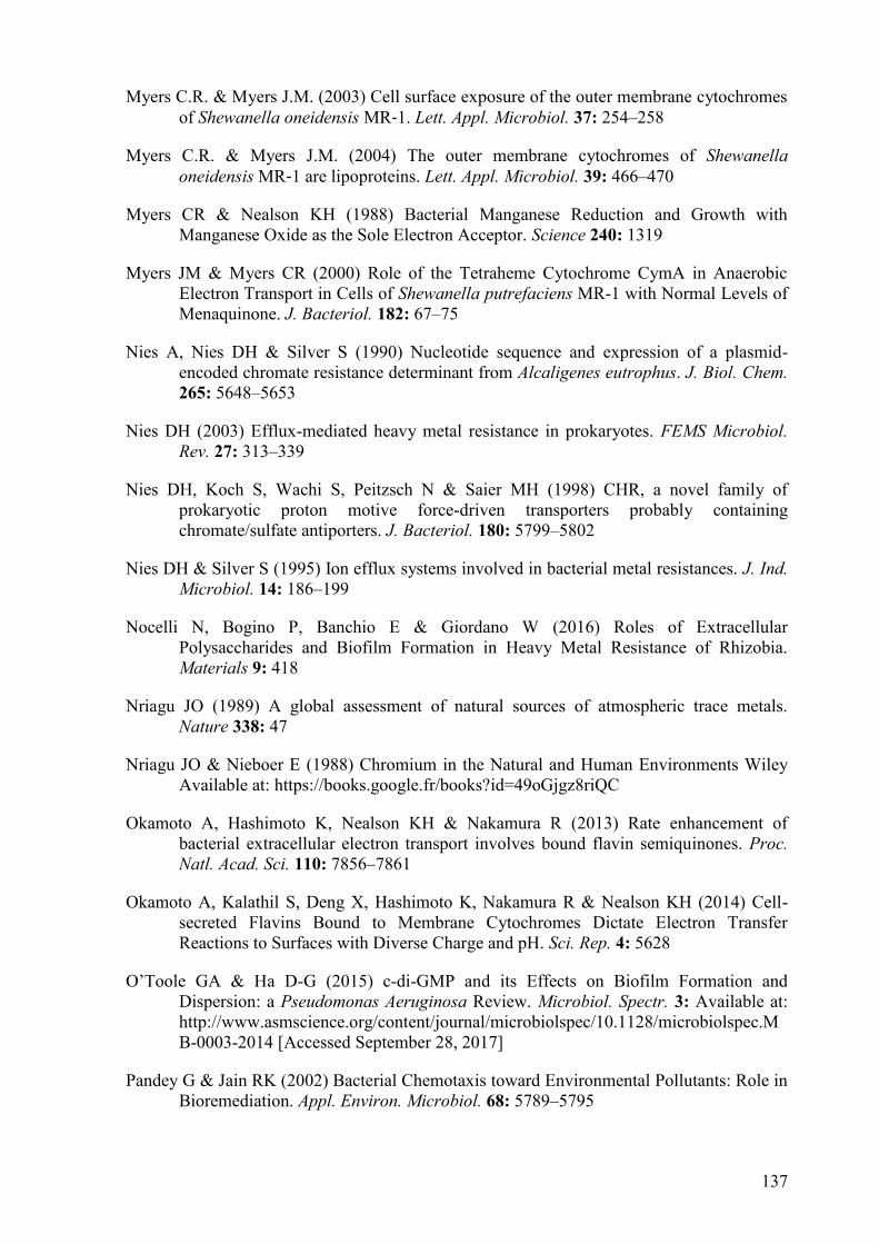
137
Myers C.R. & Myers J.M. (2003) Cell surface exposure of the outer membrane cytochromes of Shewanella oneidensis MR‐1. Lett. Appl. Microbiol. 37: 254–258
Myers C.R. & Myers J.M. (2004) The outer membrane cytochromes of Shewanella
oneidensis MR‐1 are lipoproteins. Lett. Appl. Microbiol. 39: 466–470
Myers CR & Nealson KH (1988) Bacterial Manganese Reduction and Growth with Manganese Oxide as the Sole Electron Acceptor. Science 240: 1319
Myers JM & Myers CR (2000) Role of the Tetraheme Cytochrome CymA in Anaerobic Electron Transport in Cells of Shewanella putrefaciens MR-1 with Normal Levels of Menaquinone. J. Bacteriol. 182: 67–75
Nies A, Nies DH & Silver S (1990) Nucleotide sequence and expression of a plasmid-encoded chromate resistance determinant from Alcaligenes eutrophus. J. Biol. Chem. 265: 5648–5653
Nies DH (2003) Efflux-mediated heavy metal resistance in prokaryotes. FEMS Microbiol.
Rev. 27: 313–339
Nies DH, Koch S, Wachi S, Peitzsch N & Saier MH (1998) CHR, a novel family of prokaryotic proton motive force-driven transporters probably containing chromate/sulfate antiporters. J. Bacteriol. 180: 5799–5802
Nies DH & Silver S (1995) Ion efflux systems involved in bacterial metal resistances. J. Ind.
Microbiol. 14: 186–199
Nocelli N, Bogino P, Banchio E & Giordano W (2016) Roles of Extracellular Polysaccharides and Biofilm Formation in Heavy Metal Resistance of Rhizobia. Materials 9: 418
Nriagu JO (1989) A global assessment of natural sources of atmospheric trace metals. Nature 338: 47
Nriagu JO & Nieboer E (1988) Chromium in the Natural and Human Environments Wiley Available at: https://books.google.fr/books?id=49oGjgz8riQC
Okamoto A, Hashimoto K, Nealson KH & Nakamura R (2013) Rate enhancement of bacterial extracellular electron transport involves bound flavin semiquinones. Proc.
Natl. Acad. Sci. 110: 7856–7861
Okamoto A, Kalathil S, Deng X, Hashimoto K, Nakamura R & Nealson KH (2014) Cell-secreted Flavins Bound to Membrane Cytochromes Dictate Electron Transfer Reactions to Surfaces with Diverse Charge and pH. Sci. Rep. 4: 5628
O’Toole GA & Ha D-G (2015) c-di-GMP and its Effects on Biofilm Formation and Dispersion: a Pseudomonas Aeruginosa Review. Microbiol. Spectr. 3: Available at: http://www.asmscience.org/content/journal/microbiolspec/10.1128/microbiolspec.MB-0003-2014 [Accessed September 28, 2017]
Pandey G & Jain RK (2002) Bacterial Chemotaxis toward Environmental Pollutants: Role in Bioremediation. Appl. Environ. Microbiol. 68: 5789–5795

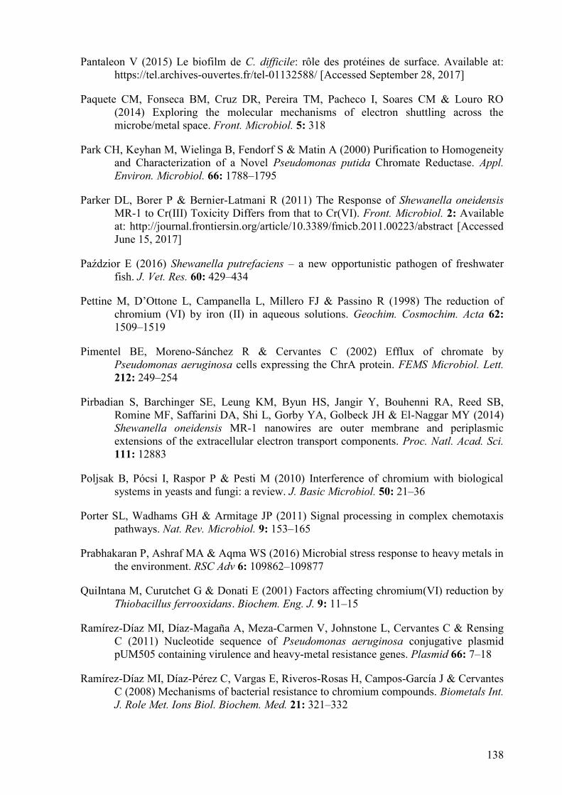
138
Pantaleon V (2015) Le biofilm de C. difficile: rôle des protéines de surface. Available at: https://tel.archives-ouvertes.fr/tel-01132588/ [Accessed September 28, 2017]
Paquete CM, Fonseca BM, Cruz DR, Pereira TM, Pacheco I, Soares CM & Louro RO (2014) Exploring the molecular mechanisms of electron shuttling across the microbe/metal space. Front. Microbiol. 5: 318
Park CH, Keyhan M, Wielinga B, Fendorf S & Matin A (2000) Purification to Homogeneity and Characterization of a Novel Pseudomonas putida Chromate Reductase. Appl.
Environ. Microbiol. 66: 1788–1795
Parker DL, Borer P & Bernier-Latmani R (2011) The Response of Shewanella oneidensis MR-1 to Cr(III) Toxicity Differs from that to Cr(VI). Front. Microbiol. 2: Available at: http://journal.frontiersin.org/article/10.3389/fmicb.2011.00223/abstract [Accessed June 15, 2017]
Paździor E (β016) Shewanella putrefaciens – a new opportunistic pathogen of freshwater fish. J. Vet. Res. 60: 429–434
Pettine M, D’Ottone L, Campanella L, Millero FJ & Passino R (1λλ8) The reduction of chromium (VI) by iron (II) in aqueous solutions. Geochim. Cosmochim. Acta 62: 1509–1519
Pimentel BE, Moreno-Sánchez R & Cervantes C (2002) Efflux of chromate by Pseudomonas aeruginosa cells expressing the ChrA protein. FEMS Microbiol. Lett. 212: 249–254
Pirbadian S, Barchinger SE, Leung KM, Byun HS, Jangir Y, Bouhenni RA, Reed SB, Romine MF, Saffarini DA, Shi L, Gorby YA, Golbeck JH & El-Naggar MY (2014) Shewanella oneidensis MR-1 nanowires are outer membrane and periplasmic extensions of the extracellular electron transport components. Proc. Natl. Acad. Sci. 111: 12883
Poljsak B, Pócsi I, Raspor P & Pesti M (2010) Interference of chromium with biological systems in yeasts and fungi: a review. J. Basic Microbiol. 50: 21–36
Porter SL, Wadhams GH & Armitage JP (2011) Signal processing in complex chemotaxis pathways. Nat. Rev. Microbiol. 9: 153–165
Prabhakaran P, Ashraf MA & Aqma WS (2016) Microbial stress response to heavy metals in the environment. RSC Adv 6: 109862–109877
QuiIntana M, Curutchet G & Donati E (2001) Factors affecting chromium(VI) reduction by Thiobacillus ferrooxidans. Biochem. Eng. J. 9: 11–15
Ramírez-Díaz MI, Díaz-Magaña A, Meza-Carmen V, Johnstone L, Cervantes C & Rensing C (2011) Nucleotide sequence of Pseudomonas aeruginosa conjugative plasmid pUM505 containing virulence and heavy-metal resistance genes. Plasmid 66: 7–18
Ramírez-Díaz MI, Díaz-Pérez C, Vargas E, Riveros-Rosas H, Campos-García J & Cervantes C (2008) Mechanisms of bacterial resistance to chromium compounds. Biometals Int.
J. Role Met. Ions Biol. Biochem. Med. 21: 321–332


139
Richardson David J., Butt Julea N., Fredrickson Jim K., Zachara John M., Shi Liang, Edwards Marcus J., White Gaye, Baiden Nanakow, Gates Andrew J., Marritt Sophie J. & Clarke Thomas A. (β01β) The ‘porin–cytochrome’ model for microbe‐to‐mineral electron transfer. Mol. Microbiol. 85: 201–212
Ringo E, Stenberg E & Strøm AR (1984) Amino acid and lactate catabolism in trimethylamine oxide respiration of Alteromonas putrefaciens NCMB 1735. Appl.
Environ. Microbiol. 47: 1084–1089
Rodionov DA, Novichkov PS, Stavrovskaya ED, Rodionova IA, Li X, Kazanov MD, Ravcheev DA, Gerasimova AV, Kazakov AE, Kovaleva G, Permina EA, Laikova ON, Overbeek R, Romine MF, Fredrickson JK, Arkin AP, Dubchak I, Osterman AL & Gelfand MS (2011) Comparative genomic reconstruction of transcriptional networks controlling central metabolism in the Shewanella genus. BMC Genomics 12: S3
Romanenko VI, Koren’kov VN (1λ77) Pure culture of bacteria using chromates and bichromates as hydrogen acceptors during development under anaerobic conditions. Mikrobiologiia 46: 7
Saffarini D ed. (2015) Bacteria-Metal Interactions Cham: Springer International Publishing Available at: http://link.springer.com/10.1007/978-3-319-18570-5 [Accessed July 31, 2017]
Saier MH (2003) Answering Fundamental Questions in Biology with Bioinformatics. ASM
News-American Society for Microbiology 69: 175–181
Satomi M, Vogel BF, Venkateswaran K & Gram L (2007) Description of Shewanella
glacialipiscicola sp. nov. and Shewanella algidipiscicola sp. nov., isolated from marine fish of the Danish Baltic Sea, and proposal that Shewanella affinis is a later heterotypic synonym of Shewanella colwelliana. Int. J. Syst. Evol. Microbiol. 57: 347–352
Scherer Judith & Nies Dietrich H. (2009) CzcP is a novel efflux system contributing to transition metal resistance in Cupriavidus metallidurans CH34. Mol. Microbiol. 73: 601–621
Schicklberger M, Sturm G & Gescher J (2013) Genomic Plasticity Enables a Secondary Electron Transport Pathway in Shewanella oneidensis. Appl. Environ. Microbiol. 79: 1150–1159
Schwalb C, Chapman SK & Reid GA (2003) The Tetraheme Cytochrome CymA Is Required for Anaerobic Respiration with Dimethyl Sulfoxide and Nitrite in Shewanella
oneidensis †. Biochemistry (Mosc.) 42: 9491–9497
Schweinitzer T & Josenhans C (2010) Bacterial energy taxis: a global strategy? Arch.
Microbiol. 192: 507–520
Sedlak DL & Chan PG (1997) Reduction of hexavalent chromium by ferrous iron. Geochim.
Cosmochim. Acta 61: 2185–2192


140
Serres MH & Riley M (2006) Genomic Analysis of Carbon Source Metabolism of Shewanella oneidensis MR-1: Predictions versus Experiments. J. Bacteriol. 188: 4601–4609
Shen H & Wang Y-T (1993) Characterization of Enzymatic Reduction of Hexavalent Chromium by Escherichia coli ATCC 33456. Appl.Environ.Microbiol: 3771–3777
Shewan, Hobbs G & Hodgkiss W (1960) A determinative scheme for the identification of certain genera of Gram-Negative Bacteria, with special reference to the Pseudomonadaceae. J. Appl. Bacteriol. 23: 379–390
Shewan JM, Gibson DM & Murray CK (1972) The Estimation of Trimethylamine in Fish Muscle Torry Research Station Available at: https://books.google.fr/books?id=rxXfcQAACAAJ
Shi L, Chen B, Wang Z, Elias DA, Mayer MU, Gorby YA, Ni S, Lower BH, Kennedy DW, Wunschel DS, Mottaz HM, Marshall MJ, Hill EA, Beliaev AS, Zachara JM, Fredrickson JK & Squier TC (2006) Isolation of a High-Affinity Functional Protein Complex between OmcA and MtrC: Two Outer Membrane Decaheme c-Type Cytochromes of Shewanella oneidensis MR-1. J. Bacteriol. 188: 4705–4714
Shi M, Wan F, Mao Y & Gao H (2015) Unraveling the Mechanism for the Viability Deficiency of Shewanella oneidensis oxyR Null Mutant. J. Bacteriol. 197: 2179–2189
Shi X & Dalal NS (1990) Evidence for a fenton-type mechanism for the generation of .OH radicals in the reduction of Cr(VI) in cellular media. Arch. Biochem. Biophys. 281: 90–95
Silver S & Phung LT (1996) Bacterial heavy metal resistance: new surprises. Annu. Rev.
Microbiol. 50: 753–789
Singh R, Gautam N, Mishra A & Gupta R (2011) Heavy metals and living systems: An overview. Indian J. Pharmacol. 43: 246–253
Somasundaram V, Philip L & Bhallamudi SM (2009) Experimental and mathematical modeling studies on Cr(VI) reduction by CRB, SRB and IRB, individually and in combination. J. Hazard. Mater. 172: 606–617
Subramanian P, Pirbadian S, El-Naggar MY & Jensen GJ (2017) The ultrastructure of Shewanella oneidensis MR-1 nanowires revealed by electron cryo-tomography. bioRxiv Available at: http://biorxiv.org/content/early/2017/01/28/103242.abstract
Summers AO (2009) Damage control: regulating defenses against toxic metals and metalloids. Cell Regul. 12: 138–144
Suzuki T, Miyata N, Horitsu H, Kawai K, Takamizawa K, Tai Y & Okazaki M (1992) NAD(P)H-dependent chromium (VI) reductase of Pseudomonas ambigua G-1: a Cr(V) intermediate is formed during the reduction of Cr(VI) to Cr(III). J. Bacteriol. 174: 5340–5345
Tauch A, Schlu A, Bischoff N, Goesmann A, Meyer F & hler AP (2003) The 79,370-bp conjugative plasmid pB4 consists of an IncP-1b backbone loaded with a chromate


141
resistance transposon, the strA-strB streptomycin resistance gene pair, the oxacillinase gene bla NPS-1, and a tripartite antibiotic efflux system of the resistance-nodulation-division family. Mol Gen Genomics: 570–584
Teitzel GM & Parsek MR (2003) Heavy Metal Resistance of Biofilm and Planktonic Pseudomonas aeruginosa. Appl. Environ. Microbiol. 69: 2313–2320
Thatoi H, Das S, Mishra J, Rath BP & Das N (2014) Bacterial chromate reductase, a potential enzyme for bioremediation of hexavalent chromium: A review. J. Environ.
Manage. 146: 383–399
Thompson MR (2007) Integrating Mass Spectrometry Based Proteomics and Bioinformatics Technologies for the Molecular Level Characterization of Shewanella oneidensis to Chromate Exposure. Available at: http://trace.tennessee.edu/utk_graddiss/318/ [Accessed August 27, 2017]
Thompson MR, VerBerkmoes NC, Chourey K, Shah M, Thompson DK & Hettich RL (2007) Dosage-Dependent Proteome Response of Shewanella oneidensis MR-1 to Acute Chromate Challenge. J. Proteome Res. 6: 1745–1757
Thormann KM, Saville RM, Shukla S, Pelletier DA & Spormann AM (2004) Initial Phases of Biofilm Formation in Shewanella oneidensis MR-1. J. Bacteriol. 186: 8096–8104
Tolker-Nielsen T, Brinch UC, Ragas PC, Andersen JB, Jacobsen CS & Molin S (2000) Development and Dynamics of Pseudomonas sp. Biofilms. J. Bacteriol. 182: 6482–6489
Tóth G, Hermann T, Da Silva MR & Montanarella L (2016) Heavy metals in agricultural soils of the European Union with implications for food safety. Environ. Int. 88: 299–309
Trevors JT, Oddie KM & Belliveau BH (1985) Metal resistance in bacteria. FEMS
Microbiol. Lett. 32: 39–54
Urone PF (1955) Stability of Colorimetric Reagent for Chromium, s-Diphenylcarbazide, in Various Solvents. Anal. Chem. 27: 1354–1355
Venkateswaran K, Moser DP, Dollhopf ME, Lies DP, Saffarini DA, MacGregor BJ, Ringelberg DB, White DC, Nishijima M, Sano H & others (1999) Polyphasic taxonomy of the genus Shewanella and description of Shewanella oneidensis sp. nov. Int. J. Syst. Evol. Microbiol. 49: 705–724
Viamajala S, Peyton BM, Sani RK, Apel WA & Petersen JN (2008) Toxic Effects of Chromium(VI) on Anaerobic and Aerobic Growth of Shewanella oneidensis MR-1. Biotechnol. Prog. 20: 87–95
Viti C, Marchi E, Decorosi F & Giovannetti L (2014) Molecular mechanisms of Cr(VI) resistance in bacteria and fungi. FEMS Microbiol. Rev. 38: 633–659
Vogel BF, Venkateswaran K, Satomi M & Gram L (2005) Identification of Shewanella
baltica as the Most Important H2S-Producing Species during Iced Storage of Danish Marine Fish. Appl. Environ. Microbiol. 71: 6689–6697

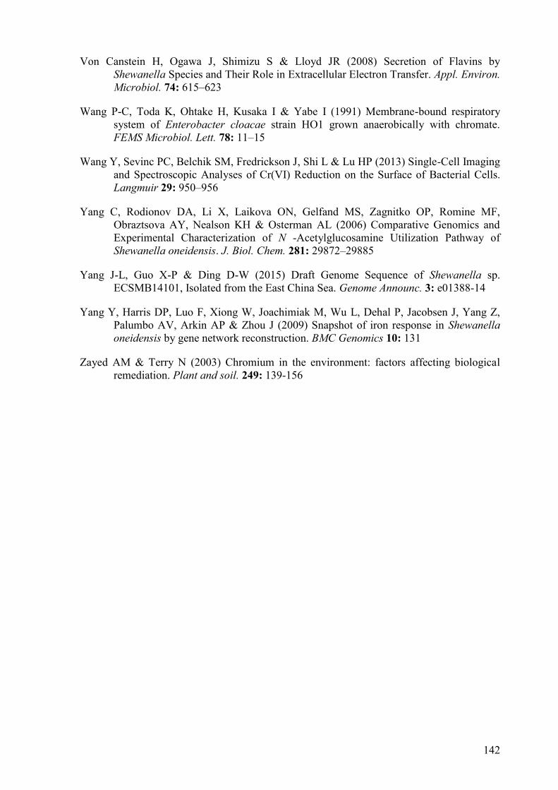
142
Von Canstein H, Ogawa J, Shimizu S & Lloyd JR (2008) Secretion of Flavins by Shewanella Species and Their Role in Extracellular Electron Transfer. Appl. Environ.
Microbiol. 74: 615–623
Wang P-C, Toda K, Ohtake H, Kusaka I & Yabe I (1991) Membrane-bound respiratory system of Enterobacter cloacae strain HO1 grown anaerobically with chromate. FEMS Microbiol. Lett. 78: 11–15
Wang Y, Sevinc PC, Belchik SM, Fredrickson J, Shi L & Lu HP (2013) Single-Cell Imaging and Spectroscopic Analyses of Cr(VI) Reduction on the Surface of Bacterial Cells. Langmuir 29: 950–956
Yang C, Rodionov DA, Li X, Laikova ON, Gelfand MS, Zagnitko OP, Romine MF, Obraztsova AY, Nealson KH & Osterman AL (2006) Comparative Genomics and Experimental Characterization of N -Acetylglucosamine Utilization Pathway of Shewanella oneidensis. J. Biol. Chem. 281: 29872–29885
Yang J-L, Guo X-P & Ding D-W (2015) Draft Genome Sequence of Shewanella sp. ECSMB14101, Isolated from the East China Sea. Genome Announc. 3: e01388-14
Yang Y, Harris DP, Luo F, Xiong W, Joachimiak M, Wu L, Dehal P, Jacobsen J, Yang Z, Palumbo AV, Arkin AP & Zhou J (2009) Snapshot of iron response in Shewanella
oneidensis by gene network reconstruction. BMC Genomics 10: 131
Zayed AM & Terry N (2003) Chromium in the environment: factors affecting biological remediation. Plant and soil. 249: 139-156


Abstract
The widespread use of the toxic heavy metal chromium (Cr) in industrial applications
resulted in large quantities of Cr being discharged into the environment, causing severe
contamination of global soil and water systems. Cr primarily exists in two stable forms,
Cr(III) and Cr(VI). The latter is highly toxic due to its strong oxidizing nature and its high
solubility. The model organism for bioremediation Shewanella oneidensis MR-1 has evolved
diverse resistance mechanisms to cope with chromate toxicity.
The first aim of the present thesis was to study the chromate resistance and reduction
mechanisms of this bacterium under semi-aerobic conditions. We showed that chrASO gene
is induced by chromate and its deletion impairs the chromate resistance and reduction
capacity of MR-1 strain, we confirmed that its product functions as an efflux pump to extrude
chromate ions from the cytoplasm protecting cells from chromate toxicity. With cymA-
deletion mutants, we revealed the involvement of the c-type cytochrome CymA in chromate
resistance and reduction. We also identified a potential chromate reductase DmsA2, as well
as two other proteins Fdh and DmsA1 that are potentially involved in chromate resistance
and reduction in MR-1 strain.
In the second part of this work, we isolated, identified and characterized two novel
Mediterranean Shewanella sp. strains, S. fidelis H76 and S. algidipiscicola H111. Both strains
are characterized by their great chromate resistance and their ability to reduce it efficiently
even at high concentrations. Although the small size of its genome and the absence of several
genes encoding enzymes known to play a role in chromate resistance and reduction, the H111
strain is the best chromate resistant strain. Interestingly, the air liquid interface biofilm
(Pellicles) of both strains reduce more efficiently chromate than their free-swimming cells.
Moreover, they can accumulate a significant amount of its reduced forms. These
characteristics make those strains, in particular H111 strain, suitable candidates for chromate
bioremediation.
Key words: Shewanella sp.,chromate, resistance, reduction, bioremediation.