Pellicle formation in Shewanella oneidensis
11
RESEARCH ARTICLE Open Access Pellicle formation in Shewanella oneidensis Yili Liang 1,2 , Haichun Gao 2,3* , Jingrong Chen 2 , Yangyang Dong 3 , Lin Wu 3 , Zhili He 1 , Xueduan Liu 1 , Guanzhou Qiu 1 , Jizhong Zhou 1,2* Abstract Background: Although solid surface-associated biofilm development of S. oneidensis has been extensively studied in recent years, pellicles formed at the air-liquid interface are largely overlooked. The goal of this work was to understand basic requirements and mechanism of pellicle formation in S. oneidensis. Results: We demonstrated that pellicle formation can be completed when oxygen and certain cations were present. Ca(II), Mn(II), Cu(II), and Zn(II) were essential for the process evidenced by fully rescuing pellicle formation of S. oneidensis from the EDTA treatment while Mg (II), Fe(II), and Fe(III) were much less effective. Proteins rather than DNA were crucial in pellicle formation and the major exopolysaccharides may be rich in mannose. Mutational analysis revealed that flagella were not required for pellicle formation but flagellum-less mutants delayed pellicle development substantially, likely due to reduced growth in static media. The analysis also demonstrated that AggA type I secretion system was essential in formation of pellicles but not of solid surface-associated biofilms in S. oneidensis. Conclusion: This systematic characterization of pellicle formation shed lights on our understanding of biofilm formation in S. oneidensis and indicated that the pellicle may serve as a good research model for studying bacterial communities. Background Most microbes in natural ecosystems exist in highly organized and functional interactive communities, which are composed of cells attached to surfaces and/or to each other either from a single species or multiple spe- cies [1-7]. Microbial communities confer a number of advantages for survival, such as nutrient availability with metabolic cooperation, acquisition of new genetic traits, and protection from the environment [4,8]. The most common microbial communities are biofilms, which refer to assemblages of cell on solid biotic or abiotic surfaces. In recent years, the subject of microbial bio- films has drawn a lot of attention and numerous studies have provided important insights into the genetic basis of biofilm development [5,7]. Pellicles, arising from the interface between air and liquid and therefore frequently called air-liquid (A-L) biofilms [9], have been well studied in an array of bacteria, such as Bacillus subtilis, Pseudomonas aerugi- nosa, and Vibrio parahaemolyticus [7,10-12]. Pellicle formation consists of at least three distinctive steps: (i) initial attachment of bacteria to the solid surface (wall of culture device) at the interface between air and liquid, (ii) development of the monolayer pellicle initiated from the attached cells, and (iii) maturation of pellicles with characteristic three-dimensional architecture [1,11]. In addition to cells, a variety of components, mainly extra- cellular polymeric substances (EPS), are needed for developing and maintaining the pellicle matrix. The most extensively studied EPS include exopolysacchar- ides, proteins, and extracellular DNA although contribu- tions of these agents to the integrity of the pellicle matrix may vary [11]. While the pellicle is generally taken into account as a special form of biofilms [5,7,13], its distinguishing characteristics justify that this type of biofilm may serve as an independent research model [12-14]. Many factors, including extracellular organelles such as flagella and type IV pili, secreted proteins, and chemi- cal agents supplemented in media such as iron and phosphate, have been shown to play important roles in * Correspondence: [email protected]; [email protected] 1 School of Minerals processing and Bioengineering, Central south University, Changsha, 410083, PR China 2 Institute for Environmental Genomics and Department of Botany and Microbiology, University of Oklahoma, Norman, 73019, USA Full list of author information is available at the end of the article Liang et al. BMC Microbiology 2010, 10:291 http://www.biomedcentral.com/1471-2180/10/291 © 2010 Liang et al; licensee BioMed Central Ltd. This is an Open Access article distributed under the terms of the Creative Commons Attribution License (http://creativecommons.org/licenses/by/2.0), which permits unrestricted use, distribution, and reproduction in any medium, provided the original work is properly cited.
Transcript of Pellicle formation in Shewanella oneidensis
RESEARCH ARTICLE Open Access
Pellicle formation in Shewanella oneidensisYili Liang1,2, Haichun Gao2,3*, Jingrong Chen2, Yangyang Dong3, Lin Wu3, Zhili He1, Xueduan Liu1, Guanzhou Qiu1,Jizhong Zhou1,2*
Abstract
Background: Although solid surface-associated biofilm development of S. oneidensis has been extensively studiedin recent years, pellicles formed at the air-liquid interface are largely overlooked. The goal of this work was tounderstand basic requirements and mechanism of pellicle formation in S. oneidensis.
Results: We demonstrated that pellicle formation can be completed when oxygen and certain cations werepresent. Ca(II), Mn(II), Cu(II), and Zn(II) were essential for the process evidenced by fully rescuing pellicle formationof S. oneidensis from the EDTA treatment while Mg (II), Fe(II), and Fe(III) were much less effective. Proteins ratherthan DNA were crucial in pellicle formation and the major exopolysaccharides may be rich in mannose. Mutationalanalysis revealed that flagella were not required for pellicle formation but flagellum-less mutants delayed pellicledevelopment substantially, likely due to reduced growth in static media. The analysis also demonstrated that AggAtype I secretion system was essential in formation of pellicles but not of solid surface-associated biofilms in S.oneidensis.
Conclusion: This systematic characterization of pellicle formation shed lights on our understanding of biofilmformation in S. oneidensis and indicated that the pellicle may serve as a good research model for studying bacterialcommunities.
BackgroundMost microbes in natural ecosystems exist in highlyorganized and functional interactive communities, whichare composed of cells attached to surfaces and/or toeach other either from a single species or multiple spe-cies [1-7]. Microbial communities confer a number ofadvantages for survival, such as nutrient availability withmetabolic cooperation, acquisition of new genetic traits,and protection from the environment [4,8]. The mostcommon microbial communities are biofilms, whichrefer to assemblages of cell on solid biotic or abioticsurfaces. In recent years, the subject of microbial bio-films has drawn a lot of attention and numerous studieshave provided important insights into the genetic basisof biofilm development [5,7].Pellicles, arising from the interface between air and
liquid and therefore frequently called air-liquid (A-L)biofilms [9], have been well studied in an array of
bacteria, such as Bacillus subtilis, Pseudomonas aerugi-nosa, and Vibrio parahaemolyticus [7,10-12]. Pellicleformation consists of at least three distinctive steps: (i)initial attachment of bacteria to the solid surface (wallof culture device) at the interface between air and liquid,(ii) development of the monolayer pellicle initiated fromthe attached cells, and (iii) maturation of pellicles withcharacteristic three-dimensional architecture [1,11]. Inaddition to cells, a variety of components, mainly extra-cellular polymeric substances (EPS), are needed fordeveloping and maintaining the pellicle matrix. Themost extensively studied EPS include exopolysacchar-ides, proteins, and extracellular DNA although contribu-tions of these agents to the integrity of the pelliclematrix may vary [11]. While the pellicle is generallytaken into account as a special form of biofilms [5,7,13],its distinguishing characteristics justify that this type ofbiofilm may serve as an independent research model[12-14].Many factors, including extracellular organelles such
as flagella and type IV pili, secreted proteins, and chemi-cal agents supplemented in media such as iron andphosphate, have been shown to play important roles in
* Correspondence: [email protected]; [email protected] of Minerals processing and Bioengineering, Central south University,Changsha, 410083, PR China2Institute for Environmental Genomics and Department of Botany andMicrobiology, University of Oklahoma, Norman, 73019, USAFull list of author information is available at the end of the article
Liang et al. BMC Microbiology 2010, 10:291http://www.biomedcentral.com/1471-2180/10/291
© 2010 Liang et al; licensee BioMed Central Ltd. This is an Open Access article distributed under the terms of the Creative CommonsAttribution License (http://creativecommons.org/licenses/by/2.0), which permits unrestricted use, distribution, and reproduction inany medium, provided the original work is properly cited.

biofilm formation [5]. However, effects of these factorson the biofilm formation process depend on the bacter-ium under study. For example, flagella facilitate surfaceadhesion for many species but it has been also observedin other species that mutations resulting in aflagellateand paralyzed nonmotile cells promote formation of amultilayer biofilm [7]. In the case of iron, results areeven more inconsistent. In P. aeruginosa and Vibrio cho-lerae, iron limitation hinders biofilm formation whereasit facilitates the process in Actinomyces naeslundii andStaphylococcus epidermidis [15,16]. It has been sug-gested that variation in effects of these factors on bio-film formation by particular species of bacteria may bereflection of the different environmental niches wherethey live [14,17-19].Shewanella oneidensis MR-1, a facultative Gram-nega-
tive anaerobe with a remarkable respiratory versatility,has been extensively studied for its biofilm development[20-26]. However, little progress has been made tounderstand biological mechanisms of pellicle formation.This work represents the initial steps in characterizingthe process in S. oneidensis. We showed that successfulpellicle formation required the availability of oxygen andthe presence of certain metal cations. A further analysison metal cations revealed that Fe(II) and Fe(III) werenot as essential as Ca(II), Cu(II), Mn(II), and Zn(II) forpellicle formation. In addition, results presented demon-strated that a type I secretion pathway of S. oneidensis isrequired for the pellicle development but not for attach-ment to abiotic surface.
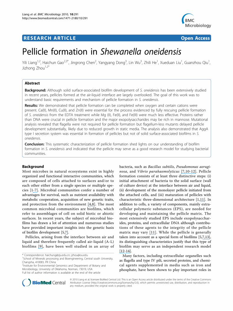
ResultsCharacteristics of S. oneidensis growth in still media underaerobic conditionsThe S. oneidensis MR-1 cells grew rapidly in LB in aflask when aeration of the media was provided by vigor-ously shaking, with a doubling time of approximately 40min at the room temperature (Figure 1A). Such growtheventually led to formation of the solid surface-asso-ciated (SSA) biofilms on the flask wall, especially aroundthe A-L interface. Cells in static media accessible toambient air, however, displayed a different growth pat-tern. Before pellicles were formed, cells lived in theplanktonic form with a much longer doubling time,approximately 2.6 h (Figure 1A). Once pellicle formationinitiated, some of the planktonic cells started to formpellicles while the rest remained in the planktonic form.During the development of pellicles, the planktonic cellsgrew at a much lower rate with a doubling time ofapproximately 6 h (Figure 1A). In this study, initiationof pellicle formation was determined by the time pointwhere the growth rate of the planktonic cells changedalthough pellicles visible to naked eyes appeared muchlater, about 12 hours after inoculation at the room
temperature. Both complex and defined media sup-ported pellicle formation of S. oneidensis. However, pel-licles from LB were thick and fairly uniform comparedto thin and porous ones from the defined medium, indi-cating an impact of nutrition on pellicle formation(Figure 1B). We therefore chose LB through the rest ofthis study unless otherwise noted.
Oxygen is required for pellicle formation in S. oneidensisAs demonstrated above, S. oneidensis initiated the pelli-cle formation process under aerobic conditions. Wethen asked whether oxygen is an essential factor for pel-licle formation of this microorganism. The pellicle for-mation assay was carried out under anaerobicconditions with lactate as the electron donor and one offollowing agents as the electron acceptors: fumarate(20 mM), nitrate (5 mM), DMSO (20 mM), TMAO (20mM), or ferrous citrate (10 mM). In all cases, the capa-city of S. oneidensis cells to form pellicles was abolished(data not shown), indicating that oxygen is required forthe process. This is in agreement with the findings thatthe lack of oxygen also resulted in a defect in SSA bio-film formation and a sudden decrease in oxygen concen-tration led to rapid detachment of SSA biofilms [25,27].To further elucidate the role of oxygen in pellicle for-
mation, dissolved oxygen concentrations (DOC) at fourdifferent depths below the surface in the unshaken cul-tures were measured in a time-course manner. Resultsrevealed that DOC at 0.5, 1, and 2 cm below the surfacein the unshaken cultures displayed a similar decliningpattern with time, decreased rapidly from approximately8 to 0.04 mg/L during the first two and half hours, andthen remained stable at 0.04 mg/L (Figure 1C). However,DOC at the depth immediately below the surface (0.1 cmbut the detector immersed in the liquid) reduced in amuch slower rate and reached the lowest level of 0.04mg/L only after the pellicle formed. These data indicatethat the majority of dissolved oxygen is likely consumedby the cells close to the surface and the cells below thesurface were grown under microaerobic/anaerobic condi-tions even before the pellicle was formed.
Proteins are essential in pellicle formation of S. oneidensisSince EPS, including proteins, polysaccharides, extracel-lular DNA, humic acid, and sugar, are important in SSAbiofilm and pellicle formation of various bacteria, wespeculated that these biopolymers may play a role inpellicle formation of S. oneidensis. To this end, effects ofproteinase K and DNase I on pellicle formation anddeveloped pellicles were assessed. The pellicles wereprevented from formation in the presence of 100 μg/mlproteinase K (Figure 2A). Consistently, 100 μg/ml of theproteinase K was able to degrade the developed pelliclesin 24 h, resulting in the semi-transparent membrane-
Liang et al. BMC Microbiology 2010, 10:291http://www.biomedcentral.com/1471-2180/10/291
Page 2 of 11
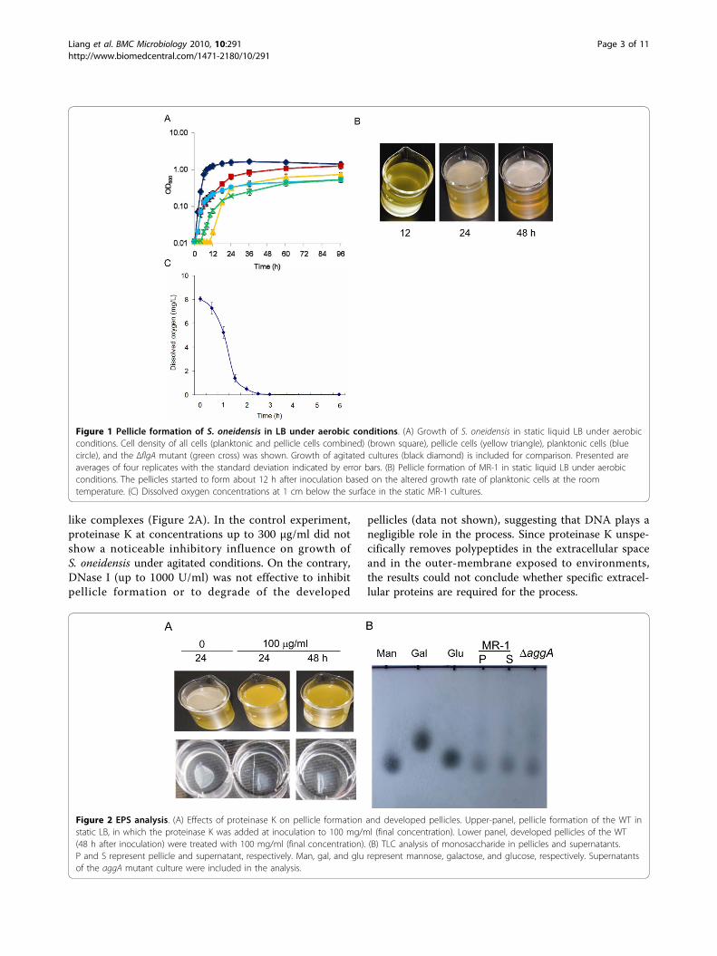
like complexes (Figure 2A). In the control experiment,proteinase K at concentrations up to 300 μg/ml did notshow a noticeable inhibitory influence on growth ofS. oneidensis under agitated conditions. On the contrary,DNase I (up to 1000 U/ml) was not effective to inhibitpellicle formation or to degrade of the developed
pellicles (data not shown), suggesting that DNA plays anegligible role in the process. Since proteinase K unspe-cifically removes polypeptides in the extracellular spaceand in the outer-membrane exposed to environments,the results could not conclude whether specific extracel-lular proteins are required for the process.
Figure 1 Pellicle formation of S. oneidensis in LB under aerobic conditions. (A) Growth of S. oneidensis in static liquid LB under aerobicconditions. Cell density of all cells (planktonic and pellicle cells combined) (brown square), pellicle cells (yellow triangle), planktonic cells (bluecircle), and the ΔflgA mutant (green cross) was shown. Growth of agitated cultures (black diamond) is included for comparison. Presented areaverages of four replicates with the standard deviation indicated by error bars. (B) Pellicle formation of MR-1 in static liquid LB under aerobicconditions. The pellicles started to form about 12 h after inoculation based on the altered growth rate of planktonic cells at the roomtemperature. (C) Dissolved oxygen concentrations at 1 cm below the surface in the static MR-1 cultures.
Figure 2 EPS analysis. (A) Effects of proteinase K on pellicle formation and developed pellicles. Upper-panel, pellicle formation of the WT instatic LB, in which the proteinase K was added at inoculation to 100 mg/ml (final concentration). Lower panel, developed pellicles of the WT(48 h after inoculation) were treated with 100 mg/ml (final concentration). (B) TLC analysis of monosaccharide in pellicles and supernatants.P and S represent pellicle and supernatant, respectively. Man, gal, and glu represent mannose, galactose, and glucose, respectively. Supernatantsof the aggA mutant culture were included in the analysis.
Liang et al. BMC Microbiology 2010, 10:291http://www.biomedcentral.com/1471-2180/10/291
Page 3 of 11
Attempts were made to solve the major polysaccharidecomponents of S. oneidensis pellicles by the thin layerchromatography (TLC) analysis. Culture supernatantsand pellicles were collected independently after 36 h ofgrowth and pellicles were then treated with 100 μg/mlproteinase K to removed cells. Polysaccharides wereextracted and subjected to TLC analysis as described inMethods. A preliminary experiment was performed withsix monosaccharides as standards, including ribose,mannose, glucose, galactose, rhamnose, and N-acetyl-glucosamine. The monosaccharides visualized on theTLC plates were close to mannose, glucose, and galac-tose (data not shown). To further confirm the observa-tion, the experiment was conducted again with thesethree monosaccharide standards only. As shown in Fig-ure 2B the major monosaccharides identified were mostlikely to be mannose in both supernatants and pellicles.To validate this result, the aggA mutant, a pellicle-lessstrain was included in the analysis and the same resultwas obtained. These data suggest that the mannose-richpolysaccharides identified in pellicles are not pelliclespecific.
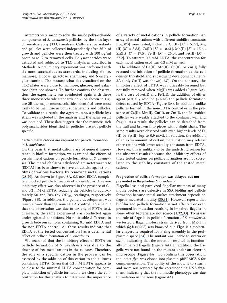
Certain metal cations are required for pellicle formationin S. oneidensisOn the basis that metal cations are of general impor-tance in biofilm formation, we examined the effects ofcertain metal cations on pellicle formation of S. oneiden-sis. The metal chelator ethylenediaminetetraacetate(EDTA) has been shown to have an activity against bio-films of various bacteria by removing metal cations[28,29]. As shown in Figure 3A, 0.3 mM EDTA comple-tely blocked pellicle formation of S. oneidensis. A severeinhibitory effect was also observed in the presence of 0.1and 0.2 mM of EDTA, reducing the pellicles to approxi-mately 50 and 70% (by OD600 readings), respectively(Figure 3B). In addition, the pellicle development wasmuch slower than the non-EDTA control. To rule outthat the observation was due to toxicity of EDTA to S.oneidensis, the same experiment was conducted againunder agitated conditions. No noticeable difference ingrowth between samples containing 0.3 mM EDTA andthe non-EDTA control. All these results indicate thatEDTA at the tested concentration has a detrimentaleffect on pellicle formation of S. oneidensis.We reasoned that the inhibitory effect of EDTA on
pellicle formation of S. oneidensis was due to theabsence of free metal cations in the cultures. Therefore,the role of a specific cation in the process can beassessed by the addition of this cation to the culturescontaining EDTA. Given that 0.3 mM EDTA appears tobe close to the minimal EDTA concentration for com-plete inhibition of pellicle formation, we chose the con-centration for this analysis to determine the importance
of a variety of metal cations in pellicle formation. Anarray of metal cations with different stability constants[log(Kc)] were tested, including Cu(II) [Kc = 5.77], Mg(II) [Kc = 8.83], Ca(II) [Kc = 10.61], Mn(II) [Kc = 15.6],Zn(II) [Kc = 17.5], Fe(II) [Kc = 25.0], and Fe(III) [Kc =27.2]. To saturate 0.3 mM EDTA, the concentration foreach metal cation used was 0.3 mM as well.The addition of Ca(II), Mn(II), Cu(II), or Zn(II) fully
rescued the initiation of pellicle formation at the celldensity threshold and subsequent development (Figure3A (only Ca(II) was shown), 3C). On the contrary, theinhibitory effect of EDTA was noticeably lessened butnot fully removed when Mg(II) was added (Figure 3A).In the case of Fe(II) and Fe(III), the addition of eitheragent partially rescued (~40%) the pellicle formationdefect caused by EDTA (Figure 3A). In addition, unlikepellicles formed in the non-EDTA control or in the pre-sence of Ca(II), Mn(II), Cu(II), or Zn(II), the Fe-enabledpellicles were weakly attached to the container wall andfragile. As a result, the pellicles can be detached fromthe wall and broken into pieces with a slight shake. Thesame results were observed with even higher levels of Fe(II) or Fe(III) (up to 0.9 mM). In solution, the additionof an extra amount of certain metal cation may releaseother cations with lower stability constants from EDTA.However, this is unlikely to be the underlying reason forthe observed results because the inhibitory effects ofthese tested cations on pellicle formation are not corre-lated to the stability constants of the tested metalcations.
Progression of pellicle formation was delayed but notprevented in flagella-less S. oneidensisFlagella-less and paralyzed flagellar mutants of manymotile bacteria are defective in SSA biofilm and pellicleformation because initial surface attachment depends onflagella-mediated motility [30,31]. However, reports thatbiofilm and pellicle formation is not affected or evenpromoted by mutation resulting in impaired flagella insome other bacteria are not scarce [1,32,33]. To assessthe role of flagella in pellicle formation of S. oneidensis,we tested a flagellum-less strain derived from MR-1 inwhich flgA(so3253) was knocked out. FlgA is a molecu-lar chaperone required for P ring assembly in the peri-plasmic space [34]. The mutant was unable to swarm orswim, indicating that the mutation resulted in function-ally impaired flagella (Figure 4A). In addition, the fla-gella were not found on the mutant under an electronmicroscope (Figure 4A). To confirm this observation,the intact flgA was cloned into plasmid pBBRMCS-5 forcomplementation. The ability of the mutant to swarmand swim was restored by the corresponding DNA frag-ment, indicating that the nonmotile phenotype was dueto mutation in the gene (Figure 4A).
Liang et al. BMC Microbiology 2010, 10:291http://www.biomedcentral.com/1471-2180/10/291
Page 4 of 11
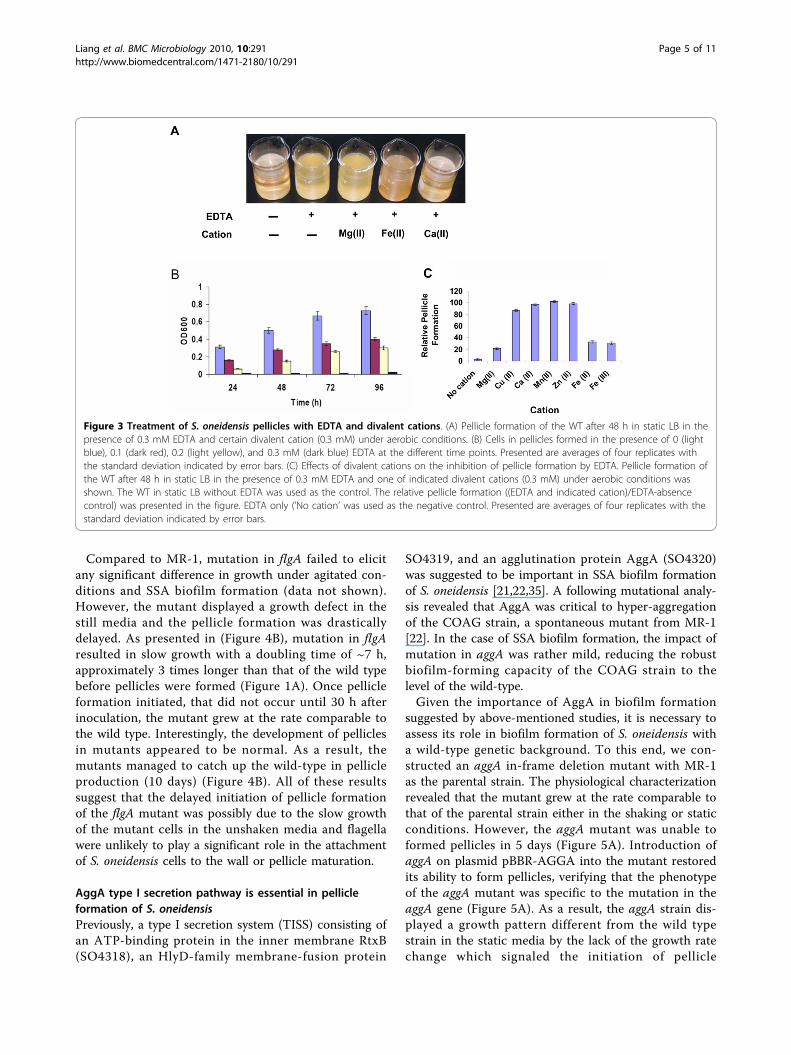
Compared to MR-1, mutation in flgA failed to elicitany significant difference in growth under agitated con-ditions and SSA biofilm formation (data not shown).However, the mutant displayed a growth defect in thestill media and the pellicle formation was drasticallydelayed. As presented in (Figure 4B), mutation in flgAresulted in slow growth with a doubling time of ~7 h,approximately 3 times longer than that of the wild typebefore pellicles were formed (Figure 1A). Once pellicleformation initiated, that did not occur until 30 h afterinoculation, the mutant grew at the rate comparable tothe wild type. Interestingly, the development of pelliclesin mutants appeared to be normal. As a result, themutants managed to catch up the wild-type in pellicleproduction (10 days) (Figure 4B). All of these resultssuggest that the delayed initiation of pellicle formationof the flgA mutant was possibly due to the slow growthof the mutant cells in the unshaken media and flagellawere unlikely to play a significant role in the attachmentof S. oneidensis cells to the wall or pellicle maturation.
AggA type I secretion pathway is essential in pellicleformation of S. oneidensisPreviously, a type I secretion system (TISS) consisting ofan ATP-binding protein in the inner membrane RtxB(SO4318), an HlyD-family membrane-fusion protein
SO4319, and an agglutination protein AggA (SO4320)was suggested to be important in SSA biofilm formationof S. oneidensis [21,22,35]. A following mutational analy-sis revealed that AggA was critical to hyper-aggregationof the COAG strain, a spontaneous mutant from MR-1[22]. In the case of SSA biofilm formation, the impact ofmutation in aggA was rather mild, reducing the robustbiofilm-forming capacity of the COAG strain to thelevel of the wild-type.Given the importance of AggA in biofilm formation
suggested by above-mentioned studies, it is necessary toassess its role in biofilm formation of S. oneidensis witha wild-type genetic background. To this end, we con-structed an aggA in-frame deletion mutant with MR-1as the parental strain. The physiological characterizationrevealed that the mutant grew at the rate comparable tothat of the parental strain either in the shaking or staticconditions. However, the aggA mutant was unable toformed pellicles in 5 days (Figure 5A). Introduction ofaggA on plasmid pBBR-AGGA into the mutant restoredits ability to form pellicles, verifying that the phenotypeof the aggA mutant was specific to the mutation in theaggA gene (Figure 5A). As a result, the aggA strain dis-played a growth pattern different from the wild typestrain in the static media by the lack of the growth ratechange which signaled the initiation of pellicle
Figure 3 Treatment of S. oneidensis pellicles with EDTA and divalent cations. (A) Pellicle formation of the WT after 48 h in static LB in thepresence of 0.3 mM EDTA and certain divalent cation (0.3 mM) under aerobic conditions. (B) Cells in pellicles formed in the presence of 0 (lightblue), 0.1 (dark red), 0.2 (light yellow), and 0.3 mM (dark blue) EDTA at the different time points. Presented are averages of four replicates withthe standard deviation indicated by error bars. (C) Effects of divalent cations on the inhibition of pellicle formation by EDTA. Pellicle formation ofthe WT after 48 h in static LB in the presence of 0.3 mM EDTA and one of indicated divalent cations (0.3 mM) under aerobic conditions wasshown. The WT in static LB without EDTA was used as the control. The relative pellicle formation ((EDTA and indicated cation)/EDTA-absencecontrol) was presented in the figure. EDTA only (’No cation’ was used as the negative control. Presented are averages of four replicates with thestandard deviation indicated by error bars.
Liang et al. BMC Microbiology 2010, 10:291http://www.biomedcentral.com/1471-2180/10/291
Page 5 of 11

formation (Figure 1A). However, the mutant was able toattach to the glass wall at the air-liquid interface, sug-gesting that AggA is not essential for this step of biofilmformation (Figure 5A). This proposal gained supportfrom the SSA biofilm formation of the mutant, whichdiffered from that of the wild type strain insignificantly(Figure 5B). All these data implicate that AggA TISS isrequired for pellicle formation, most likely at the mono-layer pellicle formation stage, which appears to be differ-ent from that in SSA biofilm formation.
Discussion and ConclusionsIn the microbial world, existence within surface-asso-ciated structured multicellular communities is the pre-vailing lifestyle [36,37]. The pellicles of facultative
bacteria formed at the liquid-air interface can be selec-tively advantageous given that respiration with oxygenas the terminal electron acceptor is the most productive.In S. oneidensis, the growth rate was promoted by betteraccess to oxygen evidenced by that the cells grew muchfaster in shaking than in static cultures. Along with theobservation that SSA biofilm formation of S. oneidensiswas inhibited under anaerobic conditions, the require-ment of oxygen for pellicle formation may mainly comefrom its facilitation of aggregation and attachment ofcells to the solid surfaces. This is consistent with pre-vious findings that oxygen promotes autoaggregation ofand sudden depletion of molecular oxygen was shownto act as the predominant trigger for initiating detach-ment of individual cells from biofilms [26,38]. We
Figure 4 The ΔflgA mutant displayed slow pellicle formation. (A) Swimming and swarming motility assays of the ΔflgA mutant. In bothpanels, the ΔflgA mutant (Upper) was compared to the WT (Lower). The ΔflgA* strain refers to the ΔflgA mutant containing pBBR-FLGA. (B)Electron micrographs of WT and the ΔflgA mutant. No flagellum was observed on the mutant. (C) Left panel, pellicle formation of the ΔflgAmutant. Right panel, the cell densities of cells in pellicles of the WT and the ΔflgA mutant. The WT, dark red; the ΔflgA mutant, light blue. Erepresents the time at which the cell density of ΔflgA mutant catches up (10 days after inoculation in the experiment). Presented are averages offour replicates with the standard deviation indicated by error bars.
Liang et al. BMC Microbiology 2010, 10:291http://www.biomedcentral.com/1471-2180/10/291
Page 6 of 11

therefore propose that an oxygen gradient established instatic cultures with the highest oxygen concentration atthe surface resulted in a larger number of cells at the A-L interface to form pellicles, which eventually induceattachment of individual cells to the abiotic surface.To form pellicles, S. oneidensis cultures require certain
divalent ions. Involvement of metals in biofilm forma-tion either as a facilitator or an inhibitor has been welldocumented. In recent years, many elegant studiesabout the susceptibility of biofilms to metals (as an inhi-bitor) have been published [39-41]. Although metals asa biofilm formation facilitator have been studied for
more than two decades, only a few metals (Ba(II), Mg(II), Ca(II), Fe(III), and Fe(III)) have been investigated[34,42,43]. In P. aeruginosa, all these metals but Ba(II)are able to protect P. aeruginosa biofilms against EDTAtreatment, presumably by stabilizing the biofilm matrix.In addition, it has been shown that there is a positivecorrelation between calcium concentration and amountof biofilm accumulation [44]. While our data supportprevious conclusions that calcium plays an importantrole in stabilizing biofilms of bacteria [34,43,44], most ofother findings are either new or surprising. Amongtested metal cations, Cu(II), Ca(II), Mn(II), and Zn(II)
Figure 5 Biofilm assay of MR-1 and aggA mutant. (A) Pellicle formation of MR-1, ΔaggA, ΔaggA* (aggA in-frame deletion mutant containingpBBR-AGGA). (B) SSA Biofilm was assessed for the strains indicated after 16 and 24 h, respectively. Cultures were prepared as described inMethods. The averaged OD readings of four independent culture tubes were given with images of representative CV-stained tubes.
Liang et al. BMC Microbiology 2010, 10:291http://www.biomedcentral.com/1471-2180/10/291
Page 7 of 11

belong to the same class, which are capable of restoringthe ability of S. oneidensis to form pellicles in the pre-sence of EDTA completely. In contrast, Mg(II) showsmild effects on relieving EDTA inhibition whereas Fe(II)and Fe(III) counteracted EDTA in a way different fromother tested cations evidenced by the fragile pellicles. Incombination, these data suggest that the relative stabilityconstants of metal cations (Cu(II) [5.77], Mg(II) [8.83],Ca(II) [10.61], Mn(II) [15.6], Zn(II) [17.5], Fe(II) [25.0],and Fe(III) [27.2]) and their affect on EDTA inhibitionare not correlated.It is particularly worth discussing roles of Fe(II) and Fe
(III) in pellicle formation of S. oneidensis. In recent years,many reports have demonstrated that the iron cationsare important, if not essential, in bacterial biofilm forma-tion [34,45-47]. In P. aeruginosa, influence of Fe(II) andFe(III) on the process was equivalent to that of Ca(II)[34]. In S. oneidensis, irons in forms of Fe(II) and Fe(III)were not only unable to neutralize the inhibitory effect ofEDTA on pellicle formation completely but also resultedin structurally impaired pellicles although these agentsindeed play a role in pellicle formation. This observationindicates that irons are not so crucial as Cu(II), Ca(II),Mn(II), and Zn(II) in pellicle formation of S. oneidensis.In fact, this may not be surprising. In Acinetobacter bau-mannii and Staphylococcus aureus, iron limitationimproved biofilm formation [48,49]. Therefore, it is pos-sible that different bacteria respond to irons in a differentway with respect to biofilm formation.Like SSA biofilms, pellicles require EPS to form a
matrix to support embedded cells. Although EPS arenow widely recognized as the essential components forbiofilm formation and development in all biofilm-form-ing microorganisms studied so far, diversity in theirindividual composition and relative abundance of certainelements is substantial [50]. For example, extracellularnucleic acids, which are not important in most biofilm-forming microorganisms, are required for SSA biofilmformation in a variety of bacteria [11,36,37,51,52]. In S.oneidensis, proteins not extracellular DNAs are requiredto pellicle formation. While essential extracellular pro-teins for S. oneidensis pellicle formation are largelyunknown, results from this study demonstrated that theAggA TISS is crucial in the process, likely at the devel-opment of the monolayer. One of substrates of thistransporter is predicted to be SO4317, a large ‘putativeRTX toxin’ [35], implicating that the protein may beinvolved in pellicle formation. In the case of polysac-charides, mannose dominates not only in pellicles butalso in supernatants, implicating that mannose-basedpolysaccharides may have a more general role in thebacterial physiology.Like in B. subtilis, mutations in S. oneidensis flagellar
genes resulting in the nonmotile phenotype significantly
delayed the initiation and development of pellicle forma-tion [17]. Here we further illustrated that neither SSAbiofilm formation nor the maturization of pellicle wasimpaired by the mutations. In agreement with findingson biofilm formation of Bacillus cereus [13], this obser-vation suggests that motility not only promotes cells tomove to surfaces where the pellicle forms but also facili-tate planktonic cells entrance into the pellicle.Overall, the results presented here provided the first
insights into pellicle formation of S. oneidensis, makingpellicle formation of S. oneidensis a simple researchmodel for biofilm formation in general. The study high-lights parallels and significant differences between thisprocess and well-documented paradigms, raising somekey questions demanding immediate investigations.These include what the major polysaccharides in S. onei-densis pellicles are, why irons result in fragile pellicles inthe presence of EDTA, and which proteins and theirsecretion pathway(s) are directly related to pellicleformation.
MethodsBacterial strains, plasmids, and culture conditionsBacterial strains and plasmids used in this study arelisted in Table 1 [53]. Escherichia coli and S. oneidensisstrains were routinely grown in LB broth or on LBplates at 37°C and the room temperature for geneticmanipulation, respectively. When needed, antibioticswere used at the following concentrations: ampicillin at50 μg/ml and gentamycin at 15 μg/ml.
Pellicle formation, measurement of growth, andquantification of pelliclesA fresh colony grown overnight on a LB plate was usedto inoculate 50 ml LB and incubated in a shaker (200rpm) to an OD600 of 0.8 at the room temperature. Thisculture was then diluted 500-fold with fresh LB, result-ing in the starting cultures. Throughout the study, allstarting cultures of S. oneidensis strains were preparedthis way. Aliquots of 30 ml starting cultures were trans-ferred to 50 ml Pyrex beakers. The beakers were keptstill for pellicle formation at the room temperature anddissolved oxygen (DO) of the cultures was recordedevery hour with an Accumet XL40 meter (Fisher Scien-tific). M1 defined medium containing 0.02% (w/v) ofvitamin-free Casamino Acids and 15 mM lactate withone of electron acceptors including fumarate (20 mM),nitrate (5 mM), trimethylamine N-oxide (TMAO) (20mM), dimethyl sulfoxide (DMSO) (20 mM) and ferrouscitrate (10 mM), was used to test pellicle formation inthe defined medium [54]. To separate cells in pellicleand underneath, cultures were withdrawn carefully forcollecting planktonic cells and the left pellicles. Forgrowth measurement, 27 parallel starting cultures were
Liang et al. BMC Microbiology 2010, 10:291http://www.biomedcentral.com/1471-2180/10/291
Page 8 of 11

used and 3 were collected at each time point and therest remained undisturbed. The cell density (OD600) ofcultures containing planktonic cells was measured firstas the planktonic cell density and measured again as theoverall cell density after cells from pellicles were addedand extensively vortexed. To quantify the pelliclesformed by the S. oneidensis wild-type and mutantstrains, cells from pellicles were collected, suspended in30 ml fresh LB, violently vortexed, and applied to thespectrometer at 600 nm.
Proteinase K and DNase I treatment of S. oneidensispelliclesS. oneidensis was statically cultured in LB broth with theaddition of proteinase K (0 μg/mL, 100 μg/mL, and 500μg/mL) or DNase I (Qiagen, 0U/mL, 100U/mL, 500U/mL and 1000U/mL) for 3 days [55]. We also investi-gated whether these 3 enzymes could dissolve estab-lished pellicles. 2-day old pellicles were rinsed with 20mM Tris-HCl (pH = 8.0) and incubated in the samebuffer supplemented with proteinase K at 37°C for 2days. Similarly, 2-day old pellicles were incubated withDNase I to examine the DNA content at room tempera-ture for 2 days.
Mutagenesis, physiological characterization andcomplementation of the resulting mutantsDeletion mutation strains were constructed using thefusion PCR method illustrated previously [56]. Primersused for mutagenesis were listed in Additional file 1. Inbrief, two DNA fragments flanking the target gene weregenerated from S. oneidensis genomic DNA by PCRwith primers 5F/5R and 3F/3R, respectively. Fusion PCRwas then performed to join these two DNA fragmentswith primers 5F/3R. The resulting single fragment wasdigested with SacI and ligated into the SacI-digested
and phosphatase-treated suicide vector pDS3.0. Theresultant vectors were electroporated into the donorstrain, E. coli WM3064 and then moved to S. oneidensisby conjugation. Integration of the mutagenesis constructinto the chromosome and resolution were performed togenerate the final deletion strains. The deletion was ver-ified by PCR and DNA sequencing.For complementation, DNA fragments containing
aggA or flgA were generated by PCR amplification withMR-1 genomic DNA as the template using primersSO4320-COM-F/SO3988-COM-R and SO3253-COM-F/SO3253-COM-R, respectively as listed in Additional file1. These fragments were digested with SacI and ligatedto SacI-digested pBBR1MCS-5 to form pBBR-AGGAand pBBR-FLGA, which was electroporated intoWM3064. Introduction of pBBR-AGGA or pBBR-FLGAinto the corresponding mutant was done by conjugation,and gentamycin-resistant colonies were selected. Thepresence of pBBR-AGGA or pBBR-FLGA in the corre-sponding mutant was confirmed by plasmid purificationand restriction enzyme digestion.
Swarm and swimming motility assayA fresh colony of tested strains was grown to an OD600
of 0.8 in LB media. The cultures (1 ml) were spottedonto a swarm LB plate (0.5% agar) or stabbed into aswimming LB plate (0.2% agar). All plates were incu-bated at the room temperature for 48 h. Images wereacquired using Alpha Innotech’s Fluorchem imagingsystem.
SSA biofilm assayThe SSA biofilm formation assay used is based on themethod previously reported [57]. In brief, 3 ml of freshLB in 15 ml glass tubes were inoculated with S. oneiden-sis strains from an overnight culture in LB at 200 rpm.
Table 1 Strains and plasmids used in this study
Strain or plasmid Relevant genotype Reference or source
E. coli
WM3064 Donor strain for conjugation; ΔdapA [53]
S. oneidensis
MR-1 Wild-type ATCC 700550
JZ3253 flgA deletion mutant derived from MR-1; Δ flgA This study
JZ4320 aggA deletion mutant derived from MR-1; ΔaggA This study
Plasmid
pDS3.0 Apr, Gmr, derivative from suicide vector pCVD442 Lab stock
pBBR1MCS-5 Gmr vector used for complementation Lab Stock
pDS-AGGA aggA deletion construct in pDS3.0 This study
pDS-FLGA flgA deletion construct in pDS3.0 This study
pBBR-AGGA pBBR1MCS-5 containing aggA of S. oneidensis This study
pBBR-FLGA pBBR1MCS-5 containing flgA of S. oneidensis This study
Liang et al. BMC Microbiology 2010, 10:291http://www.biomedcentral.com/1471-2180/10/291
Page 9 of 11

After 16, 24, 32, or 40 h of incubation at 200 rpm atroom temperature, 500 μl of 1% (wt/vol) crystal violet(CV) solution was added to each tube and incubated for15 min. Tubes were rinsed three times with 5 ml of dis-tilled H2O and air dried. Biofilm formation was quanti-fied by measuring the absorbance at 575 nm. Each assaywas performed four times.
Thin layer chromatography (TLC) analysisSupernatants and pellicles were collected after 36 h ofgrowth in static LB media. Pellicles were treated with100 μg/mL proteinase K for removal of cells. Cell-lesspellicles and supernatants were subjected to exopolysac-charide extraction and hydrolysis with trifluoroaceticacid as described previously [58]. The resulting mono-saccharides were dissolved in ddH2O in the concentra-tion of 10 mg/ml, and 2 μl of the sample was spottedonto TLC plates (silica gel 60 F254; Merck). After devel-opment in butan-1-ol-acetone-water (4:5:1), the TLCplates were dipped in the reagent aniline-diphenylaminein acetone and incubated for 2 to 5 min at 100°C.
Additional material
Additional file 1: Primers used in this study. File contains all primersused in this study
AcknowledgementsThis research was supported by Major State Basic Research DevelopmentProgram (973 Program: 2010CB833803) and National Natural ScienceFoundation of China (30870032) to HG. This research was also supported byChinese Science Foundation for Distinguished Group (No.50321402) to YL.This research was also supported by The U.S. Department of Energy underthe Genomics: GTL Program through Shewanella Federation, Office ofBiological and Environmental Research, Office of Science.
Author details1School of Minerals processing and Bioengineering, Central south University,Changsha, 410083, PR China. 2Institute for Environmental Genomics andDepartment of Botany and Microbiology, University of Oklahoma, Norman,73019, USA. 3Institute of Microbiology and College of Life Sciences, ZhejiangUniversity, Hangzhou, Zhejiang 310058, PR China.
Authors’ contributionsYL carried out pellicle formation and characterization experiments anddrafted the manuscript. HG conceived of the study, and participated in itsdesign, and directed all experiments and coordination and drafted themanuscript. JC carried out the mutagenesis experiments. YD and LW carriedout SSA biofilm and TLC assays. ZH participated in design of the study andhelped to draft the manuscript. XL and GQ participated in the design of thestudy and helped to draft the manuscript. JZ conceived of the study, andparticipated in its design and coordination and helped to draft themanuscript. All authors read and approved the final manuscript.
Received: 29 May 2010 Accepted: 16 November 2010Published: 16 November 2010
References1. O’Toole G, Kaplan HB, Kolter R: Biofilm formation as microbial
development. Ann Rev Microbiol 2000, 54:49-79.
2. Watnick P, Kolter R: Biofilm, city of microbes. J Bacteriol 2000,182:2675-2679.
3. Stoodley P, Sauer K, Davies DG, Costerton JW: Biofilms as complexdifferentiated communities. Ann Rev Microbiol 2002, 56:187-209.
4. Kolter R, Greenberg EP: Microbial sciences-The superficial life of microbes.Nature 2006, 441:300-302.
5. Goller CC, Romeo T: Environmental Influences on Biofilm Development.In Bacterial Biofilms 2008, 37-66.
6. Spormann AM: Physiology of microbes in biofilms. In Bacterial Biofilms2008, 17-36.
7. Karatan E, Watnick P: Signals, Regulatory Networks, and Materials ThatBuild and Break Bacterial Biofilms. Microbiol Mol Biol Rev 2009, 73:310-347.
8. Liu M, Alice AF, Naka H, Crosa JH: HlyU protein is a positive regulator ofrtxA1, a gene responsible for cytotoxicity and virulence in the humanpathogen Vibrio vulnificus. Infect Immun 2007, 75:3282-3289.
9. Rainey PB, Travisano M: Adaptive radiation in a heterogeneousenvironment. Nature 1998, 394:69-72.
10. Ude S, Arnold DL, Moon CD, Timms-Wilson T, Spiers AJ: Biofilm formationand cellulose expression among diverse environmental Pseudomonasisolates. Environ Microbiol 2006, 8:1997-2011.
11. Lemon KP, Earl AM, Vlamakis HC, Aguilar C, Kolter R: Biofilm developmentwith an emphasis on Bacillus subtilis. In Bacterial Biofilms 2008, 1-16.
12. Enos-Berlage JL, Guvener ZT, Keenan CE, McCarter LL: Geneticdeterminants of biofilm development of opaque and translucent Vibrioparahaemolyticus. Mol Microbiol 2005, 55:1160-1182.
13. Joshua GWP, Guthrie-Irons C, Karlyshev AV, Wren BW: Biofilm formation inCampylobacter jejuni. Microbiology 2006, 152:387-396.
14. Houry A, Briandet R, Aymerich S, Gohar M: Involvement of motility andflagella in Bacillus cereus biofilm formation. Microbiology 2010,156:1009-1018.
15. Deighton M, Borland R: Regulation of slime production in Staphylococcusepidermidis by iron limitation. Infect Immun 1993, 61:4473-4479.
16. Moelling C, Oberschlacke R, Ward P, Karijolich J, Borisova K, Bjelos N,Bergeron B: Metal-dependent repression of siderophore and biofilmformation in Actinomyces naeslundii. FEMS Microbiol Lett 2007,275:214-220.
17. Kobayashi K: Bacillus subtilis pellicle formation proceeds throughgenetically defined morphological changes. J Bacteriol 2007,189:4920-4931.
18. Solano C, Garcia B, Valle J, Berasain C, Ghigo JM, Gamazo C, Lasa I: Geneticanalysis of Salmonella enteritidis biofilm formation: critical role ofcellulose. Mol Microbiol 2002, 43:793-808.
19. Spiers AJ, Bohannon J, Gehrig SM, Rainey PB: Biofilm formation at theair-liquid interface by the Pseudomonas fluorescens SBW25 wrinklyspreader requires an acetylated form of cellulose. Mol Microbiol 2003,50:15-27.
20. Bagge D, Hjelm M, Johansen C, Huber I, Grami L: Shewanella putrefaciensadhesion and biofilm formation on food processing surfaces. ApplEnviron Microbiol 2001, 67:2319-2325.
21. De Vriendt K, Theunissen S, Carpentier W, De Smet L, Devreese B, VanBeeumen J: Proteomics of Shewanella oneidensis MR-1 biofilm revealsdifferentially expressed proteins, including AggA and RibB. Proteomics2005, 5:1308-1316.
22. De Windt W, Gao H, Kromer W, Van Damme P, Dick J, Mast J, Boon N,Zhou J, Verstraete W: AggA is required for aggregation and increasedbiofilm formation of a hyper-aggregating mutant of Shewanellaoneidensis MR-1. Microbiology 2006, 152:721-729.
23. Teal TK, Lies DP, Wold BJ, Newman DK: Spatiometabolic stratification ofShewanella oneidensis biofilms. Appl Environ Microbiol 2006, 72:7324-7330.
24. Thormann KM, Saville RM, Shukla S, Pelletier DA, Spormann AM: Initialphases of biofilm formation in Shewanella oneidensis MR-1. J Bacteriol2004, 186:8096-8104.
25. Thormann KM, Saville RM, Shukla S, Spormann AM: Induction of rapiddetachment in Shewanella oneidensis MR-1 biofilms. J Bacteriol 2005,187:1014-1021.
26. Thormann KM, Duttler S, Saville RM, Hyodo M, Shukla S, Hayakawa Y,Spormann AM: Control of formation and cellular detachment fromShewanella oneidensis MR-1 biofilms by cyclic di-GMP. J Bacteriol 2006,188:2681-2691.
27. Walters MC, Roe F, Bugnicourt A, Franklin MJ, Stewart PS: Contributions ofAntibiotic penetration, oxygen limitation, and low metabolic activity to
Liang et al. BMC Microbiology 2010, 10:291http://www.biomedcentral.com/1471-2180/10/291
Page 10 of 11
tolerance of Pseudomonas aeruginosa biofilms to ciprofloxacin andtobramycin. Antimicrob Agents Chemother 2003, 47:317-323.
28. Kite P, Eastwood K, Sugden S, Percival SL: Use of In Vivo-generatedbiofilms from hemodialysis catheters to test the efficacy of a novelantimicrobial catheter lock for biofilm eradication In Vitro. J Clin Microbio2004, 42:3073-3076.
29. Banin E, Brady KM, Greenberg EP: Chelator-induced dispersal and killingof Pseudomonas aeruginosa cells in a biofilm. Appl Environ Microbiol 2006,72:2064-2069.
30. Pratt LA, Kolter R: Genetic analysis of Escherichia coli biofilm formation:roles of flagella, motility, chemotaxis and type I pili. Mol Microbiol 1998,30:285-293.
31. Lemon KP, Higgins DE, Kolter R: Flagellar motility is critical for Listeriamonocytogenes biofilm formation. J Bacteriol 2007, 189:4418-4424.
32. Merritt PM, Danhorn T, Fuqua C: Motility and chemotaxis inAgrobacterium tumefaciens surface attachment and biofilm formation. JBacteriol 2007, 189:8005-8014.
33. Parsek MR, Tolker-Nielsen T: Pattern formation in Pseudomonas aeruginosabiofilms. Curr Opin Microbiol 2008, 11:560-566.
34. Nambu T, Kutsukake K: The Salmonella FlgA protein, a putativeperiplasmic chaperone essential for flagellar P ring formation.Microbiology 2000, 146:1171-1178.
35. Theunissen S, Vergauwen B, De Smet L, Van Beeumen J, Van Gelder P,Savvides SN: The agglutination protein AggA from Shewanella oneidensisMR-1 is a TolC-like protein and forms active channels in vitro. BiochemBiophys Res Commun 2009, 386:380-385.
36. Whitchurch CB, Tolker-Nielsen T, Ragas PC, Mattick JS: Extracellular DNArequired for bacterial biofilm formation. Science 2002, 295:1487-1487.
37. Branda SS, Vik A, Friedman L, Kolter R: Biofilms: the matrix revisited. TrendsMicrobiol 2005, 13:20-26.
38. McLean JS, Pinchuk GE, Geydebrekht OV, Bilskis CL, Zakrajsek BA, Hill EA,Saffarini DA, Romine MF, Gorby YA, Fredrickson JK, Beliaev AS: Oxygen-dependent autoaggregation in Shewanella oneidensis MR-1. EnvironMicrobiol 2008, 10:1861-1876.
39. Teitzel GM, Parsek MR: Heavy metal resistance of biofilm and planktonicPseudomonas aeruginosa. Appl Environ Microbiol 2003, 69:2313-2320.
40. Priester JH, Olson SG, Webb SM, Neu MP, Hersman LE, Holden PA:Enhanced exopolymer production and chromium stabilization inPseudomonas putida unsaturated biofilms. Appl Environ Microbiol 2006,72:1988-1996.
41. Harrison JJ, Ceri H, Turner RJ: Multimetal resistance and tolerance inmicrobial biofilms. Nature Rev Microbiol 2007, 5:928-938.
42. Turakhia MH, Characklis WG: Activity of Pseudomonas aeruginosa inbiofilms-effect of calcium. Biotechnol Bioeng 1989, 33:406-414.
43. Huang J, Pinder KL: Effects of calcium on development of anaerobicacidogenic biofilms. Biotechnol Bioeng 1995, 45:212-218.
44. Kierek K, Watnick PI: The Vibrio cholerae O139O-antigen polysaccharide isessential for Ca2+-dependent biofilm development in sea water. ProcNatl Acad Sci USA 2003, 100:14357-14362.
45. Singh PK, Parsek MR, Greenberg EP, Welsh MJ: A component of innateimmunity prevents bacterial biofilm development. Nature 2002,417:552-555.
46. Chen X, Stewart PS: Role of electrostatic interactions in cohesion ofbacterial biofilms. Appl Microbiol Biotechnol 2002, 59:718-720.
47. Berlutti F, Morea C, Battistoni A, Sarli S, Cipriani P, Superti F,Ammendolia MG, Valenti P: Iron availability influences aggregation,biofilm, adhesion and invasion of Pseudomonas aeruginosa andBurkholderia cenocepacia. Inter Journal Immunopath Ph 2005, 18:661-670.
48. Tomaras AP, Dorsey CW, Edelmann RE, Actis LA: Attachment to andbiofilm formation on abiotic surfaces by Acinetobacter baumannii:involvement of a novel chaperone-usher pili assembly system.Microbiology 2003, 149:3473-3484.
49. Johnson M, Cockayne A, Williams PH, Morrissey JA: Iron-responsiveregulation of biofilm formation in Staphylococcus aureus involves fur-dependent and fur-independent mechanisms. J Bacteriol 2005,187:8211-8215.
50. Tart AH, Wozniak DJ: Shifting paradigms in Pseudomonas aeruginosabiofilm research. In Bacterial Biofilms 2008, 193-206.
51. Spoering AL, Gilmore MS: Quorum sensing and DNA release in bacterialbiofilms. Curr Opin Microbiol 2006, 9:133-137.
52. Lappann M, Claus H, Van Alen T, Harmsen M, Elias J, Molin S, Vogel U: Adual role of extracellular DNA during biofilm formation of Neisseriameningitidis. Mol Microbiol 2010, 75:1355-1371.
53. Saltikov CW, Newman DK: Genetic identification of a respiratory arsenatereductase. Proc Natl Acad Sci USA 2003, 100:10983-10988.
54. Gao H, Wang XH, Yang ZK, Palzkill T, Zhou JZ: Probing regulon of ArcA inShewanella oneidensis MR-I by integrated genomic analyses. BMCGenomics 2008, 9:42.
55. Yap MN, Rojas CM, Yang CH, Charkowski AO: Harpin mediates cellaggregation in Erwinia chrysanthemi 3937. J Bacteriol 2006, 188:2280-2284.
56. Gao WM, Liu YQ, Giometti CS, Tollaksen SL, Khare T, Wu LY, Klingeman DM,Fields MW, Zhou J: Knock-out of SO1377 gene, which encodes themember of a conserved hypothetical bacterial protein family COG2268,results in alteration of iron metabolism, increased spontaneousmutation and hydrogen peroxide sensitivity in Shewanella oneidensisMR-1. BMC Genomics 2006, 7:76.
57. O’Toole GA, Kilter R: Flagellar and twitching motility are necessary forPseudomonas aeruginosa biofilm development. Mol Microbiol 1998,30:295-304.
58. Wall P: Thin layer Chromatography: A modern practical approach RSCpublishing; 2005.
doi:10.1186/1471-2180-10-291Cite this article as: Liang et al.: Pellicle formation in Shewanellaoneidensis. BMC Microbiology 2010 10:291.
Submit your next manuscript to BioMed Centraland take full advantage of:
• Convenient online submission
• Thorough peer review
• No space constraints or color figure charges
• Immediate publication on acceptance
• Inclusion in PubMed, CAS, Scopus and Google Scholar
• Research which is freely available for redistribution
Submit your manuscript at www.biomedcentral.com/submit
Liang et al. BMC Microbiology 2010, 10:291http://www.biomedcentral.com/1471-2180/10/291
Page 11 of 11