Power production in MFCs inoculated with Shewanella oneidensis MR1 or mixed cultures
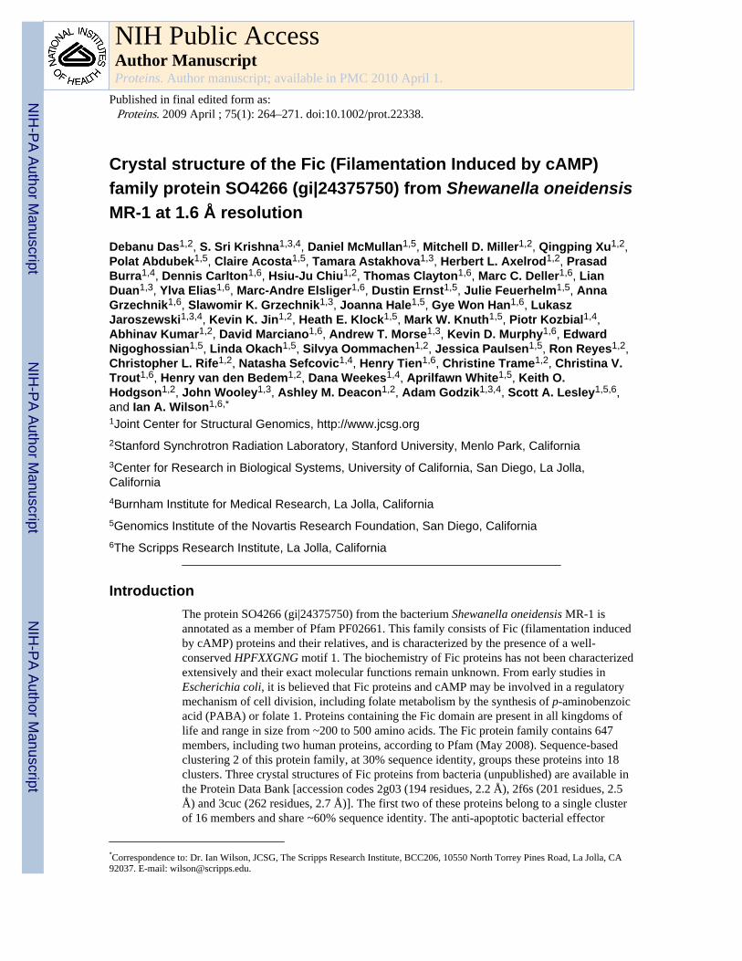
Crystal structure of the Fic (Filamentation Induced by cAMP)family protein SO4266 (gi|24375750) from Shewanella oneidensisMR-1 at 1.6 Å resolution
Debanu Das1,2, S. Sri Krishna1,3,4, Daniel McMullan1,5, Mitchell D. Miller1,2, Qingping Xu1,2,Polat Abdubek1,5, Claire Acosta1,5, Tamara Astakhova1,3, Herbert L. Axelrod1,2, PrasadBurra1,4, Dennis Carlton1,6, Hsiu-Ju Chiu1,2, Thomas Clayton1,6, Marc C. Deller1,6, LianDuan1,3, Ylva Elias1,6, Marc-Andre Elsliger1,6, Dustin Ernst1,5, Julie Feuerhelm1,5, AnnaGrzechnik1,6, Slawomir K. Grzechnik1,3, Joanna Hale1,5, Gye Won Han1,6, LukaszJaroszewski1,3,4, Kevin K. Jin1,2, Heath E. Klock1,5, Mark W. Knuth1,5, Piotr Kozbial1,4,Abhinav Kumar1,2, David Marciano1,6, Andrew T. Morse1,3, Kevin D. Murphy1,6, EdwardNigoghossian1,5, Linda Okach1,5, Silvya Oommachen1,2, Jessica Paulsen1,5, Ron Reyes1,2,Christopher L. Rife1,2, Natasha Sefcovic1,4, Henry Tien1,6, Christine Trame1,2, Christina V.Trout1,6, Henry van den Bedem1,2, Dana Weekes1,4, Aprilfawn White1,5, Keith O.Hodgson1,2, John Wooley1,3, Ashley M. Deacon1,2, Adam Godzik1,3,4, Scott A. Lesley1,5,6,and Ian A. Wilson1,6,*
1Joint Center for Structural Genomics, http://www.jcsg.org2Stanford Synchrotron Radiation Laboratory, Stanford University, Menlo Park, California3Center for Research in Biological Systems, University of California, San Diego, La Jolla,California4Burnham Institute for Medical Research, La Jolla, California5Genomics Institute of the Novartis Research Foundation, San Diego, California6The Scripps Research Institute, La Jolla, California
IntroductionThe protein SO4266 (gi|24375750) from the bacterium Shewanella oneidensis MR-1 isannotated as a member of Pfam PF02661. This family consists of Fic (filamentation inducedby cAMP) proteins and their relatives, and is characterized by the presence of a well-conserved HPFXXGNG motif 1. The biochemistry of Fic proteins has not been characterizedextensively and their exact molecular functions remain unknown. From early studies inEscherichia coli, it is believed that Fic proteins and cAMP may be involved in a regulatorymechanism of cell division, including folate metabolism by the synthesis of p-aminobenzoicacid (PABA) or folate 1. Proteins containing the Fic domain are present in all kingdoms oflife and range in size from ~200 to 500 amino acids. The Fic protein family contains 647members, including two human proteins, according to Pfam (May 2008). Sequence-basedclustering 2 of this protein family, at 30% sequence identity, groups these proteins into 18clusters. Three crystal structures of Fic proteins from bacteria (unpublished) are available inthe Protein Data Bank [accession codes 2g03 (194 residues, 2.2 Å), 2f6s (201 residues, 2.5Å) and 3cuc (262 residues, 2.7 Å)]. The first two of these proteins belong to a single clusterof 16 members and share ~60% sequence identity. The anti-apoptotic bacterial effector
*Correspondence to: Dr. Ian Wilson, JCSG, The Scripps Research Institute, BCC206, 10550 North Torrey Pines Road, La Jolla, CA92037. E-mail: [email protected].
NIH Public AccessAuthor ManuscriptProteins. Author manuscript; available in PMC 2010 April 1.
Published in final edited form as:Proteins. 2009 April ; 75(1): 264–271. doi:10.1002/prot.22338.
NIH
-PA Author Manuscript
NIH
-PA Author Manuscript
NIH
-PA Author Manuscript
protein BepA, which is a type IV secretion (T4S) system substrate, also contains an N-terminal Fic domain 3. In humans, the Fic domain is present in the Huntingtin InteractingProtein E (HYPE; Uniprot entry Q9BVA6_HUMAN), a protein of unknown function that isthought to interact with Huntingtin, one of the major proteins in the Huntington's diseaseprotein interaction network (listed as NAD- or FAD-binding) 4. Bioinformatics analysis ofprokaryotic toxin-antitoxin networks 5 suggests that Fic proteins are putative death-on-curing (Doc) toxins that are part of the Phd-Doc system. These proteins likely function asmetal-dependent nucleases or RNA-processing enzymes, 5 while more recent studiessuggest that Doc toxicity is caused by inhibition of translation elongation 6. SO4266(Uniprot entry Q8E9K5_SHEON), at 372 amino acids, is one of the largest Fic domain-containing proteins to have its structure determined. Interestingly, both HYPE and SO4266belong to the largest sequence cluster in this family (n.b. our B. thetaiotaomicronNP_811426.1 structure with PDB id 3cuc also belongs to this cluster), which comprises 466out of 647 proteins, and share ~32% sequence identity in the Fic domain.
Here, we report the crystal structure of the SO4266 protein at 1.6 Å resolution. The structurereveals a dimeric protein with additional electron density in the vicinity of the highlyconserved HPFXXGNG motif in the Fic domain of one subunit that corresponds to the N-terminus of a symmetry-related molecule. In addition, the study also reveals a C-terminalwinged-helix DNA-binding domain that sets it apart from the other Fic protein structures.The structure presented here is a representative of the largest sequence cluster and togetherwith the structures of the other Fic proteins paves the way for further structure-basedfunctional characterization.
Materials and MethodsProtein production and crystallization
The S. oneidensis MR-1 SO4266 gene (GenBank: NP_719793.1, GI:24375750) wasamplified by polymerase chain reaction (PCR) from genomic DNA (ATCC: 700550D) usingPfuTurbo (Stratagene) and primers corresponding to the predicted 5' and 3' ends. The PCRproduct was cloned into plasmid pSpeedET, which encodes an expression and purificationtag followed by a tobacco etch virus (TEV) protease cleavage site(MGSDKIHHHHHHENLYFQG) at the amino terminus of the full-length protein. LC-MSreveals a Cys at position 109, and DNA sequencing indicates a TGC codon. However, thepublished sequence for SO4266 from strain MR-1 shows a Gly (GGC codon) at thisposition. It is not clear if this discrepancy is a PCR artifact. The cloning junctions wereconfirmed by DNA sequencing. Protein expression was performed in a selenomethionine-containing medium using the Escherichia coli strain GeneHogs (Invitrogen). At the end offermentation, lysozyme was added to the culture to a final concentration of 250 μg/mL, andthe cells were harvested. After one freeze/thaw cycle, the cells were homogenized in LysisBuffer [50 mM HEPES pH 8.0, 50 mM NaCl, 10 mM imidazole, 1 mM Tris(2-carboxyethyl)phosphine hydrochloride (TCEP)] and passed through a Microfluidizer(Microfluidics). The lysate was clarified by centrifugation at 32,500 × g for 30 min andloaded onto nickel-chelating resin (GE Healthcare) preequilibrated with Lysis Buffer. Theresin was washed with Wash Buffer [50 mM HEPES pH 8.0, 300 mM NaCl, 40 mMimidazole, 10% (v/v) glycerol, 1 mM TCEP], and the protein was eluted with Elution Buffer[20 mM HEPES pH 8.0, 300 mM imidazole, 10% (v/v) glycerol, 1 mM TCEP]. The eluatewas buffer exchanged with HEPES Crystallization Buffer [20 mM HEPES pH 8.0, 200 mMNaCl, 40 mM imidazole, 1 mM TCEP] using a PD-10 column (GE Healthcare) and treatedwith 1 mg of TEV protease per 15 mg of eluted protein. The digested eluate was passed overnickel-chelating resin (GE Healthcare) pre-equilibrated with HEPES Crystallization Buffer,and the resin was washed with the same buffer. The flow-through and wash fractions werecombined and concentrated for crystallization assays to 12.4 mg/mL by centrifugal
Das et al. Page 2
Proteins. Author manuscript; available in PMC 2010 April 1.
NIH
-PA Author Manuscript
NIH
-PA Author Manuscript
NIH
-PA Author Manuscript
ultrafiltration (Millipore). SO4266 was crystallized using the nanodroplet vapor diffusionmethod 7 with standard JCSG crystallization protocols 8. Screening for diffraction wascarried out using the Stanford Automated Mounting system (SAM) 9 at the StanfordSynchrotron Radiation Laboratory (SSRL, Menlo Park, CA). The crystallization reagent thatproduced the crystal used for structure solution contained 0.2 M NaF and 20% (w/v)polyethylene glycol (PEG) 3350 at pH 7.1. PEG 200 was added as a cryoprotectant to thecrystal to a final concentration of 15% (v/v). The crystal was indexed in orthorhombic spacegroup P212121 (Table I) 10,11. The molecular weight and oligomeric state of SO4266 weredetermined using a 1 cm × 30 cm Superdex 200 column (GE Healthcare) in combinationwith static light scattering (Wyatt Technology). The mobile phase consisted of 20 mM TrispH 8.0, 150 mM NaCl, and 0.02% (w/v) sodium azide.
Data collection, structure solution, and refinementMulti-wavelength anomalous diffraction (MAD) data were collected at SSRL on beamline11-1 at wavelengths corresponding to the high energy remote (λ1), peak (λ2) and inflectionpoint (λ3) of a selenium MAD experiment using the BLU-ICE 12 data collectionenvironment. The data sets were collected at 100K using a MarMosaic 325 CCD detector(Mar USA). The MAD data were integrated and reduced using XDS 13 and scaled with theprogram XSCALE. The heavy atom substructure was solved and sites were refined usingSOLVE 14. Density modification was performed with RESOLVE 15 and ARP/wARP 16was used for automatic model building. Model completion and crystallographic refinementwere performed with the λ1 data set using COOT 17 and REFMAC5 10, respectively. Dataand refinement statistics are summarized in Table I 10,11.
Validation and depositionThe quality of the crystal structure was analyzed using the JCSG Quality Control server.This server verifies: the stereochemical quality of the model using AutoDepInputTool 18,MolProbity 19, and WHATIF 5.0 20; agreement between the atomic model and the datausing SFcheck 4.0 21 and RESOLVE 15, the protein sequence using CLUSTALW 22, atomoccupancies using MOLEMAN2 23, consistency of NCS pairs, and evaluates difference inRcryst/Rfree, expected Rfree/Rcryst and maximum/minimum B-factors by parsing therefinement log-file and PDB header. Protein quaternary structure analysis was performedusing the PISA server 24. Figure 1(B) was adapted from an analysis using PDBsum 25, andall others were prepared with PyMOL 26. Atomic coordinates and experimental structurefactors for Fic from S. oneidensis MR-1 to 1.6 Å resolution were deposited in the PDBunder the accession code 3eqx.
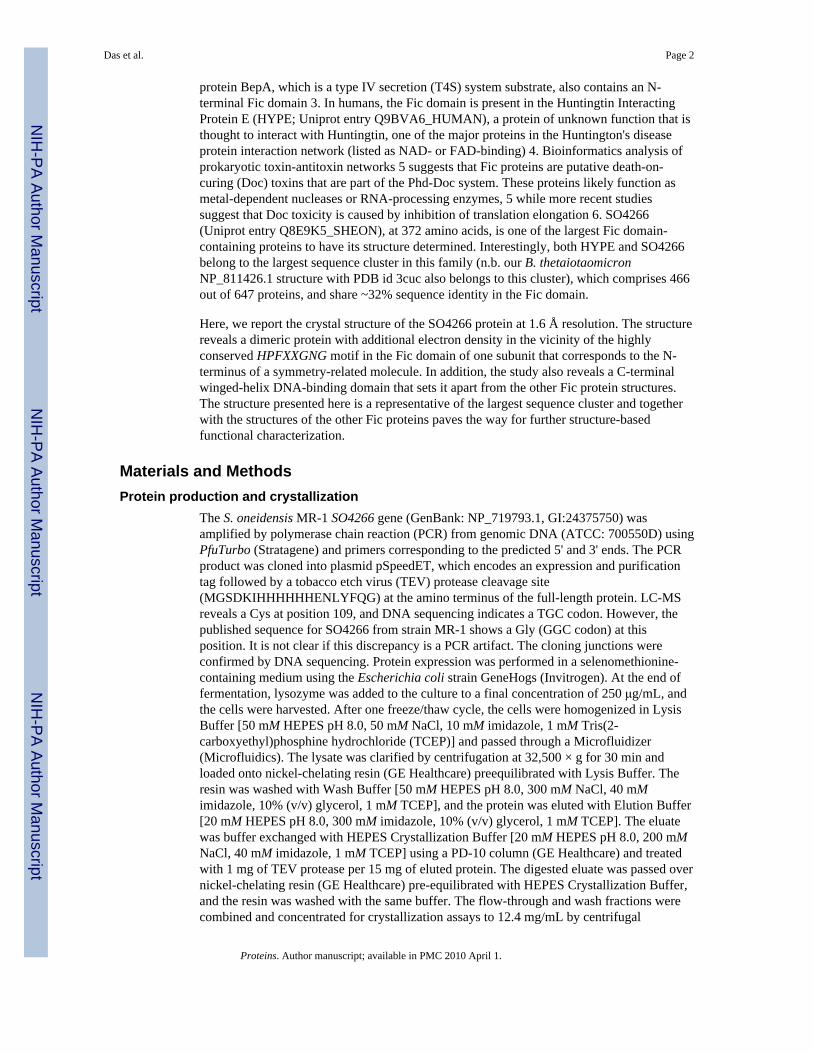
Results and DiscussionThe crystal structure of SO4266 [Fig. 1(A)] was determined by MAD phasing to a resolutionof 1.6 Å. The final model contains 2 monomers, 814 waters and 1 PGE molecule in theasymmetric unit (ASU). This dimer is the likely oligomeric association as judged fromcrystal lattice packing and assembly analysis using PISA 24, as well as size exclusionchromatography coupled with static light scattering. Residues A1, A344-A345, A371-A372,B5-B10, and B370-B372 (A and B refer to the chain identifier) and N-terminal glycine leftafter cleavage of the expression and purification tag were disordered and not modeled. TheMatthews' coefficient 27 is 2.4 Å3/Da, with an estimated solvent content of 48.4%. TheRamachandran plot produced by Molprobity 19 shows that 98.9% and 100 % of amino acidsare in the favored and allowed regions, respectively.
The secondary structure of this 372-amino acid protein is primarily α-helical (65%), with 16α-helices (H1-H16) (Fig. 1). Each monomer of SO4266 assumes an overall shape
Das et al. Page 3
Proteins. Author manuscript; available in PMC 2010 April 1.
NIH
-PA Author Manuscript
NIH
-PA Author Manuscript
NIH
-PA Author Manuscript
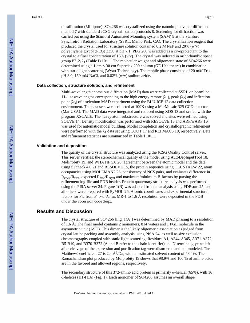
resembling a closed fist, with a back-to-back association of the monomers forming a`transcription factor-like' dimer (Fig. 2). The core of the Fic domain consist of residues100-290 (Fig. 2, magenta), with a long, mostly α-helical insert region at the N-terminus(1-100; Fig. 2, blue) that is involved in dimerization. Residues 290-370 of the C-terminusform a winged helix-turn-helix DNA-binding domain (Fig. 2, green) similar to that seen inseveral transcriptional regulators 28,29(e.g. PDB id 2d1h).
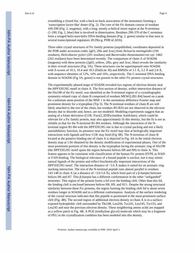
Three other crystal structures of Fic family proteins (unpublished, coordinates deposited inthe PDB under accession codes 2g03, 2f6s and 3cuc) from Neisseria meningitidis (194residues), Helicobacter pylori (201 residues) and Bacteroides thetaiotaomicron vpi- 5482(262 residues) have been determined recently. The comparison of chain A of SO4266(magenta) with these proteins (2g03, yellow, 2f6s, grey and 3cuc, blue) reveals the similarityin their overall structures (Fig. 3A). These structures can be superimposed onto SO4266with Z-scores of 10.5, 10.4 and 18.5 (DaliLite 30) and RMSDs of 3.1 Å, 3.2 Å and 2.8 Å,with sequence identities of 12%, 14% and 16%, respectively. The C-terminal DNA bindingdomain in SO4266 (Fig 3A, green) is not present in the other Fic protein crystal structures.
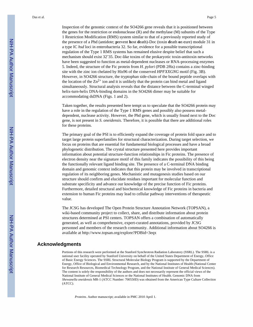
The experimentally phased maps of SO4266 revealed two regions of electron density nearthe HPFXXGNG motif in chain A. The first section of density, within interaction distance ofthe His198 of the Fic motif, was identified as the N-terminal region of a crystallographicsymmetry-related copy of chain B comprised of residues MEWQ (B1-B4) based on a peakfor a selenium atom position of the MSE 1 in the anomalous difference Fourier map andprominent density for a tryptophan (Trp 3). The N-terminal residues of chain B are stilllikely attached to the rest of the chain, but residues B5-B10 are not observed in the electrondensity due to disorder and, hence, are not modeled. Modeling experiments suggest that ananalog of a folate derivative (COE: Furo[2,3D]Pyrimidine Antifolate), which could berelevant for a Fic family protein, may also approximately fit this density, but the fit is not asreliable as that for the N-terminal B1-B4 residues. Although the interaction of the N-terminal region B1-B4 with the HPFXXGNG site is due to crystal packing and not a likelyautoinhibitory function, its presence near the Fic motif may hint at biologically importantinteractions with ligands and how COE may bind (Fig 3B). The N-terminus of chain Blocated at the putative binding site of chain A is depicted in Fig. 4A in the initial electrondensity map at 1.0σ obtained by the density modification of experimental phases. One of themost prominent portions of this density is the tryptophan facing the aromatic ring of His198(the HPFXXGNG motif spans the region between helices H8 and H9) in chain A. Thisfeature appears to be consistent with classification of the human Fic protein HYPE as NAD-or FAD-binding. The biological relevance of a bound peptide is unclear, but it may mimicnatural ligands of the protein and reflect biochemically important interactions of theHPFXXGNG motif. The interaction distance of ~3.6 Å makes it suited for an aromatic ringstacking interaction. The rest of the N-terminal peptide runs almost parallel to residues143-148 in chain A (at a distance of ~3.0-5.0 Å), which form part of a β-hairpin betweenhelices H6 and H7. This β-hairpin has a different conformation in the other “unliganded”monomer. This region of the protein forms a lid over the binding cleft. Other than this lid,the binding cleft is enclosed between helices H8, H9, and H11. Despite the strong structuralsimilarity between these Fic proteins, the region forming the binding cleft lid is about sevenresidues longer in SO4266 and in a different conformation. Analysis of the surface renderingof the crystal structure indicates that this peptide is positioned in the most prominent surfacecleft (Fig. 4B). The second region of additional electron density in chain A is in a surface-exposed hydrophobic cleft surrounded by Thr248, Leu244, Tyr241, Leu145, Tyr155, andLeu245 and near the previous electron density. These neighboring amino acids are mappedas a yellow patch in Fig. 4B. A PGE (triethylene glycol) molecule which may be a fragmentof PEG in the crystallization condition has been modeled into this density.
Das et al. Page 4
Proteins. Author manuscript; available in PMC 2010 April 1.
NIH
-PA Author Manuscript
NIH
-PA Author Manuscript
NIH
-PA Author Manuscript
Inspection of the genomic context of the SO4266 gene reveals that it is positioned betweenthe genes for the restriction or endonuclease (R) and the methylase (M) subunits of the Type1 Restriction Modification (RMS) system similar to that of a previously reported study ofthe presence of a Phd (antidote; prevent host death)-Doc (toxin death on cure) module 31 ina type IC hsd loci in enterobacteria 32. So far, evidence for a possible transcriptionalregulation of the Type 1 RMS systems has remained elusive despite belief that such amechanism should exist 32-35. Doc-like toxins of the prokaryotic toxin-antitoxin networkshave been suggested to function as metal-dependent nucleases or RNA-processing enzymes5. Indeed, the structure of the Fic protein from H. pylori (PDB 2f6s) contains a zinc-bindingsite with the zinc ion chelated by His96 of the conserved HPFXXGNG motif (Fig. 3B).However, in SO4266 structure, the tryptophan side-chain of the bound peptide overlaps withthe location of the Zn2+ ion and it is unlikely that the protein can bind metal and ligandsimultaneously. Structural analysis reveals that the distance between the C-terminal wingedhelix-turn-helix DNA-binding domains in the SO4266 dimer may be suitable foraccommodating dsDNA (Figs. 1 and 2).
Taken together, the results presented here tempt us to speculate that the SO4266 protein mayhave a role in the regulation of the Type 1 RMS genes and possibly also possess metal-dependent, nuclease activity. However, the Phd gene, which is usually found next to the Docgene, is not present in S. oneidensis. Therefore, it is possible that there are additional rolesfor these proteins.
The primary goal of the PSI is to efficiently expand the coverage of protein fold space and totarget large protein superfamilies for structural characterization. During target selection, wefocus on proteins that are essential for fundamental biological processes and have a broadphylogenetic distribution. The crystal structure presented here provides importantinformation about potential structure-function relationships in Fic proteins. The presence ofelectron density near the signature motif of this family indicates the possibility of this beingthe functionally relevant ligand binding site. The presence of a C-terminal DNA bindingdomain and genomic context indicates that this protein may be involved in transcriptionalregulation of its neighboring genes. Mechanistic and mutagenesis studies based on ourstructure should confirm and elucidate residues important for molecular function andsubstrate specificity and advance our knowledge of the precise function of Fic proteins.Furthermore, detailed structural and biochemical knowledge of Fic proteins in bacteria andextension to human Fic proteins may lead to cellular pathway interventions of therapeuticvalue.
The JCSG has developed The Open Protein Structure Annotation Network (TOPSAN), awiki-based community project to collect, share, and distribute information about proteinstructures determined at PSI centers. TOPSAN offers a combination of automaticallygenerated, as well as comprehensive, expert-curated annotations, provided by JCSGpersonnel and members of the research community. Additional information about SO4266 isavailable at http://www.topsan.org/explore?PDBid=3eqx
AcknowledgmentsPortions of this research were performed at the Stanford Synchrotron Radiation Laboratory (SSRL). The SSRL is anational user facility operated by Stanford University on behalf of the United States Department of Energy, Officeof Basic Energy Sciences. The SSRL Structural Molecular Biology Program is supported by the Department ofEnergy, Office of Biological and Environmental Research, and by the National Institutes of Health (National Centerfor Research Resources, Biomedical Technology Program, and the National Institute of General Medical Sciences).The content is solely the responsibility of the authors and does not necessarily represent the official views of theNational Institute of General Medical Sciences or the National Institutes of Health. Genomic DNA fromShewanella oneidensis MR-1 (ATCC Number: 700550D) was obtained from the American Type Culture Collection(ATCC).
Das et al. Page 5
Proteins. Author manuscript; available in PMC 2010 April 1.
NIH
-PA Author Manuscript
NIH
-PA Author Manuscript
NIH
-PA Author Manuscript
Grant Sponsor: National Institute of General Medical Sciences, Protein Structure Initiative; Grant Number: U54GM074898.
References1. Komano T, Utsumi R, Kawamukai M. Functional analysis of the fic gene involved in regulation of
cell division. Res Microbiol. 1991; 142:269–277. [PubMed: 1656497]2. Li W, Godzik A. Cd-hit: a fast program for clustering and comparing large sets of protein or
nucleotide sequences. Bioinformatics. 2006; 22:1658–1659. [PubMed: 16731699]3. Schmid MC, Scheidegger F, Dehio M, Balmelle-Devaux N, Schulein R, Guye P, Chennakesava CS,
Biedermann B, Dehio C. A translocated bacterial protein protects vascular endothelial cells fromapoptosis. PLoS Pathog. 2006; 2:e115. [PubMed: 17121462]
4. Clark HF, Gurney AL, Abaya E, Baker K, Baldwin D, Brush J, Chen J, Chow B, Chui C, CrowleyC, Currell B, Deuel B, Dowd P, Eaton D, Foster J, Grimaldi C, Gu Q, Hass PE, Heldens S, HuangA, Kim HS, Klimowski L, Jin Y, Johnson S, Lee J, Lewis L, Liao D, Mark M, Robbie E, SanchezC, Schoenfeld J, Seshagiri S, Simmons L, Singh J, Smith V, Stinson J, Vagts A, Vandlen R,Watanabe C, Wieand D, Woods K, Xie MH, Yansura D, Yi S, Yu G, Yuan J, Zhang M, Zhang Z,Goddard A, Wood WI, Godowski P, Gray A. The secreted protein discovery initiative (SPDI), alarge-scale effort to identify novel human secreted and transmembrane proteins: a bioinformaticsassessment. Genome Res. 2003; 13:2265–2270. [PubMed: 12975309]
5. Anantharaman V, Aravind L. New connections in the prokaryotic toxin-antitoxin network:relationship with the eukaryotic nonsense-mediated RNA decay system. Genome Biol. 2003; 4:R81.[PubMed: 14659018]
6. Liu M, Zhang Y, Inouye M, Woychik NA. Bacterial addiction module toxin Doc inhibits translationelongation through its association with the 30S ribosomal subunit. Proc Natl Acad Sci U S A. 2008;105:5885–5890. [PubMed: 18398006]
7. Santarsiero BD, Yegian DT, Lee CC, Spraggon G, Gu J, Scheibe D, Uber DC, Cornell EW,Nordmeyer RA, Kolbe WF, Jin J, Jones AL, Jaklevic JM, Schultz PG, Stevens RC. An approach torapid protein crystallization using nanodroplets. J Appl Crystallogr. 2002; 35:278–281.
8. Lesley SA, Kuhn P, Godzik A, Deacon AM, Mathews I, Kreusch A, Spraggon G, Klock HE,McMullan D, Shin T, Vincent J, Robb A, Brinen LS, Miller MD, McPhillips TM, Miller MA,Scheibe D, Canaves JM, Guda C, Jaroszewski L, Selby TL, Elsliger MA, Wooley J, Taylor SS,Hodgson KO, Wilson IA, Schultz PG, Stevens RC. Structural genomics of the Thermotogamaritima proteome implemented in a high-throughput structure determination pipeline. Proc NatlAcad Sci U S A. 2002; 99:11664–11669. [PubMed: 12193646]
9. Cohen AE, Ellis PJ, Miller MD, Deacon AM, Phizackerley RP. An automated system to mountcryo-cooled protein crystals on a synchrotron beamline, using compact sample cassettes and asmall-scale robot. J Appl Crystallogr. 2002; 2002:720–726.
10. The CCP4 suite: programs for protein crystallography. Acta Crystallogr D Biol Crystallogr. 1994;50:760–763. [PubMed: 15299374]
11. Tickle IJ, Laskowski RA, Moss DS. Error estimates of protein structure coordinates and deviationsfrom standard geometry by full-matrix refinement of gammaB- and betaB2-crystallin. ActaCrystallogr D Biol Crystallogr. 1998; 54:243–252. [PubMed: 9761889]
12. McPhillips TM, McPhillips SE, Chiu HJ, Cohen AE, Deacon AM, Ellis PJ, Garman E, GonzalezA, Sauter NK, Phizackerley RP, Soltis SM, Kuhn P. Blu-Ice and the Distributed Control System:software for data acquisition and instrument control at macromolecular crystallography beamlines.J Synchrotron Radiat. 2002; 9:401–406. [PubMed: 12409628]
13. Kabsch W. Automatic processing of rotation diffraction data from crystals of initially unknownsymmetry and cell constants. J Appl Crystallog. 1993; 26:795–800.
14. Terwilliger T, Berendzen J. Automated MAD and MIR structure solution. Acta Cryst. 1999;D55:849–861.
15. Terwilliger TC. Maximum-likelihood density modification. Acta Crystallogr D Biol Crystallogr.2000; 56:965–972. [PubMed: 10944333]
Das et al. Page 6
Proteins. Author manuscript; available in PMC 2010 April 1.
NIH
-PA Author Manuscript
NIH
-PA Author Manuscript
NIH
-PA Author Manuscript

16. Langer G, Cohen SX, Lamzin VS, Perrakis A. Automated macromolecular model building for X-ray crystallography using ARP/wARP version 7. Nat Protoc. 2008; 3:1171–1179. [PubMed:18600222]
17. Emsley P, Cowtan K. Coot: model-building tools for molecular graphics. Acta Crystallogr D BiolCrystallogr. 2004; 60:2126–2132. [PubMed: 15572765]
18. Yang H, Guranovic V, Dutta S, Feng Z, Berman HM, Westbrook JD. Automated and accuratedeposition of structures solved by X-ray diffraction to the Protein Data Bank. Acta Crystallogr DBiol Crystallogr. 2004; 60:1833–1839. [PubMed: 15388930]
19. Davis IW, Murray LW, Richardson JS, Richardson DC. MOLPROBITY: structure validation andall-atom contact analysis for nucleic acids and their complexes. Nucleic Acids Res. 2004;32:W615–619. [PubMed: 15215462]
20. Vriend G. WHAT IF: a molecular modeling and drug design program. J Mol Graph. 1990; 8:52–56. 29. [PubMed: 2268628]
21. Vaguine AA, Richelle J, Wodak SJ. SFCHECK: a unified set of procedures for evaluating thequality of macromolecular structure-factor data and their agreement with the atomic model. ActaCrystallogr D Biol Crystallogr. 1999; 55:191–205. [PubMed: 10089410]
22. Thompson JD, Higgins DG, Gibson TJ. CLUSTAL W: improving the sensitivity of progressivemultiple sequence alignment through sequence weighting, position-specific gap penalties andweight matrix choice. Nucleic Acids Res. 1994; 22:4673–4680. [PubMed: 7984417]
23. Kleywegt GJ. Validation of protein crystal structures. Acta Crystallogr D Biol Crystallogr. 2000;56:249–265. [PubMed: 10713511]
24. Krissinel, E.; Henrick, K. Detection of protein assemblies in crystals. Computational Life Sciences:Proceedings of the First International Symposium, Complife Berlin Heidelberg; Springer-Verlag;2005. p. 163-174.
25. Laskowski RA, Chistyakov VV, Thornton JM. PDBsum more: new summaries and analyses of theknown 3D structures of proteins and nucleic acids. Nucleic Acids Res. 2005; 33:D266–268.[PubMed: 15608193]
26. DeLano, WL. The PyMOL Molecular Graphics System. 2002.27. Matthews BW. Solvent content of protein crystals. J Mol Biol. 1968; 33:491–497. [PubMed:
5700707]28. Ohlendorf DH, Anderson WF, Matthews BW. Many gene-regulatory proteins appear to have a
similar alpha-helical fold that binds DNA and evolved from a common precursor. J Mol Evol.1983; 19:109–114. [PubMed: 6571216]
29. Aravind L, Anantharaman V, Balaji S, Babu MM, Iyer LM. The many faces of the helix-turn-helixdomain: transcription regulation and beyond. FEMS Microbiol Rev. 2005; 29:231–262. [PubMed:15808743]
30. Holm L, Park J. DaliLite workbench for protein structure comparison. Bioinformatics. 2000;16:566–567. [PubMed: 10980157]
31. Lehnherr H, Maguin E, Jafri S, Yarmolinsky MB. Plasmid addiction genes of bacteriophage P1:doc, which causes cell death on curing of prophage, and phd, which prevents host death whenprophage is retained. J Mol Biol. 1993; 233:414–428. [PubMed: 8411153]
32. Tyndall C, Lehnherr H, Sandmeier U, Kulik E, Bickle TA. The type IC hsd loci of theenterobacteria are flanked by DNA with high homology to the phage P1 genome: implications forthe evolution and spread of DNA restriction systems. Mol Microbiol. 1997; 23:729–736.[PubMed: 9157244]
33. Kulik EM, Bickle TA. Regulation of the activity of the type IC EcoR124I restriction enzyme. JMol Biol. 1996; 264:891–906. [PubMed: 9000619]
34. Loenen WA, Daniel AS, Braymer HD, Murray NE. Organization and sequence of the hsd genes ofEscherichia coli K-12. J Mol Biol. 1987; 198:159–170. [PubMed: 3323532]
35. Prakash-Cheng A, Ryu J. Delayed expression of in vivo restriction activity following conjugaltransfer of Escherichia coli hsdK (restriction-modification) genes. J Bacteriol. 1993; 175:4905–4906. [PubMed: 8393010]
Das et al. Page 7
Proteins. Author manuscript; available in PMC 2010 April 1.
NIH
-PA Author Manuscript
NIH
-PA Author Manuscript
NIH
-PA Author Manuscript
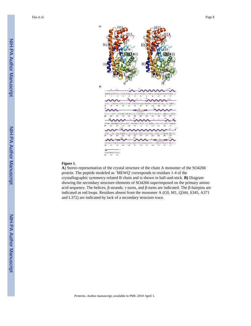
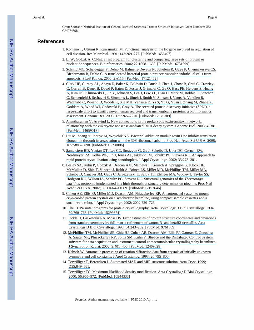
Figure 1.A) Stereo representation of the crystal structure of the chain A monomer of the SO4266protein. The peptide modeled as `MEWQ' corresponds to residues 1-4 of thecrystallographic symmetry-related B chain and is shown in ball-and-stick. B) Diagramshowing the secondary structure elements of SO4266 superimposed on the primary aminoacid sequence. The helices, β-strands, γ-turns, and β-turns are indicated. The β-hairpins areindicated as red loops. Residues absent from the monomer A (G0, M1, Q344, S345, A371and L372) are indicated by lack of a secondary structure trace.
Das et al. Page 8
Proteins. Author manuscript; available in PMC 2010 April 1.
NIH
-PA Author Manuscript
NIH
-PA Author Manuscript
NIH
-PA Author Manuscript
Figure 2.The monomers of SO4266 assume a closed fist-like shape, with a back-to-back associationof the monomers forming a “transcription factor-like” dimer. The core of the Fic domain ismade up of residues 100-290 (magenta), with a long, mostly α-helical insert region at the N-terminus (residues 1-100; blue) that is involved in dimerization. Residues 290-370 of the C-terminus form a winged helix-turn-helix (wHTH) DNA-binding domain (green) similar tothat seen in several transcriptional regulators (e.g. PDB id 2d1h). The peptide `MEWQ'bound to chain A that corresponds to residues 1-4 of the crystallographic symmetry-relatedB chain is shown (yellow).
Das et al. Page 9
Proteins. Author manuscript; available in PMC 2010 April 1.
NIH
-PA Author Manuscript
NIH
-PA Author Manuscript
NIH
-PA Author Manuscript

Figure 3.A) Superimposition of the SO4266 monomers (magenta and green) with the Fic familyproteins from Neisseria meningitidis (194 residues, 2g03, yellow), Helicobacter pylori (201residues, 2f6s, grey) and Bacteroides thetaiotaomicron vpi-5482 (262 residues, 3cuc, blue),reveal the similarity in their structures despite the absence of the C-terminal domain ofSO4266 (green) in the other Fic proteins. They align on SO4266 with RMSDs of 3.1 Å, 3.2Å and 2.8 Å, Z-scores of 10.5, 10.4 and 18.5 (DaliLite), and sequence identities of 12%,14% and 16%, respectively. The peptide is shown (yellow ball-and-stick). B) The structureof the H. pylori Fic protein (grey) contains a zinc ion (grey) interacting with His 96 of theconserved HPFXXGNG motif. In the structure of SO4266 (magenta), the tryptophan residueof the peptide (yellow ball-and-stick) partially overlaps with the location of the Zn2+ ion andit is unlikely that the protein can bind metal and the peptide at the same time (grey).
Das et al. Page 10
Proteins. Author manuscript; available in PMC 2010 April 1.
NIH
-PA Author Manuscript
NIH
-PA Author Manuscript
NIH
-PA Author Manuscript
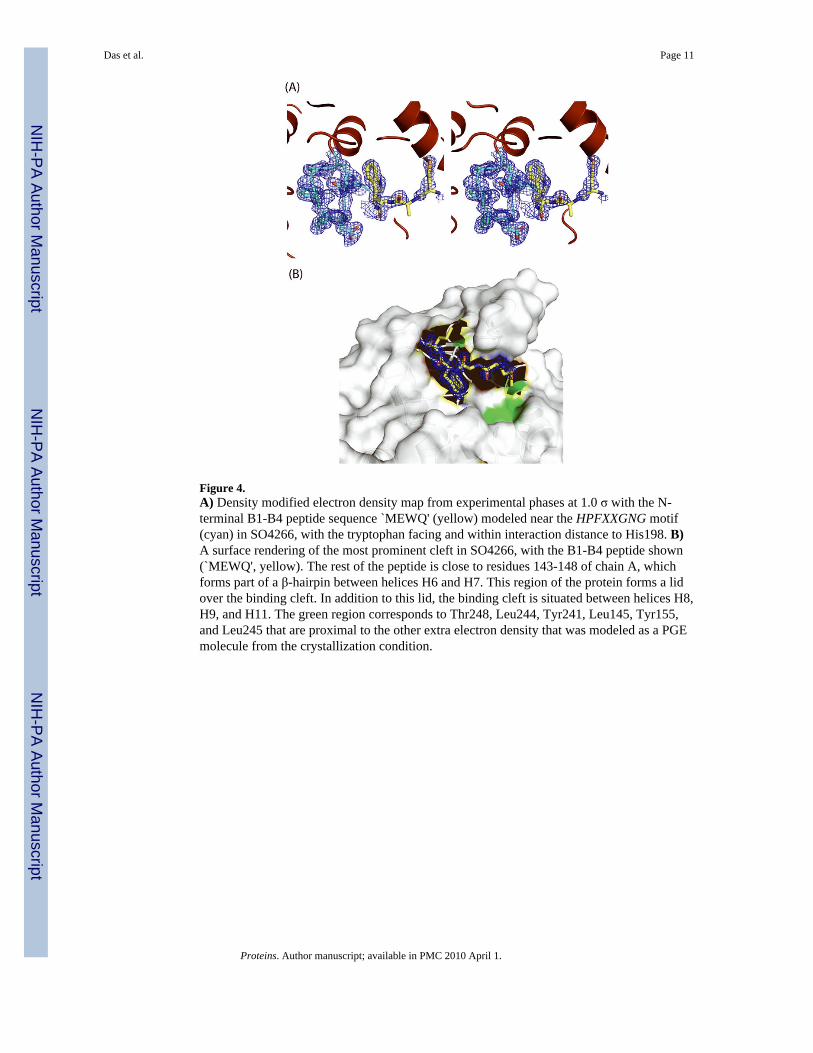
Figure 4.A) Density modified electron density map from experimental phases at 1.0 σ with the N-terminal B1-B4 peptide sequence `MEWQ' (yellow) modeled near the HPFXXGNG motif(cyan) in SO4266, with the tryptophan facing and within interaction distance to His198. B)A surface rendering of the most prominent cleft in SO4266, with the B1-B4 peptide shown(`MEWQ', yellow). The rest of the peptide is close to residues 143-148 of chain A, whichforms part of a β-hairpin between helices H6 and H7. This region of the protein forms a lidover the binding cleft. In addition to this lid, the binding cleft is situated between helices H8,H9, and H11. The green region corresponds to Thr248, Leu244, Tyr241, Leu145, Tyr155,and Leu245 that are proximal to the other extra electron density that was modeled as a PGEmolecule from the crystallization condition.
Das et al. Page 11
Proteins. Author manuscript; available in PMC 2010 April 1.
NIH
-PA Author Manuscript
NIH
-PA Author Manuscript
NIH
-PA Author Manuscript
NIH
-PA Author Manuscript
NIH
-PA Author Manuscript
NIH
-PA Author Manuscript
Das et al. Page 12
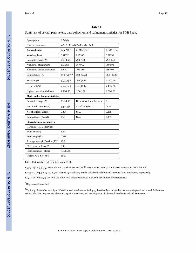
Table I
Summary of crystal parameters, data collection and refinement statistics for PDB 3eqx.
Space group P 212121
Unit cell parameters a=71.21Å, b=80.30Å, c=141.00Å
Data collection λ1 MAD Se λ2 MAD Se λ3 MAD Se
Wavelength(Å) 0.91837 0.97905 0.97935
Resolution range (Å) 29.6-1.60 29.6-1.60 29.5-1.60
Number of observations 373,102 367,969 369,908
Number of unique reflections 106,471 106,307 106,407
Completeness (%) 98.7 (98.7)a 98.0 (99.2) 98.6 (98.2)
Mean I/σ (I) 13.8 (2.0)a 16.6 (2.6) 12.5 (1.9)
Rsym on I (%) 4.2 (52.4)a 3.3 (39.0) 4.4 (51.9)
Highest resolution shell (Å) 1.66-1.60 1.66-1.60 1.66-1.60
Model and refinement statistics
Resolution range (Å) 29.6-1.60 Data set used in refinement λ 1
No. of reflections (total) 106,418b Cutoff criteria |F|>0
No. of reflections (test) 5,304 Rcryst 0.168
Completeness (%total) 99.3 Rfree 0.197
Stereochemical parameters
Restraints (RMS observed)
Bond angle (°) 1.64
Bond length (Å) 0.018
Average isotropic B-value (Å2) 16.9
ESU based on Rfree (Å) 0.08
Protein residues / atoms 731/6,005
Water / PGE molecules 814/1
ESU = Estimated overall coordinate error 10,11.
Rsym =Σ|Ii-<Ii>|/Σ|Ii|, where Ii is the scaled intensity of the ith measurement and <Ii> is the mean intensity for that reflection.
Rcryst = Σ||Fobs|-|Fcalc||/Σ|Fobs|, where Fcalc and Fobs are the calculated and observed structure factor amplitudes, respectively.
Rfree = as for Rcryst, but for 5.0% of the total reflections chosen at random and omitted from refinement.
aHighest resolution shell
bTypically, the number of unique reflections used in refinement is slightly less that the total number that were integrated and scaled. Reflections
are excluded due to systematic absences, negative intensities, and rounding errors in the resolution limits and cell parameters.
Proteins. Author manuscript; available in PMC 2010 April 1.
Copyright © 2022 FDOKUMEN