The Role of 4-Hydroxyphenylpyruvate Dioxygenase in Enhancement of SolidPhase Electron Transfer by...
41
This document was prepared in conjunction with work accomplished under Contract No. DE-AC09-96SR18500 with the U.S. Department of Energy. This work was prepared under an agreement with and funded by the U.S. Government. Neither the U. S. Government or its employees, nor any of its contractors, subcontractors or their employees, makes any express or implied: 1. warranty or assumes any legal liability for the accuracy, completeness, or for the use or results of such use of any information, product, or process disclosed; or 2. representation that such use or results of such use would not infringe privately owned rights; or 3. endorsement or recommendation of any specifically identified commercial product, process, or service. Any views and opinions of authors expressed in this work do not necessarily state or reflect those of the United States Government, or its contractors, or subcontractors.
-
Upload
independent -
Category
Documents
-
view
4 -
download
0
Transcript of The Role of 4-Hydroxyphenylpyruvate Dioxygenase in Enhancement of SolidPhase Electron Transfer by...

This document was prepared in conjunction with work accomplished under Contract No. DE-AC09-96SR18500 with the U.S. Department of Energy. This work was prepared under an agreement with and funded by the U.S. Government. Neither the U. S. Government or its employees, nor any of its contractors, subcontractors or their employees, makes any express or implied: 1. warranty or assumes any legal liability for the accuracy, completeness, or for the use or results of such use of any information, product, or process disclosed; or 2. representation that such use or results of such use would not infringe privately owned rights; or 3. endorsement or recommendation of any specifically identified commercial product, process, or service. Any views and opinions of authors expressed in this work do not necessarily state or reflect those of the United States Government, or its contractors, or subcontractors.

The Role of 4-Hydroxyphenylpyruvate Dioxygenase in Enhancement of Solid-Phase Electron Transfer by Shewanella oneidensis MR-1
Charles E. Turick1+, Alex Beliaev2, Daniel A. Lowy3, Tara E. Poppy4*,
Andrea Malony5#, and Amy A. Ekechukwu1
1Savannah River National Laboratory, Aiken, SC
2Pacific Northwest National Laboratory, Richland, WA 3Nova Research, Inc., Alexandria, VA
4University of South Carolina, Aiken, SC 5Winthrop University, Rock Hill, SC
+ Corresponding author. Environmental Science and Biotechnology Department. Savannah River National Laboratory, Building 999W, Aiken, SC. 29808. Phone: 803-819-8407. Fax: 803-819-8432. Email: [email protected] * present address- Department of Veterinary Medicine, University of Tennessee, Knoxville, TN.
# present address – South Carolina Department of Health and Environmental Control, Columbia, SC Key words: dissimilatory metal reducing bacteria, pyomelanin, electron shuttle, electron transfer, hydrous ferric oxide, iron minerals, anaerobic respiration, cyclic voltammetry.
Running title: 4-HPPD Enhances Solid-Phase Electron Transfer

Summary 1
While mechanistic details of dissimilatory metal reduction are far from being understood, it 2
is postulated that the electron transfer to solid metal oxides is mediated by outer membrane-3
associated c-type cytochromes and redox active electron shuttling compounds. This study 4
focuses on the production of homogensitate in Shewanella oneidensis MR-1, an intermediate 5
of tyrosine degradation pathway, which is a precursor of a redox cycling metabolite, 6
pyomelanin. In this study, we determined that two enzymes involved in this pathway, 4-7
hydroxyphenylpyruvate dioxygenase (4HPPD) and homogentisate 1,2-dioxygenase are 8
responsible for homogentisate production and oxidation, respectively. Inhibition of 4-HPPD 9
activity with the specific inhibitor sulcotrione ([2-(2- chloro- 4- methane sulfonylbenzoyl)-10
1,3-cyclohexanedione), and deletion of melA, a gene encoding 4-HPPD, resulted in no 11
pyomelanin production by S. oneidensis MR-1. Conversely, deletion of hmgA which 12
encodes the putative homogentisate 1,2-dioxygenase, resulted in pyomelanin overproduction. 13
The efficiency and rates, with which MR-1 reduces hydrous ferric oxide, were directly linked 14
to the ability of mutant strains to produce pyomelanin. Electrochemical studies with whole 15
cells demonstrated that pyomelanin substantially increases the formal potential (E°′) of S. 16
oneidensis MR-1. Based on this work, environmental production of pyomelanin likely 17
contributes to an increased solid-phase metal reduction capacity in Shewanella oneidensis. 18

3
Introduction
Dissimilatory metal reducing bacteria (DMRB) contribute to biogeochemical cycling of both
soluble and solid-phase metals, including Fe(III). DMRB gain energy for growth by
catalyzing Fe(III) reduction with organic electron donors (Lovley et al., 1988), even though
most of the Fe(III) in soils and sediments is found in the form of highly insoluble minerals
(Schwertmann and Taylor, 1997). The specific strategies by which DMRB transfer
electrons to insoluble minerals have not been completely characterized although substantial
evidence points at outer membrane-associated c-type cytochromes as key mediators of the
electron transfer to solid metal oxides (Lloyd, 2003; Gorby et al., 2006). Another strategy
involves environmental humic compounds which serve as terminal electron acceptors and
electron shuttles for iron reduction by DMRB (Lovley et al.,1996). Humic compounds are
capable of transferring electrons to iron minerals due to the redox cycling capabilities of the
quinone moieties that are associated with these heteropolymers (Scott et al., 1998). This
scenario, however, is dependent on the quantity and redox cycling capability of humics
present in the environment since redox activity varies for humic compounds from different
locations (Scott et al., 1998). This raises the question of how solid-phase electron transfer
can be accomplished efficiently when environmental humics are low in quantity or their
redox cycling capacities are poor.
The ability for some DMRB to produce their own extracellular electron shuttling
compounds for solid-phase mineral reduction offers another strategy for growth and survival
of these microorganisms in the environment. It was shown that production of a specific
redox cycling metabolite by Shewanella algae BrY, tentatively identified as pyomelanin,
plays a role in dissimilatory metal reduction by serving as a soluble and cell-associated

4
electron shuttle (Turick et al, 2002; 2003). Quantities of pyomelanin as low as 3 fg/cell were
calculated to be sufficient to increase iron oxide reduction rates by 10% (Turick et al., 2002).
It should be noted that several types of melanin pigments exist in nature and are produced by
various biochemical mechanisms. Pyomelanin originates from bacterial conversion of
tyrosine and/or phenylalanine as part of the fumarate pathway (Lehninger, 1975) and is
produced by numerous bacteria (Yabuuchi and Omyama, 1972; Kotob et al., 1995; Ruzafa et
al.,1995; David et al., 1996; Sanchez-Amat et al., 1998) including members of the
Shewanella genus (Fuqua et al., 1993; Coon,et al., 1994; Ruzafa et al., 1994). Complete
breakdown of these precursors to acetylacetate and fumarate requires the enzymes 4-
hydroxyphenylpyruvate dioxygenase (4-HPPD) and homogentisate oxidase (HGA-oxidase).
In the absence of HGA-oxidase [or if homogentisate (HGA) production exceeds that of
HGA-oxidase], HGA is over-produced and excreted from the cell (Coon et al., 1994). Auto-
oxidation and self-polymerization of excreted HGA results in pyomelanin, a polyaromatic
heteropolymer which consists of numerous quinone moieties (Ruzafa et al., 1995). This
process occurs when oxygen concentrations decline, (Ruzafa et al., 1995) likely at the
oxic/anoxic zones in the environment. Other types of melanins include eumelanin and
allomelanin (Prota, 1992). Eumelanin results from the conversion of tyrosine to the
eumelanin precursor 3,4-dihydroxyphenylalanine (DOPA), by the enzyme tyrosinase while
allomelanins are produced by laccase conversion of phenolic compounds.
In this study, we elucidated the process of homogentisate production by S. oneidensis
MR-1 and identified key steps involved in accumulation of pyomelanin. To better
understand the environmental relevance of this redox shuttling metabolite, we employed
genetic analysis and physiological characterization of recombinant S. oneidensis MR-1

5
strains deficient in or displaying substantially increased melanin production. The relative
significance imparted by pyomelanin on solid-phase electron transfer was also addressed
using an electrochemistry approach incorporating cyclic voltammetry to better understand the
link between genetics and physiology to biogeochemical cycling, especially in relation to
bioremediation of metal contaminated environments.
Results
Characterization of melanin production. Tyrosine supplemented lactate basal salts
medium (LBSM) resulted in production of a reddish-brown pigment during late log and early
stationary growth phases. At pH <2, the soluble cell-free pigment precipitated rapidly
displaying melanin and humic-like properties (Ellis and Griffiths, 1974). Following
precipitation, washing and dialysis, the dried (60°C) pigment resulted in a black powder with
melanin-like characteristics based on the following properties (Ellis and Griffiths, 1974):
insolublity in organic solvents (ethanol, chloroform and acetone); solubility in NaOH
solutions at pH >10; decolorization in H2O2; precipitation by FeCl3; and inability to filter
through 8-kDa dialysis membrane.
High pressure capillary electrophoresis (HPCE) analyses of spent cell-free tyrosine-
supplemented LBSM minimal medium demonstrated a peak that co-eluted with the
homogentisic acid (HGA) standard indicating production of homogensitate, the precursor of
pyomelanin (Fig. 1). In contrast, DOPA, the precursor of eumelanin was not detected.
Laccase is inhibited at elevated glucose concentrations (Frases et al., 2007), but an equivalent
degree of pigmentation was evident at both 5 and 25g/l glucose supplemented basal salts
medium with tyrosine, indicating that laccase was not involved in pigment production (data

6
not shown). These data specifically implied the production of pyomelanin and hence the
existence of 4-HPPD, which is responsible for HGA production in the tyrosine catabolic
pathway.
To confirm the role of 4-HPPD in pyomelanin production, S. oneidensis MR-1
cultures were grown in tryptic soy broth (TSB) and tyrosine supplemented LBSM for 48 hr
in the presence or absence of sulcotrione, a competitive inhibitor of 4-HPPD. Cultures
grown in LBSM with both sulcotrione and tyrosine had no pigmentation relative to cultures
without tyrosine (Table 1). Decreased pyomelanin production of spent cell free TSB was
evident from sulcotrione-treated cultures. After 48 h of growth in LBSM with sulcotrione
and no tyrosine, no pigmentation was detected (Table 1). Sulcotrione did not affect growth
in these studies. Cell densities per ml from TSB (1.6x10⁸ +3.5x10⁷) were similar to those
from TSB + sulcotrione (1.5x10⁸ +3.4x10⁷) and cell densities per ml from LBSM (4.4x10⁷
+2.6x10⁶) were similar to those from LBSM + sulcotrione (4.4x10⁷ +5.1x10⁷).
Genetic analysis of genes involved in melanin production. Obvious homologs of
tyrosinase or laccase were not identified in the genome sequence of MR-1 although a
putative melA (SO1962) gene encoding 4-HPPD was present. melA is located 120 base pairs
downstream of an ORF encoding a conserved hypothetical protein (SO1963). To identify the
potential function of SO1963, we conducted protein-protein BLAST search across the NCBI
non-redundant database. The results of the search indicated that SO1963 is 45% identical
and 61% similar to the putative homogentisate 1,2-dioxygenase from Bacillus anthracis
strain Ames and 23% identical and 42% similar to the human homogentisate 1,2-dioxygenase.
In many organisms, homogensitate 1,2-dioxygenase is a part of the tyrosine catabolic

7
pathway and is involved in the oxidation of homogentisic acid to maleyl-acetoacetate which
is subsequently assimilated through the TCA cycle. The genome sequence suggests the
presence of a complete tyrosine degradation pathway in S. oneidensis MR-1 and its
components are identified in the KEGG database (http://www.kegg.com).
To elucidate the functions of SO1962 and SO1963 in melanin production, we
generated in-frame deletion mutants lacking putative melA and hmgA genes. The resulting
mutants, ∆melA and ∆hmgA, were tested for production of pyomelanin during growth on
tyrosine supplemented LBSM and TSB media. Our results indicate that pyomelanin
production was completely abolished in the ∆melA mutant, while ∆hmgA strain deficient in
the putative HGA-oxidase displayed substantial overproduction of this electron shuttling
compound relative to the wild-type S. oneidensis MR-1 (Table 1). Interestingly, the
overproduction of pyomelanin was most notable in stationary phase when the ∆hmgA strain
was grown on minimal medium without the addition of tyrosine exceeding the wild-type
production levels by 5- to 8-fold (Table 1).
Pyomelanin production related to Fe(III)-oxide reduction. Anaerobic resting cell studies
with S. oneidensis MR-1 and H2 (as electron donor) were conducted to quantify the role of 4-
HPPD on pyomelanin production relative and subsequent HFO reduction. For LBSM grown
cultures, only those with tyrosine but without sulcotrione (melanized cells) demonstrated
enhanced HFO reduction capacities compared to cultures grown with tyrosine + sulcotrione,
no tyrosine, or sulcotrione only (non-melanized cells) (Fig. 2A). TSB grown cells with
sulcotrione (non-melanized) also had a diminished capacity for HFO reduction compared to
TSB grown cells without sulcotrione (Fig. 2B ).

8
Resting cell studies of mutant cultures and S. oneidensis MR-1 grown in TSB and
tyrosine supplemented LBSM demonstrated increased HFO reduction capacity as a function
of pyomelanin production. ∆melA strain had a diminished capacity for HFO reduction
relative to the control S. oneidensis MR-1 (Fig. 3A and 3B). In contrast, the pyomelanin
overproducing strain (∆hmgA) exceeded the HFO reduction capacity of S. oneidensis MR-1
(Fig.3A and 3B). Pyomelanin spiked cultures of ∆melA resulted in a substantial increase in
HFO reduction compared to ∆melA without supplemental pyomelanin (Fig. 3B).
Electrochemical analysis of electron transfer by pyomelanin. Electrochemical studies
were conducted in order to obtain a precise determination of electron transfer behavior at the
cell surface relative to pyomelanin concentration. Cyclic voltammograms (CV) from
concentrated whole cell suspensions demonstrated redox activity as a function of pyomelanin
content. As shown in Figure 4, the wild-type and the two mutant strains demonstrated
different CV as a function of pyomelanin production or exposure to pyomelanin during
growth.
The wild-type strain, S. oneidensis MR-1, had a formal potential (E°΄) of about
-485 mV vs. Ag/AgCl, 3 M KCl reference, henceforth Ag(RE), as calculated from the
oxidation peak (-423 mV vs. Ag(RE)) and reduction peak (-547 mV vs. Ag(RE)) (Fig.
4A). Another slight oxidation peak was produced by S. oneidensis MR-1 around 550 mV
vs. Ag(RE) but a corresponding reduction peak was not detected (Fig. 4A). Only one
redox couple was detected from ∆melA with a E°΄ of about – 566 mV vs. Ag(RE) (Fig.
4B) and the current response was diminished compared to that of S. oneidensis MR-
1(Fig.4A and 4B). Growth of ∆melA in the presence of 0.1 mg/ml of pyomelanin

9
resulted in an increase of the E°΄ to approximately – 474 mV vs. Ag(RE), similar to S.
oneidensis MR-1 and 92 mV greater than ∆melA grown without supplemental
pyomelanin (Fig. 4C). In contrast, growth in the presence of pyomelanin resulted in
increased current response of ∆melA (Fig 4C), indicating pyomelanin sorption to the cell
surface. In addition a second, small oxidation peak was detected around 470 mV vs.
Ag(RE) and may be a result of pyomelanin addition.
Two obvious redox couples were generated by CV of ∆hmgA (Fig. 4D). The formal
potential of one redox couple near -474 mV vs. Ag(RE) was similar to that of S. oneidensis
MR-1 and the pyomelanin exposed culture of ∆melA (Fig. 4A, C, and D). A second redox
couple at E°΄= 114 mV vs. Ag(RE) was likely due to excessive surface associated
pyomelanin produced by this strain. To confirm this result, dried concentrated pyomelanin
mixed with carbon paste was also analyzed. The pyomelanin/carbon paste electrode
demonstrated redox activity with an oxidation peak similar to that of the second oxidation
peak of ∆hmgA, but a more positive reduction peak relative to ∆hmgA (Fig. 4D and 4E) with
the resulting E°΄= 253 mV vs. Ag(RE) . The differences in the CV between the concentrated
pyomelanin and ∆hmgA may be indicative of how pyomelanin is incorporated onto the cell
surface.
Cell-free supernatant fluid from the suspensions analyzed above did not produce
significant electrochemical activity, demonstrating that the CV of the cell suspensions were
due to electrochemical activity at the cell surface and not electron shuttles in the bulk
solution.

10
Prolonged growth in LBSM without tyrosine; pigment production and HFO reduction.
In order to determine if pyomelanin production was possible without exogenous tyrosine,
cultures were grown in LBSM without tyrosine for extended periods. Growth in LBSM did
not result in any detectable pyomelanin production within the first 48 h by any of the strains
(Table 1). The HFO reduction rates displayed by resting cells from these cultures were also
similar (data not shown). However, when incubation was prolonged to 240 h, pyomelanin
was detected in the spent medium and cell surfaces of ∆hmgA and S. oneidensis MR-1 (Table
1). In subsequent resting cell studies carried out as above, the ∆hmgA strain reduced HFO to
the greatest degree, followed by S. oneidensis MR-1, with the least HFO reduction displayed
by ∆melA (Fig. 5).
Discussion
Bacterial activity plays an increasingly appreciated role in biogeochemical cycling of metals
in the environment. An understanding of specific mechanisms involved in this phenomenon
is necessary to more fully address dissimilatory metal reduction and its role in
biogeochemical cycling of metals and other key elements such as carbon, nitrogen, and sulfur.
DMRB, especially in the genus Shewanella, possess a complex respiratory network and
utilize a variety of strategies for metal reduction (Lloyd, 2003; Turick et al., 2003; Gorby et
al., 2006). The delineation of these mechanisms is important in order to understand their
complexity and possible synergy with the bacterial cell. Environmental nutrient availability
and its effect on specific metal reduction mechanisms is important to understand in order to
comprehend and predict biogeochemical changes, especially in relation to bioremediation.
11
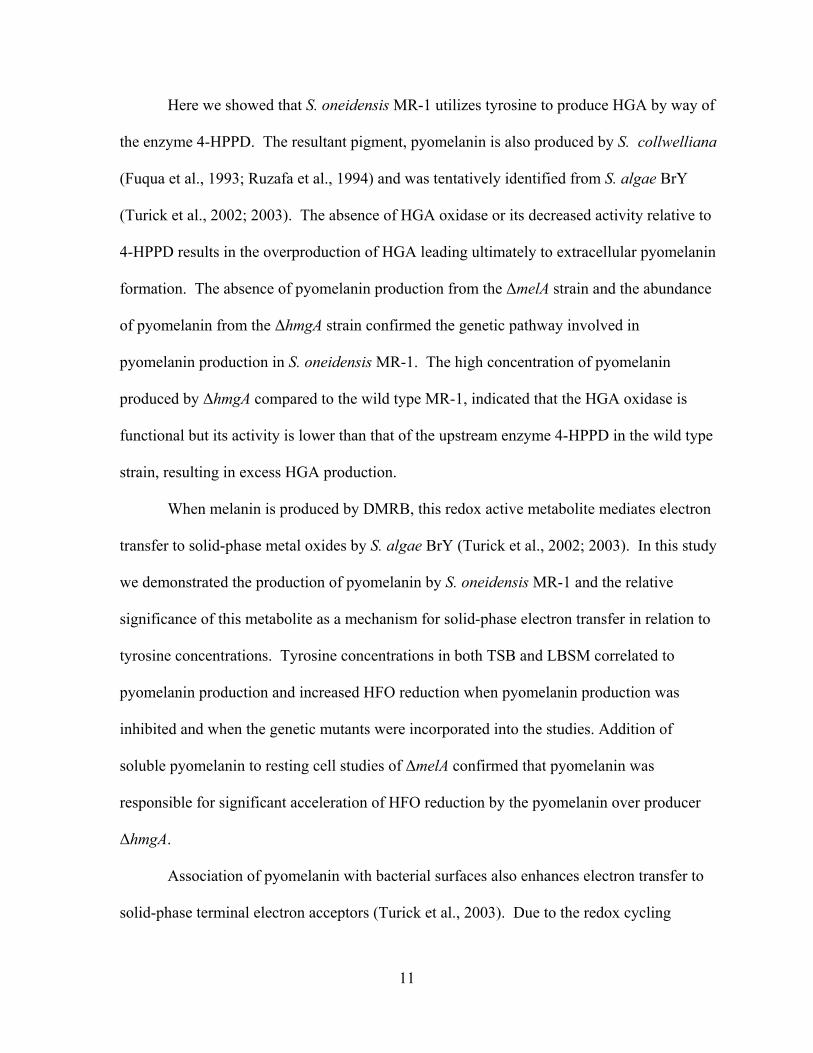
Here we showed that S. oneidensis MR-1 utilizes tyrosine to produce HGA by way of
the enzyme 4-HPPD. The resultant pigment, pyomelanin is also produced by S. collwelliana
(Fuqua et al., 1993; Ruzafa et al., 1994) and was tentatively identified from S. algae BrY
(Turick et al., 2002; 2003). The absence of HGA oxidase or its decreased activity relative to
4-HPPD results in the overproduction of HGA leading ultimately to extracellular pyomelanin
formation. The absence of pyomelanin production from the ∆melA strain and the abundance
of pyomelanin from the ∆hmgA strain confirmed the genetic pathway involved in
pyomelanin production in S. oneidensis MR-1. The high concentration of pyomelanin
produced by ∆hmgA compared to the wild type MR-1, indicated that the HGA oxidase is
functional but its activity is lower than that of the upstream enzyme 4-HPPD in the wild type
strain, resulting in excess HGA production.
When melanin is produced by DMRB, this redox active metabolite mediates electron
transfer to solid-phase metal oxides by S. algae BrY (Turick et al., 2002; 2003). In this study
we demonstrated the production of pyomelanin by S. oneidensis MR-1 and the relative
significance of this metabolite as a mechanism for solid-phase electron transfer in relation to
tyrosine concentrations. Tyrosine concentrations in both TSB and LBSM correlated to
pyomelanin production and increased HFO reduction when pyomelanin production was
inhibited and when the genetic mutants were incorporated into the studies. Addition of
soluble pyomelanin to resting cell studies of ∆melA confirmed that pyomelanin was
responsible for significant acceleration of HFO reduction by the pyomelanin over producer
∆hmgA.
Association of pyomelanin with bacterial surfaces also enhances electron transfer to
solid-phase terminal electron acceptors (Turick et al., 2003). Due to the redox cycling

12
behavior of pyomelanin, its close association with the bacterial surface presents an efficient
process that permits a low concentration of pyomelanin to be used repeatedly for electron
transfer. Our electrochemical studies were developed to determine the degree of enhanced
electron transfer conferred to the cell surface by pyomelanin. This technique has several
advantages over conventional wet chemistry techniques for determining electron transfer
kinetics. We were able to determine specific surface associated redox couples through CV
and demonstrate the contribution pyomelanin made to enhanced electron transfer at the cell
surface.
Pure cytochromes of the DMRB Goebacter sulferreducens demonstrate
electrochemical activity (Magnuson et al., 2001). In addition, cell suspensions of various
strains of S. oneidensis exhibit different electrochemical responses with CV as a result of
mutations in electron transfer capacity (Kim et al., 2002). Here, we demonstrated that
pyomelanin production also affects solid-phase electron transfer behavior of S. oneidensis.
The increase in E°΄ of ∆melA grown in the presence of pyomelanin (estimated to be the same
concentration of pyomelanin produced by S. oneidensis MR-1) essentially restored the E°΄ of
this strain to that of S. oneidensis MR-1 and the more negative redox couple of ∆hmgA. This
provided strong evidence that existing electron transfer mechanisms are enhanced through
surface complexation of pyomelanin.
A second redox couple at about +500 mV vs. Ag(RE), barely detectable in the CV
of S. oneidensis MR-1 was obvious from the CV of the pyomelanin over producer
∆hmgA and can be attributed to pyomelanin. This is based on the CV of concentrated
pyomelanin and the absence of such a redox couple from ∆melA. The difference in redox
couples between the pyomelanin over producer and pyomelanin alone may be a result of

13
pyomelanin integration onto the bacterial membrane. Peak-to-peak separation of
oxidation and reduction waves in Figs. 4D and 4E allowed for the calculation of apparent
rate constants via Laviron’s theory (Laviron, 1983; Finklea, 2001). Apparent rate
constant (ko) values obtained were 4.0 x 10⁻⁶ s-1 for ∆hmgA and 2.1 x 10⁻⁴ s-1 for
pyomelanin in carbon paste. The 50 times greater ko value obtained for concentrated
pyomelanin may be indicative of the degree of hydration or how pyomelanin is
incorporated into the cell surface, especially in relation to the potential insulating
properties of exopolysaccharides (Finkenstadt, 2005).
A key nutritional requirement for pyomelanin production is tyrosine and/or
phenylalanine, where phenylalanine is converted to tyrosine in this pathway (Lehninger,
1975). A portion of this present study focused on the complex medium TSB and the defined
LBSM (with equivalent tyrosine concentrations to TSB) in order to delineate the role of
tyrosine in both pyomelanin production and enhanced electron transfer. Enzyme inhibition
of pyomelanin production as well as directed mutagenesis studies targeted 4-HPPD. Both
approaches were successful in preventing pyomelanin production. The presence of tyrosine
as a component of growth media from previous studies with S. oneidensis MR-1 likely
contributed to pyomelanin production and hence influenced metal reduction rates. Increased
pyomelanin production from TSB compared to LBSM with supplemental tyrosine may be a
result of phenylalanine also present in TSB.
Environmental concentrations of tyrosine or phenylalanine are expected to promote
pyomelanin production and thus contribute to increased biogeochemical cycling of metals.
The presence of tyrosine and phenylalanine in soils varies and may play a useful role in
predicting biogeochemical activity in the subsurface. For instance, tyrosine and

14
phenylalanine concentrations have been shown to range from 10 – 632 mg/kg in various soils
(Martens and Loeffelmann, 2003: McLain and Martens, 2005). Since pyomelanin production
is linearly related to tyrosine concentration and minute quantities of pyomelanin are needed
per cell to increase electron transfer rates (Turick et al., 2002), tyrosine and phenylalanine
concentrations reported from environmental studies would be sufficient to produce enough
pyomelanin to accelerate metal reduction rates in the subsurface. Our evidence for
pyomelanin production from prolonged incubation suggests that lysed cells in high
concentration may provide enough amino acid precursors for pyomelanin production. The
low concentration of pyomelanin associated with S. oneidensis MR-1 from prolonged growth
in LBSM was enough to increase HFO reduction by about 10% and corroborates previous
reports (Turick et al., 2002). This scenario is likely in the environment, especially in
biofilms and could also contribute to accelerated electron transfer.
15
Experimental Procedures
Growth conditions. Cultures were maintained on tryptic soy agar throughout the study.
Tryptic soy broth (TSB) and a lactate (70 mM) basal salts medium (LBSM) (Turick et al.,
2002) with or without 300 mg/l of tyrosine were used for pyomelanin studies. Tyrosine
concentrations of LBSM were based on that of TSB (McCuen, 1988). Growth conditions
were at 25°C, 25 ml, and shaken (100 rpm) in an aerobic environment for 48- 72 h, unless
specified otherwise. Specific growth media were inoculated with 24 h cultures grown in the
same medium. To determine if pyomelanin production occurred without supplemental
tyrosine, cultures were grown in LBSM for 48 and 240h under the same conditions above.
For electrochemical studies cultures were grown in TSB (as above) for 72h. To determine
the effect of surface associated pyomelanin on electron transfer, sterile pyomelanin (0.1
mg/ml) was added to some cultures of the pyomelanin deficient strain (∆melA) after 24h of
growth.
Pigment characterization. Cell-free spent growth medium (LBSM with 300 mg/l tyrosine)
of S. oneidensis MR-1 were assayed for melanin precursors DOPA and/or HGA by high-
pressure capillary electrophoresis (HPCE) with a Celect H150 C-8 bonded phase capillary
column. Standards (DOPA and HGA) were dissolved in 4 mM ascorbate, (to prevent
oxidation of precursors to melanin pigments) to a final concentration of 4 mM each. HGA
and DOPA were also determined by colorimetric methods. DOPA analysis consisted of the
DOPA nitration method (Waite and Benedict, 1984). HGA content was determined based on
its reaction with cysteine to form 1, 4-thiazine, according to the methods of Fellman et al.
(1972). To determine if laccase activity contributed to melanin formation cultures were

16
grown with 10 and 25 g/l of glucose in BSM with 300 mg/l tyrosine because laccase is
inhibited above 20 g/l (Frases et al., 2007).
Characterization of spent cell-free media to determine humic and melanin type
properties was performed as previously described (Ellis and Griffiths, 1974; Turick et al.,
2002). Soluble pyomelanin was quantified spectrophotometrically (OD400) as previously
described (Turick et al, 2003). This technique incorporated known concentrations of
concentrated bacterial pyomelanin and pure pyomelanin produced by the autooxidation of
HGA that served as quantitative standards.
Cell associated pyomelanin was quantified initially as previously described (Turick et
al., 2003) incorporating the nitro blue tetrazolium assay (Paz et al., 1991). Results from this
assay compared well to a spectroscopic analysis (OD400) of cell suspensions, which was used
throughout the study. The methods included a known cell density of pyomelanin producing
cells blanked against an equal cell density of the pyomelanin deficient strain (∆melA).
Pyomelanin was quantified as described above and calculated per cell. Cell density was
determined with acridine orange staining and an epifluorescence microscope (Hobbie et al.,
1975)
Inhibition of pyomelanin production. Sulcotrione [2-(2- chloro- 4- methane
sulfonylbenzoyl)-1, 3-cyclohexanedione)] (Zeneca Ag. Products, Richmond, CA) is a potent
inhibitor of 4-HPPD (Schulz et al., 1993; Secor, 1988; Lee et al., 1997). S. oneidensis MR-1
was grown for 48 h in tyrosine-supplemented LBSM or TSB with 18 µM sulcotrione to test
its effects on melanin production. Sulcotrione was added continuously with a syringe pump
to maintain an 18 µM concentration. Melanin production was completely inhibited with 18

17
µM sulcotrione. Cell density and growth were not affected by the sulcotrione concentration
tested (data not shown).
Construction of melA and hmgA mutants of S. oneidensis MR-1. In-frame deletion
mutagenesis was performed using the method originally described by Link et al. (1997) with
minor modifications. Upstream and downstream fragments flanking the target locus were
PCR amplified using S. oneidensis MR-1 genomic DNA and fused via overlap extension
PCR (Ho et al., 1989). The fusion PCR amplicon was ligated into XcmI digested pDS3.1
(Wan et al., 2004). The resulting recombinant plasmids were used to transform E. coli ß-
2155 (Dehio and Meyer, 1997) or WM3063 (Saltikov et al., 2003) and subsequently
transferred to S. oneidensis MR-1 by conjugation. The primary integrants were selected by
plating on LB medium containing 7.5 µg/ml gentamycin. Selection for a second homologous
recombination to remove the plasmid sequence was accomplished by sucrose counter-
selection (Blomfield et al., 1991). Sucrose-resistant colonies were screened for sensitivity to
gentamycin and then screened for deletion of the gene of interest using PCR. The resulting
PCR amplicon was then used as the template for DNA sequencing of the deleted and
flanking regions involved in the recombination events (ACGT, Inc. Wheeling, IL).
Quantification of electron transfer. The Fe(III) oxide, hydrous ferric oxide (HFO) was
used in resting cell studies in carbonate buffer as previously described (Turick et al., 2003) in
order to quantify Fe(III) reduction by measuring Fe(II) evolution over time. An autoclaved
pyomelanin solution (1mg/ml final concentration) was spiked into selected suspensions of
∆melA in order to determine its effect on HFO reduction. Cell densities in these studies were

18
normalized to108 cells/ml as determined with acridine orange staining and an epifluorescence
microscope (Hobbie et al., 1975). Fe(II) was measured spectrophotometrically using the
ferrozine assay as described elsewhere (Turick et al., 2003).
In order to more precisely characterize the role of pyomelanin as a surface associated
electron shuttle, electrochemical studies were designed for whole bacterial cells. Cyclic
voltammetry comprises electrochemical techniques that provide information of the
electrochemical activities of materials by sweeping the potential back and forth at
predetermined rates. Potential sweeps in the positive direction result in oxidation peaks and
reduction peaks are evident when the potential is swept in the negative direction. Our studies
incorporated concentrated suspensions (10 x) of whole bacterial cells under anaerobic
conditions. Operational conditions included washed (3x) cell suspensions of cultures grown
aerobically in TSB (above). Cultures were resuspended in phosphate buffered saline (PBS)
for electrochemical studies. Cell suspensions were made anaerobic by a continuous nitrogen
purge for 15 min. prior to electrochemical analyses. Electrochemistry studies included a
Ag/AgCl reference electrode, a Pt counter electrode and carbon paste working electrode
(Bioanalytical Systems), all immersed into the anaerobic cell suspension that was blanketed
with nitrogen gas throughout the studies. Potential sweep originated in the positive direction
at a rate of 850 mV/sec for all CV in this study, using a model 100B/A potentiostat
(Bioanalytical Systems). A total of six sweeps were performed with each culture. Duplicate
CV of each assay were averaged and the 5th and 6th averaged sweeps were reported here. For
each set of experiments with each culture, working electrodes were rinsed in deionized water
followed by a methanol rinse and then sonicated for 10 minutes in deionized water. The
cleaned electrodes were then air dried prior to the next study. Each study included a CV of

19
the washed bare electrode in PBS that served as a no-cell control for the next series of
voltammetry studies for each strain analyzed. In addition, cell free liquid from each cell
suspension was analyzed (as above) for electrochemical activity in the bulk phase.
Apparent rate constant values (ko values) were derived for a 1-electron transfer
mechanism, by simulating the CV with the software provided for the Model 660a
Electrochemical Workstation by CH Instruments (Austin, TX).

20
Acknowledgements
This research was supported in part through funding by the Department of Energy
NABIR program, the Savannah River National Laboratory Independent Research and
Development Program, and Soil and Groundwater Closure Projects of the Savannah River
Site. The authors wish to thank Thomas H. Cromartie of Zenica Agrichemicals for
furnishing the sulcotrione.

21
Table 1. Production of pyomelanin by wild-type and mutant strains of Shewanella oneidensis MR-1. 1 2 3 Soluble pyomelanin (mg/ml) Cell-associated pyomelanin (fg/cell) 4 ________________________________________________ _____________________________________ 5 Strain TSB LBSM + 300
mg/l Tyrosine
(48h)
LBSM (48h)
LBSM (240h) TSB LBSM + 300 mg/l Tyrosine
(48h)
LBSM no tyrosine (240h)
MR-1 0.427 (0.046) 0.270 (0.025) ND 0.032 (0.009) 66.32 (4.63) 36.50 (4.60) 4.14 (0.964) MR-1 + sulcotrione
ND1 ND ND NI2 NI NI NI
∆melA ND ND ND ND ND ND ND ∆hmgA 0.987 (0.167) 0.680 (0.074) ND 0.162 (0.003) 153.40 (16.87) 86.44 (16.70) 37.07 (3.214)
1. ND – not detected 6 2. NI – not included in assay 7 Standard deviation in parentheses 8

22
References 1
2
Blomfield, I. C., Vaughn, V., Rest, R.F., and Eisenstein, B. I. (1991) Allelic exchange in 3
Escherichia coli using the Bacillus subtilis sacB gene and a temperature-sensitive pSC101 4
replicon. Mol. Microbiol 5:1447-1457. 5
6
Coon, S.L., Kotob, S., Jarvis, B.B., Wang, S., Fuqua, W. C., and Weiner, R.M. (1994) 7
Homogentisic acid is the product of MelA, which mediated melanogenesis in the marine 8
bacterium Shewanella colwelliana D. Appl Environ Microbiol 60:3006-3010. 9
10
Dehio, C., and Meyer, M. (1997) Maintenance of broad-host-range incompatibility group P 11
and group Q plasmids and transposition of Tn5 in Bartonella henselae following conjugal 12
plasmid transfer from Escherichia coli. J Bacteriol 179:538-540. 13
14
David, C., Daro, A., Szalai, E., Atarhouch, T., and Mergeay, M. (1996) Formation of 15
polymeric pigments in the presence of bacteria and comparison with chemical oxidative 16
coupling-II. Catabolism of tyrosine and hydroxyphenylacetic acid by Alcaligenes 17
eutrophus CH34 and mutants. Eur Polym J 32:669-697. 18
19
Ellis, D.H., and Griffiths, D.A. (1974) The location and analysis of melanins in cell walls of 20
some soil fungi. Can J Microbiol 20:1379-1386. 21
22
23
Fellman, J.H., Fujita, T.S., and Roth, E.S. (1972) Assay, properties and tissue distribution of 1
p-hydroxyphenylpyruvate hydroxylase. Biochim Biophys Acta 284:90-100. 2
3
Finkenstadt, V.L. (2005) Natural polysaccharides as electroactive polymers. Appl Microbiol 4
Biotechnol 67:735-745. 5
6
Finklea, H. O. (2001) Theory of Coupled Electron-Proton Transfer with Potential-Dependent 7
Transfer Coefficients for Redox Couples Attached to Electrodes. J Phys Chem B 105: 8
8685. 9
Frases, S., Salazar, A., Dadachova, E., and Casadevall, A. (2007) Cryptococcus neoformans 10
can utilize the bacterial melanin precursor homogentisic acid for fungal melanogenesis. 11
Appl Environ Microbiol 73:615-621. 12
13
Fuqua, W.C., and Weiner, R.M. (1993) The melA gene is essential for melanin production 14
in the marine bacterium Shewanella colwelliana. J Gen Microbiol 139:1105-1114. 15
16
Gorby, Y.A., Yanina, S., McLean, J. S., Rosso, K. M., Moyles, D., Dohnalkova, A., et 17
al., (2006) Electrically conductive bacterial nanowires produced by Shewanella 18
oneidensis strain MR-1 and other microorganisms. Proc Nat Acad Sci 103:11358-19
11363. 20
21
Ho, S. N., Hunt, H. D., Horton, R. M., Pullen, J. K., and Pease, L. R. (1989) Site-directed 22
mutagenesis by overlap extension using the polymerase chain reaction. Gene 77:51-59. 23

24
1
Hobbie, J.E., Daley, R.J., and Jasper, S. (1977) Use of Nucleopore filters for counting 2
bacteria by fluorescence microscopy. Appl Environ Microbiol 33:1225 –1228. 3
4
Kim, H.J., Park, H.S., Hyun, M.S., Chang, I.S., Kim, M., and Kim, B.H. (2002) A mediator-5
less microbial fuel cell using a metal reducing bacterium, Shewanella putrefaciens. 6
Enzyme Microbial Technol 30:145-152. 7
8
Kotob, S.I., Coon, S.L., Quintero, E.J., and Weiner, R.M. (1995) Homogentisic acid is the 9
primary precursor of melanin synthesis in Vibrio cholera, a Hyphomonas strain, and 10
Shewanella colwelliana. Appl Environ Microbiol 61:1620-1622. 11
12
Laviron, E. (1983) Electrochemical reactions with protonations at equilibrium: Part VIII. The 13
2 e, 2H+ reaction (nine-member square scheme) for a surface or for a heterogeneous 14
reaction in the absence of disproportionation and dimerization reactions. J Electroanal 15
Chem 146: 15-36. 16
17
Lee, D.L., Prisbylla, M.P., Cromartie, T.H., Dargin, D.P., Howard, S.W., Provan, W.M. et 18
al.,(1997) The discovery and structural requirements of inhibitors of p-19
hydroxyphenylpyruvate dioxygenase. Weed Sci. 45:601-609. 20
21
Lehninger, A.L. (1975) Oxidative degradation of amino acids. In: Biochemistry. pp.568-570. 22
Worth Publishers. N.Y. NY, 23

25
1
Link, A., Phillips, J., D., and Church, G. M. (1997) Methods for generating precise deletions 2
and insertions in the genome of wild-type Escherichia coli: application to open reading 3
frame characterization. J Bacteriol 179:6228-6237. 4
5
Lloyd, J. R. (2003) Microbial reduction of metals and radionuclides. FEMS Microbiol Rev 6
27:411-425. 7
8
Lovley, D.R., Coates, J.D., Blunt-Harris, E.L., Phillips, E.J.P, and Woodward, J.C. (1996) 9
Humic substances as electron acceptors for microbial respiration. Nature 382:445-448. 10
11
Lovley, D.R., and Phillips, E.J.P. (1988) Novel mode of microbial energy 12
metabolism: organic carbon oxidation coupled to dissimilatory reduction of iron or 13
manganese. Appl Environ Microbiol 54:1472-1480. 14
15
Martens, D.A., and Loeffelmann, K.L. (2003) Soil amino acid compostion quantified by acid 16
hydrolysis and anion chromatography-pulsed amperometry. J Agric Food Chem 51:6521-17
6529. 18
19
Magnuson, T.S., Isoyama, N., Hodges-Myerson, A.L., Davidson, G., Maroney, M.J. , 20
Geesey, G.G., and Lovley, D.R. (2001) Isolation, characterization and gene sequence 21
analysis of a membrane associated 89 kDa Fe(III) reducing cytochrome c from Geobacter 22
sulfurreducens. Biochem J 359:147-152. 23

26
1
McCuen, P.J. (1988) Culture media components In: Manual of BBL Products and 2
Laboratory Procedures. Power, D.A. (ed). Beckton Dickinson Systems, Cockeysville, 3
MD. pp. 293-294. 4
5
McLain, J.E.T., and Martens, D. A. (2005) Nitrous oxide flux from soil amino acid 6
mineralization. Soil Biol Biochem 37: 289-299 7
8
Paz, M.A., Fluckiger, R.A. , Boak, A., Kagan, H.M., and Gallop, P.M. (1991) Specific 9
detection of quinoproteins by redox-cycling staining. J Biol Chem 266:689-692. 10
11
Prota, G. (1992) Natural and synthetic melanins. In, Melanins and Melanogenesis. G. Prota 12
(ed). San Diego, CA, USA. Academic Press, Inc. pp. 63-87. 13
14
Ruzafa, C., Solano, F., and Sanchez-Amat, A. (1994) The protein encoded by the Shewanella 15
colwelliana melA gene is p-hydroxyphenylpyruvate dioxygenase. FEMS Microbiol Lett 16
124:179-184. 17
18
Ruzafa, C., Sanchez-Amat, A., and Solano, F. (1995) Characterization of the melanogenic 19
system in Vibrio cholerae ATCC 14035. Pigment Cell Research 8:147-152. 20
21
Saltikov, C. W., Cifuentes, A., Venkateswaran, K., and Newman, D. K. (2003) The ars 22
detoxification system is advantageous but not required for As(V) respiration by the 23
27
genetically tractable Shewanella species strain ANA-3. Appl Environ Microbiol 69:2800-1
2809. 2
3
Sanchez-Amat, A., Ruzafa, C., and Solano, F. (1998) Comparative tyrosine degradation in 4
Vibrio cholerae strains. The strain ATCC 14035 as a prokaryotic melanogenic model of 5
homogentisate-releasing cell. Comp Biochem Physiol Part B 119:557-562. 6
7
Secor, J. (1994) Inhibition of barnygrass 4-hydroxyphenylpyruvate dioxygenase by 8
sulcotrione. Plant Physiol 106:1429-1433. 9
10
Schulz, A., Ort, O. Beyer, P., and Kleninig, H. (1993) SC-0051, a 2-benzoyl-cyclohexane-11
1,3-dione bleaching herbicide, is a potent inhibitor of the enzyme p-12
hydroxyphenylpyruvate dioxygenase. FEBS Lett 318:162-166. 13
14
Schwertmann, U., and Taylor, R.M. (1977) Iron oxides. In: Minerals in Soil Environments. 15
Dixon, J.B and Weed, S.B. (eds). Madison WI: Soil Science Society of America, pp 145-16
180. 17
18
Scott, D.T., McKnight, D.M., Blunt-Harris, E. L., Kolesar, S.E., and Lovley, D.R. (1998) 19
Quinone moieties act as electron acceptors in the reduction of humic substances by 20
humics-reducing microorganisms. Envion Sci Technol 32:2984-2989. 21
22

28
Turick, C.E., Tisa, L.S., and Caccavo Jr, F. (2002) Melanin production and use as a soluble 1
electron shuttle for Fe(III) oxide reduction and as a terminal electron acceptor by 2
Shewanella algae BrY. Appl Environ Microbiol 68:2436-2444. 3
4
Turick, C.E., Cacavo, F. Jr., and Tisa, L.S. (2003) Electron transfer from Shewanella algae 5
BrY to hydrous ferric oxide is mediated by cell-associated melanin. FEMS Microbiol Lett 6
220:99-104. 7
8
Waite, J.H. and Benedict, C.V. (1984) Assay of dihydroxyphenylalanine (Dopa) in 9
invertebrate structural proteins. Meth Enzymol 107:397-413. 10
11
Wan, X. F., Verberkmoes, N. C. McCue, L. A. Stanek, D. Connelly, and H. Hauser, et al. 12
(2004) Transcriptomic and proteomic characterization of the Fur modulon in the metal-13
reducing bacterium Shewanella oneidensis. J Bacteriol 186:8385-8400. 14
15
Yabuuchi, E., and Omyama, A. 1972. Characterization of “pyomelanin”-producing strains of 16
Pseudomonas aeruginosa. Internat J Syst Bacteriol 22:53:64. 17
18

29
Figure Legends 1
2
Fig. 1. HPCE analysis for melanin precursors. After 18 h incubation of S. oneidensis MR-1, 3
spent culture supernatant liquids were analyzed as described in the experimental procedures 4
section. Panels: (A) 18 h S. oneidensis MR-1 (B) HGA and DOPA standards. 5
6
Fig. 2. Resting cell studies demonstrated inhibitory effects of sulcotrione on pyomelanin 7
production and HFO reduction by S. oneidensis MR-1. (A) Cells pregrown in LBSM (n), 8
LBSM and sulcotrione (∆), LBSM with tyrosine (g), and LBSM with tyrosine and 9
sulcotrione (); (B) Cells pregrown in tryptic soy broth with () and without (g) 10
sulcotrione. 11
12
Fig. 3. HFO reduction by 3 strains of S. oneidensis. Resting cell studies of cells pregrown in 13
(A) TSB and (B)LBSM + 300 mg/l tyrosine, demonstrated increased HFO reduction 14
efficiency by the pyomelanin over-producing strain (∆hmgA) (♦) relative to MR-1(■) and the 15
pyomelanin deficient mutant (∆melA) (▲), which demonstrated the least amount of HFO 16
reduction. Addition of pyomelanin (10µg/ml) to resting cell cultures of ∆melA (∆) pregrown 17
in LBSM demonstrated drastically increased levels of HFO reduction. 18
19
Fig. 4. Cyclic voltammetry of whole cell suspensions. Cultures grown in TSB for 72 hr, 20
washed and centrifuged 3 times in PBS then concentrated 10 fold. Voltamograms were 21
produced with a carbon paste working electrode, Pt counter electrode andAg/AgCl ref. 22
electrode. Sweep rate was 850 mV/sec. Redox activity of whole cell suspensions was a 23

30
function of pyomelanin content.(A) S. oneidensis MR-1; (B) Pyomelanin deficient strain 1
(∆melA); (C) Pyomelanin deficient strain (∆melA) incubated with 10 µg/ml pyomelanin; (D) 2
Pyomelanin overproducing strain (∆hmga). Pyomelanin increased formal potential of redox 3
couples and increased current response from cells. (E) Electrochemical activity of dried 4
pyomelanin incorporated into a carbon paste electrode. 5
6
Fig. 5. HFO reduction by 3 strains of S. oneidensis after prolonged incubation (10 days) in 7
LBSM without exogenous tyrosine. Resting cell studies of cells pregrown in LBSM but 8
without tyrosine, demonstrated increased HFO reduction efficiency by the pyomelanin over-9
producing strain (∆hmga) (♦) relative to MR-1(■) and the pyomelanin deficient mutant 10
(∆melA) (▲), which demonstrated the least amount of HFO reduction. 11

31
0
2.5
5
2.2 2.4 2.6 2.8 3Time (min)
uMA
A
1
0
40
80
2.2 2.4 2.6 2.8 3Time (min.)
mA
U
DOPA
HGA
B
2 3 4 5 Figure 1 6 7 8 9 10 11 12 13 14 15 16 17

32
1
0
0.5
1
1.5
2
0 50 100 150Time (h)
Fe(II
) (m
M)
2 3
0
0.05
0.1
0.15
0.2
0 50 100 150Time (h)
Fe(II
) (m
M)
A
B

33
Figure 2. 1 2
0
1
2
3
0 50 100 150Time (h)
Fe(II
) (m
M)
3
A

34
0
0.2
0.4
0.6
0 10 20 30 40Time (h)
Fe(II
) (m
M)
1 Figure 3. 2
B

35
-1000 -800 -600 -400 -200 0 200 400 600 800 1000
Potential (mV)
20 µA
1 2 3
Figure 4. 4
-423 mV
-547 mV
E°′= -423 mV A

36
1
-1000 -800 -600 -400 -200 0 200 400 600 800 1000
Potential (mV)
20 µA
2 Figure 4. 3
-490 mV
-643 mV
E°′= -566 mV B

37
-1000 -800 -600 -400 -200 0 200 400 600 800 1000Potential (mV)
20 µA
1 Figure 4. 2
-418 mV
-529 mV
E°′= -473 mV C

38
-1.00E-04
-8.00E-05
-6.00E-05
-4.00E-05
-2.00E-05
0.00E+00
2.00E-05
4.00E-05
6.00E-05
8.00E-05
-1000 -800 -600 -400 -200 0 200 400 600 800 1000
Potential [mV]
20 µA
1 2 Figure 4. 3
-406 mV
-543 mV
E°′= -474 mV
-339 mV
567 mV
E°′= 114 mV
D

39
-1000 -800 -600 -400 -200 0 200 400 600 800 1000
Potential (mV)
50 µA
1 2 3 Figure 4. 4 5 6 7 8 9 10 11 12 13 14 15
515 mV
17 mV
E°′= 266 mV
E

40
0
0.005
0.01
0.015
0.02
0 10 20 30 40Time (h)
Fe(II
) mM
1 Figure 5. 2 3