From Classical to Tight Binding and First Principles Methods
Upload
independentCategory
view
4download
0
Articles
Solid-Phase Library Synthesis, Screening, and Selection of Tight-BindingReduced Peptide Bond Inhibitors of a Recombinant Leishmania mexicanaCysteine Protease B
Phaedria M. St. Hilaire,*,† Lira C. Alves,‡ Fatima Herrera,§ Manat Renil,† Sanya J. Sanderson,|Jeremy C. Mottram,⊥ Graham H. Coombs,| Maria A. Juliano,‡ Luiz Juliano,‡ Jorge Arevalo,§ andMorten Meldal*,†
Department of Chemistry, Carlsberg Laboratory, Gamle Carlsberg Vej 10, DK-2500 Valby, Denmark, Department of Biophysics,Escola Paulista de Medicina, Rua Tres de Maio 100, 04044-20 Sao Paulo, Brazil, Molecular and Cellular Laboratory ofTrypanosomatids, Universidad Peruana Cayetano Heridia, Av. Honorio Delgado 430, URB Ingenieria, San Martin de Porras,Lima, Peru, Division of Infection and Immunity, Joseph Black Building, University of Glasgow, Glasgow G12 8QQ,Scotland, U.K., and Wellcome Centre for Molecular Parasitology, Anderson College, University of Glasgow,Glasgow G11 6NU, Scotland, U.K.
Received November 2, 2001
A one-bead-two-compound inhibitor library was synthesized by the split-mix method for theidentification of inhibitors of a recombinant cysteine protease from Leishmania mexicana,CPB2.8∆CTE. The inhibitor library was composed of octapeptides with a centrally locatedreduced bond introduced by reductive amination of the resin-bound amines with Fmoc aminoaldehydes. The library was screened on solid phase, and less than 1% of the library containedactive compounds. The inhibitors displayed great specificity in the subsites flanking the enzymecatalytic triad with Cha and Ile/Leu preferred in P2, Phe in P1, Cha and Ile/Leu in P1′, andIle/Leu in P2′. Some of the inhibitors were resynthesized, and the kinetics of inhibition weredetermined in solution-phase assays. Most of the inhibitors had micromolar Ki values, and afew inhibited the enzyme at nanomolar concentrations. One inhibitor, DKHF(CH2NH)LLVK(Ki ) 1 µM), was tested for antiparasite efficacy and shown to affect parasite survival with anIC50 of approximately 50 µΜ.
Introduction
The parasite Leishmania mexicana, a causative agentof cutaneous leishmaniasis, expresses in a stage-regulated manner high levels of several classes ofcysteine proteases (CPs) belonging to the papain family.In Leishmania mexicana, one group of cysteine pro-teases, CPBs (denoting cysteine protease group B),1 isexpressed from a single tandem array comprising 19gene copies (CPB1-CPB19), with individual genesencoding subtly different isoenzymes.2 CPBs are thoughtto be crucial for the survival and infectivity of theparasite in its human host and have been implicatedin a number of processes including the successfulinvasion of host macrophages by promastigotes, thetransformation of one parasitic form to another, parasitenutrition, and evasion of the host’s immune system.3-6
Inhibitors of the CPB isoenzymes have been shown toreduce the infectivity of wild-type parasites both in vitrotoward macrophages6,7 and in vivo toward BALB/c
mice,8 thus providing further evidence that these CPBisoenzymes are virulence factors.
Because of their tremendous importance in the etiol-ogy of leishmaniasis, the parasite’s CPs are attractivetargets for therapeutic agents against leishmaniasis.Traditionally, this disease has been treated using threemajor drug classes: pentavalent antimonials (e.g.,sodium stibogluconate, which inhibits the protozoalenzymes required for glycolytic and fatty acid oxidation),aromatic diamidines (e.g., pentamidine, which interfereswith nuclear metabolism), and polyene microlide anti-biotics (e.g., amphotericin B, which acts on cell mem-brane sterols and phospholipids). These chemothera-peutic modalities are generally unsatisfactory becauseof high toxicity9 and the development of drug-resistantparasites.10,11
While several strategies can be invoked for the designand selection of protease inhibitors, the development ofcysteine protease inhibitors as drug candidates isgreatly facilitated by a combinatorial library approach.Consequently, the one-bead-two-compound fluores-cence inhibitor assay for the rapid selection and iden-tification of protease inhibitors was first reported fromour laboratory.12 This assay has been used for thesuccessful identification of inhibitors of serine pro-teases,12 cysteine proteases,13,14 and matrix metallopro-teases.15 In this report, we describe the synthesis and
* To whom correspondence should be addressed. Phone: +45 33 275270/5301. Fax: +45 33 27 4708. E-mail for P.M.S.: [email protected] for M.M.: [email protected].
† Carlsberg Laboratory.‡ Escola Paulista de Medicina.§ Universidad Peruana Cayetano Heridia.| Division of Infection and Immunity, University of Glasgow.⊥ Wellcome Centre for Molecular Parasitology, University of
Glasgow.
1971J. Med. Chem. 2002, 45, 1971-1982
10.1021/jm0110901 CCC: $22.00 © 2002 American Chemical SocietyPublished on Web 04/03/2002

screening of a library of potential cysteine proteaseinhibitors comprising octapeptides with the simplestmodification of the scissile amide bond, the reducedpeptide bond, at the putative P1-P1′ bond. Reducedbond peptides are, for the most part, ideal as asparticprotease inhibitors16-20 because the proteolytically stablesecondary amine does not introduce any major struc-tural changes in the peptide, and the resulting peptidestend to bind linearly in the active site such that thetetrahedral intermediate of a good substrate is mim-icked. However, this class of compounds has not beenutilized as reversible cysteine and serine proteaseinhibitors, in contrast to aldehydes and ketones (see ref35 for a review of various classes of protease inhibitors).It is hoped that for cysteine proteases, reduced bondpeptides could be inhibitory if the side chains of theamino acids flanking the nonhydrolyzable secondaryamine encouraged tight binding of the inhibitor to theenzyme subsites, thus sterically preventing any sub-strate access to the reactive thiol. Furthermore, theincreased flexibility of the secondary amine comparedto the amide bond may be advantageous when aninduced fit is required for tight binding. We presentherein the results from the solid-phase screening of a“one-bead-two-compound” reduced bond octapeptideinhibitor library with CPB2.8∆CTE,21 an isoform of CPBcysteine protease from Leishmania mexicana lackingthe C-terminal domain.Results and Discussion
Synthesis of Fmoc Amino Aldehydes. Fmoc aminoaldehydes were synthesized by a modification of themethod of Fehrentz and Castro22 wherein the acid wasfirst converted to the Weinreb amide by reaction withO,N-dimethylhydroxylamine and subsequently reducedto generate the aldehyde (Scheme 1). In our case, weelected to synthesize the amide intermediate from thepentafluorophenyl ester, thereby circumventing thepotential problems and reduced yields associated withthe separation of the product from the byproducts of thecarbodiimide, uronium, or phosphonium salts typicallyused for activation of the acid. The yields for theformation of the amide were 95% for Lys, quantitativefor Phe, and somewhat lower for Arg (75%). The amidewas then reduced by treatment with LiAlH4 at -78 °Caccording to literature procedure.23 The yield for thereduction of the Phe analogue was 90%, whereas theyields for Arg and Lys were 69% and 58%, respectively.The reduced yield was due to unreacted starting mate-rial and loss of side chain protecting groups.
Optimization of Library Reductive AminationConditions. (Experimental details are in SupportingInformation). Reductive alkylation of resin-bound aminesusing Fmoc- or Boc-protected amino aldehydes has beensuccessfully carried out on solid phase by several groupssince the seminal work of Coy and co-workers.24,25
Typically, a single aldehyde is reacted to a single aminenucleophile and libraries of reduced-bond-containingcompounds are synthesized through parallel arrays ofcompounds that may be cleaved such that mixtures ofcompounds result, for example, in the synthesis of linearand cyclic peptides libraries of enkephalin surrogates.26
In the case of “true” libraries synthesized by the split-mix method,27,28 for the introduction of the reducedbond, each Fmoc amino aldehyde is reacted simulta-neously, in separate vessels or compartments, withseveral thousand different amine nucleophiles that areeach attached to a resin particle (see ref 29 for an earlyexample). While solid-phase synthesis has the advan-tage of driving a reaction to completion, in the case ofreductive alkylation, a balance must be sought betweenreaction completeness and overalkylation. It was there-fore decided to first optimize the conditions for reductiveamination before synthesizing the library. For theoptimization process, three different minilibraries ofresin-bound amines were initially synthesized: (Ala,Arg, Leu, Gln, Cys, Pro, His)-IMP-Pll-K(Alloc)-PEGAresin; (Asn, Met, Val, Lys, Ser, Trp)-IMP-Pll-K(Alloc)-PEGA resin, and (Thr, Ile, Asp, Glu, Phe, Gly, Tyr)-IMP-Pll-K(Alloc)-PEGA resin. Reductive alkylation of theseminilibraries with Fmoc-protected Phe, Arg, and Lysaldehydes was conducted under different conditionswhere the number of equivalents of both the aldehydeand the borohydride, the reaction time, temperature,and solvent were varied. Reaction completeness wasassessed by the Kaiser test and by MALDI-TOF massspectrometry. Mass spectrometry proved to be a usefultool in expediting the assessment of reaction complete-ness by comparison of the peak heights of the startingmaterial, products, and byproducts. Since both thereactant and the product have identical peptide spacers,their ionization and flight properties are similar. There-fore, the relative product-to-reactant signal intensitiesrepresent a pseudoquantitative measure of reactionprogress. In obtaining pseudoquantitative measure-ments from MALDI-TOF MS spectra, signal intensitiesare thought to be as accurate as peak areas for gaugingthe extent of reaction,30 and these were used in theassessment of reaction completeness. However, as ad-ditional proof, we compared the determination of reac-tion completeness using both MS and HPLC of thecleaved products (data not shown). The results weresimilar for both methods. Analysis of the three minili-braries indicated that some amino acids, particularly,Val, Ile, Cys and Pro, were difficult to reductivelyalkylate completely. There was also evidence for thedouble alkylation of Gly. The conditions that eventuallygave the most satisfactory results were the use of 7.0equiv of the Fmoc aldehyde reacting first for 45 min at50 °C and followed by the addition of 10.5 equiv ofNaCNBH3 for 150 min at 50 °C. In our hands, the bestsolvent for the reaction was a mixture of DMF/MeOH/TEOF (1:1:1) containing 1% acetic acid, since it waseffective in properly solvating the peptide and in remov-
Scheme 1a
a (i) HN(OCH3)CH3‚HCl, DhbtOH, DIPEA, CH2Cl2; (ii) LiAlH4,THF, -78 °C.
1972 Journal of Medicinal Chemistry, 2002, Vol. 45, No. 10 St. Hilaire et al.

ing the water byproduct. The extent of diastereomerformation in the library was not investigated; however,on the basis of the results of the resynthesis of hits fromthe library, it is highly likely that it did occur to varying
degrees. Finally, we were concerned that the Allocprotecting group of Lys could be reduced during thereductive amination step in light of evidence thatcyclopentenone is reduced to cyclopentanol upon treat-ment with NaBH3CN at pH 3.31 In our case, the Allocgroup could be quantitatively removed after the reduc-tive amination step.
Inhibitor Library Synthesis. The selection of in-hibitors by the enzyme from a library was monitoredby the implementation of the one-bead-two-compoundassay developed in our laboratory (Figure 1).12 In thisassay, each bead serves as a microreactor wherein aresin-bound substrate and inhibitor compete for bindingto the enzyme. Such an assay requires the introductionof two orthogonally protected functional groups on eachbead. Several approaches to the synthesis of theseorthogonally protected bifunctional arms have beenexplored.12-15 In the present case, a simple approachwherein the library is synthesized on the N-terminusof Lys while the presynthesized protected substrate isattached to the amino group on the side chain of Lysafter completion of the library synthesis has beenchosen. The synthesis of the one-bead-two-compoundreduced bond inhibitor library is shown in Scheme 2.In this synthesis, 750 mg of PEGA4000 resin (300-800µm, ca. 130 000 beads) was derivatized with NR-Fmoc-Lys(Alloc)-OPfp, and after removal of the Fmoc group,a photolabile linker32 and an ionization mass spacer,
Figure 1. Principle of the one-bead-two-compound fluores-cence-quenched assay. If the enzyme is strongly inhibited, thenthe substrate is not cleaved and beads remain dark. If thesubstrate is hydrolyzed, the beads become fluorescent.
Scheme 2
Peptide Bond Inhibitors Journal of Medicinal Chemistry, 2002, Vol. 45, No. 10 1973

PPR(Pmc)PPR(Pmc), were introduced to facilitate rapidanalysis of active compounds from the library. An eight-residue peptide was synthesized because modelingstudies (vide infra) with fluorescent-quenched sub-strates for this enzyme indicate that eight residues areoptimal for filling the active site. Randomized positionsX4′-X1′ and X2-X4 were generated using the portionmixing method, and in situ capping was achieved bycoupling a mixture of Fmoc- and Boc-protected aminoacids (92:8) after TBTU/NEM preactivation.33 Nineteengenetically encoded amino acids were used in the libraryat each coupling step; Cys was replaced by Cha. Thereduced bond was formed on solid phase by reaction ofthe resin-bound amines with 7 equiv of the NR-Fmoc-protected amino aldehyde analogues of Arg, Lys, andPhe and 10.5 equiv of NaCNBH3 in DMF/TEOF/MeOH(1:1:1) for 3.25 h at 50 °C. After completion of theladdered inhibitor library, the Alloc group was removedby treatment with Pd(PPh3)4, and a protected fluores-cent quenched substrate for L. mexicana CPB2.8∆CTE,AcY(NO2)PR(Pmc)FR(Pmc)T(tBu)GS(tBu)K(Abz)G-OH (Km ) 1.13 µM; kcat/Km ) 1300 mM s-1 for the non-acetylated analogue),34 was coupled using TBTU/NEMactivation. After complete deprotection, some librarybeads were removed and irradiated with UV light, andthe structures attached were analyzed by MS. From themass spectra, it appeared that the synthesis hadproceeded well.
Library Screening. After deprotection of the aminoacid side chains, a portion of the library (200 mg) wasincubated with L. mexicana CPB2.8∆CTE. The fluores-cence intensity of the beads was monitored with afluorescent microscope every hour for indications ofhydrolysis. After 3 h, most of the beads showed afluorescent “ring” indicating hydrolysis of the substrate.The reaction mixture was incubated for 7 h, and afterthe beads were washed, the fluorescence intensity of thebeads was assessed by inspection with a fluorescencemicroscope (Figure 2). Out of a possible 34 000 com-pounds (not including possible diastereomers), 70 darkbeads (0.21% of library) were collected and transferredto stainless steel MALDI-TOF targets and irradiatedfor sequence identification. Beads containing both puta-tive inhibitor and substrate but not treated with enzymeremained completely nonfluorescent. Beads with sub-strate only attached were also incubated with theenzyme and were brightly fluorescent within 30 min.It should be noted that some degree of quenching of the
aminobenzoyl fluorescence in cleaved substrates oc-curred because of the proximity of the o-nitrobenzylphotolabile linker to the fluorescent probe in the currentlibrary construct. This low degree of quenching, how-ever, did not affect the selection of dark beads contain-ing inhibitors of the enzyme.
Analysis of Library Hits. The structure of theinhibitors attached to the beads was analyzed byMALDI-TOF MS (Figure 3). It was possible to determinethe full structure of 41 of the inhibitors (58.6%) and fourto six amino acids in an additional 15 inhibitors (21.4%)(Table 1). From the mass spectrum it was, of course,impossible to distinguish between isobaric amino acidssuch as Leu and Ile and Lys and Gln. The identity ofthe reduced bond dipeptide was determined by compar-ing the mass difference in the spectrum correspondingto the dipeptide with the possible masses from thecombination of aldehyde and the 20 amino acids inposition 4 of the library. For example, in Figure 3, themass difference of the dipeptide is 246.22, and thatcorresponds to the reduced bond dipeptide Fψ(CH2-NH)I/L of mass 246.31. Certain dipeptide combinationswere also isobaric because the same amino acids wereused as both the amine nucleophile and the aldehyde,e.g., Rψ(CH2NH)K and Kψ(CH2NH)R, or coincidentallysuch as in Rψ(CH2NH)T and Kψ(CH2NH)E.
The inhibitor specificity of CPB2.8∆CTE was exam-ined by analyzing the frequency of the amino acid inthe enzyme subsite assuming that the inhibitor peptideswere bound such that the reduced amide bond replacedthe scissile bond of a substrate (Figure 4). From thefrequency diagram, it is clear that primarily the subsitesflanking the catalytic residues, S2, S1, S1′, and S2′,determine the specificity of the inhibitors. A similarconclusion was reached upon examination of the crystalstructure of the Rous Sarcoma virus protease S9 variantand the HIV-1 protease with a reduced bond inhibitorbased on the HIV-1 protease CA-p2 cleavage site, RVL-(CH2NH)FEANle.17 In that study, it was observed thatthe CA-p2 analogue inactivated both aspartic proteasesto a similar extent despite having differences in theconformation and interaction in the distal inhibitorresidues at P3/P4 and P3′/P4′. Additionally, recent workexamining the substrate specificity of L. mexicanaCPB2.8∆CTE, the enzyme in this study, also suggested
Figure 2. One-bead-two-compound fluorescence-quenchedassay in real time. Dark beads were isolated, washed, and thenanalyzed by MALDI-TOF MS. Figure 3. Representative MALDI-TOF mass spectrum of
dark bead from the inhibitor library. Each peak in thespectrum represents the mass of a capped truncated oligomer.The mass difference between each peak directly indicates theamino acid introduced at that position.
1974 Journal of Medicinal Chemistry, 2002, Vol. 45, No. 10 St. Hilaire et al.

that the S2 and S1′ subsites are the most crucial forspecificity.34 This observation has also formed the basisof the general design of small di-, tri-, and tetrapeptideinhibitors of several classes of proteases.35 These smallinhibitors, however, typically interact only with one-halfof the enzyme active site and furthermore generallysuffer from poor selectivity among related enzymes,bringing to debate the evolutionary purpose/advantage
of the extended enzyme active site for the majority ofcysteine proteases as well as what constitutes the mosteffective strategy for designing selective protease inhibi-tors.
More recently, inhibitors spanning parts of both theacyl and amino sides of the active site have demon-strated higher selectivity. For example, a series ofpotent, active-site spanning inhibitors of cathepsin K,which demonstrated selectivity over human cathepsinB, L, and S, were developed.36-39 Later, epoxysuccinateinhibitors with some degree of interaction with both theprimed and nonprimed sides of the active site demon-strated in vitro and in vivo selectivity for cathepsin Lover cathepsins B, C, S, and K.40 Additionally, vinylsulfonate esters and vinyl sulfones41 as well as epoxy-succinate inhibitors42 showed selectivity for cruzain overbovine cathepsin B, leishmanial cathepsin B, and pa-pain. The combinatorial methodology was used togenerate libraries of various active-site spanning inhibi-tors, resulting in potent, selective inhibitors of matrixmetalloproteases,15,43 the malaria aspartyl proteaseplasmepsin II,44 and plasmin,45 albeit less successfullyin the last two instances.
In the S2 subsite, there is a preference for hydrophobicamino acids, Cha and Ile/Leu, in keeping with thesubstrate specificity of the enzyme as determined usinga combinatorial library approach34 and from systematicmodification of the P2 residues of substrate Abz-KL-RFSQ-EDDnp,46 in which both demonstrated a prefer-ence for Leu in that position. On the other hand, therewas a surprising and overwhelming preference for thehydrophobic Phe in the S1 subsite. It was expected thatthe enzyme would maintain it’s substrate-like prefer-
Table 1. Leishmania mexicana CPB2.8∆CTE Reduced PeptideBond Inhibitor Sequences:aX4X3X2X1Ψ(CH2NH)X1′X2′X3′X4′-PPRPPR-PLL-PEGA4000 Resin
bdb P4 P3 P2 P1 P1′ P2′ P3′ P4′ inhibitorc
A1 D K/Q K/F F/K A Cha G K/Q 6A2 L/I T G F Y L/I V A 8A3 L/I W L/I F Y Cha W VA4a M L/I Cha K M N D TA4b M P Cha F L/I N D TA5 V T D F M L/I L/I AA6 D M R F M P N NA7a P P M K P L/I Cha EA7b P P L F P L/I V K/QA8 Y P P F Y D A K/QA9 D K/Q H/K/R F/R/K L/I L/I V K/Q 5A10 V S S K Y L/I Cha K/QA12 W W S F M L/I L/I AA13 K/Q A E/T K/R P G E EA14 M A D/S K/R S R R PA15 Y L/I L/I F Cha A Cha YA17 V Cha G R L/I L/I H D 18A19 S K/Q L/I R P H M VA20 Cha A E/K F H E D HA23 T P H K L/I Cha K/Q L/IA24 Cha Cha Cha K F N L/I K/Q 17B3 P W G F W W F W 12B4 M W R/F F/R A L/I L/I T 10B5 L/I V T K A L/I Cha T 16B10 D S Cha F Cha S M L/I 7B12 Cha L/I Cha F T Y L/I ChaB15 Cha F L/I F Cha G N ChaB22 L/I D G K L/I Y A P 15B23 E L/I F/K K/F Cha Cha F FC1 L/I M F F L/I L/I L/I A 14C2 P P H/R/K F/K/R F L/I Cha V 11C3 Cha N L/I F K/Q N W K/QC4 Cha A L/I F Y M Y Y 13C5 W D Cha/R F/R Y L/I W VC6 M G N R G E K/Q L/IC8 K/Q P V/K/Q F/K/K F L/I Cha K/Q 9C10 Cha F V/A K/R P L/I Cha TC11 W S W/H/R K/R/R Cha Cha Cha ChaC26 F Cha Cha K L/I S F FB9 T L/I K R L/I L/I K/QA11 R/F F/R Y L/I K/Q L/IA16 E/T K/R Cha L/I K/Q VA25 L/I/S K/R L/I W M L/IB24 R F Cha G S L/IC14 D/L/I F F L/I Cha FA18 Cha L/I W L/IA22 M W M AA26 L/I Y K/Q K/QB21 L/I Cha F K/QC9 Cha L/I M L/IC12 Cha L/I M NC13 Cha V N ChaC17 L/I L/I N L/IC19 L/I F M L/IC21 G V F FC18 F ChaC15 F Cha
a All possibilities for isobaric amino acids and reduced bonddipeptide are listed. b Three sets of beads, A-C were analyzed withset A having the darkest beads. The a,b designation denotes thetwo possible sequences from MS data that were not due to isobaricdipeptides or amino acids. c Inhibitors that were resynthesized forkinetic analysis (see Table 2 for results).
Figure 4. Frequency of inhibitor peptide amino acids inenzyme subsites based on the analysis of 56 hits. Peptidesequences are shown in Table 1. cA denotes cyclohexylalanine.
Peptide Bond Inhibitors Journal of Medicinal Chemistry, 2002, Vol. 45, No. 10 1975

ence for a basic amino acid (Arg or Lys) in the S1subsite.34 From a model of a good substrate, Y(NO2)-PKFRSNFK(Abz)G, to a model of L. mexicanaCPB2.8∆CTE (parts A and C of Figure 5), it is suggestedthat the guanidino side chain of Arg interacts with D64of the enzyme, forming a salt bridge. It is possible thatthe reduced bond secondary amine, which is protonatedat physiological (and assay) pH, can also interact withD64. In the docking of the lead inhibitor obtained fromthe library screen, DKHF(CH2NH)LLVK, into a modelof the CPB2.8∆CTE active site, the aromatic ring of thePhe at P1 lies in the narrow cavity above Cys25 formedby the bridge residues D64, G65, and G66 on one sideand N162 and H163 on the other side (parts B and D ofFigure 5). Therefore, a hydrophobic amino acid in P1 ofthe reduced bond inhibitor could confer the advantageof both strong hydrophobic interactions in addition toionic/hydrogen bonding interactions with the reducedbond amine. It is possible that having Arg or Lys in P1is necessary for catalytic activity such that the ionicinteraction of the amine or guanidino group with D64aids in the positioning of the substrate without exces-sively tight binding, since the substrate must be re-leased after hydrolysis. Alternatively, peptides contain-ing a hydrophobic residue in P1 may bind tightly to theenzyme by utilizing the cavity at S1, thus inducing apeptide conformation not conducive to catalysis. Indeed,peptides containing hydrophobic residues in P2 and P1have been shown to be nanomolar inhibitors of thisenzyme.46,47 Furthermore, it is known that the inhibi-tory proregions of several cysteine proteases possess abasic-hydrophobic-hydrophobic motif in the S3-S1subsites of the enzyme.46 In the absence of a crystal
structure of CPB2.8, it is not certain what residues ofthe proregion interact with which subsites of theenzyme. However, studies using short peptides indicatethat the presence of a basic-hydrophobic-hydrophobicmotif is important for inhibitory activity.46 Unlikesubstrates, the S1′ subsite also showed a preference forCha and Ile/Leu while the S2′ subsite was specific forIle/Leu.
The subsites further away from the catalytic triaddemonstrated less specificity. In the S4 subsite, theamino acids occurring with the highest frequency wereCha, Asp, Ile/Leu, Met, and Pro. Pro was somewhatpreferred over other amino acids in the S3 subsite, inkeeping with the substrate specificity of the enzyme.34
On the primed sites, Cha, Phe, Ile/Leu, and Met weremost frequent in S3′ while Ile/Leu and Lys/Gln werepreferred in S4′ subsite. Although the subsite frequencydiagram suggests a general preference for peptides withhydrophobic residues in every subsite, the individualinhibitors were primarily hydrophobic, polar, or acombination of both (Table 1).
Inhibition of CPB2.8∆CTE by Reduced BondInhibitors. Inhibitors were selected for resynthesis andtesting in solution-phase assays on the basis of theirdarkness in the library screening and the number ofpreferred amino acids they contained. Some inhibitorscontaining isobaric amino acids (Table 2, 5 and 13) wereresynthesized using both amino acids. Inhibitors weresynthesized on solid phase using PEGA synthesis resin(PEGA800). The synthesis generally proceeded in goodyield except for inhibitor 12, which contained severaltryptophans. In that case, several products were ob-tained including alkylated tryptophans despite the use
Figure 5. Molecular dynamics calculations of the enzyme-substrate (A) and enzyme-inhibitor (B) complexes using a homology-built model of L. mexicana CPB2.8∆CTE. Both substrate Y(NO2)PKFRVSNFK(Abz)G and inhibitor DKHF(CH2NH)LLVK bind inan extended conformation. (C, D) Schematic representation of the enzyme’s subsites and the relative modes of binding of thesubstrate and inhibitor.
1976 Journal of Medicinal Chemistry, 2002, Vol. 45, No. 10 St. Hilaire et al.
of scavengers in the cleavage cocktail. Most of theinhibitors were obtained as a mixture of two diastereo-mers (Table 2) because of racemization during thereductive amination step, and most were assayed asmixtures.
The potency of selected inhibitors against the enzymewas examined in solution-phase assays, and the resultsare listed in Table 2. On average, the hydrophobicinhibitors (12, 13a, and 14) were more potent (Ki ) 50-200 nM) than the more polar ones, although 5a, 5e, and6b also displayed good potency (Ki ) 0.5-1 µM). Sinceit was impossible to differentiate between Leu and Ileand between Lys and Gln, by mass spectrometry, someinhibitors were synthesized in the possible combinations(e.g., 5a-5e). In the P4 and P4′ positions, there is littledifference between a Lys and Gln, since they canestablish similar hydrogen bonding patterns and theenzyme active site is wide in those positions. However,there is significant difference between a Leu and Ile inthe P2′ and P3′ positions with Leu giving rise to superiorinhibitors (inhibitors 5a-5e). Conversely, Ile in P2 made13b a 10-fold better inhibitor than the correspondinganalogue 13a, with Leu in P2. The reduced peptide bondinhibitors were generally more effective than a D-aminoacid containing peptide inhibitor 20, which was anefficient inhibitor of the related enzyme cruzain.13
Modification of the scissile bond and side chains of agood substrate sometimes leads to the design of inhibi-tors. We therefore tried to compare the inhibitorypotency of substrate Y(NO2)PRFRVTGSK(Abz)G-OH (Km) 1.13 µM; kcat/Km ) 1300 mM s-1) with its reducedbond analogue PRFR(CH2NH)TGSK (19). The reducedbond peptide inhibited the activity of CPB2.8∆CTEtoward Z-FR-AMC with an inhibition constant of 8.8 µM(Table 2). The moderately decreased interaction couldbe attributed to an increased entropic penalty for the
flexibility introduced by the secondary amine but wasmost likely due to the loss of hydrophobic interactionfrom the aminobenzoyl and possible nitrotyrosyl groupswith the enzyme. It has been observed in other studiesthat the fluorescence donor and quencher groups cangreatly increase binding affinity, particularly whenplaced in the P3 and P3′/P4′ positions.46,48 This increasein affinity occurs presumably by interactions (stacking,aromatic π interactions, hydrophobic, or otherwise) withthe conserved aromatic and hydrophobic residues in ornear the active site of the enzyme: Leu 67 for thenonprimed sites and Trp 177 and Trp 181 for the primedsites (papain numbering).36,48
We then examined the interaction of substrateY(NO2)PKFRSNFK(Abz)G and inhibitor DKHF(CH2-NH)LLVK, with a homology-built model of L. mexicanaCPB2.8∆CTE to investigate their modes of binding.From Figure 5, it can be seen that both substrate andinhibitor occupy the enzyme active site in a virtuallyextended conformation. According to the modeling, thesubstrate interacts with the enzyme through key inter-actions of Lys3 with the negative charge of D61, of Phe4with the hydrophobic S2 pocket lined by L67, M68, andA139, of the guanidino group of Arg5 with D64, and ofPhe8 with the extended S1′/S2′ pocket lined by M146,H163, and N162. Ser6 is positioned in S1′/S2′ to give atight fit under the protuberance formed by C63, G23,and A21, while the Gly10 extends to interact with D18.The Lys9(Abz) extends over the shallow groove contain-ing W185 and W189. By comparison, the reduced bondinhibitor binds in a similar mode in which the amino-terminal Asp1 occupies the S4 subsite of the enzymepreviously occupied by YNO2 while Lys2 has a charge-charge interaction with D61 and D64. His3 occupies thehydrophobic S2 pocket, while Phe4 of the reduced bondprotrudes from the narrow S1 subsite above the catalytic
Table 2. Characterization of Reduced Bond Inhibitor Peptides the Ki Values for Their Inhibition of L. mexicana CPB2.8∆CTE
no.a inhibitorb mass, exptl mass, obsd ratioc yieldd (%) Kie (µM)
5a DKHF*LLVK 984.25 984.92 1.16/1 100 1.05b DKHF*IIVK 984.25 984.44 1.25/1 56.6 20.05c DKGF*LIVK 904.16 904.35 3.13/1 83.2 18.6f
5d DQHF*IIVQ 984.16 985.34 1/1 75.5 30.05e DQHF*LLVQ 984.16 984.73 1/1 82.4 0.56a DKKF*AChaGK 931.09 931.59 1.26/1 100 31.16b DKFK*AChaGK 931.09 931.23 1/1.41 100 0.67 DSChaF*ChaSMI 990.08 990.57 1/1.3 55.0 10.18 ITGF*YIVA 868.08 868.85 one peak 85.4 53.39 KPVF*FICha 1016.28 1016.95 1.13/1 93.6 13.0f
10 MWRF*AIIT 1022.32 1022.55 1/1.25 63.5 6.511 PPHF*FIChaV 994.19 994.38 1/1.5 69.8 6.612 PWGF*WWFW 1196.42 1197.25 ND 19.5 0.213a ChaALF*YMYY 1108.3 1108.64 2.7/1 34.3 1.113b ChaAIF*YMYY 1108.3 1108.40 1.6/1 44.7 0.114 IMFF*IIIA 952.31 952.38 1.3/1 96.7 0.0515 IDGK*LYAP 861.05 861.06 6.6/1 58.2 5.316 IVTK*AIChaT 883.09 883.80 one peak 100 100.0f
17 ChaChaChaK*FNIK 1093.22 1093.56 1/2.19 81.9 6.3f
18 VChaGR*LIHD 947.19 947.53 5.6/1 59.2 71.619 PKFR*SFNKg 1008.23 1008.42 1.2/1 59.2 8.820 AIWrYWAAVh 1062.16 1062.96 84.7 44.7
a Inhibitor sequences derived from variation of amino acids in the active hit are denoted a, b, c, etc. b Position of reduced bond isdenoted by *. c Ratio of first eluted diastereomer to second eluted determined from analytical HPLC of crude product. d Yield of bothdiastereomers after HPLC purification based on the assumption that 40 mg of resin (0.34 mmol/g) was evenly distributed in all the wells.e Inhibitors assayed as an approximately 1:1 mixture of diasteroemers except for 6a, 8, 12, 14, 16, and 17, which were assayed as mixturesof >90% single diastereomer. Assay conditions: 0.1 M Na phosphate, 10.0 mM DTT, pH 6.0, 37 °C, 10 min of enzyme activation. [S] )0. 7 µM Z-FR-AMC; Km ) 0.7 µM. [E] ) 1.1 nM. f Assay conditions: 0.1 M Na acetate, 2.0 mM EDTA, 200.0 mM NaCl, 10.0 mM DTT, pH5.5, 37 °C, 15 min of enzyme activation. [S] ) 18.5 µM Ac-FR-AMC; Km ) 15.0 µM. [E] ) 1.1 nM. g Sequence obtained from substrate;scissile bond is replaced by a reduced peptide bond. h D-Amino acid inhibitor of cruzain.13
Peptide Bond Inhibitors Journal of Medicinal Chemistry, 2002, Vol. 45, No. 10 1977
triad. Leu5 and Leu6 occupy the S1′ and S2′ subsites,respectively; Leu5 interacting with M146 and H163 andLeu6 interacting with C22 and A21. Val7 interacts withW185 in S3′, while the side chain of C-terminal Lys8extends out to give a charge-charge interaction withD18.
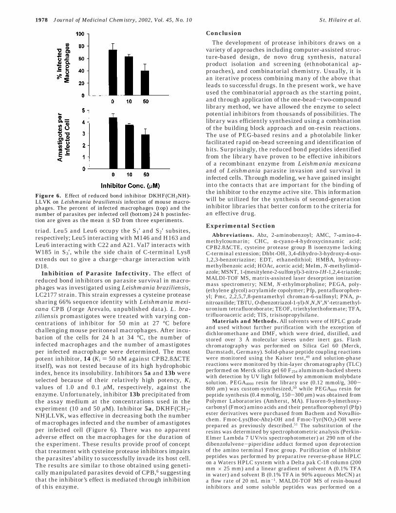
Inhibition of Parasite Infectivity. The effect ofreduced bond inhibitors on parasite survival in macro-phages was investigated using Leishmania braziliensis,LC2177 strain. This strain expresses a cysteine proteasesharing 66% sequence identity with Leishmania mexi-cana CPB (Jorge Arevalo, unpublished data). L. bra-ziliensis promastigotes were treated with varying con-centrations of inhibitor for 50 min at 27 °C beforechallenging mouse peritoneal macrophages. After incu-bation of the cells for 24 h at 34 °C, the number ofinfected macrophages and the number of amastigotesper infected macrophage were determined. The mostpotent inhibitor, 14 (Ki ) 50 nM against CPB2.8∆CTEitself), was not tested because of its high hydrophobicindex, hence its insolubility. Inhibitors 5a and 13b wereselected because of their relatively high potency, Kivalues of 1.0 and 0.1 µM, respectively, against theenzyme. Unfortunately, inhibitor 13b precipitated fromthe assay medium at the concentrations used in theexperiment (10 and 50 µM). Inhibitor 5a, DKHF(CH2-NH)LLVK, was effective in decreasing both the numberof macrophages infected and the number of amastigotesper infected cell (Figure 6). There was no apparentadverse effect on the macrophages for the duration ofthe experiment. These results provide proof of conceptthat treatment with cysteine protease inhibitors impairsthe parasites’ ability to successfully invade its host cell.The results are similar to those obtained using geneti-cally manipulated parasites devoid of CPB,6 suggestingthat the inhibitor’s effect is mediated through inhibitionof this enzyme.
Conclusion
The development of protease inhibitors draws on avariety of approaches including computer-assisted struc-ture-based design, de novo drug synthesis, naturalproduct isolation and screening (ethnobotanical ap-proaches), and combinatorial chemistry. Usually, it isan iterative process combining many of the above thatleads to successful drugs. In the present work, we haveused the combinatorial approach as the starting point,and through application of the one-bead-two-compoundlibrary method, we have allowed the enzyme to selectpotential inhibitors from thousands of possibilities. Thelibrary was efficiently synthesized using a combinationof the building block approach and on-resin reactions.The use of PEG-based resins and a photolabile linkerfacilitated rapid on-bead screening and identification ofhits. Surprisingly, the reduced bond peptides identifiedfrom the library have proven to be effective inhibitorsof a recombinant enzyme from Leishmania mexicanaand of Leishmania parasite invasion and survival ininfected cells. Through modeling, we have gained insightinto the contacts that are important for the binding ofthe inhibitor to the enzyme active site. This informationwill be utilized for the synthesis of second-generationinhibitor libraries that better conform to the criteria foran effective drug.
Experimental SectionAbbreviations. Abz, 2-aminobenzoyl; AMC, 7-amino-4-
methylcoumarin; CHC, R-cyano-4-hydroxycinnamic acid;CPB2.8∆CTE, cysteine protease group B isoenzyme lackingC-terminal extension; Dhbt-OH, 3,4-dihydro-3-hydroxy-4-oxo-1,2,3-benzotriazine; EDT, ethanedithiol; HMBA, hydroxy-methylbenzoic acid; HOAc, acetic acid; MeIm, N-methylimid-azole; MSNT, 1-(mesitylene-2-sulfonyl)-3-nitro-1H-1,2,4-triazole;MALDI-TOF MS, matrix-assisted laser desorption ionizationmass spectrometry; NEM, N-ethylmorpholine; PEGA, poly-(ethylene glycol) acrylamide copolymer; Pfp, pentafluorophen-yl; Pmc, 2,2,5,7,8-pentamethyl chroman-6-sulfonyl; PNA, p-nitroanilide; TBTU, O-(benzotriazol-1-yl)-N,N,N′,N′-tetramethyl-uronium tetrafluoroborate; TEOF, triethylorthoformate; TFA,trifluoroacetic acid; TIS, trisisopropylsilane.
Materials and Methods. All solvents were of HPLC gradeand used without further purification with the exception ofdichloromethane and DMF, which were dried, distilled, andstored over 3 Å molecular sieves under inert gas. Flashchromatography was performed on Silica Gel 60 (Merck,Darmstadt, Germany). Solid-phase peptide coupling reactionswere monitored using the Kaiser test,49 and solution-phasereactions were monitored by thin-layer chromatography (TLC)performed on Merck silica gel 60 F254 aluminum-backed sheetswith detection by UV light followed by ammonium molybdatesolution. PEGA4000 resin for library use (0.12 mmol/g, 300-800 µm) was custom-synthesized,50 while PEGA800 resin forpeptide synthesis (0.4 mmol/g, 150-300 µm) was obtained fromPolymer Laboratories (Amherst, MA). Fluoren-9-ylmethoxy-carbonyl (Fmoc) amino acids and their pentafluorophenyl (Pfp)ester derivatives were purchased from Bachem and NovaBio-chem. Fmoc-Lys(Boc-Abz)-OH and Fmoc-Tyr(NO2)-OH wereprepared as previously described.51 The substitution of theresins was determined by spectrophotometric analysis (Perkin-Elmer Lambda 7 UV/vis spectrophotometer) at 290 nm of thedibenzofulvene-piperidine adduct formed upon deprotectionof the amino terminal Fmoc group. Purification of inhibitorpeptides was performed by preparative reverse-phase HPLCon a Waters HPLC system with a Delta pak C-18 column (200mm × 25 mm) and a linear gradient of solvent A (0.1% TFAin water) and solvent B (0.1% TFA in 90% aqueous MeCN) ata flow rate of 20 mL min-1. MALDI-TOF MS of resin-boundinhibitors and some soluble peptides was performed on a
Figure 6. Effect of reduced bond inhibitor DKHF(CH2NH)-LLVK on Leishmania brasiliensis infection of mouse macro-phages. The percent of infected macrophages (top) and thenumber of parasites per infected cell (bottom) 24 h postinfec-tion are given as the mean ( SD from three experiments.
1978 Journal of Medicinal Chemistry, 2002, Vol. 45, No. 10 St. Hilaire et al.

Bruker Reflex III with a matrix of R-cyano-4-hydroxycinnamicacid. The mass of other soluble peptides was determined byES-MS recorded in the positive mode on a Fisons VG Quattroinstrument. NMR spectra were recorded on a Bruker AMX-250 spectrometer. Solution-phase kinetics of inhibition wascarried out on a Perkin-Elmer Lambda 50 fluorimeter.
Enzyme. L. mexicana CPB2.8∆CTE was expressed, puri-fied, and activated as previously described.21 The concentrationof the enzyme stock solution (11.4 µm) was determined byactive site titration with human cystatin (a generous gift fromDr. Magnus Abrahamson (University of Lund, Sweden)) usingZ-Phe-Arg-AMC as the substrate.
Solution-Phase Inhibition. Peptide inhibitors were as-sayed as according to Nicklin and Barrett52 using either Z-Phe-Arg-AMC (Km ) 0.7 µM) or 18.5 µM Ac-Phe-Arg-AMC (Km )15.0 µM) as substrate. Assay conditions were as follows: 0.1M sodium phosphate, 10.0 mM DTT, pH 6.0, 37 °C, with 10min of enzyme activation before use. [S] ) 0. 7 µM while [E]ranged from 1.1 to 4.5 nM. For the latter substrate, theconditions used were as follows: 0.1 M sodium acetate, 2.0mM EDTA, 200.0 mM NaCl, 10.0 mM DTT, pH 5.5, 37 °C,with 15 min of enzyme activation before use. [S] ) 18.5 µMwhile [E] ) ranged from 0.5 to 1.1 nM. Inhibitors were madeup as 2 mM stock solutions in DMF/H2O (1:1), and concentra-tions ranged from 0.01 to 50 µM. Precise inhibitor concentra-tions were obtained from the integration of HPLC peaks ofthe inhibitor after calibration of the HPLC at 210 nm usingtwo peptides: Y(NO2)PKFRSFNK(Abz)G-OH and AFMV-OH.Ki(app) values were obtained from the gradient of the plottingof [(vo/vi) - 1] vs [I], where vo and vi are the velocity of lessthan 2% substrate hydrolysis in the absence and in thepresence of different inhibitor concentrations [I], respectively.The Ki parameters were obtained from the equation Ki ) Ki(app)/(1 + [S]/Km). Results are the mean of two experiments thatvaried by less than 5%.
Effect of Inhibitor DKHF(CH2NH)LLVK on the Sur-vival of Leishmania-Infected Macrophages. LC2177 Leish-mania braziliensis parasites were grown in blood agar biphasicmedium at 27 °C. Promastigotes from the stationary phase(day 4) were harvested by centrifugation at 2500 rpm for 10min at 4 °C. The pellet was washed twice with M 199 mediumunder the same centrifugation conditions and resuspended in1 mL of medium for subsequent in vitro infection studies.Resident macrophages were harvested from the peritonealcavities of 6 week old Balb/c mice in ice-cold M199 mediumsupplemented with 20% (v/v) heat-inactivated fetal calf serum,plated onto microcoverslips (3 mm × 3 mm) laid in 96-wellplates, and incubated at 37 °C under an atmosphere of 5%CO2. After 2 h the wells were washed with M199 medium (3×).Predominant macrophages were kept under the same condi-tions of incubation overnight.
Parasites were incubated for 50 min at different concentra-tions of DKHF(CH2NH)LLVK inhibitor and then were washedtwice by centrifugation with M199 medium as described above.The pellet was resuspended in complete M199 medium. Theseparasites were added at a 20:1 parasite/macrophage ratio tothe wells containing attached macrophages for 1 h. Non-attached parasites were washed (3×), and the infection waspursued at 34 °C for 24 h. At the end of this period, cells werestained with Giemsa and May Grunwald solutions. Thepercentages of infected macrophages and amastigotes perinfected cell of four microcoverslips were recorded for each timepoint. Each experiment was repeated three times.
Synthesis of Fmoc Amino Aldehydes 2-4. Fmoc aminoaldehydes were synthesized according to the procedure of Wenand Crews,23 and the NMR data were in agreement withpreviously reported assignments of Phe,23 Lys, and Arg.53
Yields: Fmoc-Phe-N(Me)OMe, 99%; Fmoc-Phe-H, 90%; Fmoc-Lys(Boc)-N(Me)OMe, 95%; Fmoc-Lys(Boc)-H, 58%; Fmoc-Arg-(Pmc)-N(Me)OMe, 75%; Fmoc-Arg(Pmc)-H, 69%.
General Solid-Phase Peptide Synthesis. Synthesis ofthe inhibitor library and lead inhibitor peptides was carriedout manually in a custom-made 20-well Teflon synthesisblock54 as previously described.55 The spacer peptide, PPR-
(Pmc)PPR(Pmc), was synthesized manually in a syringe usingFmoc-Aa-OPfp esters (3 equiv). Reaction completion wasassessed by using Dhbt-OH (1 equiv), which served as bothan acylation catalyst and an indicator of the reaction com-pleteness,56 or by using the Kaiser test.49 For coupling toamines, free acids were activated for 6-8 min using TBTU(0.1 equiv less than the acid equivalent) and NEM (1.5 timesthe equivalent of acid). For coupling to the HMBA linker, freeacids were activated in situ by equivalent quantities of MSNTand N-MeIm.57 Coupling times ranged from 3 h to overnight.The photolabile linker was synthesized as previously de-scribed.32 All manipulations (synthesis and screening) ofpeptides linked to the resin via the photolabile linker werecarried out in subdued light (protected from UV radiation).
Deprotection Protocols. After each acylation, the resinwas washed with DMF (6×), and then the Fmoc protectinggroups were removed by treating the resin with a 20%piperidine/DMF (2 + 18 min) solution. The resin was thenwashed with DMF (6×). For the removal of Boc and other sidechain protecting groups, the resin was first washed with CH2-Cl2 (8×) and then treated with a cocktail consisting of TFA/thioanisole/ethanedithiol/water (87.5:5:2.5:5) initially for 10min and then for 2.5 h. The resin was then washed with 95%acetic acid (4×), DMF (2×), 5% DIPEA (2×), DMF (2×), andCH2Cl2 (6×) and then was dried in vacuo. In the case ofinhibitor peptides, the TFA filtrate was collected, the resin waswashed with 95% acetic acid (2×), and the combined filtrateswere concentrated.
Synthesis of Reduced Peptide Bond Library. Theinhibitor library was synthesized on custom-made PEGA4000
resin50 (750 mg, 0.12 mmol/g, 300-800 µm). In a 20 mLsyringe, the resin was first washed with DMF (5×), NR-FmocLys(Alloc)-OPfp (4 equiv) and DhbtOH (1 equiv) in DMFwere added, and the mixture was allowed to react for 4 h. TheFmoc group was removed, and the photolabile linker (1) wascoupled using TBTU/NEM methodology. An ionization massspacer, FmocPPR(Pmc)PPR(Pmc), was then synthesized usingthe Fmoc/OPfp method. The resin was then transferred to a20-well custom-made (2.0 mL/well capacity) library generator.The first four randomized positions in the library weregenerated by split-mix synthesis using a mixture of Fmoc andBoc amino acids (92:8, 6 equiv) in DMF (700 µL) afterpreactivation by TBTU/NEM in DMF (100 µL). After eachcoupling, the resin was pooled, mixed, and divided before Fmocremoval. The usual washing protocol followed each couplingand deprotection step. After the 4th cycle, the resin in two ofthe wells was redistributed into the remaining 18 wells andthe resin was washed with solution A (TEOF containing 1%HOAc) (6×). Aldehydes 2-4, (7 equiv) dissolved in solution B(DMF/TEOF/MeOH (1:1:1) containing 1% HOAc (800 µL) wereadded such that each aldehyde was added to six wells. After45 min at 50 °C, NaCNBH3 (10.5 equiv) in solution B (100 µL)was added and the mixture was reacted for an additional 2.5h. After completion of coupling, the resin was washed withDMF (2×) MeOH (2×), DMF (2×), and Boc2O (20% in DMF,900 µL), and excess NEM (100µL) was added for 20 min. Afterdeprotection of the N-terminal Fmoc, the remaining threepositions were coupled in a randomized manner as describedabove. After completion of the laddered inhibitor library, 200mg of the resin was transferred to a syringe and treated withPd(PPh3)4 (4 equiv) in a degassed mixture of CHCl3/AcOH/NEM (92.5:5:2.5) for a period of 4.5 h and then was washedwith CHCl3 (3×), DMF (3×), 0.5% Et2NCS2Na in DMF (1×),and DMF (4×). Substrate AcY(NO2)PR(Pmc)FR(Pmc)T(tBu)-GS(tBu)K(Abz)G-OH (3 equiv) in DMF was activated withTBTU/NEM and was added to the resin, and the mixturereacted for 5 h. The substrate and inhibitor library weredeprotected as described in the general methods section.
Synthesis of Lead Inhibitor Peptides. PEGA800 resin(800 mg, 0.34 mmol/g, 150-300 µm beads) was derivatizedwith the Rink amide linker (3 equiv) via TBTU/NRM activa-tion. The resin was then apportioned into the 20 wells (40 mg/well), and peptides were synthesized using Fmoc-Aa-OPfp (3equiv). Reductive amination was carried out as described for
Peptide Bond Inhibitors Journal of Medicinal Chemistry, 2002, Vol. 45, No. 10 1979

inhibitor library synthesis. At the end of the synthesis, theFmoc group was removed and the peptides were simulta-neously cleaved and deprotected by treatment with the fol-lowing TFA mixtures for 45 min and 2.5 h if Arg was present:mixture 1 (93% TFA, 5% H2O, 2% TIS) for peptides 5a-e, 6a,b,9, 11, 14, 17, and 18; mixture 2 (90% TFA, 5% H2O, 5%thiocresol) for peptides 8, 15, 16, and 19; mixture 3 (82.5%TFA, 5% H2O, 5% thiocresol, 2.5% EDT) for peptides 7, 10,12, and 13a,b. The peptides were purified by preparativeHPLC and analyzed by MALDI-TOF MS and analytical HPLC(Table 2).
Solid-Phase Library Screening. The library (200 mg; ca.34 000 beads) was washed (2 × 10 min) with assay buffer (100mM phosphate buffer, pH 6.0, augmented with 10 mm DTT)and then incubated with activated CPB2.8∆CTE (30 nmol) at37 °C in 5 mL of assay buffer. The fluorescence intensity ofthe beads was monitored with a fluorescent microscope everyhour for indications of hydrolysis. After 3 h, most of the beadsshowed a fluorescent “ring” indicating hydrolysis of thesubstrate. The reaction mixture was incubated for 7 h and thenwas treated with 2% aqueous TFA solution (2 × 5 min), water(2×), 2% NaHCO3 (2×), and then water (3×). The fluorescenceintensity of the beads was assessed by inspection with afluorescence microscope, and the 70 darkest beads werecollected and transferred to a stainless steel MALDI-TOFtarget for sequence identification.
MALDI-TOF Mass Spectrometry. Library Hits. Beadswere irradiated on stainless steel targets with a UV lamp for30 min. The analyte was extracted on the target from the beadsusing CH3CN/H2O (3:2, 0.5 µL) containing 1% TFA, matrixR-cyano-4-hydroxycinnamic acid (CHC, 10 mg in 1 mL of 70%acetonitrile) was added (0.5 µL), and the sample was dried witha hair dryer (ca. 50 °C). The spectrum was recorded in thepositive-ion mode on a Bruker Reflex III MALDI-TOF massspectrometer. Spectra were obtained (80-300 pulses) usingthe lowest power required to facilitate desorption and ioniza-tion.
Soluble Peptides. HPLC solutions containing the peptideswere mixed with an equal volume of CHC matrix (0.5 µL) andwere analyzed in the positive mode. Bradykinin (1060.2 mu),renin (1759.0 mu), and mellitin (2846.5 mu) were used as thestandards for internal calibration of the mass.
Modeling. Molecular dynamics calculations were carriedout on a Silicon Graphics Octane workstation using theInsightII/Discover program. A homology-based protein modelof a L. major cathepsin L-like cysteine protease (GeneBanklocus U43706, PDB identification code 1bmj)58 was built usingInsightII software (Biosym Technologies) and the crystalstructures of papain59 and cruzain60 as reference proteins. Themodel of the L. mexicana CPB2.8∆CTE isoform was obtainedby changing the amino acid residues that were different fromthose of L. major and then shortly minimizing the protein. Thecalculations were performed with the substrate Y(NO2)-PKFRSNFGK(Abz) and the inhibitor DKHF(CH2NH)LLVK toexamine the differences in binding modes of substrate andinhibitors. During all calculations, the amide backbone ofCPB2.8∆CTE was fixed, while all the side chains in contactwith the substrate/inhibitor were allowed to move. Eachcalculation was carried out as an annealing at decreasingtemperatures, 650 and then 500 and 300 K. At 500 and 300K, the CPB2.8∆CTE residues in contact with the ligand wereallowed to move freely including backbone atoms in sequenceranges of five successive amino acids or less. The ligands wereinitially energy-minimized from the extended structures andwere added in the resulting conformations approximately 15Å away from the binding site. A weak constraint of ∼3 kcal/mol (boundaries 3 and 4 Å) was added between Cys 25 thioland the scissile bond carbonyl carbon of the substrate or tothe analogous CH2 of the inhibitor. These constraints weremaintained throughout all calculations. Additional constraintsof ∼3 kcal/mol (boundaries 2 and 3 Å) were applied fordistances between the C25 sulfhydryl hydrogen and theπ-nitrogen of H163 and between the τ-NH of the H163 andthe N175 side chain carbonyl. The calculations were initiated
with 10 steps of minimization, and then 30 000-60 000 stepswere calculated at each temperature at 1 fs intervals. Thedevelopment of each calculation was monitored, and whenpersistent obstructions to the progress (i.e., distance greaterthan 5 Å between the sulfur atom and the scissile carbonylcarbon) of the calculation were observed, a new startingsubstrate conformation was used. This was obtained bymodification of one or two peptide torsion angles in theextended peptide followed by a short MD calculation alone.Only the φ-ψ angles allowed in the φ/ψ space were used asstarting conformations. As many as five independent success-ful calculations were performed for each ligand, and when thecalculations were allowed to reach equilibrium, the resultsobtained with different starting conformations converged toone or two related bound conformations. Constraints werecompletely removed from the final structure, and the complexwas subjected to extended calculations to determine whetherthe ligand remained bound to the CPB2.8∆CTE molecule in astable conformation.
Acknowledgment. This work was supported by theINCO-DCprogram(EUContractNo.ERBIC18CT970225)and the Brazilian Research Foundations FAPESP andPADCT. We acknowledge assistance from Hanne Chris-tiansen for peptide purification and from Ann-SofiSteinholtz for artwork. J.C.M. is a MRC Senior Fellow(U.K.).
Supporting Information Available: Details of reductiveamination optimization and synthesis of protected substrate.This material is available free of charge via the Internet athttp://pubs.acs.org.
References(1) Robertson, C. D.; Coombs, G. H. Multiple High Activity Cysteine
Proteases of Leishmania mexicana Are Encoded by the LmcpbGene Array. Microbiology 1994, 140, 417-424.
(2) Mottram, J. C.; Frame, M. J.; Brooks, D. R.; Tetley, L.; Hutchi-son, J. E.; Souza, A. E.; Coombs, G. H. The Multiple CpbCysteine Proteinase Genes of Leishmania mexicana EncodeIsoenzymes That Differ in Their Stage Regulation and SubstratePreferences. J. Biol. Chem. 1997, 272, 14285-14293.
(3) Coombs, G. H.; Mottram, J. C. Parasite Proteinases and AminoAcid Metabolism: Possibilities for Chemotherapeutic Exploita-tion. Parasitology 1997, 114, S61-S80.
(4) Coombs, G. H.; Mottram, J. C. Proteinases of Trypanosomes andLeishmania. In Trypanosomiasis and Leishmaniasis: Biologyand Control; Hide, G., Mottram, J. C., Coombs, G. H., Holmes,P. H., Eds.; CAB International: Oxon, U.K., 1997; pp 177-197.
(5) (a) DeSouza, L. S.; Lang, T.; Prina, E.; Hellio, R.; Antoine, J. C.Intracellular Leishmania amazonensis Amastigotes Internalizeand Degrade MHC Class II Molecules of Their Host Cells. J.Cell. Sci. 1995, 108, 3219-3231. (b) Alexander, J.; Coombs, G.H.; Mottram, J. C. Leishmania mexicana Cysteine Proteinase-Deficient Mutants Have Attenuated Virulence for Mice andPotentiate a Th1 Response. J. Immunol. 1998, 161, 6794-6801.
(6) Mottram, J. C.; Souza, A. E.; Hutchison, J. E.; Carter, R.; Frame,M. J.; Coombs, G. H. Evidence From Disruption of the LmcpbGene Array of Leishmania Mexicana That Cysteine ProteinasesAre Virulence Factors. Proc. Natl. Acad. Sci. U.S.A. 1996, 93,6008-6013.
(7) Frame, M. J.; Mottram, J. C.; Coombs, G. H. Analysis of theRoles of Cysteine Proteinases of Leishmania mexicana in theHost-Parasite Interaction. Parasitology 2000, 121, 367-377.
(8) Selzer, P. M.; Pingel, S.; Hsieh, I.; Ugele, B.; Chan, V. J.; Engel,J. C.; Bogyo, M.; Russell, D. G.; Sakanari, J. A.; McKerrow, J.H. Cysteine Protease Inhibitors as Chemotherapy: Lessons froma Parasite Target. Proc. Natl. Acad. Sci. U.S.A. 1999, 96, 11015-11022.
(9) Tropical Disease Research: Progress 1991-92: Eleventh Pro-gramme Report of the UNDP/WORLD BANK/WHO SpecialProgramme for Research and Training in Tropical Diseases(TDR); World Health Organization: Geneva, 1993; pp 77-87.
(10) Grogl, M.; Thomason, T. N.; Franke, E. D. Drug Resistance inLeishmaniasis: Its Implication in Systemic Chemotherapy ofCutaneous and Mucocutaneous Disease. Am. J. Trop. Med. Hyg.1992, 47, 117-126.
(11) Borst, P.; Ouellette, M. New Mechanisms of Drug Resistance inParasitic Protozoa. Annu. Rev. Microbiol. 1995, 49, 427-460.
1980 Journal of Medicinal Chemistry, 2002, Vol. 45, No. 10 St. Hilaire et al.

(12) Meldal, M.; Svendsen, I. Direct Visualization of Enzyme Inhibi-tors Using a Portion Mixing Inhibitor Library Containing aQuenched Fluorogenic Peptide Substrate. 1: Inhibitors forSubtilisin Carlsberg. J. Chem. Soc., Perkin Trans. 1 1995, 1591-1596.
(13) Meldal, M.; Svendsen, I.; Juliano, L.; Juliano, M. A.; Del Nery,E.; Scharfstein, J. Inhibition of Cruzipain Visualized in aFluorescence Quenched Solid-Phase Inhibitor Library. D-AminoAcid Inhibitors for Cruzipain, Cathepsin B and Cathepsin L. J.Pept. Sci. 1998, 4, 83-91.
(14) Graven, A.; St. Hilaire, P. M.; Sanderson, S. J.; Mottram, J. C.;Coombs, G. H.; Meldal, M. Combinatorial Library of PeptideIsosters Based on Diels-Alder Reactions: Identification of NovelInhibitors against a Recombinant Cysteine Protease from Leish-mania mexicana. J. Comb. Chem. 2001, 3, 441-452.
(15) Buchardt, J.; Schiødt, C. B.; Krog-Jensen, C.; Delaisse, J.-M.;Foged, N. T.; Meldal, M. Solid Phase Combinatorial Library ofPhosphinic Peptides for Discovery of Matrix MetalloproteinaseInhibitors. J. Comb. Chem 2000, 2, 624-638.
(16) Billich, S.; Knoop, M. T.; Hansen, J.; Sedlacek, J.; Mertz, R.;Moelling, K. Synthetic Peptides as Substrates and Inhibitors ofHuman Immune Deficiency Virus-1 Protease. J. Biol. Chem.1988, 263, 17905-17908.
(17) Wu, J.; Adomat, J. M.; Ridiky, T. W.; Louis, J. M.; Leis, J.;Harrison, R. W.; Weber, I. T. Structural Basis for Specificity ofRetroviral Proteases. Biochemistry 1998, 37, 4518-4526.
(18) Weber, J.; Majer, P.; Litera, J.; Urban, J.; Soucek, M.; Vondrasek,J.; Konvalinka, J.; Novek, P.; Sedlacek, J.; Strop, P.; Krausslich,H.-G.; Pichova, I. Potency Comparison of Peptidomimetic Inhibi-tors against HIV-1 and HIV-2 Proteinases: Design of EquipotentLead Compounds. Arch. Biochem. Biophys. 1997, 341, 62-69.
(19) Bateman, K. S.; Huang, K.; Anderson, S.; Lu, W.; Qasim, M. A.;Laskowski, M., Jr.; James, M. N. G. Contribution of PeptideBonds to Inhibitor-Protease Binding: Crystal Structures of theTurkey Ovomucoid Third Domain Backbone Variants OMTKY3-Pro18I and OMTKY3-Ψ[COOH]-Leu18I in Complex with Strep-tomyces Griseus Proteinase B (SGPB) and the Structure of theFree Inhibitor, OMTKY3-Ψ[CH2NH2
+]-Asp19I. J. Mol. Biol.2001, 305, 839-849.
(20) Kaltenbronn, J. S.; Hudspeth, J. P.; Lunney, E. A.; Michniewicz,B. M.; Nicolaides, E. D.; Repine, J. T.; Roark, W. H.; Stier, M.A.; Tinney, F. J.; Woo, P. K. Renin Inhibitors ContainingIsosteric Replacements of the Amide Bond Connecting the P3and P2 Sites. J. Med. Chem. 1990, 33, 838-845.
(21) Sanderson, S.; Pollock, K.; Hilley, J.; Meldal, M.; St. Hilaire, P.M.; Juliano, M. A.; Juliano, L.; Mottram, J. C.; Coombs, G. H.Expression and Characterization of a Recombinant CysteineProteinase of Leishmania mexicana. Biochem. J. 2000, 347, 383-388.
(22) Fehrentz, J. A.; Castro, B. An Efficient Synthesis of OpticallyActive R-(t-Butoxycarbonylamino)-aldehydes from R-AminoAcids. Synthesis 1983, 676-678.
(23) Wen, J. J.; Crews, C. M. Synthesis of 9-Fluorenylmethoxycar-bonyl-Protected Amino Aldehydes. Tetrahedron: Asymmetry1998, 9, 1855-1858.
(24) Sasaki, Y.; Coy, D. H. Solid Phase Synthesis of PeptidesContaining the CH2NH Peptide Bond Isostere. Peptides 1987,8, 119-121.
(25) Sasaki, Y.; Murphy, W. A.; Heiman, M. L.; Lance, V. A.; Coy, D.H. Solid-Phase Synthesis and Biological Properties of ψ[CH2-NH] Pseudopeptide Analogues of a Highly Potent SomatostatinOctapeptide. J. Med. Chem. 1987, 30, 1162-1166.
(26) Wen, J. J.; Spatola, A. F. A Systematic Approach to the Solid-Phase Synthesis of Linear and Cyclic Pseudopeptide LibrariesContaining ψ[CH2NH] Amide Bond Surrogates. J. Pept. Res.1997, 49, 3-14.
(27) Furka, A.; Sebestyen, F.; Asgedom, M.; Dibo, G. General MethodFor Rapid Synthesis of Multicomponent Peptide Mixtures. Int.J. Pept. Protein Res. 1991, 37, 487-493.
(28) Lam, K. S.; Salmon, S. E.; Hersh, E. M.; Hruby, V. J.; Kazmier-ski, W. M.; Knapp, R. J. A New Type of Synthetic PeptideLibrary for Identifying Ligand-Binding Activity. Nature (London)1991, 354, 82-84.
(29) Gordon, D. W.; Steele, J. Reductive Alkylation on a SolidPhase: Synthesis of a Piperazinedone Combinatorial Library.Bioorg. Med. Chem. Lett. 1995, 5, 47-50.
(30) Krone, J. R.; Nelson, R. W.; Dogruel, D.; Williams, P.; Granzow,R. BIA/MS: Interfacing Biomolecular Interaction Analysis withMass Spectrometry. Anal. Biochem. 1997, 244, 124-132.
(31) Borch, R. F.; Bernstein, M. D.; Durst, H. D. The Cyanohydri-doborate Anion as a Selective Reducing Agent. J. Am. Chem.Soc. 1971, 93, 2897-2904.
(32) Holmes, C.; Jones, D. Reagents for Combinatorial OrganicSynthesis: Develpoment of a New O-Nitrobenzyl PhotolabileLinker for Solid Phase Synthesis. J. Org. Chem. 1995, 60, 2318-2319.
(33) Knorr, R.; Trzeciak, A.; Bannwarth, W.; Gillessen, D. NewCoupling Reagents in Peptide Synthesis. Tetrahedron Lett. 1989,30, 1927-1930.
(34) St. Hilaire, P. M.; Alves, L. C.; Sanderson, S. J.; Mottram, J. C.;Juliano, M. A.; Coombs, G. H.; Meldal, M. The SubstrateSpecificity of a Recombinant Cysteine Protease from Leishmaniamexicana: Application of a Combinatorial Peptide Library.ChemBioChem 2000, 1, 115-122.
(35) Leung, D.; Abbenante, G.; Fairlie, D. P. Protease Inhibitors:Current Status and Future Prospects. J. Med. Chem. 2000, 43,305-341.
(36) Thompson, S. K.; Halbert, S. M.; Bossard, M. J.; Tomaszek, T.A.; Levy, M. A.; Zhao, B.; Smith, W. W.; Abdel-Meguid, S. S.;Janson, C. A.; D’Alessio, K. J.; Mcqueney, M. S.; Amegadzie, B.Y.; Hanning, C. R.; Desjarlais, R. L.; Briand, J.; Sarkar, S. K.;Huddleston, M. J.; Ijames, C. F.; Carr, S. A.; Garnes, K. T.; Shu,A.; Heys, J. R.; Bradbeer, J.; Zembryki, D.; Lee-Rykaczewski,L.; James, I. E.; Lark, M. W.; Drake, F. H.; Gowen, M.; Gleason,J. G.; Veber, D. F. Design of Potent and Selective HumanCathepsin K Inhibitors That Span the Active Site. Proc. Natl.Acad. Sci. U.S.A. 1997, 94, 14249-14254.
(37) Marquis, R. W.; Yamasita, D. S.; Ru, Y.; Locastro, S. M.; Oh,H.-J.; Erhard, K. F.; Desjarlais, R. L.; Head, M. S.; Smith, W.W.; Zhao, B.; Janson, C. A.; Abdel-Meguid, S. S.; Tomaszek, T.A.; Levy, M. A.; Veber, D. F. Conformationally Constrained 1,3-Diamino Ketones: A Series of Potent Inhibitors of the CysteineProtease Cathepsin K. J. Med. Chem. 1998, 41, 3563-3567.
(38) Lalonde, J. M.; Zhao, B.; Smith, W. W.; Janson, C. A.; Desjarlais,R. L.; Tomaszek, T. A.; Carr, T. J.; Thompson, S. K.; Oh, H.-J.;Yamashita, D. S.; Veber, D. F.; Abdel-Meguid, S. Use of Papainas a Model for the Structure-Based Design of Cathepsin KInhibitors: Crystal Structures of Two Papain-Inhibitor Com-plexes Demonstrate Binding to S′-Subsites. J. Med. Chem. 1998,41, 4567-4576.
(39) Bossard, M. J.; Tomaszek, T. A.; Levy, M. A.; Ijames, C. F.;Huddleston, M. J.; Briand, J.; Thompson, S.; Halpert, S.; Veber,D. F.; Carr, S. A.; Meek, T. D.; Tew, D. G. Mechanism ofInhibition of Cathepsin K by Potent, Selective 1,5-Diacylcarbo-hydrazides: A New Class of Mechanism-Based Inhibitors ofThiol Proteases. Biochemistry 1999, 38, 15893-15902.
(40) Katunuma, N.; Murata, E.; Kakegawa, H.; Matsui, A.; Tsuzuki,H.; Tsuge, H.; Turk, D.; Turk, V.; Fukushima, M.; Tada, Y.; Asao,T. Structure Based Development of Novel Specific Inhibitors forCathepsin L and Cathespin S in Vitro and in Vivo. FEBS Lett.1999, 458, 6-10.
(41) Roush, W. R.; Gwaltney, S. L., II; Cheng, J.; Scheidt, K. A.;Mckerrow, J. H.; Hansell, E. Vinyl Sulfonate Esters and VinylSulfonamides: Potent, Irreversible Inhibitors of Cysteine Pro-teases. J. Am. Chem. Soc. 1998, 120, 10994-10995.
(42) Roush, W. R.; Hernandez, A.; Mckerrow, J. H.; Selzer, P. M.;Hansell, E.; Engel, J. C. Design, Synthesis and Evaluation ofD-Homophenylalanyl Epoxysuccinate Inhibitors of the Trypan-osomal Cysteine Protease Cruzain. Tetrahedron 2000, 56, 9747-9762.
(43) Schiødt, C. B.; Buchardt, J.; Terp, G. E.; Christensen, U.; Brink,M.; Berger Larsen, Y.; Meldal, M.; Foged, N. T. PhosphinicPeptide Inhibitors of Macrophage Metalloelastase (MMP-12).Selectivity and Mechanism of Binding. Curr. Med. Chem. 2001,8, 967-976.
(44) Haque, T. S.; Skillman, A. G.; Lee, C. E.; Habashita, H.;Gluzman, I. Y.; Ewing, T. J. A.; Goldberg, D. E.; Kuntz, I. D.;Ellman, J. A. Potent, Low-Molecular-Weight Non-Peptide In-hibitors of Malarial Aspartyl Protease Plasmepsin II. J. Med.Chem. 1999, 42, 1428-1440.
(45) Abato, P.; Conroy, J. L.; Seto, C. T. Combinatorial Library ofSerine and Cysteine Inhibitors That Interact with Both the Sand S′ Binding Sites. J. Med. Chem. 1999, 42, 4001-4009.
(46) Alves, L. C.; St. Hilaire, P. M.; Meldal, M.; Sanderson, S. J.;Mottram, J. C.; Coombs, G. H.; Juliano, L.; Juliano, M. A.Identification of Peptides Inhibitory to Recombinant CysteineProteinase, CPB, of Leishmania mexicana. Mol. Biochem. Para-sitol. 2001, 114, 81-88.
(47) Alves, L. C.; Judice, W. A. S.; St. Hilaire, P. M.; Meldal, M.;Sanderson, S. J.; Mottram, J. C.; Coombs, G. H.; Juliano, L.;Juliano, M. A. Substrate Specificity of Recombinant CysteineProteinase, CPB, of Leishmania mexicana. Mol. Biochem. Para-sitol. 2001, 116, 1-9.
(48) St. Hilaire, P. M.; Willert, M.; Juliano, M. A.; Juliano, L.; Meldal,M. Fluorescent-Quenched Solid Phase Combinatorial Librariesin the Characterization of Cysteine Protease Substrate Specific-ity. J. Comb. Chem. 1999, 1, 509-523.
(49) Kaiser, E.; Colescott, R. L.; Bossinger, C. D.; Cook, P. I. ColorTest for Detection of Free Terminal Amino Groups in the Solid-Phase Synthesis of Peptides. Anal. Biochem. 1970, 34, 595-598.
(50) Renil, M.; Ferreras, M.; Delaisse, J. M.; Foged, N. T.; Meldal,M. PEGA Supports for Combinatorial Peptide Synthesis andSolid-Phase Enzymatic Library Assays. J. Pept. Sci. 1998, 4,195-210.
Peptide Bond Inhibitors Journal of Medicinal Chemistry, 2002, Vol. 45, No. 10 1981
(51) Meldal, M.; Breddam, K. Anthranilamide and Nitrotyrosine asa Donor Acceptor Pair in Internally Quenched FluorescentSubstrates for EndopeptidasessMulticolumn Peptide Synthesisof Enzyme Substrates for Subtilisin Carlsberg and Pepsin. Anal.Biochem. 1991, 195, 141-147.
(52) Nicklin, M. J. H.; Barrett, A. J. Inhibition of Cysteine Proteinasesand Dipeptidyl Peptidase I by Egg-White Cystatin. Biochem. J.1984, 223, 245-253.
(53) Guichard, G.; Briand, J. P.; Friede, M. Synthesis of ArginineAldehydes for the Preparation of Pseudopeptides. Pept. Res.1993, 6, 121-124.
(54) Meldal, M.; Holm, C. B.; Bojesen, G.; Jacobsen, H.; Holm, A.Multiple Column Peptide Synthesis, Part 2 (1,2). Int. J. Pept.Protein Res. 1993, 41, 250-260.
(55) Meldal, M. The Solid-Phase Enzyme Inhibitor Library Assay.In Combinatorial Peptide Library Protocols; Cabilly, S., Ed.;Humana Press: Totowa, NJ, 1998; pp 75-82.
(56) Atherton, E.; Cameron, L.; Meldal, M.; Sheppard, R. C. Self-Indicating Activated Esters for Use in Solid Phase PeptideSynthesis. Fluorenylmethoxycarbonyl Amino Acid Derivatives
of 3-Hydroxy-4-Oxodihydrobenzotriazine. J. Chem. Soc., Chem.Commun. 1986, 1763-1765.
(57) Blankemeyer-Menge, B.; Nimtz, M.; Frank, R. An EfficientMethod For Anchoring Fmoc-Amino Acids to Hydroxyl-Func-tionalised Solid Supports. Tetrahedron Lett. 1990, 31, 1701-1704.
(58) Selzer, P. M.; Chen, X.; Chan, V. J.; Cheng, M.; Kenyon, G. L.;Kuntz, I. D.; Sakanari, J. A.; Cohen, F. E.; Mckerrow, J. H.Leishmania Major: Molecular Modeling of Cysteine Proteasesand Prediction of New Nonpeptide Inhibitors. Exp. Parasitol.1997, 87, 212-221.
(59) Kamphuis, I. G.; Kalk, K. H.; Swarte, M. B.; Drenth, J. Structureof Papain Refined at 1.65 Å Resolution. J. Mol. Biol. 1984, 179,233-256.
(60) Mcgrath, M. E.; Eakin, A. E.; Engel, J. C.; Mckerrow, J. H.;Craik, C. S.; Fletterick, R. J. The Crystal Structure of Cruzain:A Therapeutic Target for Chagas’ Disease. J. Mol. Biol. 1995,247, 251-259.
JM0110901
1982 Journal of Medicinal Chemistry, 2002, Vol. 45, No. 10 St. Hilaire et al.
Copyright © 2022 FDOKUMEN