Preconcentration by SolidPhase Microextraction
13
ISSN 10619348, Journal of Analytical Chemistry, 2014, Vol. 69, No. 8, pp. 715–727. © Pleiades Publishing, Ltd., 2014. Original Russian Text © V.N. Zaitsev, M.F. Zui, 2014, published in Zhurnal Analiticheskoi Khimii, 2014, Vol. 69, No. 8, pp. 787–800. 715 Sample preparation is the most laborious and long stage of chemical analysis. It is poorly automated and significantly affects the reliability and accuracy of the determination. In the presentday analytical practice, problems of the development of methods of sample preparation have received much attention, and the requirement of their miniaturization, considerable reduction or even complete elimination of the use of toxic solvents, automation, simplicity of coupling with analytical methods have been put forward. The recently developed microextraction methods meet the presentday requirements to sample preparation, being in some parameters no worse and even superior to the classical methods of liquid–liquid and solid phase extraction. The term microextraction (ME) is usually used for analyte extraction with small volumes (up to 0.1 mL) of solvent or small weights (usually up to 5 mg) of adsorbent from liquid or gaseous samples of the vol ume up to 5–10 mL. The notion of microextraction was introduced by Janusz Pawliszyn, Professor of the University of Waterloo (Canada) in 1989. Today this method is being actively developed, a number of new technical decisions have been proposed to simplify the procedures of liquid–liquid and solidphase extrac tion and ensure a successful combination of sample preparation with presentday methods of analysis (gas and liquid chromatography, capillary electrophoresis, atomicabsorption spectrometry). Liquid–liquid and solidphase ME are used in the analysis of biological samples, environmental samples, foodstuffs, medici nal preparations, warfare agents, in toxicology, foren sic examination, etc. [1–7]. According to the physical state of the extractant, the ME methods are subdivided into solidphase microextraction (SPME) and liquid phase microex traction (LPME) (Fig. 1). LPME was described in detail in review [8]. This review is devoted to solidphase microextrac tion and its applications to analytical chemistry. In solidphase microextraction, the analytes are extracted from a vapor or a liquid phase into an adsor bent phase. Solidphase microextraction is subdivided into some versions: SPME on an absorbing coating of a microsyringe (MS) needle or rod, fiber, in a micro tube (capillary) or SPME on an adsorbent on the pipette tip, in a MS needle [9–12] (Fig. 2). All these SPME versions can be accomplished both in a flow mode and under stationary (laboratory) conditions. To intensify sample preparation in SPME, analysts recommended using rubber and liquidlike materials, on which analytes are retained by dissolution rather than through the formation of chemical bonds with the adsorbent surface as adsorbing coatings [13, 14]. This also allows the rapid isolation of analytes by ther modesorption (in GC detection) or its elution with a suitable solvent (in GC and HPLC determination). Special devices for SPME have been proposed. For example, microsyringes with a needle coated with an adsorbent either outside or inside are manufactured by the industry. Another, more widespread version of this device is syringe with a rod coated with a stationary phase inserted into the needle. This rod can move rel ative to the needle and can also be inserted into the sample after needling the septum. Same principle is used to insert an aliquot of sample into chromatograph [15–18]. Preconcentration by SolidPhase Microextraction V. N. Zaitsev* and M. F. Zui Faculty of Chemistry, Taras Shevchenko National University of Kiev, ul. Vladimirskaya 64/13, Kiev, 01601 Ukraine *email: [email protected] Received February 21, 2012; in final form, December 11, 2013 Abstract—The review is devoted to a new method of sample preparation, solidphase microextraction. Its advantages are miniaturization; considerable reduction or even complete elimination of the use of toxic sol vents; high concentration factors; low cost; simplicity of coupling with instrumental methods of analysis; and possibility of automation. The main versions of solidphase microextraction and the parameters of their opti mization are considered, such as the chemical composition of the adsorbent, the thickness of the adsorbing coating, pH, the nature and concentration of the saltingout agent, extraction time, stirring intensity, tem perature, desorption conditions, and analyte derivatization. Examples of using solidphase microextraction in the analysis of environmental samples, biological samples, and foodstuffs are presented. Keywords: solidphase microextraction, headspace microextraction, optimization parameters DOI: 10.1134/S1061934814080139 REVIEWS
Transcript of Preconcentration by SolidPhase Microextraction

ISSN 1061�9348, Journal of Analytical Chemistry, 2014, Vol. 69, No. 8, pp. 715–727. © Pleiades Publishing, Ltd., 2014.Original Russian Text © V.N. Zaitsev, M.F. Zui, 2014, published in Zhurnal Analiticheskoi Khimii, 2014, Vol. 69, No. 8, pp. 787–800.
715
Sample preparation is the most laborious and longstage of chemical analysis. It is poorly automated andsignificantly affects the reliability and accuracy of thedetermination. In the present�day analytical practice,problems of the development of methods of samplepreparation have received much attention, and therequirement of their miniaturization, considerablereduction or even complete elimination of the use oftoxic solvents, automation, simplicity of coupling withanalytical methods have been put forward. Therecently developed microextraction methods meet thepresent�day requirements to sample preparation,being in some parameters no worse and even superiorto the classical methods of liquid–liquid and solid�phase extraction.
The term microextraction (ME) is usually used foranalyte extraction with small volumes (up to 0.1 mL)of solvent or small weights (usually up to 5 mg) ofadsorbent from liquid or gaseous samples of the vol�ume up to 5–10 mL. The notion of microextractionwas introduced by Janusz Pawliszyn, Professor of theUniversity of Waterloo (Canada) in 1989. Today thismethod is being actively developed, a number of newtechnical decisions have been proposed to simplify theprocedures of liquid–liquid and solid�phase extrac�tion and ensure a successful combination of samplepreparation with present�day methods of analysis (gasand liquid chromatography, capillary electrophoresis,atomic�absorption spectrometry). Liquid–liquid andsolid�phase ME are used in the analysis of biologicalsamples, environmental samples, foodstuffs, medici�nal preparations, warfare agents, in toxicology, foren�sic examination, etc. [1–7].
According to the physical state of the extractant,the ME methods are subdivided into solid�phasemicroextraction (SPME) and liquid phase microex�traction (LPME) (Fig. 1). LPME was described indetail in review [8].
This review is devoted to solid�phase microextrac�tion and its applications to analytical chemistry. Insolid�phase microextraction, the analytes areextracted from a vapor or a liquid phase into an adsor�bent phase. Solid�phase microextraction is subdividedinto some versions: SPME on an absorbing coating ofa microsyringe (MS) needle or rod, fiber, in a micro�tube (capillary) or SPME on an adsorbent on thepipette tip, in a MS needle [9–12] (Fig. 2). All theseSPME versions can be accomplished both in a flowmode and under stationary (laboratory) conditions.
To intensify sample preparation in SPME, analystsrecommended using rubber� and liquid�like materials,on which analytes are retained by dissolution ratherthan through the formation of chemical bonds withthe adsorbent surface as adsorbing coatings [13, 14].This also allows the rapid isolation of analytes by ther�modesorption (in GC detection) or its elution with asuitable solvent (in GC and HPLC determination).
Special devices for SPME have been proposed. Forexample, microsyringes with a needle coated with anadsorbent either outside or inside are manufactured bythe industry. Another, more widespread version of thisdevice is syringe with a rod coated with a stationaryphase inserted into the needle. This rod can move rel�ative to the needle and can also be inserted into thesample after needling the septum. Same principle isused to insert an aliquot of sample into chromatograph[15–18].
Preconcentration by Solid�Phase MicroextractionV. N. Zaitsev* and M. F. Zui
Faculty of Chemistry, Taras Shevchenko National University of Kiev, ul. Vladimirskaya 64/13, Kiev, 01601 Ukraine*e�mail: [email protected]
Received February 21, 2012; in final form, December 11, 2013
Abstract—The review is devoted to a new method of sample preparation, solid�phase microextraction. Itsadvantages are miniaturization; considerable reduction or even complete elimination of the use of toxic sol�vents; high concentration factors; low cost; simplicity of coupling with instrumental methods of analysis; andpossibility of automation. The main versions of solid�phase microextraction and the parameters of their opti�mization are considered, such as the chemical composition of the adsorbent, the thickness of the adsorbingcoating, pH, the nature and concentration of the salting�out agent, extraction time, stirring intensity, tem�perature, desorption conditions, and analyte derivatization. Examples of using solid�phase microextractionin the analysis of environmental samples, biological samples, and foodstuffs are presented.
Keywords: solid�phase microextraction, headspace microextraction, optimization parameters
DOI: 10.1134/S1061934814080139
REVIEWS

716
JOURNAL OF ANALYTICAL CHEMISTRY Vol. 69 No. 8 2014
ZAITSEV, ZUI
The important quantitative characteristics ofmicroextraction are the extraction efficiency andenrichment factors, which are determined under equi�librium conditions. The extraction efficiency or therecovery R (efficiency of extraction) of an analyte inME, as well as in other extraction methods is deter�mined by the equation [19]:
(1)
where m0 is the initial amount of an analyte in the sam�ple (water phase), me is the amount of analyte in theextractant phase, Кd is the partition coefficient of theanalyte between the phases, Ve is the volume of theextractant phase, and V0 is the volume of the waterphase.
Enrichment factor К is calculated by the equation
K = сe/с0, (2)
where сe is analyte concentration in an extractantphase and с0 is analyte concentration in the initialsample.
Two obvious conclusions follow from Eq. (1):
(1) The completeness of analyte extraction isimproved with an increase in the thickness of theadsorbing coating (adsorbent weight).
(2) For the efficient preconcentration of analytesby SPME one should use adsorbents for which the dis�tribution ratios of the analyte between the
R, % me/m0 100× 100 Kd/ Kd V0/Ve+( ),×= =
adsorbent phase and the solution phase are higherthan 1 × 10³ g/mL.
Usually an extraction (adsorption) equilibrium andhigh recoveries of an analyte must but been attained inmicroextraction. As follows from Eq. (1), the values ofR depend not only on the partition coefficient of ananalyte between the phases, but also on the volumeratio of these phases. In classical extraction, a quanti�tative or substantial extraction of an analyte isrequired. As follows from Eq. (1), this requirementcan hardly be fulfilled even at the volume ratio of thephases equal to 100 and higher. In flow�injectionsolid�phase extraction, where the Vw/Ve ratio can bemuch higher than 100, recommendations for the Rvalues are reduced to 60% [6]. In microextraction, thevalues of Vw/Ve can be even higher; therefore, themethod does not assume the achievement of high Rvalues, especially in flow�injection analysis. Therecovery of an analyte in ME sample preparation isalso reduced because of restrictions on the extractioncapacity of the acceptor phase and the nonequilibriumcharacter of the process. The first factor is associatedwith the reduction of the partition coefficient of ananalyte when the saturation of the acceptor phase withan analyte is approached. Because of a small volume ofthe acceptor phase this effect should not beenneglected. Low recoveries and low reproducibilities(to RSD values of 10–20%) associated with the non�equilibrium character of microextraction untilrecently significantly restricted the intensive develop�
LPME
SPME
Drop LLME
Membrane LLME
SPME;
Dynamic SPME
Two�phase
Three�phase
Direct LPME
Headspace LPME
Direct LPME
Dispersive LPME
hollow fiber LPME
membrane bag LPME
flat�sheet membrane LPME
Dispersive LPME
Fiber SPME in coated rod, fiber
stir bar sorptive extraction
microextraction in a packed syringe or in�tip SPME
In�tube SPME, in�needle SPME
microextraction in a packed syringe or in�tip SPME
In�tube SPME, in�needle SPME
Fiber SPME on coated rod, fiber
ME
Fig. 1. Classification of microextraction methods.

JOURNAL OF ANALYTICAL CHEMISTRY Vol. 69 No. 8 2014
PRECONCENTRATION BY SOLID�PHASE MICROEXTRACTION 717
(а) (b) (c) (d)
(g) (f) (e)
Fiber holder
Coated fiber
Sample
Magnetic stirrer
Fused�silica fiber
Polymeric coating
Magnetic stirrer
Microsyringe for SPME
Water sample
Packed adsorbent
Hole in a needle
Adsorbent packed in a microsyringe
tip
Microsyringe tips
Water sample
Polymer�coated fiber
Water sample
Microsyringe for SPME
Magnetic stirrer
Adsorbent� coated
magnetic stirrer
Inside�needle polymeric coating
Fig. 2. Schematic presentation of various SPME versions: (a) fiber SPME in solution, (b) fiber headspace SPME, (c, d) SPMEin a microdozer tip filled with an adsorbent and fibers, (e) adsorption ME on coated stir�rods, (f) in�needle SPME, (g) SPME ina MS needle filled with an adsorbent.
ment of ME. A breakthrough in this direction hasoccurred in recent years, when modern autosamplers,which allowed analysts to solve the problem of lowreproducibility, and sensitive mass�selective chro�matograph detectors, which eliminated the problemof high limits of detection in ME, have come into wideuse.
Taking into account the small volume of theextracted phase, analyte extraction in ME is rarelyquantitative; therefore, from the viewpoint of theaccuracy and reproducibility of analysis, of specialimportance is the procedure of ME implementationand its automation. The equilibrium conditions ofextraction in SPME are often attained within a rela�tively long time (10–90 min). Such duration of samplepreparation is not always acceptable; therefore MEcan also be performed under nonequilibrium condi�tions, by maximally standardizing them and strictlycontrolling the parameters of ME (temperature, timeof sample preparation, pH of solution, rate of stirringthe solution, concentration of admixtures). It is desir�able to perform microextraction in the presence of aninternal standard [20–22].
The following SPME parameters can be optimized:type of SPME (headspace or classical, for laboratoryor flow�injection analysis), composition of adsorbent,thickness of adsorbing coating, parameters of aqueoussample solution (pH, nature and concentration ofsalting�out agent), time of extraction, stirring inten�
sity, temperature, and conditions of analyte desorp�tion.
ADSORBENTS FOR SPME
Coatings in SPME are created using organic andorganosilicon polymers or hybrid organo�mineraladsorbents. Polymeric coatings of polydimethylsilox�ane (PDMS), polyacrylate (PAC), polydivinylben�zene (DVB) are most often used for the preconcentra�tion of nonpolar and weakly polar compounds [23].Block copolymers, PDMS–DVB, PDMS–diphenyl�benzene, or silica�based organomineral adsorbents(for example, LiChrospher), or microporous coals(for example, carboxen) [20, 24] are also used for thispurpose. In these adsorbents a polymer film or speciesof polar organic compounds or polymers (for example,PDMS–DVB, LiChrospher RP�18) are attached tothe surface of a substrate (for example, PDMS).Hybrid adsorbents possess higher capacities comparedto polymeric adsorbents. The adsorbtion equilibriumon them is also attained more rapidly [25].
Organosilicon adsorption coatings prepared by thesol–gel technology and also polymeric coating of car�bowax (CW), carboxen (KX), PDMS–KX, or CW–DB types applied onto the surface of a capillary, metalrod, or polymeric fiber [26–29] are widely used in theSPME of polar compounds. The surface modifiers ofsilicas are crown ethers, chitosans, calixarene, andothers selective complexants. Sometimes presynthe�
718
JOURNAL OF ANALYTICAL CHEMISTRY Vol. 69 No. 8 2014
ZAITSEV, ZUI
sized porous species with functionalized surfaces areintroduced into inert silica matrices applied onto aneedle by the sol–gel technology [30–34]. Salting outis also often used to improve the efficiency of extrac�tion of polar and weakly polar analytes.
In addition to the sol–gel technology, adsorptioncoatings are also formed by electrodeposition. Forexample, this method was used to obtain polypyrrol(PP) coatings on platinum wires [35, 36]. Polypyrrolon metal surface forms a current�conductive positivelycharged polymeric coating; its adsorption propertiescan easily be varied by introducing oppositely chargedions, for example, dodecyl sulfate [37], into theadsorption system. A characteristic feature of the PP�coating is that it can be discharged and charged againunder the influence of an electric potential applied tothe Pt carrier, thus losing and acquiring a counterion.This effect is used to regulate the adsorbtion and des�orption properties of the coating. At a positive poten�tial of the electrode, anions are adsorbed as counteri�ons on the positively charged polymer and, at the neg�ative potential of the electrode, these are desorbedfrom the surface (Fig. 3a). Wu et al. in [38] treated anelectrode with a PP coating with a solution of polysty�rene sulfonate (PSS). This allowed them to extractanalytes in the cationic form on such a bilayer coating(at a negative potential applied to the Pt wire) and des�orb them at a positive potential (Fig. 3b). The coatingobtained was used for the selective SPME of dopaminefollowed by its electrochemical detection.
PHYSICAL PARAMETERS OF SPME
SPME depends on various parameters, such as thethickness of coating, time, temperature, etc.. Theproper choice of these parameters can substantiallyimprove the efficiency of microextraction.
Thickness of adsorbing coating. The typical thick�ness of adsorbing films varies from 10 to 125 µm. Anincrease in film thickness results in an increase in therecovery of an analyte with the reduction of the rate ofthe adsorption process; therefore thick coatings aremore suitable for highly volatile analytes and thinones, for low�volatile analytes.
Temperature. Both adsorption and desorption pro�cesses are accelerated at higher temperatures. Takinginto account that the affinity of an analyte to theacceptor phase is considerably higher than that to thedonor phase, heating most often improves SPME con�ditions, by approaching them to equilibrium ones. Forhighly volatile substances, it is desirable to cool SPMEdevices. The optimum temperature of SPME dependson the volatility of an analyte and on the adsorptionphase used [23].
Stirring rate. Equilibrium conditions in theadsorption of an analyte are attained more rapidly ifthe sample solution is intensely stirred.
Desorption. The thermodesorption of the targetcomponents is used in GC detection. Thermodesorp�
tion must be performed much more rapidly; it takes onan average 10–30 min. The temperature of the gaschromatograph injector must be sufficiently high forthe desorption of the least volatile analyte. In ther�modesorption one should take into account the stabil�ity of the coating at high temperatures [24]. Most oftenthermodesorption is used for volatile analytesadsorbed on polymeric coatings. For example, Walleset al. in [39] adsorbed 12 aromatic compounds fromsnow samples on a PDMS�coating of a fiber and thendesorbed them at 200°C within 5 min. In [40] acetone,toluene, phenol, benzaldehyde, and other volatileorganic compounds were extracted from urine with aDB–KS–PDMS coating and then thermodesorbed at250°C within 5 min.
TYPES OF SOLID�PHASE MICROEXTRACTION
Fiber SPME is a type of solid�phase microextrac�tion, in which the substrate for the immobilization ofa stationary phase is provided by polymer fibers, fused�silica or carbon rods, or metal wires [20, 21, 32, 34]. Inthis version, the device for SPME is similar to amicrosyringe and contains a fiber or a rod inserted intoa needle in a special holder. The needle fulfills a doublefunction: helds the fiber and protects it from mechan�ical damage during the adsorbate injection into thechromatograph.
Fiber SPME can be used for analyte preconcentra�tion in the classical and headspace version [41–43]. Inthe first case, the analyte is adsorbed directly from awater sample (Fig. 2a). Headspace fiber SPME con�sists in the adsorption of an analyte from the saturatedvapor phase over a water sample or a comminuted solidsample (Fig. 2b). Both types of fiber SPME are cou�pled with GC and HPLC and also with systems of cap�illary electrophoresis both in a flow�injection modeand under stationary (laboratory) conditions. Head�space SPME is used for the ME of volatile andmedium�volatile analytes. Among the advantages ofheadspace SPME are the efficient separation of ananalyte from nonvolatile matrix components and alsono influence of chemically aggressive components ofthe solution and of irreversibly adsorbed substances onthe adsorbing coating. This ensures a possibility of themultiple (10�to 100�fold) use of the same adsorbingmaterial for analyte preconcentration in the head�space version of fiber SPME [44, 45]. Classical fiberSPME is used to extract medium�volatile and hardly�volatile analytes directly from aqueous solutions [46].To minimize the damage of the immobilized layer, itwas recommended to exclude strongly acid andstrongly alkaline solutions. Among the drawbacks ofthe device for fiber SPME is its fragility.
In devices for SPME manufactured by Supelco,polymer coatings with adsorbing films 7–100 µm thickare used [34]. The stationary phases (polyacrylate,polydimethylsiloxane, carbowax, polydivinylbenzene,

JOURNAL OF ANALYTICAL CHEMISTRY Vol. 69 No. 8 2014
PRECONCENTRATION BY SOLID�PHASE MICROEXTRACTION 719
or polyethylene glycol derivatives) either in a quasiliq�uid state or as solid porous species uniformly distrib�uted over a matrix surface are mechanically appliedonto a mineral or a metal rod [20, 21, 32]. Because theswelling of adsorption films in SPME is quite undesir�able (it significantly decelerates adsorption–desorp�tion processes), one should control the presence oforganic solvents in the donor phase. For example,films based on polyacrylate and polydimethylsiloxanewere recommended for use only with aqueous solu�tions. Cross�linked polymeric coatings (for example,polydivinylbenzene) are stable in organic solvents ofdifferent nature.
A much wider use of present�day materials for fab�ricating adsorbing layers than that implemented inSPME devices by Supelco was described. Adsorptioncoatings from polypyrrole [47, 48] and template poly�mers [24], composite organomineral coatings basedon polymers impregnated with immunoadsorbents[49, 50] and modified silicas [51, 52], organosiloxanecoatings obtained by the sol–gel technology [53, 54]using immersion printing [55], and biocompatible(biologically inert) polymeric adsorbents for theextraction of biomolecules (peptides, polypeptides,oligonucleotides, polynucleotides, fibers) [56] werestudied.
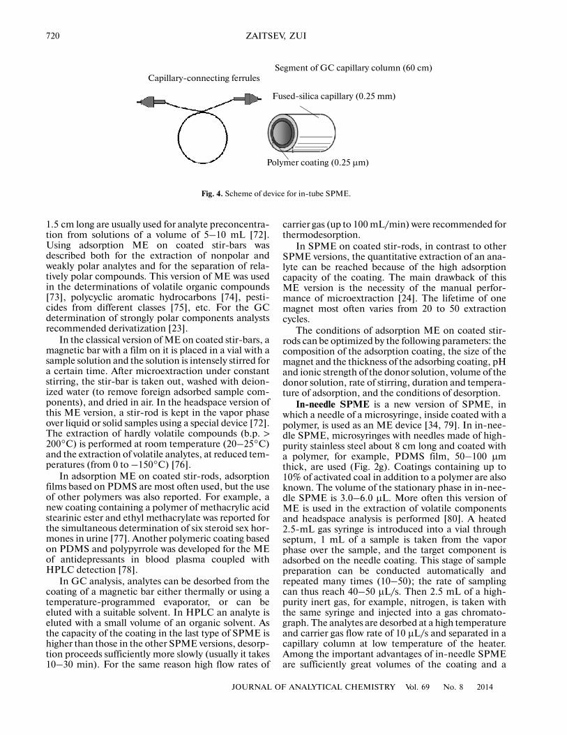
In�tube SPME. In in�tube SPME, extractiondevices are capillary tubes with polymeric coatingsapplied onto inner surfaces (Fig. 4) [57]. The tubes aresimilar to small GC capillary columns. The tubes aremost often made of fused silica. The length of the tubesvaries from 1–2 to 60 cm; their inner diameter, from0.25 to 5.0 mm; and thickness of polymeric coating,from 0.25 to 1.0 µm [34]. The coatings of the adsorb�ing tube can vary in nature. Most often polymeric (sty�rene–divinylbenzene, polyethylene glycol) or organo�silicon coatings are used [24, 52]. Coatings for fiberSPME with certain care can also be used for in�tubemicroextraction.
Both liquid and gaseous samples can be passedthrough adsorption tubes. Extraction, desorption, andinjection into a chromatograph can be convenientlyperformed with a standard autosampler [58].
To prevent moisture ingress into a GC system inSPME, the tube for analyte preconcentration fromaqueous solutions before analyte desorption mustbe dried by purging an inert gas. If elution with organicsolvent is used for desorption, the eluate ispassed through an additional drying column. Tubedrying before GC detection can take up to 30 min,which significantly extends the duration of microex�traction [59].
The following parameters can be optimized in in�tube SPME [57]: ionic strength and pH of the donorsolution, volume of donor solution, composition ofthe adsorbing coating and its thickness, eluent nature,and desorption temperature and duration.
The adsorbing coatings in in�tube SPME can varyin nature. Polypyrrol coatings were found to be effi�cient. This fact can be explained by different interac�tions between the functional groups of this coating andan analyte (dispersive, π–π, dipole–dipole, ion�exchange) [60]. In addition, analysts use polymer spe�cies obtained by the method of molecular immersionprinting [61] and special biocompatible polymers [62]as adsorbents in in�tube SPME. A capillary tube forwas described in [63–67], in which a polymer with sil�icon dioxide impregnated into it was used as anadsorbing coating. Such a combination significantlyaccelerated the attainment of an adsorption equilib�rium and, correspondingly, improved the efficiencySPME. Analytes of basic nature were separated onadsorption coatings based on hydrophobic polymers(polymethacrylates, polyethylene glycol and polyeth�ylene derivatives) with grafted acid groups [68–70].The efficiency of in�tube SPME was sometimesenhanced using tubes packed with fiber or granulatedadsorbents.
Stir bar sorptive extraction is a new type ofmicroextraction, in which the extractant is an adsorb�ing layer applied onto a bar of a magnetic stirrer (Fig.2d). This method is similar to fiber SPME; however,adsorption proceeds in a thicker layer (0.3–1.0 mm)and the adsorbent capacity is consequently increasedby 50–250 times [23, 71]. Small magnetic bars 1–
e
n
+
PSS– K+
N
H
e
N
H n
A–
0
n
PSS– K+
N
H n
A–
+
e
e
N
H
0
Polypyrrol coating Polypyrrol coating
Polypyrrol coating Polypyrrol coating
Pt Pt
PtPt
Electrochemical extraction of anions
Electrochemical extraction of cations
Anodic extraction
Cathodic extraction
Cathodic desorption
Anodic desorption
(а)
(b)
Fig. 3. Scheme electrochemically controlled SPME of(a) anions and (b) vations on a polypyrrole coating.
720
JOURNAL OF ANALYTICAL CHEMISTRY Vol. 69 No. 8 2014
ZAITSEV, ZUI
1.5 cm long are usually used for analyte preconcentra�tion from solutions of a volume of 5–10 mL [72].Using adsorption ME on coated stir�bars wasdescribed both for the extraction of nonpolar andweakly polar analytes and for the separation of rela�tively polar compounds. This version of ME was usedin the determinations of volatile organic compounds[73], polycyclic aromatic hydrocarbons [74], pesti�cides from different classes [75], etc. For the GCdetermination of strongly polar components analystsrecommended derivatization [23].
In the classical version of ME on coated stir�bars, amagnetic bar with a film on it is placed in a vial with asample solution and the solution is intensely stirred fora certain time. After microextraction under constantstirring, the stir�bar is taken out, washed with deion�ized water (to remove foreign adsorbed sample com�ponents), and dried in air. In the headspace version ofthis ME version, a stir�rod is kept in the vapor phaseover liquid or solid samples using a special device [72].The extraction of hardly volatile compounds (b.p. >200°C) is performed at room temperature (20–25°C)and the extraction of volatile analytes, at reduced tem�peratures (from 0 to –150°C) [76].
In adsorption ME on coated stir�rods, adsorptionfilms based on PDMS are most often used, but the useof other polymers was also reported. For example, anew coating containing a polymer of methacrylic acidstearinic ester and ethyl methacrylate was reported forthe simultaneous determination of six steroid sex hor�mones in urine [77]. Another polymeric coating basedon PDMS and polypyrrole was developed for the MEof antidepressants in blood plasma coupled withHPLC detection [78].
In GC analysis, analytes can be desorbed from thecoating of a magnetic bar either thermally or using atemperature�programmed evaporator, or can beeluted with a suitable solvent. In HPLC an analyte iseluted with a small volume of an organic solvent. Asthe capacity of the coating in the last type of SPME ishigher than those in the other SPME versions, desorp�tion proceeds sufficiently more slowly (usually it takes10–30 min). For the same reason high flow rates of
carrier gas (up to 100 mL/min) were recommended forthermodesorption.
In SPME on coated stir�rods, in contrast to otherSPME versions, the quantitative extraction of an ana�lyte can be reached because of the high adsorptioncapacity of the coating. The main drawback of thisME version is the necessity of the manual perfor�mance of microextraction [24]. The lifetime of onemagnet most often varies from 20 to 50 extractioncycles.
The conditions of adsorption ME on coated stir�rods can be optimized by the following parameters: thecomposition of the adsorption coating, the size of themagnet and the thickness of the adsorbing coating, pHand ionic strength of the donor solution, volume of thedonor solution, rate of stirring, duration and tempera�ture of adsorption, and the conditions of desorption.
In�needle SPME is a new version of SPME, inwhich a needle of a microsyringe, inside coated with apolymer, is used as an ME device [34, 79]. In in�nee�dle SPME, microsyringes with needles made of high�purity stainless steel about 8 cm long and coated witha polymer, for example, PDMS film, 50–100 µmthick, are used (Fig. 2g). Coatings containing up to10% of activated coal in addition to a polymer are alsoknown. The volume of the stationary phase in in�nee�dle SPME is 3.0–6.0 µL. More often this version ofME is used in the extraction of volatile componentsand headspace analysis is performed [80]. A heated2.5�mL gas syringe is introduced into a vial throughseptum, 1 mL of a sample is taken from the vaporphase over the sample, and the target component isadsorbed on the needle coating. This stage of samplepreparation can be conducted automatically andrepeated many times (10–50); the rate of samplingcan thus reach 40–50 µL/s. Then 2.5 mL of a high�purity inert gas, for example, nitrogen, is taken withthe same syringe and injected into a gas chromato�graph. The analytes are desorbed at a high temperatureand carrier gas flow rate of 10 µL/s and separated in acapillary column at low temperature of the heater.Among the important advantages of in�needle SPMEare sufficiently great volumes of the coating and a
Capillary�connecting ferrules Segment of GC capillary column (60 cm)
Fused�silica capillary (0.25 mm)
Polymer coating (0.25 µm)
Fig. 4. Scheme of device for in�tube SPME.

JOURNAL OF ANALYTICAL CHEMISTRY Vol. 69 No. 8 2014
PRECONCENTRATION BY SOLID�PHASE MICROEXTRACTION 721
shorter time of extraction compared to other types ofSPME [81]. In addition, in contrast to fiber SPME,the device for in�needle SPME is mechanically stable.
Microextraction in a packed syringe is a recentlydeveloped SPME version. It differs from in�needleSPME in using solid adsorbent species with which amicrosyringe is filled instead of a polymer coatingapplied onto the inner wall of the microsyringe needle[34, 82]. Usually 0.5–1 mg of an adsorbent is put in amicrosyringe needle of the volume up to 100 µL, andthe adsorbent is retained mechanically (Fig. 2f).Instead of adsorbent species one can also fill the nee�dle with a fibrous adsorbent [83]. Needles filled withadsorbents are used in gas syringes and in industrialdevices from Needle and Trap Device [84].
The adsorbents used in microextraction in apacked syringe are similar to those used in fiber SPME[24, 81]. The application of various hybrid materialswas described, such as silicas with grafted C2, C8, andС18 groups, polymers with molecular imprints, andsilarylene–siloxane block copolymers [85].
Different types in�needke SMPE and microextrac�tion in a packed syringe are easily automated, rapid,and applicable to the analysis of small sample volumesand to elution with small volumes of solvents.Microextraction in a packed syringe is characterizedby a higher capacity compared to fiber SPME, so thata higher efficiency of extraction and a better reproduc�ibility of the results of analysis can be reached.Microextraction in a packed syringe is used for theanalysis of complex samples, including blood plasma,urine, and other biological samples [86, 87].
This method was used to extract volatile hydrocar�bons, aldehydes and ketones, in the analysis of exhaledair (isoprene, butanal, hexanal, hexane, acetone, etc.)[84], and also to extract cannabioids from hair [88].
The supports of the stationary phase in in�tipSPME are pipette tips filled with adsorbent species(Figs. 2c and 2d) [34, 80]. This version of SPME isusually performed in laboratory (under stationaryconditions). Using multichannel pipette, one can pre�pare several samples simultaneously. Because of thedesign features of the micropipette (impossibility ofthe creation of high excess pressures or vacuum), a low
resistance to the flow is necessary for the implementa�tion of SPME in a micropipette tip. The adsorbentsused as the stationary phase should possess homoge�neous grain�size distribution, spherical particles, anda well�developed system of macropores. Monolithicphases are ideally suitable for this purpose [89]. One ofthe most widely used monolithic phases are poly�methacrylate monoliths; organomineral materials onthe basis of silica and adsorption fibers are also used[90–92]. Polymethacrylate adsorbents can bear polaracidic or basic groups; therefore, they can be used forthe extraction of analytes of various nature [93, 94].The high flow rates necessary for the implementationof in�tip SPME and the manual performance of MEmay result in the complexity of the multiple repetitionof the extraction cycle, and, therefore, low enrichmentfactors, and lower yields and lower reproducibilities incomparison to microextraction in a packed syringe.
DERIVATIZATION
Derivatization is often used by analysts to improvethe completeness and selectivity of analyte extractionin ME methods. Derivatization is most important forthe extraction of polar compounds. Derivatization isperformed either directly in a sample solution or in theinjection port of a gas or a liquid chromatograph, or onan adsorbent [24, 28, 34, 95].
In the first case, derivatization is most efficient,because the conditions, temperature, and time of thereaction can be varied. However, significant amountsof expensive derivatizing agents stable in aqueous solu�tions are often necessary to conduct the reaction. Inderivatization in an injection port, the derivatizingagent is introduced into the injector before the analyteand the analyte and agent react directly in the port. Anexample of derivatization in an injection port is pro�vided by the determination amphetamine and fenflu�ramine in blood [96]. The derivatizing agent, heptaflu�orobutanoic acid chloride, was introduced directlyinto the injector before the thermodesorption of ana�lytes from adsorption fibers. In the course of ther�modesorption the analyte reacted with the derivatizingagent in the injection port:
Another case of derivatization in the injector can beexemplified by acid�base polar analytes, which wereextracted by the adsorbent as ion pairs and thendecomposed under the conditions of increased tem�peratures in the port to form volatile derivatives of the
target components [24]. In derivatization on an adsor�bent, the adsorbent is preliminarily treated with aderivatizing agent. In this case, derivatization pro�ceeds during the adsorption of the analyte. In derivati�zation with reagents unstable to hydrolysis, either the
NH2 HN C3F7
O
+ C3F7COCl + HCl.

722
JOURNAL OF ANALYTICAL CHEMISTRY Vol. 69 No. 8 2014
ZAITSEV, ZUI
headspace version of ME is used or the analyte isderivatized on an adsorbent after its ME, by treatingthe adsorbent with a derivatizing agent solution orvapors [5, 90].
An example of on�fiber derivatization is providedby the derivatization short�chain fatty acids with1�pyrenyldiazomethane [97]:
First the fiber is placed in a 15�mg/mL solution of1�pyrenyldiazomethane for 15 min and then theimmobilized fiber is introduced into an analyte solu�tion and acids are extracted at 50°C for 30 min. Thenthe analytes are thermodesorbed into the injectionport at 260°C for 4 min.
Derivatization in solution followed by the ME oftarget component derivatives is used most widely. Forexample, the conversion of busulfan [1,4�bis�(methyl�sulfoxy)butane] into 1,4�diiodobutane in the presenceof sodium iodide was described in [98]. The majority
HCN2
RO
OH
H2C
O
R
O
+ + N2.
of derivatizing agents are sensitive to moisture; there�fore, they are added to dehydrated extracts. For exam�ple, alkyl formates unstable to hydrolysis are used forthe conversion of amino groups into carbamates [99–101] and acetic anhydride and silylating agents (trim�ethylsilylamide, hexamethyldisilazane, etc. deriva�tives), for the conversion of carbolylic acids and alco�hols into corresponding esters [102, 103]. Derivatiza�tion in solution was used for the determinationbenzoylecgonine [102] and valproic acid [104].Derivatization of cannabioids with a solution of bis�(trimethylsilyl)trifluoroacetamide was performed in adry residue of a methanolic extract of hair. In combi�nation with headspace SPME, such sample prepara�tion ensured a 5�fold increase in the sensitivity of drugdetermination [103, 105].
Preliminary processing of an adsorbent with aderivatizing agent was described in [106, 107];in these works carboxylic acids were derivatized with1�pyrenyldiazomethane in the adsorbent phase.In [108, 109] aldehydes were derivatized withadsorbed o�(2,3,4,5,6�pentafluorbenzyl)hydroxy�lamine. The studied derivatizing agents are notdecomposed by water; therefore, SPME can be used ina classical version.
The derivatization of analytes after SPME was per�formed in [110–116]; the adsorption unit after
Table 1. Use of SPME in the analysis of environmental samples
Analyte Matrix Adsorbent or membrane polymer ME version Method
of analysis References
PAH* Natural waters Silica C18 Microextraction in a packed syringe
GC−MS [117]
PAH Natural waters PDMS–DVB fiber Headspace fiber SPME
GC−MS/MS [118]
Polychlorinated hydrocarbons
Ocean waters PDMS fiber SPME The same [119]
Polychlorinated bi�phenyls, Cl and P containing pesti�cides
Seawaters The same Stir bar sorptive ex�traction
GC−MS [120]
Volatile and semiv�olatile organic compounds
Snow PDMS–DVB fiber Fiber SPME The same [40]
Phthalates Natural waters PDMS–DVB, PA, and CAR�PDMS fiber
Fiber SPME '' [121, 122]
UV filters Natural and waste waters
Silicone disks, PDMS coating
Stir bar sorptive ex�traction
'' [123]
Terpenes Plant materials PDMS–DVB fiber Fiber SPME '' [124]
Estrogens River waters PDMS coating Stir bar sorptive ex�traction
'' [125]
* PAH are polycyclic aromatic hydrocarbons.

JOURNAL OF ANALYTICAL CHEMISTRY Vol. 69 No. 8 2014
PRECONCENTRATION BY SOLID�PHASE MICROEXTRACTION 723
adsorption was put into a vial with trifluoroaceticanhydride [110,115], N�methyl�N�(tert�butyldimeth�ylsilyl)trifluoroacetamide [111], hexamethyldisila�zane [112], N�methyl�N�(trifluoroacetamide) [113],or pentafluorobenzoyl chloride [114]. In this version,the headspace derivatization of the analyte is morepreferable than the classical one, as it ensures the min�imization of possible analyte losses because of its des�orption into the liquid phase in the derivatization insolution.
Some examples of using solid�phase microextrac�tion coupled to gas and liquid chromatography arepresented in Tables 1 and 2.
COUPLING OF SPME WITH ATOMIC SPECTROMETRY
SPME can be successfully used to extract and pre�concentrate organometallic compounds of mercury,tin, arsenic, antimony, chromium, selenium, and leadfrom soils, bottom sediments, and biological samples.
Table 2. Use of SPME for the analysis of biological samples
Analyte MatrixAdsorbent
or membrane polymer for ME
ME version Method of analysis References
Amphetamine, methamphetamine, and their analogs
Hair, blood serum, urine, saliva
PDMS Fiber SPME GC−MS [126–132]
Analgetic and anaes�thetic drugs
Urine The same The same GC−TID [133]
Diazepam, lorazepam, nordiaz�epam, and ozazepam
Blood PDMS, SiO2 '' HPLC−MS/MS [134]
β�Blockers Urine and blood serum
PDMS '' HPLC [135]
Amphetamines and β�blockers
Blood serum and urine
Omegawax capillary In�tube SPME HPLC−MS [136, 137]
Benzodiazepines and hydrocortisone
Blood serum and saliva
DVB The same The same [138, 139]
Nicotine, cotinine, and other alkaloids
Urine and saliva The same '' '' [140]
Tricyclic antidepres�sants
Urine PDMS '' HPLC [68, 69]
Ketamine Urine Monolythic polymers
The same The same [64]
Angiotensin II receptor agonists
Blood plasma and urine
The same '' '' [66, 67]
Pharmaceutical preparations and their methabolites
Urine, blood, biological tissues
PDMS−DVB Stir bar sorptive ex�traction
GC−MS [141, 142]
Steroid hormones Urine The same The same HPLC−UV [143]
Phenol xenoestro�gens
Urine The same '' GC−MS [144]
Amphetamines, can�nabioids, and metha�done
Hair PDMS, Octadecyl�silica
In�needle SPME GC−MS [145–148]
Lidocaine, prilocaine, ropiv�acaine, and mepiv�acaine
Blood plasma PDMS Microextraction in a packed syringe
GC−MS [149, 150]

724
JOURNAL OF ANALYTICAL CHEMISTRY Vol. 69 No. 8 2014
ZAITSEV, ZUI
As the majority organometallic compounds are insuf�ficiently volatile, preliminary derivatization of ana�lytes before GC analysis is obligatory. An exception isprovided by strongly volatile analytes that can bedirectly determined by GC. The most widely used ver�sions of derivatization, alkylation, silylation, andreduction to hydrides are widely used for mercury,selenium, lead, arsenic, and chromium compounds.Coupling of SPME with highly sensitive methods ofdetection (ICP–AES, ICP–MS) ensure the simulta�neous determination of up to 10 organometallic com�pounds at a level of pg/g [151].
Review [152] was devoted to the application ofSPME and LPME coupled with AAS to the determi�nation of organometallic compounds. For this purposetwo fibers made of silica and treated with an acid wereproposed in addition to commercial SPME fibers (forexample, PDMS–DVB). Using such fibers, analystscan determine dimethyl�, diethyl�, and diphenylmer�cury after hydrogenation with KBH4. Procedures ofSPME coupled to electrothermal atomization (ETA)AAS were also developed for mercury, lead, arsenic,etc. compounds. Before adsorption on SPME fibers,metals are converted into volatile hydrides.
Copper in water can be determined by an ETA AASmethod after the SPME adsorbtion of its complex withoxime�5�sulfonate [153]. To enhance the sensitivity ofanalysis and improve the reproducibility of the results,the water sample was several times pumped through afluoroplastic pipe coated inside with sephiramine.
Dimethyl� and diethylmercury can be extractedfrom water samples on PDMS, PDMS–DVB, and sil�ica matrices after derivatization with tetraalkyl boratesor tetrahydroborate. For example, SPME coupled
with AAS was used to determine methylmercury in apipe after analyte derivatization into a volatile productby the generation of chlorides or hydrides [154]. Thebest coatings were found to be PDMS–DVB and sil�ica. Silica fibers with palladium coatings were pro�posed for the solid�phase microextraction of mercuryfollowed by AAS determination [155]. Methylmercuryand inorganic mercury were determined in fish tissuesby tandem gas chromatography–flame emission spec�trometry after treating samples with a methanolicKOH solution, derivatization with tetraethyl borate,and analyte extraction by fiber SPME [156].
Tetraethyllead in petrol and water was determinedby solid�phase microextraction and thermodesorptionwith AAS detection [157].
Characteristics of different versions of microex�traction are compared in Table 3. The further develop�ment of microextraction is associated with using newcoatings, for example, chirally active phases, variouscyclodextrine derivatives, new ion exchangers, newstationary phases for HPLC, sol–gel porous silicas,immunoaffine adsorbents, and immersion printingpolymers. The future of microextraction is associatedwith new biocompatible adsorbents and monolithiccoatings for the direct analysis (without sample prepa�ration) of biological samples; therefore, the directdetermination of analytes will become simpler. Theenrichment factors in microextraction on coated stir�bars, in needles, and in tips are higher than those inheadspace LPME, hollow fiber LPME, fiber SPME,and in�tube SPME; so that the former methods can beused for the preconcentration of nonvolatile analytes.The application of ME methods leads to the simplifi�cation of sampling and sample preparation with a
Table 3. Comparison of ME versions
Parameter Single�drop LPME
Hollow fiber LPME
Fiber SPME
Stir bar sorptive ex�traction
Dynamic SPME
Microextraction in a packed sy�
ringe
Extraction time, min
5–20 5–60 15–60 30–200 15–30 2–10
Optimal type of analyte
Volatile, medi�um�volatile, and nonpolar organic com�pounds
Medium�vola�tile, hardly volatile, non�polar, and po�lar organic compounds
Volatile, medi�um�volatile, and nonpolar organic com�pounds
Volatile, medium�volatile, and nonpo�lar organic com�pounds
Medium�vola�tile, nonvola�tile, nonpolar, and polar organic com�pounds
Medium�vola�tile, nonvolatile, nonpolar, and polar organic compounds
Complexity Low Low Low Low High Low
Cost of equipment
Low Low Moderate Relatively high High Moderate
Reproducibili�ty
Satisfactory Satisfactory Good Good Excellent Good
Notes – Can be automated
Can be automated
Can be automated Automated Can be automated

JOURNAL OF ANALYTICAL CHEMISTRY Vol. 69 No. 8 2014
PRECONCENTRATION BY SOLID�PHASE MICROEXTRACTION 725
simultaneous increase in the accuracy of the analysis;the stage of sample purification is excluded.
The further automation of microextraction may beinteresting. It will lead to the improvement of thereproducibility and sensitivity of analysis, the reduc�tion of its cost, and an increase in the throughput ofthe systems. A more efficient coupling of sampling andsample preparation to methods of instrumental analy�sis will ensure a wider use of automated flow systemsfor the analysis of environmental samples, foodstuffs,pharmaceutical preparations, and biological samples.
REFERENCES
1. Ridgway, K., Lalljie, S.P.D., and Smith, R.M.,J. Chromatogr., A, 2007, vol. 1153, p. 36.
2. David, F. and Sandra, P., J. Chromatogr., A, 2007,vol. 1152, p. 54.
3. Pico, Y., Fernandez, M., Ruiz, M.J., and Font, G.,J. Biochem. Biophys. Meth., 2007, vol. 70, p. 17.
4. David, F., Hoeck, E., and Sandra, P., Anal. Bioanal.Chem., 2007, vol. 387, p. 141.
5. Quintana, J.B. and Rodriguez, I., Anal. Bioanal.Chem., 2006, vol. 384, p. 1447.
6. Domini, C.E., Hristozov, D., Almagro, B.,Roman, I.P., Prats, S., and Canals, A., Chromato�graphic Science Series, Chromatographic Analysis of theEnvironment, Boca Raton: CRC, 2006, 3rd ed.
7. Hyötyläinen, T., J. Chromatogr., A, 2007, vol. 1153,p. 14.
8. Krylov, V.A., Krylov, A.V., Mosyagin, P.V., and Mat�kivskaya, Yu.O., J. Anal. Chem., 2011, vol. 66, no. 4,p. 331.
9. Saito, Y., Kawazoe, M., Imaizumi, M.,Morishima, Y., Nakao, Y., Hatano, K.,Hayashida, M., and Jinno, K., Anal. Sci., 2002,vol. 18, p. 7.
10. Dietz, C. and Sanz, J., C’amara, C., J. Chromatogr., A,2006, vol. 1103, p. 183.
11. Globig, D. and Weickhardt, C., Anal. Bioanal. Chem.,2005, vol. 381, p. 656.
12. Olejniczak, J. and Staniewski, J., Anal. Chim. Acta,2007, vol. 588, p. 64.
13. Psillakis, E. and Kalogerakis, N., Trends Anal. Chem.,Pers. Ed. 2002, vol. 21, p. 53.
14. Pedersen�Bjergaard, S. and Rasmussen, R.E., Anal.Chem., 1999, vol. 71, p. 2650.
15. Baltussen, E., Cramers, C., and Sandra, P., Anal. Bio�anal. Chem., 2002, vol. 373, p. 3.
16. Fontanals, N., Marce, R.M., and Borrull, F., J. Chro�matogr., A, 2007, vol. 1152, p. 14.
17. Tena, M.T. and Carrillo, J.D., Trends Anal. Chem.,Pers. Ed., 2007, vol. 26, p. 206.
18. Musteata, F.M. and Pawliszyn, J., Trends Anal. Chem.,2007, vol. 26, p. 36.
19. Spietelun, A., Pilarczyk, M., Kloskowski, A., andNamiesnik, J., Chem. Soc. Rev., 2010, vol. 39, p. 4524.
20. Ouyang, G. and Pawliszyn, J., Trends Anal. Chem.,Pers. Ed., 2006, vol. 25, p. 692.
21. Chen, Y., O’Reilly, J., Wang, Y., and Pawliszyn, J.,Analyst, 2004, vol. 129, p. 702.
22. Chen, Y. and Pawliszyn, J., Anal. Chem., 2004, vol. 79,p. 5897.
23. Hyötyläinen, T. and Riekkola, M.�L., Anal. Chim.Acta, 2008, vol. 614, p. 27.
24. Kataoka, H., Anal. Bioanal. Chem., 2010, vol. 396,p. 339.
25. Walles, M., Mullett, W.M., and Pawliszyn, J., J. Chro�matogr., A, 2004, vol. 1025, p. 85.
26. Dietz, C., Sanz, J., and Camara, C., J. Chromatogr., A,2006, vol. 1103, p. 183.
27. Kumar, A., Gaurav, M.A.K., Tewary, D.K., andSingh, B., Anal. Chim. Acta, 2008, vol. 610, p. 1.
28. Ulrich, S., J. Chromatogr., A, 2000, vol. 902, p. 167.29. Bianchi, F., Bisceglie, F., Careri, M., Di Berardino, S.,
Mangia, A., and Musci, M., J. Chromatogr., A, 2008,vol. 1196, p. 15.
30. Bianchi, F., Careri, M., Mangia, A., Mattarozzi, M.,and Musci, M., J. Chromatogr., A, 2008, vol. 1196,p. 41.
31. Theodoridis, G., Koster, E.H., and de Jong, G.J.,J. Chromatogr., B, 2000, vol. 745, p. 49.
32. Pawliszyn, J., Adv. Exp. Med. Biol., 2001, vol. 488,p. 73.
33. Vas, G. and Vekey, K., J. Mass. Spectrom., 2004,vol. 39, p. 233.
34. Pragst, F., Anal. Bioanal. Chem., 2007, vol. 388,p. 1393.
35. Bagheri, H., Es�haghi, A., and Rouini, M.R., J. Chro�matogr., B, 2005, vol. 818, p. 147.
36. Wu, J. and Pawliszyn, J., J. Chromatogr., A, 2001,vol. 909, p. 37.
37. Mohammadi, A., Yamini, Y., and Alizadeh, N.,J. Chromatogr., A, 2005, vol. 1063, p. 1.
38. Wu, J., Mullett, W.M., and Pawliszyn, J., Anal. Chem.,2002, vol. 74, p. 4855.
39. Walles, M., Mullett, W.M., and Pawliszyn, J., J. Chro�matogr., A, 2004, vol. 1025, p. 85.
40. Kos, G. and Ariya, P.A., Anal. Bioanal. Chem., 2006,vol. 385, p. 57.
41. Kussano, M., Mendez, E., and Furton, K.G., Anal.Bioanal. Chem., 2011, vol. 400, p. 1817.
42. Aulakh, J.S., Malik, A.K., Kaur, V., and Schmitt�Kopplin, P., CRC Crit. Rev. Anal. Chem., 2005, vol. 35,p. 71.
43. Bicchi, C., Cordero, C., Liberto, E., Sgorbini, B., andRubiolo, P., J. Chromatogr., A, 2008, vol. 1184, p. 220.
44. Pawliszyn, J., Solid Phase Microextraction: Theory andPractice, New York: Wiley, 1997.
45. Phillips, M., Herrera, J., Krishnan, S., Zain, M.,Greenberg, J., and Cataneo, R.N., J. Chromatogr., B,1999, vol. 729, p. 75.
46. Brown, S.D., Rhodes, D.I., and Prichard, B.J., Foren�sic Sci. Int., 2007, vol. 171, p. 142.
47. Guillot, S., Kelly, M.T., Fenet, H., and Larroque, M.,J. Chromatogr., A, 2006, vol. 1101, p. 46.
48. Alizadeh, N., Mohammadi, A., and Tabrizchi, M.,J. Chromatogr., A, 2008, vol. 1183, p. 21.
726
JOURNAL OF ANALYTICAL CHEMISTRY Vol. 69 No. 8 2014
ZAITSEV, ZUI
49. Chaves, A.R., Chiericato, Junior G., andQueiroz, M.E., J. Chromatogr., B, 2009, vol. 877,p. 587.
50. Lord, H.L., Rajabi, M., Safari, S., and Pawliszyn, J.,J. Pharm. Biomed. Anal., 2006, vol. 40, p. 769.
51. Lord, H.L., Rajabi, M., Safari, S., and Pawliszyn, J.,J. Pharm. Biomed. Anal., 2007, vol. 44, p. 506.
52. Walles, M., Mullett, W.M., and Pawliszyn, J., J. Chro�matogr., A, 2004, vol. 1025, p. 85.
53. Mullett, W.M. and Pawliszyn, J., J. Anal. Chem., 2002,vol. 74, p. 1081.
54. Li, X., Zeng, Z., Hu, M., and Mao, M., J. Sep. Sci.,2005, vol. 28, p. 2489.
55. Zhou, X., Li, X., and Zeng, Z., J. Chromatogr., A,2006, vol. 1104, p. 359.
56. Hu, M., Pan, J., Hu, Y., and Li, G., J. Chromatogr., A,2009, vol. 1216, p. 190.
57. Vuckovic, D., Shiery, R., Chen, Y., Sidisky, L.,Aurand, C., Stenerson, K., and Pawliszyn, J., Anal.Chim. Acta, 2009, vol. 638, p. 175.
58. Kataoka, H., Trends Anal. Chem., Pers. Ed., 2003,vol. 22, p. 232.
59. Spietelun, A., Pilarczyk, M., Kloskowski, A., andNamiestnik, J., Chem. Soc. Rev., 2010, vol. 39, p. 4524.
60. Wu, J., Lord, H.L., and Pawliszyn, J., Talanta, 2001,vol. 54, p. 655.
61. Mullett, W.M., Martin, P., and Pavliszyn, J., Anal.Chem., 2001, vol. 73, p. 2383.
62. Mullett, W.M., Levsen, K., Lubda, D., andPawliszyn J., J. Chromatogr., A, 2002, vol. 963, p. 325.
63. Queiroz, M.E.C., Oliveria, E.B., Breton, F., andPawliszyn, J., J. Chromatogr., A, 2007, vol. 1174, p. 72.
64. Fan, Y., Feng, Y.Q., Da, S.L., and Gao, X.P., Analyst,2004, vol. 129, p. 1065.
65. Fan, Y., Feng, Y.Q., Da, S.L., and Shi, Z.G., Anal.Chim. Acta, 2004, vol. 523, p. 251.
66. Nie, J., Zhang, M., Fan, Y., Wen, Y., Xiang, B., andFeng, Y.Q., J. Chromatogr., B, 2005, vol. 828, p. 62.
67. Zhang, M., Wei, F., Feng, Y.Q., Nie, J., andFeng, Y.Q., J. Chromatogr., A, 2006, vol. 1102, p. 294.
68. Saito, Y., Kawazoe, M., Hayashida, M., and Jinno, K.,Analyst, 2000, vol. 125, p. 807.
69. Jinno, K., Kawazoe, M., Saito, Y., Takeichi, T., andHayashida, M., Electrophoresis, 2001, vol. 22, p. 3785.
70. Saito, Y., Nakao, Y., Imaizumi, M., Takeichi, T.,Kiso, Y., and Kiyokatsu, J., Fresenius J. Anal. Chem.,2000, vol. 368, p. 641.
71. Chen, Y., Guo, Z., Wang, X., and Qiu, C., J. Chro�matogr., A, 2008, vol. 1184, p. 191.
72. Kawaguchi, M., Ito, R., Saito, K., and Nakazawa, H.,J. Pharm. Biomed. Anal., 2006, vol. 40, p. 500.
73. Demeestere, K., Dewulf, J., De Witte, B., andVan Langenhove, H., J. Chromatogr., A, 2007,vol. 1153, p. 130.
74. Garcia�Falcon, M.S., Cancho�Grande, B., andSimal�Gandara, J., Water Res., 2004, vol. 38, p. 1679.
75. Sero, Dio P. and Nogueira, J.M.F., Anal. Chim. Acta,2004, vol. 517, p. 21.
76. Sánchez�Rojas, F., Bosch�Ojeda, C., and Cano�Pavón, J.M., Chromatografia, 2009, vol. 69, p. 79.
77. Huang, X., Yuan, D., and Huang, B., Talanta, 2008,vol. 75, p. 172.
78. Melo, L.P., Nogueiram, A.M., Lancas, F.M., andQueiroz, M.E., Anal. Chim. Acta, 2009, vol. 633, p. 57.
79. Risticevic, S., Niri, V.H., Vuckovic, D., andPawlyszyn, J., Anal. Bioanal. Chem., 2009, vol. 393,p. 781.
80. Nerin, C., Salafranca, J., Aznar, M., and Battle, R.,Anal. Bioanal. Chem., 2009, vol. 393, p. 809.
81. Duan, C., Shen, Z., Wu, D., and Guan, Y., TrendsAnal. Chem., Pers. Ed., 2011, vol. 30, no. 10, p. 1568.
82. Blomberg, L.G., Anal. Bioanal. Chem., 2009, vol. 393,p. 797.
83. Saito, Y., Ueta, I., Ogawa, M., Abe, A., Yogo, K., Shi�rai, S., and Jinno, K., Anal. Bioanal. Chem., 2009,vol. 393, p. 861.
84. Mieth, M., Kischkel, S., Schubert, J.K., Hein, D., andMiekisch, W., Anal. Chem., 2009, vol. 81, p. 5851.
85. Abdel�Rehim, M., Dahlgren, M., Claude, S., Tabac�chi, R., and Blomberg, L., J. Liq. Chromatogr. Relat.Technol., 2006, vol. 29, p. 2537.
86. Jinno, K., Ogawa, M., Ueta, I., and Saito, Y., TrendsAnal. Chem., Pers. Ed., 2007, vol. 26, p. 27.
87. Saito, Y., Ueta, I., Ogawa, M., Abe, A., Yago, K., Shi�rai, S., and Jinno, K., Anal. Bioanal. Chem., 2009,vol. 393, p. 861.
88. Musshoff, F., Lachenmeier, D.W., Kroener, L., andMadea, B., Forensic Sci. Int., 2003, vol. 133, no. 1,p. 32.
89. Xu, L., Shi, Z.G., and Feng, Y.Q., Anal. Bioanal.Chem., 2011, vol. 399, p. 3345.
90. Hasegawa, C., Kumazawa, T., Lee, X.P., Fuji�shiro, M., Kuriki, A., Marumo, A., Seno, H., andSato, K., Rapid Commun. Mass Spectrom., 2005,vol. 20, p. 537.
91. El�Beqqali, A., Kussak, A., and Abdel�Rehim, M.,J. Chromatogr., A, 2006, vol. 1114, p. 234.
92. Abdel�Rehim, M., J. Chromatogr., B, 2004, vol. 801,p. 317.
93. Wu, Y.B., Wu, J.H., Shi, Z.G., and Feng, Y.Q., J.Chromatogr., B, 2009, vol. 877, p. 1847.
94. Wu, J.Y., Shi, Z.G., and Feng, Y.Q., J. Agric. FoodChem., 2009, vol. 57, p. 3981.
95. Kawaguchi, M., Sakui, N., Okanouchi, N., Ito, R.,Saito, K., Izumi, S., Makino, T., and Nakazawa, H., J.Chromatogr., B, 2005, vol. 820, p. 49.
96. Namera, A., Yashiki, M., Liu, J., Okajima, K.,Hara, K., Imamura, T., and Kojima, T., Forensic Sci.Int., 2000, vol. 109, p. 215.
97. Mills, G.A., Walker, V., and Mughal, H., J. Chro�matogr., B, 1999, vol. 730, p. 113.
98. Abdel�Rehim, M., Hassan, Z., Blomberg, L., andHassan, M., Drug Monit., 2003, vol. 25, p. 400.
99. Jonsson, J., Kronstrand, R., and Hatanpaa, M., J.Forensic Sci., 1996, vol. 41, p. 148.
100. Sporkert, F., Pragst, F., and Rasanan, I., Tallin:Tallinna Raamatutrükikoja, 2001, p. 429.
101. Chafer�Pericas, C., Campins�Falco, P., and Herraez�Hernandez, R., Anal. Biochem., 2004, vol. 333, p. 328.
102. Hall, B.J., Parikh, A.R., and Brodbelt, J.S., J. ForensicSci., 1999, vol. 44, p. 527.
JOURNAL OF ANALYTICAL CHEMISTRY Vol. 69 No. 8 2014
PRECONCENTRATION BY SOLID�PHASE MICROEXTRACTION 727
103. Staerk, U. and Külpmann, W.R., J. Chromatogr., B,2000, vol. 745, p. 399.
104. Deng, C., Li, N., Jim, J., Yang, B., Duan, G., andZhang, X., Rapid Commun. Mass Spectrom., 2006,vol. 20, p. 1281.
105. Nadulski, T. and Pragst, F., J. Chromatogr., B, 2007,vol. 846, p. 78.
106. Mills, G.A., Walker, V., and Mughal, H., J. Chro�matogr., B, 1999, vol. 730, p. 113.
107. Sporkert, F., Pragst, F., Hübner, S., and Mills, G., J.Chromatogr., B, 2002, vol. 772, p. 45.
108. Deng, C., Zhang, W., Zhang, J., and Zhang, X., J.Chromatogr., B, 2004, vol. 805, p. 235.
109. Deng, C. and Zhang, X., Rapid Commun. Mass Spec�trom., 2004, vol. 8, p. 1715.
110. Musshoff, F., Junker, H.P., Lachenmeier, D.W.,Kroener, L., and Madea, B., J. Anal. Toxicol., 2002,vol. 26, p. 554.
111. Rodriguez, I., Carpinteiro, J., Quintana, J.B.,Carro, A.M., Lorenzo, R.A., and Cela, R., J. Chro�matogr., A, 2004, vol. 1024, p. 1.
112. Engelmann, M.D., Hinz, D., and Wenclawiak, B.W.,Anal. Bioanal. Chem., 2003, vol. 375, p. 460.
113. Musshoff, F., Junker, H.P., Lachenmeier, D.W.,Kroener, L., and Madea, B., J. Chromatogr. Sci., 2002,vol. 40, p. 359.
114. Koster, E.H., Bruins, C.H., and de Jong, G.J., Ana�lyst, 2002, vol. 127, p. 598.
115. Jurado, C., Gimenez, M.P., Soriano, T.,Menendez, M., and Repetto, M., J. Anal. Toxicol.,2000, vol. 24, p. 11.
116. Leinonen, A., Vuorensola, K., Lepola, L.M.,Kuuranne, T., Kotiaho, T., Ketola, R.A., and Kosti�ainen, R., Anal. Chim. Acta, 2006, vol. 559, p. 166.
117. El�Beqqali, A., Kussak, A., and Abdel�Rehim, M.,J. Chromatogr., A, 2006, vol. 34, p. 114.
118. Fernández�González, V., Concha�Granna, E., Muni�ategui�Lorenzo, S., Lopez�Mahia, P., and Prada�Rodriguez, D., J. Chromatogr., A, 2007, vol. 48,p. 1176.
119. Zeng, E.Y., Tsukada, D., and Diehl, D.W., Environ.Sci. Technol., 2004, vol. 38, p. 5737.
120. Perez�Carrera, E., Leon�Leon, V.M., Gomez�Parra,A., and Gonzalez�Mazo, E., J. Chromatogr., A, 2007,vol. 82, p. 1170.
121. Polo, M., Llompart, M., Garcia�Jares, C., andCela, R., J. Chromatogr., A, 2005, vol. 1072, p. 63.
122. Pen~alver, A., Pocurull, E., Borrull, F., andMarcé, R.M., J. Chromatogr., A, 2000, vol. 872, p. 191.
123. Negreira, N., Rodriguez, I., Rubí, E., and Cela, R.,Anal. Bioanal. Chem., 2011, vol. 400, p. 603.
124. Bouvier�Brown, N.C., Holzinger, R., Palitzsch, K.,and Goldstein, A.H., J. Chromatogr., A, 2007, vol. 113,p. 1161.
125. Kawaguchi, M., Ishii, Y., Sakui, N., Okanouchi, N.,Itoinoue, R., Saito, K., and Nakazawa, H., J. Chro�matogr., A, 2004, p. 1049.
126. Liu, J., Hara, K., Kashimura, S., Kashiwagi, M., andKageura, M., J. Chromatogr., B, 2001, vol. 758, p. 95.
127. Namera, A., Yashiki, M., Liu, J., Okajiama, K.,Hara, K., Imamura, T., and Kojima, T., Forensic Sci.Int., 2000, vol. 109, p. 215.
128. Okajiama, K., Namera, A., Yashiki, M., Tsukue, I.,and Kojima, T., Forensic Sci. Int., 2001, vol. 116, p. 15.
129. Lee, M.R., Song, Y.S., Hwang, B.H., and Chou, C.C.,J. Chromatogr., A, 2000, vol. 896, p. 265.
130. Jurado, C., Gimenez, M.P., Soriano, T.,Menendez, M., and Repetto, M., J. Anal. Toxicol.,2000, vol. 24, p. 11.
131. Namera, A., Yashiki, M., Kojima, T., and Ueki, M.,J. Chromatogr. Sci., 2002, vol. 40, p. 19.
132. Huang, M.K., Liu, C., and Huang, S.D., Analyst,2002, vol. 127, p. 1203.
133. Raikos, N., Theodorikis, G., Alexiadou, E., Gika, H.,Argiriadou, H., and Parlapani, H., Tsoukalih, J. Sep.Sci., 2009, vol. 32, p. 1018.
134. Vuckovic, D., Cudjoe, E., Hein, D., and Pawliszyn, J.,Anal. Chem., 2008, vol. 80, p. 6870.
135. Hu, M., Pan, J., Hu, Y., and Li, G., J. Chromatogr., A,2009, vol. 1216, p. 190.
136. Kataoka, H., Lord, H.L., and Pawliszhyn, J., J. Anal.Toxicol., 2000, vol. 24, p. 257.
137. Kataoka, H., Lord, H.L., Yamamoto, S.,Narimatsu, S., and Pawliszhyn, J., J. Microcolumn.Sep., 2000, vol. 12, p. 493.
138. Wei, F., Fan, Y., Zhang, M., and Fengy, Q., Electro�phoresis, 2005, vol. 26, p. 3141.
139. Kataoka, H., Matsuura, E., and Mitani, K., J. Pharm.Biomed. Anal., 2007, vol. 44, p. 160.
140. Kataoka, H., Inoue, R., Yagi, K., and Saito, K.,J. Pharm. Biomed. Anal., 2009, vol. 49, p. 108.
141. Tienpont, B., David, F., Benijts, T., and Sandra, P.,J. Pharm. Biomed. Anal., 2003, vol. 32, p. 569.
142. Crifasi, J.A., Bruder, M.F., Long, C.W., andJanssen, K., J. Anal. Toxicol., 2006, vol. 30, p. 581.
143. Tienpont, B., David, F., Desmet, K., and Sandra, P.,Anal. Bioanal. Chem., 2002, vol. 373, p. 46.
144. Kawaguchi, M., Sakui, N., Okanouchi, N., Ito, R.,Saito, K., Izumi, S., Makino, T., and Nakazawa, H., J.Chromatogr., B, 2005, vol. 820, p. 49.
145. Musshoff, F., Lachenmeier, D.W., Kroener, L., andMadea, B., Forensic Sci. Int., 2003, vol. 133, p. 32.
146. Musshoff, F., Lachenmeier, D.W., Kroener, L., andMadea, B., J. Chromatogr., A, 2002, vol. 958, p. 231.
147. Lachenmeier, D.W., Kroener, L., Musshoff, F., andMadea, B., Rapid Commun. Mass Spectrom., 2003,vol. 17, p. 472.
148. Miyaguchi, H., Iwata, Y.T., Kanamori, T.,Tsujikawa, K., Kuwayama, K., and Inoue, H., J. Chro�matogr., A, 2009, vol. 1216, p. 4063.
149. Abdel�Rehim, M., J. Chromatogr., B, 2004, vol. 801,p. 317.
150. Alturn, Z., Abdel�Rehim, M., and Blomberg, L.G.,J. Chromatogr., B, 2004, vol. 813, p. 129.
151. Díez, S. and Bayona, J.M., J. Chromatogr. Sci., 2006,vol. 44, p. 458.
152. Farrukh, M.A. http://www.intechopen.com. AccessedDecember 27, 2013.
153. Koki, K., Isoshi, N., Fumihide, M., and Kunio, O.,Bunseki Kagaku, 2003, vol. 52, no. 10, p. 917.
154. Fragueiro, S., Lavilla, I., and Bendicho, C., Anal. At.Spectrom., 2004, vol. 19, p. 250.
155. Romero, V., Costas�Mora, I., Lavilla, I., and Bendi�cho, C., Spectrochim. Acta, B, 2011, vol. 66, p. 156.
156. Grinberg, P., Campos, R.C., Mester, Z., andSturgeon, R.E., Spectrochim. Acta, B, 2003, vol. 58, no. 3,p. 427.
157. Fragueiro, M.S., Alava1Moreno, F., Lavilla, I.,Bendicho C., J. Anal. Atom. Spectrom. 2000. V. 15. P. 705.
Translated by E. Rykova