
The Use of Functional Nucleic Acids in SolidPhase Fluorimetric Assays
34
Chapter 12 The Use of Functional Nucleic Acids in Solid-Phase Fluorimetric Assays Nicholas Rupcich, Razvan Nutiu, Yutu Shen, Yingfu Li, and John D. Brennan Abstract The past 15 years have seen a revolution in the area of functional nucleic acid (FNA) research since the demonstration that single-stranded RNA and DNA species can be used for both ligand binding and catalysis. An emerging area of application for such species is in the development of solid-phase fluorimetric assays for biosensing, proteomics, and drug screening purposes. In this chapter, the methods for immobilization of functional nucleic acids are briefly reviewed, with emphasis on emerging technologies such as sol-gel encapsulation. Methods for generating fluorescence signals from aptamers and nucleic acid enzymes are then described, and the use of such species in solid-phase fluorimetric assays is then discussed. Unique features of sol-gel based materials for the development of solid-phase assays are highlighted, and some emerging applications of immobilized FNA species are discussed. 12.1 Introduction The past 40 years have seen an ever-growing use of selective biological recognition elements, such as proteins or single-stranded DNAs, for the development of solid- phase bioassays and biosensors. Immobilization of proteins (mainly enzymes and antibodies) has resulted in a wide range of sensors for analytes that span small molecules and metals to proteins and viruses. Immobilization of single-stranded DNA 1,2 and molecular beacons 3 has allowed the development of highly selective genosensors and gene arrays that can be used for detection of pathogenic organ- isms, genetic markers, or single nucleotide polymorphisms. However, there were drawbacks of protein and DNA-based sensors, including the poor stability of N. Rupcich, R. Nutiu, Y. Shen, Y. Li, and J.D. Brennan Department of Chemistry, McMaster University, Hamilton, Ontario, L8S 4M1, Canada [email protected] Y. Li Department of Biochemistry and Biomedical Sciences, McMaster University, Hamilton Ontario, L8N 3Z5, Canada Y. Li and Y. Lu (eds.), Functional Nucleic Acids for Analytical Applications, 309 © Springer Science + Business Media, LLC 2009
Transcript of The Use of Functional Nucleic Acids in SolidPhase Fluorimetric Assays
Chapter 12The Use of Functional Nucleic Acids in Solid-Phase Fluorimetric Assays
Nicholas Rupcich, Razvan Nutiu, Yutu Shen, Yingfu Li, and John D. Brennan
Abstract The past 15 years have seen a revolution in the area of functional nucleic acid (FNA) research since the demonstration that single-stranded RNA and DNA species can be used for both ligand binding and catalysis. An emerging area of application for such species is in the development of solid-phase fluorimetric assays for biosensing, proteomics, and drug screening purposes. In this chapter, the methods for immobilization of functional nucleic acids are briefly reviewed, with emphasis on emerging technologies such as sol-gel encapsulation. Methods for generating fluorescence signals from aptamers and nucleic acid enzymes are then described, and the use of such species in solid-phase fluorimetric assays is then discussed. Unique features of sol-gel based materials for the development of solid-phase assays are highlighted, and some emerging applications of immobilized FNA species are discussed.
12.1 Introduction
The past 40 years have seen an ever-growing use of selective biological recognition elements, such as proteins or single-stranded DNAs, for the development of solid-phase bioassays and biosensors. Immobilization of proteins (mainly enzymes and antibodies) has resulted in a wide range of sensors for analytes that span small molecules and metals to proteins and viruses. Immobilization of single-stranded DNA1,2 and molecular beacons3 has allowed the development of highly selective genosensors and gene arrays that can be used for detection of pathogenic organ-isms, genetic markers, or single nucleotide polymorphisms. However, there were drawbacks of protein and DNA-based sensors, including the poor stability of
N. Rupcich, R. Nutiu, Y. Shen, Y. Li, and J.D. BrennanDepartment of Chemistry, McMaster University, Hamilton, Ontario, L8S 4M1, Canada [email protected]
Y. LiDepartment of Biochemistry and Biomedical Sciences, McMaster University, HamiltonOntario, L8N 3Z5, Canada
Y. Li and Y. Lu (eds.), Functional Nucleic Acids for Analytical Applications, 309© Springer Science + Business Media, LLC 2009

310 N. Rupcich et al.
many immobilized proteins and a lack of versatility for nucleic acid-based sensors beyond detection of complementary DNA, which restricted the development of sensors for many small-molecule- and protein-based analytes.
The development of functional nucleic acids (FNA), and in particular molecular beacons (MB), DNA/RNA aptamers, ribozymes, and deoxyribozymes (also known as DNA enzymes, DNAzymes, or catalytic DNA) during the past 15 years has led to a revolution in the area of biosensors and bioassays. Aptamers, which are single-stranded nucleic acids that are generated by in vitro selection,4,5 have been reported for a variety of ligands (targets) including metabolites and proteins.6,7 The high affinity of aptamers,8,9 and their properties of precise molecular recognition,10,11 along with the simplicity of in vitro selection and the inherent stability of DNA species, make aptamers attractive as molecular receptors and sensing elements. Deoxyribozymes, which are single-stranded DNA molecules with catalytic capabilities,12–16 have also been created by in vitro selection, and can perform diverse chemical transformations such as RNA cleavage,17,18 RNA ligation,19,20 DNA cleavage,21 DNA ligation,22,23 and DNA phosphorylation.24,25 These species possess many unique properties relative to proteins, including chemical stability, the ability to withstand denaturation and renaturation cycles, cost efficiency, ease of site-specific labeling, as well as the simplicity of the in vitro selection method for DNAzyme generation. These features have led to these species becoming desirable alternatives to traditional protein-based enzymes and ribozymes for applications such as fluorescence-based biosensing.26
In addition to the advantages outlined above, DNA aptamers and DNAzymes have two other key advantages over proteins as biorecognition elements. First, it is possible to tune the properties of such species to provide desired levels of affinity, selectivity, or catalytic rate for a wide range of analytes, or to allow operation under unusual conditions of temperature or pH,27 which is generally not possible with proteins. A second major advantage of such species is the ability to prepare the optimized FNA by standard solid-phase synthetic methods. This ability allows for the incorporation of specially modified nucleic acids during synthesis, which makes it easy to place signaling molecules (i.e., fluorophores, quenchers, or redox species) into specific locations within the DNA sequence to make the FNA species compatible with fluo-rescence or electrochemical sensors, or to incorporate biotinylated nucleotides into specific locations for immobilization to avidin-coated surfaces.
To realize the full potential of biological recognition elements for applications such as multianalyte biosensing, metabolite profiling, reporting enzymatic activity, or affin-ity capture of specific analytes, it is necessary to immobilize such species on a suit-able surface or within a suitable material. Both proteins28,29 and single-stranded DNA species1,2 have been immobilized onto a range of surfaces, typically through physical adsorption, covalent binding, or affinity-based interactions such as that between avidin and biotin,2 to create analytical devices such as microarrays, affinity columns, and biosensors. However, proteins often suffer from issues related to control of orientation and denaturation during and after covalent immobilization,28,30 while modification of the DNA is often required to conjugate functional groups to allow covalent binding to occur.31 Physiosorption of either proteins or DNA, while simpler to perform, can
12 The Use of Functional Nucleic Acids in Solid-Phase Fluorimetric Assays 311
lead to dissociation of bound biomolecules under certain pH and ionic strength condi-tions, and may prevent proper folding of proteins or functional nucleic acids such as aptamers or DNAzymes.30,31 Furthermore, proteins and DNA immobilized by such methods are prone to degradation by proteases and nucleases, respectively. Because of these issues, there is a need for new methods for immobilization of functional biomolecules and, in particular, functional nucleic acids.
An emerging method for bioimmobilization is entrapment of biomolecules into sol-gel-derived silica materials. Although widely reported as a method for immobiliza-tion of both soluble and membrane-bound proteins,32–36 reports on the entrapment of DNA into sol-gel-derived materials are few and are generally restricted to studies on the nature of nucleotide–silica interactions37 or the use of DNA as a silica templating agent.38 While it has been suggested that DNA-based biorecognition elements could potentially be entrapped with sol-gel-derived materials,35,39 until recently there was only one report on DNA immobilization involving sol-gel materials.40
In this chapter, we provide a brief overview of methods to immobilize FNA species onto solid surfaces, and describe in some detail the methods used to generate fluorescence signals from immobilized aptamer and DNAzyme species and the use of tethered FNA species for biosensing and proteomics applications. A brief overview of the sol-gel entrapment method is then provided, followed by a description of work that has been done using entrapped DNA aptamers and DNA enzymes. The use of sol-gel entrapment methods for coimmobilization of both FNA and protein species is also described as a means to develop a new enzyme activity assay, with emphasis on the use of layered immobilization constructs for assay optimization. Finally, a brief discussion of emerging applications of immo-bilized FNA species is provided, highlighting new directions for optical biosensing and high-throughput screening.
12.2 Traditional Methods for DNA Immobilization
Methods for the immobilization of functional nucleic acids have been reviewed recently,31 and hence only a brief overview of traditional immobilization methods is presented here. It is worth noting that most immobilization methods have been developed with the goal of using the immobilized nucleic acid for detection of complementary DNA strands that bind through Watson–Crick base-pairing interactions. However, most methods have been shown to be amenable to FNAs, although in certain cases special precautions must be taken to achieve optimal per-formance for such species.
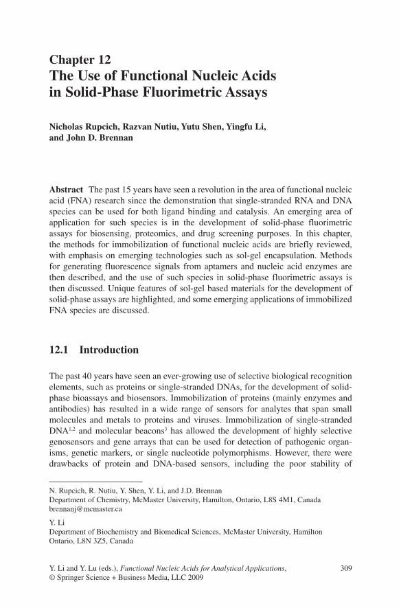
As noted by Di Giusto and King,31 there are four general methods for DNA or RNA immobilization, which are adsorption onto activated surfaces, entrapment of nucleic acids into polymeric matrixes, affinity binding to suitably modified sur-faces, and covalent attachment of nucleic acids to surfaces (Fig. 12.1). These meth-ods can be employed with a wide range of surfaces, including polymers, gels, silica, graphite, metal oxides, or pure metals such as gold, and may also be utilized for
312 N. Rupcich et al.
immobilization onto specialized substrates such as silica or gold nanoparticles or carbon nanotubes to form fiberoptic sensors, microarrays, electrochemical sensors, or bead-based assays.
Adsorption is the simplest method to implement and generally makes use of hydrophobic, electrostatic, or hydrogen bonding interactions between the nucleic acid and the surface. However, it is not possible to control the orientation of the FNA, and binding through multiple points of contact can restrict conforma-tional mobility; this can be particularly problematic for FNAs as such species often require significant conformational changes to generate an analytical signal (see Section 12.3.1). It is also possible that adsorption can alter the active confor-mation of FNAs, causing them to lose binding affinity. In addition, small changes in pH or ionic strength can lead to desorption of the FNA, which makes calibration of sensors difficult.
Matrix entrapment generally employs water-soluble organic polymers such as acrylamide, or electroactive polymers such as polypyrrole or polyanaline to produce a conductive polymer matrix. The FNA can either be entrapped or covalently bound to the matrix, with the latter method circumventing potential issues with leaching of the FNA. A potential advantage of such materials is the ability to form a three-dimensional layer of the nucleic acid, which can lead to much higher binding site density. However, a disadvantage is the difficulty in controlling pore size so as to retain conformational mobility and accessibility while avoiding leaching. These issues can be addressed using inorganic or organic/inorganic composite polymers for entrapment of FNA; this possibility is described in more detail in Section 12.4.
The availability of a wide range of modified nucleotides and the ability to insert such species into specific locations within a nucleic acid via standard solid-phase synthetic methods makes it straightforward to immobilize nucleic acids with a specific orientation using either affinity interactions or covalent attachment strate-gies. Affinity-based methods for immobilization of FNA almost exclusively involve the use of the high-affinity avidin–biotin interaction, which has a dissociation constant of 10−15 M. An advantage of this technique is the ability to label the FNA with biotin at either at the 3′- or 5′-end, or even in the center of the nucleic acid strand, to control the orientation of the FNA on the surface. In certain cases the need for an avidin underlayer can restrict electron transfer and thus compromise electrochemical sensors, but this is not an issue for fluorescence-based sensors.
Fig. 12.1 Four general methods for DNA and RNA immobilization
12 The Use of Functional Nucleic Acids in Solid-Phase Fluorimetric Assays 313
The most prevalent method for immobilization of FNA is through covalent attachment to surfaces. Amine- and carboxylic acid-modified nucleic acids are readily available and can be immobilized to suitably modified surfaces (aldehyde, epoxy, amine) using appropriate coupling agents. Thiol-modified nucleic acids can also be utilized for direct binding to gold surfaces through coordination bonds. In many cases the surface can be initially derivatized with a spacer such as a short polyethylene glycol linker to minimize unwanted interactions between the FNA and the surface, improving performance. An important attribute of covalent immo-bilization is the ability to control the density of the nucleic acid to provide either high binding site density or a higher degree of conformational flexibility for the bound biomolecule.31
An issue with the immobilization of FNA species onto surfaces by any of the above methods is the potential for degradation by exonucleases when such species are to be used for in vivo analysis. Two methods exist to overcome this problem. First, the FNA can be placed in a polymer matrix or within a semipermeable membrane to exclude nucleases on the basis of size; this is an acceptable method for small-molecule sensing applications but is not appropriate for larger analytes. Another approach is to use a nucleic acid that has been modified to increase its resistance to degradation by nucleases. As noted by Di Giusto and King,31 species such as locked nucleic acids (LNA), containing a 2′-O,4′-C-methylene bridge, or 2′-O,4′-C-C-ethylene-bridged nucleic acids (ENA) can significantly reduce the rate of degradation, as can oligo-2′-O-methylribonucleotides or other variants at the 2′-sugar position. The use of phosphorothioate backbone nucleic acids or pep-tide nucleic acids (PNA) also protect against nuclease attack, but in some cases can alter the energetics of hybridization, making it necessary to optimize temperature and ionic strength conditions for optimal selectivity and affinity.
12.3 Assays Utilizing Immobilized Functional Nucleic Acids
A wide range of assays that utilize immobilized FNAs have been reported in the past 10 years, and these have been reviewed extensively.31,41 Applications that utilize tethered nucleic acids have ranged from the use of immobilized FNA species as affinity chromatography phases42,43 to their use as selective capture agents for matrix-assisted laser desorption/ionization (MALDI) mass spectrometry.44 A range of colorimetric sensors have been developed based on DNA-directed assembly or disassembly of gold nanoparticle aggregates,45,46 whereas FNA species modified with redox probes have been widely employed for electrochemical sensors.47 Immobilized FNA species have also been used for label-free sensing using impedance,48 surface plasmon resonance,49 quartz crystal microbalance,50 or cantilever-based transduction methods.51
In this section a brief review of various fluorescence-signaling methods is pro-vided, followed by an overview of fluorescence assays that have been developed

314 N. Rupcich et al.
using tethered FNA species. Advantages and disadvantages of various fluorescence assay formats are described, along with interesting features of the different immo-bilization formats as supports for different assay platforms. In the interests of space, only assays utilizing planar surfaces are described. However, it is noted that there is a large body of work that utilizes FNA and other species bound to nanoparticles for a range of assays, as reviewed by Lu52 and Mirkin.53
12.3.1 Methods of Fluorescence Signaling
The use of FNAs for fluorescence assay and sensor development has been recently reviewed,54 and only key features of different systems are highlighted here. Fluorescence-based sensors have been developed utilizing a range of fluorescence signal readouts, including changes in intensity, wavelength, polarization, or fluo-rescence resonance energy transfer (FRET) signals.
Perhaps the most straightforward method for developing FNA-based solid-phase fluorescence assays is to use FNA species to detect fluorescent analytes, or as cap-ture agents for fluorescence-based competitive binding assays.77 Several examples exist that involve capture of small fluorescent molecules or fluorescently labeled proteins,55 or the capture of proteins using photoaptamers followed by labeling.56 These assays are described in more detail below.
More advanced fluorescence sensor formats utilize fluorescently labeled FNA species that undergo alterations in fluorescence anisotropy57 or intensity58 upon lig-and binding. However, in most cases the total change in signal can be quite small (i.e., Jhaveri et al., reported an intensity increase of only 45% upon binding of ATP to the ATP-binding aptamer).58
More recently, specially designed nucleotide sequences have been prepared with both a fluorophore and a quencher moiety present. Such systems initially have a fluorescently labeled nucleotide (F) in close proximity to a quencher (dabcyl, blackhole quencher, nanogold) -modified nucleotide (Q), resulting in a low fluo-rescence signal. A range of probes have been utilized, with more recent examples including species such as quantum dots,59 which have particular advantages for multiplexed assays.60 Alteration of the nucleotide conformation, either by hybridi-zation with a complementary target, binding of a nonnucleic acid target or catalytic cleavage or an engineered active site, results in spatial separation of the fluorophore and quencher, increasing the fluorophore–quencher distance and producing a large increase in fluorescence intensity.54 In a related method, fluorescently tagged MB or aptamer species have been bound to metal surfaces to produce metal-dependent quenching of the signal.61 Upon analyte binding, the fluorophore moves away from the surface, and fluorescence emission intensity is enhanced.
Early work on dequenching-based fluorescence-signaling DNA systems cen-tered around the use of molecular beacons for detection of complementary DNA species.3,62,63 These probes undergo self-complementary hybridization to form a hairpin structure that places the F and Q moieties in close proximity (Fig. 12.2a).

12 The Use of Functional Nucleic Acids in Solid-Phase Fluorimetric Assays 315
The stem is normally five to seven nucleotides long while the loop of the hairpin is designed to be complementary to the desired target. Upon binding of target the MB moves from the “closed” to the “open” state, which leads to separation of the F and Q moieties, accompanied by a several-fold increase in emission intensity. MB systems can be designed to have high selectivity for perfectly complementary targets relative to those with single nucleotide polymorphisms (SNPs), making them highly useful for genosensing. Furthermore, the use of different labels allows for multiplexing of the emission signal to allow multianalyte sensing. MB systems can also be modified with a fluorophore–quencher pair by hybridization of F-DNA and Q-DNA to primer regions that extend beyond the normal stem region to form tripartite molecular beacons,64 allowing for facile screening of different F-Q pairs to optimize signaling.
While MB species are excellent recognition elements for genosensing, they are not directly amenable to detection of nonnucleic acid targets. A method to extend the utility of MB species is to replace the loop region with an aptamer sequence to create an aptamer beacon, so that binding of the target to the aptamer results in a switch from the closed to the open state.65 Several variants on this concept have been reported, but all rely on disruption or formation of a stem that controls the distance between F- and Q-labeled nucleotides (Fig. 12.2b).
A related concept for generating a change in fluorescence is to simply label the terminal nucleotides in an aptamer sequence with a fluorophore and quencher, and to then make use of conformational changes in the aptamer upon target binding to change the spatial separation of the terminal nucleotides.66 However, this method often results in a loss in intensity upon target binding, which leads to poorer detection limits. Furthermore, this method requires that the aptamer undergo a fairly specific conformational change to ensure that the terminal nucleotides undergo large changes
Fig. 12.2 Signaling DNA probes. (a) Molecular beacons for detection of nucleic acid targets. Aptamer beacons (b) and structure-switching signaling aptamers (c) for detection of nonnucleic acid targets. F, fluorophore; Q, quencher

316 N. Rupcich et al.
in separation upon target binding; this is not always the case, and it is difficult to engineer into existing aptamers.
A more versatile method for converting aptamers to fluorescence-signaling aptamers is the creation of structure-switching signaling aptamers67 (see Fig. 12.2c). This method exploits the fact that all aptamers can adopt two conformations: a weak duplex structure with a complementary DNA and a stronger complexed structure with the target. In this case a F-DNA strand is hybridized to a primer region extend-ing from the aptamer, while a Q-DNA strand is hybridized over a region spanning the primer and aptamer regions, with the quencher adjacent to the fluorophore. Upon binding of a target, the aptamer undergoes a structural switch that destabi-lizes hybridization to the Q-DNA, resulting in displacement of the quencher and an increase in fluorescence intensity. An advantage of this method is that a single aptamer can be labeled with a variety of different fluorophores or quenchers by simply altering the F-DNA and Q-DNA species. Thus, individual aptamers can be labeled with different F/Q pairs to allow for multiplexed detection of different ana-lytes based on unique excitation and/or emission wavelengths for each aptamer.
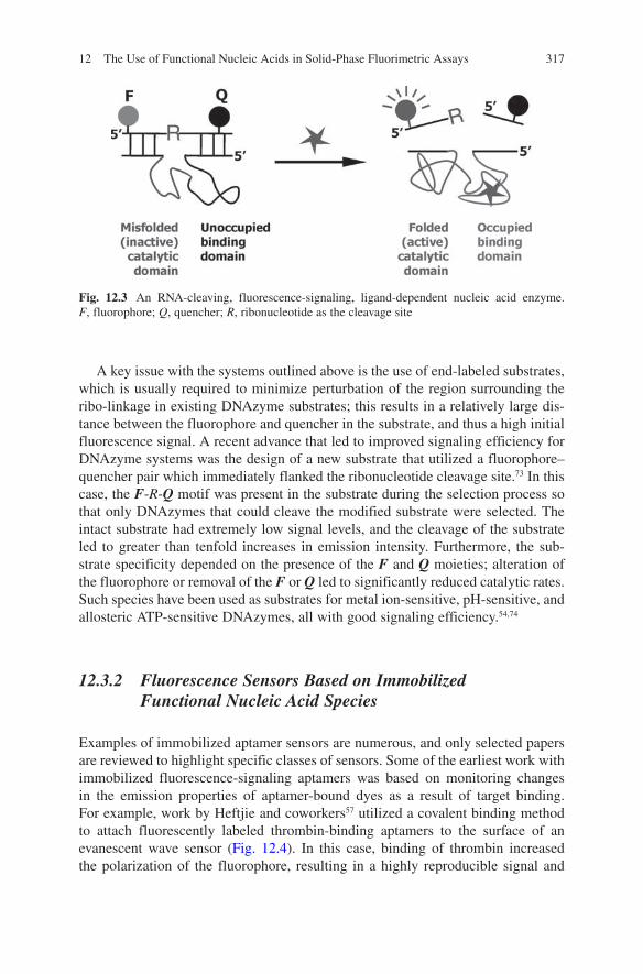
A range of methods have also been developed to create fluorescence-signaling ribozymes and deoxyribozymes. The basis of most signaling enzyme systems is to have binding of a target molecule elicit catalytic activity, which then results in either cleavage of a RNA-bearing substrate or ligation of two nucleic acid strands, with the former method being by far the most widely employed.54 By labeling the substrate nucleotide(s) with a fluorophore–quencher (or donor–acceptor) pair (Fig. 12.3), the spatial proximity of the F-Q (or D-A) pair is altered by cleavage or ligation, generating a signal. This approach has been used for detection of met-als, small molecules, nucleic acids, and proteins. As an example, a DNAzyme for Pb(II) was developed by Li and Lu and used for fluorimetric detection of lead.68,69 A DNA-based substrate bearing a fluorophore, quencher, and ribonucleotide cleav-age site was annealed to the lead-dependent DNAzyme. Addition of Pb(II) acti-vated the catalytic DNA, resulting in cleavage of the ribo-linkage and an increase in fluorescence.
Catalytic molecular beacons have also been developed using an MB motif to control the activity of a DNAzyme.70 In the absence of target, a F/Q-labeled substrate containing a ribonucleotide is only partially annealed to the DNAzyme because of blockage of one of the substrate-binding arms as a result of formation of a closed stem, preventing cleavage. Upon target binding, the MB stem dissoci-ates and the substrate-binding arm associates with the F/Q-labeled substrate. Upon full hybridization with the DNAzyme, the ribo-linkage is cleaved, and fluorescence emission increases.
The concept of using an aptamer rather than a MB for allosteric control of ribozyme and DNAzyme catalysis has recently been reported. In this case, the aptamer is linked to the nucleic acid enzyme by a “communication module,” which allows the aptamer to inhibit the catalytic action of the enzyme.71,72 Upon target binding, the aptamer adopts a folded conformation that releases a substrate-binding sequence within the communication module, resulting in full substrate binding and catalysis (Fig. 12.3).
12 The Use of Functional Nucleic Acids in Solid-Phase Fluorimetric Assays 317
A key issue with the systems outlined above is the use of end-labeled substrates, which is usually required to minimize perturbation of the region surrounding the ribo-linkage in existing DNAzyme substrates; this results in a relatively large dis-tance between the fluorophore and quencher in the substrate, and thus a high initial fluorescence signal. A recent advance that led to improved signaling efficiency for DNAzyme systems was the design of a new substrate that utilized a fluorophore–quencher pair which immediately flanked the ribonucleotide cleavage site.73 In this case, the F-R-Q motif was present in the substrate during the selection process so that only DNAzymes that could cleave the modified substrate were selected. The intact substrate had extremely low signal levels, and the cleavage of the substrate led to greater than tenfold increases in emission intensity. Furthermore, the sub-strate specificity depended on the presence of the F and Q moieties; alteration of the fluorophore or removal of the F or Q led to significantly reduced catalytic rates. Such species have been used as substrates for metal ion-sensitive, pH-sensitive, and allosteric ATP-sensitive DNAzymes, all with good signaling efficiency.54,74
12.3.2 Fluorescence Sensors Based on Immobilized Functional Nucleic Acid Species
Examples of immobilized aptamer sensors are numerous, and only selected papers are reviewed to highlight specific classes of sensors. Some of the earliest work with immobilized fluorescence-signaling aptamers was based on monitoring changes in the emission properties of aptamer-bound dyes as a result of target binding. For example, work by Heftjie and coworkers57 utilized a covalent binding method to attach fluorescently labeled thrombin-binding aptamers to the surface of an evanescent wave sensor (Fig. 12.4). In this case, binding of thrombin increased the polarization of the fluorophore, resulting in a highly reproducible signal and
Fig. 12.3 An RNA-cleaving, fluorescence-signaling, ligand-dependent nucleic acid enzyme. F, fluorophore; Q, quencher; R, ribonucleotide as the cleavage site

318 N. Rupcich et al.
excellent detection limits (0.7 πmol thrombin) but a relatively limited dynamic range (three orders of magnitude).
A more common approach for detection of analytes is through the use of immobilized molecular beacons or aptamer beacons. The first reports in this area focused on the development of fiberoptic sensors that utilized MB systems bound by affinity or covalent interactions. For example, Tan’s group developed a bioti-nylated MB that was bound to a planar silica surface derivatized with physisorbed avidin that was cross-linked with glutaraldehyde.3 The MB-derivatized slide was incorporated into an internal reflection fluorescence instrument to allow evanescent excitation of the MB fluorescence. Upon introduction of complementary DNA, a 2.5-fold enhancement in fluorescence was observed over a period of ~30 min (versus a tenfold enhancement in 20 min in solution). The sensor could be used to detect sub-nanomolar levels of DNA. In a follow-up study, the MB was bound to streptavidin-coated optical fibers and used to detect γ-actin mRNA sequences via changes in evanescently excited fluorescence.75 A detection limit of 1.1 nM was obtained, although the signaling magnitude and response kinetics were again much worse than that in solution, possibly due to a fraction of MB that was either initially unfolded or inaccessible to target.
This use of fiberoptic sensors has also been extended to aptamer beacons, which are formed by replacing the loop of the traditional MB with an aptamer. For example, Yu and coworkers developed a thrombin-binding MB and interfaced it to an optical fiber by affinity interactions.76 The sensor showed a thrombin-dependent increase in emission intensity, and again the performance characteristics were worse than in solution.
12.3.3 Multianalyte Arrays Utilizing Immobilized Molecular Beacon Species
The examples noted above, while demonstrating the potential of MB and aptamer beacon systems for sensing, were relatively limited in performance owing to the ability to detect only a single species. Given the fact that conventional DNA
Fig. 12.4 A fluorescent sensor with an immobilized, fluorophore-labeled aptamer. The target is detected via changes in the evanescent-wave-induced fluorescence anisotropy of the immobilized aptamer

12 The Use of Functional Nucleic Acids in Solid-Phase Fluorimetric Assays 319
microarray technology was relatively mature by the year 2000, it was clear that this would be a major direction for the application of immobilized FNA species. Early work on the development of a multianalyte sensor platform used a hybrid of fiberoptic and array technologies. Walt and coworkers reported on the development of aptamer-linked silica microspheres that were distributed randomly within micro-wells etched into the distal tip of an imaging fiber bundle.77 Thrombin-binding aptamers were bound to one set of beads and a second oligonucleotide was bound on separate beads to account for nonspecific binding. The beads were incubated with a mixture of fluorescently labeled thrombin and nonlabeled thrombin (analyte) in a competitive assay format, captured on the tip of the imaging fiber bundle and imaged. The fiber array could detect 10 nM thrombin with a dynamic range of three orders of magnitude, although there was some interference from albumin observed with the sensor.
More recent work on FNA-based microarrays has focused on the development of multianalyte arrays fabricated by contact or noncontact printing of multiple aptam-ers in a spatially defined pattern on a planar surface, or by synthesis of aptamers on chip. The methods used to immobilize DNA onto arrays are reviewed in detail by Heise and Bier,78 and thus are not covered here.
There are many examples of FNA microarrays, and only a selection of studies is presented here. Chapter 11 of this volume covers the development and application of aptamer arrays in more detail. Early microarray studies focused on fabrication of arrays of molecular beacons and utilized a variety of substrates and immobi-lization chemistries. For example, MBs have been covalently immobilized onto three- dimensional photopolymerized acrylamide gel arrays,79 and were able to discriminate complementary and noncomplementary DNA with a fivefold differ-ence in intensity, with a 100-fold higher signal than similar two-dimensional arrays. More recently, Yao and Tan developed a MB array using biotinylated linkers to bind MBs to avidin-coated surfaces.80 The length of the linker, pH, and ionic strength were optimized to extend the MB away from the surface of the substrate and maxi-mize signaling capacity, and once optimized the array provided a 5.5-fold enhance-ment upon target binding, a significant improvement relative to approximately twofold enhancements observed in earlier work with immobilized MB systems (see above). Work by Malayer and coworkers81 demonstrated the development of an MB array for detection of the 16S rRNA of the bacterium Francisella tularensis. In this case, the MB was covalently immobilized onto aldehyde- or hydrogel-modified slides and was used to discriminate complementary and mismatched sequences. It was demonstrated that the MB array showed poor discrimination between matched targets and those with up to three mismatches, particularly if mismatches were in terminal regions of the targets. Interestingly, the ability to discriminate between matched and mismatched targets improved as the temperature of the assay was increased to a value just below the melting temperature of the matched target. However, this study clearly demonstrated that use of single MB sequences for determination of SNPs could be fraught with poor selectivity, arguing for the need for multiplexed analysis. The most recent study to employ MB arrays involved the development of an array to detect Escherichia coli O157:H7.82 This system uses

320 N. Rupcich et al.
wavelength-shifting MB systems based on the use of a donor–acceptor pair rather than the conventional fluorophore–quencher system employed in the majority of MB arrays. The ability to detect hybridization via a color shift avoids false-negative signals that may arise from photobleaching or washing-off of donor probes. The array could detect as little as 1 ng/μl of E. coli marker genes, with changes in the ratio of red to green fluorescence (due to FRET) being as high as 30-fold in the presence of ~10 ng/μl cDNA.
An interesting variation on traditional MB systems that makes clever use of immobilized MBs is the modulation of fluorescence as a result of quenching by metal surfaces.83 Such systems utilize MBs bound to nanoparticle or planar gold surfaces via thiol-modified terminal groups. In the “closed” state, the fluorophore on the other terminus is quenched by its close proximity to the gold surface via met-al-induced quenching. Upon binding to a target (genes from methicillin-resistant Staphylococcus aureus, MRSA), the probe moves several nanometers away from the surface, and the fluorescence is enhanced by a factor of up to 20-fold, as shown in Fig. 12.5.61 Further optimization of the density of the MB on the surface (via addition of mercaptopropanol spacers) and the nucleotides in the stem of the MB led to a significant increase in signal enhancement (100-fold) relative to the unopti-mized system.84 An important aspect of the optimization was the ability to provide sufficient space to allow hybridization to occur, while at the same time capping the gold surface to prevent unwanted interactions between the DNA and the metal. This platform could be used to produce microarrays that could discriminate SNPs with a tenfold difference in intensity relative to complementary DNA. However, the immobilized MB could be reused only a limited number of times because of loss of the MB from the gold surface during regeneration.
Most recently, this methodology has been extended to catalytic molecular beacons on gold surfaces.85 A thiolated version of the Pb(II) dependent DNAzyme (17E) was immobilized onto a Au surface along with mercaptohexanol spacer molecules and hybridized with a complementary fluorophore-labeled substrate nucleotide to form a double-stranded structure. Introduction of lead results in cleav-age of the substrate and release of the fluorophore into solution, where it is detected
Fig. 12.5 A biosensor with molecular beacon immobilized on gold surface, which acts as the quencher

12 The Use of Functional Nucleic Acids in Solid-Phase Fluorimetric Assays 321
after removal of the DNAzyme modified substrate. In this format, an eightfold increase in intensity was observed upon addition of Pb(II), although selectivity rela-tive to other divalent metal ions was poor (only threefold difference in intensity). While such a scheme does not make use of metal-induced quenching, it shows the potential of using self-assembled DNAzyme monolayers for generation of fluores-cence signals.
12.3.4 Multianalyte Arrays Utilizing Immobilized Aptamers
Although significant work has been done on MB microarrays, these systems are inherently limited to detection of DNA or RNA targets. To overcome this issue, a large number of groups have developed a range of aptamer-based microarrays for detection of proteins and small molecules.
Several studies have utilized nonlabeled aptamer arrays for the detection of fluorescently labeled proteins, or for capture of proteins followed by on-array labeling or detection using fluorescently labeled secondary antibodies. For exam-ple, Lindner and coworkers reported on a comparison between DNA aptamers and monoclonal antibodies for detection of labeled proteins on microarrays.86 This study demonstrated that aptamers could be used for protein detection with perform-ance that was equivalent to or better than antibody-based arrays, although it must be noted that this study utilized a limited number of aptamers. It will be necessary to prepare arrays with a much larger number of aptamers to a wide range of analytes to make this technology competitive with antibody arrays.
A series of studies from Ellington’s group have focused on the development of protein-sensing RNA aptamer microarrays. An early study utilized lysozyme- or ricin-binding aptamers on beads, which were dispensed into the wells of an array-based “electronic tongue”.87 Biotinylated agarose beads were derivatized with streptavidin and used to capture biotinylated aptamers. Bound proteins were detected based on the emission of a label on the protein and could be regener-ated and reused over many assay cycles. A more conventional RNA microarray was fabricated by preparing biotinylated RNA aptamers for lysozyme by in vivo transcription and spotting these onto streptavidin-coated slides.88 A total of 24 different lysozyme-binding aptamers, derived from an automated in vitro selec-tion protocol, were spotted along with two clone pools and tested for binding to fluorescently labeled lysozyme. In this manner, it was possible to rapidly identify the highest affinity aptamer from the set of clones or to select clones with specific affinities or dynamic ranges. Overall, this study showed that automated selection combined with high-throughput screening of aptamer affinity on microarrays could be used to rapidly identify optimal aptamers for desired targets. Detailed methods for the fabrication, processing, and analysis of such microarrays have recently been reported,89 as have methods for optimizing aptamer arrays for multiprotein analysis, including choice of surface chemistry, orientation of the aptamer, and type of assay and washing buffer.55

322 N. Rupcich et al.
An interesting variant on aptamer microarrays is the use of photoaptamer arrays for protein detection. Early work by Gold and coworkers suggested that arrays of aptamers with photoreactive 5-bromodeoxyuridine groups could be used to capture target proteins onto specific locations on an array. Irradiation with UV light could then be used to photo-cross-link the protein permanently to the aptamer, allowing for harsh washing steps to remove nonspecifically bound proteins. Protein-specific stains could then be added to determine the presence and/or concentration of protein in the original sample.90 To demonstrate the concept, photoaptamer microarrays were devel-oped by pin-printing photoaptamers onto amine-derivatized slides to allow covalent binding and were used for protein detection.56 Introduction of a sample of proteins was performed, followed by photo-cross-linking and washing. The array was then exposed to either a general protein-binding stain such as NHS-AlexaFluor555 or to a specific fluorescently tagged antibody to allow for general or specific detection of bound proteins, respectively. The arrays were demonstrated to be capable of detecting proteins in either buffer or diluted serum, and in optimal cases had detection limits in the low fM range. Use of antibody-based staining allowed for more selective deter-mination of proteins, as the protein had to bind selectively to both the photoaptamer and secondary antibody, minimizing false positives. Furthermore, the use of a cross-linking reaction increased the stringency of the array based assay, as nonprotein species that might nonselectively adsorb to the aptamer were not covalently captured and thus were removed during the harsh washing step.
Two issues with the this aptamer array system are the need for multiple washing steps and for labeling of captured targets, either before or after capture or for devel-opment of competitive binding assays. This need results in added complexity for the assay and significantly limits that application of such arrays for small-molecule analysis, as neither staining nor secondary antibody binding is possible in this case. To overcome these issues, significant work has been done to utilize signaling aptamers in microarray-based assays for both protein and small-molecule analysis.
An early study employed signaling aptamers composed of either RNA or DNA to prepare a microarray on a planar waveguide surface.91 The array was excited evanescently by total internal reflection, and the emission was detected through a polarizing beamsplitter to produce two images, one for parallel polarized light and one for perpendicularly polarized light, thus allowing for a fluorescence anisotropy based assay on the array. Initial studies showed that the binding of thrombin to array elements containing thrombin-binding aptamer resulted in a significant increase in anisotropy for these spots and resulted in a detection limit of ~1 nM. A microarray was constructed using four aptamers to different proteins and demonstrated excel-lent selectivity for detection of different proteins in cell lysates and serum, although some cross reactivity was noticed. The authors also reported that the RNA aptamers had poor stability relative to DNA aptamers owing to the presence of ribonucleases in the biological samples tested, and suggested that modified RNA species would be required to overcome this problem.
A second signaling aptamer array system was developed using a strategy wherein a RNA aptamer against human immunodeficiency virus (HIV)-1 Tat was split into two nonfunctional units, one of which (the anchor oligo) was immobilized on

12 The Use of Functional Nucleic Acids in Solid-Phase Fluorimetric Assays 323
aldehyde-coated slides in an array pattern by covalent interactions. In the presence of target, the other half of the aptamer (hybridizing oligo), which was labeled with a fluorescent probe, reassembled on the surface, resulting in an increase in fluo-rescence intensity.92 The assay was shown to be amenable to detection of protein in buffer, but was unable to detect proteins in HeLa cell extracts even with added RNase inhibitors to minimize degradation of the RNA aptamer, again highlighting the need for modified RNA aptamers. A further disadvantage of this assay format relative to the methods described above was the need to add the hybridizing oligo in solution and to perform washing steps to remove nonselectively bound oligo species to minimize fluorescence background signals.
An interesting demonstration of an immobilized signaling aptamer system for protein detection involved the use of DNA tiles containing a fluorescently labeled thrombin aptamer sequence to allow self-assembly of DNA tiles onto a surface via assembly of “sticky ends” (Fig. 12.6).93 This approach resulted in a periodic DNA structure on the surface that had a thrombin-binding aptamer every 27 nm within the array. The signaling aptamer underwent an increase in intensity greater than twofold upon thrombin binding, leading to a useable signal. An interesting aspect of this work was the ability to capture the analyte in solution in the first step, followed by assembly of the DNA tiles along with the captured protein to produce a fluorescent pattern on a surface. This method produced higher overall intensity values relative to adding protein to a preformed array, presumably due to the ability to avoid surface effects such as inaccessibility of bound aptamers.
A final example of a signaling aptamer array system utilized a polythiophene–DNA aptamer aggregate for detection of thrombin.94 The basis of this system is the formation of a tight complex between polythiophene and ssDNA, which results in effective quenching of polythiophene fluorescence. Upon binding of either complementary DNA or a protein target, the DNA adopts a different structure, which leads to a significant enhancement in polythiophene emission. An array was fabricated with Cy-3-labeled antithrombin aptamers via covalent immobilization of the DNA, which was preassociated with the polymer, to an aldehyde surface. Addition of thrombin resulted in a conformational change in the DNA that allowed for dequenching of the polymer fluorescence and hence efficient energy transfer to the Cy-3 labels. The presence of several Cy-3-labeled DNA aptamers within the polymer–aptamer complex led to enhanced emission from the acceptor and resulted
Fig. 12.6 A biosensor that uses DNA tiles containing a fluorescently labeled aptamer (Reproduced with permission from ref. 93. Copyright [2006] Wiley)
324 N. Rupcich et al.
in a detection limit in the low pM range. The array also demonstrated excellent selectivity for thrombin over nontarget proteins, likely because of the inability of such proteins to induce the necessary conformational change in the aptamer to gen-erate a signal. In this manner, issues with signals arising from nonspecific binding of proteins were avoided.
While there are several examples of signaling aptamer microarrays, as noted above, it is noteworthy that there are few reports of arrays fabricated with signaling deoxyribozymes, although Ellington did report on a DNAzyme array that utilized radioactivity for detection,95 while Breaker and coworkers reported on riboswitch arrays for analysis of complex mixtures.96 Such species might be expected to pro-vide good resistance against changes in emission owing to nonspecific binding, because only specific binding should lead to catalysis and thus generation of a signal. As noted below, there are solid-phase assays involving signaling aptamers and DNAzymes that are moving in this direction.
12.4 Sol-gel Immobilization Methods
12.4.1 The Sol-gel Process for Biomolecule Entrapment
While there has been significant progress in the development of DNA immobilization protocols during the past 20 years, as noted here, many of the methods result in monolayer or submonolayer coverage and thus can suffer from poor sensitivity. In the case of FNAs, there is also the potential for surface-mediated changes in FNA conformation and mobility, which could potentially reduce activity and long-term stability and lead to the degradation of FNA by nucleases.31
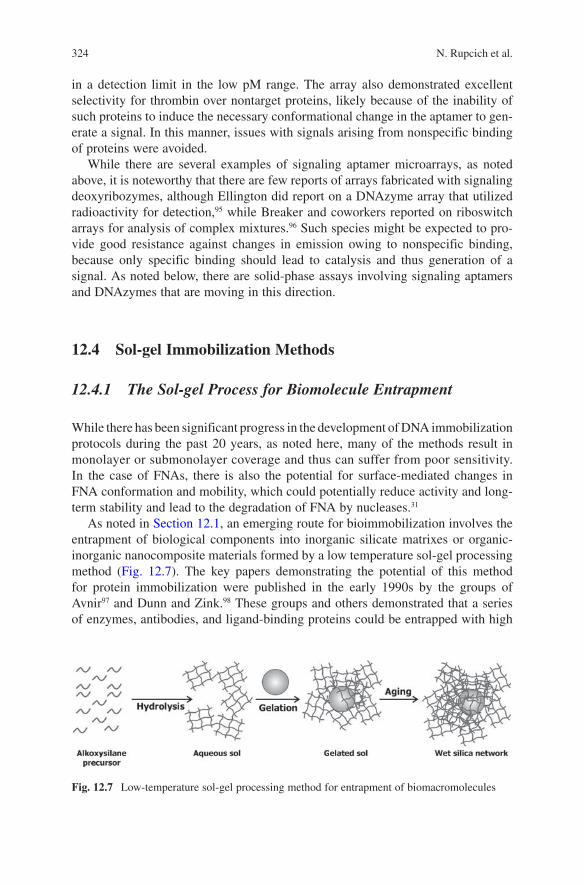
As noted in Section 12.1, an emerging route for bioimmobilization involves the entrapment of biological components into inorganic silicate matrixes or organic-inorganic nanocomposite materials formed by a low temperature sol-gel processing method (Fig. 12.7). The key papers demonstrating the potential of this method for protein immobilization were published in the early 1990s by the groups of Avnir97 and Dunn and Zink.98 These groups and others demonstrated that a series of enzymes, antibodies, and ligand-binding proteins could be entrapped with high
Fig. 12.7 Low-temperature sol-gel processing method for entrapment of biomacromolecules

12 The Use of Functional Nucleic Acids in Solid-Phase Fluorimetric Assays 325
activity into glasses derived from alkoxysilanes such as tetraethyl orthosilicate (TEOS) using a general protocol similar to that outlined in Fig. 12.7. The formation of the material begins with the partial or complete hydrolysis of a suitable precur-sor to form an aqueous sol, which is an aqueous suspension of silica nanostructures (oligomers, colloids, fibrils, etc.). The precursors can consist of tetraalkoxysilanes, mono-, di-, or tri-alkyl alkoxysilanes, or may comprise functional groups such as alkenyl, aryl, amino, carboxyl, or thiol as well as redox-active moieties, flavins, or quinones. Other metal centers such as Al, Ti, V, or Ce may also be used to alter the material properties. The hydrolysis reaction itself can be either acid- or base catalyzed, and more advanced precursors, such as diglycerylsilane,99 can be hydrolyzed at neutral pH and liberate the biocompatible byproduct glycerol, which helps to stabilize biomolecules.
Following hydrolysis, the sol is mixed with a buffered aqueous solution containing the biomolecule along with any catalysts, drying control additives, polymers, tem-plating agents, redox species, or fillers that may be required to modify the properties of the final material. The change in pH along with the presence of added salts and catalysts promotes a large-scale polymerization reaction over a period of minutes to hours, resulting in gelation of the sol and entrapment of the biomolecule. The initial gels are soft and have high water content (50–80%) and large pores (up to 200 nm diameter). Aging of the wet silica network over a period of days to weeks promotes further condensation and strengthens the network. During this stage, entrapped alcohol and/or water resulting from the initial hydrolysis and condensation reac-tions will be removed from the matrix, causing the matrix to shrink by 10–30%, the pore diameters to decrease by about 25%, and the relative proportion of siloxane to silanol groups to increase owing to coarsening of the material.100 Finally, the aged material can be partially dried, resulting in the loss of most of the interstitial water, further cross-linking of the matrix, shrinkage of the pores to the range of 2–20 nm, and overall shrinkage of the material by 10–85% of its initial volume, depending on the types of precursors and processing conditions used.
In addition to enzymes and antibodies, sol-gel materials have been used to entrap a wide variety of biological species, including regulatory proteins, membrane-bound proteins, and even whole cells.32–36 However, this immobilization platform did not gain significant popularity for the entrapment of DNA species, mainly because most DNA sensors were targeted to binding of large complementary DNA strands, and thus immobilization by simple size exclusion was not possible.
With the emergence of functional nucleic acids, and the use of these species for detection of small molecules, the sol-gel platform became amenable to entrapment of FNA species for small-molecule sensing. Potential advantages of sol-gel-derived silicate materials for immobilization of FNA species are as follows: (a) they can be made to be optically transparent, making them ideal for the development of opti-cal sensors; (b) they are open to a wide variety of chemical modifications based on the inclusion of various polymer additives, redox modifiers, and organically modified silanes to create organic–inorganic composite materials; (c) they have a tunable pore size and pore distribution, which allows small molecules and ions to diffuse into the matrix while large biomolecules remain trapped in the pores,
326 N. Rupcich et al.
allowing size-dependent bioanalysis; and (d) they can be made in a variety of forms (thin films, bulk glasses, powders, microarrays, etc.) to allow for development of novel bioanalytical devices. Specific examples of FNA entrapment within sol-gel materials, including both aptamers and DNAzymes, are provided in the following sections.
12.4.2 Immobilization of Molecular Beacons onto Sol-gel Materials
The earliest work involving the interfacing of FNA species and sol-gel supports involved the immobilization of biotinylated molecular beacons onto sol-gel mate-rials that contained entrapped biotin-derivatized BSA which were coated, post gelation, with a layer of streptavidin. The SA layer acted as a bridge to link the MB species to the sol-gel material.101 The advantages of utilizing sol-gel materi-als, relative to formation of MB monolayers on solid supports, included a faster response time, improved long-term stability for the immobilized MB, and a higher loading of MB owing to the high surface area of the sol-gel material, which led to improved detection limits. The sol-gel immobilized MB showed high sensitivity for detection of target DNA and good selectivity against single-base mismatches, indicating good potential for SNP analysis. Although the sol-gel method showed significant advantages for immobilization of biotinylated FNA species, there is no follow-up study reported in which this method is used for the immobilization of either aptamers or DNAzymes.
12.4.3 Entrapment of DNA Aptamers Within Sol-gel Materials
Our groups provided the first report on the entrapment of a functional nucleic acid within a sol-gel-derived material.102 In this work, a structure-switching sig-naling aptamer was entrapped into a range of sol-gel materials derived from the biocompatible precursors sodium silicate or diglycerylsilane. A number of factors were found to affect the performance of the entrapped aptamer, including the type of precursor, the presence of additives, and the length of the Q-DNA. Our results showed that entrapment in any material resulted in a slight reduction in the signal-ing level relative to solution (tenfold vs 12-fold), with signaling levels decreasing dramatically if cationic silanes were included in the material. The rate of signal evolution was also decreased by a factor of approximately tenfold, and up to 50% of the aptamer was able to leach from the material. However, of the aptamer that was retained, more than 90% of the entrapped aptamer remained accessible to externally added small molecules. Overall, the results indicated that aptamers could be entrapped with retention of conformational freedom, although inclusion of

12 The Use of Functional Nucleic Acids in Solid-Phase Fluorimetric Assays 327
cationic silanes, such as aminopropyltriethoxysilane (APTES), resulted in the DNA backbone of the aptamer electrostatically binding to the matrix, with an inability to undergo a structural switch upon introduction of ATP.
As shown in Fig. 12.8a, the entrapped ATP-binding aptamer retained selectivity for ATP over other nucleotide triphosphates (such as GTP). The aptamer also retained sensitivity to ATP concentration that was similar to that observed in solu-tion. Importantly, the dynamic range of the entrapped aptamer matched well with the physiological range of ATP, and thus the sensor could potentially be used for direct detection of ATP without the need for sample dilution. The aptamer retained full signaling capability for at least 30 days when aged in buffer solution, and required 3 months before signaling ability was completely lost, likely owing to continued evolution of the sol-gel matrix, which could lead to pore collapse and subsequent restriction of dynamic motion for the entrapped DNA.103
One of the key advantages of entrapping the aptamer within the silica matrix was that it provided a steric barrier to entry of digestive enzymes which could degrade the biomolecule. Addition of DNAse I to the aptamer in solution results in an increased fluorescence signal (5.5-fold in 1 h, eightfold maximum) from DNA deg-radation (see Fig. 12.8b), due to dehybridization of the QDNA and/or release of the fluorescein-labeled nucleotide, causing an overall increase in distance between the fluorescein- and dabcyl-labeled nucleotides. Digestion of the FNA immobilized by standard avidin–biotin affinity interactions was essentially identical to that obtained in solution, indicating no protection of the aptamer in this case. On the other hand, the entrapped aptamer underwent only a very minor change in fluorescence inten-sity (~18%), likely because of digestion of a small fraction (<2%) of the aptamer
Fig. 12.8 Performance of ATP-binding DNA aptamer entrapped in silica sol-gel. (a) Comparison of selectivity of the aptamer and an inactive mutant. All analyses were done using 0.5 mM of the specified analyte: , aptamer + ATP; , blank; , mutant + ATP; , aptamer + GTP. (b) Changes in emission intensity of structure-switching aptamers upon exposure to DNase: , aptamer in solu-tion; , aptamer immobilized on a streptavidin-coated microwell; , aptamer entrapped in sodium silicate; , DNase free control. (Reproduced with permission from ref. 102. Copyright [2005] American Chemical Society)

328 N. Rupcich et al.
that resided close to the surface of the silica monolith. These results indicate that the DNA was not accessible to the DNAse I, and thus was well protected from digestion owing to the mesoporous silica matrix.
12.4.4 Entrapment of DNA Enzymes in Sol-gel Materials
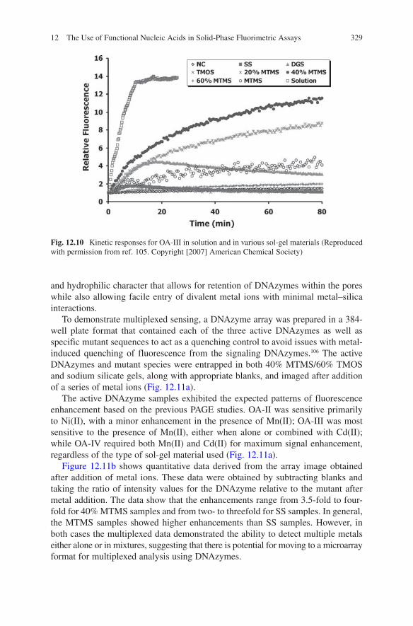
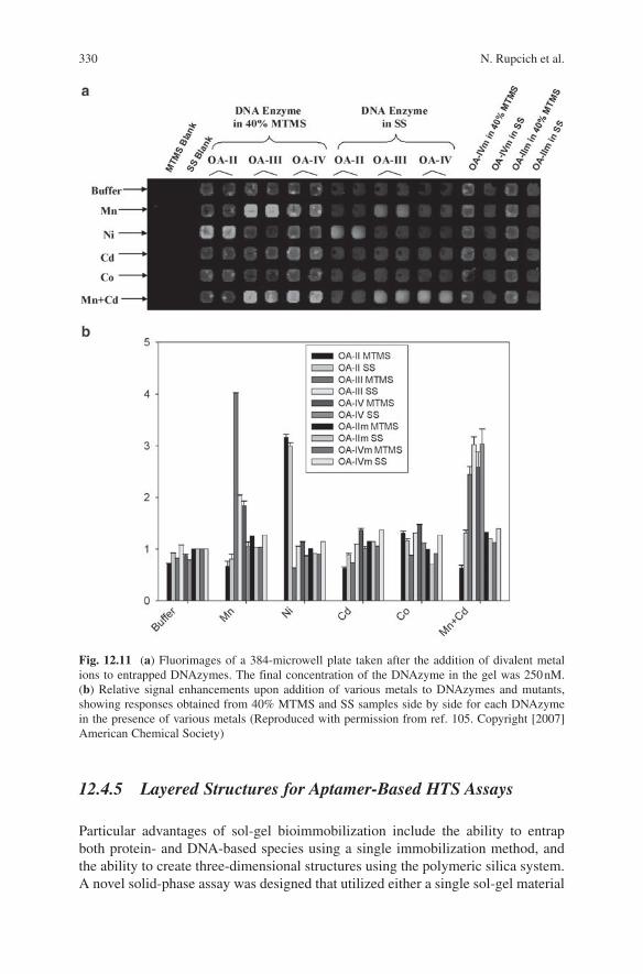
To further assess the potential of sol-gel based materials for entrapment of FNA species, three fluorescence-signaling DNA enzymes, denoted as OA-II, OA-III, and OA-IV,104 were examined (the secondary structures of these DNAzymes are shown in Fig. 12.9), each of which contains the F-R-Q element described above and exhibits distinct profiles of divalent metal ion specificities. PAGE studies previously showed that OA-II was active in the presence of Mg(II) and Ni(II), OA-III strictly used Mn(II), while OA-IV required both Mn(II) and Cd(II) to produce cleavage of the ribo-linkage. The signaling abilities of the deoxyribozymes were examined both in solution and in a range of polar and organic–inorganic composite sol-gel materi-als to determine if materials that were not optimal for protein entrapment (i.e., those that were partially hydrophobic and evolved alcohol byproducts) might be suit-able for the entrapment of metal sensing DNAzymes.105 Figure 12.10 shows the rela-tive fluorescence enhancement obtained for OA-III upon addition of divalent metals to the enzymes in solution and when entrapped in the various sol-gel materials.
In solution, the signal enhancement was about 14-fold, with full signal develop-ment within 10 min. Upon entrapment, the signal enhancements and rates of signal development were highly dependent on the type of sol-gel material used. As shown in Fig. 12.10, some of the materials resulted in essentially no signal enhance-ments upon addition of metals, while other materials resulted in significant signal enhancements; all materials showed slower responses than in solution. Overall, the data showed that composite materials formed from 40% methyltrimethoxysilane (MTMS) and 60% tetramethylorthosilicate (TMOS) provided the best performance for the entrapped DNAzyme, resulting in the highest signal enhancements, highest signaling rates and lowest degree of leaching (similar observations were made with OA-II and OA-IV). It is likely that such materials have a balance of hydrophobic
Fig. 12.9 Putative secondary structures of metal sensing deoxyribozymes: OA-II, OA-III, and OA-IV (Reproduced with permission from ref. 105. Copyright [2007] American Chemical Society)
12 The Use of Functional Nucleic Acids in Solid-Phase Fluorimetric Assays 329
and hydrophilic character that allows for retention of DNAzymes within the pores while also allowing facile entry of divalent metal ions with minimal metal–silica interactions.
To demonstrate multiplexed sensing, a DNAzyme array was prepared in a 384-well plate format that contained each of the three active DNAzymes as well as specific mutant sequences to act as a quenching control to avoid issues with metal-induced quenching of fluorescence from the signaling DNAzymes.106 The active DNAzymes and mutant species were entrapped in both 40% MTMS/60% TMOS and sodium silicate gels, along with appropriate blanks, and imaged after addition of a series of metal ions (Fig. 12.11a).
The active DNAzyme samples exhibited the expected patterns of fluorescence enhancement based on the previous PAGE studies. OA-II was sensitive primarily to Ni(II), with a minor enhancement in the presence of Mn(II); OA-III was most sensitive to the presence of Mn(II), either when alone or combined with Cd(II); while OA-IV required both Mn(II) and Cd(II) for maximum signal enhancement, regardless of the type of sol-gel material used (Fig. 12.11a).
Figure 12.11b shows quantitative data derived from the array image obtained after addition of metal ions. These data were obtained by subtracting blanks and taking the ratio of intensity values for the DNAzyme relative to the mutant after metal addition. The data show that the enhancements range from 3.5-fold to four-fold for 40% MTMS samples and from two- to threefold for SS samples. In general, the MTMS samples showed higher enhancements than SS samples. However, in both cases the multiplexed data demonstrated the ability to detect multiple metals either alone or in mixtures, suggesting that there is potential for moving to a microarray format for multiplexed analysis using DNAzymes.
Fig. 12.10 Kinetic responses for OA-III in solution and in various sol-gel materials (Reproduced with permission from ref. 105. Copyright [2007] American Chemical Society)
330 N. Rupcich et al.
12.4.5 Layered Structures for Aptamer-Based HTS Assays
Particular advantages of sol-gel bioimmobilization include the ability to entrap both protein- and DNA-based species using a single immobilization method, and the ability to create three-dimensional structures using the polymeric silica system. A novel solid-phase assay was designed that utilized either a single sol-gel material
Fig. 12.11 (a) Fluorimages of a 384-microwell plate taken after the addition of divalent metal ions to entrapped DNAzymes. The final concentration of the DNAzyme in the gel was 250 nM. (b) Relative signal enhancements upon addition of various metals to DNAzymes and mutants, showing responses obtained from 40% MTMS and SS samples side by side for each DNAzyme in the presence of various metals (Reproduced with permission from ref. 105. Copyright [2007] American Chemical Society)
12 The Use of Functional Nucleic Acids in Solid-Phase Fluorimetric Assays 331
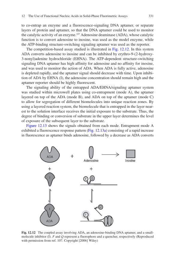
to co-entrap an enzyme and a fluorescence-signaling DNA aptamer, or separate layers of protein and aptamer, so that the DNA aptamer could be used to monitor the catalytic activity of an enzyme.107 Adenosine deaminase (ADA), whose catalytic function is to convert adenosine to inosine, was used as the model enzyme, while the ATP-binding structure-switching signaling aptamer was used as the reporter.
The competition-based assay studied is illustrated in Fig. 12.12. In this system ADA converts adenosine to inosine and can be inhibited by erythro-9-(2-hydroxy-3-nonyl)adenine hydrochloride (EHNA). The ATP-dependent structure-switching signaling DNA aptamer has high affinity for adenosine and no affinity for inosine, and was used to monitor the action of ADA. When ADA is fully active, adenosine is depleted rapidly, and the aptamer signal should decrease with time. Upon inhibi-tion of ADA by EHNA (I), the adenosine concentration should remain high and the aptamer reporter should be highly fluorescent.
The signaling ability of the entrapped ADA/EHNA/signaling aptamer system was studied within microwell plates using co-entrapment (mode A), the aptamer layered on top of the ADA (mode B), and ADA on top of the aptamer (mode C) to allow for segregation of different biomolecules into unique reaction zones. By using a layered reaction system, the biomolecule that is entrapped in the layer near-est to the solution interface receives the initial exposure to the substrate. Thus, the degree of binding or conversion of substrate in the upper layer determines the level of exposure of the subsequent layer to the substrate.
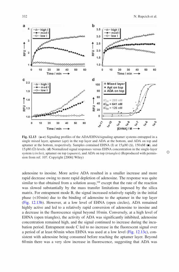
Figure 12.13 shows the signals obtained from each mode. Entrapment mode A exhibited a fluorescence response pattern (Fig. 12.13a) consisting of a rapid increase in fluorescence as aptamer binds adenosine, followed by a decrease as ADA converts
Fig. 12.12 The coupled assay involving ADA, an adenosine-binding DNA aptamer, and a small-molecule inhibitor (I). F and Q represent a fluorophore and a quencher, respectively (Reproduced with permission from ref. 107. Copyright [2006] Wiley)
332 N. Rupcich et al.
adenosine to inosine. More active ADA resulted in a smaller increase and more rapid decrease owing to more rapid depletion of adenosine. The response was quite similar to that obtained from a solution assay,108 except that the rate of the reaction was slowed substantially by the mass transfer limitations imposed by the silica matrix. For entrapment mode B, the signal increased relatively rapidly in the initial phase (<10 min) due to the binding of adenosine to the aptamer in the top layer (Fig. 12.13b). However, at a low level of EHNA (open circles), ADA remained highly active and led to a relatively rapid conversion of adenosine to inosine and a decrease in the fluorescence signal beyond 10 min. Conversely, at a high level of EHNA (open triangles), the activity of ADA was significantly inhibited, adenosine concentration remained high, and the signal continued to increase during the incu-bation period. Entrapment mode C led to no increase in the fluorescent signal over a period of at least 60 min when EHNA was used at a low level (Fig. 12.13c), con-sistent with adenosine being consumed before reaching the aptamer layer. Beyond 60 min there was a very slow increase in fluorescence, suggesting that ADA was
Fig. 12.13 (a–c) Signaling profiles of the ADA/EHNA/signaling aptamer systems entrapped in a single mixed layer, aptamer (apt) in the top layer and ADA at the bottom, and ADA on top and aptamer at the bottom, respectively. Samples contained EHNA (I) at 15 μM ( ), 150 nM ( ), and 15 pM ( ) levels. (d) Normalized signal responses versus EHNA concentration in the single-layer system (circles), aptamer on top (squares), and ADA on top (triangles) (Reproduced with permis-sion from ref. 107. Copyright [2006] Wiley)

12 The Use of Functional Nucleic Acids in Solid-Phase Fluorimetric Assays 333
not able to fully consume all adenosine that was added. When ADA was strongly inhibited by EHNA (open triangles), the fluorescence response showed a relatively slow increase over the first 20 min, followed by a more rapid increase in intensity over a period of 40 min as adenosine penetrated into the aptamer layer. The slope of the response increased with increasing inhibitor concentration, reflecting the loss of ADA activity. It is noted that even after 2 h the system did not show the maximum expected signal, indicating that full equilibration of substrate into the bottom layer likely required a longer time. The data convincingly demonstrate that both ADA and the signaling aptamer remain active within silica and that the signaling aptamer can report the activity of ADA, regardless of which entrapment option is used.
The ability of each entrapment method to generate accurate inhibition constants was also assessed. For entrapment modes A and B, fitting the fluorescence responses to the concentration change of EHNA (see Fig. 12.13d, circles and squares) accord-ing to the Hill equation resulted in inaccurate IC
50 values of 269 ± 15 and 641 ±
9 nM, respectively. Using a KM
value of 106 μM for ADA entrapped in silica,109 KI
values of 26 and 61 nM were obtained, which are approximately fourfold to ten-fold larger than the literature value of 6 nM.110 The increase in the K
I value is most
likely because the mixed system never attains equilibrium with respect to either the aptamer–adenosine or ADA–adenosine reactions, whereas the system with the aptamer in the top layer results in substrate depletion in the ADA layer. On the other hand, entrapment mode C (triangles in Fig. 12.13d) produced an IC
50 of 126 ± 7 and
a KI of 12 nM, both of which were close to the reported values. The better accuracy
of these values when ADA is in the top layer is most likely the result of the ability of the inhibitor to penetrate through the thinner upper layer (1.1 vs 2.3 mm for the mixed system) and thus establish equilibrium with the enzyme.
The use of layered entrapment systems for the separation of coupled assays can offer additional advantages. First, the spatial separation of biomolecules may allow biological species that are incompatible (such as a DNase and a DNA aptamer) to be coimmobilized, which would not be possible using a single monolith. Second, the thickness of each layer can either be increased to introduce a delay time (which provides time to perform multiple assay steps before reading), or decreased to accelerate the overall reaction. Finally, the concentration of biomolecule within each layer and the chemical composition of the individual silica layers can also be optimized to ensure that the molar ratio and activity of reagents is optimized. As aptamers can be generated by in vitro selection for virtually any target of inter-est6 and structure-switching signaling aptamers can be either rationally designed for any known aptamer67 or selected directly when there is no existing aptamer,111 it is conceivable that many specific signaling aptamer–protein enzyme pairs can be developed in the future. Finally, while the foregoing example utilized aptamers that bind to the substrate of an enzyme, it is likely that even more straightforward assay formats could be developed with aptamers which bind products of enzymatic reactions,112 since in this case there is no competition between the aptamer and enzyme for the product molecules. For example, a recently reported ADP-binding aptamer could be used to develop a universal solid-phase kinase assay to comple-ment the previously described solution-based kinase assay.113 Other aptamers that

334 N. Rupcich et al.
could selectively bind NAD(P)H over NAD(P)+ could also be used for a variety of redox enzyme assays, further extending the potential of this method for both sens-ing and high-throughput drug screening.
12.5 Emerging Applications
The examples outlined here demonstrate a wide range of potential applications for solid-phase assays using fluorescence-signaling FNAs, including biosensing, proteomics, and high-throughput screening using fiberoptic, waveguide, and micro-array formats. In this section, a few emerging applications of DNA aptamers are considered, including recent advances in colorimetric detection using FNA species bearing gold nanoparticles for the development of “dipstick” tests, innovative signaling methods involving rolling-circle amplification, and emerging applications for FNA microarrays.
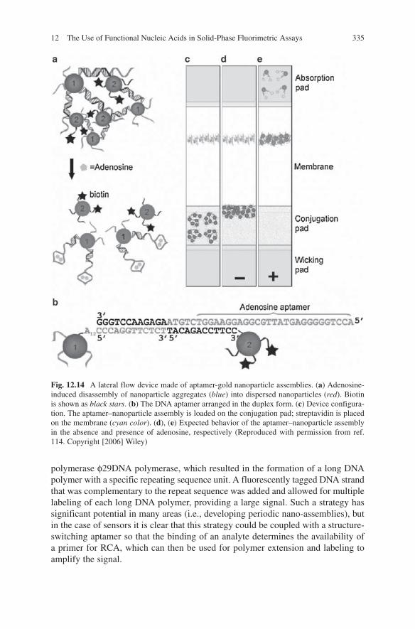
As already noted, an emerging area of interest in aptamer and DNAzyme-based detection is the development of rapid colorimetric assays, which in ideal cases removes the need for instrumentation to allow reading of a signal.46 A recent article by Liu and coworkers114 described the use of a lateral flow device containing ATP- or cocaine-binding aptamers bound to two types of DNA-modified gold nanopar-ticles, one (particle 1) containing a short sequence complementary to a primer extension of the aptamer, and the other (particle 2) containing a biotinylated DNA sequence that was complementary to the aptamer (Fig. 12.14a, b).
In the absence of target, the two types of particles form a large, multiparticle complex that is unable to migrate through the membrane of the lateral flow device. Upon addition of ATP or cocaine, the aptamer undergoes a structural switch to dissociate the DNA bound to particle 2 (similar to the structure switching signal-ing aptamers described above). The dissociated particle is small enough to move through the membrane of the flow device and is captured on a small strip of streptavidin placed at the top of the membrane (see Fig. 12.14c–e). The dipstick test allowed for rapid detection of target analytes present in both buffer and serum sam-ples and showed good sensitivity for targets using detection with the naked eye.
Another recent report described the coupling of structure-switching signaling aptamers to cellulose as a first step in the development of bioactive paper materials for pathogen detection.115 In this case, both physiosorption and covalent coupling methods were used to immobilize the ATP-dependent aptamer onto cellulose, and the fluorescence enhancement upon addition of ATP was determined. Aptamers that were adsorbed to the cellulose were easily washed off; however, covalently coupled aptamers bound to oxidized cellulose retained their structure-switching ability and were able to signal the presence of ATP.
Li and coworkers116 also reported on a new method for amplifying the signals from aptamer-based sensors by using rolling-circle amplification (RCA). In this method, gold nanoparticles were used to immobilize DNA aptamers via thiol–gold interactions. A circular DNA template was added along with the special DNA
12 The Use of Functional Nucleic Acids in Solid-Phase Fluorimetric Assays 335
polymerase φ29DNA polymerase, which resulted in the formation of a long DNA polymer with a specific repeating sequence unit. A fluorescently tagged DNA strand that was complementary to the repeat sequence was added and allowed for multiple labeling of each long DNA polymer, providing a large signal. Such a strategy has significant potential in many areas (i.e., developing periodic nano-assemblies), but in the case of sensors it is clear that this strategy could be coupled with a structure-switching aptamer so that the binding of an analyte determines the availability of a primer for RCA, which can then be used for polymer extension and labeling to amplify the signal.
Fig. 12.14 A lateral flow device made of aptamer-gold nanoparticle assemblies. (a) Adenosine-induced disassembly of nanoparticle aggregates (blue) into dispersed nanoparticles (red). Biotin is shown as black stars. (b) The DNA aptamer arranged in the duplex form. (c) Device configura-tion. The aptamer–nanoparticle assembly is loaded on the conjugation pad; streptavidin is placed on the membrane (cyan color). (d), (e) Expected behavior of the aptamer–nanoparticle assembly in the absence and presence of adenosine, respectively (Reproduced with permission from ref. 114. Copyright [2006] Wiley)

336 N. Rupcich et al.
Another area that is rapidly evolving is the further development of microarray technologies. As noted above, it is possible to form DNA with “sticky ends” to allow formation of DNA patterns on surfaces.93 A very recent report has extended this idea to the formation of self-assembled DNA tile nano-arrays for multiplexed biosensing.117 In this case, “A” tile-forming DNA strands bearing red or green fluo-rescent dyes are mixed in a specific ratio to produce a given color for encoding, and then mixed with a specific “B” detection tile-forming DNA that has a specific aptamer and a blue dye. The A and B tiles self-assemble to form a “blue-masked” region on a tiled array. Solutions containing various (A1 + A2):Bi combinations are then mixed so that each unique detection element resides within a specific color-coded tile. Addition of target releases the blue dye, revealing the specific coded color produced by the associated A tiles. In this way, multiplexed analysis can be performed. Such tile arrays can be self-assembled onto glass slides to produce solid-phase multiplexed fluorescence sensors.
A final example of an emerging application for microarrays is the use of “arrays of microarrays” to provide a means for ultrahigh-throughput screening. This approach relies on the formation of microarrays within the wells of microtiter plates.118 Such an approach has been utilized with protein and small-molecule arrays to allow for multiplexed screening of protein–ligand interactions. In such a system, each well can carry up to 200 different species within an array, and each well can be interrogated with a different analyte to allow a “many versus many” screening platform to be developed. While this approach has not yet been applied to FNA species, it is clear that such an approach could be used in conjunction with the aptamer-based screening approaches outlined above to provide a highly multi-plexed solid-phase assay for rapid compound screening.
12.6 Conclusions and Perspectives
In the past 15 years, a vast array of fluorescence sensors and assays have been developed with functional nucleic acids as the molecular recognition element. FNA species have several advantages over antibodies, including a wider versatility of targets (i.e., toxins, metals, whole cells, etc.), higher stability, ease of modifica-tion, and facile signal development, and, as shown here, ease of immobilization. Furthermore, it is possible to modify FNA species to make them less susceptible to degradation by nucleases, making them amenable to in vivo sensing. As a result, it is likely that such species will continue to expand in terms of their applications in the area of diagnostics and screening.
At this point, the majority of studies involving signaling FNA species have been done in solution and many investigations have utilized only “clean” samples. While such studies have been important in optimizing the signaling and binding proper-ties of FNA species, there is a clear need to extend these studies to real samples to better evaluate performance in actual sensing settings. In the past decade there has been increasing emphasis on the development of solid-phase fluorescence assays

12 The Use of Functional Nucleic Acids in Solid-Phase Fluorimetric Assays 337
that utilize FNA, particularly for biosensor and microarray applications and more recently for high-throughput screening. However, once again most of the studies have been performed using well-behaved model systems, highlighting the need to expand such assays to more complex samples.
An emerging method for bioimmobilization is entrapment of biomolecules within sol-gel-derived materials. Although this approach has only been demon-strated in a few reports, there are significant opportunities for developing sensing devices, including multicomponent microarrays, via this method. The ability to entrap high amounts of FNA in a three-dimensional polymeric matrix has the potential to lead to increases in signal-to-noise ratios from small array elements, while the potential for layering of protein and FNA species could provide a method for novel microarray architectures for high-throughput screening. However, there is still much work to be done to better understand the properties of FNA within sol-gel materials so as to optimize entrapped aptamer performance. Advanced studies of aptamer dynamics, conformational mobility, and accessibility, along with studies on the long-term stability of entrapped FNA are needed to achieve this goal. Even so, the future of this method is bright.
References
1. Luderer, F. and Walschus, U. (2005) Immobilization of oligonucleotides for biochemical sensing by self-assembled monolayers: thiol-organic bonding on gold and silanization on silica surfaces. Top. Curr. Chem. 260:37–56.
2. Smith, C.L., Milea, J.S. and Nguyen, G.H. (2006) Immobilization of nucleic acids using biotin-strept(avidin) systems. Top. Curr. Chem. 261:63–90.
3. Fang, X., Liu, X., Schuster, S. and Tan, W. (1999) Designing a novel molecular beacon for surface-immobilized DNA hybridization studies. J. Am. Chem. Soc. 121:2921–2922.
4. Ellington, A.D. and Szostak, J.W. (1990) In vitro selection of RNA molecules that bind specific ligands. Nature (Lond.) 346:818–822.
5. Tuerk, C. and Gold, L. (1990) Systematic evolution of ligands by exponential enrichment: RNA ligands to bacteriophage T4 DNA polymerase. Science 249:505–510.
6. Wilson, D.S. and Szostak, J.W. (1999) In vitro selection of functional nucleic acids. Annu. Rev. Biochem. 68:611–647.
7. Famulok, M. (1999) Oligonucleotide aptamers that recognize small molecules. Curr. Opin. Struct. Biol. 9:324–329.
8. Green, L.S., Jellinek, D., Bell, C., Beebe, L.A., Fesitner, B.D., Gill, S.C., Jucker, F.M. and Janjic, N. (1995) Nuclease-resistant nucleic acid ligands to vascular permeability factor/vascular endothelial growth factor. Chem. Biol. 2:683–695.
9. Pagratis, N.C., Bell, C., Chang, Y.-F., Jennings, S., Fitzwater, T., Jellinek, D. and Dang, C. (1997) Potent 2′-amino-, and 2′-fluoro-2′-deoxyribonucleotide RNA inhibitors of keratinocyte growth factor. Nat. Biotechnol. 15:68–73.
10. Jenison, R.D., Gill, S.C., Pardi, A. and Polisky, B. (1994) High-resolution molecular discrimi-nation by RNA. Science 263:1425–1429.
11. Geiger, A., Burgstaller, P., von der Eltz, H., Roeder, A. and Famulok, M. (1996) RNA aptamers that bind L-arginine with sub-micromolar dissociation constants and high enantioselectivity. Nucleic Acids Res. 24:1029–1036.
12. Achenbach, J.C., Chiuman, W., Cruz, R.P. and Li, Y. (2004) DNAzymes: from creation in vitro to application in vivo. Curr. Pharm. Biotechnol. 5:321–336.
338 N. Rupcich et al.
13. Silverman, S.K. (2004) Deoxyribozymes: DNA catalysts for bioorganic chemistry. Org. Biomol. Chem. 2:2701–2706.
14. Joyce, G.F. (2004) Directed evolution of nucleic acid enzymes. Annu. Rev. Biochem. 73:791–836.
15. Breaker, R.R. (2004) Natural and engineered nucleic acids as tools to explore biology. Nature (Lond.) 432:838–845.
16. Peracchi, A. (2005) DNA catalysis: potential, limitations, open questions. ChemBioChem 6:1316–1322.
17. Breaker, R.R. and Joyce, G.F. (1994) A DNA enzyme that cleaves RNA. Chem. Biol. 1:223–229.
18. Santoro, S.W. and Joyce, G.F. (1997) A general purpose RNA-cleaving DNA enzyme. Proc. Natl. Acad. Sci. USA 94:4262–4266.
19. Flynn-Charlebois, A., Wang, Y., Prior, T.K., Rashid, I., Hoadley, K.A., Coppins, R.L., Wolf, A.C. and Silverman, S.K. (2003) Deoxyribozymes with 2′-5′ RNA ligase activity. J. Am. Chem. Soc. 125:2444–2454.
20. Wang, Y. and Silverman, S.K. (2003) Deoxyribozymes that synthesize branched and lariat RNA. J. Am. Chem. Soc. 125:6880–6881.
21. Carmi, N., Balkhi, S.R. and Breaker, R.R. (1998) Cleaving DNA with DNA. Proc. Natl. Acad. Sci. USA 95:2233–2237.
22. Cuenoud, B. and Szostak, J.W. (1995) A DNA metalloenzyme with DNA ligase activity. Nature (Lond.) 375:611–614.
23. Sreedhara, A., Li, Y. and Breaker, R.R. (2004) Ligating DNA with DNA. J. Am. Chem. Soc. 126:3454–3460.
24. Li, Y. and Breaker, R.R. (1999) Phosphorylating DNA with DNA. Proc. Natl. Acad. Sci. USA 96:2746–2751.
25. Achenbach, J.C., Jeffries, G.A., McManus, S.A., Billen, L.P. and Li, Y. (2005) Secondary-structure characterization of two proficient kinase deoxyribozymes. Biochemistry 44:3765–3774.
26. Nutiu, R., Mei, S.H.J., Liu, Z. and Li, Y. (2004) Engineering DNA aptamers and DNA enzymes with fluorescence-signaling properties. Pure Appl. Chem. 76:1547–1561.
27. Liu, Z., Mei, S.H.J., Brennan J.D. and Li, Y. (2003) An assemblage of signaling DNA enzymes with intriguing metal-ion specificities and pH dependences. J. Am. Chem. Soc. 125:7539–7545.
28. Camarero, J.A. (2006) New developments for the site-specific attachment of protein to sur-faces. Biophys. Rev. Lett. 1:1–28.
29. Weetall, H.H. (1993) Preparation of immobilized proteins covalently coupled through silane coupling agents to inorganic supports. Appl. Biochem. Biotechnol. 41:157–188.
30. Vandenberg, E.T., Brown, R.S. and Krull, U.J. (1994) Immobilization of proteins for biosensor development. In: Veliky, I.A. and Mclean, R.J.C. (eds.) Immobilized biosystems in theory and practical applications. Blackie, Glasgow, pp. 129–231.
31. Di Giusto, D.A. and King, G.C. (2006) Special-purpose modifications and immobilized func-tional nucleic acids for biomolecular interactions. Top. Curr. Chem. 261:131–168.
32. Gill, I. (2001) Bio-doped nanocomposite polymers: sol-gel bioencapsulates. Chem. Mater. 13:3404–3421.
33. Jin, W. and Brennan, J.D. (2002) Properties and applications of proteins encapsulated within sol-gel derived materials. Anal. Chim. Acta 461:1–36.
34. Pierre, A.C. (2004) The sol-gel encapsulation of enzymes. Biocat. Biotrans. 22:145–170.35. Avnir, D., Coradin, T., Lev. O. and Livage, J. (2006) Recent bio-applications of sol-gel materi-
als. J. Mater. Chem. 16:1013–1030.36. Besanger, T.R. and Brennan, J.D. (2006) Entrapment of membrane proteins in sol-gel derived
silica. J. Sol-gel Sci. Technol. 40:209–225.37. Pierre, A., Bonnet, J., Vekris, A. and Portier, J. (2001) Encapsulation of deoxyribonucleic acid
molecules in silica and hybrid organic-silica gels. J. Mater. Sci. Mater. Med. 12:51–55.38. Numata, M., Sugiyasu, K., Hasegawa, T. and Shinkai, S. (2004) Sol-gel reaction using DNA
as a template: an attempt toward transcription of DNA into inorganic materials. Angew. Chem. Int. Ed. Engl. 43:3279–3283.
12 The Use of Functional Nucleic Acids in Solid-Phase Fluorimetric Assays 339
39. Gill, I. and Ballesteros, A. (2000) Bioencapsulation within synthetic polymers (part 1): sol-gel encapsulated biologicals. Trends Biotechnol. 18:282–296.
40. Li, J., Tan, W., Wang, K., Yang, X., Tang, Z. and He, X. (2001) Optical DNA biosensor based on molecular beacon immobilized on sol-gel membrane. Proc. SPIE 4414 (International Conference on Sensor Technology: ISTC 2001) 2001:27–30.
41. Navani, N.K. and Li, Y. (2006) Nucleic acid aptamers and enzymes as sensors. Curr. Opin. Chem. Biol. 10:272–281.
42. Deng, Q., German, I., Buchanan, D. and Kennedy, R.T. (2001) Retention and separation of adenosine and analogues by affinity chromatography with an aptamer stationary phase. Anal. Chem. 73:5415–5421.
43. Rehder, M.A. and McGown, L.B. (2001) Open-tubular capillary electrochromatography of bovine b-lactoglobulin variants A and B using an aptamer stationary phase. Electrophoresis 22:3759–3764.
44. Dick, L.W. and McGown, L.B. (2004) Aptamer-enhanced laser desorption/ionization for affinity mass spectrometry. Anal. Chem. 76:3037–3041.
45. Elghanian, R., Storhoff, J.J., Mucic, R.C., Letsinger, R.L. and Mirkin, C.A. (1997) Selective colorimetric detection of polynucleotides based on the distance-dependent optical properties of gold nanoparticles. Science 277:1078–1080.
46. Liu, J. and Lu, Y. (2003) A colorimetric lead biosensor using DNAzyme-directed assembly of gold nanoparticles. J. Am. Chem. Soc. 125:6642–6643.
47. Di Giusto, D.A., Wlassoff, W.A., Giesebrecht, S., Gooding, J.J. and King, G.C. (2004) Multipotential electrochemical detection of primer extension reactions on DNA self-assem-bled monolayers. J. Am. Chem. Soc. 126:4120–4121.
48. Rodriguez, M.C., Kawde, A.N. and Wang, J. (2005) Aptamer biosensor for label-free imped-ance spectroscopy detection of proteins based on recognition-induced switching of the surface charge. Chem. Commun. 34:4267–4269.
49. Li, Y., Lee, H.J. and Corn, R.M. (2006) Fabrication and characterization of RNA aptamer microarrays for the study of protein–aptamer interactions with SPR imaging. Nucleic Acids Res. 34:6416–6424.
50. Liss, M., Petersen, B., Wolf, H. and Prohaska, E. (2002) An aptamer-based quartz crystal protein biosensor. Anal. Chem. 74:4488–4495.
51. Savran, C.A., Knudsen, S.M., Ellington, A.D. and Manalis, S.R. (2004) Micromechanical detection of proteins using aptamer-based receptor molecules. Anal. Chem. 76:3194–3198.
52. Lu, Y. and Liu, J. (2006) Functional DNA nanotechnology: emerging applications of DNAzymes and aptamers. Curr. Opin. Biotechnol. 17:580–588.
53. Rosi, N.L. and Mirkin, C.A. (2005) Nanostructures in biodiagnostics. Chem. Rev. 105:1547–1562.
54. Nutiu, R., Billen, L.P. and Li, Y. (2006). Fluorescence-signaling nucleic acid-based sensors. In: Silverman, S.K. (ed.) Nucleic acid switches and sensors. Landes Bioscience/Springer, New York, pp. 49–74.
55. Cho, E.J., Collett, J.R., Szafranska, A.E. and Ellington, A.D. (2006) Optimization of aptamer microarray technology for multiple protein targets. Anal. Chim. Acta 564:82–90.
56. Bock, C., Coleman, M., Collins, B., Davis, J., Foulds, G., Gold, L., Greef, C., Heil, J., Heilig, J.S., Hicke, B., Nelson Hurst, M., Husar, G., Miller, D., Ostroff, R., Petach, H., Schneider, D., Vant-Hull, B., Waugh, S., Weiss, A. and Wilcox, S.K. (2004) Photoaptamer arrays applied to multiplexed proteomic analysis. Proteomics 4:609–618.
57. Potyrailo, R.A., Conrad, R.C., Ellington, A.D. and Hieftje, G.M. (1998) Adapting selected nucleic acid ligands (aptamers) to biosensors. Anal. Chem. 70:3419–3425.
58. Jhaveri, S., Kirby, R., Conrad, R., Maglott, E., Bowser, M., Kennedy, R.T., Glick, G. and Ellington, A.D. (2000) Designed signaling aptamers that transducer molecular recognition to changes in fluorescence intensity. J. Am. Chem. Soc. 122:2469–2473.
59. Levy, M., Cater, S.F. and Ellington, A.D. (2005) Quantum-dot aptamer beacons for the detec-tion of proteins. ChemBioChem 6:2163–2166.
60. Liu, J. and Lu, Y. (2007) Quantum dot encoding of aptamer-linked nanostructures for one-pot simultaneous detection of multiple analytes. Anal. Chem. 79:4120–4125.

340 N. Rupcich et al.
61. Du, H., Disney, M.D., Miller, B.L. and Krauss, T.D. (2003) Hybridization-based unquenching of DNA hairpins on Au surfaces: prototypical “molecular beacon” biosensors. J. Am. Chem. Soc. 125:4012–4013.
62. Tyagi, S. and Kramer, F.R. (1996) Molecular beacons: probes that fluoresce upon hybridiza-tion. Nat. Biotechnol. 14:303–308.
63. Tan, W., Fang, X., Li, J. and Liu, X. (2000) Molecular beacons: a novel DNA probe for nucleic acid and protein studies. Chem. Eur. J. 6:1107–1111.
64. Nutiu, R. and Li, Y. (2002) Tripartite molecular beacons. Nucleic Acids Res. 30:e94.65. Hamaguchi, N., Ellington, A. and Stanton, M. (2001) Aptamer beacons for the direct detection
of proteins. Anal. Biochem. 294:126–131.66. Li, J.J., Fang, X. and Tan, W. (2002) Molecular aptamer beacons for real-time protein recogni-
tion. Biochem. Biophys. Res. Commun. 292:31–40.67. Nutiu, R. and Li, Y. (2003) Structure-switching signaling aptamers. J. Am. Chem. Soc. 125:
4771–4778.68. Li, J. and Lu, Y. (2000) A highly sensitive and selective catalytic DNA biosensor for lead ions.
J. Am. Chem. Soc. 122:10466–10467.69. Liu, J., Brown, A.K., Meng, X., Cropek, D.M., Istok, J.D., Watson, D.B. and Lu, Y. (2007)
A catalytic beacon sensor for uranium with parts-per trillion sensitivity and millionfold selectivity. Proc. Natl. Acad. Sci. USA 104:2056–2061.
70. Stojanovic, M.N., de Prada, P. and Landry, D.W. (2001) Catalytic molecular beacons. ChemBioChem 2:411–415.
71. Hartig, J.S., Najafi-Shoushtari, S.H., Gruene, I., Yan, A., Ellington, A.D. and Famulok, M. (2002) Protein-dependent ribozymes report molecular interactions in real time. Nat. Biotechnol. 20:717–722.
72. Achenbach, J.C., Nutiu, R. and Li, Y. (2005) Structure-switching allosteric deoxyribozymes. Anal. Chim. Acta 534:41–51.
73. Mei, S.H., Liu, Z., Brennan, J.D. and Li, Y. (2003) An efficient RNA-cleaving DNA enzyme the synchronized catalysis with fluorescence signaling. J. Am. Chem. Soc. 125:412–420.
74. Chiuman, W. and Li, Y. (2007) Efficient signaling platforms built from a small catalytic DNA and doubly labeled fluorogenic substrates. Nucleic Acids Res. 35:401–405.
75. Liu, X. and Tan, W. (1999) A fiber-optic evanescent wave DNA biosensor based on novel molecular beacons. Anal. Chem. 71:5054–5059.
76. Yu, S., Cai, X., Tan, X., Zhu, Y. and Lu, B. (2001) Fiber optic biosensor using aptamer as receptors. Proc. SPIE 4414 (International Conference on Sensor Technology: ISTC 2001) 2001:35–37.
77. Lee, M. and Walt, D.R. (2000) A fiber-optic microarray biosensor using aptamers as receptors. Anal. Biochem. 282:142–146.
78. Heise, C. and Bier, F.F. (2006) Immobilization of DNA on microarrays. Top. Curr. Chem. 261:1–25.
79. Wang, H., Li, J., Liu, Q., Liu, H. and Lu, Z. (2001) Label-free DNA hybridization detection with molecular beacon immobilized in photopolymerized acrylamide gel microarray. Proc. SPIE 4601 (Micromachining and Microfabrication Process Technology and Devices), 256–259.
80. Yao, G. and Tan, W. (2004) Molecular-beacon-based array for sensitive DNA analysis. Anal. Biochem. 331:216–223.
81. Ramachandran, A., Flinchbaugh, J., Ayoubi, P., Olah, G.A. and Malayer, J.R. (2004) Target discrimination by surface-immobilized molecular beacons designed to detect Francisella tularensis. Biosens. Bioelectron. 19:727–736.
82. Kim, H., Kane, M.D., Kim, S., Dominguez, W., Applegate, B.M. and Savikhin, S. (2007) A molecular beacon DNA microarray system for rapid detection of E. coli O157:H7 that eliminates the risk of a false negative signal. Biosens. Bioelectron. 22:1041–1047.
83. Maxwell, D.J., Taylor, J.R. and Nie, S. (2002) Self-assembled nanoparticle probes for recognition and detection of biomolecules. J. Am. Chem. Soc. 124:9606–9612.
84. Du, H., Strohsahl, C.M., Camera, J., Miller, B.L. and Krauss, T.D. (2005) Sensitivity and specificity of metal surface-immobilized “molecular beacon” biosensors. J. Am. Chem. Soc. 127:7932–7940.

12 The Use of Functional Nucleic Acids in Solid-Phase Fluorimetric Assays 341
85. Swearingen, C.B., Wernette, D.P., Cropek, D.M., Lu, Y., Sweedler, J.V. and Bohn, P.W. (2005) Immobilization of a catalytic DNA molecular beacon on Au for Pb(II) detection. Anal. Chem. 77:442–448.
86. Stadtherr, K., Wolf, H. and Lindner, P. (2005) An aptamer-based protein biochip. Anal. Chem. 77:4548–4554.
87. Kirby, R., Cho, E.J., Gehrke, B., Bayer, T., Park, Y.S., Neikirk, D.P., McDevitt, J.T. and Ellington, A.D. (2004) Aptamer-based sensor arrays for the detection and quantitation of proteins. Anal. Chem. 76:4066–4078.
88. Collett, J.R., Cho, E.J., Lee, J.F., Levy, M., Hood, A.J., Wan, C. and Ellington, A.D. (2004) Functional RNA microarrays for high-throughput screening of antiprotein aptamers. Anal. Biochem. 338:113–123.
89. Collett, J.R., Cho, E.J. and Ellington, A.D. (2005) Production and processing of aptamer microarrays. Methods 37:4–15.
90. Brody, E.N., Willis, M.C., Smith, J.D., Jayasena, S., Zichi, D. and Gold, L. (1999) The use of aptamers in large arrays for molecular diagnostics. Mol. Diagn. 4:381–388.
91. McCauley, T.G., Hamaguchi, N. and Stanton, M. (2003) Aptamer-based biosensor arrays for detection and quantification of biological macromolecules. Anal. Biochem. 319:244–250.
92. Yamamoto-Fujita, R. and Kumar, P.K.R. (2005) Aptamer-derived nucleic acid oligos: appli-cation to develop nucleic acid chips to analyze proteins and small ligands. Anal. Chem. 77:5460–5466.
93. Lin, C., Katilius, E., Liu, Y., Zhang, J. and Yan, H. (2006) Self-assembled signalling aptamer DNA arrays for protein detection. Angew. Chem. Int. Ed. 45:5295–5301.
94. Abérem, M.B., Najari, A., Ho, H.-A., Gravel, J.-F., Nobert, P., Boudreau, D. and Leclerc, M. (2006) Protein detecting arrays based on cationic polythiophene–DNA–aptamer complexes. Adv. Mater. 18:2703–2707.
95. Hesselbert, J.R., Robertson, M.P., Knudsen, S.M. and Ellington, A.D. (2003) Simultaneous detection of diverse analytes with an aptazyme ligase array. Anal. Biochem. 312:106–112.
96. Seetharaman, S., Zivarts, M., Sudarsan, N. and Breaker, R.R. (2001) Immobilized RNA switches for the analysis of complex chemical and biological mixtures. Nat. Biotechnol. 19:336–341.
97. Braun, S., Rappoport, S., Zusman, R., Avnir, D. and Ottolenghi, M. (1990) Biochemically active sol-gel-glasses: the trapping of enzymes. Mater. Lett. 10:1–5.
98. Ellerby, L.M., Nishida, C.R., Nishida, F., Yamanaka, S., Dunn, B., Valentine, J.S. and Zink, J.I. (1992) Encapsulation of proteins in transparent porous silicate glasses prepared by the sol-gel method. Science 255:1113–1115.
99. Brook, M.A., Chen, Y., Guo, K., Zhang, Z. and Brennan, J.D. (2004) Sugar-modified silanes: precursors for silica monoliths. J. Mater. Chem. 14:1469–1479.
100. Brinker, C.J. and Scherer, G.W. (1990) Sol-gel science: the physics and chemistry of sol-gel processing. Academic Press, San Diego, CA.
101. Wang, H., Li, J., Liu, Q., Liu, H. and Lu, Z. (2001) Label-free DNA hybridization detec-tion with molecular beacon immobilized in photopolymerized acrylamide gel microarray. Proc. SPIE 4601 (Micromachining and Microfabrication Process Technology and Devices), 256–259.
102. Rupcich, N., Nutiu, R., Li, Y. and Brennan, J.D. (2005) Entrapment of fluorescent signaling DNA aptamers in sol-gel derived silica. Anal. Chem. 77:4300–4307.
103. Sui, X., Cruz-Aguado, J.A., Chen, Y., Zhang, Z., Brook, M.A. and Brennan, J.D. (2005) Properties of human serum albumin entrapped in sol-gel-derived silica bearing covalently tethered sugars. Chem. Mater. 17:1174–1182.
104. Chiuman, W. and Li, Y. (2006) Revitalization of six abandoned catalytic DNA species reveals a common three-way junction framework and diverse catalytic cores. J. Mol. Biol. 357:748–754.
105. Shen, Y., Mackey, G., Rupcich, N., Gloster, N.D., Chiuman, W., Li, Y. and Brennan, J.D. (2007) Entrapment of fluorescence-signaling DNA enzymes in sol-gel derived materials for metal ion sensing. Anal. Chem. 79:3494–3503.
342 N. Rupcich et al.
106. Rupcich, N., Chiuman, W., Nutiu, R., Mei, S., Flora, K.K., Li, Y. and Brennan, J.D. (2006) Quenching of fluorophore-labeled DNA oligonucleotides by divalent metal ions: implications for selection, design and applications of signaling aptamers and signaling deoxyribozymes. J. Am. Chem. Soc. 128:780–790.
107. Rupcich, N., Nutiu, R., Li, Y. and Brennan, J.D. (2006) Solid-phase enzyme activity assay utilizing an entrapped fluorescence-signaling DNA aptamer. Angew. Chem. Int. Ed. Engl. 45:3295–3299.
108. Elowe, N.H., Nutiu, R., Allali-Hassani, A., Cechetto, J.D., Hughes, D.W., Li, Y. and Brown, E.D. (2006) Screening made simple for a difficult target with a signaling aptamer for deaminase activity. Angew. Chem. Int. Ed. Engl. 45:5648–5652.
109. Hodgson, R., Besanger, T.R., Brook M.A. and Brennan, J.D. (2005) Inhibitor screening using immobilized enzyme-reactor chromatography/mass spectrometry. Anal. Chem. 77:7512–7519.
110. Agarwal, R.P. (1982) Inhibitors of adenosine deaminase. Pharmacol. Ther. 17:399–429.111. Nutiu, R. and Li, Y. (2005). In vitro selection of structure-switching signaling aptamers.
Angew. Chem. Int. Ed. Engl. 44:1061–1065.112. Nutiu, R., Yu, J.M.Y. and Li, Y. (2004a) Signaling aptamers for monitoring enzymatic activ-
ity and for inhibitor screening. ChemBioChem 5:1139–1144.113. Srinivasan, J., Cload, S.T., Hamaguchi, N., Kurz, J., Keene, S., Kurz, M., Boomer, R.,
Blanchard, J., Epstein, D., Wilson, C. and Diener, J.L. (2004) ADP-specific sensors enable universal assay of protein kinase activity. Chem. Biol. 11:499–508.
114. Liu, J., Mazumdar, D. and Lu, Y. (2006) A simple and sensitive dipstick test in serum based on lateral flow separation of aptamer-linked nanostructures. Angew. Chem. Int. Ed. 45:7955–7959.
115. Su, S., Nutiu, R., Filipe, C.D.M., Li, Y. and Pelton, R. (2007) Adsorption and covalent coupling of ATP-binding DNA aptamers onto cellulose. Langmuir 23:1300–1302.
116. Zhao, W., Gao, Y., Kandadai, S.A., Brook, M.A. and Li, Y. (2006) DNA polymerization on gold nanoparticles through rolling circle amplification: towards novel scaffolds for three-dimensional periodic nanoassemblies. Angew. Chem. Int. Ed. 45:2409–2413.
117. Lin, C., Liu, Y. and Yan, H. (2007) Self-assembled combinatorial encoding nanoarrays for multiplexed biosensing. Nano Lett. 7:507–512.
118. Jones, R.B., Gordus, A., Krall, J.A. and MacBeath, G. (2006) A quantitative protein interaction network for the ErbB receptors using protein microarrays. Nature (Lond.) 439:168–174.