IN VITRO ASSAYS FOR HAZARD IDENTIFICATION OF ...
213
IN VITRO ASSAYS FOR HAZARD IDENTIFICATION OF NANOPARTICLES Samantha K. Kloet 13937-kloet-layout.indb 1 28/10/2016 09:28
-
Upload
khangminh22 -
Category
Documents
-
view
0 -
download
0
Transcript of IN VITRO ASSAYS FOR HAZARD IDENTIFICATION OF ...
IN VITRO ASSAYS FOR HAZARDIDENTIFICATION OF NANOPARTICLES
Samantha K. Kloet
13937-kloet-layout.indb 1 28/10/2016 09:28
Thesis committee Promotor Prof. Dr I.M.C.M. Rietjens Professor of Toxicology Wageningen University Co-promotors Dr J. Louisse Assistant professor, Sub-department of Toxicology Wageningen University
Dr N.W. van den Brink Associate professor, Sub-department of Toxicology Wageningen University Other members Prof. Dr A.A. Koelmans, Wageningen University Prof. Dr A.H. Piersma, Utrecht University Dr A.G. Oomen, National Institute for Public Health and the Environment (RIVM), Bilthoven Dr T. Hamers, VU University Amsterdam This research was conducted under the auspices of the Graduate School VLAG (Advanced studies in Food Technology, Agrobiotechnology, Nutrition and Health Sciences).
13937-kloet-layout.indb 2 28/10/2016 09:28
IN VITRO ASSAYS FOR HAZARDIDENTIFICATION OF NANOPARTICLES
Samantha K. Kloet
THESIS
submitted in fulfilment of the requirements for the degree of doctor
at Wageningen University
by the authority of the Rector Magnificus
Prof. Dr A.P.J. Mol,
in the presence of the
Thesis Committee appointed by the Academic Board
to be defended in public
on Wednesday 7th of December 2016
at 11 a.m. in the Aula.
13937-kloet-layout.indb 3 28/10/2016 09:28
Samantha Kristina KloetIn vitro assays for hazard identification of nanoparticles211 pages.
PhD thesis, Wageningen University, Wageningen, NL (2016) With references, with summary in English
ISBN: 978-94-6257-941-5DOI: 10.18174/390912
13937-kloet-layout.indb 4 28/10/2016 09:28

TABLE OF CONTENTS
Chapter 1 General introduction, aim and outline of the thesis 7
Chapter 2 Progress and future of in vitro models to study translocation of nanoparticles
31
Chapter 3 Translocation of positively and negatively charged polystyrene nanoparticles in an in vitro placental model
79
Chapter 4 Assessment of the potential developmental toxicity of positively and negatively charged polystyrene nanoparticles using the embryonic stem cell differentiation assay
105
Chapter 5 Effects of nanoparticle forms of metal (oxide) food additives and related nanoparticles on cell viability, TNF-α production and mitochondria-related parameters in RAW264.7 macrophages
123
Chapter 6 Cellular interactions of silver nanoparticles with systematic variation in size and surface coating with macrophage RAW 264.7 cells
145
Chapter 7 General discussion, future perspectives and conclusions 179
Chapter 8 Summary 201
Appendix Acknowledgements
About the author
List of publications
Overview of completed training activities
208
210
211
212
13937-kloet-layout.indb 5 28/10/2016 09:28

12
34
56
78
910
1112
A B C D E F G G H
13937-kloet-layout.indb 6 28/10/2016 09:28
Chapter 1
General introduction, aim and outline of the thesis
12
34
56
78
910
1112
A B C D E F G G H
13937-kloet-layout.indb 7 28/10/2016 09:28
8
Chapter 1
BACKGROUND
In recent years technological innovations have increasingly developed and applied nanoma-terials (NMs) which are structures that have at least one external dimension in the nanoscale (1-100 nm) (Stone et al. 2009). In this thesis the focus lies on nanoparticles (NPs) which are NMs with all three external dimensions (length, height, width) in the nanoscale (ISO/TS 80004-2:2015). Due to their small size and relatively large surface area to volume ratio, NPs have physicochemical characteristics diff erent from those of the bulk material, such as diff er-ent thermal, mechanical, electrical, chemical, catalytic and optical properties, which makes engineered NPs so interesting for applications (Auff an et al. 2009, Kango S. 2013). For exam-ple, bulk gold is considered to be inert but gold NPs are very eff ective catalysts (Auff an et al. 2009). Due to their specifi c properties, engineered NPs off er numerous interesting opportuni-ties in many sectors of society including medicine, agriculture and food technology.
Since NPs are incorporated more and more in a variety of consumer products, it is likely that the general public may be exposed to NP-containing products such as personal care products, food and food packaging materials, textiles and medicine. In order to assess whether exposure to these NPs may pose a human health risk, NPs should be assessed for their safety. Insight into the potential human health risks posed by NPs is essential for sustainable development and safe use of innovative products based on these materials. To develop such insights, toxic properties (hazards) of NPs should be identifi ed and dose-response relationships established. Despite the fact that there are a number of publications concerning undesirable eff ects of NPs, various gaps still exist in the knowledge on the intrinsic hazards of NPs, the potential human exposures to NPs and the relationship between exposure and adverse health eff ects (risks) of NPs (Oberdorster et al. 2007, Dhawan et al. 2010, Hussain et al. 2015). Existing information on potential adverse health eff ects is mainly based on animal experiments, although in vitro studies with cell lines have recently added to the insight in potential hazards of NPs. NPs have been shown, for example, to induce cytotoxic, genotoxic, infl ammatory and oxidative stress responses in diff erent mammalian cell lines (Nel et al. 2006, Elsabahy et al. 2013, Watson et al. 2014).
Th ere is no doubt that the safety testing of all these materials requires a high number of exper-imental animals given the large numbers and variety of NPs developed and applied. Testing all these NPs in animal bioassays is undesirable for scientifi c, ethical and societal reasons. Th ere is a strong societal demand to reduce the use of experimental animals for safety testing of chemicals, including NPs. In order to reduce in vivo testing, alternative methods for testing of chemicals are currently being developed and validated (Van der Jagt et al. 2004, Macharia Th euri et al. 2016). It is desirable that the safety and risk assessment of NPs would be largely based on studies using in vitro models instead of in vivo models as this would also reduce costs and time needed to test the large numbers of NPs.
13937-kloet-layout.indb 8 28/10/2016 09:28
9
General introducti on, aim and outline of the thesis
1Use of in vitro models may also help to defi ne structure activity relationships and facilitate read-across within classes of NPs. Previous research has indicated that NP toxicity can be re-lated to a certain extent to their physicochemical properties (Fourches et al. 2010). Diff erent specifi c biological responses resulting from exposure to NPs, such as reduction in cell viabil-ity, are shown to be aff ected by NP physicochemical properties, such as size, surface charge and surface area (Duran et al. 2015, Recordati et al. 2016). It would be of great value to get more insight in these relationships, not only to facilitate read across within classes of NPs, but also to be able to set priorities for safety testing and/or to develop safer NPs.
AIM OF THE THESIS
Th e aim of the present thesis was to investigate the potential of in vitro alternative testing strategies to detect hazards of NPs, focusing on toxicokinetic as well as toxicodynamic end-points. Toxicokinetic studies focused on translocation of NPs across in vitro models of the placental barrier, while toxicodynamic studies were directed at two endpoints that represent potential hazards of NPs that have not been well characterized yet: developmental toxicity and immunotoxicity. Th e model NPs and in vitro model systems used in the present thesis are described in more detail in the following sections.
NANOPARTICLES
Th e next sections provide background information about the diff erent types of NPs that have been used in this thesis. A fi rst group of model NPs consisted of polystyrene NPs (PS-NPs). A second group of model NPs consisted of metal and metal oxide NPs.
Polystyrene NPs (PS-NPs)During the last decades, NPs made of polymers like polystyrene are increasingly used in food packaging due to their functionality, light weight, ease of processing and low costs (Arora et al. 2010). PS-NPs are also used in industrial applications, including sensors and drug deliv-ery systems (Phosphorex , Simon et al. 2008, Arora and Padua 2010, Lee et al. 2014, Chiu et al. 2015). At present it is unknown whether humans are exposed to such PS-NPs due to e.g. migration into food and drinks (Chaudhry et al. 2008). It is also not clear whether such a migration would have a consequence for the safety of the food.
Fluorescent PS-NPs are oft en used as model NPs in nanotoxicological studies to investigate interactions between NPs and cells because of their commercial availability, high quality, easy detection by means of fl uorescence measurements and wide variety of available sizes and sur-face chemistries (surface charges) (Varela et al. 2012). PS-NPs have been reported to enter dif-ferent cell types like renal cells (Monti et al. 2015), macrophages (Xia et al. 2008), hepatocytes
13937-kloet-layout.indb 9 28/10/2016 09:28
10
Chapter 1
(Johnston et al. 2010), gastric cells (Forte et al. 2016) and lung cells (Geys et al. 2006, Yacobi et al. 2008, Salvati et al. 2011, Deville et al. 2015). Results from these studies have shown that the in vitro internalisation of NPs depends on several factors like cell type, NP size, surface charge as well as the presence or absence of serum in the cell culture medium. Evaluation of studies with PS-NPs with diff erent size (50 and 100 nm) and charge (negative and positive) in an in vitro intestinal cell model, suggests that ingested PS-NPs, depending on their size and charge, could potentially translocate across the intestinal barrier (Walczak et al. 2014).
Although polystyrene itself is considered to be inert, several studies have found toxic eff ects of PS-NPs. Aft er internalisation in human gastric adenocarcinoma epithelial cells, PS-NPs of 44 and 100 nm aff ected gene expression, and caused infl ammatory responses and morpho-logical alterations, especially for the smaller sized PS-NPs (Forte et al. 2016). In another study, 20 nm carboxylated PS-NPs were taken up passively, stimulated IL-8 secretion, and induced oxidative burst in human monocytes while 1000 nm PS-NPs entered cells both passively and actively (Prietl et al. 2014). Th e eff ect of size has also been demonstrated by Bhattacharjee et al. who investigated polymer NPs of 45 and 90 nm. Th e 45 nm NPs showed a higher response in terms of cytotoxicity, ROS production and TNF-α release, compared to the 90 nm NPs (Bhattacharjee et al. 2012). Surface charge is another factor that could determine cytotoxicity of NPs (Bhattacharjee et al. 2010, Frohlich 2012). In the present thesis, aminated PS-NPs (positively charged) and carboxylated PS-NPs (negatively charged) were used. Th e present thesis studied whether these diff erences in surface charge would aff ect their toxicokinetic and toxicodynamic characteristics.
Metal and metal oxide NPsTh e other model NPs used in the present thesis were metal and metal oxide NPs. Several met-al and metal oxide NPs are possible constituents of approved food additives. Food additives, such as titanium dioxide (TiO2) (E171), iron(III) oxide (Fe2O3) (E172), and silicon dioxide (SiO2) (E551) consist of metal oxide particles of variable sizes. Also metal NPs may be present in food additives, and for this silver (Ag) (E174) is an example. At present the specifi cations of these food additives do not exclude the presence of the compounds in their nanoform, and food additives are excluded from the regulation that states that all ingredients that are present in nano-form should be indicated in the list of ingredients as ‘nano’ (2011). In addition to the food-borne NPs, also some related metal oxide NPs were used in the present thesis. Th ese included manganese oxide (Mn2O3), copper oxide (CuO), chromium oxide (Cr2O3), cobalt oxide (Co3O4) and nickel oxide (NiO) NPs, to which humans may be exposed via products like paints, coatings, batteries, catalysts and construction materials (Siddiqui et al. 2012, Tav-ares et al. 2014, Dang et al. 2015).
13937-kloet-layout.indb 10 28/10/2016 09:28
11
General introducti on, aim and outline of the thesis
1Titanium dioxide (TiO2).TiO2 is used in food as a colorant (E171), and as such may end up in food in the nanoform (Periasamy et al. 2015, Grande et al. 2016). Chewing gum and candies contain the highest amount of particulate TiO2. Of the chewing gum and candies many tested samples contained 0.01 to 1 mg titanium per serving (Weir et al. 2012, Peters et al. 2014). TiO2 NPs are also used in glazes, paper, fi bers, plastics, pharmaceuticals, cosmetics, sunscreens and toothpastes due to their ability to confer opacity and whiteness (Weir et al. 2012). It has been shown that 25-38% of the TiO2 of a food-grade TiO2 was present as NPs smaller than 200 nm (Weir et al. 2012). TiO2 NPs are normally present as a mixture of anatase and rutile crystal forms, of which the anatase form has been found to be more chemically reactive (Warheit et al. 2007).
Concerns have been raised that TiO2 NPs may present a diff erent bioactivity than bulk TiO2 and cause adverse eff ects to human health (Shi et al. 2013). It has been found that TiO2 NPs can induce genotoxicity, cytotoxicity, formation of reactive oxygen species (ROS) and induce cytokine production as demonstrated in vitro and in vivo (Gerloff et al. 2012, Montiel-Davalos et al. 2012, Shi et al. 2013). Also exposure to a food grade TiO2 which included about 25% of the TiO2 as NPs <100 nm, has been found to cause a loss of microvilli of Caco-2BBe1 cells (Faust et al. 2014). Although TiO2 NPs have been studied extensively in recent years, a lot of issues concerning possible health eff ects still remain to be elucidated to support risk assessment and management (Shi et al. 2013).
Iron oxide (Fe2O3).Fe2O3 is used in food products as coloring agent (E172), in paints and in the cosmetic and per-sonal care industry, to add color to products like eye shadow, lipstick and mineral make-up. In recent years there has also been an increased interest for the use of Fe2O3 NPs for biomedical, magnetic, catalytic and electronic applications (Valdiglesias et al. 2016). New applications are for example drug delivery systems or magnetic resonance imaging contrast agents (Baetke et al. 2015). Together with these promising applications, concerns have been raised regarding the onset of unexpected adverse health eff ects following exposure. In vitro and in vivo studies report on several adverse eff ects of Fe2O3 NPs, such as a decrease in cell viability (Karlsson et al. 2009, Ying et al. 2010), increased ROS production (Alili et al. 2015), induction of apoptosis (Kim et al. 2013), cell cycle alterations (Wu et al. 2011), cell membrane disruptions (Rajiv et al. 2016), cytoskeleton modifi cations (Wu et al. 2008) and autophagy (Shi et al. 2015). How-ever, a recent review concluded that the potential human health eff ects of Fe2O3 NPs remain unknown, since the currently available information on their potential toxicity is still scarce and contradictory, and human epidemiological studies do not exist (Valdiglesias et al. 2016).
Silicon dioxide (SiO2).SiO2 is used as an anti-caking additive in several food products (Chaudhry et al. 2008, Dek-
13937-kloet-layout.indb 11 28/10/2016 09:28
12
Chapter 1
kers et al. 2013). Coff ee creamers, instant soups and seasoning mixes are products that con-tain the highest amount of nano-sized SiO2 (Dekkers et al. 2011). Analysis of isolated SiO2 samples from commercial food products showed the presence of SiO2 NPs of about 10-50 nm in size (Athinarayanan et al. 2014). Also, it was demonstrated that 33% of the food additive SiO2 (E551) consisted of 10- to 200-nm spherical particles (Dekkers et al. 2011). Th e health risks of using nano-sized SiO2 particles as food additives are largely unknown.
When food products containing SiO2 were tested in an in vitro digestion model, which sim-ulated the conditions of the human gastrointestinal tract, it was found that SiO2 NPs (5-200 nm) were still present in the intestinal content upon 4 hours and 15 minutes of incubation (Peters et al. 2012). Th is indicates that it is most likely that upon consumption of foods con-taining SiO2, the gut epithelium is exposed to SiO2 NPs (5-200 nm) (Peters et al. 2012). SiO2 NPs can translocate across the intestinal barrier, as demonstrated in a rat in vivo study in which 6.6-9.7% of SiO2 NPs of 20 and 100 nm were absorbed into the systemic circulation when orally administered at a single dose of 500 or 1000 mg/kg bw (Lee et al. 2014).
Several in vitro and in vivo studies have shown adverse eff ects of SiO2 NPs. Yamashita et al. demonstrated that 70 nm SiO2 NPs reduced the growth of fetuses in pregnant mice upon intravenous injection, while the 300 and 1000 nm particles did not induce these eff ects (Ya-mashita et al. 2011). In endothelial cells, 20 nm SiO2 NPs were shown to induce ROS produc-tion which resulted in apoptosis via JNK/p53 dependent mitochondrial pathways (Liu et al. 2010). Exposure of bone marrow-derived dendritic cells to SiO2 NPs (14 nm) induced partial maturation of the cells, activation of the infl ammasome and apoptosis in a substantial fraction of cells (Winter et al. 2011). Human lung normal fi broblast treated with high concentrations of isolated SiO2 containing NPs (10–50 nm), showed cellular damage, mitochondrial mem-brane potential depletion, and ROS generation. Th e cell cycle distribution was altered and the expression of antioxidant enzymes and stress response proteins were modulated (Athina-rayanan et al. 2014).
Silver (Ag).Th e food additive silver (E174) is used as coloring agent to give a metallic appearance to for example cake decorating sweets. Silver NPs are used in food supplements, in the form of aqueous suspensions of Ag NPs that claim to enhance the body’s immune system and to have antimicrobial activity (Griffi th et al. 2015). Furthermore, Ag NPs are used as antibacterial coatings in refrigerators and food packaging materials, wound dressings, clothes and deodo-rants (Nanoparticle Products Inventory). In vitro studies report on various adverse eff ects of Ag NPs, including for example a decrease in cell viability (McShan et al. 2014), increased ROS production (Rahman et al. 2009), DNA damage (Huk et al. 2014), cell cycle alterations (Asharani et al. 2009), apoptosis (Foldbjerg et al. 2009) and inhibition of stem cell diff erenti-
13937-kloet-layout.indb 12 28/10/2016 09:28
13
General introducti on, aim and outline of the thesis
1ation (Park et al. 2011). In vivo studies with Ag NPs have been done with rats (Vandebriel et al. 2014, Boudreau et al. 2016, Garcia et al. 2016) and mice (Su et al. 2013). In a 28-day repeat-ed-dose toxicity study with rats, intravenously administration of Ag NPs induced suppression of the functional immune system (Vandebriel et al. 2014). Factors known to infl uence toxicity are concentration, dispersion, size and surface functionalization of the Ag NPs (Duran et al. 2015, Recordati et al. 2016).
Manganese oxide (Mn2O3).Human exposure to Mn2O3 NPs is not so much via the food, but is more likely to take place as occupational exposure since Mn2O3 NPs are mainly used in industry, for example as compo-site in wastewater treatment, as catalysts, sensors, electronic components or voltage sensitive material (Negahdary et al. 2015). So far, a limited number of studies investigating the eff ects of Mn2O3 has been performed. Some in vitro studies classifi ed Mn2O3 NPs as one of the most toxic metal oxide NPs and it has been hypothesized that oxidative stress can be a mechanism underlying their cytotoxicity (Zhang et al. 2012, Liu et al. 2013). Recently, 70 nm Mn2O3 NPs at a dose of 400 ppm in the diet were found to reduce sex hormone levels and sperm produc-tion and caused testicular damage in Wistar adult male rats (Negahdary et al. 2015).
Copper oxide (CuO).CuO NPs are used in catalysts, gas sensors, conductors, batteries and for applications in antimicrobial textiles, paints and plastics metallic coatings (Ren et al., 2009; Dastjerdi and Montazer, 2010; Delgado et al., 2011; Chang, Zhang et al. 2012). Exposure to CuO is mainly through inhalation (Ahamed et al. 2015). In vitro studies have shown that CuO NPs can cause cytotoxicity, DNA damage, ROS production, and infl ammatory and genotoxic eff ects (Karlsson et al. 2008, Karlsson et al. 2009, Perreault et al. 2012, Zhang et al. 2012). CuO NPs are reported to cause oxidative stress in cells (Karlsson et al. 2008, Zhang et al. 2012, Mitra et al. 2013).
Chromium oxide (Cr2O3).Cr2O3 NPs are used as a pigment in paints, inks, and glasses. It is the colourant in “chrome green” and “institutional green” (Senapati et al. 2015). Furthermore Cr2O3 NPs are used in a wide variety of applications such as: coating materials for thermal protection, wear resistance materials, solar energy application, digital recording system and electrochromic material (Abdullah et al. 2014, Meenambika et al. 2014). Exposure to Cr2O3 NPs is mainly through in-halation (Senapati et al. 2015). In vitro studies have shown that in human lung epithelial cells, exposure to 30 nm Cr2O3 NPs caused DNA damage, which was investigated by the comet assay and the cytokinesis block micronucleus assay (Senapati et al. 2015). In L929 cells, 37 nm Cr2O3 NPs had signifi cant cytotoxic eff ects, induced ROS production and caspase-3 induc-tion, indicating that exposure to Cr2O3 NPs induced apoptosis (Alarifi et al. 2016).
13937-kloet-layout.indb 13 28/10/2016 09:28
14
Chapter 1
Cobalt oxide (Co3O4).Co3O4 NPs fi nd applications in catalysts, sensors, magnetism, energy storage and in pigments (Papis et al. 2009). Since Co3O4 NPs are used in automotive exhaust catalysts, the NPs may get released into the air, and thus may be inhaled by humans (Verstraelen et al. 2014). In vitro studies have shown that exposure of HepG2 cells to Co3O4 NPs resulted in an induction of cytotoxicity and genotoxicity through ROS formation and oxidative stress (Alarifi et al. 2016). In another study using human lymphocytes, a decrease in cell viability and an increase in cell membrane damage was observed aft er exposure to Co3O4 NPs. In addition chromosomal ab-errations were observed in human lymphocytes exposed to 100 µg/ml Co3O4 NPs for 24 hours (Rajiv et al. 2016). One other study investigated both the in vitro eff ects of 60 nm Co3O4 NPs in normal PBMCs and the in vivo eff ects in Swiss mice that were subcutaneously injected by a three-days interval for 15 and 30 days with Co3O4 NPs (Chattopadhyay et al. 2015). Co3O4 NPs induced signifi cantly the intracellular production of ROS and cytokines and apoptosis in normal PBMCs exposed in vitro. Th e in vivo results showed toxicity demonstrated by a sig-nifi cant rise in lactate dehydrogenase (LDH), serum glutamate oxalate transaminase (SGOT) and creatinine level in the 200–1000 µg/kg bw treated groups (Chattopadhyay et al. 2015).
Nickel oxide (NiO).While occupational nickel exposure is a known cause of pulmonary alveolitis, fi brosis, and cancer, the health risks of NiO NPs are not so well understood (Glista-Baker et al. 2014). NiO is an important industrial material used for solar cells, lithium-ion batteries, electronic components, as pigment for glasses, catalysts and ceramic materials (Song et al. 2008, Horie et al. 2011). Compared to bulk NiO, NiO NPs possess unique properties such as early oxida-tion and melting phenomena, which can be used in industrial products promoting innovative applications (Song et al. 2008, Oukarroum et al. 2015). Possible exposure to NiO NPs can occur via inhalation (Shimada et al. 2009). In vitro studies have reported eff ects of NiO NPs exposure including cytotoxicity (Ada et al. 2010), oxidative stress (Horie et al. 2011), DNA damage (Kawanishi et al. 2002), apoptosis (Siddiqui et al. 2012) and induction of infl ammato-ry cytokines (Morimoto et al. 2010). Some in vivo studies have demonstrated that NiO NPs of 26 nm (Nishi et al. 2009, Morimoto et al. 2010), 27 nm (Ogami et al. 2009), and 20 nm (Horie et al. 2011) were able to induce infl ammatory eff ects aft er intratracheally instillation in rats.
IN VITRO MODELS FOR THE STUDY OF TOXICOKINETICS OF NANOPARTI-CLES
In the present thesis, fi rst the use of in vitro models for studies on the toxicokinetics of NPs is investigated. Toxicokinetics describe the uptake and fate of chemicals and NPs in the body as a result of absorption, distribution, metabolism, and excretion (ADME) processes, which to-gether determine the internal exposure and the potential adverse health eff ect. Th e four main
13937-kloet-layout.indb 14 28/10/2016 09:28
15
General introducti on, aim and outline of the thesis
1routes via which NPs may enter the human body are oral, inhalation, dermal and systemic intravenous (IV) injection (Oberdorster et al. 2007, Krug et al. 2011). Several studies have indicated that translocation of NPs across the gut, lung and skin barriers is possible, enabling NP uptake upon oral, inhalation and dermal exposure, respectively (Landsiedel et al. 2012). Also, NPs have been shown to translocate across other barriers, such as the blood-brain bar-rier and the placental barrier (Landsiedel et al. 2012). In order to estimate the in vivo internal exposure, several in vitro models have been used to study NP translocation across the lung, gut, skin, blood-brain and placental barrier (Mahler et al. 2012, Prow et al. 2012, George et al. 2015, Poulsen et al. 2015). A detailed evaluation of the performance of such in vitro models is presented in chapter 2 of this thesis.
Since part of the toxicodynamic studies of the present thesis were directed at testing the pos-sible developmental toxicity of NPs, an important toxicokinetic aspect studied in the present thesis was placental translocation of NPs. At present, research on placental translocation of nanomaterials and their eff ects on the placenta and the developing fetus is still in its infancy (Saunders 2009, Buerki-Th urnherr et al. 2012, Juch et al. 2013). Th e perfused isolated human placenta ex vivo model which maintains the complexity of the intact placenta, has oft en been used to study placental transfer (Saunders 2009). A drawback of the isolated ex vivo human placenta model is that experiments with this model are technically challenging and require large quantities of substances for testing. Th erefore, models using placental cells in an in vitro transwell system are being developed that may serve as in vitro alternatives for the isolated human placenta ex vivo model. Generally, these models are easier to handle and require less test material. Available human placental choriocarcinoma cell lines for such models include BeWo b30, Jar and JEG-3 cell lines, which have been applied to study placental translocation of a variety of drugs and compounds (Manley et al. 2005, Ikeda et al. 2011, Blazquez et al. 2014). Th e BeWo b30 cell line is most commonly used in in vitro models of the placental barrier (Saunders 2009) and was also applied in the present thesis. Th is cell line was derived from a human choriocarcinoma and can be grown on transwell inserts to form a cell layer that separates an apical from a basolateral compartment, representing the maternal and the fetal side, respectively.
A few studies have applied the BeWo b30 transwell model to study translocation of NPs, in-cluding PEGylated gold NPs of 10-30 nm (Myllynen et al. 2008), iron oxide and silica NPs of 23-38 nm (Correia Carreira et al. 2013), dexamethasone loaded Poly lactic-co-glycolic acid (PLGA) NPs of 140-289 nm (Ali et al. 2013), rhodamine labelled silica NPs of 25 and 50 nm (Sonnegaard Poulsen et al. 2013) and neutral PS-NPs of 50 and 100 nm (Cartwright et al. 2012). Th e 50 nm PS-NPs were translocated to a higher extent to the fetal compartment than the 100 nm PS-NPs, indicating that transport of PS-NPs in this in vitro model of the placental barrier is, like in the ex vivo placental model (Wick et al. 2010), dependent on the
13937-kloet-layout.indb 15 28/10/2016 09:28
16
Chapter 1
size of the NPs (Cartwright et al. 2012). Th ese studies focused on the eff ect of size on the po-tential translocation, while no studies have been performed on the eff ects of surface charge,another important property of NPs driving their toxicokinetics. In the present thesis, we aimed therefore to assess the infl uence of surface charge on placental translocation. For this, the translocation of positively and negatively charged 50 nm PS-NPs was studied in the BeWo b30 placental model.
IN VITRO MODELS FOR THE STUDY OF TOXICODYNAMICS OF NANOPARTI-CLES
In the present thesis toxicodynamic studies were directed at two endpoints that represent potential hazards of NPs that have not yet been well characterized. Th ese included develop-mental toxicity and immunotoxicity.
Developmental toxicity of nanoparticlesDevelopmental toxicity refers to the adverse eff ects of compounds on developmental process-es including the induction of structural abnormalities (malformations), growth retardation, death, and behavioural and functional abnormalities. To date, data on developmental toxicity of NPs are limited and remain insuffi cient as a basis for risk assessment for pregnant women and their children (Powers et al. 2013, Hougaard et al. 2015). For NPs to reach the fetus, they fi rst need to translocate at the portal of entry to the systemic circulation and then traverse the placental barrier. NPs have been shown to be able to pass physiological barriers, such as that of the intestines and the placenta (Wick et al. 2010, Walczak et al. 2015), indicating that systemic exposure, including fetal exposure is feasible and that the fetus is hence a potential target of NPs.
To assess possible adverse eff ects of NPs on the developing fetus, in vivo developmental toxici-ty studies can be performed in for example rats (Ema et al. 2016, Garcia et al. 2016). However, because of economic and ethical constrains with in vivo studies, alternatives for in vivo toxic-ity studies are being developed and validated (Jelinek et al. 1985, Fort et al. 2002, zur Nieden et al. 2004, Spielmann et al. 2006, Park et al. 2009, de Jong et al. 2011). Due to the complexity of the developmental process, it is a challenge to develop non-animal based methods that cover the whole developmental process (de Jong et al., 2011). Although the implementation of alternative methods for in vivo toxicity testing in regulatory frameworks is a relatively slow process, several in vitro tests that may function adequately in hazard assessment for develop-mental toxicity are available.
Currently, three in vitro developmental toxicity tests have been validated according to EC-VAM’s (European Centre for the Validation of Alternative Methods) criteria: the whole em-
13937-kloet-layout.indb 16 28/10/2016 09:28
17
General introducti on, aim and outline of the thesis
1bryo culture (WEC), the limb bud micromass (MM) and the embryonic stem cell test (EST) (Genschow et al. 2002, Augustine-Rauch et al. 2010). Of these three tests only the EST does not require pregnant animals to obtain embryonic tissue (de Jong et al. 2011). Th e EST uses pluripotent mouse embryonic stem cells, derived from the inner cell mass of blastocysts. Th e test is based on the capacity of murine embryonic stem (ES) cells (cell line D3) to diff erentiate into contracting myocardial cells under specifi c cell culture conditions (Campagnolo et al., 2013). Th e treatment-induced inhibition of the formation of beating cardiomyocytes in em-bryoid bodies (EBs) outgrowth is used as a toxicological endpoint to assess the embryotoxic potential of a compound (Buesen et al., 2004). Disturbances of ES cell diff erentiation follow-ing a treatment indicate an embryotoxic potential, since embryotoxic compounds can inter-fere with these diff erentiation processes (ECVAM 2012). Th e EST is relatively easy to carry out and the endpoints used in the assay do not require extensive knowledge on morphological development (de Jong, 2012). So far, some studies have used the diff erentiation assay of the EST to test NPs for in vitro developmental toxicity potencies, such as the studies testing amor-phous silica NPs (Park et al. 2009), gold and cobalt ferrite NPs (Di Guglielmo et al. 2010), and oxidized single-wall carbon nanotubes (SWCNTs) (Pietroiusti et al. 2011). So far, the EST has never been applied to evaluate the eff ects of surface charge of NPs on embryotoxicity. Th e present thesis aims to study the embryotoxicity of diff erentially charged PS-NPs in the EST, which can be related to the toxicokinetic studies in the in vitro BeWo b30 placental transwell system using the same PS-NPs.
Immunotoxicity of nanoparticlesA second toxicodynamic endpoint of interest is the potential immunotoxicity of NPs. Giv-en that immune responses are highly complex processes, in vitro models generally focus on specifi c sub-characteristics of this endpoint. Intestinal and colonic macrophages represent the largest population of mononuclear phagocytes in the body. Gut macrophages are part of the fi rst-line defence mechanisms, playing a major role in the innate immune response (Ma et al. 2003, Smith et al. 2011, Elsabahy and Wooley 2013). NP-induced macrophage cell death may decrease the innate immune response. NPs have been shown to induce infl ammato-ry responses, like induction of proinfl ammatory cytokines and oxidative stress responses, in diff erent mammalian cell lines which may point at possible immunotoxicity (Nel et al. 2006, Elsabahy and Wooley 2013, Watson et al. 2014). NPs can interact with various components of the immune system and either enhance or inhibit its function (Elsabahy and Wooley 2013). For example ZnO NPs tested in primary and immortalized immune cells were observed to re-lease Zn2+ from the ZnO NPs which triggered the production of ROS, resulting in autophagic death of immune cells (Johnson et al. 2015). Intravenous administration of Ag NPs to rats in a 28-day repeated-dose toxicity study induces suppression of the functional immune system, as observed by a reduction in KHL-specifi c IgG and a reduced thymus weight (Vandebriel
13937-kloet-layout.indb 17 28/10/2016 09:28

18
Chapter 1
et al. 2014). Further, NPs have been reported to attract macrophages and neutrophils that in response can produce ROS (Krug et al. 2011). Activation of cytokine production, such as TNF-α, by macrophages upon exposure to NPs may lead to infl ammatory reactions.
In this thesis, eff ects of NPs on RAW264.7 cells were characterized, assessing cell viability, mitochondria related parameters and release of TNF-α. RAW264.7 cells are macrophages and can be considered an in vitro model for this important cell type in the innate immune function. Focus was on mitochondria-related parameters, as previous research indicated that mitochondria play an important role in NP-induced toxicity and ROS production (Shvedova et al. 2012, Bhattacharjee et al. 2013). Th e NPs chosen for these studies were the metal oxide NPs. Endpoints studied included cell viability, oxidative stress, mitochondrial permeability transition pore (MPTP) opening, ATP levels and TNF-α production. Th e MPTP opening refers to a change in the inner mitochondrial membrane permeability (Bonora et al. 2014). An increase in MPTP opening results in a rapid loss of mitochondrial membrane potential, a decrease in ATP production and fi nally in the loss of cell integrity (Ly et al. 2003). Th e production of ROS within cells (oxidative stress) can be a main cause leading to cell death (Elsaesser et al. 2012). Th e NPs’ ability to generate oxidative stress can be due to their high surface reactivity (Krug et al. 2011), the presence of transition metal ions in the case of metal oxides (Kim et al. 2014), or the fact that they can interact with the mitochondrial membrane and disrupt the electron transport chain (Bhattacharjee et al. 2012).
PHYSICOCHEMICAL CHARACTERIZATION
It is important to note that the assessment of toxicokinetics and -dynamics of NPs using in vitro assays is more complex than with regular chemicals. Factors like size, shape, composi-tion, surface modifi cations, surface charge, interactions with salts, proteins and other macro-molecules can have a major infl uence on the kinetics and dynamics of NPs (Oberdorster et al. 2005, Bouwmeester et al. 2011, Landsiedel et al. 2012). Surface charge has an eff ect on the potential for aggregation and agglomeration of the NPs, infl uencing the uptake and translo-cation of NPs by organisms (Graf et al. 2012). Due to their high surface energy, NPs adsorb biomolecules upon contact with biological environments, forming the so-called protein coro-na (Cedervall et al. 2007). Th e formation of the protein corona can infl uence the biodistribu-tion and clearance (toxicokinetics) of NPs as well as their possible interactions with cellularmacromolecules and cells, indicating that it can also infl uence their toxicity and pathophysi-ology (Meissner et al. 2009, Tenzer et al. 2013).
Not only the NP material, size and surface properties have been shown to play a role in de-termining the composition of the protein corona, also exposure time and the type of phys-iological fl uid (e.g. human or murine, plasma or serum) are critical aspects (Fadeel 2012,
13937-kloet-layout.indb 18 28/10/2016 09:28
19
General introducti on, aim and outline of the thesis
1Westmeier et al. 2016). Th is implies that when developing and validating possible in vitro alternative model systems for testing kinetics and dynamics of NPs, their characterisation in the actual medium or matrix in which they are tested and the relevance of this medium for the in vivo situation is of utmost importance (Gunsolus et al. 2016). Th erefore, in the present thesis, the NPs were characterized in the medium used for the respective assay using dynamic light scattering (DLS), zeta potential and scanning electron microscopic (SEM) techniques.
OUTLINE OF THE THESIS
As outlined above the aim of the present thesis was to investigate the potential of in vitro testing strategies to detect hazards of NPs, focusing on toxicokinetic as well as toxicodynamic endpoints. Model NPs used for the studies included PS-NPs with diff erent surface charges, and metal (oxide) NPs, of which some are possible constituents of specifi c food additives. Th e work is presented in seven chapters. Th e present chapter, chapter 1, provides an introduction and background information on the work discussed in this thesis.
Chapter 2 presents a review about the performance of in vitro models for studies on toxicoki-netics of NPs. Th e chapter describes models that mimic the diff erent barriers of the human body, with a focus on the lung, gut, skin and placental barrier, thus providing an overview of the state-of-the-art of using in vitro model systems to characterize the translocation of NPs across these diff erent barriers.
In chapter 3 the BeWo b30 model for placental transfer was selected to study the role of NP charge on their placental translocation, using PS-NPs with positive and negative charge. Th e BeWo b30 model was used to get an initial qualitative impression about the capacity of PS-NPs to translocate across the placental barrier, which can be used to defi ne priorities for further in vivo studies on placental translocation of NPs and developmental toxicity.
Chapter 4 investigated whether the same PS-NPs as tested in chapter 3 for placental trans-location, are able to cause in vitro developmental toxicity in the ES-D3 cell diff erentiation assay of the EST. Th is study also focussed on the eff ect that charge may have on the in vitro developmental toxicity. Combining the results from Chapter 3 and 4 allows the integration of kinetic considerations with the in vitro developmental toxicity assay to improve the pre-diction of the in vivo hazards as done previously by Li et al. (Li et al. 2015, Li et al. 2015) for a series of azoles.
In chapter 5, toxic eff ects of a series of metal (oxide) NPs were tested in macrophage RAW264.7 cells in order to obtain insight in the eff ect of these NPs on cells that present a model for the innate immune response. In these macrophage RAW264.7 cells the eff ects of the metal (oxide)
13937-kloet-layout.indb 19 28/10/2016 09:28
20
Chapter 1
NPs was characterized on cell viability, TNF-α production and mitochondria-related para-meters like formation of reactive oxygen species (ROS), mitochondrial permeability transi-tion pore (MPTP) opening, and intracellular ATP, since previous research has indicated that mitochondria play an important role in NP-induced toxicity (Park et al. 2008, Sharma et al. 2011, Shvedova et al. 2012, Bhattacharjee et al. 2013).
In chapter 6 we used the in vitro RAW264.7 model to characterize the eff ect of surface charge and size of NPs in this in vitro model system using Ag NPs with two diff erent sizes (20 and 50 nm) and with three diff erent surface coatings (chitosan, bovine serum albumin, polyvi-nylpyrrolidone). Th e eff ects of these Ag NPs on cell viability, TNF-α production, formation of ROS, mitochondrial permeability transition pore (MPTP) opening, and intracellular ATP were measured. All together the results of the work presented in this thesis will contribute to the further development and use of in vitro testing strategies for the safety testing of NPs and provide insight into selected potential hazards of the tested NPs.
Chapter 7 discusses the fi ndings described in the thesis and presents suggestions for future research.
13937-kloet-layout.indb 20 28/10/2016 09:28
21
General introducti on, aim and outline of the thesis
1REFERENCES
(2011). “Regulation EU no 1169/2011 on the provision of food information to consumers. Offi cial Journal of the European Union. .” Offi cial Journal of the European Union from http://eur-lex.europa.eu/legal-content/EN/TXT/PDF/?uri=CELEX:32011R1169&from=EN
Abdullah, M. M., F. M. Rajab and S. M. Al-Abbas (2014). “Structural and optical characterization of Cr2O3 nano-structures: Evaluation of its dielectric properties.” AIP Advances.
Ada, K., M. Turk, S. Oguztuzun, M. Kilic, M. Demirel, N. Tandogan, E. Ersayar and O. Latif (2010). “Cytotoxicity and apoptotic eff ects of nickel oxide nanoparticles in cultured HeLa cells.” Folia histochemica et cytobiologica / Polish Academy of Sciences, Polish Histochemical and Cytochemical Society 48(4): 524-529.
Ahamed, M., M. J. Akhtar, H. A. Alhadlaq and S. A. Alrokayan (2015). “Assessment of the lung toxicity of copper oxide nanoparticles: current status.” Nanomedicine (Lond) 10(15): 2365-2377.
Alarifi , S., D. Ali and S. Alkahtani (2016). “Mechanistic investigation of toxicity of chromium oxide nanoparticles in murine fi brosarcoma cells.” Int J Nanomedicine 11: 1253-1259.
Ali, H., I. Kalashnikova, M. A. White, M. Sherman and E. Rytting (2013). “Preparation, characterization, and trans-port of dexamethasone-loaded polymeric nanoparticles across a human placental in vitro model.” Int J Pharm 454(1): 149-157.
Alili, L., S. Chapiro, G. U. Marten, A. M. Schmidt, K. Zanger and P. Brenneisen (2015). “Eff ect of Fe3O4 Nanoparti-cles on Skin Tumor Cells and Dermal Fibroblasts.” Biomed Res Int 2015: 530957.
Arora, A. and G. W. Padua (2010). “Review: nanocomposites in food packaging.” J Food Sci 75(1): R43-49.Asharani, P. V., M. P. Hande and S. Valiyaveettil (2009). “Anti-proliferative activity of silver nanoparticles.” BMC cell
biology 10: 65.Athinarayanan, J., V. S. Periasamy, M. A. Alsaif, A. A. Al-Warthan and A. A. Alshatwi (2014). “Presence of nanosilica
(E551) in commercial food products: TNF-mediated oxidative stress and altered cell cycle progression in human lung fi broblast cells.” Cell biology and toxicology 30(2): 89-100.
Auff an, M., J. Rose, J. Y. Bottero, G. V. Lowry, J. P. Jolivet and M. R. Wiesner (2009). “Towards a defi nition of inorganic nanoparticles from an environmental, health and safety perspective.” Nat Nanotechnol 4(10): 634-641.
Augustine-Rauch, K., C. X. Zhang and J. M. Panzica-Kelly (2010). “In vitro developmental toxicology assays: A review of the state of the science of rodent and zebrafi sh whole embryo culture and embryonic stem cell assays.” Birth defects research. Part C, Embryo today : reviews 90(2): 87-98.
Baetke, S. C., T. Lammers and F. Kiessling (2015). “Applications of nanoparticles for diagnosis and therapy of cancer.” Th e British journal of radiology 88(1054): 20150207.
Bhattacharjee, S., L. H. de Haan, N. M. Evers, X. Jiang, A. T. Marcelis, H. Zuilhof, I. M. Rietjens and G. M. Alink (2010). “Role of surface charge and oxidative stress in cytotoxicity of organic monolayer-coated silicon nanoparticles towards macrophage NR8383 cells.” Part Fibre Toxicol 7: 25.
Bhattacharjee, S., D. Ershov, K. Fytianos, J. Gucht, G. Alink, I. Rietjens, A. Marcelis and H. Zuilhof (2012). “Cytotox-icity and cellular uptake of tri-block copolymer nanoparticles with diff erent size and surface characteristics.” Particle and Fibre Toxicology 9(1): 11.
Bhattacharjee, S., I. M. Rietjens, M. P. Singh, T. M. Atkins, T. K. Purkait, Z. Xu, S. Regli, A. Shukaliak, R. J. Clark, B. S. Mitchell, G. M. Alink, A. T. Marcelis, M. J. Fink, J. G. Veinot, S. M. Kauzlarich and H. Zuilhof (2013). “Cytotoxicity of surface-functionalized silicon and germanium nanoparticles: the dominant role of surface charges.” Nanoscale 5(11): 4870-4883.
Blazquez, A. G., O. Briz, E. Gonzalez-Sanchez, M. J. Perez, C. I. Ghanem and J. J. Marin (2014). “Th e eff ect of acet-aminophen on the expression of BCRP in trophoblast cells impairs the placental barrier to bile acids during maternal cholestasis.” Toxicol Appl Pharmacol 277(1): 77-85.
Bonora, M., J. M. Bravo-San Pedro, G. Kroemer, L. Galluzzi and P. Pinton (2014). “Novel insights into the mitochon-drial permeability transition.” Cell cycle 13(17): 2666-2670.
Boudreau, M. D., M. S. Imam, A. M. Paredes, M. S. Bryant, C. K. Cunningham, R. P. Felton, M. Y. Jones, K. J. Davis and G. R. Olson (2016). “Diff erential Eff ects of Silver Nanoparticles and Silver Ions on Tissue Accumulation, Distribution, and Toxicity in the Sprague Dawley Rat Following Daily Oral Gavage Administration for 13 Weeks.” Toxicol Sci 150(1): 131-160.
13937-kloet-layout.indb 21 28/10/2016 09:28

22
Chapter 1
Bouwmeester, H., I. Lynch, H. J. Marvin, K. A. Dawson, M. Berges, D. Braguer, H. J. Byrne, A. Casey, G. Chambers, M. J. Clift , G. Elia, T. F. Fernandes, L. B. Fjellsbo, P. Hatto, L. Juillerat, C. Klein, W. G. Kreyling, C. Nickel, M. Riediker and V. Stone (2011). “Minimal analytical characterization of engineered nanomaterials needed for hazard assessment in biological matrices.” Nanotoxicology 5(1): 1-11.
Buerki-Th urnherr, T., M. von and P. Wick (2012). “Knocking at the door of the unborn child: engineered nanoparti-cles at the human placental barrier.” Swiss medical weekly 142: 0.
Cartwright, L., M. S. Poulsen, H. M. Nielsen, G. Pojana, L. E. Knudsen, M. Saunders and E. Rytting (2012). “In vitro placental model optimization for nanoparticle transport studies.” Int J Nanomedicine 7: 497-510.
Cedervall, T., I. Lynch, S. Lindman, T. Berggard, E. Th ulin, H. Nilsson, K. A. Dawson and S. Linse (2007). “Under-standing the nanoparticle-protein corona using methods to quantify exchange rates and affi nities of proteins for nanoparticles.” Proc Natl Acad Sci U S A 104(7): 2050-2055.
Chattopadhyay, S., S. K. Dash, S. Tripathy, B. Das, D. Mandal, P. Pramanik and S. Roy (2015). “Toxicity of cobalt oxide nanoparticles to normal cells; an in vitro and in vivo study.” Chem Biol Interact 226: 58-71.
Chaudhry, Q., M. Scotter, J. Blackburn, B. Ross, A. Boxall, L. Castle, R. Aitken and R. Watkins (2008). “Applications and implications of nanotechnologies for the food sector.” Food Addit Contam Part A Chem Anal Control Expo Risk Assess 25(3): 241-258.
Chiu, H. W., T. Xia, Y. H. Lee, C. W. Chen, J. C. Tsai and Y. J. Wang (2015). “Cationic polystyrene nanospheres induce autophagic cell death through the induction of endoplasmic reticulum stress.” Nanoscale 7(2): 736-746.
Correia Carreira, S., L. Walker, K. Paul and M. Saunders (2013). “Th e toxicity, transport and uptake of nanoparticles in the in vitro BeWo b30 placental cell barrier model used within NanoTEST.” Nanotoxicology.
Dang, T., T. Le, T. Hoang and T. Mai (2015). “Synthesis of nanostructured manganese oxides based materials and application for supercapacitor.” Advances in Natural Sciences; Nanoscience and Nanotechnology 6.
de Jong, E., M. Barenys, S. A. B. Hermsen, A. Verhoef, B. C. Ossendorp, J. G. M. Bessems and A. H. Piersma (2011). “Comparison of the mouse Embryonic Stem cell Test, the rat Whole Embryo Culture and the Zebrafi sh Em-bryotoxicity Test as alternative methods for developmental toxicity testing of six 1,2,4-triazoles.” Toxicology and Applied Pharmacology 253(2): 103-111.
Dekkers, S., H. Bouwmeester, P. M. Bos, R. J. Peters, A. G. Rietveld and A. G. Oomen (2013). “Knowledge gaps in risk assessment of nanosilica in food: evaluation of the dissolution and toxicity of diff erent forms of silica.” Nanotoxicology 7(4): 367-377.
Dekkers, S., P. Krystek, R. J. Peters, D. P. Lankveld, B. G. Bokkers, P. H. van Hoeven-Arentzen, H. Bouwmeester and A. G. Oomen (2011). “Presence and risks of nanosilica in food products.” Nanotoxicology 5(3): 393-405.
Deville, S., R. Penjweini, N. Smisdom, K. Notelaers, I. Nelissen, J. Hooyberghs and M. Ameloot (2015). “Intracellular dynamics and fate of polystyrene nanoparticles in A549 Lung epithelial cells monitored by image (cross-) correlation spectroscopy and single particle tracking.” Biochim Biophys Acta 1853(10 Pt A): 2411-2419.
Dhawan, A. and V. Sharma (2010). “Toxicity assessment of nanomaterials: methods and challenges.” Analytical and Bioanalytical Chemistry 398(2): 589-605.
Di Guglielmo, C., D. R. Lopez, J. De Lapuente, J. M. Mallafre and M. B. Suarez (2010). “Embryotoxicity of cobalt ferrite and gold nanoparticles: a fi rst in vitro approach.” Reprod Toxicol 30(2): 271-276.
Duran, N., C. P. Silveira, M. Duran and D. S. Martinez (2015). “Silver nanoparticle protein corona and toxicity: a mini-review.” Journal of nanobiotechnology 13: 55.
ECVAM. (2012). “INVITTOX Protocols No. 113 Embryonic Stem Cell Test.” Retrieved August 26, 2013, from http://ecvam-dbalm.jrc.ec.europa.eu/public_view_doc.cfm?id=DC5ABDF7AC30F1B7DF7EF27E87D-68AAC7180BB0BC12CB10496CDA74B54630A05A3291B895581F634.
Elsabahy, M. and K. L. Wooley (2013). “Cytokines as biomarkers of nanoparticle immunotoxicity.” Chemical Society reviews 42(12): 5552-5576.
Elsaesser, A. and C. V. Howard (2012). “Toxicology of nanoparticles.” Adv Drug Deliv Rev 64(2): 129-137.Ema, M., K. S. Hougaard, A. Kishimoto and K. Honda (2016). “Reproductive and developmental toxicity of car-
bon-based nanomaterials: A literature review.” Nanotoxicology 10(4): 391-412.Fadeel, B. (2012). “Clear and present danger? Engineered nanoparticles and the immune system.” Swiss medical
weekly 142: w13609.Faust, J. J., K. Doudrick, Y. Yang, P. Westerhoff and D. G. Capco (2014). “Food grade titanium dioxide disrupts in-
testinal brush border microvilli in vitro independent of sedimentation.” Cell biology and toxicology 30(3): 169-188.
13937-kloet-layout.indb 22 28/10/2016 09:28
23
General introducti on, aim and outline of the thesis
1Foldbjerg, R., P. Olesen, M. Hougaard, D. A. Dang, H. J. Hoff mann and H. Autrup (2009). “PVP-coated silver
nanoparticles and silver ions induce reactive oxygen species, apoptosis and necrosis in THP-1 monocytes.” Toxicol Lett 190(2): 156-162.
Fort, D. J. and R. R. Paul (2002). “Enhancing the predictive validity of Frog Embryo Teratogenesis Assay--Xenopus (FETAX).” Journal of applied toxicology : JAT 22(3): 185-191.
Forte, M., G. Iachetta, M. Tussellino, R. Carotenuto, M. Prisco, M. De Falco, V. Laforgia and S. Valiante (2016). “Poly-styrene nanoparticles internalization in human gastric adenocarcinoma cells.” Toxicol In Vitro 31: 126-136.
Fourches, D., D. Pu, C. Tassa, R. Weissleder, S. Y. Shaw, R. J. Mumper and A. Tropsha (2010). “Quantitative nano-structure-activity relationship modeling.” ACS Nano 4(10): 5703-5712.
Frohlich, E. (2012). “Th e role of surface charge in cellular uptake and cytotoxicity of medical nanoparticles.” Int J Nanomedicine 7: 5577-5591.
Garcia, T., D. Lafuente, J. Blanco, D. J. Sanchez, J. J. Sirvent, J. L. Domingo and M. Gomez (2016). “Oral subchronic exposure to silver nanoparticles in rats.” Food Chem Toxicol 92: 177-187.
Genschow, E., H. Spielmann, G. Scholz, A. Seiler, N. Brown, A. Piersma, M. Brady, N. Clemann, H. Huuskonen, F. Paillard, S. Bremer and K. Becker (2002). “Th e ECVAM international validation study on in vitro embryo-toxicity tests: results of the defi nitive phase and evaluation of prediction models. European Centre for the Validation of Alternative Methods.” Alternatives to laboratory animals : ATLA 30(2): 151-176.
George, I., S. Vranic, S. Boland, A. Courtois and A. Baeza-Squiban (2015). “Development of an in vitro model of human bronchial epithelial barrier to study nanoparticle translocation.” Toxicol In Vitro 29(1): 51-58.
Gerloff , K., I. Fenoglio, E. Carella, J. Kolling, C. Albrecht, A. W. Boots, I. Forster and R. P. Schins (2012). “Distinctive toxicity of TiO2 rutile/anatase mixed phase nanoparticles on Caco-2 cells.” Chemical research in toxicology 25(3): 646-655.
Geys, J., L. Coenegrachts, J. Vercammen, Y. Engelborghs, A. Nemmar, B. Nemery and P. H. Hoet (2006). “In vitro study of the pulmonary translocation of nanoparticles: a preliminary study.” Toxicol Lett 160(3): 218-226.
Glista-Baker, E. E., A. J. Taylor, B. C. Sayers, E. A. Th ompson and J. C. Bonner (2014). “Nickel nanoparticles cause exaggerated lung and airway remodeling in mice lacking the T-box transcription factor, TBX21 (T-bet).” Part Fibre Toxicol 11: 7.
Graf, C., Q. Gao, I. Schutz, C. N. Noufele, W. Ruan, U. Posselt, E. Korotianskiy, D. Nordmeyer, F. Rancan, S. Had-am, A. Vogt, J. Lademann, V. Haucke and E. Ruhl (2012). “Surface functionalization of silica nanoparticles supports colloidal stability in physiological media and facilitates internalization in cells.” Langmuir 28(20): 7598-7613.
Grande, F. and P. Tucci (2016). “Titanium Dioxide Nanoparticles: a Risk for Human Health?” Mini reviews in me-dicinal chemistry 16(9): 762-769.
Griffi th, R. D., B. J. Simmons, M. A. Yazdani Abyaneh, F. N. Bray, L. A. Falto-Aizpurua and K. Nouri (2015). “Colloi-dal Silver: Dangerous and Readily Available.” JAMA dermatology 151(6): 667-668.
Gunsolus, I. L. and C. L. Haynes (2016). “Analytical Aspects of Nanotoxicology.” Anal Chem 88(1): 451-479.Horie, M., H. Fukui, K. Nishio, S. Endoh, H. Kato, K. Fujita, A. Miyauchi, A. Nakamura, M. Shichiri, N. Ishida, S.
Kinugasa, Y. Morimoto, E. Niki, Y. Yoshida and H. Iwahashi (2011). “Evaluation of acute oxidative stress induced by NiO nanoparticles in vivo and in vitro.” Journal of occupational health 53(2): 64-74.
Hougaard, K. S., L. Campagnolo, P. Chavatte-Palmer, A. Tarrade, D. Rousseau-Ralliard, S. Valentino, M. V. Park, W. H. de Jong, G. Wolterink, A. H. Piersma, B. L. Ross, G. R. Hutchison, J. S. Hansen, U. Vogel, P. Jackson, R. Slama, A. Pietroiusti and F. R. Cassee (2015). “A perspective on the developmental toxicity of inhaled nanoparticles.” Reprod Toxicol 56: 118-140.
Huk, A., E. Izak-Nau, B. Reidy, M. Boyles, A. Duschl, I. Lynch and M. Dusinska (2014). “Is the toxic potential of nanosilver dependent on its size?” Part Fibre Toxicol 11: 65.
Hussain, S. M., D. B. Warheit, S. P. Ng, K. K. Comfort, C. M. Grabinski and L. K. Braydich-Stolle (2015). “At the Crossroads of Nanotoxicology in vitro: Past Achievements and Current Challenges.” Toxicol Sci 147(1): 5-16.
Ikeda, K., N. Utoguchi, H. Tsutsui, S. Yamaue, M. Homemoto, E. Nakao, Y. Hukunaga, K. Yamasaki, M. Myotoku and Y. Hirotani (2011). “In vitro approaches to evaluate placental drug transport by using diff erentiating JEG-3 human choriocarcinoma cells.” Basic & clinical pharmacology & toxicology 108(2): 138-145.
Jelinek, R., M. Peterka and Z. Rychter (1985). “Chick embryotoxicity screening test--130 substances tested.” Indian J Exp Biol 23(10): 588-595.
13937-kloet-layout.indb 23 28/10/2016 09:28
24
Chapter 1
Johnson, B. M., J. A. Fraietta, D. T. Gracias, J. L. Hope, C. J. Stairiker, P. R. Patel, Y. M. Mueller, M. D. McHugh, L. J. Jablonowski, M. A. Wheatley and P. D. Katsikis (2015). “Acute exposure to ZnO nanoparticles induces autophagic immune cell death.” Nanotoxicology 9(6): 737-748.
Johnston, H. J., M. Semmler-Behnke, D. M. Brown, W. Kreyling, L. Tran and V. Stone (2010). “Evaluating the uptake and intracellular fate of polystyrene nanoparticles by primary and hepatocyte cell lines in vitro.” Toxicol Appl Pharmacol 242(1): 66-78.
Juch, H., L. Nikitina, P. Debbage, G. Dohr and M. Gauster (2013). “Nanomaterial interference with early human pla-centa: Sophisticated matter meets sophisticated tissues.” Reproductive Toxicology 41(0): 73-79.
Kango S., K. S., Celli A., Njuguna J., Habibi Y., Kumar R. (2013). “Surface modifi cation of inorganic nanoparticles for development of organic–inorganic nanocomposites—A review “ Progress in Polymer Science 38: 1232-1261.
Karlsson, H. L., P. Cronholm, J. Gustafsson and L. Moller (2008). “Copper oxide nanoparticles are highly toxic: a comparison between metal oxide nanoparticles and carbon nanotubes.” Chemical research in toxicology 21(9): 1726-1732.
Karlsson, H. L., J. Gustafsson, P. Cronholm and L. Moller (2009). “Size-dependent toxicity of metal oxide particles--a comparison between nano- and micrometer size.” Toxicol Lett 188(2): 112-118.
Kawanishi, S., S. Oikawa, S. Inoue and K. Nishino (2002). “Distinct mechanisms of oxidative DNA damage induced by carcinogenic nickel subsulfi de and nickel oxides.” Environ Health Perspect 110 Suppl 5: 789-791.
Kim, C. S., H. D. Nguyen, R. M. Ignacio, J. H. Kim, H. C. Cho, E. H. Maeng, Y. R. Kim, M. K. Kim, B. K. Park and S. K. Kim (2014). “Immunotoxicity of zinc oxide nanoparticles with diff erent size and electrostatic charge.” Int J Nanomedicine 9 Suppl 2: 195-205.
Kim, Y., S. D. Kong, L. H. Chen, T. R. Pisanic, 2nd, S. Jin and V. I. Shubayev (2013). “In vivo nanoneurotoxicity screening using oxidative stress and neuroinfl ammation paradigms.” Nanomedicine 9(7): 1057-1066.
Krug, H. F. and P. Wick (2011). “Nanotoxicology: An Interdisciplinary Challenge.” Angew Chem Int Ed Engl.Landsiedel, R., E. Fabian, L. Ma-Hock, W. Wohlleben, K. Wiench, F. Oesch and B. van Ravenzwaay (2012). “Toxico-/
biokinetics of nanomaterials.” Arch Toxicol.Lee, J. A., M. K. Kim, H. J. Paek, Y. R. Kim, M. K. Kim, J. K. Lee, J. Jeong and S. J. Choi (2014). “Tissue distribution and
excretion kinetics of orally administered silica nanoparticles in rats.” Int J Nanomedicine 9 Suppl 2: 251-260.Lee, J. Y., J. S. Kim, H. J. Cho and D. D. Kim (2014). “Poly(styrene)-b-poly(DL-lactide) copolymer-based nanoparti-
cles for anticancer drug delivery.” Int J Nanomedicine 9: 2803-2813.Li, H., B. Flick, I. M. Rietjens, J. Louisse, S. Schneider and B. van Ravenzwaay (2015). “Extended evaluation on the
ES-D3 cell diff erentiation assay combined with the BeWo transport model, to predict relative developmental toxicity of triazole compounds.” Arch Toxicol.
Li, H., I. M. Rietjens, J. Louisse, M. Blok, X. Wang, L. Snijders and B. van Ravenzwaay (2015). “Use of the ES-D3 cell diff erentiation assay, combined with the BeWo transport model, to predict relative in vivo developmental toxicity of antifungal compounds.” Toxicol In Vitro 29(2): 320-328.
Liu, R., H. Y. Zhang, Z. X. Ji, R. Rallo, T. Xia, C. H. Chang, A. Nel and Y. Cohen (2013). “Development of structure-ac-tivity relationship for metal oxide nanoparticles.” Nanoscale 5(12): 5644-5653.
Liu, X. and J. Sun (2010). “Endothelial cells dysfunction induced by silica nanoparticles through oxidative stress via JNK/P53 and NF-kappaB pathways.” Biomaterials 31(32): 8198-8209.
Ly, J. D., D. R. Grubb and A. Lawen (2003). “Th e mitochondrial membrane potential (deltapsi(m)) in apoptosis; an update.” Apoptosis 8(2): 115-128.
Ma, J., T. Chen, J. Mandelin, A. Ceponis, N. E. Miller, M. Hukkanen, G. F. Ma and Y. T. Konttinen (2003). “Regulation of macrophage activation.” Cellular and molecular life sciences : CMLS 60(11): 2334-2346.
Macharia Th euri, S., J. Ngonyo and M. Wangari Kagai (2016). “Alternatives to the use of animals in research, educa-tion and testing.” Altex 33(2): 183-185.
Mahler, G. J., M. B. Esch, E. Tako, T. L. Southard, S. D. Archer, R. P. Glahn and M. L. Shuler (2012). “Oral exposure to polystyrene nanoparticles aff ects iron absorption.” Nat Nanotechnol 7(4): 264-271.
Manley, S. W., H. Li and R. H. Mortimer (2005). “Th e BeWo choriocarcinoma cell line as a model of iodide transport by placenta.” Placenta 26(5): 380-386.
McShan, D., P. C. Ray and H. Yu (2014). “Molecular toxicity mechanism of nanosilver.” Journal of food and drug analysis 22(1): 116-127.
13937-kloet-layout.indb 24 28/10/2016 09:28
25
General introducti on, aim and outline of the thesis
1Meenambika, R., S. Ramalingom and T. Chithambara Th anu (2014). “Structural and Morphological properties of
Cr2O3 Nanoparticles synthesized by novel solvent free method.” International Journal of Engineering Re-search and Applications 4(2): 20-23.
Meissner, T., A. Potthoff and V. Richter (2009). “Physico-chemical characterization in the light of toxicological ef-fects.” Inhal Toxicol 21 Suppl 1: 35-39.
Mitra, S., T. Keswani, N. Ghosh, S. Goswami, A. Datta, S. Das, S. Maity and A. Bhattacharyya (2013). “Copper induced immunotoxicity promote diff erential apoptotic pathways in spleen and thymus.” Toxicology 306: 74-84.
Monti, D. M., D. Guarnieri, G. Napolitano, R. Piccoli, P. Netti, S. Fusco and A. Arciello (2015). “Biocompatibility, uptake and endocytosis pathways of polystyrene nanoparticles in primary human renal epithelial cells.” Journal of biotechnology 193: 3-10.
Montiel-Davalos, A., J. L. Ventura-Gallegos, E. Alfaro-Moreno, E. Soria-Castro, E. Garcia-Latorre, J. G. Caban-as-Moreno, M. del Pilar Ramos-Godinez and R. Lopez-Marure (2012). “TiO(2) nanoparticles induce dys-function and activation of human endothelial cells.” Chemical research in toxicology 25(4): 920-930.
Morimoto, Y., A. Ogami, M. Todoroki, M. Yamamoto, M. Murakami, M. Hirohashi, T. Oyabu, T. Myojo, K. Nishi, C. Kadoya, S. Yamasaki, H. Nagatomo, K. Fujita, S. Endoh, K. Uchida, K. Yamamoto, N. Kobayashi, J. Nakani-shi and I. Tanaka (2010). “Expression of infl ammation-related cytokines following intratracheal instillation of nickel oxide nanoparticles.” Nanotoxicology 4(2): 161-176.
Myllynen, P. K., M. J. Loughran, C. V. Howard, R. Sormunen, A. A. Walsh and K. H. Vahakangas (2008). “Kinetics of gold nanoparticles in the human placenta.” Reprod Toxicol 26(2): 130-137.
Nanoparticle Products Inventory, P. “Th e project on emerging nanotechnologies.” from http://www.nanotechproject.org/cpi/.
Negahdary, M., Z. Arefi an, H. A. Dastjerdi and M. Ajdary (2015). “Toxic eff ects of Mn2O3 nanoparticles on rat testis and sex hormone.” Journal of natural science, biology, and medicine 6(2): 335-339.
Nel, A., T. Xia, L. Madler and N. Li (2006). “Toxic potential of materials at the nanolevel.” Science 311(5761): 622-627.Nishi, K., Y. Morimoto, A. Ogami, M. Murakami, T. Myojo, T. Oyabu, C. Kadoya, M. Yamamoto, M. Todoroki, M.
Hirohashi, S. Yamasaki, K. Fujita, S. Endo, K. Uchida, K. Yamamoto, J. Nakanishi and I. Tanaka (2009). “Ex-pression of cytokine-induced neutrophil chemoattractant in rat lungs by intratracheal instillation of nickel oxide nanoparticles.” Inhal Toxicol 21(12): 1030-1039.
Oberdorster, G., E. Oberdorster and J. Oberdorster (2005). “Nanotoxicology: an emerging discipline evolving from studies of ultrafi ne particles.” Environ Health Perspect 113(7): 823-839.
Oberdorster, G., V. Stone and K. Donaldson (2007). “Toxicology of nanoparticles: A historical perspective.” Nano-toxicology 1(1): 2-25.
Ogami, A., Y. Morimoto, T. Myojo, T. Oyabu, M. Murakami, M. Todoroki, K. Nishi, C. Kadoya, M. Yamamoto and I. Tanaka (2009). “Pathological features of diff erent sizes of nickel oxide following intratracheal instillation in rats.” Inhal Toxicol 21(10): 812-818.
Oukarroum, A., L. Barhoumi, M. Samadani and D. Dewez (2015). “Toxic eff ects of nickel oxide bulk and nanoparti-cles on the aquatic plant Lemna gibba L.” Biomed Res Int 2015: 501326.
Papis, E., F. Rossi, M. Raspanti, I. Dalle-Donne, G. Colombo, A. Milzani, G. Bernardini and R. Gornati (2009). “En-gineered cobalt oxide nanoparticles readily enter cells.” Toxicol Lett 189(3): 253-259.
Park, E. J., J. Yi, K. H. Chung, D. Y. Ryu, J. Choi and K. Park (2008). “Oxidative stress and apoptosis induced by tita-nium dioxide nanoparticles in cultured BEAS-2B cells.” Toxicol Lett 180(3): 222-229.
Park, M. V., W. Annema, A. Salvati, A. Lesniak, A. Elsaesser, C. Barnes, G. McKerr, C. V. Howard, I. Lynch, K. A. Dawson, A. H. Piersma and W. H. de Jong (2009). “In vitro developmental toxicity test detects inhibition of stem cell diff erentiation by silica nanoparticles.” Toxicol Appl Pharmacol 240(1): 108-116.
Park, M. V., A. M. Neigh, J. P. Vermeulen, L. J. de la Fonteyne, H. W. Verharen, J. J. Briede, H. van Loveren and W. H. de Jong (2011). “Th e eff ect of particle size on the cytotoxicity, infl ammation, developmental toxicity and genotoxicity of silver nanoparticles.” Biomaterials 32(36): 9810-9817.
Park, M. V. D. Z., W. Annema, A. Salvati, A. Lesniak, A. Elsaesser, C. Barnes, G. McKerr, C. V. Howard, I. Lynch, K. A. Dawson, A. H. Piersma and W. H. de Jong (2009). “In vitro developmental toxicity test detects inhibition of stem cell diff erentiation by silica nanoparticles.” Toxicology and Applied Pharmacology 240(1): 108-116.
13937-kloet-layout.indb 25 28/10/2016 09:28

26
Chapter 1
Periasamy, V. S., J. Athinarayanan, A. M. Al-Hadi, F. A. Juhaimi, M. H. Mahmoud and A. A. Alshatwi (2015). “Identi-fi cation of titanium dioxide nanoparticles in food products: induce intracellular oxidative stress mediated by TNF and CYP1A genes in human lung fi broblast cells.” Environ Toxicol Pharmacol 39(1): 176-186.
Perreault, F., S. P. Melegari, C. H. da Costa, A. L. de Oliveira Franco Rossetto, R. Popovic and W. G. Matias (2012). “Genotoxic eff ects of copper oxide nanoparticles in Neuro 2A cell cultures.” Science of Th e Total Environ-ment 441(0): 117-124.
Peters, R., E. Kramer, A. G. Oomen, Z. E. Rivera, G. Oegema, P. C. Tromp, R. Fokkink, A. Rietveld, H. J. Marvin, S. Weigel, A. A. Peijnenburg and H. Bouwmeester (2012). “Presence of nano-sized silica during in vitro diges-tion of foods containing silica as a food additive.” ACS Nano 6(3): 2441-2451.
Peters, R. J., G. van Bemmel, Z. Herrera-Rivera, H. P. Helsper, H. J. Marvin, S. Weigel, P. C. Tromp, A. G. Oomen, A. G. Rietveld and H. Bouwmeester (2014). “Characterization of titanium dioxide nanoparticles in food prod-ucts: analytical methods to defi ne nanoparticles.” J Agric Food Chem 62(27): 6285-6293.
Phosphorex Commercial Applications Use Polystyrene Nanoparticles. 2013 ; Available from: http://www.phos-phorex.com/1/post/2013/07/commercial-applications-use-polystyrene-nanoparticles.html.
Pietroiusti, A., M. Massimiani, I. Fenoglio, M. Colonna, F. Valentini, G. Palleschi, A. Camaioni, A. Magrini, G. Sir-acusa, A. Bergamaschi, A. Sgambato and L. Campagnolo (2011). “Low Doses of Pristine and Oxidized Sin-gle-Wall Carbon Nanotubes Aff ect Mammalian Embryonic Development.” ACS Nano 5(6): 4624-4633.
Poulsen, M. S., T. Mose, L. L. Maroun, L. Mathiesen, L. E. Knudsen and E. Rytting (2015). “Kinetics of silica nanopar-ticles in the human placenta.” Nanotoxicology 9 Suppl 1: 79-86.
Powers, C. M., A. S. Bale, A. D. Kraft , S. L. Makris, J. Trecki, J. Cowden, A. Hotchkiss and P. A. Gillespie (2013). “Developmental neurotoxicity of engineered nanomaterials: identifying research needs to support human health risk assessment.” Toxicol Sci 134(2): 225-242.
Prietl, B., C. Meindl, E. Roblegg, T. R. Pieber, G. Lanzer and E. Frohlich (2014). “Nano-sized and micro-sized poly-styrene particles aff ect phagocyte function.” Cell biology and toxicology 30(1): 1-16.
Prow, T. W., N. A. Monteiro-Riviere, A. O. Inman, J. E. Grice, X. Chen, X. Zhao, W. H. Sanchez, A. Gierden, M. A. Kendall, A. V. Zvyagin, D. Erdmann, J. E. Riviere and M. S. Roberts (2012). “Quantum dot penetration into viable human skin.” Nanotoxicology 6(2): 173-185.
Rahman, M. F., J. Wang, T. A. Patterson, U. T. Saini, B. L. Robinson, G. D. Newport, R. C. Murdock, J. J. Schlager, S. M. Hussain and S. F. Ali (2009). “Expression of genes related to oxidative stress in the mouse brain aft er exposure to silver-25 nanoparticles.” Toxicol Lett 187(1): 15-21.
Rajiv, S., J. Jerobin, V. Saranya, M. Nainawat, A. Sharma, P. Makwana, C. Gayathri, L. Bharath, M. Singh, M. Kumar, A. Mukherjee and N. Chandrasekaran (2016). “Comparative cytotoxicity and genotoxicity of cobalt (II, III) oxide, iron (III) oxide, silicon dioxide, and aluminum oxide nanoparticles on human lymphocytes in vitro.” Human & experimental toxicology 35(2): 170-183.
Recordati, C., M. De Maglie, S. Bianchessi, S. Argentiere, C. Cella, S. Mattiello, F. Cubadda, F. Aureli, M. D’Amato, A. Raggi, C. Lenardi, P. Milani and E. Scanziani (2016). “Tissue distribution and acute toxicity of silver aft er single intravenous administration in mice: nano-specifi c and size-dependent eff ects.” Part Fibre Toxicol 13: 12.
Salvati, A., C. Aberg, T. dos Santos, J. Varela, P. Pinto, I. Lynch and K. A. Dawson (2011). “Experimental and theoret-ical comparison of intracellular import of polymeric nanoparticles and small molecules: toward models of uptake kinetics.” Nanomedicine 7(6): 818-826.
Saunders, M. (2009). “Transplacental transport of nanomaterials.” Wiley Interdiscip Rev Nanomed Nanobiotechnol 1(6): 671-684.
Senapati, V. A., A. K. Jain, G. S. Gupta, A. K. Pandey and A. Dhawan (2015). “Chromium oxide nanoparticle-induced genotoxicity and p53-dependent apoptosis in human lung alveolar cells.” Journal of applied toxicology : JAT 35(10): 1179-1188.
Sharma, V., D. Anderson and A. Dhawan (2011). “Zinc oxide nanoparticles induce oxidative stress and genotoxicity in human liver cells (HepG2).” J Biomed Nanotechnol 7(1): 98-99.
Shi, H., R. Magaye, V. Castranova and J. Zhao (2013). “Titanium dioxide nanoparticles: a review of current toxico-logical data.” Part Fibre Toxicol 10: 15.
Shi, M., L. Cheng, Z. Zhang, Z. Liu and X. Mao (2015). “Ferroferric oxide nanoparticles induce prosurvival autophagy in human blood cells by modulating the Beclin 1/Bcl-2/VPS34 complex.” Int J Nanomedicine 10: 207-216.
13937-kloet-layout.indb 26 28/10/2016 09:28
27
General introducti on, aim and outline of the thesis
1Shimada, M., W. N. Wang, K. Okuyama, T. Myojo, T. Oyabu, Y. Morimoto, I. Tanaka, S. Endoh, K. Uchida, K. Ehara,
H. Sakurai, K. Yamamoto and J. Nakanishi (2009). “Development and evaluation of an aerosol generation and supplying system for inhalation experiments of manufactured nanoparticles.” Environ Sci Technol 43(14): 5529-5534.
Shvedova, A. A., A. Pietroiusti, B. Fadeel and V. E. Kagan (2012). “Mechanisms of carbon nanotube-induced toxicity: focus on oxidative stress.” Toxicol Appl Pharmacol 261(2): 121-133.
Siddiqui, M. A., M. Ahamed, J. Ahmad, M. A. Majeed Khan, J. Musarrat, A. A. Al-Khedhairy and S. A. Alrokayan (2012). “Nickel oxide nanoparticles induce cytotoxicity, oxidative stress and apoptosis in cultured human cells that is abrogated by the dietary antioxidant curcumin.” Food Chem Toxicol 50(3-4): 641-647.
Simon, P., Q. Chaudhry and D. Bakos (2008). “Migration of engineered nanoparticles from polymer packaging to food - a physicochemical view.” J Food Nutr Res 47(3): 105-113.
Smith, P. D., L. E. Smythies, R. Shen, T. Greenwell-Wild, M. Gliozzi and S. M. Wahl (2011). “Intestinal macrophages and response to microbial encroachment.” Mucosal immunology 4(1): 31-42.
Song, P., D. Wen, X. Guo and T. Korakianitis (2008). “Oxidation investigation of nickel nanoparticles.” physical chem-istry chemical physics 10(33): 5057-5065.
Sonnegaard Poulsen, M., T. Mose, L. Leth Maroun, L. Mathiesen, L. Ehlert Knudsen and E. Rytting (2013). “Kinetics of silica nanoparticles in the human placenta.” Nanotoxicology.
Spielmann, H., A. Seiler, S. Bremer, L. Hareng, T. Hartung, H. Ahr, E. Faustman, U. Haas, G. J. Moff at, H. Nau, P. Vanparys, A. Piersma, J. R. Sintes and J. Stuart (2006). “Th e practical application of three validated in vitro embryotoxicity tests. Th e report and recommendations of an ECVAM/ZEBET workshop (ECVAM work-shop 57).” Alternatives to laboratory animals : ATLA 34(5): 527-538.
Stone, V., H. Johnston and R. P. Schins (2009). “Development of in vitro systems for nanotoxicology: methodological considerations.” Crit Rev Toxicol 39(7): 613-626.
Su, C. L., T. T. Chen, C. C. Chang, K. J. Chuang, C. K. Wu, W. T. Liu, K. F. Ho, K. Y. Lee, S. C. Ho, H. E. Tseng, H. C. Chuang, T. J. Cheng and G. Taiwan CardioPulmonary Research (2013). “Comparative proteomics of inhaled silver nanoparticles in healthy and allergen provoked mice.” International journal of nanomedicine 8: 2783-2799.
Tavares, K., A. Caloto-Oliveira, D. Vicentini, S. Melegari and W. Matias (2014). “Acute toxicity of copper and chromi-um oxide nanoparticles to Daphnia similis.” Ecotoxicolo. Environ. Contam. 9(1): 43-50.
Tenzer, S., D. Docter, J. Kuharev, A. Musyanovych, V. Fetz, R. Hecht, F. Schlenk, D. Fischer, K. Kiouptsi, C. Reinhardt, K. Landfester, H. Schild, M. Maskos, S. K. Knauer and R. H. Stauber (2013). “Rapid formation of plasma protein corona critically aff ects nanoparticle pathophysiology.” Nat Nanotechnol.
Valdiglesias, V., N. Fernandez-Bertolez, G. Kilic, C. Costa, S. Costa, S. Fraga, M. J. Bessa, E. Pasaro, J. P. Teixeira and B. Laff on (2016). “Are iron oxide nanoparticles safe? Current knowledge and future perspectives.” Journal of trace elements in medicine and biology : organ of the Society for Minerals and Trace Elements.
Van der Jagt, K., S. Munn, J. Torslov and J. de Bruijn (2004). “Alternative Approaches can reduce the use of test animals under REACH. Addendum to the report “assessment of additional testing needs under REACH. Eff ects of (Q)SARs, Risk Based Testing and Volunary Industry Initiatives.” JRC Report EUR: 25.
Vandebriel, R. J., E. C. Tonk, L. J. de la Fonteyne-Blankestijn, E. R. Gremmer, H. W. Verharen, L. T. van der Ven, H. van Loveren and W. H. de Jong (2014). “Immunotoxicity of silver nanoparticles in an intravenous 28-day repeated-dose toxicity study in rats.” Part Fibre Toxicol 11: 21.
Varela, J. A., M. G. Bexiga, C. Aberg, J. C. Simpson and K. A. Dawson (2012). “Quantifying size-dependent interac-tions between fl uorescently labeled polystyrene nanoparticles and mammalian cells.” Journal of nanobio-technology 10: 39.
Verstraelen, S., S. Remy, E. Casals, P. De Boever, H. Witters, A. Gatti, V. Puntes and I. Nelissen (2014). “Gene expres-sion profi les reveal distinct immunological responses of cobalt and cerium dioxide nanoparticles in two in vitro lung epithelial cell models.” Toxicol Lett 228(3): 157-169.
Walczak, A. P., E. Kramer, P. J. Hendriksen, R. Helsdingen, M. van der Zande, I. M. Rietjens and H. Bouwmeester (2015). “In vitro gastrointestinal digestion increases the translocation of polystyrene nanoparticles in an in vitro intestinal co-culture model.” Nanotoxicology 9(7): 886-894.
Walczak, A. P., E. Kramer, P. J. Hendriksen, P. Tromp, J. P. Helsper, M. van der Zande, I. M. Rietjens and H. Bouw-meester (2014). “Translocation of diff erently sized and charged polystyrene nanoparticles in in vitro intesti-nal cell models of increasing complexity.” Nanotoxicology: 1-9.
13937-kloet-layout.indb 27 28/10/2016 09:28
28
Chapter 1
Warheit, D. B., T. R. Webb, K. L. Reed, S. Frerichs and C. M. Sayes (2007). “Pulmonary toxicity study in rats with three forms of ultrafi ne-TiO2 particles: diff erential responses related to surface properties.” Toxicology 230(1): 90-104.
Watson, C., J. Ge, J. Cohen, G. Pyrgiotakis, B. P. Engelward and P. Demokritou (2014). “High-throughput screening platform for engineered nanoparticle-mediated genotoxicity using CometChip technology.” ACS Nano 8(3): 2118-2133.
Weir, A., P. Westerhoff , L. Fabricius, K. Hristovski and N. von Goetz (2012). “Titanium dioxide nanoparticles in food and personal care products.” Environ Sci Technol 46(4): 2242-2250.
Westmeier, D., R. H. Stauber and D. Docter (2016). “Th e concept of bio-corona in modulating the toxicity of engi-neered nanomaterials (ENM).” Toxicol Appl Pharmacol 299: 53-57.
Wick, P., A. Malek, P. Manser, D. Meili, X. Maeder-Althaus, L. Diener, P. A. Diener, A. Zisch, H. F. Krug and U. von Mandach (2010). “Barrier capacity of human placenta for nanosized materials.” Environmental health per-spectives 118(3): 432-436.
Winter, M., H. D. Beer, V. Hornung, U. Kramer, R. P. Schins and I. Forster (2011). “Activation of the infl ammasome by amorphous silica and TiO2 nanoparticles in murine dendritic cells.” Nanotoxicology 5(3): 326-340.
Wu, J. and J. Sun (2011). “Investigation on mechanism of growth arrest induced by iron oxide nanoparticles in PC12 cells.” J Nanosci Nanotechnol 11(12): 11079-11083.
Wu, W., Q. He and C. Jiang (2008). “Magnetic iron oxide nanoparticles: synthesis and surface functionalization strat-egies.” Nanoscale research letters 3(11): 397-415.
Xia, T., M. Kovochich, M. Liong, J. I. Zink and A. E. Nel (2008). “Cationic polystyrene nanosphere toxicity depends on cell-specifi c endocytic and mitochondrial injury pathways.” ACS Nano 2(1): 85-96.
Yacobi, N. R., L. Demaio, J. Xie, S. F. Hamm-Alvarez, Z. Borok, K. J. Kim and E. D. Crandall (2008). “Polystyrene nanoparticle traffi cking across alveolar epithelium.” Nanomedicine 4(2): 139-145.
Yamashita, K., Y. Yoshioka, K. Higashisaka, K. Mimura, Y. Morishita, M. Nozaki, T. Yoshida, T. Ogura, H. Nabeshi, K. Nagano, Y. Abe, H. Kamada, Y. Monobe, T. Imazawa, H. Aoshima, K. Shishido, Y. Kawai, T. Mayumi, S. Tsunoda, N. Itoh, T. Yoshikawa, I. Yanagihara, S. Saito and Y. Tsutsumi (2011). “Silica and titanium dioxide nanoparticles cause pregnancy complications in mice.” Nat Nanotechnol 6(5): 321-328.
Ying, E. and H. M. Hwang (2010). “In vitro evaluation of the cytotoxicity of iron oxide nanoparticles with diff erent coatings and diff erent sizes in A3 human T lymphocytes.” Th e Science of the total environment 408(20): 4475-4481.
Zhang, H., Z. Ji, T. Xia, H. Meng, C. Low-Kam, R. Liu, S. Pokhrel, S. Lin, X. Wang, Y. P. Liao, M. Wang, L. Li, R. Rallo, R. Damoiseaux, D. Telesca, L. Madler, Y. Cohen, J. I. Zink and A. E. Nel (2012). “Use of metal oxide nanopar-ticle band gap to develop a predictive paradigm for oxidative stress and acute pulmonary infl ammation.” ACS Nano 6(5): 4349-4368.
zur Nieden, N. I., G. Kempka and H. J. Ahr (2004). “Molecular multiple endpoint embryonic stem cell test--a possi-ble approach to test for the teratogenic potential of compounds.” Toxicol Appl Pharmacol 194(3): 257-269.
13937-kloet-layout.indb 28 28/10/2016 09:28

13937-kloet-layout.indb 29 28/10/2016 09:28
12
34
56
78
910
1112
A B C D E F G G H
13937-kloet-layout.indb 30 28/10/2016 09:28
12
34
56
78
910
1112
A B C D E F G G H
Chapter 2
Progress and future of in vitro models to study translocation of nanoparticles
Hedwig M. BraakhuisSamantha K. Kloet
Sanja KezicFrieke Kuper
Margriet V. D. Z. ParkSusann Bellmann
Meike van der ZandeSéverine Le Gac
Petra KrystekRuud J. B. Peters
Ivonne M. C. M. RietjensHans Bouwmeester
Based on: Archives of Toxicology (2015) 89(9):1469-95
13937-kloet-layout.indb 31 28/10/2016 09:28
32
Chapter 2
ABSTRACT
Th e increasing use of nanoparticles in products likely results in increased exposure of both workers and consumers. Because of their small size, there are concerns that nanoparticles unintentionally cross the barriers of the human body. Several in vivo rodent studies show that, dependent on the exposure route, time, and concentration, and their characteristics, nanoparticles can cross the lung, gut, skin, and placental barrier. Th is review aims to evaluate the performance of in vitro models that mimic the barriers of the human body, with a focus on the lung, gut, skin, and placental barrier. For these barriers, in vitro models of varying complexity are available, ranging from single- cell-type monolayer to multi-cell (3D) models. Only a few studies are available that allow comparison of the in vitro translocation to in vivo data. Th is situation could change since the availability of analytical detection techniques is no longer a limiting factor for this comparison. It was concluded that to further develop in vitro models to be used in risk assessment, the current strategy to improve the models to more closely mimic the human situation by using co-cultures of diff erent cell types and microfl uid-ic approaches to better control the tissue microenvironments are essential. At the current state of the art, the in vitro models do not yet allow prediction of absolute transfer rates but they do support the defi nition of relative transfer rates and can thus help to reduce animal testing by setting priorities for subsequent in vivo testing.
13937-kloet-layout.indb 32 28/10/2016 09:28
33
Progress and future of in vitro models to study translocati on of nanoparti cles
2
INTRODUCTION
Nanoparticles have attractive and novel properties compared with their bulk counterparts and are therefore used in an increasing number of consumer products (Nanotechnologies 2014). Examples are zinc oxide and titanium dioxide nanoparticles in sunscreens and silver nanoparticles in food packaging material, textiles, and cosmetics, but many more have been identifi ed (Bouwmeester et al. 2014). Th e increasing use of nanoparticles in products likely re-sults in increasing exposure of both workers and consumers. Because of the unique propertiesof nanoparticles that are related to their small size, concerns arise that nanoparticles would unintentionally cross the barriers of the human body, which would result in internal exposure to nanoparticles potentially leading to adverse eff ects.
Several in vivo studies have been performed to assess the distribution of nanoparticles aft er inhalation, oral exposure, skin exposure, and intravenous injection (Balasubramanian et al. 2010, Braakhuis et al. 2014a, Creutzenberg et al. 2012, De Jong et al. 2008, Elder et al. 2006, Geraets et al. 2012, Kreyling et al. 2009, Leite-Silva et al. 2013, Ma-Hock et al. 2012, Oberdor-ster et al. 2004, Semmler et al. 2004, Takenaka et al. 2001, van der Zande et al. 2012, 2014). Th ese studies show that, depending on the exposure route, time, concentration, as well as on their characteristics, nanoparticles can cross the lung, gut, skin, and placental barrier.
Information on the kinetics of nanoparticles in the human body is essential for risk assess-ment purposes, because of their potency to accumulate. Th e overall resultant of absorption, distribution, metabolism, and excretion (ADME), i.e. internal exposure, will determine target tissue doses and will be critical for the ultimate systemic adverse health eff ects (Geraets et al. 2014). Even in cases of low absorption of nanoparticles, the oft en chronic nature of the expo-sure (inhalatory, oral or dermal) might result in internal accumulation of the nanoparticles potentially reaching levels that might give rise to health concerns (van Kesteren et al. 2014). Th e current risk assessment of nanoparticles (and chemicals) mainly relies on in vivo studies using animal models (EFSA 2011). While these in vivo studies provide unique information on the distribution of nanoparticles in a whole organism, the number of animal studies should be reduced as much as possible for several reasons (Hartung et al. 2013). First, the use of animals is ethically debatable. Secondly, animal models do not fully simulate the physiology of humans. Lastly, given the great number of and variety in diff erent nanoparticles, it is im-possible and economically not feasible to test all of them through in vivo studies. Th erefore, in vitro models have been developed to study the translocation of nanoparticles (Hartung et al. 2013) and estimate the in vivo internal exposure. However, before such in vitro models can reliably be used in risk assessment of nanoparticles, they need to be well described and vali-dated (Kandarova and Letasiova 2011, Worth and Balls 2004) using in vivo data (Genschow et al. 2002).
13937-kloet-layout.indb 33 28/10/2016 09:28
34
Chapter 2
To obtain reliable NP kinetic data from in vitro or in vivo studies, robust analytical detection methods should be used in the experiments. Over the last couple of years, the quality of NP characterization methods used in kinetic studies has been improved, but much is to be gained here. Th erefore, the current state of the knowledge on analytical detection methods was reviewed and directions for further improvement and incorporation in in vitro or in vivo studies were proposed.
Th is review aimed to evaluate the existence and performance of in vitro models that mimic the barriers of the human body. Where possible the observed translocation in vitro was com-pared to the in vivo translocation to compare to what extent the in vitro results mimic the in vivo situation. In addition, the placental barrier was included that protects the unborn foetus from exposure via the maternal circulation. For an overview on the status of alternatives for regulatory toxicology in general, we refer to the 2014 JRC Science and Policy Report by Worth et al. (2014). First the diff erent in vitro models that are currently in use to study the transfer of nanoparticles via inhalation, oral uptake, skin uptake, and placental uptake were described. In vitro models that are used only to assess the toxicity of nanoparticles, but not to measure translocation, are excluded from this review. Aft er defi ning the in vitro models available to study transfer across the diff erent barriers, the results of the in vitro models were compared with available in vivo data and discuss their predictive value. Finally, recommendations were given for the future development of relevant in vitro models.
Introduction to the lung barrierTh e main function of the lungs is to transport oxygen from the atmosphere into the blood-stream and to release carbon dioxide from the bloodstream into the atmosphere. During in-halation, air travels from the mouth or nose through the nasopharynx, oropharynx, larynx, and trachea. Th e trachea divides into two main bronchi, which branch to the left and right lungs and subsequently subdivide into a system of bronchi and bronchioles until the alveoli where the gas exchange takes place. Th e airways are lined by ciliated respiratory epithelium, which is covered by a mucus layer. Th e mucociliary movement is an important clearance mechanism, especially to remove inhaled (nano)particles. Deeper in the airways, the clear-ance is slower, given the increased pathway length and decreased mucous velocity (Geiser and Kreyling 2010). Nanoparticles, especially those that dissolve readily such as ZnO, may be able to translocate the mucus layer and reach the epithelial cells and thus cause local damage (Frieke Kuper et al. 2015, Landsiedel et al.2014a, Vandebriel and De Jong 2012). Th ey may also be able to cross the epithelial barrier and reach underlying interstitium with its blood and lymph vasculature.
Th e alveoli are lined by a single epithelial layer under which is an interstitium with extracel-lular matrix, blood capillaries, and stromal cells. Th e epithelial layer is covered by surfactant
13937-kloet-layout.indb 34 28/10/2016 09:28

35
Progress and future of in vitro models to study translocati on of nanoparti cles
2
at the alveolar luminal side. Alveolar type I cells form the structure of the alveolar wall. Th ese cells are very thin to improve the gas exchange. In addition, type I cells have tight junctions to prevent chemicals and particles from entering the bloodstream. Besides type I cells, alveolar type II cells secrete pulmonary surfactant to lower the surface tension. Finally, for clearance of particles and pathogens from the lungs, alveolar macrophages are present (Klein et al. 2011, Möller et al. 2010).
To cross the lung–blood barrier, nanoparticles must deposit in the alveolar region. Th e depo-sition of inhaled particles depends on the morphology of the lungs, the respiratory condi-tions, and the physicochemical properties of the particles. Th e most important physico-chemical properties of inhaled particles that infl uence deposition are (agglomerate) size, size distribution, density, shape, charge, and hygroscopicity (Braakhuis et al. 2014b, Carvalho et al. 2011, Pilcer and Amighi 2010). When the agglomerate size of nanoparticles is <100 nm but >10 nm, a considerable part will deposit in the alveolar region (about 30 % of the particles) (Asgharian et al. 2009, ICRP 1994, Oberdorster 1989). Below 30 nm, the deposition shift s from the alveoli more towards to tracheobronchial region (Braakhuis et al. 2014b).
Once deposited in the alveoli, nanoparticles can be cleared from the lungs by alveolar macrophages. However, single nanoparticles and agglomerates of <100 nm are less effi ciently phagocytized by alveolar macrophages compared with microparticles or large agglomerates of >1 µm (Bakand et al. 2012, Muhlfeld et al. 2008, Phalen et al. 2010). Aft er uptake of the par-ticles, macrophages can move gradually upward by the mucociliary escalator, are subsequent-ly swallowed, and enter the gastrointestinal tract. If not cleared by phagocytosis, nanoparticles can be taken up by the alveolar epithelium and reach the pulmonary interstitium from which they are transported to the local lymph nodes, or reach the blood circulation (Borm et al. 2006). Translocated particles may subsequently reach organs where they can be taken up and might cause damage (Braakhuis et al. 2014b).
Introduction to the intestinal barrierTh e primary functions of the human gastrointestinal (GI) tract are related to the digestion and absorption of nutrients and electrolytes, and to water homeostasis. Th e GI tract is respon-sive to internal stimuli as well as to (microbe) stimuli from the lumen content. Th e GI epithe-lial layer forms a tight, but selective barrier: nutrients are absorbed effi ciently, while microbes, for example, are not. Anatomically the gut wall can be divided into the mucosa, submucosa, muscularis externa, and serosa. At the lumen site, the gut wall consists of a mucosa, which is a combined mucus and cell epithelial layer. Th e composition of the mucus and the type of cells is variable along the GI tract and refl ects the specialized function of each region. Th e submucosa is a layer of connective tissue that contains lymphatic and blood vessels as well as ganglion and nerve cells. In the next layer, the muscularis externa, the main smooth muscles
13937-kloet-layout.indb 35 28/10/2016 09:28

36
Chapter 2
of the gut are found. Th e thickness of the muscle layer varies. Th e serosa is a squamous epi-thelium (mesothelium) which sits on connective tissue and is continuous with the abdominal peritoneum.
Th e small intestine is the site where most of the chemical and mechanical digestion takes place and where almost all of the absorption of nutrients and electrolytes is carried out. Th e wall of the small intestine is lined with absorptive mucosa. Th e mucosal surface is extended by the presence of crypts and villi. Th e most common epithelial cell is the enterocyte, its major function being to absorb nutrients. Th e second cell type is the mucus-secreting goblet cell, the mucus acts as a lubricant and protects the mucosa from irritation. Lastly, the gut-associated lymphoid tissue (GALT) includes several specialized cells including Peyer’s patches, M cells, and intraepithelial lymphocytes, which are part of the intestinal immune system.
Following ingestion, translocation of particles into and across the gastrointestinal mucosa can occur via four diff erent: (1) via endocytosis, through enterocytes, (2) via the M cell-rich layer of Peyer’s patches (small intestinal lymphoid aggregates), (3) via persorption, where particles can trans- locate through a ‘hole’ left in the epithelium when enterocytes shed from the villous tip, and (4) via the paracellular route, where nanoparticles pass across tight junctions of the epithelial cell layer (Powell et al. 2010). While the exposure of the gastrointestinal mucosa to engineered nanoparticles might pose yet unresolved heath issues, it is important to realize that people in the Western world are daily exposed to sub-micrometre-sized mineral particles (Powell et al. 1996, 2010). Th ese particles have been observed to be composed of aluminosili-cates, titanium dioxide, and a small percentage of non-aluminium-containing silicates such as silica (SiO2) and magnesium trisilicate (talc) (Dekkers et al. 2011, Powell et al. 1996).
Introduction to the skin barrierTh e skin is a barrier towards loss of water and ingress of microorganisms, UV radiation, and potentially harmful chemicals. Although the permeability of skin is an order of magnitude less than that of the intestinal epithelial cell layer, due to its large surface of almost 2 m2 and likelihood of dermal exposure in everyday life, the skin can pose an important absorption route for nanoparticles. Th e barrier function of the skin is generally attributed to its upper layer, the stratum corneum. Th e stratum corneum consists of stacked layers of corneocytes, enucleated fl attened cells that are surrounded by impermeable cornifi ed envelope and em-bedded in organized lipid bilayers. Th ere are three potential routes by which a penetrant can diff use across the stratum corneum: (1) across the lipid bilayers (intercellular route; Fig. 1a), (2) across the corneocytes and lipid bilayers (intracellular route; Fig. 1b), and (3) along hair follicles and sweat glands (Fig. 1c).
13937-kloet-layout.indb 36 28/10/2016 09:28
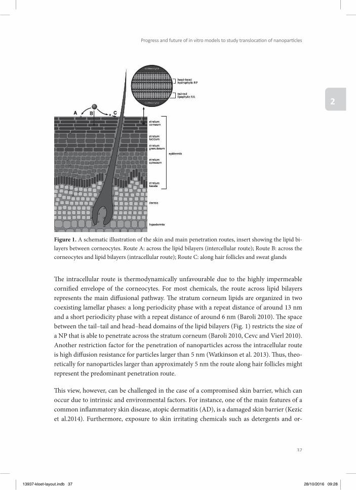
37
Progress and future of in vitro models to study translocati on of nanoparti cles
2
Th e intracellular route is thermodynamically unfavourable due to the highly impermeable cornifi ed envelope of the corneocytes. For most chemicals, the route across lipid bilayers represents the main diff usional pathway. Th e stratum corneum lipids are organized in two coexisting lamellar phases: a long periodicity phase with a repeat distance of around 13 nm and a short periodicity phase with a repeat distance of around 6 nm (Baroli 2010). Th e space between the tail–tail and head–head domains of the lipid bilayers (Fig. 1) restricts the size of a NP that is able to penetrate across the stratum corneum (Baroli 2010, Cevc and Vierl 2010). Another restriction factor for the penetration of nanoparticles across the intracellular route is high diff usion resistance for particles larger than 5 nm (Watkinson et al. 2013). Th us, theo-retically for nanoparticles larger than approximately 5 nm the route along hair follicles might represent the predominant penetration route.
Th is view, however, can be challenged in the case of a compromised skin barrier, which can occur due to intrinsic and environmental factors. For instance, one of the main features of a common infl ammatory skin disease, atopic dermatitis (AD), is a damaged skin barrier (Kezic et al.2014). Furthermore, exposure to skin irritating chemicals such as detergents and or-
Figure 1. A schematic illustration of the skin and main penetration routes, insert showing the lipid bi-layers between corneocytes. Route A: across the lipid bilayers (intercellular route); Route B: across the corneocytes and lipid bilayers (intracellular route); Route C: along hair follicles and sweat glands
13937-kloet-layout.indb 37 28/10/2016 09:28
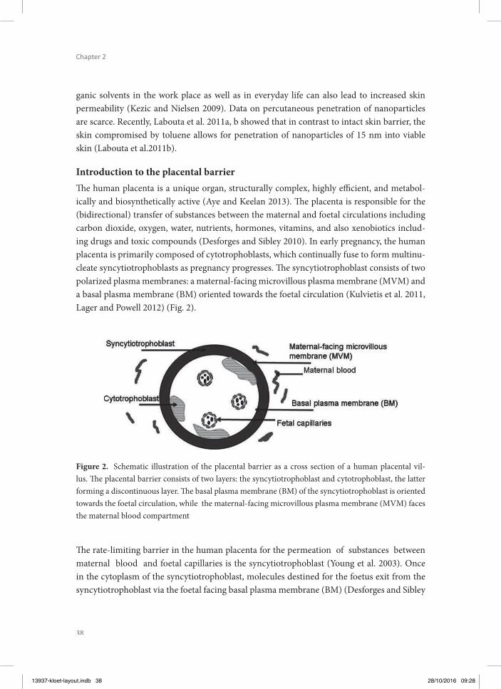
38
Chapter 2
ganic solvents in the work place as well as in everyday life can also lead to increased skin permeability (Kezic and Nielsen 2009). Data on percutaneous penetration of nanoparticles are scarce. Recently, Labouta et al. 2011a, b showed that in contrast to intact skin barrier, the skin compromised by toluene allows for penetration of nanoparticles of 15 nm into viable skin (Labouta et al.2011b).
Introduction to the placental barrierTh e human placenta is a unique organ, structurally complex, highly effi cient, and metabol-ically and biosynthetically active (Aye and Keelan 2013). Th e placenta is responsible for the (bidirectional) transfer of substances between the maternal and foetal circulations including carbon dioxide, oxygen, water, nutrients, hormones, vitamins, and also xenobiotics includ-ing drugs and toxic compounds (Desforges and Sibley 2010). In early pregnancy, the human placenta is primarily composed of cytotrophoblasts, which continually fuse to form multinu-cleate syncytiotrophoblasts as pregnancy progresses. Th e syncytiotrophoblast consists of two polarized plasma membranes: a maternal-facing microvillous plasma membrane (MVM) and a basal plasma membrane (BM) oriented towards the foetal circulation (Kulvietis et al. 2011, Lager and Powell 2012) (Fig. 2).
Figure 2. Schematic illustration of the placental barrier as a cross section of a human placental vil-lus. Th e placental barrier consists of two layers: the syncytiotrophoblast and cytotrophoblast, the latter forming a discontinuous layer. Th e basal plasma membrane (BM) of the syncytiotrophoblast is oriented towards the foetal circulation, while the maternal-facing microvillous plasma membrane (MVM) faces the maternal blood compartment
Th e rate-limiting barrier in the human placenta for the permeation of substances between maternal blood and foetal capillaries is the syncytiotrophoblast (Young et al. 2003). Once in the cytoplasm of the syncytiotrophoblast, molecules destined for the foetus exit from the syncytiotrophoblast via the foetal facing basal plasma membrane (BM) (Desforges and Sibley
13937-kloet-layout.indb 38 28/10/2016 09:28
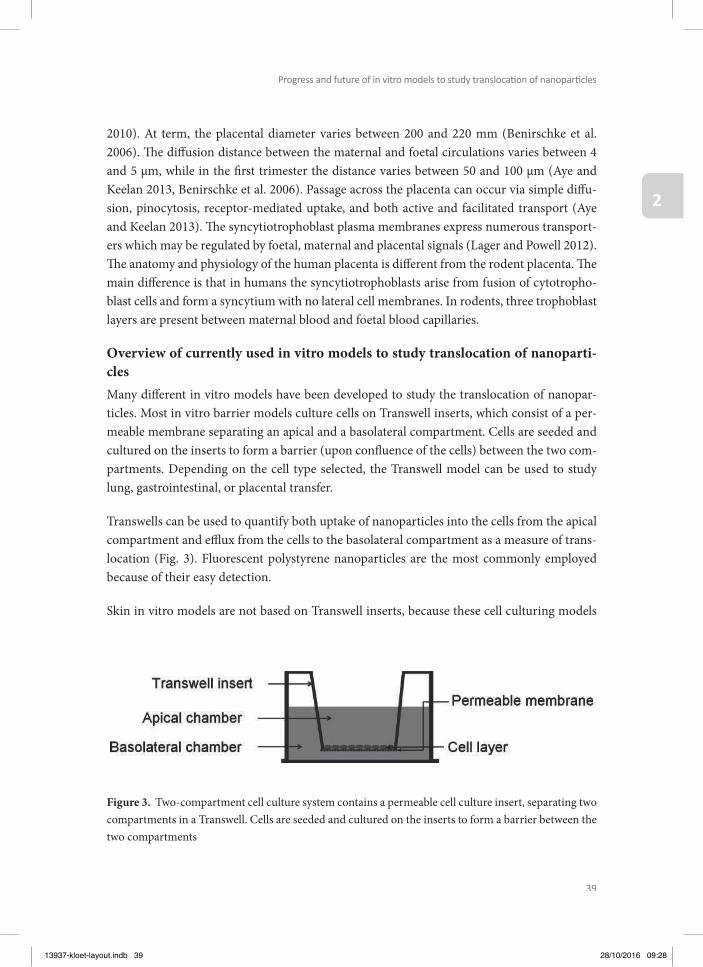
39
Progress and future of in vitro models to study translocati on of nanoparti cles
2
2010). At term, the placental diameter varies between 200 and 220 mm (Benirschke et al. 2006). Th e diff usion distance between the maternal and foetal circulations varies between 4 and 5 µm, while in the fi rst trimester the distance varies between 50 and 100 µm (Aye and Keelan 2013, Benirschke et al. 2006). Passage across the placenta can occur via simple diff u-sion, pinocytosis, receptor-mediated uptake, and both active and facilitated transport (Aye and Keelan 2013). Th e syncytiotrophoblast plasma membranes express numerous transport-ers which may be regulated by foetal, maternal and placental signals (Lager and Powell 2012). Th e anatomy and physiology of the human placenta is diff erent from the rodent placenta. Th e main diff erence is that in humans the syncytiotrophoblasts arise from fusion of cytotropho-blast cells and form a syncytium with no lateral cell membranes. In rodents, three trophoblast layers are present between maternal blood and foetal blood capillaries.
Overview of currently used in vitro models to study translocation of nanoparti-clesMany diff erent in vitro models have been developed to study the translocation of nanopar-ticles. Most in vitro barrier models culture cells on Transwell inserts, which consist of a per-meable membrane separating an apical and a basolateral compartment. Cells are seeded and cultured on the inserts to form a barrier (upon confl uence of the cells) between the two com-partments. Depending on the cell type selected, the Transwell model can be used to study lung, gastrointestinal, or placental transfer.
Transwells can be used to quantify both uptake of nanoparticles into the cells from the apical compartment and effl ux from the cells to the basolateral compartment as a measure of trans-location (Fig. 3). Fluorescent polystyrene nanoparticles are the most commonly employed because of their easy detection.
Skin in vitro models are not based on Transwell inserts, because these cell culturing models
Figure 3. Two-compartment cell culture system contains a permeable cell culture insert, separating two compartments in a Transwell. Cells are seeded and cultured on the inserts to form a barrier between the two compartments
13937-kloet-layout.indb 39 28/10/2016 09:28
40
Chapter 2
lack the principal barrier, the stratum corneum. Th erefore, ex vivo skin models are mostly used for the in vitro assessment of nanoparticles translocation. Also for the other barriers, especially for the placental barrier and to a lesser extent for the lung and intestinal barrier, ex vivo models are available to test the translocation of nanoparticles.
Table 1 presents an overview of in vitro barrier models currently used to study the transloca-tion of nanoparticles aft er inhalation, oral intake, dermal exposure, and across the placenta.
Lung in vitro modelsTh e most frequently used lung epithelial cells lines are A549, Calu-3, H441, and 16HBE14o-. Of these cell lines, Calu-3, H441, and 16HBE14o- form tight junctions, but A549 do not (Lehmann et al. 2011, Chowdhury et al. 2010, Geys et al. 2006, Hermanns et al. 2004, George et al. 2015). In in vitro translocation studies, primary rat alveolar cells have also been frequently used (Fazlollahi et al. 2011, Geys et al. 2009, Yacobi et al. 2008).
Besides diff erences in cell types, in vitro lung barrier models diff er in whether they are submerged or cultured at the air–liquid interface. Submerged models have the advan-tage of being technically simple. However, the culture medium can alter the properties of the nanoparticles, and subsequently their uptake and eff ects. Air–liquid models mimic more realistically the inhalation exposure; therefore, many air–liquid models have been developed recently (Blank et al. 2006, Brandenberger et al. 2010, Frohlich et al. 2013, Herzog et al. 2013, Holder and Marr 2013, Lenz et al. 2009, 2013, Raemy et al. 2011, Rothen-Rutishauser et al. 2009, Savi et al. 2008, Xie et al. 2012). Disadvantages are the complexity of the system to maintain constant temperature and humidifi cation, and the high costs in comparison with submerged models.
In recent years, co-culture models containing more than one cell type are used to mimic the lung barrier more closely compared with monocultures (Klein et al. 2011). Most models use lung epithelial cells as a basis, those being either primary cells or immortalized cell lines. To obtain co-culture models, diff erent cell types are added to the basic model. Th e fi rst type of co-culture models includes, in addition to epithelial cells, endothelial cells to mimic the alveolar-capillary barrier (Bermudez et al. 2002, Chowdhury et al. 2010, Hermanns et al. 2010, Hermanns et al. 2004, Papritz et al. 2010). Th is type of model can be extended by the addition of alveolar macrophages, mast cells, and/or type II alveolar cells. Th e second type of models does not include endothelial cells, but adds dendritic cells and macrophages to the epithelial cell layer (Brandenberger et al. 2010, Muller et al. 2010, Rothen-Rutishauser et al. 2008, Rothen-Rutishauser et al. 2005). Th is type of model can also be completed with type II alveolar cells. Th e third type of models includes, in addition to the epithelial cells, fi broblasts instead of endothelial cells, which can be extended by adding dendritic cells.
13937-kloet-layout.indb 40 28/10/2016 09:28
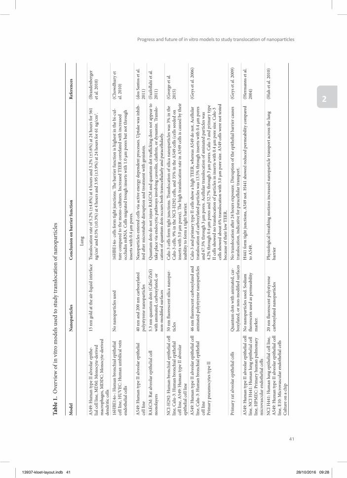
41
Progress and future of in vitro models to study translocati on of nanoparti cles
2
Tabl
e 1.
Ove
rvie
w o
f in
vitr
o m
odel
s use
d to
stud
y tr
anslo
catio
n of
nan
opar
ticle
s
Mod
elN
anop
artic
les
Con
clus
ion
on b
arri
er fu
nctio
nR
efer
ence
s
Lung
A54
9: H
uman
type
II a
lveo
lar e
pith
e-lia
l cel
l lin
e, M
DM
: Mon
ocyt
e-de
rived
m
acro
phag
es, M
DD
C: M
onoc
yte-
deriv
ed
dend
ritic
cells
15 n
m g
old
at th
e ai
r-liq
uid
inte
rfac
eTr
anslo
catio
n ra
te o
f 5.2
% (±
4.8%
) at 4
hou
rs a
nd 5
.2%
(±5.
6%) a
t 24
hour
s for
561
ng
/cm
2 and
0.5
% (±
0.3%
) at 4
hou
rs a
nd 3
.95
(±3.
9%) a
t 24
hour
s for
61
ng/c
m2 .
(Bra
nden
berg
er
et a
l. 20
10)
16H
BE14
o-: H
uman
bro
nchi
al e
pith
elia
l ce
ll lin
e, H
UV
EC: H
uman
um
bilic
al v
ein
endo
thel
ial c
ells
No
nano
part
icle
s use
d16
HBE
14o-
cells
form
tigh
t jun
ctio
ns. Th
e b
arrie
r fun
ctio
n is
high
est i
n th
e bi
-cul
-tu
re co
mpa
red
to th
e m
ono-
cultu
res.
Incr
ease
d TE
ER co
rrel
ated
with
incr
ease
d oc
clud
in m
RNA
. Cel
ls m
igra
ted
trou
gh in
sert
s with
1.0
µm
por
es b
ut n
ot th
roug
h in
sert
s with
0.4
µm
por
es.
(Cho
wdh
ury
et
al. 2
010)
A54
9: H
uman
type
II a
lveo
lar e
pith
elia
l ce
ll lin
e40
nm
and
200
nm
car
boxy
late
d po
lyst
yren
e na
nopa
rtic
les
Nan
opar
ticle
s ent
ered
cells
via
act
ive
ener
gy d
epen
dent
pro
cess
es. U
ptak
e w
as in
hib-
ited
aft e
r mic
rotu
bule
disr
uptio
n an
d tr
eatm
ent w
ith g
enist
ein.
(dos
San
tos e
t al.
2011
)RA
ECM
: Rat
alv
eola
r epi
thel
ial c
ell
mon
olay
ers
5.3
nm q
uant
um d
ots (
CdS
e/Zn
S)
with
am
inat
ed, c
arbo
xyla
ted,
or
non-
mod
ifi ed
surf
aces
Qua
ntum
dot
s do
not i
njur
e RA
ECM
and
qua
ntum
dot
traffi
cki
ng d
oes n
ot ap
pear
to
take
pla
ce v
ia e
ndoc
ytic
pat
hway
s inv
olvi
ng c
aveo
lin, c
lath
rin, o
r dyn
amin
. Tra
nslo
-ca
tion
of q
uant
um d
ots o
ccur
s bot
h tr
ansc
ellu
larly
and
par
acel
lula
rly.
(Faz
lolla
hi e
t al.
2011
)
NC
I-H
292:
Hum
an b
ronc
hial
epi
thel
ial c
ell
line,
Cal
u-3:
Hum
an b
ronc
hial
epi
thel
ial
cell
line,
A54
9: H
uman
type
II a
lveo
lar
epith
elia
l cel
l lin
e
50 n
m fl
uore
scen
t sili
ca n
anop
ar-
ticle
sC
alu-
3 ce
lls fo
rm ti
ght j
unct
ions
. Tra
nslo
catio
n of
silic
a na
nopa
rtic
les w
as 3
% in
the
Cal
u-3
cells
, 9%
in th
e N
CI-
H29
2 ce
lls, a
nd 3
5% in
the
A54
9 ce
lls (c
ells
seed
ed o
n in
sert
s with
3.0
µm
por
es).
Th e
high
tran
sloca
tion
rate
in A
549
cells
is c
ause
d by
thei
r in
abili
ty to
form
a ti
ght b
arrie
r.
(Geo
rge
et a
l. 20
15)
A54
9: H
uman
type
II a
lveo
lar e
pith
elia
l cel
l lin
e, C
alu-
3: H
uman
bro
nchi
al e
pith
elia
l ce
ll lin
ePr
imar
y pn
eum
ocyt
es ty
pe II
46 n
m fl
uore
scen
t car
boxy
late
d an
d am
inat
ed p
olys
tyre
ne n
anop
artic
les
Cal
u-3
and
prim
ary
type
II ce
lls sh
ow a
hig
h TE
ER, w
here
as A
549
do n
ot. A
cellu
lar
tran
sloca
tion
of c
arbo
xyla
ted
part
icle
s was
13.
5% th
roug
h in
sert
s with
0.4
µm
por
es
and
67.5
% th
roug
h 3
µm p
ores
. Ace
llula
r tra
nslo
catio
n of
am
inat
ed p
artic
les w
as
4.2%
thro
ugh
0.4
µm p
ores
and
52.
7% th
roug
h 3
µm p
ores
. Cal
u-3
and
prim
ary
type
II
cells
show
ed n
o tr
anslo
catio
n of
par
ticle
s in
inse
rts w
ith 0
.4 µ
m p
ore
size.
Cal
u-3
cells
show
ed a
bout
6%
tran
sloca
tion
with
3.0
µm
por
e siz
e. A
549
cells
wer
e no
t tes
ted
beca
use
of th
eir l
ow T
EER.
(Gey
s et a
l. 20
06)
Prim
ary
rat a
lveo
lar e
pith
elia
l cel
lsQ
uant
um d
ots w
ith a
min
ated
, car
-bo
xyla
ted,
or n
on-m
odifi
ed su
rfac
esN
o tr
anslo
catio
n aft
er 2
4 ho
urs e
xpos
ure.
Disr
uptio
n of
the
epith
elia
l bar
rier c
ause
s tr
anslo
catio
n, in
dica
tive
for p
arac
ellu
lar t
rans
port
.(G
eys e
t al.
2009
)
A54
9: H
uman
type
II a
lveo
lar e
pith
elia
l cel
l lin
e, N
CI H
441:
Hum
an lu
ng e
pith
elia
l cel
l lin
e, H
PMEC
: Prim
ary
hum
an p
ulm
onar
y m
icro
vasc
ular
end
othe
lial c
ells
No
nano
part
icle
s use
d. S
odiu
m
fl uor
esce
in u
sed
as p
erm
eabi
lity
mar
ker.
H44
1 fo
rm ti
ght j
unct
ions
, A54
9 no
t. H
441
show
ed re
duce
d pe
rmea
bilit
y co
mpa
red
to A
549.
(Her
man
ns e
t al.
2004
)
NC
I H44
1: H
uman
lung
epi
thel
ial c
ell l
ine,
A54
9: H
uman
type
II a
lveo
lar e
pith
elia
l cel
l lin
e, E1
0: M
icro
vasc
ular
end
othe
lial c
ells
Cultu
re o
n a
chip
20 n
m fl
uore
scen
t pol
ysty
rene
ca
rbox
ylat
ed n
anop
artic
les
Phys
iolo
gica
l bre
athi
ng m
otio
ns in
crea
sed
nano
part
icle
tran
spor
t acr
oss t
he lu
ng
barr
ier
(Huh
et a
l. 20
10)
13937-kloet-layout.indb 41 28/10/2016 09:28
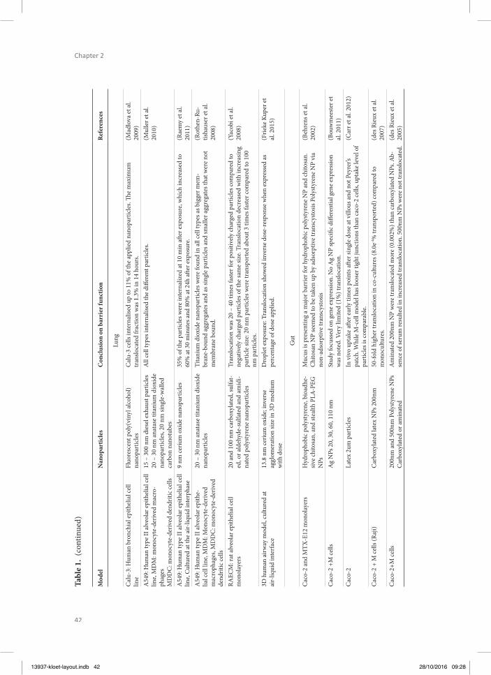
42
Chapter 2
Tabl
e 1.
(co
ntin
ued)
Mod
elN
anop
artic
les
Con
clus
ion
on b
arri
er fu
nctio
nR
efer
ence
s
Lung
Cal
u-3:
Hum
an b
ronc
hial
epi
thel
ial c
ell
line
Fluo
resc
ent p
oly(
viny
l alc
ohol
) na
nopa
rtic
les
Cal
u-3
cells
inte
rnal
ised
up to
11%
of t
he ap
plie
d na
nopa
rtic
les.
Th e
max
imum
tr
anslo
cate
d fr
actio
n w
as 1
.3%
in 1
4 ho
urs.
(Mad
lova
et a
l. 20
09)
A54
9: H
uman
type
II a
lveo
lar e
pith
elia
l cel
l lin
e, M
DM
: mon
ocyt
e-de
rived
mac
ro-
phag
esM
DD
C: m
onoc
yte-
deriv
ed d
endr
itic c
ells
15 –
300
nm
die
sel e
xhau
st p
artic
les
20 –
30
nm a
nata
se ti
tani
um d
ioxi
de
nano
part
icle
s, 20
nm
sing
le-w
alle
d ca
rbon
nan
otub
es
All
cell
type
s int
erna
lised
the
diff e
rent
par
ticle
s. (M
ulle
r et a
l. 20
10)
A54
9: H
uman
type
II a
lveo
lar e
pith
elia
l cel
l lin
e, Cu
lture
d at
the
air-
liqui
d in
terp
hase
9 nm
ceriu
m o
xide
nan
opar
ticle
s35
% o
f the
par
ticle
s wer
e in
tern
aliz
ed at
10
min
aft e
r exp
osur
e, w
hich
incr
ease
d to
60
% at
30
min
utes
and
80%
at 2
4h a
ft er e
xpos
ure.
(Rae
my
et a
l. 20
11)
A54
9: H
uman
type
II a
lveo
lar e
pith
e-lia
l cel
l lin
e, M
DM
: Mon
ocyt
e-de
rived
m
acro
phag
es, M
DD
C: m
onoc
yte-
deriv
ed
dend
ritic
cells
20 –
30
nm a
nata
se ti
tani
um d
ioxi
de
nano
part
icle
sTi
tani
um d
ioxi
de n
anop
artic
les w
ere
foun
d in
all
cell
type
s as b
igge
r mem
-br
ane-
boun
d ag
greg
ates
and
as s
ingl
e pa
rtic
les a
nd sm
alle
r agg
rega
tes t
hat w
ere
not
mem
bran
e bo
und.
(Rot
hen-
Ru-
tisha
user
et a
l. 20
08)
RAEC
M: r
at a
lveo
lar e
pith
elia
l cel
l m
onol
ayer
s20
and
100
nm
car
boxy
late
d, su
lfat-
ed, o
r ald
ehyd
e-su
lfate
d an
d am
idi-
nate
d po
lyst
yren
e na
nopa
rtic
les
Tran
sloca
tion
was
20
– 40
tim
es fa
ster
for p
ositi
vely
char
ged
part
icle
s com
pare
d to
ne
gativ
ely
char
ged
part
icle
s of t
he sa
me
size.
Tran
sloca
tion
decr
ease
d w
ith in
crea
sing
part
icle
size
: 20
nm p
artic
les w
ere
tran
spor
ted
abou
t 3 ti
mes
fast
er co
mpa
red
to 1
00
nm p
artic
les.
(Yac
obi e
t al.
2008
)
3D h
uman
air
way
mod
el, c
ultu
red
at
air-
liqui
d in
terf
ace
13.8
nm
ceriu
m o
xide
; inv
erse
ag
glom
erat
ion
size
in 3
D m
ediu
m
with
dos
e
Dro
plet
exp
osur
e: T
rans
loca
tion
show
ed in
vers
e do
se-r
espo
nse
whe
n ex
pres
sed
as
perc
enta
ge o
f dos
e ap
plie
d.
(Frie
ke K
uper
et
al. 2
015)
Gut
Cac
o-2
and
MTX
-E12
mon
olay
ers
Hyd
roph
obic
pol
ysty
rene
, bio
adhe
-siv
e ch
itosa
n, a
nd st
ealth
PLA
-PEG
N
Ps
Muc
us is
pre
sent
ing
a m
ajor
bar
rier f
or h
ydro
phob
ic p
olys
tyre
ne N
P an
d ch
itosa
n.
Chi
tosa
n N
P se
emed
to b
e ta
ken
up b
y ad
sorp
tive
tran
scys
tosis
Pol
ysty
rene
NP
via
non-
adso
rptiv
e tr
ansc
ysto
sis
(Beh
rens
et a
l. 20
02)
Cac
o-2
+M ce
llsA
g N
Ps 2
0, 3
0, 6
0, 1
10 n
mSt
udy
focu
ssed
on
gene
exp
ress
ion.
No
Ag
NP
spec
ifi c d
iff er
entia
l gen
e ex
pres
sion
was
not
ed. V
ery
limite
d (1
%) t
rans
loca
tion.
(Bou
wm
eest
er e
t al
. 201
1)C
aco-
2La
tex
2um
par
ticle
sIn
viv
o up
take
aft e
r ear
ly ti
mes
poi
nts a
ft er s
ingl
e do
se at
vill
ous a
nd n
ot P
eyre
r’s
patc
h. W
hile
M-c
ell m
odel
has
loos
er ti
ght j
unct
ions
than
cac
o-2
cells
, upt
ake
leve
l of
part
icle
s is c
ompa
rabl
e.
(Car
r et a
l. 20
12)
Cac
o-2
+ M
cells
(Raj
i)C
arbo
xyla
ted
late
x N
Ps 2
00nm
50-fo
ld h
ighe
r tra
nslo
catio
n in
co-c
ultu
res (
8.0e
-4%
tran
spor
ted)
com
pare
d to
m
onoc
ultu
res.
(des
Rie
ux e
t al.
2007
)C
aco-
2+M
cells
200n
m a
nd 5
00nm
Pol
ysty
rene
NPs
C
arbo
xyla
ted
or a
min
ated
Am
inat
ed 2
00nm
NP
wer
e tr
anslo
cate
d m
ore
(0.0
02%
) tha
n ca
rbox
ylat
ed N
Ps. A
b-se
nce
of se
rum
resu
lted
in in
crea
sed
tran
sloca
tion.
500
nm N
Ps w
ere
not t
rans
loca
ted.
(des
Rie
ux e
t al.
2005
)
13937-kloet-layout.indb 42 28/10/2016 09:28

43
Progress and future of in vitro models to study translocati on of nanoparti cles
2
Tabl
e 1.
(co
ntin
ued)
Mod
elN
anop
artic
les
Con
clus
ion
on b
arri
er fu
nctio
nR
efer
ence
s
Gut
Cac
o-2
cell
rat i
leum
(ex
vivo
), an
d sy
stem
-ic
bio
dist
ribut
ion
aft e
r ora
l gav
age.
Rhod
amin
e B
labe
led
carb
oxyl
ated
ch
itosa
n gr
aft e
d na
nopa
rtic
les (
RhB-
CC
NP)
(300
, 600
, and
100
0 nm
) and
sim
ilar Z
eta
pote
ntia
ls (-
35 m
V)
RhB-
CC
NP-
BSA
with
smal
ler s
izes
(300
nm
) dem
onst
rate
d el
evat
ed in
test
inal
ab
sorp
tion,
com
pare
d to
the
othe
rs.
(He
et a
l. 20
12)
Cac
o-2+
HT2
9-M
TXIn
sulin
load
ed ch
itosa
n na
nopa
r-tic
les.
CSK
pep
tide
mod
ifi ca
tion
show
ed e
nhan
ced
tran
spor
t. Th
e pr
esen
ce o
f muc
us
incr
ease
d th
e tr
anslo
catio
n of
bot
h m
odifi
ed a
nd u
nmod
ifi ed
nan
opar
ticle
s.(J
in e
t al.
2012
)
Cac
o-2
M ce
llsC
hito
san-
DN
A n
anop
artic
les
Liga
nd d
ecor
atio
n ha
s the
mos
t dra
mat
ic e
ff ect
on
the
tran
scyt
osis
rate
: tra
nsfe
rrin
m
odifi
catio
n en
hanc
es tr
ansp
ort t
hrou
gh b
oth
mod
els b
y 3-
to 5
-fold
. Tr
ansp
ort t
hrou
gh th
e M
-cel
l co-
cultu
re m
odel
is 5
-fold
that
of t
he in
test
inal
epi
the-
lial m
onol
ayer
, with
at le
ast 8
0% o
f the
chito
san-
DN
A n
anop
artic
les t
aken
up
in th
e fi r
st 3
0 m
in.
(Kad
iyal
a et
al.
2010
)
Cac
o-2
+ H
T29-
MTX
+ M
cells
50
and
200
nm
pol
ysty
rene
NPs
, ca
rbox
ylat
ed, a
min
ated
, or p
lain
Co-
expo
sure
of m
onol
ayer
s to
eith
er 5
0 or
200
nm
NPs
and
iron
- asc
orba
te so
lutio
n re
sulte
d in
incr
ease
d tr
anslo
catio
n of
iron
50
nm N
PS tr
anslo
cate
d eq
ual i
n bo
th co
(c
aco
and
HT2
9) a
nd tr
icul
ture
mod
els.
200n
m N
Ps tr
anslo
cate
d sig
nifi c
antly
mor
e in
tri c
ultu
res,
only
at 3
7ºC
at 3
7 an
d 4°
C.
(Mah
ler e
t al.
2012
)
Cac
o-2
Poly
elec
trol
yte
com
plex
nan
opar
-tic
les o
f spe
rmin
e (S
PM) w
ith
poly
acry
lic a
cid
(PA
A) p
olym
er.
Perm
eatio
n en
hanc
ing
eff e
cts f
ollo
win
g ex
posu
re to
NPs
wer
e as
soci
ated
with
a
reve
rsib
le d
ecre
ase
in T
EER
valu
es, s
ugge
stin
g a
para
cellu
lar p
erm
eatio
n pa
thw
ay b
y re
vers
ible
ope
ning
of t
he ti
ght j
unct
ions
. H
ighe
r per
mea
tion
enha
ncin
g pr
ofi le
s of S
PM-P
AA
NPs
, as c
ompa
red
to S
PM so
lu-
tion
or P
AA
NPs
pre
pare
d by
ioni
c gel
atio
n w
ith M
gCl(2
) (M
g-PA
A N
Ps).
(Mak
hlof
et a
l. 20
11)
Cac
o-2
with
and
with
out H
T29-
MTX
with
an
d w
ithou
t M
cells
50 a
nd 1
00 n
m p
olys
tyre
ne N
Ps,
carb
oxyl
ated
, am
inat
ed, o
r pla
inTh
e in
corp
orat
ion
of m
ucus
in th
e m
odel
redu
ced
the
tran
sloca
tion
of 5
0nm
NPs
co
mpa
red
to C
aco-
2 on
ly. Th
e in
corp
orat
ion
of M
cells
resu
lted
in a
tran
sloca
tion
leve
ls cl
ose
thos
e as
obs
erve
d fo
r the
mon
ocul
ture
. Th e
surf
ace
char
ge o
f the
NP
was
ve
ry im
port
ant f
or th
e (c
hang
es) i
n tr
anslo
catio
n in
the
diff e
rent
mod
els.
100n
m N
Ps
wer
e tr
anslo
cate
d at
a v
ery
low
leve
l.
(Wal
czak
et a
l. 20
14)
Cac
o-2
with
HT2
9-M
TX co
uple
d to
in
vitr
o di
gest
ion
50 p
olys
tyre
ne N
Ps, c
arbo
xyla
ted,
am
inat
ed, o
r pla
in.
Incu
batio
n of
the
NPs
in th
e in
vitr
o di
gest
ion
mod
el re
sulte
d in
incr
ease
d tr
anslo
ca-
tion
leve
ls co
mpa
red
to ‘n
on d
iges
ted’
NPs
.(W
alcz
ak e
t al.
2015
)Ex
viv
o m
ice
inte
stin
al ti
ssue
in U
ssin
g ch
ambe
rsH
ydro
dyna
mic
size
of 1
30 n
m sl
ices
ex
pose
d fo
r 2 h
to 5
0 μg
/ml..
Loca
l Ti s
pots
in ti
ssue
s wer
e ob
serv
ed, i
ncre
ased
per
mea
bilit
y of
4kD
a de
xtra
ns a
ft er
Ti N
P ex
posu
re. N
o Ti
tran
sloca
tion
coul
d be
det
ecte
d(B
run
et a
l. 20
14)
Ex v
ivo
proc
ine
inte
stin
al ti
ssue
s in
Uss
ing
cham
bers
50 a
nd 2
00nm
pol
ysty
rene
nan
opar
-tic
les e
xpos
ed fo
r 4 h
to 1
00 μ
g/m
l.En
trap
men
t of N
Ps in
supe
rfi ci
al p
art o
f the
tiss
ue w
as o
bser
ved.
Tra
nslo
catio
n no
t de
term
ined
(Sch
impe
l et a
l. 20
14)
Skin
Full
thic
knes
s hum
an sk
in, h
andm
ade
vert
ical
diff
usio
n ce
llFe
oxi
de, <
10
nmN
Ps w
ere
able
to p
enet
rate
into
via
ble
skin
laye
rs.
(Bar
oli 2
010)
13937-kloet-layout.indb 43 28/10/2016 09:28
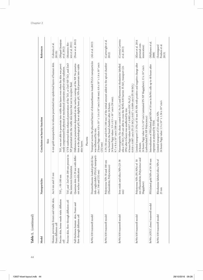
44
Chapter 2
Tabl
e 1.
(co
ntin
ued)
Mod
elN
anop
artic
les
Con
clus
ion
on b
arri
er fu
nctio
nR
efer
ence
s
Skin
Hum
an, p
revi
ously
froz
en a
nd v
iabl
e sk
in,
Fran
z diff
usio
n ce
llAu
6 n
m a
nd 1
5 nm
6-nm
gol
d na
nopa
rtic
les i
n to
luen
e pe
netr
ated
into
epi
derm
al la
yers
of h
uman
skin
.(L
abou
ta e
t al.
2011
b)Po
rcin
e sk
in, h
ome-
mad
e st
atic
diff
usio
n ce
llTi
O2, >
50-1
00 n
mTi
O2 w
as n
ot a
ble
to p
enet
rate
into
via
ble
skin
laye
rs e
ven
whe
n th
e sk
in w
as p
revi
-ou
sly co
mpr
omise
d by
phy
sical
or m
echa
nica
l con
stra
ins a
nd/o
r sol
ar ra
diat
ion.
(Miq
uel-J
eanj
ean
et a
l. 20
12)
Porc
ine
skin
, fl o
w-t
hrou
gh d
iff us
ion
cell
TiO
2 an
d Z
nO 6
0-20
0 nm
pre
sent
in
the
suns
cree
n fo
rmul
atio
nU
VB-
sunb
urne
d sk
in e
nhan
ced
pene
trat
ion
of th
e Ti
O2 o
r ZnO
NP.
TiO
2 and
ZnO
N
P pe
netr
ated
into
the
viab
le sk
in la
yers
but
not
in re
cept
or fl
uid.
(Mon
teiro
-Riv
-ie
re e
t al.
2011
)Fu
ll th
ickn
ess h
uman
ski
n, F
ranz
and
fl o
w-t
hrou
gh d
iff us
ion
cell
Qua
ntum
dot
s 13-
29 n
m w
ith d
iff er
-en
t sur
face
mod
ifi ca
tion
Diff
eren
ces i
n pe
netr
atio
n de
pend
ed o
n th
e su
rfac
e co
atin
gs o
f the
NP.
No
pene
tra-
tion
at th
e ph
ysio
logi
cal p
H, b
ut at
slig
htly
bas
ic p
H, N
Ps d
id p
enet
rate
into
via
ble
skin
laye
rs.
(Pro
w e
t al.
2012
)
Plac
enta
BeW
o b3
0 tr
answ
ell m
odel
D
exam
etha
sone
-load
ed p
oly(
d,l-l
ac-
tide-
cogl
ycol
ide)
(PLG
A) n
anop
arti-
cles
( 14
6 an
d 23
2 nm
)
Tran
spor
t acr
oss t
he p
lace
ntal
bar
rier o
f dex
amet
haso
ne-lo
aded
PLG
A n
anop
artic
les
was
infl u
ence
d by
size
. 2
hour
s Pap
p va
lues
: 6.0
x 1
0-5 ±
1.6
x 1
0-5 cm
/s (1
46 n
m);
4.8
x 10
-5 ±
1.6
x 1
0-5 cm
/s
(232
nm
)
(Ali
et a
l. 20
13)
BeW
o b3
0 tr
answ
ell m
odel
Poly
styr
ene
NPs
50
and
100
nm
(Flu
ores
brite
; Pol
ysci
ence
s)3.
5% (5
0 nm
) and
0.6
% (1
00 n
m) o
f the
initi
al a
mou
nt a
dded
to th
e ap
ical
cham
ber
was
foun
d in
the
basa
l cha
mbe
r aft e
r 24
hour
s.24
hou
rs P
app
valu
es: 3
.9 ±
0.5
× 1
0-5 cm
/s (5
0 nm
);9.
7 ±
9.4
x 10
-6 cm
/s (1
00 n
m)
(Car
twrig
ht e
t al.
2012
)
BeW
o b3
0 tr
answ
ell m
odel
Iron
oxi
de a
nd si
lica
NPs
(23-
38
nm)
Nan
omeg
netit
e N
a-ol
eate
-coa
ted
(OC
-Fe 3O
4) and
Flu
ores
cent
rhod
amin
e-la
belle
d sil
ica
(Fl-S
iO2) N
Ps a
re a
ble
to cr
oss t
he B
eWo
b30
barr
ier.
Fl-S
iO2 tr
ansp
ort (
24-
29%
) aft e
r 6 h
ours
was
una
ff ect
ed b
y siz
e. 6
hour
s Pap
p va
lues
: 0.0
17 ±
0.0
02 cm
/s (O
C-F
e 3O4);
0.01
8 ±
0.00
7 cm
/s (F
l-25
SiO
2); 0.
017
± 0.
009
cm/s
(Fl-5
0 Si
O2)
(Cor
reia
Car
reira
et
al.
2013
)
BeW
o b3
0 tr
answ
ell m
odel
Poly
styr
ene
NPs
(PS-
NPs
) of
50
nm (F
luor
esbr
ite; P
olys
cien
ce a
nd
Mag
sphe
re)
Lim
ited
tran
spor
t (3-
15%
) of 5
0 nm
PS-
NPs
with
pos
itive
and
neg
ativ
e ch
arge
aft e
r 24
hou
rs.
24 h
ours
Pap
p va
lue:
0.3
x 1
0-6 cm
/s (a
min
ated
PS-
NP
Mag
sphe
re);
13 x
10-6
cm/s
(c
arbo
xyla
ted
PS-N
P Po
lysc
ienc
e)
(Klo
et e
t al.
2014
pe
rson
al co
m-
mun
icat
ion)
BeW
o (A
TCC
clon
e) tr
answ
ell m
odel
PEG
ylat
ed g
old
NPs
of 1
0-30
nm
Inte
rnal
isatio
n of
PEG
ylat
ed g
old
NPs
of 1
0 nm
in B
eWo
cells
up
to 4
8 ho
urs a
ft er
expo
sure
as a
naly
sed
by T
EM.
(Myl
lyne
n et
al.
2008
)Be
Wo
b30
tran
swel
l mod
elRh
odam
ine
labe
led
silic
a N
Ps o
f 25
nm
Lim
ited
tran
spor
t of 2
5 nm
silic
a N
Ps.
24 h
ours
Pap
p va
lue:
1.5
x 1
0-6 ±
1.5
6 x
10-6
cm/s
(S
onne
gaar
d Po
ulse
n et
al.
2013
)
13937-kloet-layout.indb 44 28/10/2016 09:28
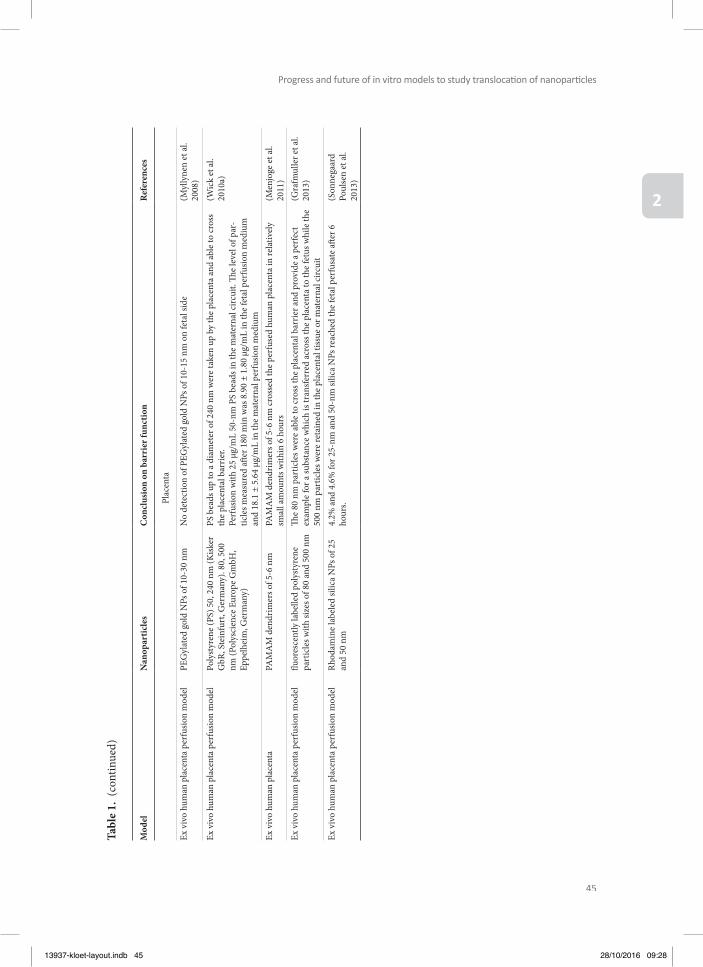
45
Progress and future of in vitro models to study translocati on of nanoparti cles
2
Tabl
e 1.
(co
ntin
ued)
Mod
elN
anop
artic
les
Con
clus
ion
on b
arri
er fu
nctio
nR
efer
ence
s
Plac
enta
Ex v
ivo
hum
an p
lace
nta
perf
usio
n m
odel
PEG
ylat
ed g
old
NPs
of 1
0-30
nm
No
dete
ctio
n of
PEG
ylat
ed g
old
NPs
of 1
0-15
nm
on
feta
l sid
e(M
ylly
nen
et a
l. 20
08)
Ex v
ivo
hum
an p
lace
nta
perf
usio
n m
odel
Poly
styr
ene
(PS)
50,
240
nm
(Kisk
er
GbR
, Ste
infu
rt, G
erm
any)
. 80,
500
nm
(Pol
ysci
ence
Eur
ope
Gm
bH,
Eppe
lhei
m, G
erm
any)
PS b
eads
up
to a
dia
met
er o
f 240
nm
wer
e ta
ken
up b
y th
e pl
acen
ta a
nd a
ble
to cr
oss
the
plac
enta
l bar
rier.
Perf
usio
n w
ith 2
5 μg
/mL
50-n
m P
S be
ads i
n th
e m
ater
nal c
ircui
t. Th
e le
vel o
f par
-tic
les m
easu
red
aft e
r 180
min
was
8.9
0 ±
1.80
μg/
mL
in th
e fe
tal p
erfu
sion
med
ium
an
d 18
.1 ±
5.6
4 μg
/mL
in th
e m
ater
nal p
erfu
sion
med
ium
(Wic
k et
al.
2010
a)
Ex v
ivo
hum
an p
lace
nta
PAM
AM
den
drim
ers o
f 5-6
nm
PAM
AM
den
drim
ers o
f 5-6
nm
cros
sed
the
perf
used
hum
an p
lace
nta
in re
lativ
ely
smal
l am
ount
s with
in 6
hou
rs(M
enjo
ge e
t al.
2011
)Ex
viv
o hu
man
pla
cent
a pe
rfus
ion
mod
elfl u
ores
cent
ly la
belle
d po
lyst
yren
e pa
rtic
les w
ith si
zes o
f 80
and
500
nm
Th e
80 n
m p
artic
les w
ere
able
to cr
oss t
he p
lace
ntal
bar
rier a
nd p
rovi
de a
per
fect
ex
ampl
e fo
r a su
bsta
nce
whi
ch is
tran
sfer
red
acro
ss th
e pl
acen
ta to
the
fetu
s whi
le th
e 50
0 nm
par
ticle
s wer
e re
tain
ed in
the
plac
enta
l tiss
ue o
r mat
erna
l circ
uit
(Gra
fmul
ler e
t al.
2013
)
Ex v
ivo
hum
an p
lace
nta
perf
usio
n m
odel
Rhod
amin
e la
bele
d sil
ica
NPs
of 2
5 an
d 50
nm
4.2%
and
4.6
% fo
r 25-
nm a
nd 5
0-nm
silic
a N
Ps re
ache
d th
e fe
tal p
erfu
sate
aft e
r 6
hour
s. (S
onne
gaar
d Po
ulse
n et
al.
2013
)
13937-kloet-layout.indb 45 28/10/2016 09:28
46
Chapter 2
A limited number of nanoparticles have been studied to test translocation across the in vitro lung barrier. Th ese include polystyrene nanoparticles, titanium dioxide nanoparticles, quan-tum dots, cerium dioxide nanoparticles, gold nanoparticles, silica nanoparticles, diesel parti-cles, and single-walled carbon nanotubes (Table 1). Th e translocation rate of nanoparticles is higher in models with cells that do not form tight junctions compared with cells that do (Geys et al. 2006, Hermanns et al. 2004, George et al. 2015). Probably, the higher translocation rate is a consequence of paracellular transport, which will not occur in a healthy lung but might occur in a damaged or infl amed lung. Th e pore size of the permeable membrane of the in-serts also infl uences the translocation rate of nanoparticles: the larger the pore size, the high-er the translocation rate (Geys et al. 2006). Th erefore, for each single type of nanoparticles, the translocation across the diff erent types of inserts should be tested without cells to assess whether the nanoparticles are not withheld by the insert itself.
Ex vivo tissues as model for the lung are not addressed here. Th e precision-cut lung slices (PCLS) taken from human and rodent lungs have been used to study translocation and toxic-ity of nanomaterials. Th e use of PCLS for translocation of nanoparticle-mediated drug deliv-ery has recently been reviewed by Paranjpe and Muller-Goymann (Paranjpe and Muller-Goy-mann 2014).
Gut in vitro modelsOrally ingested nanoparticles are exposed to continuously changing conditions while tran-siting through the gastrointestinal tract, which infl uences their nature and characteristics (Bellmann et al. 2015). In vitro GI models aim to mimic the gastrointestinal environment as closely as possible, to generate physiologically relevant results. Th ese models focus either on the aspects of dynamically changing GI conditions during digestion by simulation of the transit of nanoparticles along the GI tract from the mouth towards the large intestine (diges-tion models), or on mimicking translocation and uptake behaviour (in vitro human intestinal epithelium models) (Lefebvre et al. 2014).
Human digestion models can fi rst be simple and static, and mimic only gastric or small in-testinal conditions, in which materials are incubated with simulated gastric fl uids, simulated small intestinal fl uids or buff ers at static pH values (Mwilu et al. 2013, Minekus et al. 2014). More complex static models oft en include most of the relevant GI conditions, i.e. the oral, gas-tric, small intestinal (and large intestinal conditions) (Oomen et al. 2003, Van de Wiele et al. 2007, Versantvoort et al. 2005). Recently, these models have been used to assess the fate of 60-nm silver nanoparticles, and nanometre-sized silica (synthetic amorphous silica) nanopar-ticles during digestion (Peters et al. 2012, Walczak et al. 2013). In contrast to static models, more complex dynamic models simulate successive changes in conditions (i.e. pH, secretion of digestive fl uids) and transit times (Helbig et al. 2013, Kong and Singh 2010, Minekus et
13937-kloet-layout.indb 46 28/10/2016 09:28

47
Progress and future of in vitro models to study translocati on of nanoparti cles
2
al. 1995, Wickham et al. 2009, Zangenberg et al. 2001). Such a dynamic computer controlled model was used to study the behaviour of engineered nanoclay materials (Newsome 2014).
Translocation and uptake of nanoparticles can be addressed by cellular models that can also be divided in relatively simple models or more complex ones (Lefebvre et al. 2014). Amongst the variety of cell models that are available in vitro, intestinal Caco-2 cells (human epithelial colorectal adenocarcinoma cells) are the most commonly used cell type (Miret et al. 2004) in nanoparticle translocation studies. Caco-2 cells are regarded as model cells for enterocytes, the most abundant epithelial cell type in the intestine. Several examples can be found in the literature where monocultures of Caco-2 cells have been used to study the in vitro trans-location of nanoparticles, (mainly polystyrene, but also silicon, silver, and organic nanoparti-cles) (Bhattacharjee et al. 2013, des Rieux et al. 2007, des Rieux et al. 2005, Mahler et al. 2009, Natoli et al. 2012, Nkabinde et al. 2012, Walczak et al. 2014).
A potential drawback of Caco-2 monolayers is the lack of a mucus layer (at least in con-ventional models), which can, however, be introduced by co-culturing Caco-2 cells with HT29-MTX cells (human colon adenocarcinoma mucus-secreting cells) (Behrens et al. 2002, Mahler et al. 2009, Scaldaferri et al. 2012, Walczak et al. 2014). Th e mucus layer consists of mucin glycoproteins that form viscoelastic gels, and it is thinner in the small intestine com-pared with the large intestine. Th ough this makes the small intestine a prominent place for nanoparticle uptake, the mucus layer also represents a hindrance allowing selective passage of materials. Th e mucus can entrap nanoparticles (and thus reduce their translocation) because it poses a physical barrier due to its thickness, density, negative charge, and constant renewal (Cone 2009, Crater and Carrier 2010, Szentkuti and Lorenz 1995). In addition, mucus has a protective function for bio-relevant fl uids present in the lumen of the gut, and direct expo-sure of Caco-2 cells (without a mucus layer) to lumen content stimulants reduces the barrier function of Caco-2 cells models (Ingels et al. 2002). Th is is probably due to the lack of mucus layer making the Caco-2 cells much more sensitive to direct exposure to the low pH and high osmolality of the buff ers (Westerhout et al. 2014).
To further increase the complex anatomy of the human gut epithelium, human intestine mi-crofold (M) cells are introduced in epithelial monolayers. While M cells in total compose less than 1 percent of the small intestine epithelial cell layer, they are responsible for the uptake and translocation of relatively larger particles (Antunes et al. 2013, Bouwmeester et al. 2011, des Rieux et al. 2007, des Rieux et al. 2005, Kerneis et al. 1997, Martinez-Argudo et al.2007, Walczak et al. 2014). Recently, in vitro digestion models have been linked to in vitro gut epi-thelial models to study fi rst the digestion of nanoparticles and next the bioavailability of the digested particles in the intestines (Walczak et al. 2015) (see Table 1).
13937-kloet-layout.indb 47 28/10/2016 09:28
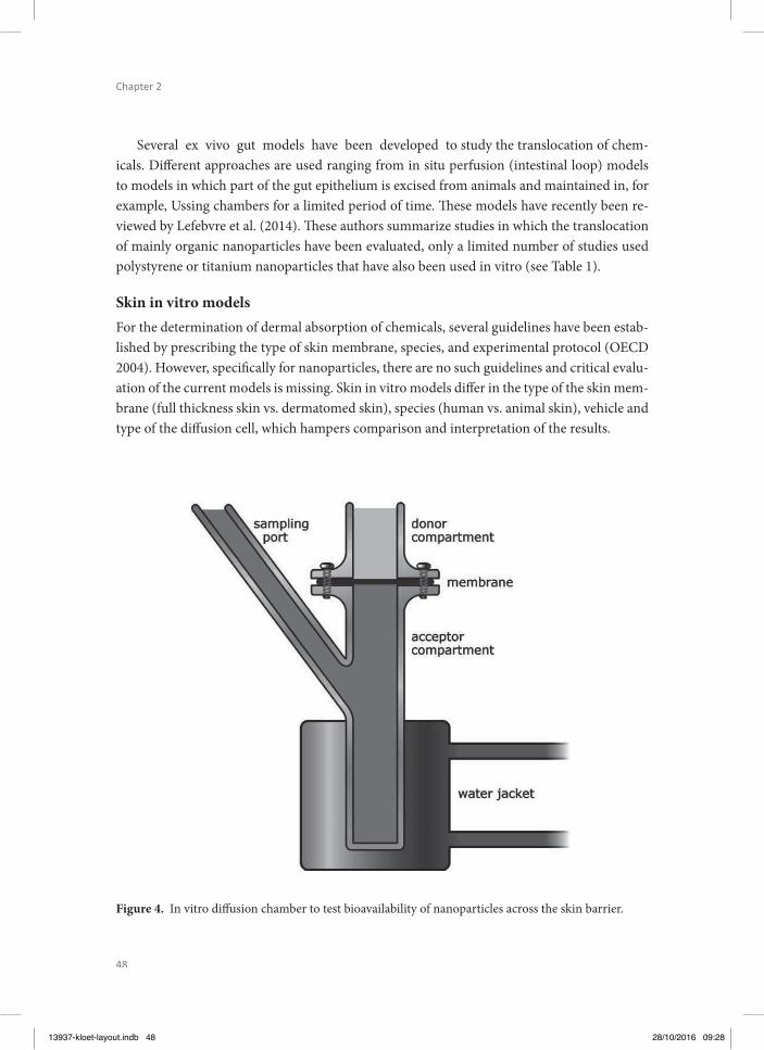
48
Chapter 2
Several ex vivo gut models have been developed to study the translocation of chem-icals. Diff erent approaches are used ranging from in situ perfusion (intestinal loop) models to models in which part of the gut epithelium is excised from animals and maintained in, for example, Ussing chambers for a limited period of time. Th ese models have recently been re-viewed by Lefebvre et al. (2014). Th ese authors summarize studies in which the translocation of mainly organic nanoparticles have been evaluated, only a limited number of studies used polystyrene or titanium nanoparticles that have also been used in vitro (see Table 1).
Skin in vitro modelsFor the determination of dermal absorption of chemicals, several guidelines have been estab-lished by prescribing the type of skin membrane, species, and experimental protocol (OECD 2004). However, specifi cally for nanoparticles, there are no such guidelines and critical evalu-ation of the current models is missing. Skin in vitro models diff er in the type of the skin mem-brane (full thickness skin vs. dermatomed skin), species (human vs. animal skin), vehicle and type of the diff usion cell, which hampers comparison and interpretation of the results.
Figure 4. In vitro diff usion chamber to test bioavailability of nanoparticles across the skin barrier.
13937-kloet-layout.indb 48 28/10/2016 09:28
49
Progress and future of in vitro models to study translocati on of nanoparti cles
2
In contrast to the lung, intestinal and placental barrier, no Transwells are used for percu-taneous penetration studies. Th e reported in vitro studies on percutaneous penetration of nanoparticles have been performed by using either a Franz static cell or a fl ow-through dif-fusion cells. In both systems, the skin membrane is clamped between two chambers, one of which contains a vehicle supplemented with the investigated chemical (donor chamber) and the other one a receptor fl uid from which the penetrated chemical will be sampled (Fig. 4) (Ja-kasa and Kezic 2008). In several studies, human reconstructed skin models have been applied although there were concerns regarding a less well-developed barrier in these models and the absence of the follicular penetration route that might play an important role for translocation of nanoparticles (Labouta et al. 2011a, b). In in vitro assays, usually a cryopreserved skin is used, which might, however, lead to changes of the skin barrier and shrinkage of the hair follicles (Labouta et al. 2011a).
Although human skin is regarded as a gold standard, a large number of studies on percutane-ous penetration of nanoparticles use diff erent animal models including mice, rat, and pig skin (Labouta and Schneider 2013). However, structural and morphological diff erences between human and animal skin, especially concerning the density of the hair follicles, thickness of the skin layers, skin lipid composition, and structure, could certainly aff ect the NP percutaneous penetration (Labouta and Schneider 2013). Another problem encountered by using hairy an-imals is damage of the skin barrier due to hair clipping.
Placenta in vitro modelsModels for studying transplacental transport have long been based on the perfused isolated human placenta studied in ex vivo study designs. Th e perfused isolated human placenta ex vivo model presents a directly relevant alternative that maintains the complexity of the in-tact placenta (Saunders 2009). Transport studies in the ex vivo intact placenta are technically challenging and require large quantities of substances to be tested. Th erefore, models using representative placental cell lines in Transwell study designs are being developed as in vitro alternatives. Most commonly used human placental cell lines are the BeWo, Jar, and JEG-3 cell lines, which have been applied to study transplacental transfer of a variety of drugs and compounds. Th e most popular cellular model consists of the BeWo cell line, representing a choriocarcinoma-derived placental cell line that strongly resembles cytotrophoblastic cells. Th e BeWo b30 subclone can be grown on Transwell inserts to form confl uent cell layers, en-abling the quantifi cation of both uptake into the cells from the apical compartment (maternal side) and effl ux from the cells to the basolateral compartment (foetal side) (Buerki-Th urnherr et al. 2012). In some studies these models have been validated by comparison of the transport rate across the Transwell cellular BeWo b30 layer and the transport rates detected in ex vivo placental models for the same compounds (Li et al. 2013, Poulsen et al. 2009).
13937-kloet-layout.indb 49 28/10/2016 09:28
50
Chapter 2
Although the type and nature of the nanoparticles studied in in vitro placental models are diff erent and the number of studies is limited, some general observations can be made. Nanoparticles can be transported across the placental barrier, where their size and type of nanoparticles infl uence the effi ciency of the transport. Furthermore, in contrast to drugs and other xenobiotic bulk chemicals, transport of several nanoparticles across the placental bar-rier appears to be highly variable. Th e latter conclusion can be derived from the observation that for the nanoparticles studied so far the amount (%) of the nanoparticles transported across the BeWo b30 cell layer varies from 0.6 to 29 % in 6 h (Table 1). Comparison of this transport rate to that reported in the BeWo model for several chemicals mounting up to 30 % in 2 h reveals that the transport of nanoparticles across the BeWo cell layer, just as that of chemicals, may be limited or signifi cantly depending on the type of nanoparticles. Th is im-plies that a validated in vitro model to quantify the potential transport of nanoparticles across the placental barrier would be of high value to set priorities for further in vivo testing, thereby avoiding in vivo testing of all newly developed nanoparticles.
General remarks on currently used in vitro models to study translocation of nanoparticlesCurrently used in vitro models to study translocation of nanoparticles are mostly based on cell culturing on Transwell inserts or on the use of ex vivo tissues. Transwell inserts can diff er in the type of plastic they are made of and in the pore size of the permeable membrane. Th ese diff erences between the Transwell inserts infl uence the translocation rate (Cartwright et al. 2012, Geys et al. 2006). Transwell inserts with a larger pore size give increased translocation rates compared with inserts with smaller pore sizes. In addition, some types of plastic of the Transwell inserts yield decreased translocation due to nanoparticle adsorption on the material surface. Th erefore, for each single type of nanoparticles, the translocation across the diff erent types of inserts should be tested without cells to assess whether the nanoparticles are not withheld by the insert itself.
Next to this, diff erent cell types are used, which clearly infl uences the translocation and the mechanism of translocation. When cell types lack the formation of tight junctions, nanopar-ticles can easily pass between the cells resulting in a higher translocation rate compared with cell types that do form tight junctions. To prevent paracellular transport, epithelial cells form-ing tight junctions are preferred. As already discussed, the incorporation of a mucus layer in gut epithelial models decreases the translocation (Walczak et al. 2014). Th is mucus layer might be a very important barrier for nanoparticles in vivo because of electrostatic repul-sion (for negatively charged nanoparticles) and mucus entrapment (for positively charged nanoparticles) (Husain et al. 2001, Lai et al. 2007, Norris et al. 1998, Szentkuti and Lorenz 1995). Similarly, such a mucus layer might also be essential for in vitro lung barrier models, since nanoparticles fi rst come in contact with the pulmonary surfactant if they are deposited
13937-kloet-layout.indb 50 28/10/2016 09:28
51
Progress and future of in vitro models to study translocati on of nanoparti cles
2
in the lungs. Lastly, the use of specifi c cells types in co-culture on top of an epithelial layer can alter the translocation rate of the nanoparticles. For instance, alveolar macrophages in lung models can decrease the translocation rate by uptake of nanoparticles, while M cells in intes-tinal models can enhance translocation. Th e addition of these specifi c cell types can improve the in vitro models by more closely mimicking the in vivo situation in which, for example, the lungs are protected from particles via phagocytosis by alveolar macrophages.
Some reported in vitro models use primary cells, while others use immortalized cell lines. On one hand, primary cells have a more diff erentiated phenotype compared with cell lines. On the other hand, the isolation of primary cells is oft en experimentally challenging: the cells dediff erentiate aft er isolation, diff er from batch to batch, and proliferate to a limited extent. Cell lines are easy to work with, well characterized and more homogenous, but they show only few characteristics of diff erentiated cells. Overall they only poorly represent the in vivo sit-uation (Hartung et al. 2002, Klein et al. 2011). In in vitro lung translocation studies, primary rat alveolar cells have been frequently used (Fazlollahi et al. 2011, Geys et al. 2009, Yacobi et al. 2008). Recent, biologically complex human intestinal tissues have been cultured using human (induced) pluripotent stem cells, resulting in human intestinal organoids that have secretory and absorptive functions (Brugmann and Wells 2013). Comparable systems have now been modifi ed into mature epithelial, functional and polarized monolayers grown on Transwell membranes, which are suitable for translocation studies (VanDussen et al. 2014).
Ex vivo models maintain the complexity of the physiological barriers (Saunders 2009). How-ever, transport studies in ex vivo models are technically challenging, time consuming, and can require large amounts of the test substance.
Advances in the fi eld of miniaturized and microfl uidic devices have recently led to the con-cept of organ-on-a-chip models (Bhatia and Ingber 2014, Huh et al. 2012, Moraes et al. 2012, van der Meer and van den Berg 2012), which can be seen as hybrid devices combining cells and microfabricated structures aiming to recapitulate the dynamic physical, cellular, and functional features of human tissues (Ardavin et al. 2001, Huh et al. 2010. 2013, Schimek et al. 2013, Wagner et al. 2013). Th ese devices fi rst provide a high control on the cell microenvi-ronment (e.g. physical and chemical parameters) together with dynamic culture conditions, since they are embedded in a microfl uidic format (Whitesides 2006). Furthermore, these de-vices can include active elements, which allow exposing cells to mechanical stimuli and surface strains, by stretching the substrate on which they are grown (Sinha et al. 2015). Th ese approaches mostly rely on 3D cell culture conditions, which are acknowledged to better mim-ic the in vivo conditions compared with conventional monolayer models (Harink et al. 2013).
13937-kloet-layout.indb 51 28/10/2016 09:28
52
Chapter 2
Over the years, examples of organ-on-a-chip models in the literature have diversifi ed and, for instance, include gut (Kim and Ingber 2013), lung (Huh et al. 2007, 2010, Nalayanda et al. 2010), or even blood–brain barriers (Griep et al.2013, Huh et al. 2013, Wolff et al. 2015). Th ese models can be of particular interest to the fi eld of nanotoxicology. Huh et al., for in-stance, have developed a complex lung-on-a-chip model, which recapitulates movements associated with breathing that is accompanied by cyclic stretching of cells (Huh et al. 2010). Th is device has been applied for NP testing, showing that breathing motion is essential for such nanotoxicological assays. Th e same group, using the same principle, later proposed a gut-on-a-chip model integrating peristaltic motion associated with digestion. Interestingly, exposed to a combination of dynamic culture and mechanical strain, Caco-2 cells were pro-ducing mucus and microvilli features were formed (Kim and Ingber 2013). Finally, organ-on-a-chip devices are amenable to parallelization and automation; they are easily coupled to virtually any detection technique, and diff erent organ models can even be combined on one device (Wagner et al. 2013) to eventually mimic the whole body/organism. Th e availability of such complex and in vivo-like models is expected to facilitate the implementation of the 3R’s legislation regarding animal experimentation (Marx et al. 2012, van de Stolpe and den Toonder 2013).
Accuracy of current in vitro translocation models compared to in vivo dataIn vitro translocation studies show mostly limited transport of nanoparticles across the lungs, gut, skin, and placenta. In the sections below, we discuss the comparison between in vitro data and available in vivo data. We included in vivo studies that measured the bioavailability of similar nanoparticles compared to their in vitro counterparts. However, most in vivo data come from separate studies in which not exactly the same type of nanoparticle is used: for instance, the nanoparticles diff er in size. Another marked diff erence is that the in vitro models are composed of cells (oft en cell lines) with a human origin, while the in vivo models mostly are rodents. Th erefore, we fi rst compared data from studies using nanoparticles of the same chemical composition although they sometimes diff ered for other characteristics. In Table 2, we included studies that used exactly the same nanoparticles in both an in vivo and an in vitro design.
Lung barrierTh e translocation rate between in vitro lung barrier models and in vivo inhalation studies diff ers depending on the tested nanoparticles (Table 2). In addition, some studies determined the cellular uptake and not the translocation rate of nanoparticles. Th e information on their localization can also be used in the comparison between in vitro and in vivo data.
13937-kloet-layout.indb 52 28/10/2016 09:28

53
Progress and future of in vitro models to study translocati on of nanoparti cles
2
For the most frequently used polystyrene nanoparticles, the translocation rate, both in vitro and in vivo, is very low 1 day aft er exposure. Depending on the pore size of the inserts, the translocation amounts to 0–6 % in vitro against 0.05–2 % in vivo. However, one in vivo study shows accumulation over time of polystyrene nanoparticles in the thymus (Sarlo et al. 2009), which cannot be demonstrated in an in vitro lung model. One in vitro study reported a size dependent translocation rate (Yacobi et al. 2008), which is in line with the size-dependent distribution shown aft er oropharyngeal aspiration (Sarlo et al. 2009). Also for gold nanopar-ticles, the in vitro translocation rate ranges from 0.5 to 5.2 % (Brandenberger et al. 2010). For comparison this can be compared to the in vivo translocation to the liver of 0.7 to 2.8 %; clearly, this is an underrepresentation of the complete systemic availability (Sadauskas et al. 2009, Sung et al. 2011, Takenaka et al. 2006, Yu et al. 2007). For cerium dioxide, quantum dots, silica, and titanium dioxide nanoparticles, diff erent translocation rates are found in vi-tro and in vivo. For cerium dioxide, the uptake rate in vivo is very low, and only about 10 % of the inhaled cerium dioxide was detected in the lungs (Geraets et al. 2012), while 80 % of the particles was internalized in cells in vitro (Raemy et al. 2011). Aft er inhalation exposure, quantum dots could be detected in the liver and kidney at relatively high amounts of 15 and 5 %, respectively (Ma-Hock et al. 2012). However, in vitro, no translocation of quantum dots across primary rat alveolar epithelial cells was observed (Fazlollahi et al. 2011, Geys et al. 2009). Silica nanoparticles were able to translocate across monolayers of Calu-3, NCI-H292, and A549 cells in vitro (George et al. 2015), but were not detected in tracheobronchial lymph nodes aft er inhalation exposure (Arts et al. 2007). However, the silica content was not mea-sured in any other tissue or organ besides the lung and lymph nodes, so there might have been some in vivo translocation to the blood, liver, and other organs. Finally, titanium dioxide nanoparticles were internalized by A549 epithelial cells, monocyte-derived macrophages, and monocyte-derived dendritic cells in vitro (Muller et al. 2010, Rothen-Rutishauser et al. 2008), while the in vivo studies showed that most nanoparticles retained in the alveolar space in the lung-lining fl uid directly aft er exposure, and were found later mostly inside alveolar macro-phages (Creutzenberg et al. 2012, Geiser et al. 2005, Oberdorster et al. 1994).
13937-kloet-layout.indb 53 28/10/2016 09:28
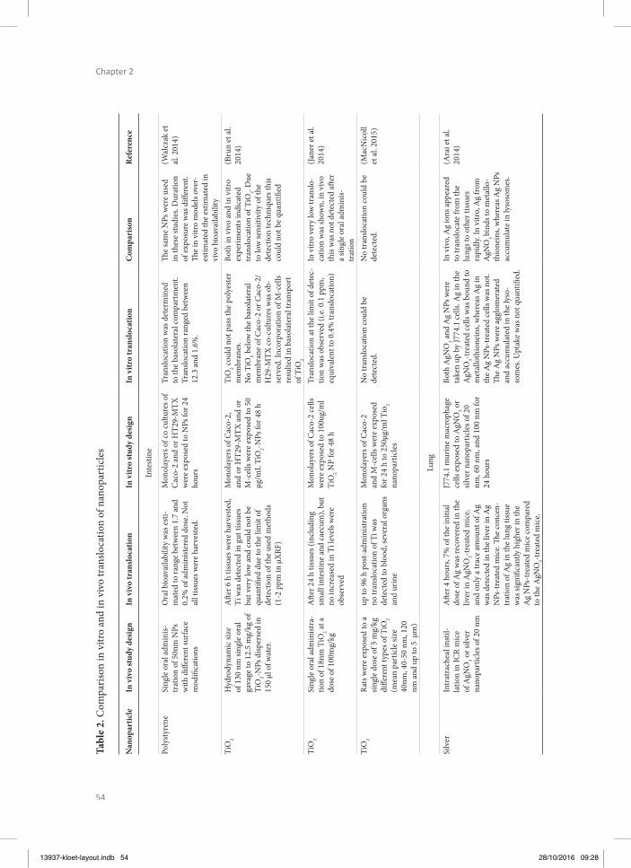
54
Chapter 2
Tabl
e 2.
Com
paris
on in
vitr
o an
d in
viv
o tr
anslo
catio
n of
nan
opar
ticle
s
Nan
opar
ticle
In v
ivo
stud
y de
sign
In v
ivo
tran
sloc
atio
nIn
vitr
o st
udy
desi
gnIn
vitr
o tr
ansl
ocat
ion
Com
pari
son
Ref
eren
ce
Inte
stin
e
Poly
styr
ene
Sing
le o
ral a
dmin
is-tr
atio
n of
50n
m N
Ps
with
diff
eren
t sur
face
m
odifi
catio
ns
Ora
l bio
avai
labi
lity
was
est
i-m
ated
to ra
nge
betw
een
1.7
and
0.2%
of a
dmin
ister
ed d
ose.
Not
al
l tiss
ues w
ere
harv
este
d.
Mon
olay
ers o
f co
cultu
res o
f C
aco-
2 an
d or
HT2
9-M
TX
wer
e ex
pose
d to
NPs
for 2
4 ho
urs
Tran
sloca
tion
was
det
erm
ined
to
the
baso
late
ral c
ompa
rtm
ent.
Tran
sloca
tion
rang
ed b
etw
een
12.3
and
1.6
%.
Th e
sam
e N
Ps w
ere
used
in
thes
e st
udie
s. D
urat
ion
of e
xpos
ure
was
diff
eren
t. Th
e in
vitr
o m
odel
s ove
r-es
timat
ed th
e es
timat
ed in
vi
vo b
ioav
aila
bilit
y
(Wal
czak
et
al. 2
014)
TiO
2H
ydro
dyna
mic
size
of
130
nm
sing
le o
ral
gava
ge to
12.
5 m
g/kg
of
TiO
2-NPs
disp
erse
d in
15
0 μl
of w
ater
.
Aft e
r 6 h
tiss
ues w
ere
harv
este
d,
Ti w
as d
etec
ted
in g
ut ti
ssue
s bu
t ver
y lo
w a
nd co
uld
not b
e qu
antifi
ed
due
to th
e lim
it of
de
tect
ion
of th
e us
ed m
etho
ds
(1-2
ppm
in μ
XRF
)
Mon
olay
ers o
f Cac
o-2,
an
d or
HT2
9-M
TX a
nd o
r M
-cel
ls w
ere
expo
sed
to 5
0 μg
/mL
TiO
2-NPs
for 4
8 h
TiO
2 coul
d no
t pas
s the
pol
yest
er
mem
bran
es.
No
TiO
2 bel
ow th
e ba
sola
tera
l m
embr
ane
of C
aco-
2 or
Cac
o-2/
H29
-MTX
co-c
ultu
res w
as o
b-se
rved
. Inc
orpo
ratio
n of
M-c
ells
resu
lted
in b
asol
ater
al tr
ansp
ort
of T
iO2
Both
in v
ivo
and
in v
itro
expe
rimen
ts in
dica
ted
tran
sloca
tion
of T
iO2. D
ue
to lo
w se
nsiti
vity
of t
he
dete
ctio
n te
chni
ques
this
coul
d no
t be
quan
tifi e
d
(Bru
n et
al.
2014
)
TiO
2Si
ngle
ora
l adm
inist
ra-
tion
of 1
8nm
TiO
2 at a
do
se o
f 100
mg/
kg
Aft e
r 24
h tis
sues
(inc
ludi
ng
smal
l int
estin
e an
d ca
ecum
), bu
t no
incr
ease
d in
Ti l
evel
s wer
e ob
serv
ed
Mon
olay
ers o
f Cac
o-2
cells
w
ere
expo
sed
to 1
00ug
/ml
TiO
2 NP
for 4
8 h
Tran
sloca
tion
at th
e lim
it of
det
ec-
tion
was
obs
erve
d (i.
e. 0.
1 pp
m,
equi
vale
nt to
0.4
% tr
anslo
catio
n)
In v
itro
very
low
tran
slo-
catio
n w
as sh
own,
in v
ivo
this
was
not
det
ecte
d aft
er
a sin
gle
oral
adm
inis-
trat
ion
(Jan
er e
t al.
2014
)
TiO
2Ra
ts w
ere
expo
sed
to a
sin
gle
dose
of 5
mg/
kg
diff e
rent
type
s of T
iO2
(mea
n pa
rtic
le si
ze
40nm
, 40-
50 n
m, 1
20
nm a
nd u
p to
5 µ
m)
up to
96
h po
st-a
dmin
istra
tion
no tr
anslo
catio
n of
Ti w
as
dete
cted
to b
lood
, sev
eral
org
ans
and
urin
e
Mon
olay
ers o
f Cac
o-2
and
M-c
ells
wer
e ex
pose
d fo
r 24
h to
250
µg/m
l Tio
2 na
nopa
rtic
les
No
tran
sloca
tion
coul
d be
de
tect
ed.
No
tran
sloca
tion
coul
d be
de
tect
ed.
(Mac
Nic
oll
et a
l. 20
15)
Lung
Silv
er
Intr
atra
chea
l ins
til-
latio
n in
ICR
mic
e of
AgN
O3 o
r silv
er
nano
part
icle
s of 2
0 nm
Aft e
r 4 h
ours
, 7%
of t
he in
itial
do
se o
f Ag
was
reco
vere
d in
the
liver
in A
gNO
3-tre
ated
mic
e, an
d on
ly a
trac
e am
ount
of A
g w
as d
etec
ted
in th
e liv
er in
Ag
NPs
-tre
ated
mic
e. Th
e co
ncen
-tr
atio
n of
Ag
in th
e lu
ng ti
ssue
w
as si
gnifi
cant
ly h
ighe
r in
the
Ag
NPs
-tre
ated
mic
e co
mpa
red
to th
e A
gNO
3-tre
ated
mic
e.
J774
.1 m
urin
e m
acro
phag
e ce
lls e
xpos
ed to
AgN
O3 o
r sil
ver n
anop
artic
les o
f 20
nm, 6
0 nm
, and
100
nm
for
24 h
ours
Both
AgN
O3 a
nd A
g N
Ps w
ere
take
n up
by
J774
.1 ce
lls. A
g in
the
AgN
O3-t
reat
ed ce
lls w
as b
ound
to
met
allo
thio
nein
s, w
here
as A
g in
th
e A
g N
Ps-t
reat
ed ce
lls w
as n
ot.
Th e
Ag
NPs
wer
e ag
glom
erat
ed
and
accu
mul
ated
in th
e ly
so-
som
es. U
ptak
e w
as n
ot q
uant
ifi ed
.
In v
ivo,
Ag
ions
appe
ared
to
tran
sloca
te fr
om th
e lu
ngs t
o ot
her t
issue
s ra
pidl
y. In
vitr
o, A
g fr
om
AgN
O3 bi
nds t
o m
etal
lo-
thio
nein
s, w
here
as A
g N
Ps
accu
mul
ate
in ly
soso
mes
.
(Ara
i et a
l. 20
14)
13937-kloet-layout.indb 54 28/10/2016 09:28
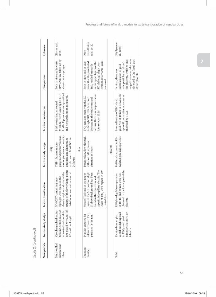
55
Progress and future of in vitro models to study translocati on of nanoparti cles
2
Tabl
e 2.
(con
tinue
d)
Nan
opar
ticle
In v
ivo
stud
y de
sign
In v
ivo
tran
sloc
atio
nIn
vitr
o st
udy
desi
gnIn
vitr
o tr
ansl
ocat
ion
Com
pari
son
Ref
eren
ce
Lung
Mul
ti-w
alle
d ca
rbon
nan
o-tu
bes
Oro
phar
ynch
eal a
spira
-tio
n in
C57
BL6
mic
e of
un
coat
ed a
nd a
lum
ini-
um co
ated
MW
CN
T of
0.
5 –
40 µ
m le
ngth
MW
CN
T-co
ntai
ning
mac
-ro
phag
es w
ere
foun
d in
the
alve
olar
regi
on a
nd a
roun
d th
e ai
rway
s of t
he lo
wer
lung
. Tiss
ue
dist
ribut
ion
was
not
mea
sure
d.
THP-
1 hu
man
mac
ro-
phag
es a
nd p
rimar
y hu
man
m
onoc
ytes
wer
e ex
pose
d to
un
coat
ed a
nd a
lum
iniu
m
oxid
e co
ated
MW
CN
T fo
r 24
hou
rs
Both
coat
ed a
nd u
ncoa
ted
MW
CN
T w
ere
take
n up
by
THP-
1 ce
lls. U
ptak
e w
as n
ot q
uant
ifi ed
an
d no
tran
sloca
tion
mea
sure
d.
Both
in v
ivo
and
in v
itro,
M
WC
NTs
are
take
n up
by
alve
olar
mac
roph
ages
.
(Tay
lor e
t al.
2014
)
Skin
Tita
nium
di
oxid
ePi
g sk
in e
xpos
ed fo
r 48
hrs
to co
ated
TiO
2 pa
rtic
les 1
4–16
nm
.
Mos
t of T
i fou
nd in
the
uppe
r la
yers
of t
he S
C, h
owev
er sl
ight
Ti
abo
ve b
ackg
roun
d ha
s bee
n fo
und
in th
e ep
ider
mis
and
supe
rfi ci
al p
apill
ary
derm
is. Th
e
leve
ls of
TiO
2 wer
e hi
gher
in U
V
trea
ted
skin
Porc
ine
skin
, fl o
w-t
hrou
gh
diff u
sion
cell,
exp
osur
e du
ratio
n 24
hou
rs
TiO
2 rem
ains
mai
nly
in th
e SC
. A
lthou
gh T
iO2 N
Ps h
ave
been
de
tect
ed in
the
epid
erm
is an
d de
rmis,
ther
e w
as n
o pe
netr
atio
n in
to re
cept
or fl
uid
Both
in v
itro
and
in v
ivo
show
that
the
pene
trat
ed
TiO
2 resid
ed p
rimar
ily
in th
e up
per l
ayer
s of t
he
SC, a
lthou
gh sl
ight
pen
-et
ratio
n in
to v
iabl
e la
yers
oc
curr
ed.
(Mon
-te
iro-R
ivie
re
et a
l. 20
11)
Plac
enta
Gol
dEx
viv
o hu
man
pla
-ce
ntal
mod
el e
xpos
ed
to P
EGyl
ated
gol
d na
nopa
rtic
les f
or 1
or
6 ho
urs
PEG
ylat
ed g
old
nano
part
icle
s of
10,
15,
and
30
nm w
ere
not
obse
rved
in th
e fo
etal
par
t of t
he
plac
enta
BeW
o ce
lls e
xpos
ed to
PE-
Gyl
ated
gol
d na
nopa
rtic
les
Inte
rnal
isatio
n of
PEG
ylat
ed
gold
NPs
of 1
0 nm
in B
eWo
cells
up
to 4
8 ho
urs a
ft er e
xpos
ure
as
anal
yzed
by
TEM
.
In v
itro,
ther
e w
as
inte
rnal
izat
ion
of g
old
nano
part
icle
s in
cells
of
the
plac
enta
, whi
le e
x vi
vo
no g
old
nano
part
icle
s wer
e ob
serv
ed in
the
foet
al p
art
of th
e pl
acen
ta
(Myl
lyne
n et
al
., 20
08)
13937-kloet-layout.indb 55 28/10/2016 09:28
56
Chapter 2
Th e above-mentioned studies did not test exactly the same nanoparticles for the in vitro and in vivo set-ups. Arai et al. (2014) did test the same silver nanoparticles in vitro and in vivo. However, they did not measure the in vitro NP translocation but the presence of silver mate-rial in cells. Th ey concluded that, in vivo, silver ions appear to translocate from the lungs toother tissues rapidly. In vitro, Ag from AgNO3 binds to metallothioneins, whereas Ag nanopar-ticles accumulate in lysosomes (Arai et al. 2014). Multi- walled carbon nanotubes (MWCNTs) were jointly tested in vitro and in vivo. However, this study focused on the development of pulmonary fi brosis and did not measure tissue distribution or translocation. Th e results did show, however, that MWCNTs are taken up by alveolar macrophages both in vivo and in vitro (Taylor et al. 2014).
Intestinal barrierSeveral in vivo oral studies have been performed, but only a very limited number of those in vivo studies can directly be compared to in vitro studies. Here, we focus only on studies that evaluated the uptake of food-relevant nanoparticles such as silica and titanium dioxide. In addition, studies that used model materials such as polystyrene are discussed, as polystyrene nanoparticles have also been used in vitro (Table 1).
Some studies investigated the in vitro and in vivo translocation of titanium dioxide across the gut epithelium. Six hours aft er a single oral administration of 130-nm TiO2, titanium could be detected in gut tissues, but due to the low concentrations, it could not be quantifi ed (Brun et al. 2014). Janer et al. (2014) did not observe any increase in tissue concentration in vivo 24 h aft er a single dose of 100 mg/kg TiO2 of 18 nm, while very low translocation was observed in vitro (Janer et al. 2014). In another study, rats were exposed to 5 mg/kg of diff erent types of TiO2 nanoparticles (mean particle size 40 nm, 40–50 nm, 120 nm and up to 5 µm), and up to 96 h post-administration, no translocation of titanium was detected to blood, several organs and urine. Also, no translocation was observed in vitro (MacNicoll et al. 2015). In rats exposed for 30 days to 200 mg/kg body weight (bw) 75-nm TiO2 nanoparticles, no increased titanium levels were detected in blood, liver kidney and spleen, while eff ects on liver and other organs have been reported (Wang et al. 2013). Titanium nanoparticles were also orally administered to rats, with a dose of 1 or 2 mg/kg TiO2 nanoparticles with a primary size of 20–60 nm (large agglomerates were present) for 5 days (Tassinari et al. 2014). Only a limit-ed number of tissues were collected; interestingly, in spleen elevated Ti concentrations were found in the high dose group. Detailed analysis revealed the presence of 130-nm (sp ICP-MS) or 200–400 nm (SEM-EDX) materials in spleens (Tassinari et al. 2014).
Like titanium dioxide, silica is commonly used as food additives. Th erefore, the oral up-take of silica nanoparticles was studied aft er 28 and 90 days of exposure to food-grade syn-thetic amorphous silica. Limited uptake was observed: only aft er 90-day exposure to 2500
13937-kloet-layout.indb 56 28/10/2016 09:28

57
Progress and future of in vitro models to study translocati on of nanoparti cles
2
mg/kg bw elevated Si levels were found in spleen (van der Zande et al. 2014). More studies are available that focussed on model polystyrene nanoparticles, and they generally highlight the dependence of uptake and accumulation of polystyrene nanoparticles on several factors, including their size, surface charge, and type of coating material (Araujo et al. 2014, Hillery et al. 1994, Hillyer and Albrecht 2001, Hussain and Florence 1998, Hussain et al. 1997, Jani et al. 1989). In general, smaller polystyrene nanoparticles were taken up across the GI tract to a higher extent than the larger ones (Jani et al. 1990); the non-ionic more than the carboxylated ones (Jani et al. 1989) and 407 poloxamer-coated more than 188 poloxamer-coated nanopar-ticles (Hillery and Florence 1996, Hussain et al. 1997).
Th e estimated oral bioavailability of 50-nm polystyrene nanoparticles varied between stud-ies from 0.2 to 1.7 % (Walczak et al., submitted) to 6.6 % (Jani et al. 1990). Strikingly, much higher bioavailability (23 %) was reported for 500-nm polystyrene nanoparticles (Hussain et al. 1997), while their 1-µm-size counterpart’s nanoparticles had a lower uptake (2 × 10⁻⁶ % detected in lymph fl uid) (Seifert et al. 1996). Also, the amounts of polystyrene nanoparticles associated with intestinal tissues that were reported by Walczak (between 0.38 and 0.74 % depending on the type of polystyrene nanoparticles, calculated as a sum of the small- and large intestinal walls), were lower than the ones reported by others for 60-nm nanoparti-cles (between 1.5 and 10 %, depending on the type (i.e. surface chemistry) of polystyrene nanoparticles used) (Hillery and Florence 1996, Hillery et al. 1994). Comparison of the oral in vivo bioavailability (0.2 and 1.7 %) (Walczak 2015), with the in vitro translocation values (1.6 to 12.3 %) of the same 50-nm polystyrene nanoparticles (Walczak et al. 2014), shows lower uptake values in the in vivo model. Th erefore, the in vitro model used by Walczak et al. appears to overestimate the in vivo translocation.
Skin barrierIn the literature, various in vitro assays (based on ex vivo skin tissue) have been used to de-termine percutaneous penetration of nanoparticles. However, the scarcity of in vivo human data hampers proper evaluation of these models. Th e vast majority of in vitro studies focused on TiO2 and ZnO nanoparticles found in sunscreens. Most, although not all in vitro studies fi nd that these nanoparticles do not penetrate beyond the superfi cial layers of the stratum corneum (Cross et al. 2007, Mavon et al. 2007, Wu et al. 2009, Zvyagin et al. 2008). One of the disadvantages of in vitro assays is that exposure duration is limited to 24 h, although long-term exposures would be closer to the real-life situation. Wu et al. (2009) compared in vitro and in vivo penetration of nanoscale TiO2 (4 nm and 60 nm) in two animal models (hairless mice and pigs). Aft er in vitro dermal exposure, TiO2 nanoparticles were not de-tected beyond the stratum corneum. However, in vivo, 30-day dermal exposure to the same nanoparticles in hairless mice revealed, that in contrast to short-term in vitro exposure, TiO2
13937-kloet-layout.indb 57 28/10/2016 09:28
58
Chapter 2
nanoparticles do reach viable skin layers (Wu et al. 2009). Furthermore, aft er 60-day dermal exposure, TiO2 nanoparticles could penetrate through the skin, reach diff erent tissues and induce diverse pathological lesions in several major organs (Wu et al. 2009). Deeper, although minimal, penetration into epidermal layers has also been found for ZnO nanoparticles in an in vivo study in human volunteers (Leite-Silva et al. 2013). In a parallel in vitro–in vivo study, Monteiro- Riviere et al. (2011) investigated the penetration of TiO2 and ZnO nanoparticles in UVB-damaged porcine skin. Under both conditions, TiO2 and ZnO NP predominantly resid-ed in the stratum corneum, although small amounts of TiO2 and ZnO were also detected in the viable skin layers. On the other side, not all in vivo studies report penetration of nanopar-ticles beyond the stratum corneum (Monteiro- Riviere et al. 2011). For instance, Zvyagin et al. found in an in vivo study with human skin no penetration of ZnO (26–30 nm) into the viable layers (Zvyagin et al. 2008). Th is is consistent with the fi ndings reported by Mavon et al. show-ing no penetration of TiO2 into the viable epidermal layers of human skin either in vivo or in vitro (Mavon et al.2007). Obviously, the discrepancy in the results cannot be explained solely by the diff erences between in vitro and in vivo data but also by the characteristics of the used nanoparticles (size, coating), exposure duration and sensitivity of the detection methods.
Another frequently investigated NP is silver, which is used on a large scale in medicinal and consumer products. Larese et al. found in an in vitro study with human skin that smaller silver nanoparticles (30 nm) can penetrate across the stratum corneum into the upper layers of the epidermis (Larese et al. 2009). Th is is consistent with in vivo data obtained in human volunteers (George et al. 2014) showing that silver nanoparticles could penetrate as deep as the reticular dermis. Zhu et al. showed, using a highly sensitive detection method, that the penetration depth of Ag nanoparticles could exceed the stratum corneum thickness (Zhu et al. 2015).
Placental barrierAs described above, the transport effi ciency of nanoparticles across the human placenta is likely to be diff erent from that in rodents (Wick et al. 2010). To study the translocation over the human placental barrier, dual recirculation human placental (ex vivo) perfusion models are used. In such a design the translocation of 50, 80-, 240-, or 500-nm polystyrene was stud-ied, under highly controlled conditions (i.e. translocation of a marker compound) (Wick et al.2010). Aft er a single administration, 50-, 80-, and 240-nm polystyrene nanoparticles were observed in the foetal circulation (foetal to maternal ratios 0.4; 0.4; 0.1, respectively), while the 500-nm polystyrene nanoparticles were retained in the placenta (Wick et al. 2010). In vitro similar results were found, where the translocation rate of 50-nm polystyrene nanopar-ticles was larger compared with 100-nm nanoparticles (Cartwright et al., 2012). Myllynen et al. perfused ex vivo human placentas with 10-, 15-, and 30-nm PEGylated gold nanoparticles
13937-kloet-layout.indb 58 28/10/2016 09:28
59
Progress and future of in vitro models to study translocati on of nanoparti cles
2
(for up to 6 h) and did not observe any PEGylated gold nanoparticles in the foetal part of the placenta (Myllynen et al. 2008). In vitro, they observed internalization of PEGylated gold nanoparticles of 10 nm in BeWo cells up to 48 h aft er exposure (Myllynen et al. 2008).
In rodents, fl uorescent polystyrene nanoparticles were administered via the extraembryonic tissue. Th e embryos were ex vivo incubated for 12 h with 20-, 100-, and 500- nm carboxylated and 200-nm amino-conjugated/terminated polystyrene nanoparticles. Th e 20-nm carboxyl-ated nanoparticles were distributed in the embryonic and extraembryonic germ layers of ec-toderm, mesoderm, and endoderm. Th e 100 and 500-nm carboxylated polystyrene nanopar-ticles accumulated in extraembryonic tissue. Interestingly, the 200-nm amino-conjugated particles can pass into the embryos (Tian et al. 2009). For 5-nm gold nanoparticles, 0.018 % of the administered dose (118 µg/kg bw intravenous) administered to rats at day 19 of gestation was detectable in the embryo (Takahashi and Matsuoka 1981).
In addition, studies with silica nanoparticles showed that 70 nm, but not 300 or 1000 nm reached the brain and liver of the foetus aft er i.v. administration to the mother (Yamashita et al. 2011), while there was limited transport of 25-nm silica nanoparticles in vitro (Sonnegaard Poulsen et al. 2013). In the same study of Yamashita et al. 63-nm TiO2 nanoparticles were detected by TEM in brains and livers of foetuses (Yamashita et al. 2011). In another study, surface modifi ed 28–30-nm iron oxide nanoparticles were administered (intraperitoneal) to mice from gestation days 9–16. Nanoparticles with a positive zeta potential in water [coated with hydrophilic polyethyleneimine (PEI)] were detected in the livers of foetuses 1 day aft er dosing of the dams, while negatively charged nanoparticles (coated with acrylic acid) could not be found in the foetuses (Di Bona et al. 2014). In vitro, iron oxide nanoparticles were able to cross a BeWo cell layer (Correia Carreira et al. 2013).
Based on these studies, it can be concluded that nanoparticles can pass the placenta of rodents and humans (ex vivo). Th is translocation is size- and surface charge- dependent. Although there are few studies available to compare the in vitro translocation to the in vivo or ex vivo translocation, for polystyrene nanoparticles, size-dependent translocation has been observed both ex vivo and in vitro. However, for gold nanoparticles and iron oxide nanopar-ticles the in vitro translocation was higher compared with the ex vivo or in vivo translocation, while for silica nanoparticles it was lower.
13937-kloet-layout.indb 59 28/10/2016 09:28
60
Chapter 2
ANALYTICAL TECHNIQUES TO QUANTIFY AND CHARACTERIZE NANOPAR-TICLE TRANSLOCATION IN VITRO AND IN VIVO
Meaningful interpretation and comparison of the results obtained using diff erent in vitro ex-periments and extrapolation to in vivo data require reliable characterization of the nanopar-ticles and their aggregates, as well as matrix-based infl uences on the nanoparticles. Th erefore, appropriate analytical techniques should be applied to determine the nanoparticle size distri-bution, composition, and concentration in the experimental samples.
Widely used methods to detect nanoparticles in liquid dispersions are dynamic light scat-tering (DLS), centrifugal liquid sedimentation (CLS) (Braun et al. 2011, Cascio et al. 2015, Murdock et al. 2008, Nickel et al. 2014, Powers et al. 2006) and nanoparticle tracking analysis (NTA) (Filipe et al. 2010, Vasco et al. 2010). Th ese methods allow determining an average size or size distribution related to the measured intensity signal. NTA is able to count and size nanoparticles in aqueous media at µg/L to mg/L concentrations (Filipe et al. 2010, Vasco et al. 2010). Both DLS and NTA are highly dependent on the polydispersity of the nanoparticle suspension and material properties of the particles since the scattered light of the indivi-dual particles must be suffi ciently strong for detection. CLS is more robust since particles are size-separated before their actual detection and sizing. Transmission and scanning electron microscopy (TEM, SEM) are techniques to visualize nanoparticles (Dudkiewicz et al. 2011, Zhang et al. 2012). If pure nanoparticle dispersions are analysed, EM is currently the only technique that reliably covers the entire size range down to 1 nm. In cells or tissues, the min-imal particle size that can be detected is around 20 nm, depending on the electron density of the nanoparticles (De Jong et al. 2010). Furthermore, EM distinguishes size aggregates and primary particles. Other imaging techniques are atomic force microscopy (AFM) (Brown et al. 2013) and particle- induced X-ray emission (PIXE) spectroscopy (Lozano et al. 2012, 2013).
Elemental information about the sample can also be obtained by atomic spectrometry meth-ods such as inductively coupled plasma optical emission spectrometry (ICP-OES) (Elzey et al. 2012) and ICP mass spectrometry (ICP-MS) (Krystek 2012, Krystek et al. 2013), especially in single particle mode (sp ICP-MS) (Laborda et al. 2014, Pace et al. 2011, Peters et al. 2014a). From these studies it becomes clear that he smallest particle sizes that now can be determined are around 20 nm for silver and gold nanoparticles. For TiO2 and SiO2, nanoparticles size detection limits are around 50 and 200 nm; however, recent experiments suggest that the size detection limit may become lower in the next few years. Another promising possibility is to combine size-specifi c techniques, that separate particles from each other, such as hydrody-namic chromatography (HDC) or fi eld-fl ow fractionation (FFF) with atomic spectroscopy techniques, that characterize particles, such as ICP-MS (Bednar et al. 2013, Hassellov et al.
13937-kloet-layout.indb 60 28/10/2016 09:28

61
Progress and future of in vitro models to study translocati on of nanoparti cles
2
2008, Peters et al. 2014b, Striegel and Brewer 2012, Von der Kammer et al. 2011). Currently, asymmetric fl ow fi eld-fl ow fractionation (AF4) is the most successfully used variant of FFF (Zattoni et al. 2014). In in vitro testing, AF4 coupled to ICP-MS becomes a powerful tool to investigate, for example, time-dependent uptake of medium-sized silver nanoparticles (Krys-tek et al. 2015). Laser ablation (LA)-ICP-MS has been used to quantify gold nanoparticles in single cells (Wang et al. 2014).
Most of the detection methods described above generally require sample preparation proce-dures. However, only little information concerning sample preparation techniques is available in the literature (Szakal et al. 2014). Aqueous media containing nanoparticles only need lim-ited sample preparation; samples may be sonicated or tip-sonicated to suspend materials and proteins such as bovine serum albumin (BSA), or detergents such as sodium dodecylsulphate (SDS) may be added to stabilize nanoparticle suspensions (Jensen et al. 2011). For other ma-trices, matrix removal or nanoparticle isolation from the matrix can be achieved by physical processes such as centrifugation, fi ltration, column techniques or cloud point extraction, or by chemical or enzymatic destruction of the matrix (Loeschner et al. 2014, Peters et al. 2014a, b).
As mentioned not only size, but also, surface chemistry and charge, and composition of the so-called protein corona have been shown to signifi cantly aff ect the translocation of nanopar-ticles. Th e nanoparticle surface charge (or zeta potential) is routinely determined, but a cor-rect interpretation might be hampered by interactions of the matrix with these measurements. More details analysis of the surface of the nanoparticles, for example, by matrix-assisted laser desorption/ionization time-of-fl ight mass spectrometry (MALDI-TOF) should be consid-ered (Walczak et al.2014). Methods for the assessment of protein composition range from simple gel electrophoreses experiments to a full characterization of the biomolecules that are present using mass spectrometry-based techniques (Lesniak et al. 2010, Lundqvist et al. 2008, Tedja et al. 2012).
Overall, diff erent techniques are available to determine the nanoparticle size distribution, composition and concentration in experimental samples. Attention should be paid to the lim-its of the chosen techniques and to adequate sample preparation; this should be incorporated in studies reporting translocation data.
Conclusions on comparison of in vitro translocation models with in vivo dataWhile comparing the in vitro translocation to in vivo data, we noticed that very few studies tested exactly the same nanoparticles in in vitro and in in vivo settings. It seems that most studies are performed either in vitro or in vivo. Th erefore, we focused our comparison on the nanoparticle chemical composition although the nanoparticles studied sometimes diff er in
13937-kloet-layout.indb 61 28/10/2016 09:28
62
Chapter 2
other particle characteristics such as size. Interestingly, this approach revealed that for some nanoparticles, the in vitro translocation is similar to the in vivo translocation. Examples are the translocation of polystyrene and gold nanoparticles in in vitro lung barrier models, and the translocation of titanium dioxide and silver nanoparticles in in vitro skin models. Howev-er, for other nanoparticles, major diff erences seem to exist between the in vitro translocation rate and the in vivo translocation.
Th ese diff erences in translocation may be caused by the many diff erences existing between the in vitro barrier models and the in vivo study designs. First of all, the in vitro models mostly use an acute exposure of maximum 24 h and a short post-exposure time, while, for example, in vivo inhalation studies vary from 1 h to 13 weeks of inhalation exposure, 5 days a week, 6 h per day with post-exposure periods of up to 1 year. Th e relative short in vitro exposure durations might imply that only a fraction the nanoparticles that have been taken up (intracellularly) has been translocated. Longterm exposures are not conceivable in an in vitro set-up, although this would be a more realistic exposure scenario. Second, exposure concentrations between the in vitro and in vivo studies diff er. In in vitro experiments, high particle concentrations are sometimes used to be able to detect the nanoparticles in the baso-lateral compartment. Th ese high concentrations might increase the agglomeration state of the nanoparticles and damage the epithelial barriers resulting in a diff erent translocation mech-anism compared with the in vivo situation. Clearly also in vivo dose selection can be criti-cal, as it has been shown in rats that exposure to high concentrations to silica nanoparticles cause gelation (agglomeration) of silica nanoparticles in the gut (van der Zande et al. 2014). Th ird, in vitro models have a single basolateral compartment, while in vivo, nanoparticles can translocate to various tissues and organs. In the in vitro set-up, saturation might occur while under in vivo conditions nanoparticles can continue translocating, since they are taken up in organs and removed from the blood. On the other hand, this distribution of nanoparticles to various tissues can hamper translocation studies in vivo. Fourth, current in vitro models are almost all static, while in vivo exposure is dynamic. Fift h, nanoparticles adsorb proteins and/or phospholipids in biological fl uids such as serum or lung-lining fl uid (Landsiedel et al. 2014b). Th ese proteins or phospholipids form a corona around the particles that aff ects their uptake and bioavailability (Lesniak et al. 2012). Th e proteins that are encountered in in vitro models, for example in foetal calf serum, are completely diff erent from the proteins in vivo (i.e. rodent or human). Sixth, many in vitro lung models are submerged, which might alter the nanoparticle characteristics and thus the translocation rate, while most in vivo studies rely on inhalation exposure.
13937-kloet-layout.indb 62 28/10/2016 09:28

63
Progress and future of in vitro models to study translocati on of nanoparti cles
2
CONCLUSIONS
Th e aim of this review was to evaluate the performance of in vitro models that mimic diff erent physiological barriers found in the human body by comparing—when possible— the in vitro translocation of nanoparticles to their in vivo translocation across the lung, gut, skin, and pla-cental barriers. For all these barriers, a great variety of in vitro models are available to evaluate the translocation of nanoparticles, ranging in complexity from single-cell-type monolayer to multi-cell (3D) models. Many studies that use in vitro models on inserts focus on the toxicity of nanoparticles, do not include their translocation, and were thus not included in this review. Clearly, for a correct interpretation of the observed toxicity, the translocation (or systemic availability, internal concentration) is a crucial parameter. In recent years, the avail-ability of analytical detection methods to quantify and characterize the nanoparticles in in vitro settings has improved considerably, which provides highquality data that are valuable in studying the relationship between physiochemical properties of the nanoparticles and their translocation. Th e improved analytical chemical detection methods also contributed to an increase in in vivo uptake data (bioavailability) of nanoparticles.
Here, while comparing the in vitro translocation to in vivo data, we noticed that very few studies tested exactly the same materials in both settings. Comparing data obtained using nanoparticles of the same chemical composition, we found that for some nanoparticles, the in vitro translocation is similar to the in vivo bioavailability. Examples are the translocation of polystyrene and gold nanoparticles in in vitro lung barrier models, and that of titanium dioxide and silver nanoparticles in in vitro skin models that are both in line with the in vivo data. However, for other nanoparticles, major diff erences were found between the in vitro and the in vivo translocation rate. As discussed in the sections above, many diff erences exist in the experimental set-up between in vitro and in vivo study design that probably account for the poor correlation between these two types of studies. Especially, the changes in the phys-icochemical characteristics of the nanoparticles caused by the presence of lung-lining fl uid, mucus, serum protein, and lipoproteins that form a corona should be taken into account, as these dramatically alter their recognition, uptake and translocation (Lesniak et al. 2012, Treuel et al. 2013).
Risk assessment of nanoparticles (as is true for chemicals in general) still heavily relies on in vivo studies using experimental animals. However, the latter must be reduced as far as possible, for numerous reasons. Th erefore, there is an urgent need to validate existing in vitro models using data from animal models, although these animal models do not fully simulate the physiology of humans. How can we establish an in vitro barrier model that has value for the risk assessment of nanoparticles for humans? Ideally, in vitro models refl ect the key mechanisms of corresponding in vivo end points, which cannot always be accomplished and
13937-kloet-layout.indb 63 28/10/2016 09:28
64
Chapter 2
may not be required if in vitro models reliably detect nanoparticles that are of concern in vivo (Landsiedel et al. 2014b). Th e predictive value of in vitro models can be better assessed by testing exactly the same nanoparticles simultaneously in both in vitro and in vivo assays. If the ranking of the tested nanoparticles from the lowest translocation rate to the highest translo-cation rate is the same in both cases, the in vitro models provide information on the internal exposure, which is critical for the ultimate systemic adverse eff ects. Th en, the in vitro models can be considered as suitable for the risk assessment of nanoparticles and will in addition help to reduce animal testing by setting priorities for subsequent in vivo testing.
A crucial factor for both translocation studies is the sensitivity of the analytical technique used for the quantifi cation of the translocation. While this is of importance also for in vivo studies, in vitro studies are more vulnerable for poorly performing methods. Th e observed low translocation rates combined with the relatively short exposure durations in vitro pose great analytical challenges. Th e absence of acute in vitro translocation should always be inter-preted in relation to the sensitivity of the detection technique (both in terms of concentra-tion and nanoparticle size). Th e introduction of in vitro models that allow chronic exposures are promising in that a chronic exposure better refl ects the real-life human exposure.
In vitro models with a high predictive value do not necessarily have to be complex, but can consist of a single cell type, as long as they give a similar ranking of nanoparticles as obtained in the in vivo situation. However, the mechanism in these models might be completely diff er-ent from the in vivo situation. To obtain more insight into the mechanisms behind nanopar-ticle uptake and translocation, in vitro models should be further developed to become phys-iologically very close to the in vivo human conditions. Such in vitro models must be more complex because the human lung, gut, skin, and placental barrier consist of multiple cell types, which are exposed to a low concentration of nanoparticles. A physiologically realistic model will increase the confi dence in the NP testing outcome, but it will also be elaborate and expensive to develop and maintain.
When in vitro models are available that either have a high human predictive value or are phys-iologically similar to the human situation, experimental data can be used to develop in silico models that will eventually be able to predict the human in vivo bioavailability of nanoparti-cles from their in vitro translocation rate. Th e (improved) in vitro models still needs valida-tion, most likely using animal data, clearly alternative approaches need to be developed for this (making use of available human data).
We conclude that the current in vitro models to study the translocation of nanoparticles do not (yet) allow correlating to the reported in vivo translocation because of many diff erences between the in vitro and in vivo study designs. However, the use of in vitro models is very
13937-kloet-layout.indb 64 28/10/2016 09:28
65
Progress and future of in vitro models to study translocati on of nanoparti cles
2
promising since they are currently further improved to mimic the in vivo situation more closely by, for example, using co-cultures of diff erent cell types and implementing them in a microfl uidic format. When these models are further validated by testing exactly the same nanoparticles in an in vivo set-up as in the in vitro model, then, they can be used to determine the internal exposure (bioavailability) of nanoparticles and to set priorities for nanoparticles testing.
ACKNOWLEDGEMENTS
Th is work is supported by NanoNextNL, a micro- and nanotechnology consortium of the Government of Th e Netherlands, and 130 partners.
13937-kloet-layout.indb 65 28/10/2016 09:28
66
Chapter 2
REFERENCES
Ali H, Kalashnikova I, White MA, Sherman M, Rytting E (2013) Preparation, characterization, and transport of dexamethasone- loaded polymeric nanoparticles across a human placental in vitro model. Int J Pharm 454(1):149–157. doi:10.1016/j. ijpharm.2013.07.010
Antunes F, Andrade F, Araujo F, Ferreira D, Sarmento B (2013) Establishment of a triple co-culture in vitro cell models to study intestinal absorption of peptide drugs. Eur J Pharm Biopharm 83(3):427–435. doi:10.1016/j.ejpb.2012.10.003
Arai Y, Miyayama T, Hirano S (2014) Diff erence in the toxicity mechanism between ion and nanoparticle forms of silver in the mouse lung and in macrophages. Toxicology 328:84–92. doi:10.1016/j.tox.2014.12.014
Araujo F, Shrestha N, Shahbazi MA et al (2014) Th e impact of nanoparticles on the mucosal translocation and transport of GLP-1 across the intestinal epithelium. Biomaterials 35(33):9199– 9207. doi:10.1016/j.bio-materials.2014.07.026
Ardavin C, del Hoyo GM, Martin P et al (2001) Origin and diff erentiation of dendritic cells. Trends Immunol 22(12):691–700. doi:10.1016/S1471-4906(01)02059-2
Arts JH, Muijser H, Duistermaat E, Junker K, Kuper CF (2007) Five-day inhalation toxicity study of three types of synthetic amorphous silicas in Wistar rats and post-exposure evaluations for up to 3 months. Food Chem Toxicol 45(10):1856–1867. doi:10.1016/j.fct.2007.04.001
Asgharian B, Price O, Miller F et al (2009) Multiple-path particle dosimetry model (MPPD v 2.11): a model for human and rat airway particle dosimetry. In: Applied Research Associates (ARA) HIfHS, National Institute for Public Health and the Environment (RIVM), and Ministry of Housing, Spatial Planning and the Envi-ronment (ed). V2.11 edn. Applied Research Associates (ARA), Raleigh, North Carolina
Aye IL, Keelan JA (2013) Placental ABC transporters, cellular toxicity and stress in pregnancy. Chem Biol Interact 203(2):456–466. doi:10.1016/j.cbi.2013.03.007
Bakand S, Hayes A, Dechsakulthorn F (2012) Nanoparticles: a review of particle toxicology following inhalation exposure. Inhal Toxicol 24(2):125–135. doi:10.3109/08958378.2010.642021
Balasubramanian SK, Jittiwat J, Manikandan J, Ong CN, Yu LE, Ong WY (2010) Biodistribution of gold nanopar-ticles and gene expression changes in the liver and spleen aft er intravenous administration in rats. Bioma-terials 31(8):2034–2042. doi:10.1016/j.biomaterials.2009.11.079
Baroli B (2010) Penetration of nanoparticles and nanomaterials in the skin: fi ction or reality? J Pharm Sci 99(1):21–50. doi:10.1002/ jps.21817
Bednar AJ, Poda AR, Mitrano DM et al (2013) Comparison of on-line detectors for fi eld fl ow fractionation analysis of nanomaterials. Talanta 104:140–148. doi:10.1016/j.talanta.2012.11.008
Behrens I, Pena AI, Alonso MJ, Kissel T (2002) Comparative uptake studies of bioadhesive and non-bioadhesive nanoparticles in human intestinal cell lines and rats: the eff ect of mucus on particle adsorption and trans-port. Pharm Res 19(8):1185–1193
Bellmann S, Carlander D, Fasano A et al (2015) Mammalian gastrointestinal tract parameters modulating the integ-rity, surface properties, and absorption of food-relevant nanomaterials. Wiley Interdiscip Rev Nanomed Nanobiotechnol. doi:10.1002/ wnan.1333
Benirschke K, Kaufmann P, Baergen RN (2006) Pathology of the human placenta. Springer, New YorkBermudez LE, Sangari FJ, Kolonoski P, Petrofsky M, Goodman J (2002) Th e effi ciency of the translocation of Myco-
bacterium tuberculosis across a bilayer of epithelial and endothelial cells as a model of the alveolar wall is a consequence of transport within mononuclear phagocytes and invasion of alveolar epithelial cells. Infect Immun 70(1):140–146
Bhatia SN, Ingber DE (2014) Microfl uidic organs-on-chips. Nat Biotechnol 32(8):760–772. doi:10.1038/nbt.2989Bhattacharjee S, Ershov D, Gucht J et al (2013) Surface charge-specifi c cytotoxicity and cellular uptake of tri-block
copolymer nanoparticles. Nanotoxicology 7(1):71–84. doi:10.3109/17435390.2011.633714Blank F, Rothen-Rutishauser BM, Schurch S, Gehr P (2006) An optimized in vitro model of the respiratory tract
wall to study particle cell interactions. J Aerosol Med 19(3):392–405. doi:10.1089/jam.2006.19.392Borm PJ, Robbins D, Haubold S et al (2006) Th e potential risks of nanomaterials: a review carried out for ECETOC.
Part Fibre Toxicol 3:11. doi:10.1186/1743-8977-3-11
13937-kloet-layout.indb 66 28/10/2016 09:28
67
Progress and future of in vitro models to study translocati on of nanoparti cles
2
Bouwmeester H, Poortman J, Peters RJ et al (2011) Characterization of translocation of silver nanoparticles and eff ects on whole genome gene expression using an in vitro intestinal epithelium coculture model. ACS Nano 5(5):4091–4103. doi:10.1021/ nn2007145
Bouwmeester H, Brandhoff P, Marvin HJ, Weigel S, Peters RJ (2014) State of the safety assessment and current use of nanomaterials in food and food production. Trends Food Sci Technol 40(2):200–210
Braakhuis HM, Gosens I, Krystek P et al (2014a) Particle size dependent deposition and pulmonary infl ammation aft er shortterm inhalation of silver nanoparticles. Part Fibre Toxicol11(1):16. doi:10.1186/s12989-014-0049-1
Braakhuis HM, Park MV, Gosens I, De Jong WH, Cassee FR (2014b) Physicochemical characteristics of nano-materials that aff ect pulmonary infl ammation. Part Fibre Toxicol 11(1):18. doi:10.1186/1743-8977-11-18
Brandenberger C, Rothen-Rutishauser B, Muhlfeld C et al (2010) Eff ects and uptake of gold nanoparticles deposited at the air-liq- uid interface of a human epithelial airway model. Toxicol Appl Pharmacol 242(1):56–65. doi:10.1016/j.taap.2009.09.014
Braun A, Couteau O, Franks K et al (2011) Validation of dynamic light scattering and centrifugal liquid sedimenta-tion methods for nanoparticle characterisation. Adv Powder Technol 22:766–770
Brown SC, Boyko V, Meyers G, Voetz M, Wohlleben W (2013) Toward advancing nano-object count metrology: a best practice framework. Environ Health Perspect 121(11–12):1282–1291. doi:10.1289/ehp.1306957
Brugmann SA, Wells JM (2013) Building additional complexity to in vitro-derived intestinal tissues. Stem Cell Res Th er 4(Suppl1):S1. doi:10.1186/scrt362
Brun E, Barreau F, Veronesi G et al (2014) Titanium dioxide nanoparticle impact and translocation through ex vivo, in vivo and in vitro gut epithelia. Particle Fibre Toxicol. doi:10.1186/1743-8977-11-13
Buerki-Th urnherr T, von Mandach U, Wick P (2012) Knocking at the door of the unborn child: engineered nanopar-ticles at the human placental barrier. Swiss Med Wkly 142:w13559. doi:10.4414/ smw.2012.13559
Carr KE, Smyth SH, McCullough MT, Morris JF, Moyes SM (2012) Morphological aspects of interactions between microparticles and mammalian cells: intestinal uptake and onward movement. Prog Histochem Cyto-chem 46(4):185–252. doi:10.1016/j. proghi.2011.11.001
Cartwright L, Poulsen MS, Nielsen HM et al (2012) In vitro placental model optimization for nanoparticle transport studies. Int J Nanomed 7:497–510. doi:10.2147/ijn.s26601
Carvalho TC, Peters JI, Williams RO 3rd (2011) Infl uence of particle size on regional lung deposition—what evi-dence is there? Int J Pharm 406(1–2):1–10. doi:10.1016/j.ijpharm.2010.12.040
Cascio C, Geiss O, Franchini F et al (2015) Detection, quantifi cation and derivation of number size distribution of silver nanoparticles in antimicrobial consumer products. J Anal At Spectrom. doi:10.1039/c4ja00410h
Cevc G, Vierl U (2010) Nanotechnology and the transdermal route: a state of the art review and critical appraisal. J Control Release 141(3):277–299. doi:10.1016/j.jconrel.2009.10.016
Chowdhury F, Howat WJ, Phillips GJ, Lackie PM (2010) Interactions between endothelial cells and epithelial cells in a combined cell model of airway mucosa: eff ects on tight junction permeability. Exp Lung Res 36(1):1–11. doi:10.3109/01902140903026582
Cone RA (2009) Barrier properties of mucus. Adv Drug Deliv Rev 61(2):75–85. doi:10.1016/j.addr.2008.09.008Correia Carreira S, Walker L, Paul K, Saunders M (2013) Th e toxicity, transport and uptake of nanoparticles in the
in vitro BeWo b30 placental cell barrier model used within NanoTEST. Nanotoxicology. doi:10.3109/17435390.2013.833317
Crater JS, Carrier RL (2010) Barrier properties of gastrointestinal mucus to nanoparticle transport. Mac-romol Biosci10(12):1473–1483. doi:10.1002/mabi.201000137
Creutzenberg O, Bellmann B, Korolewitz R et al (2012) Change in agglomeration status and toxicokinetic fate of various nanoparticles in vivo following lung exposure in rats. Inhal Toxicol 24(12):821–830. doi:10.3109/08958378.2012.721097
Cross SE, Innes B, Roberts MS, Tsuzuki T, Robertson TA, McCormick P (2007) Human skin penetration of sunscreen nanoparticles: in vitro assessment of a novel micronized zinc oxide formulation. Skin Pharmacol Physiol 20(3):148–154. doi:10.1159/000098701
De Jong WH, Hagens WI, Krystek P, Burger MC, Sips AJ, Geertsma RE (2008) Particle size-dependent organ distribu-tion of gold nanoparticles aft er intravenous administration. Biomaterials 29(12):1912–1919. doi:10.1016/j.biomaterials.2007.12.037
13937-kloet-layout.indb 67 28/10/2016 09:28
68
Chapter 2
De Jong WH, Burger MC, Verheijen MA, Geertsma RE (2010) Detection of the presence of gold nanoparticles in organs by transmission electron microscopy. Materials 3:4681–4694
Dekkers S, Krystek P, Peters RJ et al (2011) Presence and risks of nanosilica in food products. Nanotoxicology 5(3):393–405. doi:10.3109/17435390.2010.519836
des Rieux A, Fievez V, Th eate I, Mast J, Preat V, Schneider YJ (2007) An improved in vitro model of human intestinal follicle-associated epithelium to study nanoparticle transport by M cells. Eur J Pharm Sci 30(5):380–391. doi:10.1016/j.ejps.2006.12.006
Desforges M, Sibley CP (2010) Placental nutrient supply and fetal growth. Int J Dev Biol 54(2–3):377–390. doi:10.1387/ ijdb.082765md
Di Bona KR, Xu Y, Ramirez PA et al (2014) Surface charge and dosage dependent potential developmental tox-icity and biodistribution of iron oxide nanoparticles in pregnant CD-1 mice. Reprod Toxicol 50:36–42. doi:10.1016/j.reprotox.2014.09.010
dos Santos T, Varela J, Lynch I, Salvati A, Dawson KA (2011) Eff ects of transport inhibitors on the cellular uptake of carboxylated polystyrene nanoparticles in diff erent cell lines. PLoS One 6(9):e24438. doi:10.1371/journal.pone.0024438
Dudkiewicz A, Tiede K, Loeschner K et al (2011) Characterization of nanomaterials in food by electronmicroscopy. Trends Anal Chem 30:28–43
EFSA (2011) Scientifi c opinion on guidance on the risk assessment of the application of nanoscience and nanotech-nologies in the food and feed chain. EFSA J 9(5):36. doi:10.2903/j.efsa.2011.2140
Elder A, Gelein R, Silva V et al (2006) Translocation of inhaled ultrafi ne manganese oxide particles to the central nervous system. Environ Health Perspect 114(8):1172–1178
Elzey S, Tsai DH, Rabb SA, Yu LL, Winchester MR, Hackley VA (2012) Quantifi cation of ligand packing density on gold nano- particles using ICP-OES. Anal Bioanal Chem 403(1):145–149. doi:10.1007/s00216-012-5830-0
Fazlollahi F, Sipos A, Kim YH et al (2011) Translocation of PEGylated quantum dots across rat alveolar epithelial cell mon- olayers. Int J Nanomed 6:2849–2857. doi:10.2147/IJN.S26051
Filipe V, Hawe A, Jiskoot W (2010) Critical evaluation of Nanoparticle Tracking Analysis (NTA) by NanoSight for the measurement of nanoparticles and protein aggregates. Pharm Res 27(5):796–810. doi:10.1007/s11095-010-0073-2
Frieke Kuper C, Grollers-Mulderij M, Maarschalkerweerd T et al (2015) Toxicity assessment of aggregated/agglom-erated cerium oxide nanoparticles in an in vitro 3D airway model: the infl uence of mucociliary clearance. Toxicol In Vitro 29(2):389–397. doi:10.1016/j.tiv.2014.10.017
Frohlich E, Bonstingl G, Hofl er A et al (2013) Comparison of two in vitro systems to assess cellular eff ects of nanopar-ticles-con- taining aerosols. Toxicol In Vitro 27(1):409–417. doi:10.1016/j. tiv.2012.08.008
Geiser M, Kreyling WG (2010) Deposition and biokinetics of inhaled nanoparticles. Part Fibre Toxicol 7:2. doi:10.1186/1743-8977-7-2
Geiser M, Rothen-Rutishauser B, Kapp N et al (2005) Ultrafi ne particles cross cellular membranes by nonphagocytic mechanisms in lungs and in cultured cells. Environ Health Perspect 113(11):1555–1560
Genschow E, Spielmann H, Scholz G et al (2002) Th e ECVAM international validation study on in vitro em-bryotoxicity tests: results of the defi nitive phase and evaluation of prediction models. European Centre for the Validation of Alternative Methods. Altern Lab Anim 30(2):151–176
George R, Merten S, Wang TT, Kennedy P, Maitz P (2014) In vivo analysis of dermal and systemic absorption of silver nanoparticles through healthy human skin. Australas J Dermatol 55(3):185–190. doi:10.1111/ajd.12101
George I, Vranic S, Boland S, Courtois A, Baeza-Squiban A (2015) Development of an in vitro model of human bronchial epithelial barrier to study nanoparticle translocation. Toxicol In Vitro 29(1):51–58. doi:10.1016/j.tiv.2014.08.003
Geraets L, Oomen AG, Schroeter JD, Coleman VA, Cassee FR (2012) Tissue distribution of inhaled microand na-no-sized cerium oxide particles in rats: results from a 28-day exposure study. Toxicol Sci 127(2):463–473. doi:10.1093/toxsci/kfs113
Geraets L, Oomen AG, Krystek P et al (2014) Tissue distribution and elimination aft er oral and intravenous adminis-tration of diff erent titanium dioxide nanoparticles in rats. Part Fibre Toxicol 11:30. doi:10.1186/1743-8977-11-30
Geys J, Coenegrachts L, Vercammen J et al (2006) In vitro study of the pulmonary translocation of nanoparticles: a preliminary study. Toxicol Lett 160(3):218–226. doi:10.1016/j.toxlet.2005.07.005
13937-kloet-layout.indb 68 28/10/2016 09:28
69
Progress and future of in vitro models to study translocati on of nanoparti cles
2
Geys J, De Vos R, Nemery B, Hoet PH (2009) In vitro translocation of quantum dots and infl uence of oxidative stress. Am J Physiol Lung Cell Mol Physiol 297(5):L903–L911. doi:10.1152/ ajplung.00029.2009
Grafmuller S, Manser P, Krug HF, Wick P, vonMandach U (2013) Determination of the transport rate of xenobiot-ics and nanomaterials across the placenta using the ex vivo human placental perfusion model. J Vis Exp. doi:10.3791/50401
Griep LM, Wolbers F, de Wagenaar B et al (2013) BBB on chip: microfl uidic platform to mechanically and biochem-ically modulate blood-brain barrier function. Biomed Microdevices 15(1):145–150. doi:10.1007/s10544-012-9699-7
Harink B, Le Gac S, Truckenmuller R, van Blitterswijk C, Habibovic P (2013) Regeneration-on-a-chip? Th e perspec-tives on use of microfl uidics in regenerative medicine. Lab Chip 13(18):3512– 3528. doi:10.1039/c3lc50293g
Hartung T, Balls M, Bardouille C et al (2002) Good cell culture practice. ECVAM good cell culture practice task force report 1. Altern Lab Anim 30(4):407–414
Hartung T, Luechtefeld T, Maertens A, Kleensang A (2013) Integrated testing strategies for safety assessments. AL-TEX 30(1):3–18
Hassellov M, Readman JW, Ranville JF, Tiede K (2008) Nanoparticle analysis and characterization methodologies in environmental risk assessment of engineered nanoparticles. Ecotoxicology 17(5):344–361. doi:10.1007/s10646-008-0225-x
He C, Yin L, Tang C, Yin C (2012) Size-dependent absorption mechanism of polymeric nanoparticles for oral delivery of protein drugs. Biomaterials 33(33):8569–8578. doi:10.1016/j. biomaterials.2012.07.063
Helbig A, Silletti E, van Aken GA et al (2013) Lipid digestion of protein stabilized emulsions investigated in a dynam-ic in vitro gastro-intestinal model system. Food Dig 4:58–68
Hermanns MI, Unger RE, Kehe K, Peters K, Kirkpatrick CJ (2004) Lung epithelial cell lines in coculture with human pulmonary microvascular endothelial cells: development of an alveolo-capillary barrier in vitro. Lab Invest 84(6):736–752. doi:10.1038/ labinvest.3700081
Hermanns MI, Kasper J, Dubruel P, Pohl C, Uboldi C, Vermeersch V, Fuchs S, Unger RE, Kirkpatrick CJ (2010) An impaired alveolar-capillary barrier in vitro: eff ect of proinfl ammatory cytokines and consequences on nanocarrier interaction. J R Soc Interface 7(Suppl 1):S41–S54. doi:10.1098/rsif.2009.0288. focus
Herzog F, Clift MJ, Piccapietra F et al (2013) Exposure of silver-nanoparticles and silver-ions to lung cells in vitro at the air-liquid interface. Part Fibre Toxicol 10(1):11. doi:10.1186/1743-8977-10-11
Hillery AM, Florence AT (1996) Th e eff ect of adsorbed poloxamer188 and 407 surfactants on the intestinal up-take of 60-nm polystyrene particles aft er oral administration in the rat. Int J Pharm 132(1–2):123–130. doi:10.1016/0378-5173(95)04353-5
Hillery AM, Jani PU, Florence AT (1994) Comparative, quantitative study of lymphoid and non-lymphoid uptake of 60 nm polystyrene particles. J Drug Target 2(2):151–156. doi:10.3109/10611869409015904
Hillyer JF, Albrecht RM (2001) Gastrointestinal persorption and tissue distribution of diff erently sized colloidal gold nanoparticles. J Pharm Sci 90(12):1927–1936
Holder AL, Marr LC (2013) Toxicity of silver nanoparticles at the air-liquid interface. Biomed Res Int 2013:328934. doi:10.1155/2013/328934
Huh D, Fujioka H, Tung YC et al (2007) Acoustically detectable cellular-level lung injury induced by fl uid mechanical stresses in microfl uidic airway systems. Proc Natl Acad Sci USA 104(48):18886–18891. doi:10.1073/pnas.0610868104
Huh D, Matthews BD, Mammoto A, Montoya-Zavala M, Hsin HY, Ingber DE (2010) Reconstituting organ-level lung functions on a chip. Science 328(5986):1662–1668. doi:10.1126/ science.1188302
Huh D, Torisawa YS, Hamilton GA, Kim HJ, Ingber DE (2012) Microengineered physiological biomimicry: organs-on-chips. Lab Chip 12(12):2156–2164. doi:10.1039/c2lc40089h
Huh D, Kim HJ, Fraser JP et al (2013) Microfabrication of human organs-on-chips. Nat Protoc 8(11):2135–2157. doi:10.1038/ nprot.2013.137
Hussain N, Florence AT (1998) Utilizing bacterial mechanisms of epithelial cell entry: invasin-induced oral uptake of latex nanoparticles. Pharm Res 15(1):153–156
Hussain N, Jani PU, Florence AT (1997) Enhanced oral uptake of tomato lectin-conjugated nanoparticles in the rat. Pharm Res14(5):613–618
Hussain N, Jaitley V, Florence AT (2001) Recent advances in the understanding of uptake of microparticulates across the gastrointestinal lymphatics. Adv Drug Deliv Rev 50(1–2):107–142
13937-kloet-layout.indb 69 28/10/2016 09:28
70
Chapter 2
ICRP (1994) Human respiratory tract model for radiological protection. ICRP Publication 66, vol 24. International Commission on Radiological Protection, p 1–3
Ingels F, Deferme S, Destexhe E, Oth M, Van den Mooter G, Augustijns P (2002) Simulated intestinal fl uid as trans-port medium in the Caco-2 cell culture model. Int J Pharm 232(1–2):183–192
Jakasa I, Kezic S (2008) Evaluation of in vivo animal and in vitro models for prediction of dermal absorption in man. Hum Exp Toxicol 27(4):281–288. doi:10.1177/0960327107085826
Janer G, Mas del Molino E, Fernandez-Rosas E, Fernandez A, Vazquez-Campos S (2014) Cell uptake and oral ab-sorption of titanium dioxide nanoparticles. Toxicol Lett 228(2):103–110. doi:10.1016/j.toxlet.2014.04.014
Jani P, Halbert GW, Langridge J, Florence AT (1989) Th e uptake and translocation of latex nanospheres and micro-spheres aft er oral administration to rats. J Pharm Pharmacol 41(12):809–812
Jani P, Halbert GW, Langridge J, Florence AT (1990) Nanoparticle uptake by the rat gastrointestinal mucosa: quanti-tation and particle size dependency. J Pharm Pharmacol 42(12):821–826
Jensen KA, Kembouche Y, Christiansen E et al (2011) Final protocol for producing suitable manufactures nanoma-terial exposure media. Th e generic NANOGENOTOX dispersion protocol, Standard Operation Procedure (SOP). http://www.nanogenotox. eu/
Jin Y, Song Y, Zhu X et al (2012) Goblet cell-targeting nanoparticles for oral insulin delivery and the infl uence of mucus on insulin transport. Biomaterials 33(5):1573–1582. doi:10.1016/j. biomaterials.2011.10.075
Kadiyala I, Loo Y, Roy K, Rice J, Leong KW (2010) Transport of chitosan-DNA nanoparticles in human intesti-nal M-cell model versus normal intestinal enterocytes. Eur J Pharm Sci 39(1–3):103–109. doi:10.1016/j.ejps.2009.11.002
Kandarova H, Letasiova S (2011) Alternative methods in toxicology: pre-validated and validated methods. Interdis-cip Toxicol4(3):107–113. doi:10.2478/v10102-011-0018-6
Kerneis S, Bogdanova A, Kraehenbuhl JP, Pringault E (1997) Conversion by Peyer’s patch lymphocytes of human enterocytes into M cells that transport bacteria. Science 277(5328):949–952
Kezic S, Nielsen JB (2009) Absorption of chemicals through compromised skin. Int Arch Occup Environ Health 82(6):677–688. doi:10.1007/s00420-009-0405-x
Kezic S, Novak N, Jakasa I et al (2014) Skin barrier in atopic dermatitis. Front Biosci (Landmark Ed) 19:542–556Kim HJ, Ingber DE (2013) Gut-on-a-Chip microenvironment induces human intestinal cells to undergo villus diff er-
entiation. Integr Biol (Camb) 5(9):1130–1140. doi:10.1039/c3ib40126jKlein SG, Hennen J, Serchi T, Blomeke B, Gutleb AC (2011) Potential of coculture in vitro models to study infl am-
matory and sensitizing eff ects of particles on the lung. Toxicol In Vitro 25(8):1516–1534. doi:10.1016/j.tiv.2011.09.006
Kong F, Singh RP (2010) A human gastric simulator (HGS) to study food digestion in human stomach. J Food Sci 75(9):E627–E635. doi:10.1111/j.1750-3841.2010.01856.x
Kreyling WG, Semmler-Behnke M, Seitz J et al (2009) Size dependence of the translocation of inhaled iridium and carbon nanoparticle aggregates from the lung of rats to the blood and secondary target organs. Inhal Toxicol 21(Suppl 1):55–60. doi:10.1080/08958370902942517
Krystek P (2012) A review on approaches to biodistribution studies about gold and silver engineered nanoparticles by inductively couples plasma mass spectrometry. Microchem J 105:39–43. doi:10.1016/j.microc.2012.02.008
Krystek P, Braakhuis HM, Park MVDZ, Jong WHd (2013) Inductively coupled plasma-mass spectrometry in bio-distribution studies of (Engineered) nanoparticles encyclopaedia of analytical chemistry. Wiley, New York
Krystek P, Kettler K, van der Wagt B, De Jong WH (2015) Exploring infl uences on the cellular uptake of medi-um-sized silver nanoparticles into THP-1 cells. Microchem J 120:45–50
Kulvietis V, Zalgeviciene V, Didziapetriene J, Rotomskis R (2011) Transport of nanoparticles through the placental barrier. Tohoku J Exp Med 225(4):225–234
Laborda F, Bolea E, Jimenez-Lamana J (2014) Single particle inductively coupled plasma mass spectrometry: a pow-erful tool for nanoanalysis. Anal Chem 86(5):2270–2278. doi:10.1021/ ac402980q
Labouta HI, Schneider M (2013) Interaction of inorganic nanoparticles with the skin barrier: current status and critical review. Nanomedicine 9(1):39–54. doi:10.1016/j.nano.2012.04.004
Labouta HI, Liu DC, Lin LL et al (2011a) Gold nanoparticle penetration and reduced metabolism in human skin by toluene. Pharm Res 28(11):2931–2944. doi:10.1007/s11095-011-0561-z
Labouta HI, el-Khordagui LK, Kraus T, Schneider M (2011b) Mechanism and determinants of nanoparticle penetra-tion through human skin. Nanoscale 3(12):4989–4999. doi:10.1039/ c1nr11109d
13937-kloet-layout.indb 70 28/10/2016 09:28
71
Progress and future of in vitro models to study translocati on of nanoparti cles
2
Lager S, Powell TL (2012) Regulation of nutrient transport across the placenta. J Pregnancy 2012:179827. doi:10.1155/2012/179827
Lai SK, O’Hanlon DE, Harrold S et al (2007) Rapid transport of large polymeric nanoparticles in fresh undiluted human mucus. Proc Natl Acad Sci USA 104(5):1482–1487. doi:10.1073/ pnas.0608611104
Landsiedel R, Ma-Hock L, Hofmann T et al (2014a) Application of short-term inhalation studies to assess the inhalation toxicity of nanomaterials. Part Fibre Toxicol 11:16. doi:10.1186/1743-8977-11-16
Landsiedel R, Sauer UG, Ma-Hock L, Schnekenburger J, Wiemann M (2014b) Pulmonary toxicity of nanomaterials: a critical comparison of published in vitro assays and in vivo inhalation or instillation studies. Nanomedicine (Lond) 9(16):2557–2585. doi:10.2217/nnm.14.149
Larese FF, D’Agostin F, Crosera M et al (2009) Human skin penetration of silver nanoparticles through intact and damaged skin. Toxicology 255(1–2):33–37. doi:10.1016/j.tox.2008.09.025
Lefebvre DE, Venema K, Gombau L et al (2014) Utility of models of the gastrointestinal tract for assessment of the digestion and absorption of engineered nanomaterials released from food matrices. Nanotoxicology. doi:10.3109/17435390.2014.948091
Lehmann AD, Daum N, Bur M, Lehr CM, Gehr P, Rothen-Rut- ishauser BM (2011) An in vitro triple cell co-cul-ture model with primary cells mimicking the human alveolar epithelial barrier. Eur J Pharm Biopharm 77(3):398–406. doi:10.1016/j. ejpb.2010.10.014
Leite-Silva VR, Le Lamer M, Sanchez WY et al (2013) Th e eff ect of formulation on the penetration of coated and uncoated zinc oxide nanoparticles into the viable epidermis of human skin in vivo. Eur J Pharm Biopharm 84(2):297–308. doi:10.1016/j. ejpb.2013.01.020
Lenz AG, Karg E, Lentner B et al (2009) A dose-controlled system for air-liquid interface cell exposure and application to zinc oxide nanoparticles. Part Fibre Toxicol 6:32. doi:10.1186/1743-8977-6-32
Lenz AG, Karg E, Brendel E et al (2013) Infl ammatory and oxidative stress responses of an alveolar epithelial cell line to airborne zinc oxide nanoparticles at the air-liquid interface: a comparison with conventional, submerged cell-culture conditions. Biomed Res Int 2013:652632. doi:10.1155/2013/652632
Lesniak A, Campbell A, Monopoli MP, Lynch I, Salvati A, Dawson KA (2010) Serum heat inactivation aff ects protein corona composition and nanoparticle uptake. Biomaterials 31(36):9511–9518. doi:10.1016/j.biomateri-als.2010.09.049
Lesniak A, Fenaroli F, Monopoli MP, Aberg C, Dawson KA, Salvati A (2012) Eff ects of the presence or absence of a protein corona on silica nanoparticle uptake and impact on cells. ACS Nano 6(7):5845–5857. doi:10.1021/nn300223w
Li H, van Ravenzwaay B, Rietjens IM, Louisse J (2013) Assessment of an in vitro transport model using BeWo b30 cells to predict placental transfer of compounds. Arch Toxicol 87(9):1661–1669. doi:10.1007/s00204-013-1074-9
Loeschner K, Brabrand MS, Sloth JJ, Larsen EH (2014) Use of alkaline or enzymatic sample pretreatment prior to characterization of gold nanoparticles in animal tissue by single-particle ICPMS. Anal Bioanal Chem 406(16):3845–3851. doi:10.1007/ s00216-013-7431-y
Lozano O, Mejia J, Masereel B, Toussaint O, Lison D, Lucas S (2012) Development of a PIXE analysis method for the determination of the biopersistence of SiC and TiC nanoparticles in rat lungs. Nanotoxicology 6(3):263–271. doi:10.3109/17435390.2011.572301
Lozano O, Olivier T, Dogne JM, Lucas S (2013) Th e use of PIXE for engineered nanomaterials quantifi cation in com-plex matrices. J Phys Conf Ser 429:012010
Lundqvist M, Stigler J, Elia G, Lynch I, Cedervall T, Dawson KA (2008) Nanoparticle size and surface properties determine the protein corona with possible implications for biological impacts. Proc Natl Acad Sci USA 105(38):14265–14270. doi:10.1073/pnas.0805135105
MacNicoll A, Kelly M, Aksoy H, Kramer E, Bouwmeester H, Chaudhry Q (2015) A study of the uptake and biodistri-bution of nano-titanium dioxide using in vitro and in vivo models of oral intake. J Nanopart Res 17(66):20
Madlova M, Jones SA, Zwerschke I, Ma Y, Hider RC, Forbes B (2009) Poly(vinyl alcohol) nanoparticle stability in biological media and uptake in respiratory epithelial cell layers in vitro. Eur J Pharm Biopharm 72(2):437–443. doi:10.1016/j. ejpb.2009.01.009
Mahler GJ, Esch MB, Glahn RP, Shuler ML (2009) Characterization of a gastrointestinal tract microscale cell culture analog used to predict drug toxicity. Biotechnol Bioeng 104(1):193–205. doi:10.1002/bit.22366
13937-kloet-layout.indb 71 28/10/2016 09:28
72
Chapter 2
Mahler GJ, Esch MB, Tako E et al (2012) Oral exposure to polystyrene nanoparticles aff ects iron absorption. Nat Nanotechnol 7(4):264–271. doi:10.1038/nnano.2012.3
Ma-Hock L, Brill S, Wohlleben W et al (2012) Short term inhalation toxicity of a liquid aerosol of CdS/Cd(OH) core shell quan- tum dots in male Wistar rats. Toxicol Lett 208(2):115–124. doi:10.1016/j.toxlet.2011.10.011
Makhlof A, Werle M, Tozuka Y, Takeuchi H (2011) A mucoadhesive nanoparticulate system for the simultaneous delivery of macromolecules and permeation enhancers to the intestinal mucosa. J Control Release 149(1):81–88. doi:10.1016/j. jconrel.2010.02.001
Martinez-Argudo I, Sands C, Jepson MA (2007) Translocation of enteropathogenic Escherichia coli across an in vitro M cell model is regulated by its type III secretion system. Cell Microbiol 9(6):1538–1546. doi:10.1111/j.1462-5822.2007.00891.x
Marx U, Walles H, Hoff mann S et al (2012) ‘Human-on-a-chip’ developments: a translational cutting-edge alternative to systemic safety assessment and effi ciency evaluation of substances in laboratory animals and man? Altern Lab Anim 40(5):235–257
Mavon A, Miquel C, Lejeune O, Payre B, Moretto P (2007) In vitro percutaneous absorption and in vivo stra-tum corneum distribution of an organic and a mineral sunscreen. Skin Pharmacol Physiol 20(1):10–20. doi:10.1159/000096167
Menjoge AR, Rinderknecht AL, Navath RS et al (2011) Transfer of PAMAM dendrimers across human placen-ta: prospects of its use as drug carrier during pregnancy. J Control Release 150(3):326–338. doi:10.1016/j.jconrel.2010.11.023
Minekus M, Marteau P, Havenaar R, Huis in’t Veld JHJ (1995) A multi compartmental dynamic computer-con-trolled model simulating the stomach and small intestine. Altern Lab Anim (ATLA) 23:197–209
Minekus M, Alminger M, Alvito P et al (2014) A standardised static in vitro digestion method suitable for food—an international consensus. Food Funct 5(6):1113–1124. doi:10.1039/ c3fo60702j
Miquel-Jeanjean C, Crepel F, Raufast V et al (2012) Penetration study of formulated nanosized titanium dioxide in models of damaged and sun-irradiated skins. Photochem Photobiol 88(6):1513–1521. doi:10.1111/j.1751-1097.2012.01181.x
Miret S, Abrahamse L, de Groene EM (2004) Comparison of in vitro models for the prediction of compound ab-sorption across the human intestinal mucosa. J Biomol Screen 9(7):598–606. doi:10.1177/1087057104267162
Möller W, Kreyling WG, Schmid O, Semmler-Behnke M, Schulz H (2010) Deposition, retention and clearance, and translocation of inhaled fi ne and nano-sized particles in the respiratory tract. In: Gehr P, Mühlfeld C, Rothen-Rutishauser B, Blank F (eds) Particle-Lung Interactions, 2nd edn. Informa Healthcare USA Inc, New York, p 338
Monteiro-Riviere NA, Wiench K, Landsiedel R, Schulte S, Inman AO, Riviere JE (2011) Safety evaluation of sun-screen formulations containing titanium dioxide and zinc oxide nanoparticles in UVB sunburned skin: an in vitro and in vivo study. Toxicol Sci 123(1):264–280. doi:10.1093/toxsci/kfr148
Moraes C, Mehta G, Lesher-Perez SC, Takayama S (2012) Organs- on-a-chip: a focus on compartmentalized mi-crodevices. Ann Biomed Eng 40(6):1211–1227. doi:10.1007/s10439-011-0455-6
Muhlfeld C, Gehr P, Rothen-Rutishauser B (2008) Translocation and cellular entering mechanisms of nanoparticles in the respiratory tract. Swiss Med Wkly 138(27–28):387–391
Muller L, Riediker M, Wick P, Mohr M, Gehr P, Rothen-Rutishauser B (2010) Oxidative stress and infl ammation response aft er nanoparticle exposure: diff erences between human lung cell monocultures and an ad-vanced three-dimensional model of the human epithelial airways. J R Soc Interface 7(Suppl 1):S27– S40. doi:10.1098/rsif.2009.0161.focus
Murdock RC, Braydich-Stolle L, Schrand AM, Schlager JJ, Hussain SM (2008) Characterization of nanomateri-al dispersion in solution prior to in vitro exposure using dynamic light scattering technique. Toxicol Sci 101(2):239–253. doi:10.1093/toxsci/kfm240
Mwilu SK, El Badawy AM, Bradham K et al (2013) Changes in silver nanoparticles exposed to human synthetic stomach fl uid: eff ects of particle size and surface chemistry. Sci Total Environ 447:90–98. doi:10.1016/j.scitotenv.2012.12.036
Myllynen PK, Loughran MJ, Howard CV, Sormunen R, Walsh AA, Vahakangas KH (2008) Kinetics of gold nanopar-ticles in the human placenta. Reprod Toxicol 26(2):130–137. doi:10.1016/j. reprotox.2008.06.008
13937-kloet-layout.indb 72 28/10/2016 09:28
73
Progress and future of in vitro models to study translocati on of nanoparti cles
2
Nalayanda DD, Wang Q, Fulton WB, Wang TH, Abdullah F (2010) Engineering an artifi cial alveolar-capillary mem-brane: a novel continuously perfused model within microchannels. J Pediatr Surg 45(1):45–51. doi:10.1016/j.jpedsurg.2009.10.008
Nanotechnologies PoE (2014) Consumer Products Inventory. http://www.nanotechproject.org/cpi. Accessed June 2014
Natoli M, Leoni BD, D’Agnano I, Zucco F, Felsani A (2012) Good Caco-2 cell culture practices. Toxicol In Vitro 26(8):1243–1246. doi:10.1016/j.tiv.2012.03.009
Newsome R (2014) 2013 IFT International Food Nanoscience Conference: proceedings. Compr Rev Food Sci Food Saf 13(2):190–228. doi:10.1111/1541-4337.12055
Nickel C, Angelstorf J, Bienert R et al (2014) Dynamic light-scattering measurement comparability of nanomaterial suspensions. J Nanopart Res 16:1–12
Nkabinde LA, Shoba-Zikhali LN, Semete-Makokotlela B et al (2012) Permeation of PLGA nanoparticles across dif-ferent in vitro models. Curr Drug Deliv 9(6):617–627
Norris DA, Puri N, Sinko PJ (1998) Th e eff ect of physical barriers and properties on the oral absorption of particu-lates. Adv Drug Deliv Rev 34(2–3):135–154
Oberdorster G (1989) Dosimetric principles for extrapolating results of rat inhalation studies to humans, using an inhaled Ni compound as an example. Health Phys 57(Suppl 1):213–220
Oberdorster G, Ferin J, Lehnert BE (1994) Correlation between particle size, in vivo particle persistence, and lung injury. Environ Health Perspect 102(Suppl 5):173–179
Oberdorster G, Sharp Z, Atudorei V et al (2004) Translocation of inhaled ultrafi ne particles to the brain. Inhal Toxicol 16(6–7):437–445. doi:10.1080/08958370490439597
OECD (2004) Guidelines for the testing of chemicals, section 4. Test no.428: skin absorption: in vitro methodOomen AG, Tolls J, Sips AJ, Van den Hoop MA (2003) Lead speciation in artifi cial human digestive fl uid. Arch En-
viron Contam Toxicol 44(1):107–115. doi:10.1007/s00244-002-1225-0Pace HE, Rogers NJ, Jarolimek C, Coleman VA, Higgins CP, Ranville JF (2011) Determining transport effi ciency for
the purpose of counting and sizing nanoparticles via single particle inductively coupled plasma mass spec-trometry. Anal Chem 83(24):9361–9369. doi:10.1021/ac201952t
Papritz M, Pohl C, Wübbeke C, Moisch M, Hofmann H, Hermanns MI, Th iermann H, Kirkpatrick CJ, Kehe K (2010) Side-specifi c eff ects by cadmium exposure: apical and basolateral treatment in a coculture model of the blood-air barrier. Toxicol Appl Pharmacol 245(3):361–369. doi:10.1016/j. taap.2010.04.002
Paranjpe M, Muller-Goymann CC (2014) Nanoparticle-mediated pulmonary drug delivery: a review. Int J Mol Sci 15(4):5852–5873. doi:10.3390/ijms15045852
Peters R, Kramer E, Oomen AG et al (2012) Presence of nano-sized silica during in vitro digestion of foods contain-ing silica as a food additive. ACS Nano 6(3):2441–2451. doi:10.1021/nn204728k
Peters RJ, Rivera ZH, van Bemmel G, Marvin HJ, Weigel S, Bouwmeester H (2014a) Development and validation of single particle ICP-MS for sizing and quantitative determination of nano- silver in chicken meat. Anal Bioanal Chem 406(16):3875–3885. doi:10.1007/s00216-013-7571-0
Peters RJ, van Bemmel G, Herrera-Rivera Z et al (2014b) Characterization of titanium dioxide nanoparticles in food products: analytical methods to defi ne nanoparticles. J Agric Food Chem 62(27):6285–6293. doi:10.1021/jf5011885
Phalen RF, Mendez LB, Oldham MJ (2010) New developments in aerosol dosimetry. Inhal Toxicol 22(Suppl 2):6–14. doi:10.3109/08958378.2010.516031
Pilcer G, Amighi K (2010) Formulation strategy and use of excipients in pulmonary drug delivery. Int J Pharm 392(1–2):1–19. doi:10.1016/j.ijpharm.2010.03.017
Poulsen MS, Rytting E, Mose T, Knudsen LE (2009) Modeling placental transport: correlation of in vitro BeWo cell permeability and ex vivo human placental perfusion. Toxicol In Vitro 23(7):1380–1386. doi:10.1016/j.tiv.2009.07.028
Powell JJ, Ainley CC, Harvey RS et al (1996) Characterisation of inorganic microparticles in pigment cells of human gut associated lymphoid tissue. Gut 38(3):390–395
Powell JJ, Faria N, Th omas-McKay E, Pele LC (2010) Origin and fate of dietary nanoparticles and microparticles in the gastrointestinal tract. J Autoimmun 34(3):J226–J233. doi:10.1016/j. jaut.2009.11.006
13937-kloet-layout.indb 73 28/10/2016 09:28
74
Chapter 2
Powers KW, Brown SC, Krishna VB, Wasdo SC, Moudgil BM, Roberts SM (2006) Research strategies for safety evalu-ation of nanomaterials. Part VI. Characterization of nanoscale particles for toxicological evaluation. Toxicol Sci 90(2):296–303. doi:10.1093/toxsci/kfj 099
Prow TW, Monteiro-Riviere NA, Inman AO et al (2012) Quantum dot penetration into viable human skin. Nanotox-icology 6(2):173–185. doi:10.3109/17435390.2011.569092
Raemy DO, Limbach LK, Rothen-Rutishauser B et al (2011) Cerium oxide nanoparticle uptake kinetics from the gas-phase into lung cells in vitro is transport limited. Eur J Pharm Biopharm 77(3):368–375. doi:10.1016/j.ejpb.2010.11.017
Rieux A, Ragnarsson EG, Gullberg E, Preat V, Schneider YJ, Artursson P (2005) Transport of nanoparticles across an in vitro model of the human intestinal follicle associated epithelium. Eur J Pharm Sci 25(4–5):455–465. doi:10.1016/j.ejps.2005.04.015
Rothen-Rutishauser BM, Kiama SG, Gehr P (2005) A three-dimensional cellular model of the human respiratory tract to study the interaction with particles. Am J Respir Cell Mol Biol 32(4):281–289
Rothen-Rutishauser B, Mueller L, Blank F, Brandenberger C, Muehlfeld C, Gehr P (2008) A newly developed in vitro model of the human epithelial airway barrier to study the toxic potential of nanoparticles. ALTEX 25(3):191–196
Rothen-Rutishauser B, Grass RN, Blank F et al (2009) Direct combination of nanoparticle fabrication and exposure to lung cell cultures in a closed setup as a method to simulate accidental nanoparticle exposure of humans. Environ Sci Technol 43(7):2634–2640
Sadauskas E, Jacobsen NR, Danscher G et al (2009) Biodistribution of gold nanoparticles in mouse lung following intratracheal instillation. Chem Cent J 3:16. doi:10.1186/1752-153X-3-16
Sarlo K, Blackburn KL, Clark ED et al (2009) Tissue distribution of 20 nm, 100 nm and 1000 nm fl uorescent poly-styrene latex nanospheres following acute systemic or acute and repeat airway exposure in the rat. Toxicology 263(2–3):117–126. doi:10.1016/j.tox.2009.07.002
Saunders M (2009) Transplacental transport of nanomaterials. Wiley Interdiscip Rev Nanomed Nanobiotechnol 1(6):671–684. doi:10.1002/wnan.53
Savi M, Kalberer M, Lang D et al (2008) A novel exposure system for the effi cient and controlled deposition of aerosol particles onto cell cultures. Environ Sci Technol 42(15):5667–5674
Scaldaferri F, Pizzoferrato M, Gerardi V, Lopetuso L, Gasbarrini A (2012) Th e gut barrier: new acquisitions and therapeutic approaches. J Clin Gastroenterol 46(Suppl):S12–S17. doi:10.1097/MCG.0b013e31826ae849
Schimek K, Busek M, Brincker S et al (2013) Integrating biological vasculature into a multi-organ-chip microsystem. Lab Chip 13(18):3588–3598. doi:10.1039/c3lc50217a
Schimpel C, Teubl B, Absenger M et al (2014) Development of an advanced intestinal in vitro triple culture permea-bility model to study transport of nanoparticles. Mol Pharm 11(3):808–818. doi:10.1021/mp400507g
Seifert J, Haraszti B, Sass W (1996) Th e infl uence of age and particle number on absorption of polystyrene particles from the rat gut. J Anat 189:483–486
Semmler M, Seitz J, Erbe F et al (2004) Long-term clearance kinetics of inhaled ultrafi ne insoluble iridium particles from the rat lung, including transient translocation into secondary organs. Inhal Toxicol 16(6–7):453–459. doi:10.1080/08958370490439650
Sinha R, Le Gac S, Verdonschot N, van den Berg A, Koopman B, Rouwkema J (2015) A medium throughput de-vice to study the eff ects of combinations of surface strains and fl uid-fl ow shear stresses on cells. Lab Chip 15(2):429–439. doi:10.1039/ c4lc01259c
Sonnegaard Poulsen M, Mose T, Leth Maroun L, Mathiesen L, Ehlert Knudsen L, Rytting E (2013) Kinetics of silica nanoparticles in the human placenta. Nanotoxicology. doi:10.3109/17435390.2013.812259
Striegel AM, Brewer AK (2012) Hydrodynamic chromatography.Annu Rev Anal Chem (Palo Alto Calif) 5:15–34. doi:10.1146/ annurev-anchem-062011-143107
Sung JH, Ji JH, Park JD et al (2011) Subchronic inhalation toxicity of gold nanoparticles. Part Fibre Toxicol 8:16. doi:10.1186/1743-8977-8-16
Szakal C, Roberts SM, Westerhoff P et al (2014) Measurement of nanomaterials in foods: integrative consideration of challenges and future prospects. ACS Nano 8(4):3128–3135. doi:10.1021/ nn501108g
Szentkuti L, Lorenz K (1995) Th e thickness of the mucus layer in diff erent segments of the rat intestine. Histochem J 27(6):466–472
13937-kloet-layout.indb 74 28/10/2016 09:28

75
Progress and future of in vitro models to study translocati on of nanoparti cles
2
Takahashi S, Matsuoka O (1981) Cross placental transfer of 198Au colloid in near term rats. J Radiat Res 22(2):242–249
Takenaka S, Karg E, Roth C et al (2001) Pulmonary and systemic distribution of inhaled ultrafi ne silver particles in rats. Environ Health Perspect 109(Suppl 4):547–551
Takenaka S, Karg E, Kreyling WG et al (2006) Distribution pattern of inhaled ultrafi ne gold particles in the rat lung. Inhal Toxicol 18(10):733–740. doi:10.1080/08958370600748281
Tassinari R, Cubadda F, Moracci G et al (2014) Oral, short-term exposure to titanium dioxide nanoparticles in Sprague-Dawley rat: focus on reproductive and endocrine systems and spleen. Nanotoxicology 8(6):654–662. doi:10.3109/17435390.2013.822114
Taylor AJ, McClure CD, Shipkowski KA et al (2014) Atomic layer deposition coating of carbon nanotubes with aluminum oxide alters pro-fi brogenic cytokine expression by human mononuclear phagocytes in vitro and reduces lung fi brosis in mice in vivo. PLoS One 9(9):e106870. doi:10.1371/journal.pone.0106870
Tedja R, Lim M, Amal R, Marquis C (2012) Eff ects of serum adsorption on cellular uptake profi le and consequent im-pact of titanium dioxide nanoparticles on human lung cell lines. ACS Nano 6(5):4083–4093. doi:10.1021/nn3004845
Tian F, Razansky D, Estrada GG et al (2009) Surface modifi cation and size dependence in particle translocation during early embryonic development. Inhal Toxicol 21(Suppl 1):92–96. doi:10.1080/08958370902942624
Treuel L, Jiang X, Nienhaus GU (2013) New views on cellular uptake and traffi cking of manufactured nanoparticles. J R Soc Interface 10(82):20120939. doi:10.1098/rsif.2012.0939
van de Stolpe A, den Toonder J (2013) Workshop meeting report Organs-on-Chips: human disease models. Lab Chip 13(18):3449–3470. doi:10.1039/c3lc50248a
Van de Wiele TR, Oomen AG, Wragg J et al (2007) Comparison of fi ve in vitro digestion models to in vivo experi-mental results: lead bioaccessibility in the human gastrointestinal tract. J Environ Sci Health A Tox Hazard Subst Environ Eng 42(9):1203–1211. doi:10.1080/10934520701434919
van der Meer AD, van den Berg A (2012) Organs-on-chips: breaking the in vitro impasse. Integr Biol (Camb) 4(5):461–470. doi:10.1039/c2ib00176d
van der Zande M, Vandebriel RJ, Van Doren E et al (2012) Distribution, elimination, and toxicity of silver nanopar-ticles and silver ions in rats aft er 28-day oral exposure. ACS Nano 6(8):7427–7442. doi:10.1021/nn302649p
van der Zande M, Vandebriel RJ, Groot MJ et al (2014) Sub-chronic toxicity study in rats orally exposed to nano-structured silica. Part Fibre Toxicol 11:8. doi:10.1186/1743-8977-11-8
van Kesteren PCE, Cubadda F, Bouwmeester H et al (2014) Novel insights into the risk assessment of the nanomate-rial synthetic amorphous silica, additive E551, in food. Nanotoxicology. doi:10.3109/17435390.2014.940408
Vandebriel RJ, De Jong WH (2012) A review of mammalian toxicity of ZnO nanoparticles. Nanotechnol Sci Appl 5:61–71. doi:10.2147/nsa.s23932
VanDussen KL, Marinshaw JM, Shaikh N et al (2014) Development of an enhanced human gastrointestinal epithelial culture system to facilitate patient-based assays. Gut. doi:10.1136/ gutjnl-2013-306651
Vasco F, Hawe A, Jiskoot W (2010) Critical evaluation of nanoparticle tracking analysis (NTA) by nanosight for the measurement of nanoparticles and protein aggregates. Pharm Res 27:796–810
Versantvoort CH, Oomen AG, Van de Kamp E, Rompelberg CJ, Sips AJ (2005) Applicability of an in vitro diges-tion model in assessing the bioaccessibility of mycotoxins from food. Food Chem Toxicol 43(1):31–40. doi:10.1016/j.fct.2004.08.007
Von der Kammer F, Legros S, Hofmann T, Larsen EH, Loeschner K (2011) Separation characterization of nanoparti-cles in complex food and environmental samples by fi eld-fl ow fractionation. Trends Anal Chem 30:425–436
Wagner I, Materne EM, Brincker S et al (2013) A dynamic multi- organ-chip for long-term cultivation and substance testing proven by 3D human liver and skin tissue co-culture. Lab Chip 13(18):3538–3547. doi:10.1039/c3lc50234a
Walczak AP (2015) Development of an integrated in vitro model for the prediction of oral bioavailability of nanopar-ticles. Wageningen University, Wageningen.
Walczak AP, Fokkink R, Peters R et al (2013) Behaviour of silver nanoparticles and silver ions in an in vitro human gastrointestinal digestion model. Nanotoxicology 7(7):1198–1210. doi:10.3109/17435390.2012.726382
13937-kloet-layout.indb 75 28/10/2016 09:28
76
Chapter 2
Walczak AP, Kramer E, Hendriksen PJ et al (2014) Translocation of diff erently sized and charged polystyrene nanoparticles in in vitro intestinal cell models of increasing complexity. Nanotoxicology. doi:10.3109/17435390.2014.944599
Walczak AP, Kramer E, Hendriksen PJ et al (2015) In vitro gastrointestinal digestion increases the translocation of polystyrene nanoparticles in an in vitro intestinal co-culture model. Nanotoxicology. doi:10.3109/17435390.2014.988664
Wang Y, Chen Z, Ba T et al (2013) Susceptibility of young and adult rats to the oral toxicity of titanium dioxide nanoparticles. Small 9(9–10):1742–1752. doi:10.1002/smll.201201185
Wang M, Zheng LN, Wang B et al (2014) Quantitative analysis of gold nanoparticles in single cells by laser ablation inductively coupled plasma-mass spectrometry. Anal Chem 86(20):10252–10256. doi:10.1021/ac502438n
Watkinson AC, Bunge AL, Hadgraft J, Lane ME (2013) Nanoparticles do not penetrate human skin–a theoretical perspective. Pharm Res 30(8):1943–1946. doi:10.1007/s11095-013-1073-9
Westerhout J, van de Steeg E, Grossouw D et al (2014) A new approach to predict human intestinal absorption using porcine intestinal tissue and biorelevant matrices. Eur J Pharm Sci 63:167–177. doi:10.1016/j.ejps.2014.07.003
Whitesides GM (2006) Th e origins and the future of microfl uidics.Nature 442(7101):368–373. doi:10.1038/na-ture05058
Wick P, Malek A, Manser P et al (2010) Barrier capacity of human placenta for nanosized materials. Environ Health Perspect118(3):432–436. doi:10.1289/ehp.0901200
Wickham M, Faulks R, Mills C (2009) In vitro digestion methods for assessing the eff ect of food structure on allergen breakdown. Mol Nutr Food Res 53(8):952–958. doi:10.1002/ mnfr.200800193
Wolff A, Antfolk M, Brodin B, Tenje M (2015) In vitro blood-brain barrier models-an overview of established models and new microfl uidic approaches. J Pharm Sci. doi:10.1002/jps.24329
Worth AP, Balls M (2004) Th e principles of validation and the ECVAM validation process. Altern Lab Anim 32(Suppl 1B):623–629
Worth A, Barroso J, Bremer S et al (2014) Alternative methods for regulatory toxicology—a state of the art review. JRC Sci Policy Rep EUR 26797:1–475
Wu J, Liu W, Xue C et al (2009) Toxicity and penetration of TiO2 nanoparticles in hairless mice and porcine skin aft er subchronic dermal exposure. Toxicol Lett 191(1):1–8. doi:10.1016/j. toxlet.2009.05.020
Xie Y, Williams NG, Tolic A et al (2012) Aerosolized ZnO nanoparticles induce toxicity in alveolar type II epithelial cells at the air-liquid interface. Toxicol Sci 125(2):450–461. doi:10.1093/ toxsci/kfr251
Yacobi NR, Demaio L, Xie J et al (2008) Polystyrene nanoparticle traffi cking across alveolar epithelium. Nanomedi-cine 4(2):139145. doi:10.1016/j.nano.2008.02.002
Yamashita K, Yoshioka Y, Higashisaka K et al (2011) Silica and titanium dioxide nanoparticles cause pregnancy complications in mice. Nat Nanotechnol 6(5):321–328. doi:10.1038/ nnano.2011.41
Young AM, Allen CE, Audus KL (2003) Effl ux transporters of the human placenta. Adv Drug Deliv Rev 55(1):125–132
Yu LE, Yung L-YL, Ong C-N et al (2007) Translocation and eff ects of gold nanoparticles aft er inhalation exposure in rats. Nanotoxicology 1(3):235–242. doi:10.1080/17435390701763108
Zangenberg NH, Mullertz A, Kristensen HG, Hovgaard L (2001) A dynamic in vitro lipolysis model. II: evaluation of the model. Eur J Pharm Sci 14(3):237–244
Zattoni A, Roda B, Borghi F, Marassi V, Reschiglian P (2014) Flow fi eld-fl ow fractionation for the analysis of nanopar-ticles used in drug delivery. J Pharm Biomed Anal 87:53–61. doi:10.1016/j. jpba.2013.08.018
Zhang Z, Kong F, Vardhanabhuti B, Mustapha A, Lin M (2012) Detection of engineered silver nanoparticle con-tamination in pears. J Agric Food Chem 60(43):10762–10767. doi:10.1021/ jf303423q
Zhu Y, Choe CS, Ahlberg S et al (2015) Penetration of silver nanoparticles into porcine skin ex vivo using fl uores-cence lifetime imaging microscopy, Raman microscopy, and surface-enhanced Raman scattering microsco-py. J Biomed Opt 20(5):051006. doi:10.1117/1.jbo.20.5.051006
Zvyagin AV, Zhao X, Gierden A, Sanchez W, Ross JA, Roberts MS (2008) Imaging of zinc oxide nanoparticle pen-etration in human skin in vitro and in vivo. J Biomed Opt 13(6):064031. doi:10.1117/1.3041492
13937-kloet-layout.indb 76 28/10/2016 09:28

13937-kloet-layout.indb 77 28/10/2016 09:28
12
34
56
78
910
1112
A B C D E F G G H
13937-kloet-layout.indb 78 28/10/2016 09:28
12
34
56
78
910
1112
A B C D E F G G H
Chapter 3
Translocation of positively and negatively charged polystyrene nanoparticles in an in vitro placental model
Samantha K. KloetAgata P. Walczak
Jochem LouisseHans H.J. van den Berg
Hans BouwmeesterPeter Tromp
Remco G. FokkinkIvonne M.C.M. Rietjens
Based on: Toxicology In Vitro (2015) 29:1701-1710
13937-kloet-layout.indb 79 28/10/2016 09:28
80
Chapter 3
ABSTRACT
To obtain insight in translocation of nanoparticles across the placental barrier, translocation was studied for one positively and two negatively charged polystyrene nanoparticles (PS-NPs) of similar size in an in vitro model. Th e model consisted of BeWo b30 cells, derived from a human choriocarcinoma grown on a transwell insert forming a cell layer that separates an apical from a basolateral compartment. PS-NPs were characterized with respect to size, surface charge, morphology and protein corona. Translocation of PS-NPs was not related to PS-NP charge. Two PS-NPs were translocated across the BeWo transwell model to a lower ex-tent than amoxicillin, a model compound known to be translocated over the placental barrier to only a limited extent, whereas one PS-NP showed a slightly higher translocation. Studies on the eff ect of transporter inhibitors on the translocation of the PS-NPs indicated that their translocation was not mediated by known transporters and mainly dependent on passive dif-fusion. It is concluded that the BeWo b30 model can be used as an effi cient method to get an initial qualitative impression about the capacity of NPs to translocate across the placental barrier and set priorities in further in vivo studies on translocation of NPs to the fetus.
13937-kloet-layout.indb 80 28/10/2016 09:28
81
Translocati on of positi vely and negati vely charged polystyrene nanoparti cles in an in vitro placental model
3
INTRODUCTION
Nanoparticles (NPs) are incorporated in a variety of consumer products, such as electronics, clothing, food, cosmetics and medicines. Consequently, there is an increasing potential for workers and consumers to be exposed to various NPs. Due to their small size and relatively large surface area compared to volume, NPs have physico-chemical characteristics diff erent from the bulk material (Auff an et al. 2009). Although these novel characteristics drive the development of numerous applications in many fi elds of technology and medicine, they may also have unforeseen (adverse) health eff ects (Oberdorster 2004, Stampfl et al. 2011). It is known that size and surface functionalization are major factors that infl uence kinetics, in-cluding rate of uptake, distribution, and excretion of NPs, and thereby their possible toxicity (Oberdorster et al. 2005, Sager et al. 2009, Park et al. 2011, Bhattacharjee et al. 2013). In order to bypass in vivo toxicity testing of every type of NP, effi cient in vitro research methods with prognostic potential would be of great value. Th erefore, suitable in vitro methods are needed that can be used to predict the in vivo kinetics of NPs in order to predict in vivo internal ex-posure. At present, research on transplacental transfer of nanomaterials and their eff ects on the placenta and the developing fetus is still in its infancy (Saunders 2009, Buerki-Th urnherr et al. 2012, Juch et al. 2013). However, the fetus may be exposed to NPs upon translocation across the placenta, (Menjoge et al. 2011).
Th e aim of the present study was to obtain insight in translocation of NPs across the placental barrier by studying translocation of positively and negatively charged polystyrene NPs (PS-NPs) across an in vitro model. Th e model consisted of BeWo b30 cells, derived from a human choriocarcinoma, grown on a transwell insert forming a cell layer that separates an apical compartment from a basolateral compartment. When grown on transwell inserts, BeWo b30 cells retain functional adenosine triphosphate binding cassette (ABC) transporters specifi c to the apical and basolateral membranes, such as P-glycoprotein (Pgp), breast cancer resistance protein (BCRP) or multidrug resistance-associated protein 1 (MRP-1) (Utoguchi et al. 2000, Atkinson et al. 2003, Ceckova et al. 2006, Evseenko et al. 2006, Milane et al. 2009, Mitra et al. 2010, Wick et al. 2010). Furthermore, these cells express placental diff erentiation mark-ers, such as human chorionic gonadotrophin (HCG) (Takeuchi et al. 1990) and the major cytochrome P450 isoforms (CYP1A1 and 1A2) present in placenta (Avery et al. 2003). In our previous studies the BeWo b30 transwell model was shown to be useful to study placental translocation of drugs and nutrients, and translocation of these compounds across the BeWo b30 layer in the transwell model appeared to correlate with their translocation in an ex vivo human placental model (Li et al. 2013).
In the present study the BeWo b30 transwell model was used to study possible placental trans-location of NPs. Th e model NPs selected for this study were 50 nm PS-NPs with positive or
13937-kloet-layout.indb 81 28/10/2016 09:28

82
Chapter 3
negative charge. PS-NPs are used in various products like food packaging, drug delivery sys-tems, sensors, paints and electronics spacers in LCD displays (Phosphorex , Simon et al. 2008, Arora et al. 2010, Lee et al. 2014, Chiu et al. 2015). PS-NPs were characterized by measuring their size and polydispersity index (PDI) by dynamic light scattering (DLS), their surface po-tential (as an indication of surface charge) by ζ-potential measurements, their protein corona by SDS-PAGE and their morphology by scanning electron microscopy (SEM).
MATERIALS AND METHODS
ChemicalsFluorescent red PS-NPs were obtained from Magsphere Inc. (Cat nr: AMFR050NM, CA-FR050NM). Fluorescent Yellow-Green carboxylated 50 nm microspheres were obtained from Polysciences (Cat nr. 16661-10). 3-(4,5-Dimethylthiazol-2-yl)-2,5-diphenyltetrazolium bro-mide (MTT), fl uorescein, antipyrine, amoxicillin, β-mercaptoethanol and Dulbecco's mod-ifi ed Eagle's medium (DMEM) (Cat nr. D5071 and D5671) were purchased from Sigma-Al-drich (Zwijndrecht, the Netherlands). Penicillin-streptomycin, L-glutamine, trypsin-EDTA, fetal calf serum (FCS), phosphate-buff ered saline (PBS), and Hank’s balanced salt solution (HBSS) were obtained from Invitrogen (Breda, the Netherlands). Laemmli Sample Buff er, Precision Plus Protein Dual Color Standards marker, Bio-Safe Coomassie Stain G-250 and Mini-PROTEAN TGX Gels were obtained from Bio-rad (Veenendaal, the Netherlands).
Cell cultureTh e choriocarcinoma BeWo cell line (clone b30) was kindly provided by the Institute of Pub-lic Health of the Faculty of Health Sciences (University of Copenhagen, Denmark) with per-mission from Dr. Alan Schwartz (Washington University, St. Louis, MO) and confi rmed to be mycoplasma negative. Cells were cultured at 37°C with 5% CO2 in a humidifi ed atmosphere in DMEM supplemented with 10% (v/v) heat inactivated FCS, 1% (v/v) penicillin-streptomy-cin and 1% (v/v) L-glutamine. Cells were subcultured every 3 to 4 days when reaching 80% confl uency using trypsin-EDTA aft er rinsing with PBS.
Particle characterization Th e size (hydrodynamic diameter) of the PS-NPs was determined by dynamic light scattering (DLS) using a Cobolt Samba 300mW DPSS laser at a wavelength of 532 nm as a light source. Measurements were performed on freshly prepared NP solutions at 10 μg/ml in nanopure water and in cell culture medium containing 10% FCS. For each sample, twenty measure-ments were made and the detector intensity data (measured with an ALV/SO SIPD Single Photon Detector with ALV Static and Dynamic Fiber optics) were processed with an AL-V5000/60X0 external correlator and ALV-5000/E soft ware (all from ALV-GmbH, Germany).
13937-kloet-layout.indb 82 28/10/2016 09:28
83
Translocati on of positi vely and negati vely charged polystyrene nanoparti cles in an in vitro placental model
3
Data were analysed with the Aft erALV soft ware and mean diameter and polydispersity index (PDI) were determined for each PS-NP sample. Additionally the ζ-potential of the PS-NPs was measured at a concentration of 100 µg/ml using the Malvern Zetasizer 2000 (Malvern Instruments, UK). Th ree independent measurements, each containing 5 sub-measurements, were used to establish the average ζ-potential and standard deviation (SD) for each sample. Morphology of the PS-NPs was characterised by scanning electron microscopy (SEM). To this end suspensions of the PS-NPs (20 µg/mL) in water were studied using SEM. Of each sus-pension 20 μl was deposited on a nickel-coated polycarbonate fi lter, which was mounted on an aluminium specimen holder. Th e droplets were left to dry. Subsequently the fi lters with PS-NPs were coated with a 5 nm layer of chromium with an Emitech K575X turbo sputter coater. Th e high resolution fi eld emission gun scanning electron microscope (FEG-SEM) used in this study was a Tescan MIRA-LMH FEG-SEM (Tescan, Brno, Czech Republic). Th e microscope was operated at an accelerating voltage of 15 kV, working distance (WD) of 10 mm and spot size of 5 nm. Th e mean diameter ± SD of 50-500 particles is given.
Protein corona detection For the protein corona characterization, methods were adapted from previous similar stud-ies (Lundqvist et al. 2008, Lesniak et al. 2010). Briefl y, PS-NPs were suspended in DMEM cell culture medium containing 10% FCS at a concentration of 1.66 mg/ml and incubated for 24 hours at 37°C. Th e samples were centrifuged 40 min at 18000 rcf at 15°C to pellet the NP-protein complexes. Th e pellet was resuspended in 1 ml PBS, transferred to a new tube and centrifuged again. Th is washing procedure was repeated three times to eliminate non-fi rmly bound proteins, using a new tube in every wash step to avoid artifacts from proteins bound to the tube wall. Finally, aft er the last centrifugation, the supernatant was removed and the pellets were resuspended carefully with 70 µl of a 1:1 (v/v) mixture of PBS and 2x Laemmli Sample Buff er containing 5% β-mercaptoethanol. Next, the samples were boiled for 5 min at 98°C to denature the proteins and unbind the proteins from the PS-NPs and then cooled down immediately on ice. Samples were again centrifuged 40 min at 18000 rcf at 15°C to sep-arate the PS-NPs from proteins desorbed from their surface. Of the supernatant containing the desorbed proteins 20 µl was loaded on the gel. In this way the amount of protein loaded in each lane was normalized for the weight of the diff erent PS-NPs in the sample. A second gel was loaded with samples that were normalized for amount of protein in each lane. As we could not measure the protein content because of interference of β-mercaptoethanol which was present and required in all samples, an indirect measurement of total protein present in the samples was used. To this end the acquired gel image of the fi rst gel was digitally anal-ysed for the pixel intensity of each whole lane. We thus obtained an estimate of the amount of protein loaded in each sample. Th e pixel intensity of all lanes was expressed relative to the pixel intensity of the lane with the lowest pixel intensity. Th is resulted in a factor by which the
13937-kloet-layout.indb 83 28/10/2016 09:28

84
Chapter 3
samples should be diluted in order to load the same amount of protein in all lanes of the sec-ond gel. One-dimensional polyacrylamide gel electrophoresis was performed using standard methods on the Bio-Rad Mini-Protean system, at 90 V for about 80 min, until the proteins neared the end of the gel (Mini-PROTEAN TGX Gel, 12% polyacrylamide 1 mm). A marker sample was included on each gel. Th e gels were washed in 40% methanol in water containing 10% acetic acid for 15 min and subsequently stained with Bio-Safe Coomassie Stain G-250 for 1.5 hours. Aft erwards, the gels were washed extensively and stored overnight in distilled wa-ter. Gels were scanned using the Odyssey scanner (Li-cor ISO 9001) and analysed by further quantifi cation of the band intensity of the lanes with the Odyssey soft ware (Li-cor Bioscienc-es). Experiments were repeated three times.
MTT assayBeWo b30 cells were seeded at 1x10⁴ cells/well in 100 µl culture medium in 96-well plates and incubated at 37°C and 5% CO2 for 24 hours, followed by exposure to 100 µl of PS-NP solutions with concentrations ranging from 0 µg/ml to 100 µg/ml. Aft er 24 hours 10 µl of MTT solution in PBS (5 mg/ml) was added to the medium to reach a fi nal concentration of 0.5 mg/ml MTT. Aft er 1.5 hours incubation, the MTT solution was removed and 100 µl/well pure DMSO were added to lyse the cells and to dissolve the formazan crystals. Th e plates were shaken for 10 minutes protected from light and subsequently absorbance was measured at 562 and 620 (background) nm using a SpectraMax M2 Microplate Reader (Molecular De-vices LLC, Sunnyvale, CA USA). For each PS-NP concentration cellular MTT reduction to formazan is expressed as percentage of the corresponding DMEM control reading (0 µg/ml). Th e absence of possible interference of particle fl uorescence with the absorbance readings was confi rmed by reading the absorbance of wells with diff erent concentrations of NPs aft er adding MTT and this indicated there was no interference of the particles with the absorbance readings.
Translocation studies Originally transwell membranes are coated with placental collagen before seeding the BeWo b30 cells (Bode et al. 2006). However, previous studies showed that this collagen coating in-hibits NP transfer across the permeable membrane (Cartwright et al. 2012). Th erefore, no collagen coating was used in the translocation studies. Th is will present a model presenting the highest possible translocation and thus preventing underestimation of the translocation. BeWo b30 cells were seeded at a density of 100.000 cells/cm2 on 12-well transwell inserts containing a polycarbonate membrane (0.4 µm pore size and a surface area of 1.12 cm2) (Corning, NY, product number 3401) with daily refreshment of the medium as described by (Poulsen et al. 2009). At day 6 post-seeding the BeWo b30 cell layers were washed twice with prewarmed (37oC) HBSS without phenol red aft er which transepithelial electrical resis-
13937-kloet-layout.indb 84 28/10/2016 09:28
85
Translocati on of positi vely and negati vely charged polystyrene nanoparti cles in an in vitro placental model
3
tance (TEER) measurements were performed just before exposure using a Millicell ERS-2 Volt-Ohm Meter (Millipore, USA). Cell layers showing TEER values >160 ohm cm2 were chosen for translocation experiments, as these were values reported by others indicating a intact barrier (Liu et al. 1997, Li et al. 2013). Aft er TEER measurement, HBSS was removed and replaced by exposure medium.
Translocation of the two reference compounds amoxicillin and antipyrine (both at fi nal con-centration 100 µM) was assessed by adding apically 0.5 ml of the test compound dissolved in HBSS and basolaterally 1.5 ml of HBSS. In order to ensure the quality of the BeWo model, we always include both antipyrine and amoxicillin in our transport studies, using the circum-stances (HBSS without protein) as used in our validation study (Li et al., 2013). In that study, (Li et al., 2013) showed that the transport of antipyrine and amoxicillin is not aff ected by the presence of proteins in the medium. At selected time points a sample of 0.2 ml was taken from the basolateral compartment and replaced by an equal volume of HBSS. At the end of each experiment 0.2 ml was also taken from the apical compartment. Subsequently cells were washed three times with HBSS without phenol red, TEER values were determined to check for an intact barrier. If the TEER value aft er the translocation study was not lower than before the translocation study, the experiment was considered valid, and the membrane with cells was dissected and transferred to an Eppendorf tube containing 250 µl 65% (v/v) methanol to determine the amount of compound accumulated in the cells using HPLC.
Translocation of PS-NPs in time was tested by adding 0.5 ml of the freshly prepared vor-texed PS-NP dilutions in phenol red free DMEM supplemented with 10% FCS to the apical compartment. At selected time points, samples of 100 µl were taken from the apical and ba-solateral compartments and at the end of the incubation the membranes with BeWo b30 cell layers were washed three times with HBSS (37 °C) without phenol red and TEER values were measured to check cell layer integrity. Th en membranes were dissected and transferred to an Eppendorf tube containing 400 µl cold nanopure water. Samples were sonicated in order to lyse the cells and samples of 100 µl were taken and added to a 96 wells plate to measure fl uorescence for PS-NP quantifi cation. PS-NP quantifi cation was done by fl uorescence mea-surements using a calibration curve (Magsphere PS-NPs: λex=540, λem=630, Polysciences PS-NPs λex=485, λem=530) using a SpectraMax M2 Microplate Reader (Molecular Devices LLC, Sunnyvale, CA USA).
Th e role of specifi c transporters in the translocation of PS-NPs was tested by performing the translocation experiments in the presence or absence of the transporter inhibitors verapam-il (inhibitor of PgP), KO143 (inhibitor of human breast cancer resistance protein; BCRP), MK571 (inhibitor of multidrug resistance-associated protein 1; MRP-1), chlorpromazine (in-hibitor of caveolin-mediated endocytosis) or methyl-β-cyclodextrin (MβCD) (inhibitor of clathrin-mediated endocytosis). Table S1 presents an overview of the diff erent transporter
13937-kloet-layout.indb 85 28/10/2016 09:28
86
Chapter 3
inhibitors used, their transporter specifi city, their concentrations generally used in literature to study transporter inhibition and relevant references. All inhibitors were diluted from a 250 times concentrated stock solution in DMSO in cell culture medium, except for MβCD which was diluted from a 250 times concentrated stock solution in PBS. Transporter inhibitors were also added to the basolateral compartment containing 1.5 ml culture medium, using the same concentrations as applied in the apical compartment. Subsequently, the plate was incubated in a humidifi ed atmosphere with 5% CO2 at 37 °C. Aft er 24 hours, samples of 100 µl were taken and PS-NPs were quantifi ed by fl uorescence measurements in the same way as previously described.
High performance liquid chromatography analysisTo determine the transport rate of amoxicillin and antipyrine, the collected samples were quantifi ed using High Performance Liquid Chromatography (HPLC) as was described by (Li et al. 2013).
Data analysisTh e apparent permeability coeffi cients (Papp; cm/s) were calculated according to:
Papp= (dQ / dt) / (A x C0)
In which dQ/dt is the transport velocity [nmol/s or µg/s] in the initial phase when appearance in the basolateral chamber is linear in time, A - the surface area of the membrane insert [cm2] and C0 - the initial concentration of the compound in the donor chamber (nmol/ml or µg/ml) (Artursson et al. 1991).
Statistical analysisEach experiment was repeated at least three or four times and the results are shown as mean ± standard deviation (SD). Statistical analyses were performed using GraphPad Prism® version 5, using a one-way analysis of variance ANOVA test and post hoc Tukey test to determine sig-nifi cant diff erences between the groups. Groups were considered signifi cantly diff erent when p<0.05.
RESULTS
Physico-chemical characterization of the PS-NPsTh is study included three types of PS-NPs, one 50 nm positively charged PS-NP and two 50 nm negatively charged PS-NPs. Th e two 50 nm negatively charged PS-NPs were included because previous studies of our group revealed that these are translocated to a diff erent extent across a Caco-2 monolayer (Walczak et al. 2014). Several physico-chemical properties of the nanoparticles were characterized.
13937-kloet-layout.indb 86 28/10/2016 09:28
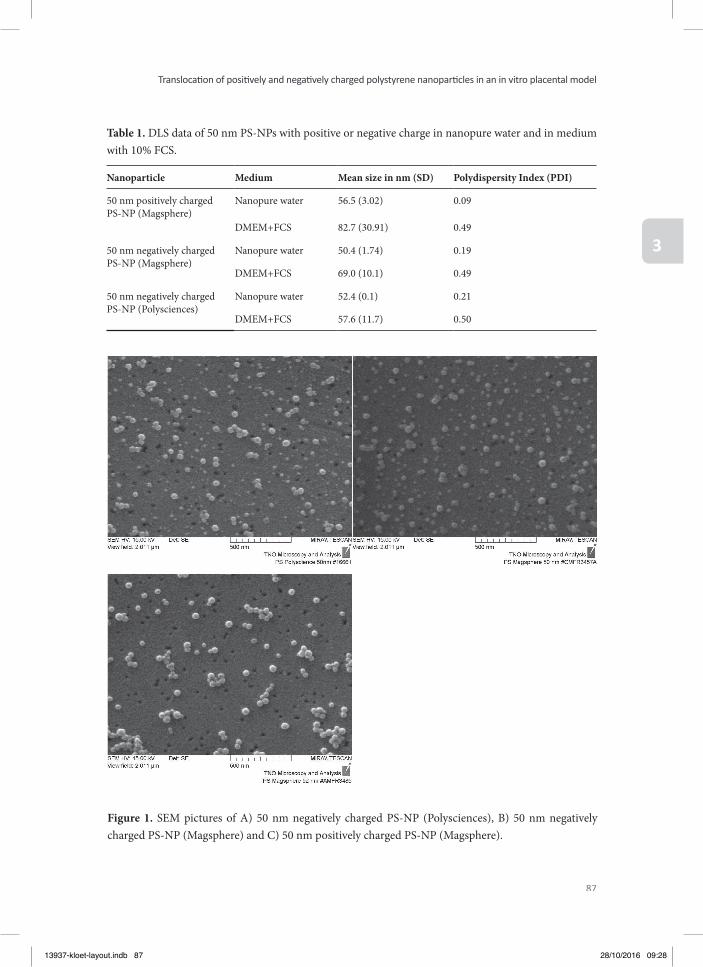
87
Translocati on of positi vely and negati vely charged polystyrene nanoparti cles in an in vitro placental model
3
Figure 1. SEM pictures of A) 50 nm negatively charged PS-NP (Polysciences), B) 50 nm negatively charged PS-NP (Magsphere) and C) 50 nm positively charged PS-NP (Magsphere).
Table 1. DLS data of 50 nm PS-NPs with positive or negative charge in nanopure water and in medium with 10% FCS.
Nanoparticle Medium Mean size in nm (SD) Polydispersity Index (PDI)
50 nm positively charged PS-NP (Magsphere)
Nanopure water 56.5 (3.02) 0.09
DMEM+FCS 82.7 (30.91) 0.49
50 nm negatively charged PS-NP (Magsphere)
Nanopure water 50.4 (1.74) 0.19
DMEM+FCS 69.0 (10.1) 0.49
50 nm negatively charged PS-NP (Polysciences)
Nanopure water 52.4 (0.1) 0.21
DMEM+FCS 57.6 (11.7) 0.50
13937-kloet-layout.indb 87 28/10/2016 09:28
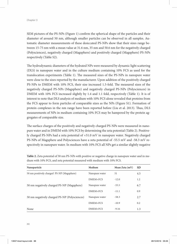
88
Chapter 3
SEM pictures of the PS-NPs (Figure 1) confi rm the spherical shape of the particles and their diameter of around 50 nm, although smaller particles can be observed in all samples. Au-tomatic diameter measurements of these desiccated PS-NPs show that their sizes range be-tween 15-75 nm with a mean value at 31.6 nm, 35 nm and 50.6 nm for the negatively charged (Polysciences), negatively charged (Magsphere) and positively charged (Magsphere) PS-NPs respectively (Table S2).
Th e hydrodynamic diameters of the hydrated NPs were measured by dynamic light scattering (DLS) in nanopure water and in the culture medium containing 10% FCS as used for the translocation experiments (Table 1). Th e measured sizes of the PS-NPs in nanopure water were close to the sizes reported by the manufacturer. Upon addition of the positively charged PS-NPs to DMEM with 10% FCS, their size increased 1.5-fold. Th e measured sizes of the negatively charged PS-NPs (Magsphere) and negatively charged PS-NPs (Polysciences) in DMEM with 10% FCS increased slightly by 1.4 and 1.1 fold, respectively (Table 1). It is of interest to note that DLS analysis of medium with 10% FCS alone revealed that proteins from the FCS appear to form particles of comparable sizes as the NPs (Figure S1). Formation of protein complexes in the nm range have been reported before (Liu et al. 2013). Th us, DLS measurements of NPs in medium containing 10% FCS may be hampered by the protein ag-gregates of comparable size.
Th e surface charges of the positively and negatively charged PS-NPs were measured in nano-pure water and in DMEM with 10% FCS by determining the zeta potential (Table 2). Positive-ly charged PS-NPs had a zeta potential of +51.0 mV in nanopure water. Negatively charged PS-NPs of Magsphere and Polysciences have a zeta potential of -55.5 mV and -58.3 mV re-spectively in nanopure water. In medium with 10% FCS all NPs get a similar slightly negative
Table 2. Zeta potential of 50 nm PS-NPs with positive or negative charge in nanopure water and in me-dium with 10% FCS, and zeta potential measured with medium with 10% FCS.
Nanoparticle Medium Mean Zeta (mV) SD
50 nm positively charged PS-NP (Magsphere) Nanopure water 51 4.5
DMEM+FCS -12.0 1.2
50 nm negatively charged PS-NP (Magsphere) Nanopure water -55.5 6.7
DMEM+FCS -11.1 0.9
50 nm negatively charged PS-NP (Polysciences) Nanopure water -58.3 2.7
DMEM+FCS -10.9 0.2
None DMEM+FCS -9.16 1.3
13937-kloet-layout.indb 88 28/10/2016 09:28
89
Translocati on of positi vely and negati vely charged polystyrene nanoparti cles in an in vitro placental model
3
charge. Th is may be explained by the fact that the majority of particles present in these sam-ples are protein aggregates from the 10% FCS so that the zeta potential may refl ect the overall charge of the protein aggregates rather than those of the PS-NPs (Table 2). Alternatively the similar charges in the presence of FCS may be ascribed to the protein corona dominating the overall charge.
Protein corona detectionFigure 2A shows a representative picture of the SDS-PAGE gel with proteins desorbed from the diff erent PS-NPs aft er their incubation in medium with 10% FCS. Each lane contains the amount of protein obtained from the same amount of PS-NP and thus, given the similar size of the PS-NPs, presents the amount of protein desorbed from a presumably similar surface area of the PS-NPs. Proteins desorbed from the protein corona of NPs are present in lanes 2-4. Figure 2B shows a protein gel prepared to contain the same amount of protein in each lane. Th e band intensities of the lanes containing NPs were further quantifi ed using the Od-yssey soft ware and graphs thus obtained are also presented in Figure 2. Th ese results reveal diff erences in the amount but not the nature of the proteins in the corona of the three diff erent PS-NPs (Figure 2A+B lanes 2-4).
From the 50 nm positively charged PS-NPs (lane 3) only a small amount of protein corona was obtained. Th e control (lane 5) shows the DMEM sample without nanoparticles which was treated by the same experimental procedure as NP samples. Hardly any protein is detected in this control, which corroborates that the proteins which are detected in the other samples are coming from the protein corona of the PS-NPs. Lane 6 shows the proteins present in the me-dium containing 10% FCS and comparison of this pattern to that of the PS-NPs reveals that not all medium proteins contribute to the protein corona to the same relative extent at which they are present in the medium, indicating at least some preferential binding of proteins to the NPs.
Cell viability studiesBefore testing the transplacental translocation of the PS-NPs, the concentration of the PS-NPs to be tested in the BeWo b30 transwell translocation studies were chosen based on results from cell viability studies. Th e cell viability of BeWo b30 cells upon 24 hours exposure to increasing PS-NP concentrations was determined using the MTT assay. Only cells exposed to the positively charged PS-NPs showed a decrease in MTT reduction, an eff ect that becomes statistically signifi cant at concentrations of 20 µg/ml and higher (Figure 3), whereas no eff ect was found upon exposure to the negatively charged PS-NPs. Based on these results the con-centration of PS-NPs to be tested in the translocation studies was chosen at 10 µg/ml.
13937-kloet-layout.indb 89 28/10/2016 09:28
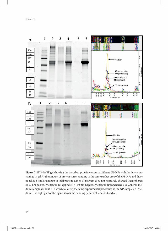
90
Chapter 3
Figure 2. SDS-PAGE gel showing the desorbed protein corona of diff erent PS-NPs with the lanes con-taining: in gel A) the amount of protein corresponding to the same surface area of the PS-NPs and those in gel B) a similar amount of total protein. Lanes: 1) marker; 2) 50 nm negatively charged (Magsphere); 3) 50 nm positively charged (Magsphere); 4) 50 nm negatively charged (Polysciences); 5) Control: me-dium sample without NPs which followed the same experimental procedure as the NP-samples; 6) Me-dium. Th e right part of the fi gure shows the banding pattern of lanes 2-4 and 6.
13937-kloet-layout.indb 90 28/10/2016 09:28
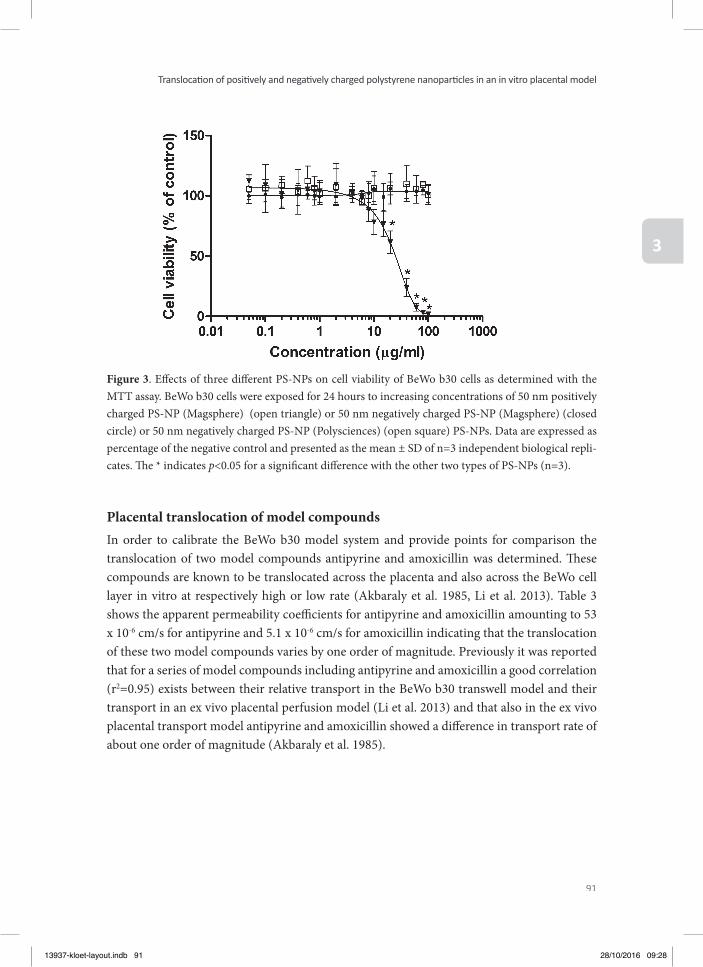
91
Translocati on of positi vely and negati vely charged polystyrene nanoparti cles in an in vitro placental model
3
Figure 3. Eff ects of three diff erent PS-NPs on cell viability of BeWo b30 cells as determined with the MTT assay. BeWo b30 cells were exposed for 24 hours to increasing concentrations of 50 nm positively charged PS-NP (Magsphere) (open triangle) or 50 nm negatively charged PS-NP (Magsphere) (closed circle) or 50 nm negatively charged PS-NP (Polysciences) (open square) PS-NPs. Data are expressed as percentage of the negative control and presented as the mean ± SD of n=3 independent biological repli-cates. Th e * indicates p<0.05 for a signifi cant diff erence with the other two types of PS-NPs (n=3).
Placental translocation of model compoundsIn order to calibrate the BeWo b30 model system and provide points for comparison the translocation of two model compounds antipyrine and amoxicillin was determined. Th ese compounds are known to be translocated across the placenta and also across the BeWo cell layer in vitro at respectively high or low rate (Akbaraly et al. 1985, Li et al. 2013). Table 3 shows the apparent permeability coeffi cients for antipyrine and amoxicillin amounting to 53 x 10-6 cm/s for antipyrine and 5.1 x 10⁻⁶ cm/s for amoxicillin indicating that the translocation of these two model compounds varies by one order of magnitude. Previously it was reported that for a series of model compounds including antipyrine and amoxicillin a good correlation (r2=0.95) exists between their relative transport in the BeWo b30 transwell model and their transport in an ex vivo placental perfusion model (Li et al. 2013) and that also in the ex vivo placental transport model antipyrine and amoxicillin showed a diff erence in transport rate of about one order of magnitude (Akbaraly et al. 1985).
13937-kloet-layout.indb 91 28/10/2016 09:28
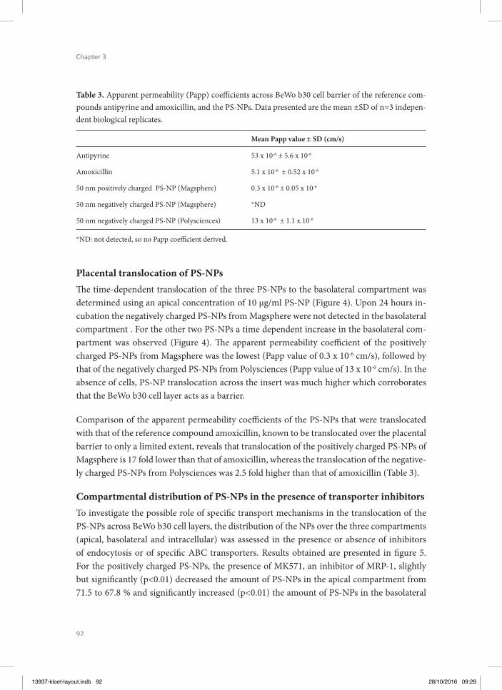
92
Chapter 3
Table 3. Apparent permeability (Papp) coeffi cients across BeWo b30 cell barrier of the reference com-pounds antipyrine and amoxicillin, and the PS-NPs. Data presented are the mean ±SD of n=3 indepen-dent biological replicates.
Mean Papp value ± SD (cm/s)
Antipyrine 53 x 10-6 ± 5.6 x 10⁻⁶
Amoxicillin 5.1 x 10⁻⁶ ± 0.52 x 10⁻⁶
50 nm positively charged PS-NP (Magsphere) 0.3 x 10⁻⁶ ± 0.05 x 10⁻⁶
50 nm negatively charged PS-NP (Magsphere) *ND
50 nm negatively charged PS-NP (Polysciences) 13 x 10-6 ± 1.1 x 10⁻⁶
*ND: not detected, so no Papp coeffi cient derived.
Placental translocation of PS-NPsTh e time-dependent translocation of the three PS-NPs to the basolateral compartment was determined using an apical concentration of 10 µg/ml PS-NP (Figure 4). Upon 24 hours in-cubation the negatively charged PS-NPs from Magsphere were not detected in the basolateral compartment . For the other two PS-NPs a time dependent increase in the basolateral com-partment was observed (Figure 4). Th e apparent permeability coeffi cient of the positively charged PS-NPs from Magsphere was the lowest (Papp value of 0.3 x 10⁻⁶ cm/s), followed by that of the negatively charged PS-NPs from Polysciences (Papp value of 13 x 10-6 cm/s). In the absence of cells, PS-NP translocation across the insert was much higher which corroborates that the BeWo b30 cell layer acts as a barrier.
Comparison of the apparent permeability coeffi cients of the PS-NPs that were translocated with that of the reference compound amoxicillin, known to be translocated over the placental barrier to only a limited extent, reveals that translocation of the positively charged PS-NPs of Magsphere is 17 fold lower than that of amoxicillin, whereas the translocation of the negative-ly charged PS-NPs from Polysciences was 2.5 fold higher than that of amoxicillin (Table 3).
Compartmental distribution of PS-NPs in the presence of transporter inhibitorsTo investigate the possible role of specifi c transport mechanisms in the translocation of the PS-NPs across BeWo b30 cell layers, the distribution of the NPs over the three compartments (apical, basolateral and intracellular) was assessed in the presence or absence of inhibitors of endocytosis or of specifi c ABC transporters. Results obtained are presented in fi gure 5. For the positively charged PS-NPs, the presence of MK571, an inhibitor of MRP-1, slightly but signifi cantly (p<0.01) decreased the amount of PS-NPs in the apical compartment from 71.5 to 67.8 % and signifi cantly increased (p<0.01) the amount of PS-NPs in the basolateral
13937-kloet-layout.indb 92 28/10/2016 09:28
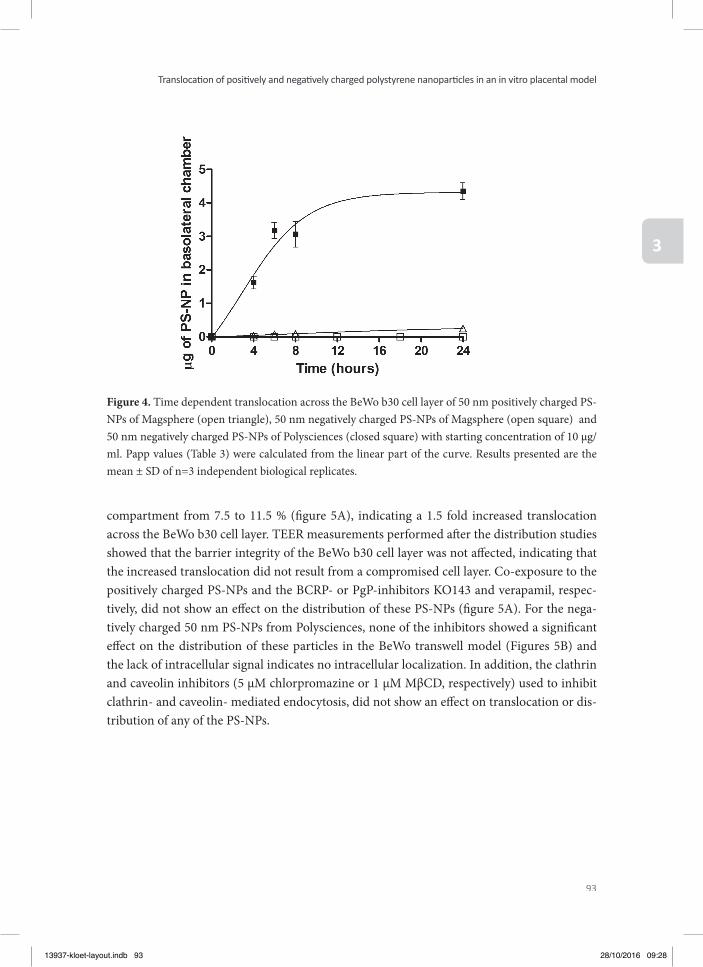
93
Translocati on of positi vely and negati vely charged polystyrene nanoparti cles in an in vitro placental model
3
compartment from 7.5 to 11.5 % (fi gure 5A), indicating a 1.5 fold increased translocation across the BeWo b30 cell layer. TEER measurements performed aft er the distribution studies showed that the barrier integrity of the BeWo b30 cell layer was not aff ected, indicating that the increased translocation did not result from a compromised cell layer. Co-exposure to the positively charged PS-NPs and the BCRP- or PgP-inhibitors KO143 and verapamil, respec-tively, did not show an eff ect on the distribution of these PS-NPs (fi gure 5A). For the nega-tively charged 50 nm PS-NPs from Polysciences, none of the inhibitors showed a signifi cant eff ect on the distribution of these particles in the BeWo transwell model (Figures 5B) and the lack of intracellular signal indicates no intracellular localization. In addition, the clathrin and caveolin inhibitors (5 µM chlorpromazine or 1 µM MβCD, respectively) used to inhibit clathrin- and caveolin- mediated endocytosis, did not show an eff ect on translocation or dis-tribution of any of the PS-NPs.
Figure 4. Time dependent translocation across the BeWo b30 cell layer of 50 nm positively charged PS-NPs of Magsphere (open triangle), 50 nm negatively charged PS-NPs of Magsphere (open square) and 50 nm negatively charged PS-NPs of Polysciences (closed square) with starting concentration of 10 µg/ml. Papp values (Table 3) were calculated from the linear part of the curve. Results presented are the mean ± SD of n=3 independent biological replicates.
13937-kloet-layout.indb 93 28/10/2016 09:28
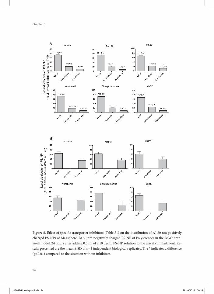
94
Chapter 3
Figure 5. Eff ect of specifi c transporter inhibitors (Table S1) on the distribution of A) 50 nm positively charged PS-NPs of Magsphere; B) 50 nm negatively charged PS-NP of Polysciences in the BeWo tran-swell model, 24 hours aft er adding 0.5 ml of a 10 µg/ml PS-NP solution to the apical compartment. Re-sults presented are the mean ± SD of n=4 independent biological replicates. Th e * indicates a diff erence (p<0.01) compared to the situation without inhibitors.
13937-kloet-layout.indb 94 28/10/2016 09:28
95
Translocati on of positi vely and negati vely charged polystyrene nanoparti cles in an in vitro placental model
3
DISCUSSION
To obtain insight in passage of NPs across the placental barrier, translocation of one positively and two diff erent types of negatively charged PS-NPs across an in vitro model of the placen-tal barrier was studied. Th e two negatively charged PS-NPs were included because previous studies within our group revealed they were translocated to a diff erent extent across a Caco-2 monolayer, a model for intestinal translocation (Walczak et al. 2014). Th e model in the pres-ent study consisted of cells of the BeWo cell line (clone b30) grown on a transwell insert, and forming a cell layer that separates an apical compartment from a fetal compartment.
Th e characterization of the PS-NPs was done by measuring the size and polydispersity index (PDI) by dynamic light scattering (DLS), the ζ-potential (as an indication of surface charge) by ζ-potential measurements, the protein corona by SDS-PAGE and shape and the num-ber-based size distribution by scanning electron microscopy (SEM). Th e results obtained re-veal that in the presence of 10% FCS in cell culture medium, measurement of ζ-potential may be hampered by the presence of free protein particles as the ζ-potential of medium without or with PS-NPs present, resulted in the same negative ζ-potential. Alternatively the similar charges of the diff erently charged NPs in the presence of FCS may be ascribed to their protein corona dominating the underlying PS-NP’s charge. SDS-PAGE analysis of this protein corona revealed that there were diff erences in mainly the amount but not in the nature of the proteins in the corona of the diff erently charged PS-NPs. Th e proteins desorbed from the PS-NPs were to a large extent a refl ection of what was detected in the medium containing 10% FCS. Th is result was somewhat diff erent from a previous study that reported that surface properties of PS-NPs signifi cantly aff ected the composition of the protein corona (Lundqvist et al. 2008). In this latter study using diff erent commercially available PS-NPs and mass spectrometry instead of SDS-PAGE, a fraction (about 35%) of the proteins in the corona were specifi cally detected on the positively charged PS-NPs or on the negatively charged PS-NPs, the others being similar.
In the present study it appeared that in spite of similar size (SEM and DLS), ζ-potential and type of proteins in the protein corona, the diff erently charged NPs displayed a remarkable diff erence in cytotoxicity, with only the PS-NPs with an original positive charge inducing cytotoxicity. Th e relatively higher cytotoxicity of positively as compared to negatively charged NPs was reported before for tri-block copolymer NPs (Bhattacharjee et al. 2012) but also for other type of NPs like gold NPs (Liu et al. 2013) and silicon NPs (Bhattacharjee et al. 2013). Th us for interaction with the cells, the original diff erence in charges or surface chemistry of the particles as prepared and measured in water, is apparently still of infl uence. Th e higher cytotoxicity of positive NPs might be due to a stronger interaction with cells compared with negative NPs, because of the electrostatic attraction towards the negatively charged cell mem-brane (Kim et al. 2009).
13937-kloet-layout.indb 95 28/10/2016 09:28

96
Chapter 3
Translocation of the diff erent PS-NPs across the BeWo cell layer was not found to be relat-ed to PS-NP charge. A remarkable diff erence in translocation was found between the two 50 nm negatively charged PS-NPs from diff erent manufacturers. For the PS-NPs from Poly-sciences, the apparent permeability coeffi cient was slightly higher than that of amoxicillin, with a Papp value of 13 x 10-6 cm/s (table 3). When testing the negatively charged PS-NPs from Magsphere, no PS-NPs could be detected in the basolateral compartment upon adding a concentration of 10 µg/ml in the apical compartment and 24 hours incubation. A 30-fold diff erence was found in translocation of the same types of negatively charged PS-NPs across a Caco-2 monolayer in a similar transwell model system (Walczak et al. 2014). Since none of the characterized parameters, including size, ζ-potential and protein corona revealed re-markable diff erences between the two negatively charged NPs, the diff erence may originate from the chemical groups on the surface of the NPs generating the negative charge. Although both PS-NPs are reported by the respective manufacturers to contain carboxylic groups from vinylic co-monomers, including acrylic acid, methacrylic acid and itaconic acid, further de-tails on the chemical characteristics of the NP surface remained unknown. In this respect it is of interest that variation in steric shielding of the charge on a surface moiety due to subtle diff erences in the chemical structure of the charge bearing moiety may aff ect the cellular up-take and toxicity of fl uorescent tri-block copolymer nanoparticles (Bhattacharjee et al. 2011). In this previous study it was demonstrated that three diff erent positively charged tri-block copolymer nanoparticles showed diff erent cellular uptake and toxicity in spite of a similar overall size and ζ-potential, and that only a diff erent shielding of the charge due to subtle diff erences in the charge generating surface moiety infl uenced the cellular uptake and toxicity (Bhattacharjee et al. 2011). Th is factor may explain the diff erent translocation rates of the two negatively charged 50 nm PS-NPs in this study, given the diff erent carboxylic groups that have been used in the production of these diff erent PS-NPs.
Th e translocation studies presented in this study revealed that the PS-NPs were translocated across the BeWo b30 transwell model to only a limited extent. We compared these results to reports in the literature studying the translocation of NPs across the placental barrier. A few studies also applied the BeWo b30 transwell model studying translocation of PEGylated gold NPs of 10-30 nm (Myllynen et al. 2008), iron oxide and silica NPs of 23-38 nm (Cor-reia Carreira et al. 2013), dexamethasone loaded PLGA NPs of 140-289 nm (Ali et al. 2013), rhodamine labelled silica NPs of 25 and 50 nm (Sonnegaard Poulsen et al. 2013) and poly-styrene NPs of 50 and 100 nm (Cartwright et al. 2012). Th ese studies generally report that the NPs are translocated across the BeWo cell layer aft er a few hours of incubation to the basolateral compartment albeit in relatively small amounts. Th e results of the present study are in line with these observations, and suggest that translocation of NPs across the in vitro placental barrier cannot be excluded. Literature data obtained in ex vivo human placental
13937-kloet-layout.indb 96 28/10/2016 09:28
97
Translocati on of positi vely and negati vely charged polystyrene nanoparti cles in an in vitro placental model
3
perfusion models also report the potential of NPs to cross the placental barrier (Wick et al. 2010, Menjoge et al. 2011, Grafmuller et al. 2013, Sonnegaard Poulsen et al. 2013) and are in line with this conclusion.
In order to investigate the role of specifi c transporters in the translocation of the PS-NPs across the BeWo b30 cell layer, translocation studies were also performed in the absence and presence of transporter inhibitors. Th e presence of PgP, BCRP, MRP-1 has been confi rmed in the BeWo b30 cells (Atkinson et al. 2003, Ceckova et al. 2006, Evseenko et al. 2006, Milane et al. 2009, Mitra and Audus 2010, Wick et al. 2010) but inhibitors of PgP and BCRP at a con-centration of 50 and 2.5 µM, respectively, showed no eff ect on translocation of PS-NPs across the BeWo b30 cell layers. MK571 (24 µM), an inhibitor of MRP-1, signifi cantly increased the basolateral translocation of positively charged PS-NP by 1.5 fold but did not infl uence the translocation of the negatively charged PS-NPs so in general the translocation of the PS-NPs tested appeared to occur preferentially by passive diff usion.
In vitro models are attractive for high throughput testing of various NPs. However, it should be kept in mind that in vitro models like the BeWo b30 transwell model applied in the current study have their limitations (Nikitina et al. 2013). For example, given that the physiology of the placental barrier in vivo varies in time, it is diffi cult to classify the BeWo cell layer in a rep-resenting specifi c stage of pregnancy. Furthermore, the BeWo transwell model is a simplifi ed placental barrier compared to the complex in vivo placental barrier since it lacks the multi-ple layers of diff erent cell types (cytotrophoblasts and syncytiotrophoblasts) and extracellular matrix including collagen, known to constitute the in vivo placental barrier. Th is implies that extrapolation of in vitro results to the in vivo situation should acknowledge uncertainties in the predictions made (Mathiesen et al. 2014). To this end we have included the model com-pounds amoxicillin and antipyrine, known to be translocated across the placental barrier to respectively only a limited or large extent (Akbaraly et al. 1985, Li et al. 2013) to enable qual-itative interpretation of the results. Th is comparison revealed that for positively charged PS-NPs translocation was 17 fold lower and for negatively charged PS-NPs from Polysciences 2.5 fold higher than that of amoxicillin, while for the negatively charged PS-NPs from Magsphere no translocation was found under the same test conditions.
In conclusion, the translocation studies of PS-NPs in the present study and the comparison of the data with translocation data of model compounds, for which the model has been validated with an ex vivo placental model, reveal that the in vitro BeWo b30 model can be used as a fast method to get an initial qualitative impression about the capacity of NPs to translocate across the placental barrier and set priorities for further in vivo studies on translocation of NPs to the fetus.
13937-kloet-layout.indb 97 28/10/2016 09:28
98
Chapter 3
ACKNOWLEDGEMENTS
Th is work is supported by NanoNextNL, a micro and nanotechnology consortium of the Government of the Netherlands and 130 partners.
13937-kloet-layout.indb 98 28/10/2016 09:28
99
Translocati on of positi vely and negati vely charged polystyrene nanoparti cles in an in vitro placental model
3
REFERENCES
Akbaraly, J. P., S. Guibert, J. J. Leng and J. Auzerie (1985). "Transplacental transfer of 5 antibiotics by in vitro human placental perfusion." Pathol Biol (Paris) 33(5): 368-372.
Ali, H., I. Kalashnikova, M. A. White, M. Sherman and E. Rytting (2013). "Preparation, characterization, and trans-port of dexamethasone-loaded polymeric nanoparticles across a human placental in vitro model." Int J Pharm 454(1): 149-157.
Arora, A. and G. W. Padua (2010). "Review: nanocomposites in food packaging." J Food Sci 75(1): R43-49.Artursson, P. and J. Karlsson (1991). "Correlation between oral drug absorption in humans and apparent drug per-
meability coeffi cients in human intestinal epithelial (Caco-2) cells." Biochem Biophys Res Commun 175(3): 880-885.
Atkinson, D. E., S. L. Greenwood, C. P. Sibley, J. D. Glazier and L. J. Fairbairn (2003). "Role of MDR1 and MRP1 in trophoblast cells, elucidated using retroviral gene transfer." American journal of physiology. Cell physiology 285(3): C584-591.
Auff an, M., J. Rose, J. Y. Bottero, G. V. Lowry, J. P. Jolivet and M. R. Wiesner (2009). "Towards a defi nition of inorganic nanoparticles from an environmental, health and safety perspective." Nat Nanotechnol 4(10): 634-641.
Avery, M. L., C. E. Meek and K. L. Audus (2003). "Th e presence of inducible cytochrome P450 types 1A1 and 1A2 in the BeWo cell line." Placenta 24(1): 45-52.
Bhattacharjee, S., D. Ershov, K. Fytianos, J. Gucht, G. Alink, I. Rietjens, A. Marcelis and H. Zuilhof (2012). "Cytotox-icity and cellular uptake of tri-block copolymer nanoparticles with diff erent size and surface characteristics." Particle and Fibre Toxicology 9(1): 11.
Bhattacharjee, S., D. Ershov, J. V. Gucht, G. M. Alink, I. M. Rietjens, H. Zuilhof and A. T. Marcelis (2011). "Surface charge-specifi c cytotoxicity and cellular uptake of tri-block copolymer nanoparticles." Nanotoxicology.
Bhattacharjee, S., I. M. Rietjens, M. P. Singh, T. M. Atkins, T. K. Purkait, Z. Xu, S. Regli, A. Shukaliak, R. J. Clark, B. S. Mitchell, G. M. Alink, A. T. Marcelis, M. J. Fink, J. G. Veinot, S. M. Kauzlarich and H. Zuilhof (2013). "Cytotoxicity of surface-functionalized silicon and germanium nanoparticles: the dominant role of surface charges." Nanoscale 5(11): 4870-4883.
Bhattacharjee, S., E. J. van Opstal, G. M. Alink, A. T. M. Marcelis, H. Zuilhof and I. M. C. M. Rietjens (2013). "Surface charge-specifi c interactions between polymer nanoparticles and ABC transporters in Caco-2 cells." Journal of Nanoparticle Research 15(6).
Bode, C. J., H. Jin, E. Rytting, P. S. Silverstein, A. M. Young and K. L. Audus (2006). "In vitro models for studying trophoblast transcellular transport." Methods Mol Med 122: 225-239.
Brand, W., P. A. van der Wel, M. J. Rein, D. Barron, G. Williamson, P. J. van Bladeren and I. M. Rietjens (2008). "Me-tabolism and transport of the citrus fl avonoid hesperetin in Caco-2 cell monolayers." Drug metabolism and disposition: the biological fate of chemicals 36(9): 1794-1802.
Buerki-Th urnherr, T., M. von and P. Wick (2012). "Knocking at the door of the unborn child: engineered nanoparti-cles at the human placental barrier." Swiss medical weekly 142: 0.
Cartwright, L., M. S. Poulsen, H. M. Nielsen, G. Pojana, L. E. Knudsen, M. Saunders and E. Rytting (2012). "In vitro placental model optimization for nanoparticle transport studies." Int J Nanomedicine 7: 497-510.
Ceckova, M., A. Libra, P. Pavek, P. Nachtigal, M. Brabec, R. Fuchs and F. Staud (2006). "Expression and functional activity of breast cancer resistance protein (bcrp, abcg2) transporter in the human choriocarcinoma cell line bewo" Clinical and Experimental Pharmacology and Physiology 33(1-2): 58-65.
Chiu, H. W., T. Xia, Y. H. Lee, C. W. Chen, J. C. Tsai and Y. J. Wang (2015). "Cationic polystyrene nanospheres induce autophagic cell death through the induction of endoplasmic reticulum stress." Nanoscale 7(2): 736-746.
Correia Carreira, S., L. Walker, K. Paul and M. Saunders (2013). "Th e toxicity, transport and uptake of nanoparticles in the in vitro BeWo b30 placental cell barrier model used within NanoTEST." Nanotoxicology.
dos Santos, T., J. Varela, I. Lynch, A. Salvati and K. A. Dawson (2011). "Eff ects of Transport Inhibitors on the Cellular Uptake of Carboxylated Polystyrene Nanoparticles in Diff erent Cell Lines." PLoS One 6(9): e24438.
Evseenko, D. A., J. W. Paxton and J. A. Keelan (2006). "ABC drug transporter expression and functional activity in trophoblast-like cell lines and diff erentiating primary trophoblast." American Journal of Physiology - Regu-latory, Integrative and Comparative Physiology 290(5): R1357-R1365.
13937-kloet-layout.indb 99 28/10/2016 09:28

100
Chapter 3
Grafmuller, S., P. Manser, H. F. Krug, P. Wick and U. von Mandach (2013). "Determination of the transport rate of xenobiotics and nanomaterials across the placenta using the ex vivo human placental perfusion model." J Vis Exp(76).
Juch, H., L. Nikitina, P. Debbage, G. Dohr and M. Gauster (2013). "Nanomaterial interference with early human pla-centa: Sophisticated matter meets sophisticated tissues." Reproductive Toxicology 41(0): 73-79.
Kim, W. J., A. C. Bonoiu, T. Hayakawa, C. Xia, M. A. Kakimoto, H. E. Pudavar, K. S. Lee and P. N. Prasad (2009). "Hyperbranched polysiloxysilane nanoparticles: surface charge control of nonviral gene delivery vectors and nanoprobes." Int J Pharm 376(1-2): 141-152.
Lee, J. Y., J. S. Kim, H. J. Cho and D. D. Kim (2014). "Poly(styrene)-b-poly(DL-lactide) copolymer-based nanoparti-cles for anticancer drug delivery." Int J Nanomedicine 9: 2803-2813.
Lesniak, A., A. Campbell, M. P. Monopoli, I. Lynch, A. Salvati and K. A. Dawson (2010). "Serum heat inactivation aff ects protein corona composition and nanoparticle uptake." Biomaterials 31(36): 9511-9518.
Li, H., B. van Ravenzwaay, I. M. Rietjens and J. Louisse (2013). "Assessment of an in vitro transport model using BeWo b30 cells to predict placental transfer of compounds." Arch Toxicol 87(9): 1661-1669.
Liu, F., M. J. Soares and K. L. Audus (1997). "Permeability properties of monolayers of the human trophoblast cell line BeWo." American Journal of Physiology - Cell Physiology 273(5): C1596-C1604.
Liu, W., J. Rose, S. Plantevin, M. Auff an, J. Y. Bottero and C. Vidaud (2013). "Protein corona formation for nanoma-terials and proteins of a similar size: hard or soft corona?" Nanoscale 5(4): 1658-1668.
Liu, X., N. Huang, H. Li, Q. Jin and J. Ji (2013). "Surface and Size Eff ects on Cell Interaction of Gold Nanoparticles with Both Phagocytic and Nonphagocytic Cells." Langmuir 29(29): 9138-9148.
Lundqvist, M., J. Stigler, G. Elia, I. Lynch, T. Cedervall and K. A. Dawson (2008). "Nanoparticle size and surface properties determine the protein corona with possible implications for biological impacts." Proceedings of the National Academy of Sciences 105(38): 14265-14270.
Mathiesen, L., T. A. Morck, G. Zuri, M. H. Andersen, C. Pehrson, M. Frederiksen, T. Mose, E. Rytting, M. S. Poulsen, J. K. Nielsen and L. E. Knudsen (2014). "Modelling of Human Transplacental Transport as Performed in Copenhagen, Denmark." Basic & clinical pharmacology & toxicology.
Menjoge, A. R., A. L. Rinderknecht, R. S. Navath, M. Faridnia, C. J. Kim, R. Romero, R. K. Miller and R. M. Kannan (2011). "Transfer of PAMAM dendrimers across human placenta: prospects of its use as drug carrier during pregnancy." J Control Release 150(3): 326-338.
Milane, A., S. Vautier, H. Chacun, V. Meininger, G. Bensimon, R. Farinotti and C. Fernandez (2009). "Interactions between riluzole and ABCG2/BCRP transporter." Neuroscience Letters 452(1): 12-16.
Mitra, P. and K. L. Audus (2010). "MRP isoforms and BCRP mediate sulfate conjugate effl ux out of BeWo cells." International Journal of Pharmaceutics 384(1-2): 15-23.
Myllynen, P. K., M. J. Loughran, C. V. Howard, R. Sormunen, A. A. Walsh and K. H. Vahakangas (2008). "Kinetics of gold nanoparticles in the human placenta." Reprod Toxicol 26(2): 130-137.
Nikitina, L., G. Dohr and H. Juch (2013). "Studying nanoparticle interaction with human placenta: Festina lente!" Nanotoxicology.
Oberdorster, E. (2004). "Manufactured nanomaterials (fullerenes, C60) induce oxidative stress in the brain of juvenile largemouth bass." Environ Health Perspect 112(10): 1058-1062.
Oberdorster, G., E. Oberdorster and J. Oberdorster (2005). "Nanotoxicology: an emerging discipline evolving from studies of ultrafi ne particles." Environ Health Perspect 113(7): 823-839.
Park, M. V., A. M. Neigh, J. P. Vermeulen, L. J. de la Fonteyne, H. W. Verharen, J. J. Briede, H. van Loveren and W. H. de Jong (2011). "Th e eff ect of particle size on the cytotoxicity, infl ammation, developmental toxicity and genotoxicity of silver nanoparticles." Biomaterials 32(36): 9810-9817.
Phosphorex Commercial Applications Use Polystyrene Nanoparticles. 2013 ; Available from: http://www.phos-phorex.com/1/post/2013/07/commercial-applications-use-polystyrene-nanoparticles.html.
Poulsen, M. S., E. Rytting, T. Mose and L. E. Knudsen (2009). "Modeling placental transport: correlation of in vitro BeWo cell permeability and ex vivo human placental perfusion." Toxicol In Vitro 23(7): 1380-1386.
Sager, T. M. and V. Castranova (2009). "Surface area of particle administered versus mass in determining the pul-monary toxicity of ultrafi ne and fi ne carbon black: comparison to ultrafi ne titanium dioxide." Part Fibre Toxicol 6: 15.
Saunders, M. (2009). "Transplacental transport of nanomaterials." Wiley Interdiscip Rev Nanomed Nanobiotechnol 1(6): 671-684.
13937-kloet-layout.indb 100 28/10/2016 09:28
101
Translocati on of positi vely and negati vely charged polystyrene nanoparti cles in an in vitro placental model
3
Simon, P., Q. Chaudhry and D. Bakos (2008). "Migration of engineered nanoparticles from polymer packaging to food - a physicochemical view." J Food Nutr Res 47(3): 105-113.
Sonnegaard Poulsen, M., T. Mose, L. Leth Maroun, L. Mathiesen, L. Ehlert Knudsen and E. Rytting (2013). "Kinetics of silica nanoparticles in the human placenta." Nanotoxicology.
Stampfl , A., M. Maier, R. Radykewicz, P. Reitmeir, M. Göttlicher and R. Niessner (2011). "Langendorff Heart: A Model System To Study Cardiovascular Eff ects of Engineered Nanoparticles." ACS Nano 5(7): 5345-5353.
Takeuchi, Y., R. Sakakibara and M. Ishiguro (1990). "Synthesis and secretion of human chorionic gonadotropin and its subunits in choriocarcinoma cells: a comparative study with normal placental cells." Mol Cell Endocrinol 69(2-3): 145-156.
Ushigome, F., H. Takanaga, H. Matsuo, S. Yanai, K. Tsukimori, H. Nakano, T. Uchiumi, T. Nakamura, M. Kuwano, H. Ohtani and Y. Sawada (2000). "Human placental transport of vinblastine, vincristine, digoxin and progester-one: contribution of P-glycoprotein." European Journal of Pharmacology 408(1): 1-10.
Utoguchi, N., G. A. Chandorkar, M. Avery and K. L. Audus (2000). "Functional expression of P-glycoprotein in pri-mary cultures of human cytotrophoblasts and BeWo cells." Reprod Toxicol 14(3): 217-224.
Walczak, A. P., E. Kramer, P. J. Hendriksen, P. Tromp, J. P. Helsper, M. van der Zande, I. M. Rietjens and H. Bouw-meester (2014). "Translocation of diff erently sized and charged polystyrene nanoparticles in in vitro intesti-nal cell models of increasing complexity." Nanotoxicology: 1-9.
Wick, P., A. Malek, P. Manser, D. Meili, X. Maeder-Althaus, L. Diener, P. A. Diener, A. Zisch, H. F. Krug and U. von Mandach (2010). "Barrier capacity of human placenta for nanosized materials." Environmental health per-spectives 118(3): 432-436.
13937-kloet-layout.indb 101 28/10/2016 09:28

102
Chapter 3
SUPPLEMENTARY MATERIALS CHAPTER 3
Table S1. Transporter inhibitors used in the PS-NP distribution studies.
Transporter inhibitor TargetConcentration used in present study (µM)
Reference
Methyl-β-cyclodextrin Caveolin 1 (Bhattacharjee et al. 2013)
Chlorpromazine Clathrin 5 (dos Santos et al. 2011)
Verapamil PgP 50 (Ushigome et al. 2000)
KO143 BCRP 2.5 (Brand et al. 2008) (Ceckova et al. 2006)
MK571 MRP-1/MRP-2 24 (Brand et al. 2008)
Table S2. Automatic diameter measurements of PS-NPs using Scandium SIS image analysis soft ware (Olympus Soft Imaging Solutions GmbH, Germany). Th e mean diameter ± SD and range of 50-500 particles is given (in nm).
Nanoparticle Mean SD Range
50 nm negatively charged PS-NP (Polysciences) 31.6 13.6 15 - 70
50 nm negatively charged PS-NP (Magsphere) 35.0 15.3 15 - 70
50 nm positively charged PS-NP (Magsphere) 50.6 9.3 25 - 75
13937-kloet-layout.indb 102 28/10/2016 09:28
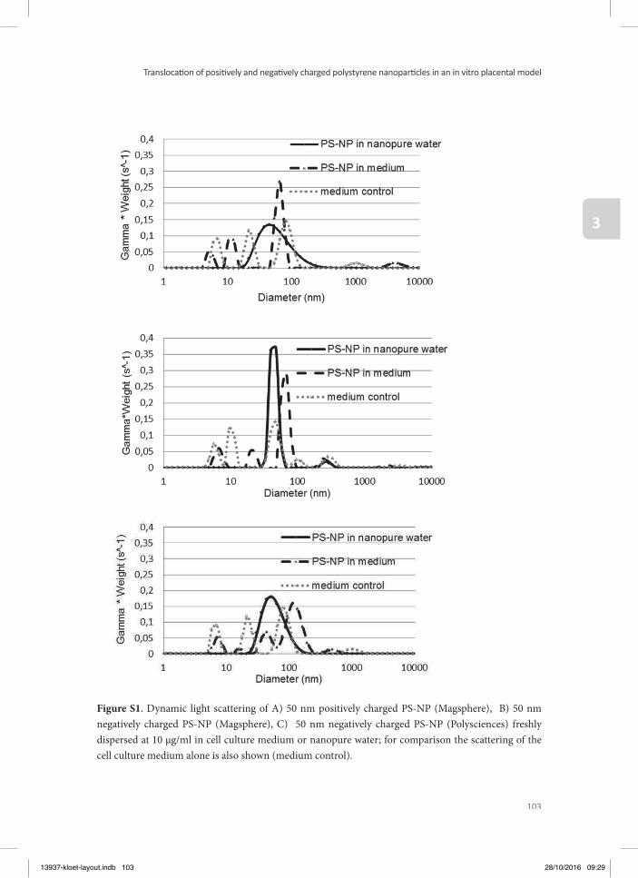
103
Translocati on of positi vely and negati vely charged polystyrene nanoparti cles in an in vitro placental model
3
Figure S1. Dynamic light scattering of A) 50 nm positively charged PS-NP (Magsphere), B) 50 nm negatively charged PS-NP (Magsphere), C) 50 nm negatively charged PS-NP (Polysciences) freshly dispersed at 10 µg/ml in cell culture medium or nanopure water; for comparison the scattering of the cell culture medium alone is also shown (medium control).
13937-kloet-layout.indb 103 28/10/2016 09:29

12
34
56
78
910
1112
A B C D E F G G H
13937-kloet-layout.indb 104 28/10/2016 09:29

12
34
56
78
910
1112
A B C D E F G G H
Chapter 4
Assessment of the potential developmental toxicity of positively and negatively charged polystyrene
nanoparticles using the embryonic stem cell differentiation assay
Samantha K. KloetHans H.J. van den Berg
Leonie F.H. FransenIgnacio Miro Estruch
Vasileios VasileiadisLaura de Haan
Ivonne M.C.M. RietjensJochem Louisse
Submitted for publication to Toxicology Letters
13937-kloet-layout.indb 105 28/10/2016 09:29
106
Chapter 4
ABSTRACT
Knowledge on the relation between physicochemical characteristics of nanoparticles (NPs) and their toxicity would be of great help for setting priorities for NP toxicity testing. Th e present study aimed to assess the role of surface charge of NPs on in vitro developmental tox-icity, by determining the eff ects of positively or negatively charged polystyrene nanoparticles (PS-NPs) in the embryonic stem (ES-D3) cell diff erentiation assay. Th e results show that the negatively charged PS-NPs did not aff ect ES-D3 cell diff erentiation, whereas the positively charged PS-NPs showed a concentration-dependent inhibition of ES-D3 cell diff erentiation, indicating that the eff ects of NPs on ES-D3 cell diff erentiation may depend on their surface charge. In order to assess whether NP-induced eff ects on ES-D3 cell diff erentiation may be related to other physicochemical properties, we performed a literature search on the reported potencies of NPs in these models. Th is analysis shows that the potency of NPs may depend on size, surface charge or coating, but that currently available data are too limited to indicate a clear relation between physicochemical characteristics and their in vitro developmental toxic-ity. Th e results also reveal that additional experimental data, as presented in the present study, may help to further advance the fi eld.
13937-kloet-layout.indb 106 28/10/2016 09:29
107
Assessment of the potenti al developmental toxicity of positi vely and negati vely charged polystyrene nanoparti cles
using the embryonic stem cell diff erenti ati on assay
4
INTRODUCTION
Th e rapid increase of nanotechnology has several benefi ts for society, yet there is an increas-ing concern that exposure to nanoparticles (NPs) may result in signifi cant adverse eff ects (Oberdorster et al. 2007, Gebel et al. 2013). NPs have been shown to be able to pass physio-logical barriers, such as that of the intestines and the placenta (Braakhuis et al. 2015). Th is indicates that systemic exposure, including fetal exposure, is feasible and that the fetus is a potential target for NPs. To assess possible adverse eff ects of NPs on the developing fetus, in vivo developmental toxicity studies can be performed in for example rats or mice. However, because of economic and ethical constrains with in vivo studies, alternatives for in vivo tox-icity studies are being developed and validated (Jelinek et al. 1985, Fort and Paul 2002, zur Nieden et al. 2004, Spielmann et al. 2006, Park et al. 2009, de Jong et al. 2011).
One of the in vitro alternative assays for developmental toxicity testing is the embryonic stem cell test (EST) which has been validated by the European Centre for the Validation of Alterna-tive Methods (ECVAM). In particular the diff erentiation assay of the EST has been shown to be useful in assessing relative developmental toxicity potencies of chemicals and NPs (Gen-schow et al. 2004, de Jong et al. 2011, Pietroiusti et al. 2011, Li et al. 2015). In this assay, the chemical- or NP-induced inhibition of blastocyst-derived mouse embryonic stem (ES-D3) cells developing into contracting cardiomyocytes, is used as an in vitro endpoint of develop-mental toxicity (de Jong et al. 2009, Campagnolo et al. 2013). Considering the large variety of NPs that have not been tested yet for their developmental toxicity, knowledge on the relation between physicochemical characteristics of NPs, such as size or surface charge, and their tox-icity would be of great help for setting priorities for NP toxicity testing.
To increase our knowledge on the relation between physicochemical properties of NPs and their developmental toxicity potential, the present study aimed to assess the role of surface charge in the in vitro developmental toxicity potential of NPs. To this end, we assessed the eff ects of one 50 nm positively charged polystyrene NP (PS-NP) and two 50 nm negatively charged PS-NPs on the viability and diff erentiation of ES-D3 cells. Further, we made an over-view of the available literature on NP-induced eff ects on ES-D3 cell diff erentiation, in order to assess whether also other physicochemical characteristics may relate to the in vitro devel-opmental toxicity potential of NPs.
METHODS
Materials WST-1 Cell Proliferation Reagent was purchased from Roche (Woerden, Th e Netherlands). Phosphate buff ered saline (PBS), non-essential amino acids, penicillin–streptomycin and
13937-kloet-layout.indb 107 28/10/2016 09:29
108
Chapter 4
L-glutamine were obtained from Invitrogen (Breda, Th e Netherlands). Non-enzymatic cell dissociation solution, 0.1% gelatine, murine leukemia inhibiting factor (LIF) and β-mercap-toethanol were purchased from Sigma–Aldrich (Zwijndrecht, Th e Netherlands). AdvanceS-TEMTM Low Osmo DMEM was obtained from Hyclone (Hyclone; Logan, USA) and foetal calf serum (FCS) was obtained from ATCC (USA).
NanoparticlesTh is study included three diff erent PS-NPs, one 50 nm positively charged PS-NP and two 50 nm negatively charged PS-NPs. Positively and negatively charged fl uorescent red PS-NPs were obtained from Magsphere Inc. (California, USA) (Cat nr: CAFR050NM, AMFR050NM respectively). Negatively charged fl uorescent Yellow-Green carboxylated 50 nm PS-NPs were obtained from Polysciences (Warrington, USA) (Cat nr. 16661-10). All the PS-NPs used had an average particle size of 50 nm as indicated by the manufacturers’ specifi cation.
Particle characterizationTh e size (hydrodynamic diameter) of the NPs was determined by dynamic light scattering (DLS) using a Cobolt Samba 300mW DPSS laser at a wavelength of 532 nm as a light source. Measurements were performed on freshly prepared NP solutions at 100 μg/ml in nanopure water and in cell culture medium containing 20% FCS. For each sample, twenty measurements were made and the detector intensity data (measured with an ALV/SO SIPD Single Photon Detector with ALV Static and Dynamic Fiber optics) were processed with an ALV5000/60X0 external correlator and ALV-5000/E soft ware (all from ALV-GmbH, Germany). Data were analysed with the Aft erALV soft ware and mean diameter and polydispersity index (PDI) were determined for each NP sample. Additionally, the ζ-potential of the NPs was measured at a concentration of 100 µg/ml using the Malvern Zetasizer 2000 (Malvern Instruments, UK).
Cell cultureTh e murine ES-D3 cell line was purchased from ATCC (Wesel, Germany). Th e cells were maintained in cell culture fl asks (Corning, the Netherlands) that were pre-coated with 0.1% gelatin, using AdvanceSTEMTM Low Osmo DMEM, supplemented with 20% FCS, 2 mM L-glutamine and 50U/50µg penicillin-streptomycin and leukemia inhibiting factor (LIF 1000 U/ml), in a humidifi ed atmosphere at 37°C and 5% CO2. LIF serves to keep the embryonic stem cells in the pluripotent state, by inhibiting their diff erentiation. Cells were subcultured every 2 to 3 days using non-enzymatic cell dissociation solution to detach the cells when reaching 60-80% confl uence. Experiments were performed using ES-D3 cells between pas-sages 4 and 18.
13937-kloet-layout.indb 108 28/10/2016 09:29
109
Assessment of the potenti al developmental toxicity of positi vely and negati vely charged polystyrene nanoparti cles
using the embryonic stem cell diff erenti ati on assay
4
Assessment of cell viability To assess the eff ects of the PS-NPs on cell viability, ES-D3 cells were seeded at 2 x 10⁴ cells/ml (one-day exposure) or 1 x 10³ cells/ml (fi ve-day exposure) in 100 μL culture medium in 96-well plates (Greiner bio-one) in the absence of LIF and incubated at 37°C and 5% CO2 for 24 hours to allow cell adherence. Th en the cells were exposed to 100 μL of PS-NPs at con-centrations up to 100 μg/ml and subsequently incubated for one or fi ve days at 37°C and 5% CO2 in a humidifi ed atmosphere. Culture medium was used as a negative control and 200 µM CuSO4 was used as a positive control. Aft er incubation, 20 μL WST-1 reagent was added to each well and the plates were incubated for another 3 hours. Th en absorbance was measured at 450 nm and 620 nm (background) using a SpectraMax M2 Microplate Reader (Molecular Devices LLC, Sunnyvale, CA USA). Th e cell viability was expressed as percentage of the neg-ative control, with the control set at 100% viability.
Diff erentiation assay with ES-D3 cellsTh e potential of PS-NPs to inhibit ES-D3 cell diff erentiation was evaluated using the ES-D3 cell diff erentiation assay using culture medium without LIF. In this assay, the eff ect of chemi-cals or NPs is assessed on the diff erentiation of ES-D3 cells into contracting cardiomyocytes. Similar to other studies (van Dartel et al. 2009, van Dartel et al. 2009, van Dartel et al. 2011), we exposed diff erentiating ES-D3 cells during day 3-10 of the 10-day diff erentiation process. On day 0, droplets of 20 μl ES-D3 cell suspension (3.75 x 10⁴ cells/ml) in the absence of PS-NPs were placed as hanging drops on the inner side of the lid of a 96-well plate. Sterile lids of Eppendorf tubes were placed on each corner of the plate to prevent contact of the drops with the plate. Th e wells of the 96-well plate were fi lled with 250 μl PBS and the plate was sealed with Micropore tape (3M, Neuss, Germany) to prevent evaporation of the hanging drops. Plates were incubated for three days at 37°C and 5% CO2 in a humidifi ed atmosphere to allow for the formation of embryonic bodies (EBs). Th ese EBs were transferred aft er 3 days to non-tissue culture treated Petri dishes (Greiner, diameter 6 cm) with 5 ml of medium in the absence (negative control) or presence of PS-NPs. On day 5, the EBs were transferred to a 24-well plate (Corning) with 1 ml of medium in the absence or presence of PS-NPs, plating one EB per well. On day 10, the number of wells containing contracting cardiomyocytes was determined by visual inspection using a light microscope. Tests were accepted for further analysis if at least 21 of the 24 wells of the negative control contained contracting cardiomy-ocytes. For each test compound, three independent assays were performed, at concentrations ranging from 0.1 to 50 μg/ml. For each concentration, the fraction of the EBs that diff erenti-ated into contracting cardiomyocytes was determined, with a value of 1 implying that all EBs in one 24-well plate diff erentiated into contracting cardiomyocytes.
13937-kloet-layout.indb 109 28/10/2016 09:29
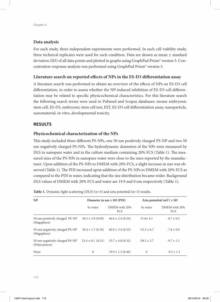
110
Chapter 4
Data analysisFor each study, three independent experiments were performed. In each cell viability study, three technical replicates were used for each condition. Data are shown as mean ± standard deviation (SD) of all data points and plotted in graphs using GraphPad Prism® version 5. Con-centration-response analysis was performed using GraphPad Prism® version 5.
Literature search on reported eff ects of NPs in the ES-D3 diff erentiation assayA literature search was performed to obtain an overview of the eff ects of NPs on ES-D3 cell diff erentiation, in order to assess whether the NP-induced inhibition of ES-D3 cell diff eren-tiation may be related to specifi c physicochemical characteristics. For this literature search the following search terms were used in Pubmed and Scopus databases: mouse embryonic stem cell, ES-D3, embryonic stem cell test, EST, ES-D3 cell diff erentiation assay, nanoparticle, nanomaterial, in vitro, developmental toxicity.
RESULTS
Physicochemical characterization of the NPsTh is study included three diff erent PS-NPs, one 50 nm positively charged PS-NP and two 50 nm negatively charged PS-NPs. Th e hydrodynamic diameters of the NPs were measured by DLS in nanopure water and in the culture medium containing 20% FCS (Table 1). Th e mea-sured sizes of the PS-NPs in nanopure water were close to the sizes reported by the manufac-turer. Upon addition of the PS-NPs to DMEM with 20% FCS, a slight increase in size was ob-served (Table 1). Th e PDI increased upon addition of the PS-NPs to DMEM with 20% FCS as compared to the PDI in water, indicating that the size distribution became wider. Background DLS values of DMEM with 20% FCS and water are 19.9 and 0 nm respectively (Table 1).
Table 1. Dynamic light scattering (DLS) (n=3) and zeta potential (n=3) results.
NP Diameter in nm ± SD (PDI) Zeta potential (mV) ± SD
In water DMEM with 20% FCS
In water DMEM with 20% FCS
50 nm positively charged PS-NP (Magsphere)
56.5 ± 3.0 (0.09) 66.6 ± 2.4 (0.34) 51.0± 4.5 -8.7 ± 0.2
50 nm negatively charged PS-NP (Magsphere)
50.4 ± 1.7 (0.19) 60.9 ± 5.4 (0.33) -55.5 ± 6.7 -7.8 ± 0.9
50 nm negatively charged PS-NP (Polysciences)
52.4 ± 0.1 (0.21) 55.7 ± 4.8 (0.32) -58.3 ± 2.7 -9.7 ± 1.1
None 0 19.9 ± 1.2 (0.46) 0 -9.2 ± 1.3
13937-kloet-layout.indb 110 28/10/2016 09:29
111
Assessment of the potenti al developmental toxicity of positi vely and negati vely charged polystyrene nanoparti cles
using the embryonic stem cell diff erenti ati on assay
4
Th e surface charges of the positively and negatively charged PS-NPs were measured in nano-pure water and in DMEM with 20% FCS by determining the zeta potential (Table 1). Positive-ly charged PS-NPs had a zeta potential of +51.0 mV in nanopure water. Negatively charged PS-NPs of Magsphere and Polyscience had a zeta potential of -55.5 mV and -58.3 mV, respec-tively, in nanopure water. In DMEM with 20% FCS all NPs got a slightly negative charge of -8 to -9 mV, similar to the charge measured for DMEM with 20% FCS without PS-NPs (Table 1).
Eff ects of PS-NPs on ES-D3 cell viability and ES-D3 cell diff erentiationTo evaluate the eff ects of the PS-NPs on ES-D3 cell viability, the WST-1 assay was performed on ES-D3 cells exposed for one day or fi ve days to the PS-NPs (Figure 1). Th e positively charged PS-NPs caused a concentration-dependent decrease in cell viability, which was more prominent aft er the 5-day exposure than aft er the 1-day exposure (Figure 1A), indicated by EC50 values of 2.0 and 71 µg/ml, respectively. For both negatively charged PS-NPs no eff ect on cell viability was observed up to the highest concentrations tested (100 μg/ml) (Figure 1B and 1C).
To study the in vitro developmental toxicity of the PS-NPs, their eff ect on the diff erentia-tion of ES-D3 cells into contracting cardiomyocytes was evaluated (Figure 1). Th e positively charged PS-NPs showed a concentration-dependent inhibition of the diff erentiation of ES-D3 cells into beating cardiomyocytes, with an EC50 of 1.2 µg/ml, whereas both the negatively charged PS-NPs did not aff ect the diff erentiation of the ES-D3 cells up to the highest concen-tration tested (50 μg/ml) (Figure 1B and 1C).
Th e results indicate a clear eff ect of NP charge on both viability and diff erentiation of the ES-D3 cells. Th e results for the negatively charged PS-NPs do not seem to point at a hazard for developmental toxicity of these NPs. Th e results for the positively charged PS-NPs may in-dicate a possible hazard for developmental toxicity. Since cell diff erentiation was inhibited at concentrations that also inhibited cell viability upon a 5-day exposure, NP-induced inhibition of ES-D3 cell diff erentiation may be due to cytotoxic eff ects.
13937-kloet-layout.indb 111 28/10/2016 09:29
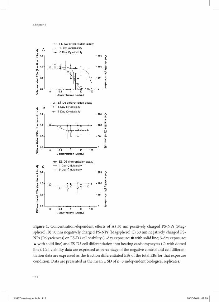
112
Chapter 4
Figure 1. Concentration-dependent eff ects of A) 50 nm positively charged PS-NPs (Mag-sphere), B) 50 nm negatively charged PS-NPs (Magsphere) C) 50 nm negatively charged PS-NPs (Polysciences) on ES-D3 cell viability (1-day exposure: with solid line; 5-day exposure: with solid line) and ES-D3 cell diff erentiation into beating cardiomyocytes ( with dotted line). Cell viability data are expressed as percentage of the negative control and cell diff eren-tiation data are expressed as the fraction diff erentiated EBs of the total EBs for that exposure condition. Data are presented as the mean ± SD of n=3 independent biological replicates.
13937-kloet-layout.indb 112 28/10/2016 09:29
113
Assessment of the potenti al developmental toxicity of positi vely and negati vely charged polystyrene nanoparti cles
using the embryonic stem cell diff erenti ati on assay
4
Reported eff ects of other NPs on ES-D3 cell diff erentiationIn order to obtain further insight in the role of physicochemical characteristics of NPs on their potential developmental toxicity, we performed a literature search on the NP-induced inhibition of ES-D3 cell diff erentiation into contracting cardiomyocytes. Table 2 presents an overview of the literature data obtained, showing the EC50 values for the inhibition of ES-D3 cell diff erentiation, including EC50 values for cell viability, as well as the NP physicochemical properties.
For silicon dioxide NPs and silver NPs, diff erent sizes have been tested in the ES-D3 cell dif-ferentiation assay. For silicon dioxide NPs, the two largest particles (80 and 400 nm) did not inhibit ES-D3 cell diff erentiation up to the highest concentration tested (100 µg/ml), whereas the two smallest particles (10 and 30 nm) inhibited ES-D3 cell diff erentiation with EC50 val-ues of 59 and 29 µg/ml, respectively (Table 2). For silver NPs all four sizes (7, 20, 80 and 113 nm) inhibited ES-D3 cell diff erentiation, and the smaller the NPs, the higher their potency, except for the two largest particles, which showed a similar potency (Table 2). For other NPs, the eff ect of coating on the in vitro developmental toxicity has been assessed. Table 2 shows that the coating of NPs had an eff ect on the toxicity of cobalt ferrite and titanium dioxide NPs, but not on zinc oxide NPs. Th is literature search also indicated that the present study is the only study so far that has assessed the eff ect of NP charge on the in vitro developmental tox-icity of NPs, showing that positively charged PS-NPs are more potent than negatively charged PS-NPs. Th e review also reveals however that the eff ect of surface charge may vary with the type of NPs investigated, since the silicon dioxide, silver and titanium dioxide NPs that were reported to inhibit ES-D3 cell diff erentiation were negatively charged, while the negatively charged PS-NPs of the present study did not aff ect ES-D3 cell diff erentiation.
A comparison of eff ect concentrations on the inhibition of ES-D3 cell diff erentiation with ef-fect concentrations on the decrease of ES-D3 cell viability reveals that for various NPs (cobalt ferrite NPs, gold NPs, silicon dioxide NPs and titanium dioxide NPs) ES-D3 cell diff erentia-tion is already inhibited at concentrations that do not yet aff ect cell viability, whereas for other NPs (PS-NPs, silver NPs and zinc oxide NPs), ES-D3 cell diff erentiation is only inhibited at concentrations that also aff ect cell viability. Th ese diff erences between NPs may point to diff erences in modes of action underlying their inhibition of ES-D3 cell diff erentiation. In summary, the review shows that, in spite of the limited available data, smaller NPs may be more potent than larger NPs made of the same core material and that NP coating and surface charge can aff ect the NP-induced decrease of ES-D3 cell viability and inhibition of ES-D3 cell diff erentiation, albeit in an NP-type specifi c way.
13937-kloet-layout.indb 113 28/10/2016 09:29
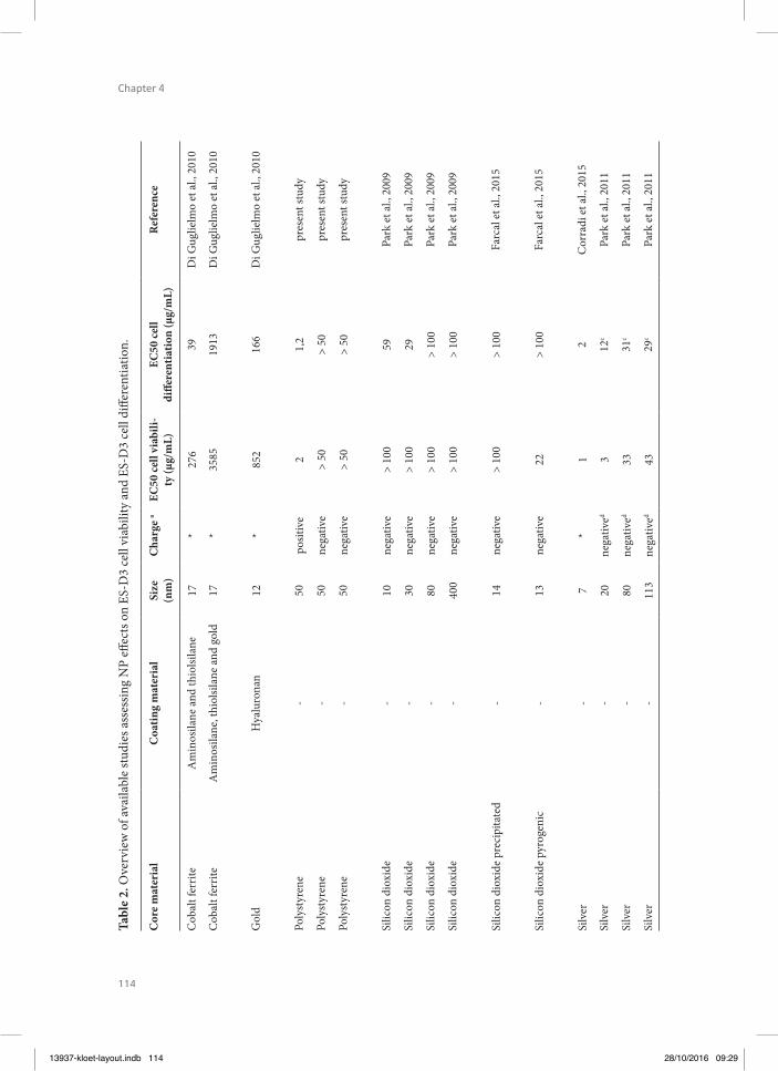
114
Chapter 4
Tabl
e 2.
Ove
rvie
w o
f ava
ilabl
e st
udie
s ass
essin
g N
P eff
ect
s on
ES-D
3 ce
ll vi
abili
ty a
nd E
S-D
3 ce
ll di
ff ere
ntia
tion.
Cor
e m
ater
ial
Coa
ting
mat
eria
lSi
ze
(nm
)C
harg
e a
EC50
cell
viab
ili-
ty (µ
g/m
L)EC
50 ce
ll di
ff ere
ntia
tion
(µg/
mL)
Ref
eren
ce
Cob
alt f
errit
eA
min
osila
ne a
nd th
iolsi
lane
17*
276
39D
i Gug
lielm
o et
al.,
201
0
Cob
alt f
errit
eA
min
osila
ne, t
hiol
silan
e an
d go
ld17
*35
8519
13D
i Gug
lielm
o et
al.,
201
0
Gol
dH
yalu
rona
n12
*85
216
6D
i Gug
lielm
o et
al.,
201
0
Poly
styr
ene
-50
posit
ive
21,
2pr
esen
t stu
dy
Poly
styr
ene
-50
nega
tive
> 50
> 50
pres
ent s
tudy
Poly
styr
ene
-50
nega
tive
> 50
> 50
pres
ent s
tudy
Silic
on d
ioxi
de-
10ne
gativ
e>
100
59Pa
rk e
t al.,
200
9
Silic
on d
ioxi
de-
30ne
gativ
e>
100
29Pa
rk e
t al.,
200
9
Silic
on d
ioxi
de-
80ne
gativ
e>
100
> 10
0Pa
rk e
t al.,
200
9
Silic
on d
ioxi
de-
400
nega
tive
> 10
0>
100
Park
et a
l., 2
009
Silic
on d
ioxi
de p
reci
pita
ted
-14
nega
tive
> 10
0>
100
Farc
al e
t al.,
201
5
Silic
on d
ioxi
de p
yrog
enic
-13
nega
tive
22>
100
Farc
al e
t al.,
201
5
Silv
er-
7*
12
Cor
radi
et a
l., 2
015
Silv
er-
20ne
gativ
ed3
12c
Park
et a
l., 2
011
Silv
er-
80ne
gativ
ed33
31c
Park
et a
l., 2
011
Silv
er-
113
nega
tived
4329
cPa
rk e
t al.,
201
1
13937-kloet-layout.indb 114 28/10/2016 09:29
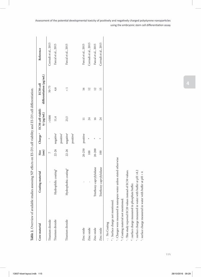
115
Assessment of the potenti al developmental toxicity of positi vely and negati vely charged polystyrene nanoparti cles
using the embryonic stem cell diff erenti ati on assay
4
Tabl
e 2.
Ove
rvie
w o
f ava
ilabl
e st
udie
s ass
essin
g N
P eff
ect
s on
ES-D
3 ce
ll vi
abili
ty a
nd E
S-D
3 ce
ll di
ff ere
ntia
tion.
Cor
e m
ater
ial
Coa
ting
mat
eria
lSi
ze
(nm
)C
harg
e a
EC50
cell
viab
ili-
ty (µ
g/m
L)EC
50 ce
ll di
ff ere
ntia
tion
(µg/
mL)
Ref
eren
ce
Tita
nium
dio
xide
-7
*>1
000
50-7
5C
orra
di e
t al.,
201
5
Tita
nium
dio
xide
Hyd
roph
ilic c
oatin
gb22
-26
nega
tivee
11,4
4,6
Farc
al e
t al.,
201
5
posit
ivef
Tita
nium
dio
xide
Hyd
roph
obic
coat
ingb
22-2
6ne
gativ
ee23
,5<
1Fa
rcal
et a
l., 2
015
posit
ivef
Zinc
oxi
de-
20-2
50po
sitiv
e11
16Fa
rcal
et a
l., 2
015
Zinc
oxi
de-
100
*24
12C
orra
di e
t al.,
201
5
Zinc
oxi
deTr
ieth
oxy-
capr
ylyl
silan
e20
-200
*16
12Fa
rcal
et a
l., 2
015
Zinc
oxi
deTr
ieth
oxy-
capr
ylyl
silan
e10
0*
2415
Cor
radi
et a
l., 2
015
- :
No
Coa
ting
* : S
urfa
ce ch
arge
not
men
tione
d.a :
Cha
rges
wer
e m
easu
red
in n
anop
ure
wat
er u
nles
s sta
ted
othe
rwise
b : C
oatin
g m
ater
ial n
ot m
entio
ned.
c : Th
is st
udy
repo
rted
EC
20 v
alue
s ins
tead
of E
C50
val
ues.
d : su
rfac
e ch
arge
mea
sure
d in
pho
spha
te b
uff e
r. e :
surf
ace
char
ge m
easu
red
in w
ater
with
buff
er a
t pH
>8.
2f :
surf
ace
char
ge m
easu
red
in w
ater
with
buff
er a
t pH
< 4
13937-kloet-layout.indb 115 28/10/2016 09:29
116
Chapter 4
DISCUSSION
Th e present study aimed to assess the role of NP surface charge on the in vitro developmental toxicity potential of NPs as measured in the ES-D3 cell diff erentiation assay. Th e results show that the positively charged PS-NPs caused a concentration-dependent inhibition of ES-D3 cell diff erentiation while the two negatively charged PS-NPs did not show any eff ect, indi-cating that NP charge may play an important role in NP-induced developmental toxicity. In order to obtain insight whether also other physicochemical characteristics of NPs may relate to their in vitro developmental toxicity potency, we performed a literature search on the reported eff ects of NPs on ES-D3 cell diff erentiation. Th is analysis indicated that besides surface charge, also size and coating are important NP characteristics that may determine the developmental toxicity potential of NPs.
Eff ect concentrations of the positively charged PS-NPs in the ES-D3 cell diff erentiation assay were close to cytotoxic concentrations as measured in cell viability studies (Figure 1A), which indicates that the inhibition of the ES-D3 cell diff erentiation may be caused by non-specifi c eff ects. Similar eff ects have also been found for silver NPs and zinc oxide NPs (Table 2). In the validation study of the EST, most developmental toxicants that were selected based on pos-itive developmental toxicity eff ects in in vivo studies appeared to cause inhibition of ES-D3 cell diff erentiation at concentrations that also caused cytotoxicity (Marx-Stoelting et al. 2009). Th erefore, the positively charged PS-NPs tested in the present study may present a develop-mental hazard in vivo, even though the mode of action underlying this developmental toxicity may be related to cytotoxicity.
Knowledge on the relation between physicochemical characteristics of NPs, such as size or surface charge, and their toxicity would be of great help for setting priorities for NP toxicity testing. Although the experiments in the present study show that positively charged PS-NPs are more toxic than negatively charged PS-NPs, it appeared that this may not be generalizable to other NPs, following from the fact that the negatively charged PS-NPs of the present study did not aff ect ES-D3 cell diff erentiation, while silicon dioxide, silver and titanium dioxide NPs that were reported to inhibit ES-D3 cell diff erentiation were also negatively charged (Table 2). Th e reported surface charges of these NPs were measured in either nanopure water or phos-phate buff er. Th is implies that the negative charge did not arise from serum proteins which may aff ect the NP charge, as observed in the present study when NP charge was measured in medium with FCS. To obtain better insight in the role of NP surface charge on developmental toxicity more studies are needed that assess the eff ects of NPs with diff erent charge that are made of the same core material. Th e eff ect of surface charge on the potential to aff ect cell viability has been demonstrated before in other cell lines for tri-block copolymer NPs (Bhat-tacharjee et al. 2012), gold NPs (Liu et al. 2013) and silicon NPs (Bhattacharjee et al. 2013),
13937-kloet-layout.indb 116 28/10/2016 09:29
117
Assessment of the potenti al developmental toxicity of positi vely and negati vely charged polystyrene nanoparti cles
using the embryonic stem cell diff erenti ati on assay
4
generally showing higher cytotoxicity for positively charged NPs compared to negatively charged NPs. Whether this implies that positively charged silicon dioxide, silver and titanium dioxide NPs would show more potent eff ects on ES-D3 cell viability and diff erentiation than reported for their negative counterparts in literature (Table 2) remains to be investigated.
Our literature search showed that for two diff erent types of NPs (silicon dioxide NPs and silver NPs), it has been shown that the eff ects on ES-D3 cell viability and diff erentiation were found at lower concentrations for relatively small NPs than for larger NPs of the same core material, indicating the role of NP size in their eff ects on ES-D3 cell viability and diff erenti-ation. Th e smaller the diameter of a spherical NP the higher the surface area which is oft en accompanied by increased chemical reactivity with cells (Nel et al. 2009). It has also been re-ported that cellular uptake of NPs is dependent on their size, showing a higher cellular uptake for relatively small NPs (Kreyling et al. 2009, Dos Santos et al. 2011), indicating that the intra-cellular concentration of small-sized NPs may be higher than the intracellular concentration of the larger sized NPs, which may also explain their higher toxic potency. However, when comparing toxic potencies of NPs of diff erent sizes, it must be taken into account that when expressing exposure on a mass-basis (µg/ml) the number of NPs is larger for small-sized NPs than for large-sized NPs at the same concentration. Th is may need to be accounted for when comparing the toxicity potencies of NPs of diff erent sizes.
Our analysis of the available literature also indicated that the in vitro developmental toxicity potency of NPs may also largely depend on the coating applied. However, from the available data it cannot be concluded whether the toxicity potential is dependent on the coating alone, or whether that is dependent on the combination of the core material and the coating. Th ere-fore, although the limited data available indicate that charge, size and coating of NPs may be important characteristics that determine the developmental toxicity potential of NPs, more (systematic) studies are needed to assess how physicochemical characteristics of NPs relate to their developmental toxicity potency. Th is information may help to prioritize NPs for in vitro and in vivo developmental toxicity testing.
Th e results of the present study indicate that the positively charged PS-NPs may pose a hazard for embryonic development. However, to possibly become a risk for embryonic development, NPs need to be able to reach the developing embryo in the in vivo situation. Th erefore, with oral exposure, NPs would need to translocate across the intestinal barrier as well as the pla-cental barrier. Th e bioavailability upon oral exposure has been reported to be low for many NPs, because of a limited translocation across the intestinal barrier (Walczak et al. 2014). As far as we know, no in vivo study using oral exposure has shown so far that NPs can cause de-velopmental toxicity. However, in vivo studies using intravenous injection of NPs have shown that NPs can cause adverse eff ects to the developing embryo, indicating that NPs can trans-
13937-kloet-layout.indb 117 28/10/2016 09:29
118
Chapter 4
locate across the placenta in the in vivo situation (Semmler-Behnke et al. 2008, Yamashita et al. 2011, Yang et al. 2012, Qi et al. 2014). In an earlier study, we have assessed the potential of the same PS-NPs as used in the present study to translocate across an in vitro barrier of hu-man placental (BeWo) cells (Kloet et al. 2015). Th e results of these studies indicated that the positively charged PS-NPs hardly translocated across the BeWo cell layer, which may indicate that fetal exposure of these PS-NPs is low. Th erefore, although the present study indicates the potential of positively charged PS-NPs to cause developmental toxicity, concentrations shown in the present study to be required to exert these eff ects may not be reached in the in vivo situation, because of the low placental transfer. Th erefore, in order to predict which NPs may cause developmental toxicity in the in vivo situation, data obtained in the in vitro ES-D3 cell diff erentiation assay should be combined with placental translocation data from the BeWo model in order to select the NPs that are expected to have the highest potency in vivo. Th ese combined data may then be used to select and prioritize NPs that should be assessed in vivo for potential developmental toxicity.
ACKNOWLEDGEMENTS
Th is work is supported by NanoNextNL, a micro and nanotechnology consortium of the Government of the Netherlands and 130 partners.
13937-kloet-layout.indb 118 28/10/2016 09:29
119
Assessment of the potenti al developmental toxicity of positi vely and negati vely charged polystyrene nanoparti cles
using the embryonic stem cell diff erenti ati on assay
4
REFERENCES
Bhattacharjee, S., D. Ershov, K. Fytianos, J. Gucht, G. Alink, I. Rietjens, A. Marcelis and H. Zuilhof (2012). “Cytotox-icity and cellular uptake of tri-block copolymer nanoparticles with diff erent size and surface characteristics.” Particle and Fibre Toxicology 9(1): 11.
Bhattacharjee, S., I. M. Rietjens, M. P. Singh, T. M. Atkins, T. K. Purkait, Z. Xu, S. Regli, A. Shukaliak, R. J. Clark, B. S. Mitchell, G. M. Alink, A. T. Marcelis, M. J. Fink, J. G. Veinot, S. M. Kauzlarich and H. Zuilhof (2013). “Cytotoxicity of surface-functionalized silicon and germanium nanoparticles: the dominant role of surface charges.” Nanoscale 5(11): 4870-4883.
Braakhuis, H. M., S. K. Kloet, S. Kezic, F. Kuper, M. V. Park, S. Bellmann, M. van der Zande, S. Le Gac, P. Krystek, R. J. Peters, I. M. Rietjens and H. Bouwmeester (2015). “Progress and future of in vitro models to study trans-location of nanoparticles.” Arch Toxicol 89(9): 1469-1495.
Campagnolo, L., I. Fenoglio, M. Massimiani, A. Magrini and A. Pietroiusti (2013). “Screening of nanoparticle em-bryotoxicity using embryonic stem cells.” Methods Mol Biol 1058: 49-60.
de Jong, E., M. Barenys, S. A. B. Hermsen, A. Verhoef, B. C. Ossendorp, J. G. M. Bessems and A. H. Piersma (2011). “Comparison of the mouse Embryonic Stem cell Test, the rat Whole Embryo Culture and the Zebrafi sh Em-bryotoxicity Test as alternative methods for developmental toxicity testing of six 1,2,4-triazoles.” Toxicology and Applied Pharmacology 253(2): 103-111.
de Jong, E., J. Louisse, M. Verwei, B. J. Blaauboer, J. J. van de Sandt, R. A. Woutersen, I. M. Rietjens and A. H. Piersma (2009). “Relative developmental toxicity of glycol ether alkoxy acid metabolites in the embryonic stem cell test as compared with the in vivo potency of their parent compounds.” Toxicol Sci 110(1): 117-124.
Dos Santos, T., J. Varela, I. Lynch, A. Salvati and K. A. Dawson (2011). “Quantitative Assessment of the Comparative Nanoparticle-Uptake Effi ciency of a Range of Cell Lines.” Small.
Fort, D. J. and R. R. Paul (2002). “Enhancing the predictive validity of Frog Embryo Teratogenesis Assay--Xenopus (FETAX).” J Appl Toxicol 22(3): 185-191.
Gebel, T., R. Marchan and J. G. Hengstler (2013). “Th e nanotoxicology revolution.” Arch Toxicol 87(12): 2057-2062.Genschow, E., H. Spielmann, G. Scholz, I. Pohl, A. Seiler, N. Clemann, S. Bremer and K. Becker (2004). “Validation of
the embryonic stem cell test in the international ECVAM validation study on three in vitro embryotoxicity tests.” Altern Lab Anim 32(3): 209-244.
Jelinek, R., M. Peterka and Z. Rychter (1985). “Chick embryotoxicity screening test--130 substances tested.” Indian J Exp Biol 23(10): 588-595.
Kloet, S. K., A. P. Walczak, J. Louisse, H. H. van den Berg, H. Bouwmeester, P. Tromp, R. G. Fokkink and I. M. Rietjens (2015). “Translocation of positively and negatively charged polystyrene nanoparticles in an in vitro placental model.” Toxicol In Vitro 29(7): 1701-1710.
Kreyling, W. G., M. Semmler-Behnke, J. Seitz, W. Scymczak, A. Wenk, P. Mayer, S. Takenaka and G. Oberdorster (2009). “Size dependence of the translocation of inhaled iridium and carbon nanoparticle aggregates from the lung of rats to the blood and secondary target organs.” Inhal Toxicol 21 Suppl 1: 55-60.
Li, H., I. M. Rietjens, J. Louisse, M. Blok, X. Wang, L. Snijders and B. van Ravenzwaay (2015). “Use of the ES-D3 cell diff erentiation assay, combined with the BeWo transport model, to predict relative in vivo developmental toxicity of antifungal compounds.” Toxicol In Vitro 29(2): 320-328.
Liu, X., N. Huang, H. Li, Q. Jin and J. Ji (2013). “Surface and Size Eff ects on Cell Interaction of Gold Nanoparticles with Both Phagocytic and Nonphagocytic Cells.” Langmuir 29(29): 9138-9148.
Marx-Stoelting, P., E. Adriaens, H. J. Ahr, S. Bremer, B. Garthoff , H. P. Gelbke, A. Piersma, C. Pellizzer, U. Reuter, V. Rogiers, B. Schenk, S. Schwengberg, A. Seiler, H. Spielmann, M. Steemans, D. B. Stedman, P. Vanparys, J. A. Vericat, M. Verwei, F. van der Water, M. Weimer and M. Schwarz (2009). “A review of the implementation of the embryonic stem cell test (EST). Th e report and recommendations of an ECVAM/ReProTect Workshop.” Altern Lab Anim 37(3): 313-328.
Nel, A. E., L. Madler, D. Velegol, T. Xia, E. M. Hoek, P. Somasundaran, F. Klaessig, V. Castranova and M. Th ompson (2009). “Understanding biophysicochemical interactions at the nano-bio interface.” Nat Mater 8(7): 543-557.
Oberdorster, G., V. Stone and K. Donaldson (2007). “Toxicology of nanoparticles: A historical perspective.” Nano-toxicology 1(1): 2-25.
13937-kloet-layout.indb 119 28/10/2016 09:29

120
Chapter 4
Park, M. V., W. Annema, A. Salvati, A. Lesniak, A. Elsaesser, C. Barnes, G. McKerr, C. V. Howard, I. Lynch, K. A. Dawson, A. H. Piersma and W. H. de Jong (2009). “In vitro developmental toxicity test detects inhibition of stem cell diff erentiation by silica nanoparticles.” Toxicol Appl Pharmacol 240(1): 108-116.
Pietroiusti, A., M. Massimiani, I. Fenoglio, M. Colonna, F. Valentini, G. Palleschi, A. Camaioni, A. Magrini, G. Sir-acusa, A. Bergamaschi, A. Sgambato and L. Campagnolo (2011). “Low Doses of Pristine and Oxidized Sin-gle-Wall Carbon Nanotubes Aff ect Mammalian Embryonic Development.” ACS Nano 5(6): 4624-4633.
Qi, W., J. Bi, X. Zhang, J. Wang, J. Wang, P. Liu, Z. Li and W. Wu (2014). “Damaging eff ects of multi-walled carbon nanotubes on pregnant mice with diff erent pregnancy times.” Sci Rep 4: 4352.
Semmler-Behnke, M., W. G. Kreyling, J. Lipka, S. Fertsch, A. Wenk, S. Takenaka, G. Schmid and W. Brandau (2008). “Biodistribution of 1.4- and 18-nm gold particles in rats.” Small 4(12): 2108-2111.
Spielmann, H., A. Seiler, S. Bremer, L. Hareng, T. Hartung, H. Ahr, E. Faustman, U. Haas, G. J. Moff at, H. Nau, P. Vanparys, A. Piersma, J. R. Sintes and J. Stuart (2006). “Th e practical application of three validated in vitro embryotoxicity tests. Th e report and recommendations of an ECVAM/ZEBET workshop (ECVAM work-shop 57).” Altern Lab Anim 34(5): 527-538.
van Dartel, D. A., J. L. Pennings, L. J. de la Fonteyne, K. J. Brauers, S. Claessen, J. H. van Delft , J. C. Kleinjans and A. H. Piersma (2011). “Concentration-dependent gene expression responses to fl usilazole in embryonic stem cell diff erentiation cultures.” Toxicol Appl Pharmacol 251(2): 110-118.
van Dartel, D. A., J. L. Pennings, P. J. Hendriksen, F. J. van Schooten and A. H. Piersma (2009). “Early gene expression changes during embryonic stem cell diff erentiation into cardiomyocytes and their modulation by monobu-tyl phthalate.” Reprod Toxicol 27(2): 93-102.
van Dartel, D. A. M., N. J. L. Zeijen, L. J. J. de la Fonteyne, F. J. van Schooten and A. H. Piersma (2009). “Disentangling cellular proliferation and diff erentiation in the embryonic stem cell test, and its impact on the experimental protocol.” Reproductive Toxicology 28(2): 254-261.
Walczak, A. P., E. Kramer, P. J. Hendriksen, P. Tromp, J. P. Helsper, M. van der Zande, I. M. Rietjens and H. Bouw-meester (2014). “Translocation of diff erently sized and charged polystyrene nanoparticles in in vitro intesti-nal cell models of increasing complexity.” Nanotoxicology: 1-9.
Yamashita, K., Y. Yoshioka, K. Higashisaka, K. Mimura, Y. Morishita, M. Nozaki, T. Yoshida, T. Ogura, H. Nabeshi, K. Nagano, Y. Abe, H. Kamada, Y. Monobe, T. Imazawa, H. Aoshima, K. Shishido, Y. Kawai, T. Mayumi, S. Tsunoda, N. Itoh, T. Yoshikawa, I. Yanagihara, S. Saito and Y. Tsutsumi (2011). “Silica and titanium dioxide nanoparticles cause pregnancy complications in mice.” Nat Nanotechnol 6(5): 321-328.
Yang, H., C. Sun, Z. Fan, X. Tian, L. Yan, L. Du, Y. Liu, C. Chen, X. J. Liang, G. J. Anderson, J. A. Keelan, Y. Zhao and G. Nie (2012). “Eff ects of gestational age and surface modifi cation on materno-fetal transfer of nanoparticles in murine pregnancy.” Sci Rep 2: 847.
zur Nieden, N. I., G. Kempka and H. J. Ahr (2004). “Molecular multiple endpoint embryonic stem cell test--a possi-ble approach to test for the teratogenic potential of compounds.” Toxicol Appl Pharmacol 194(3): 257-269.
13937-kloet-layout.indb 120 28/10/2016 09:29

13937-kloet-layout.indb 121 28/10/2016 09:29

12
34
56
78
910
1112
A B C D E F G G H
13937-kloet-layout.indb 122 28/10/2016 09:29
12
34
56
78
910
1112
A B C D E F G G H
Chapter 5
Effects of nanoparticle forms of metal (oxide) food additives and related nanoparticles on cell viability,
TNF-α production and mitochondria-related parameters in RAW264.7 macrophages
Samantha K. KloetSunday Makama
Hans H.J. van den BergGang Wu
Georgios SaviolakisHeike Vogt
Aikaterini ManolesouNico van den Brink
Ivonne M.C.M. RietjensJochem Louisse
Submitted for publication to In Vitro Toxicology
13937-kloet-layout.indb 123 28/10/2016 09:29
124
Chapter 5
ABSTRACT
Specifi cations of food additives like titanium dioxide, iron oxide, silicon dioxide and silver do not exclude the material of commerce being present in part in nanoform. Th e aim of the present study was to investigate toxic eff ects of the nanoform of these metal (oxide) food additives and other nanoparticles (NPs), being manganese oxide, copper oxide, chromium oxide, cobalt oxide and nickel oxide NPs, using an in vitro model of macrophage RAW264.7 cells. Eff ects on cell viability, reactive oxygen species (ROS) production, mitochondrial per-meability transition pore (MPTP) opening, ATP, and TNF-α were assessed. Th e results indi-cate that most of the tested NPs show no eff ects on the selected endpoints up to the highest concentration tested. Of the food additive related NPs, silicon dioxide and silver NPs were most toxic, showing a decrease in cell viability at concentrations of 100 µg/ml and higher. Altogether, the present study shows no or limited eff ects of the NP forms of metal (oxide) food additives on cell viability, ROS production, MPTP opening, ATP levels and TNF-α pro-duction in RAW264.7 macrophages. Eff ects were only observed at high concentrations that may not be physiologically relevant, indicating that related adverse eff ects upon exposure to the respective NPs may be limited.
13937-kloet-layout.indb 124 28/10/2016 09:29
125
Eff ects of nanoparti cle forms of metal (oxide) food additi ves and related nanoparti cles on cell viability, TNF-α
producti on and mitochondria-related parameters in RAW264.7 macrophages
5
INTRODUCTION
Several food additives, such as titanium dioxide (TiO2) (E171), iron (III) oxide (Fe2O3) (E172), silicon dioxide (SiO2) (E551) and silver (Ag) (E174), contain metal or metal oxide particles of variable sizes (Athinarayanan et al. 2014, Periasamy et al. 2015). At present the specifi cations of these food additives do not exclude the presence of the compounds in their nanoform, be-cause food additives are excluded from the regulation that states that all ingredients that are present in nano-form should be indicated in the list of ingredients as ‘nano’ (2011). A recent study investigated the morphology and dimensions of the food additive SiO2 in several com-mercial food products, showing the presence of spherical SiO2 nanoparticles (NPs) of about 10-50 nm in size (Athinarayanan et al. 2014). Other studies found that a certain food grade TiO2 contained ~25-38% of the TiO2 as NPs smaller than 200 nm (Weir et al. 2012, Faust et al. 2014). Th ese fi ndings indicate that studies on the health eff ects of nanoforms of current food additives are of high interest. At the same time, safety considerations for nanoscale materials in food products have not been well developed yet (Bouwmeester et al. 2009). As the health eff ects of NPs are largely unknown, toxicity studies with nanomaterials that measure such health eff ects are urgently needed.
Th e aim of the present study was to investigate the toxic eff ects of the nanoform of metal (oxide) food additives TiO2, Fe2O3, SiO2 and Ag using an in vitro model consisting of mac-rophage RAW264.7 cells. Further, the eff ects of other metal oxide NPs of manganese oxide (Mn2O3), copper oxide (CuO), chromium oxide (Cr2O3), cobalt oxide (Co3O4) and nickel oxide (NiO), to which we may be exposed via products like paints, catalysts, construction materials, coatings and batteries were quantifi ed (Siddiqui et al. 2012, Tavares et al. 2014, Dang et al. 2015). Th e possible NP-induced adverse eff ects on macrophage RAW264.7 cells may indicate possible immunotoxic responses and hazards since macrophages play a major role in the innate immune response, providing a fi rst line of defence (Ma et al. 2003, Elsabahy et al. 2013). NP-induced macrophage cell toxicity may therefore decrease the innate immune response. Also, activation of cytokine production, such as tumor necrosis factor-α (TNF-α), by macrophages exposed to NPs is undesired, since this may lead to infl ammatory reactions.
To assess eff ects of the NPs on the macrophages, cell viability, mitochondria-related parame-ters and release of the infl ammatory mediator TNF-α were assessed in NP-exposed RAW264.7 cells. Th is study focussed on the eff ects of the NPs on mitochondria-related parameters, since previous research indicated that mitochondria play an important role in NP-induced toxicity (Park et al. 2008, Sharma et al. 2011, Shvedova et al. 2012, Bhattacharjee et al. 2013). Espe-cially cationic NPs have been reported to damage the mitochondria, leading to disturbance of intracellular adenosine 5′-triphosphate (ATP) and reactive oxygen species (ROS) production, which may trigger apoptotic reactions that ultimately lead to the observed toxicity (Bhat-
13937-kloet-layout.indb 125 28/10/2016 09:29
126
Chapter 5
tacharjee et al. 2013). Th erefore, in the present study the eff ects of the metal oxide NPs on ROS production, mitochondrial permeability transition pore (MPTP) opening and intracel-lular ATP levels were assessed, in addition to eff ects on cell viability and production of TNF-α.
MATERIALS AND METHODS
Materials High glucose Dulbecco´s Modifi ed Eagle Medium (DMEM/F-12), Phosphate Buff ered Sa-line (PBS), Fetal Calf Serum (FCS), Hank’s Balanced Salt Solution (HBSS) and trypsin-ED-TA were obtained from Invitrogen (Breda, the Netherlands). Bovine Serum Albumin (BSA), 3-(4,5-Dimethylthiazol-2-yl)-2,5-diphenyltetrazolium bromide (MTT) and copper sulfate were purchased from Sigma Aldrich (Zwijndrecht, the Netherlands). WST-1 Cell Prolifera-tion Reagent was purchased from Roche (Woerden, Th e Netherlands). Information on size and specifi c surface area of the NPs that were used in the present study as provided by the manufacturers are presented in table 1. Fe2O3 NPs, TiO2 anatase and TiO2 rutile NPs, and Cr2O3 NPs were obtained from US Research Nanomaterials (Houston, Texas, USA). Mn2O3 NPs were obtained from Auer Remy (Hamburg, Germany). SiO2 NPs and NiO NPs were ob-tained from Skyspring (Houston, Texas, USA). Co3O4 NPs and CuO NPs were obtained from Sigma Aldrich (Zwijndrecht, Th e Netherlands). Ag NPs were obtained from NanoComposix Inc. (San Diego, California, USA).
Preparation of NPs for in vitro studiesAll NPs were delivered in powdered form except for Ag NPs which came as a stable 5 mg/ml aqueous suspension in nanopure water. Th e working suspensions were prepared according to a method described by Zhang et al. (2012). Briefl y, a 5 mg/ml stock suspension in nanopure water of each powdered NP was prepared in a sterile glass vial. Th ese suspensions were soni-cated in a bath sonicator (VWR international, Leuven, Belgium) for 20 minutes. Aft erwards, an equal volume of 4% BSA in nanopure water was added to stabilise the suspension. Th ese suspensions were left for one hour at room temperature to equilibrate. Aft er vortexing the NP stock suspension, further dilutions were prepared with 2% BSA in nanopure water, except for the Ag NP stock suspension which was further diluted in nanopure water. Finally, the BSA-coated NP suspensions were 12.5x diluted in cell culture medium containing 10% FCS, reaching the fi nal exposure NP concentrations between 0 and 200 µg/ml.
NP characterizationTh e size (hydrodynamic diameter) of the NPs was determined by dynamic light scattering (DLS) using a Cobolt Samba 300mW DPSS laser at a wavelength of 532 nm. Measurements were performed on freshly prepared NP suspensions at 10 μg/ml in nanopure water and in cell culture medium containing 10% FCS. For each sample, twenty measurements were made
13937-kloet-layout.indb 126 28/10/2016 09:29
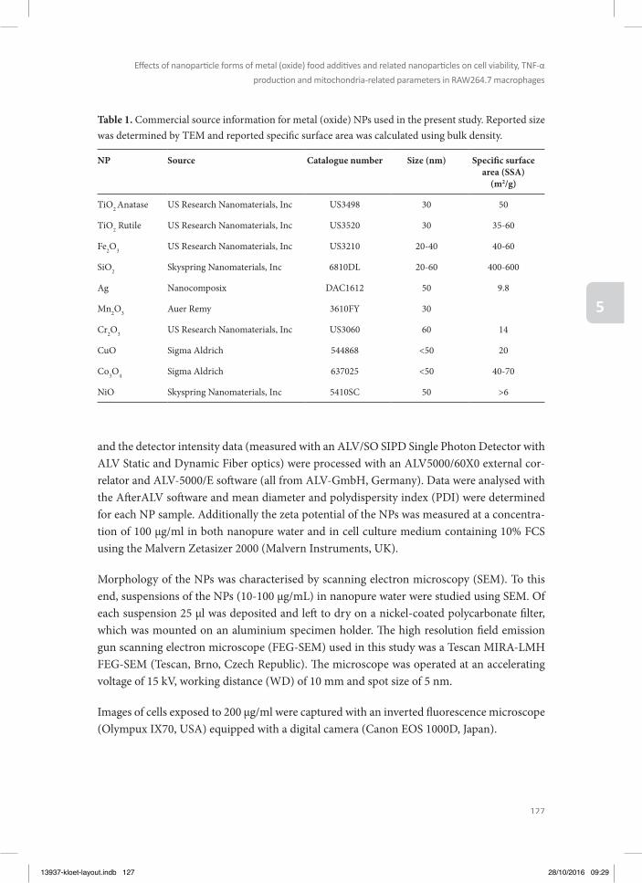
127
Eff ects of nanoparti cle forms of metal (oxide) food additi ves and related nanoparti cles on cell viability, TNF-α
producti on and mitochondria-related parameters in RAW264.7 macrophages
5
and the detector intensity data (measured with an ALV/SO SIPD Single Photon Detector with ALV Static and Dynamic Fiber optics) were processed with an ALV5000/60X0 external cor-relator and ALV-5000/E soft ware (all from ALV-GmbH, Germany). Data were analysed with the Aft erALV soft ware and mean diameter and polydispersity index (PDI) were determined for each NP sample. Additionally the zeta potential of the NPs was measured at a concentra-tion of 100 µg/ml in both nanopure water and in cell culture medium containing 10% FCS using the Malvern Zetasizer 2000 (Malvern Instruments, UK).
Morphology of the NPs was characterised by scanning electron microscopy (SEM). To this end, suspensions of the NPs (10-100 µg/mL) in nanopure water were studied using SEM. Of each suspension 25 μl was deposited and left to dry on a nickel-coated polycarbonate fi lter, which was mounted on an aluminium specimen holder. Th e high resolution fi eld emission gun scanning electron microscope (FEG-SEM) used in this study was a Tescan MIRA-LMH FEG-SEM (Tescan, Brno, Czech Republic). Th e microscope was operated at an accelerating voltage of 15 kV, working distance (WD) of 10 mm and spot size of 5 nm.
Images of cells exposed to 200 µg/ml were captured with an inverted fl uorescence microscope (Olympux IX70, USA) equipped with a digital camera (Canon EOS 1000D, Japan).
Table 1. Commercial source information for metal (oxide) NPs used in the present study. Reported size was determined by TEM and reported specifi c surface area was calculated using bulk density.
NP Source Catalogue number Size (nm) Specifi c surface area (SSA)
(m2/g)
TiO2 Anatase US Research Nanomaterials, Inc US3498 30 50
TiO2 Rutile US Research Nanomaterials, Inc US3520 30 35-60
Fe2O3 US Research Nanomaterials, Inc US3210 20-40 40-60
SiO2 Skyspring Nanomaterials, Inc 6810DL 20-60 400-600
Ag Nanocomposix DAC1612 50 9.8
Mn2O3 Auer Remy 3610FY 30
Cr2O3 US Research Nanomaterials, Inc US3060 60 14
CuO Sigma Aldrich 544868 <50 20
Co3O4 Sigma Aldrich 637025 <50 40-70
NiO Skyspring Nanomaterials, Inc 5410SC 50 >6
13937-kloet-layout.indb 127 28/10/2016 09:29
128
Chapter 5
Cell culture and assessment of cell viabilityRAW264.7 cells (LGC, ATCC, Teddington, UK) were cultured at 37°C with 5% CO2 in a hu-midifi ed atmosphere in 10 ml DMEM supplemented with 10% (v/v) FCS in a 75 cm2 culture fl ask (Corning, USA). Cells were subcultured every 3 to 4 days when reaching 80% confl uen-cy using trypsin-EDTA aft er rinsing with PBS.
To assess eff ects of the NPs on cell viability, RAW264.7 cells were seeded at 2x10⁴ cells/well in 100 µl culture medium with 10% FCS in 96-well plates (Greiner bio-one) and incubated at 37°C and 5% CO2 for 24 hours. Aft er this, the medium was removed and cells were sub-sequently exposed to 100 µl of NP suspensions using concentrations ranging from 0 to 200 µg/ml in culture medium with 10% FCS. Aft er 24 hours exposure, 10 µl of WST solution was added to the medium. Aft er 45 min incubation, the 96-well plate was shaken for 5 minutes (protected from light) and subsequently absorbance was measured at 450 and 620 nm (back-ground) using a SpectraMax M2 Microplate Reader (Molecular Devices LLC, Sunnyvale, CA USA). For each experimental condition, formazan formation was expressed as percentage of the formazan formation of the solvent control. Ag NPs showed a concentration-dependent interference in this WST assay. Th erefore, instead of WST solution, 5 µl of a 5 mg/mL tetra-zolium salt 3-(4,5-dimethylthiazol-2-yl)-2,5-diphenyltetrazolium bromide (MTT) solution in PBS was added to each well in the experiments on Ag NPs and incubated for 1.5 hours. Th e exposure media containing MTT was then removed and 100 µL DMSO was added to solu-bilize the formazan crystals, which was achieved by shaking the plates protected from light for 10 min. Th e dissolved formazan was quantifi ed by measuring the absorption at 562 nm and 620 nm (background) using a SpectraMax M2 Microplate Reader. For each experimental condition, formazan formation was expressed as percentage of the formazan formation of the solvent control. Copper sulfate (100 µM) was used as the positive control.
Assessment of mitochondrial permeability transition pore (MPTP) openingTh e MPTP opening refers to a change in the inner mitochondrial membrane permeability (Bonora et al. 2014). An increase in MPTP opening results in an increase in the mitochon-drial membrane permeability, leading to rapid loss of the mitochondrial membrane potential, a decrease in ATP production, and fi nally to the loss of cell integrity (Ly et al. 2003). Aft er 24 hours exposure to the NPs under similar conditions as with the cell viability assay, MPTP opening was measured using the MitoProbe Transition Pore Assay Kit (Invitrogen, Breda, Th e Netherlands, cat nr. M34153). Fluorescence was measured at 485 and 530 nm (background) using a SpectraMax M2 Microplate Reader. Ionomycin, which was supplied with the kit, was used as the positive control (15 minutes incubation at a concentration of 10 µM). In the assay, an increase in MPTP opening causes a decrease in the fl uorescent signal which correlates with an increase in the mitochondrial membrane permeability, so with a decrease in the mitochon-drial membrane integrity.
13937-kloet-layout.indb 128 28/10/2016 09:29
129
Eff ects of nanoparti cle forms of metal (oxide) food additi ves and related nanoparti cles on cell viability, TNF-α
producti on and mitochondria-related parameters in RAW264.7 macrophages
5
Assessment of intracellular ROS productionTo determine the eff ects of the NPs on intracellular ROS production, RAW264.7 cells were seeded at 2x10⁴ cells/well in 100 µl culture medium with 10% FCS in 96-well plates and in-cubated at 37°C and 5% CO2 for 24 hours, followed by removal of the medium and addition of a solution of 20 µM 2,7-dichlorodihydrofl uorescein diacetate (DCFDA) in medium with 10% FCS. Aft er 45 minutes the DCFDA solution was removed and cells were exposed for 24 hours to the NPs under similar conditions as with the cell viability assay. Fluorescence was measured using a SpectraMax M2 Microplate Reader at 485 and 538 nm (background) wave-lengths. H2O2 (10 mM) was used as the positive control.
Assessment of intracellular ATP levelsTh e eff ects of NPs on intracellular ATP levels was determined. Aft er 24 hours exposure to the NPs under similar conditions as with the cell viability assay, the ATP levels were determined using the ATP bioluminescent somatic cell assay kit (Sigma Aldrich, Zwijndrecht, Th e Neth-erlands, cat nr. FLASC) by measuring luminescence using the Luminoscan Ascent (Th ermo electroncorporation). 2,4-Dinitrophenol (4 mM) was used as the positive control.
Assessment of TNF-α productionTo determine the eff ects of the NPs on TNF-α production, the sandwich ELISA mouse TNF-α kit (Invitrogen, Breda, Th e Netherlands, cat nr. KMC3011C) was used. Aft er 24 hours expo-sure to the NPs under similar conditions as with the cell viability assay, plates were centri-fuged (centrifuge model 4k10 from Sigma) at 1200 rpm for 5 minutes at 4°C. Subsequently, the supernatants were collected and frozen at -80°C. Samples were thawed and added to mi-crotiter strips, coated with monoclonal antibodies specifi c for mouse TNF-α. Th e samples were diluted twice with standard diluent buff er from the kit and the secondary antibody (bi-otinylated mouse TNF-α Biotin Conjugate) was added. Aft er 90 minutes of incubation, the wells were washed four times with wash buff er (to remove any unbound secondary antibody) aft er which a 100 μl solution containing Streptavidin-HRP was added, which binds to the biotinylated secondary antibody. Th e plate was covered with a plastic cover and stored for 30 minutes at room temperature and the wells were subsequently washed four times with wash buff er delivered with the kit. Stabilized chromogen was added to each well and the plate was stored for 20 to 25 minutes in the dark. Subsequently, a stop solution was added to each well and the absorbance of each well was read at 450 nm, using a SpectraMax M2 Microplate Reader. A series of TNF-α concentrations were included in duplicate to each 96 wells plate in order to defi ne a calibration curve enabling quantifi cation of TNF-α. Lipopolysaccharide (LPS, 0.1 μg/ml) was used as the positive control.
13937-kloet-layout.indb 129 28/10/2016 09:29

130
Chapter 5
Data analysisFor each study, three or four independent experiments were performed (n= 3-4). In each ex-periment three technical replicates were included, which were averaged per experiment. Data are shown as mean ± standard deviation (SD) of all data points per experiment and plotted in graphs using GraphPad Prism® version 5. Statistical analyses among mean values of the diff erent treatments were performed by one-way analysis of variance followed by a Tukey’s Multiple Comparison Test using GraphPad Prism® version 5.
RESULTS
Physicochemical characterization of the NPsNP size, zeta potential and morphology were characterized using SEM, DLS and zeta po-tential analyses. Figure 1 shows the SEM pictures of the diff erent NPs. Ag NPs are rather spherical shaped and monodispersed, while the other NPs have variable shapes and form agglomerates (Figure 1).
Dynamic light scattering (DLS) measurements were performed to determine the hydrody-namic diameter of the diff erent NPs in nanopure water and cell culture medium with 10% FCS (Table 2). All NPs agglomerated when dispersed in water or cell culture medium with
Figure 1. SEM pictures (magnifi cation 200 000x) of the NPs used in the present study.
13937-kloet-layout.indb 130 28/10/2016 09:29
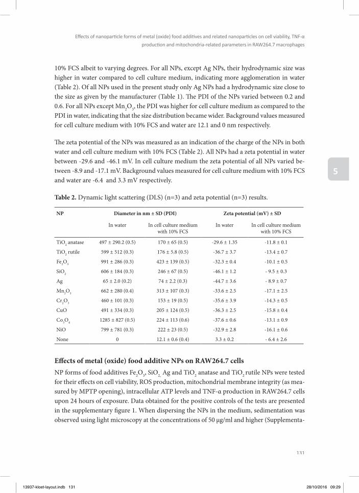
131
Eff ects of nanoparti cle forms of metal (oxide) food additi ves and related nanoparti cles on cell viability, TNF-α
producti on and mitochondria-related parameters in RAW264.7 macrophages
5
10% FCS albeit to varying degrees. For all NPs, except Ag NPs, their hydrodynamic size was higher in water compared to cell culture medium, indicating more agglomeration in water (Table 2). Of all NPs used in the present study only Ag NPs had a hydrodynamic size close to the size as given by the manufacturer (Table 1). Th e PDI of the NPs varied between 0.2 and 0.6. For all NPs except Mn2O3, the PDI was higher for cell culture medium as compared to the PDI in water, indicating that the size distribution became wider. Background values measured for cell culture medium with 10% FCS and water are 12.1 and 0 nm respectively.
Th e zeta potential of the NPs was measured as an indication of the charge of the NPs in both water and cell culture medium with 10% FCS (Table 2). All NPs had a zeta potential in water between -29.6 and -46.1 mV. In cell culture medium the zeta potential of all NPs varied be-tween -8.9 and -17.1 mV. Background values measured for cell culture medium with 10% FCS and water are -6.4 and 3.3 mV respectively.
Table 2. Dynamic light scattering (DLS) (n=3) and zeta potential (n=3) results.
NP Diameter in nm ± SD (PDI) Zeta potential (mV) ± SD
In water In cell culture medium with 10% FCS
In water In cell culture medium with 10% FCS
TiO2 anatase 497 ± 290.2 (0.5) 170 ± 65 (0.5) -29.6 ± 1.35 -11.8 ± 0.1
TiO2 rutile 599 ± 512 (0.3) 176 ± 5.8 (0.5) -36.7 ± 3.7 -13.4 ± 0.7
Fe2O3 991 ± 286 (0.3) 423 ± 139 (0.5) -32.3 ± 0.4 -10.1 ± 0.5
SiO2 606 ± 184 (0.3) 246 ± 67 (0.5) -46.1 ± 1.2 - 9.5 ± 0.3
Ag 65 ± 2.0 (0.2) 74 ± 2.2 (0.3) -44.7 ± 3.6 - 8.9 ± 0.7
Mn2O3 662 ± 280 (0.4) 313 ± 107 (0.3) -33.6 ± 2.5 -17.1 ± 2.5
Cr2O3 460 ± 101 (0.3) 153 ± 19 (0.5) -35.6 ± 3.9 -14.3 ± 0.5
CuO 491 ± 334 (0.3) 205 ± 124 (0.5) -36.3 ± 2.5 -15.8 ± 0.4
Co3O4 1285 ± 827 (0.5) 224 ± 113 (0.6) -37.6 ± 0.6 -13.1 ± 0.9
NiO 799 ± 781 (0.3) 222 ± 23 (0.5) -32.9 ± 2.8 -16.1 ± 0.6
None 0 12.1 ± 0.6 (0.4) 3.3 ± 0.2 - 6.4 ± 2.6
Eff ects of metal (oxide) food additive NPs on RAW264.7 cells NP forms of food additives Fe2O3, SiO2, Ag and TiO2 anatase and TiO2 rutile NPs were tested for their eff ects on cell viability, ROS production, mitochondrial membrane integrity (as mea-sured by MPTP opening), intracellular ATP levels and TNF-α production in RAW264.7 cells upon 24 hours of exposure. Data obtained for the positive controls of the tests are presented in the supplementary fi gure 1. When dispersing the NPs in the medium, sedimentation was observed using light microscopy at the concentrations of 50 µg/ml and higher (Supplementa-
13937-kloet-layout.indb 131 28/10/2016 09:29
132
Chapter 5
ry fi gure 2). Th e results reveal that of the diff erent NP forms of food additives only SiO2 and Ag NPs cause a signifi cant concentration-dependent decrease in cell viability (Figure 2A). No eff ect on cell viability was noticed when RAW264.7 cells were exposed to the other NPs to concentrations up to 200 μg/ml (Figure 2A). Besides aff ecting cell viability, Ag NPs and SiO2 NPs also signifi cantly decreased the intracellular ATP levels in a concentration-depen-dent manner (Figure 2D). SiO2 NPs caused at the highest concentration tested an increase in TNF-α production by the cells (Figure 2E). None of the food-related NPs showed statistically signifi cant eff ects on mitochondrial membrane integrity (Figure 2C). Only Fe2O3 NPs signifi -cantly induced ROS production at the highest concentration tested (Figure 2B).
Eff ects of other (non-food-related) metal oxide NPs on RAW264.7 cells To assess possible adverse eff ects of other metal oxide NPs, the eff ects of an additional series of metal oxide NPs were characterized on RAW264.7 cells, being Mn2O3, CuO, Cr2O3, Co3O4
and NiO NPs. Results obtained reveal that only exposure to Mn2O3 and CuO NPs induced sig-nifi cant eff ects in RAW264.7 cells (Figure 3). No eff ects were found upon exposure to Cr2O3, Co3O4 and NiO NPs up to concentrations of 200 µg/ml. Exposure to Mn2O3 and CuO NPs resulted in a statistically signifi cant decrease in cell viability (starting from 200 and 50 µg/ml respectively; Figure 3A), a decrease of mitochondrial integrity (starting from 100 and 50 µg/ml respectively; Figure 3C) and a decrease in intracellular ATP levels (starting from 50 and 100 µg/ml respectively; Figure 3D). Mn2O3 and CuO NPs also caused a signifi cant increase in ROS production (Figure 3B). Further, a concentration-dependent increase of TNF-α produc-tion upon Mn2O3 NPs exposure was found, but these eff ects were not statistically signifi cant (Figure 3E).
13937-kloet-layout.indb 132 28/10/2016 09:29
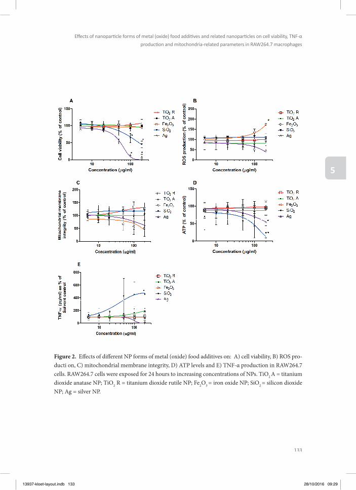
133
Eff ects of nanoparti cle forms of metal (oxide) food additi ves and related nanoparti cles on cell viability, TNF-α
producti on and mitochondria-related parameters in RAW264.7 macrophages
5
Figure 2. Eff ects of diff erent NP forms of metal (oxide) food additives on: A) cell viability, B) ROS pro-ducti on, C) mitochondrial membrane integrity, D) ATP levels and E) TNF-α production in RAW264.7 cells. RAW264.7 cells were exposed for 24 hours to increasing concentrations of NPs. TiO2 A = titanium dioxide anatase NP; TiO2 R = titanium dioxide rutile NP; Fe2O3 = iron oxide NP; SiO2 = silicon dioxide NP; Ag = silver NP.
13937-kloet-layout.indb 133 28/10/2016 09:29
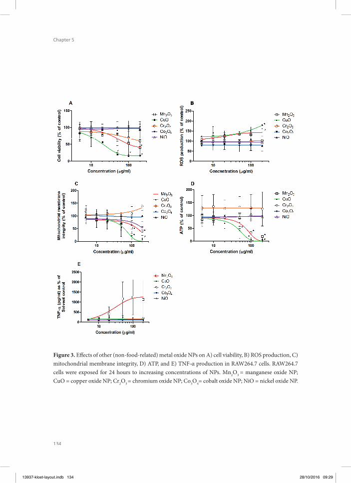
134
Chapter 5
Figure 3. Eff ects of other (non-food-related) metal oxide NPs on A) cell viability, B) ROS production, C) mitochondrial membrane integrity, D) ATP, and E) TNF-α production in RAW264.7 cells. RAW264.7 cells were exposed for 24 hours to increasing concentrations of NPs. Mn2O3 = manganese oxide NP; CuO = copper oxide NP; Cr2O3 = chromium oxide NP; Co3O4= cobalt oxide NP; NiO = nickel oxide NP.
13937-kloet-layout.indb 134 28/10/2016 09:29
135
Eff ects of nanoparti cle forms of metal (oxide) food additi ves and related nanoparti cles on cell viability, TNF-α
producti on and mitochondria-related parameters in RAW264.7 macrophages
5
DISCUSSION
Th e present study assessed the adverse eff ects of metal (oxide) NPs on macrophages in vitro, to study possible hazards of such NPs. To this end the eff ects of a series of food-related and other (non-food-related) metal (oxide) NPs on cell viability, ROS production, mitochondrial integrity, intracellular ATP levels and TNF-α production in RAW264.7 macrophages were assessed. Th e results indicate that of the food additive related NPs, SiO2 and Ag NPs were the most toxic, showing a signifi cant decrease of cell viability at concentrations of 100 µg/ml and higher, and a decrease of intracellular ATP at 200 µg/ml (Figure 2). In addition, SiO2 NPs in-duced increased TNF-α production at 200 µg/ml. From the series of other (non-food-related) metal oxide NPs, CuO and Mn2O3 NPs were the most toxic, aff ecting mitochondrial integrity, ROS production, ATP levels and cell viability at concentrations of 50 µg/ml and higher. Al-together, the present study shows no or limited eff ects of the NP forms of metal (oxide) food additives and related NPs on cell viability, ROS production, mitochondrial integrity, intracel-lular ATP levels and TNF-α production in RAW264.7 macrophages.
In the present study, eff ects for most NPs were only detected at high concentrations, if de-tected at all. At concentrations of 50 µg/ml and higher, metal oxide NP sedimentation was observed (supplementary fi gure 2), and it has been reported that toxicity of NPs may be af-fected by their agglomeration (Oberdorster 2010, Albanese et al. 2011, Zook et al. 2011, Ca-ballero-Díaz et al. 2013). However, since these NP agglomerations appeared to be co-localized with the RAW264.7 marcrophages and since they were not dispersed in the medium, this may indicate that these agglomerations are not the result of dispersity problems, but that the mac-rophages may have internalised the NPs. Th erefore, the toxicity of NPs to the macrophages as observed in the present study for SiO2, Ag, CuO and Mn2O3 NPs may be related to a high NP uptake, as has been reported before for gold NPs (Zhang et al. 2011).
It has been hypothesized that oxidative stress is an important mechanism underlying the cytotoxicity of NPs, including Mn2O3 and CuO NPs (Carlson et al. 2008, Park et al. 2009, Foucaud et al. 2010). NPs’ ability to generate oxidative stress can be due to their high surface reactivity (Krug et al. 2011), the presence of transition metal ions in the case of metal oxides and metal NPs (Kim et al. 2014), the fact that they can interact with the mitochondrial mem-brane and disrupt the electron transport chain (Elsaesser et al. 2012), and because they can attract macrophages and neutrophils that in response produce ROS (Krug et al. 2011). For Mn2O3 NPs and CuO NPs, we indeed found an increase in ROS production upon exposure of the cells (Figure 3B), which was accompanied by a decrease in cell viability at high expo-sure concentrations. Th e increase in ROS production upon Mn2O3 NPs and CuO NPs expo-sure was clearly associated with a decrease in mitochondrial membrane integrity (Figure 3C) and a decrease in ATP levels (Figure 3D) suggesting that the ROS production may originate
13937-kloet-layout.indb 135 28/10/2016 09:29
136
Chapter 5
from impairment of the membrane integrity resulting in uncoupling of the electron transport chain. For the other two most cytotoxic NPs tested in the present study, the food-related SiO2 NPs and Ag NPs, no increased ROS production was found (Figure 2B). Also, no eff ects on mitochondrial membrane integrity were observed for these NPs (Figure 2C) and only slight eff ects on ATP levels (Figure 2D), which may be a secondary eff ect because of dying cells. Th is indicates that the food-related Ag NPs and SiO2 NPs likely have a diff erent mode of action underlying their cytotoxicity than the non-food related Mn2O3 NPs and CuO NPs.
Of the NPs used in the present study, Ag NPs and CuO NPs were the most toxic ones. CuO and Ag NPs have been reported before as being signifi cantly cytotoxic to A549 cells and Calu-3 epithelial cells and Raw264.7 macrophages (Karlsson et al. 2008, Haase et al. 2011, Rotoli et al. 2012). It is known that ions are released from Ag and CuO NPs at diff erent rates, making the understanding of the delivered dose challenging (Ponti et al. 2009, Misra et al. 2012). Discussion remains about the role of this ion dissolution in the mode of action underlying the toxicity (Karlsson et al. 2008, Adamczyk et al. 2016). Although toxicity of CuO NPs may be (partly) caused by dissolution of Cu ions, a study of Karlsson et al. (2008) showed that the toxicity of CuO NPs cannot be solely explained by Cu ions released into the cell medium. For example, cell viability of A549 cells decreased more when exposed to 40 µg/mL of Cu from CuO NPs (50 µg/mL CuO) than when exposed to 40 µg/mL of Cu from CuCl2 (85 µg/mL CuCl2) (Karlsson et al. 2008). Regarding Ag NPs, no toxic eff ect was observed when HeLa cells were exposed to Ag ions (as silver nitrate) up to 8 µg/mL silver nitrate whereas Ag NPs caused toxicity starting from 2.0 µg/mL (Kaba and Egorova 2015). For U937 cells on the other hand, Ag ions showed similar toxicity as Ag NPs in a concentration range of 0.5-8 µg/mL (Kaba et al. 2015). So although it is known that ions can be released from these NPs, it remains still a topic of investigation how these ions contribute to the toxicity observed.
Our results showed that SiO2 NPs were the most toxic ones among the tested food-related NPs. SiO2 NPs are known to exert a decrease in cell viability in primary human monocyte-de-rived macrophages and MH-S and RAW264.7 cells (Farcal et al. 2015, Di Cristo et al. 2016). Exposure to food-related NPs is expected to be mainly via the gastrointestinal tract. Upon oral exposure, NP characteristics may change because of gastrointestinal digestion (Rogers et al. 2012, Walczak et al. 2015). When food products containing SiO2 were tested in an in vitro digestion model, simulating the conditions of the human gastrointestinal tract, it was found that SiO2 NPs (5-200 nm) were still present in the intestinal content (Peters et al. 2012). Th is indicates that it is most likely that upon consumption of foods containing particulate SiO2, the gut epithelium is exposed to SiO2 NPs (5-200 nm) (Peters et al. 2012). To enter the systemic circulation, NPs need to be absorbed in the gastrointestinal tract. Intestinal absorption large-ly depends on the size of NPs, surface charge and hydrophobicity (Bouwmeester et al. 2011, Bhattacharjee et al. 2013). In an in vivo study in rats it was shown that 6.6-9.7% of SiO2 NPs
13937-kloet-layout.indb 136 28/10/2016 09:29

137
Eff ects of nanoparti cle forms of metal (oxide) food additi ves and related nanoparti cles on cell viability, TNF-α
producti on and mitochondria-related parameters in RAW264.7 macrophages
5
of 20 and 100 nm were absorbed into the systemic circulation when orally administered at a single dose of 500 and 1000 mg/kg (Lee et al. 2014), indicating that SiO2 NPs can get trans-located across the intestinal barrier. An in vitro study showed that 50 nm SiO2 NPs were able to translocate across an intestinal barrier based on results obtained in a Caco-2 cell transwell model system (Sakai-Kato et al. 2014).
With an estimated daily intake of SiO2 in the nano-size range of 1.8 mg/kg bw/day (van der Zande et al. 2014, van Kesteren et al. 2014), the daily intake of a 70 kg person would be 126 mg. In the adult human the gastrointestinal tract represents a considerable surface area of about 200 m2 which has the potential to interact with ingested NPs (Bergin et al. 2013). In addition the gastrointestinal tract has localised representations of the adaptive immune system in the form of gut-associated lymphoid tissue (GALT) aggregates (Peyer’s Patches) (Bergin and Witzmann 2013). Th ese Peyer’s patches account for 1% of the total surface area of the gastrointestinal tract (des Rieux et al. 2006). About 9 litres of fl uid pass through the GI system each day (Sherwood L. 2014). Th us, upon oral intake of 126 mg SiO2 per day this would lead to a concentration of 14 µg/ml, which is lower than the lowest observed adverse eff ect concentration of 100 µg/mL in the present study.
If 100% of the estimated daily intake of SiO2 NPs (126 mg) would be transported from the intestines into the blood, assuming 5 liters of blood and a 100% clearance per day, a maximal concentration of 25 µg/ml would be reached if NPs would not distribute to tissues. Also this concentration is lower than the lowest observed adverse eff ect concentration of 100 µg/mL defi ned in the present study. Considering that the oral bioavailability is lower than 10% and that NPs also distribute to tissues (Lee et al. 2014), concentrations higher than 2.5 µg/mL are not likely to be reached in a real-life exposure scenario. Th is concentration is far below the concentrations of SiO2 causing in vitro toxicity in the present study, suggesting that in vivo toxic eff ects of food additive related SiO2 NPs on macrophages are expected to be limited. Th is is all in line with the results from an oral repeated-dose toxicity study in which male Wistar rats were exposed to SiO2 NPs via the diet for 28 or 84 days, showing no toxicity at the highest dose level tested (2500 mg/kg bw/day) (van der Zande et al. 2014).
It has to be kept in mind that NPs may not only aff ect the gastrointestinal cells directly but may also infl uence the microbiome, although the evaluation of eff ects of NPs on the micro-biome is in its early infancy (Pietroiusti et al. 2015). For example, the cytotoxicity of Ag NPs on Escherichia coli as representative for intestinal bacteria was compared with the cytotoxicity of Ag NPs on gastrointestinal cells. Th is revealed that Ag NPs aff ected bacteria at lower con-centrations than those that aff ected gastrointestinal cells, indicating that Ag NPs may cause adverse eff ects by selectively aff ecting the gut microbiota (Frohlich et al. 2016). In a study us-ing an in vitro colon model (Taylor et al. 2015), TiO2 , ZnO and CeO2 NPs induced signifi cant
13937-kloet-layout.indb 137 28/10/2016 09:29

138
Chapter 5
phenotypic changes of the gut microbial community, inlcuding the production of short chain fatty acids which are the key metabolites in the control of energy homeostatis and which have been implicated in the development of obesity (Schwiertz et al. 2010). For other food related metal (oxide) NPs such as SiO2, Fe2O3 and Ag NPs, such eff ects on the gut microbiota and related short chain fatty acid production remain to be investigated.
CONCLUSION
In conclusion, the present study shows no or limited eff ects of the NP forms of metal (ox-ide) food additives on cell viability, ROS production, mitochondrial integrity, ATP levels and TNF-α production in RAW264.7 macrophages. Of the food additive related NPs, SiO2 and Ag NPs were the most toxic, showing a decrease of cell viability at concentrations of 100 µg/ml and higher. Eff ects were only observed at high concentrations that may not be physiologically relevant, indicating that related adverse eff ects upon exposure to the respective NPs may be limited.
ACKNOWLEDGEMENTS
Th is work is supported by NanoNextNL, a micro and nanotechnology consortium of the Government of the Netherlands and 130 partners.
13937-kloet-layout.indb 138 28/10/2016 09:29
139
Eff ects of nanoparti cle forms of metal (oxide) food additi ves and related nanoparti cles on cell viability, TNF-α
producti on and mitochondria-related parameters in RAW264.7 macrophages
5
REFERENCES
(2011). “Regulation EU no 1169/2011 on the provision of food information to consumers. Offi cial Journal of the European Union. .” Offi cial Journal of the European Union from http://eur-lex.europa.eu/legal-content/EN/TXT/PDF/?uri=CELEX:32011R1169&from=EN
Adamczyk, Z., M. Ocwieja, H. Mrowiec, S. Walas and D. Lupa (2016). “Oxidative dissolution of silver nanoparticles: A new theoretical approach.” J Colloid Interface Sci 469: 355-364.
Albanese, A. and W. C. Chan (2011). “Eff ect of gold nanoparticle aggregation on cell uptake and toxicity.” ACS Nano 5(7): 5478-5489.
Athinarayanan, J., V. S. Periasamy, M. A. Alsaif, A. A. Al-Warthan and A. A. Alshatwi (2014). “Presence of nanosilica (E551) in commercial food products: TNF-mediated oxidative stress and altered cell cycle progression in human lung fi broblast cells.” Cell biology and toxicology 30(2): 89-100.
Bergin, I. L. and F. A. Witzmann (2013). “Nanoparticle toxicity by the gastrointestinal route: evidence and knowledge gaps.” International journal of biomedical nanoscience and nanotechnology 3(1-2).
Bhattacharjee, S., D. Ershov, J. Gucht, G. M. Alink, I. M. Rietjens, H. Zuilhof and A. T. Marcelis (2013). “Surface charge-specifi c cytotoxicity and cellular uptake of tri-block copolymer nanoparticles.” Nanotoxicology 7(1): 71-84.
Bhattacharjee, S., I. M. Rietjens, M. P. Singh, T. M. Atkins, T. K. Purkait, Z. Xu, S. Regli, A. Shukaliak, R. J. Clark, B. S. Mitchell, G. M. Alink, A. T. Marcelis, M. J. Fink, J. G. Veinot, S. M. Kauzlarich and H. Zuilhof (2013). “Cytotoxicity of surface-functionalized silicon and germanium nanoparticles: the dominant role of surface charges.” Nanoscale 5(11): 4870-4883.
Bonora, M. and P. Pinton (2014). “Th e mitochondrial permeability transition pore and cancer: molecular mecha-nisms involved in cell death.” Frontiers in oncology 4: 302.
Bouwmeester, H., S. Dekkers, M. Y. Noordam, W. I. Hagens, A. S. Bulder, C. de Heer, S. E. ten Voorde, S. W. Wi-jnhoven, H. J. Marvin and A. J. Sips (2009). “Review of health safety aspects of nanotechnologies in food production.” Regul Toxicol Pharmacol 53(1): 52-62.
Bouwmeester, H., J. Poortman, R. J. Peters, E. Wijma, E. Kramer, S. Makama, K. Puspitaninganindita, H. J. Marvin, A. A. Peijnenburg and P. J. Hendriksen (2011). “Characterization of translocation of silver nanoparticles and eff ects on whole-genome gene expression using an in vitro intestinal epithelium coculture model.” ACS Nano 5(5): 4091-4103.
Caballero-Díaz, E., C. Pfeiff er, L. Kastl, P. Rivera-Gil, B. Simonet, M. Valcárcel, J. Jiménez-Lamana, F. Laborda and W. J. Parak (2013). “Th e Toxicity of Silver Nanoparticles Depends on Th eir Uptake by Cells and Th us on Th eir Surface Chemistry.” Particle & Particle Systems Characterization 30(12): 1079-1085.
Carlson, C., S. M. Hussain, A. M. Schrand, L. K. Braydich-Stolle, K. L. Hess, R. L. Jones and J. J. Schlager (2008). “Unique cellular interaction of silver nanoparticles: size-dependent generation of reactive oxygen species.” J Phys Chem B 112(43): 13608-13619.
Dang, T., T. Le, T. Hoang and T. Mai (2015). “Synthesis of nanostructured manganese oxides based materials and application for supercapacitor.” Advances in Natural Sciences; Nanoscience and Nanotechnology 6.
des Rieux, A., V. Fievez, M. Garinot, Y. J. Schneider and V. Preat (2006). “Nanoparticles as potential oral delivery systems of proteins and vaccines: a mechanistic approach.” J Control Release 116(1): 1-27.
Di Cristo, L., D. Movia, M. G. Bianchi, M. Allegri, B. M. Mohamed, A. P. Bell, C. Moore, S. Pinelli, K. Rasmussen, J. Riego-Sintes, A. Prina-Mello, O. Bussolati and E. Bergamaschi (2016). “Proinfl ammatory Eff ects of Py-rogenic and Precipitated Amorphous Silica Nanoparticles in Innate Immunity Cells.” Toxicol Sci 150(1): 40-53.
Elsabahy, M. and K. L. Wooley (2013). “Cytokines as biomarkers of nanoparticle immunotoxicity.” Chemical Society reviews 42(12): 5552-5576.
Elsaesser, A. and C. V. Howard (2012). “Toxicology of nanoparticles.” Adv Drug Deliv Rev 64(2): 129-137.Farcal, L., F. Torres Andon, L. Di Cristo, B. M. Rotoli, O. Bussolati, E. Bergamaschi, A. Mech, N. B. Hartmann, K.
Rasmussen, J. Riego-Sintes, J. Ponti, A. Kinsner-Ovaskainen, F. Rossi, A. Oomen, P. Bos, R. Chen, R. Bai, C. Chen, L. Rocks, N. Fulton, B. Ross, G. Hutchison, L. Tran, S. Mues, R. Ossig, J. Schnekenburger, L. Cam-pagnolo, L. Vecchione, A. Pietroiusti and B. Fadeel (2015). “Comprehensive In Vitro Toxicity Testing of a Panel of Representative Oxide Nanomaterials: First Steps towards an Intelligent Testing Strategy.” PLoS One 10(5): e0127174.
13937-kloet-layout.indb 139 28/10/2016 09:29
140
Chapter 5
Faust, J. J., K. Doudrick, Y. Yang, P. Westerhoff and D. G. Capco (2014). “Food grade titanium dioxide disrupts in-testinal brush border microvilli in vitro independent of sedimentation.” Cell biology and toxicology 30(3): 169-188.
Foucaud, L., S. Goulaouic, A. Bennasroune, P. Laval-Gilly, D. Brown, V. Stone and J. Falla (2010). “Oxidative stress induction by nanoparticles in THP-1 cells with 4-HNE production: stress biomarker or oxidative stress signalling molecule?” Toxicol In Vitro 24(6): 1512-1520.
Frohlich, E. E. and E. Frohlich (2016). “Cytotoxicity of Nanoparticles Contained in Food on Intestinal Cells and the Gut Microbiota.” International journal of molecular sciences 17(4).
Haase, A., J. Tentschert, H. Jungnickel, P. Graf, A. Mantion, F. Draude, J. Plendl, M. E. Goetz, S. Galla, A. Masic, A. F. Th uenemann, A. Taubert, H. F. Arlinghaus and L. A (2011). “Toxicity of silver nanoparticles in human macrophages: uptake, intracellular distribution and cellular responses.” Journal of Physics: Conference se-ries 304.
Kaba, S. I. and E. M. Egorova (2015). “In vitro studies of the toxic eff ects of silver nanoparticles on HeLa and U937 cells.” Nanotechnology, science and applications 8: 19-29.
Karlsson, H. L., P. Cronholm, J. Gustafsson and L. Moller (2008). “Copper oxide nanoparticles are highly toxic: a comparison between metal oxide nanoparticles and carbon nanotubes.” Chemical research in toxicology 21(9): 1726-1732.
Kim, C. S., H. D. Nguyen, R. M. Ignacio, J. H. Kim, H. C. Cho, E. H. Maeng, Y. R. Kim, M. K. Kim, B. K. Park and S. K. Kim (2014). “Immunotoxicity of zinc oxide nanoparticles with diff erent size and electrostatic charge.” Int J Nanomedicine 9 Suppl 2: 195-205.
Krug, H. F. and P. Wick (2011). “Nanotoxicology: An Interdisciplinary Challenge.” Angew Chem Int Ed Engl.Lee, J. A., M. K. Kim, H. J. Paek, Y. R. Kim, M. K. Kim, J. K. Lee, J. Jeong and S. J. Choi (2014). “Tissue distribution and
excretion kinetics of orally administered silica nanoparticles in rats.” Int J Nanomedicine 9 Suppl 2: 251-260.Ly, J. D., D. R. Grubb and A. Lawen (2003). “Th e mitochondrial membrane potential (deltapsi(m)) in apoptosis; an
update.” Apoptosis 8(2): 115-128.Ma, J., T. Chen, J. Mandelin, A. Ceponis, N. E. Miller, M. Hukkanen, G. F. Ma and Y. T. Konttinen (2003). “Regulation
of macrophage activation.” Cellular and molecular life sciences : CMLS 60(11): 2334-2346.Misra, S. K., A. Dybowska, D. Berhanu, S. N. Luoma and E. Valsami-Jones (2012). “Th e complexity of nanoparticle
dissolution and its importance in nanotoxicological studies.” Th e Science of the total environment 438: 225-232.
Oberdorster, G. (2010). “Safety assessment for nanotechnology and nanomedicine: concepts of nanotoxicology.” J Intern Med 267(1): 89-105.
Park, E. J. and K. Park (2009). “Oxidative stress and pro-infl ammatory responses induced by silica nanoparticles in vivo and in vitro.” Toxicol Lett 184(1): 18-25.
Park, E. J., J. Yi, K. H. Chung, D. Y. Ryu, J. Choi and K. Park (2008). “Oxidative stress and apoptosis induced by tita-nium dioxide nanoparticles in cultured BEAS-2B cells.” Toxicol Lett 180(3): 222-229.
Periasamy, V. S., J. Athinarayanan, A. M. Al-Hadi, F. A. Juhaimi, M. H. Mahmoud and A. A. Alshatwi (2015). “Identi-fi cation of titanium dioxide nanoparticles in food products: induce intracellular oxidative stress mediated by TNF and CYP1A genes in human lung fi broblast cells.” Environ Toxicol Pharmacol 39(1): 176-186.
Peters, R., E. Kramer, A. G. Oomen, Z. E. Rivera, G. Oegema, P. C. Tromp, R. Fokkink, A. Rietveld, H. J. Marvin, S. Weigel, A. A. Peijnenburg and H. Bouwmeester (2012). “Presence of nano-sized silica during in vitro diges-tion of foods containing silica as a food additive.” ACS Nano 6(3): 2441-2451.
Pietroiusti, A., A. Magrini and L. Campagnolo (2015). “New frontiers in nanotoxicology: Gut microbiota/microbi-ome-mediated eff ects of engineered nanomaterials.” Toxicology and applied pharmacology.
Ponti, J., E. Sabbioni, B. Munaro, F. Broggi, P. Marmorato, F. Franchini, R. Colognato and F. Rossi (2009). “Genotoxic-ity and morphological transformation induced by cobalt nanoparticles and cobalt chloride: an in vitro study in Balb/3T3 mouse fi broblasts.” Mutagenesis 24(5): 439-445.
Rogers, K. R., K. Bradham, T. Tolaymat, D. J. Th omas, T. Hartmann, L. Ma and A. Williams (2012). “Alterations in physical state of silver nanoparticles exposed to synthetic human stomach fl uid.” Th e Science of the total environment 420: 334-339.
13937-kloet-layout.indb 140 28/10/2016 09:29
141
Eff ects of nanoparti cle forms of metal (oxide) food additi ves and related nanoparti cles on cell viability, TNF-α
producti on and mitochondria-related parameters in RAW264.7 macrophages
5
Rotoli, B. M., O. Bussolati, A. L. Costa, M. Blosi, L. Di Cristo, P. P. Zanello, G. Bianchi, R. Visigalli and E. Bergamaschi (2012). “Comparative eff ects of metal oxide nanoparticles on human airway epithelial cells and macro-phages.” Journal of nanoparticle research : an interdisciplinary forum for nanoscale science and technology 14(1069).
Sakai-Kato, K., M. Hidaka, K. Un, T. Kawanishi and H. Okuda (2014). “Physicochemical properties and in vitro intestinal permeability properties and intestinal cell toxicity of silica particles, performed in simulated gas-trointestinal fl uids.” Biochim Biophys Acta 1840(3): 1171-1180.
Schwiertz, A., D. Taras, K. Schafer, S. Beijer, N. A. Bos, C. Donus and P. D. Hardt (2010). “Microbiota and SCFA in lean and overweight healthy subjects.” Obesity 18(1): 190-195.
Sharma, V., D. Anderson and A. Dhawan (2011). “Zinc oxide nanoparticles induce oxidative stress and genotoxicity in human liver cells (HepG2).” J Biomed Nanotechnol 7(1): 98-99.
Sherwood L. (2014). Human Physiology from cells to systems. Canada, Cengage Learning.Shvedova, A. A., A. Pietroiusti, B. Fadeel and V. E. Kagan (2012). “Mechanisms of carbon nanotube-induced toxicity:
focus on oxidative stress.” Toxicol Appl Pharmacol 261(2): 121-133.Siddiqui, M. A., M. Ahamed, J. Ahmad, M. A. Majeed Khan, J. Musarrat, A. A. Al-Khedhairy and S. A. Alrokayan
(2012). “Nickel oxide nanoparticles induce cytotoxicity, oxidative stress and apoptosis in cultured human cells that is abrogated by the dietary antioxidant curcumin.” Food Chem Toxicol 50(3-4): 641-647.
Tavares, K., A. Caloto-Oliveira, D. Vicentini, S. Melegari and W. Matias (2014). “Acute toxicity of copper and chromi-um oxide nanoparticles to Daphnia similis.” Ecotoxicolo. Environ. Contam. 9(1): 43-50.
Taylor, A. A., I. M. Marcus, R. L. Guysi and S. L. Walker (2015). “Metal oxide nanoparticles induce minimal pheno-typic changes in a model colon gut microbiota.” Environ Eng Sci 32: 602-612.
van der Zande, M., R. J. Vandebriel, M. J. Groot, E. Kramer, Z. E. Herrera Rivera, K. Rasmussen, J. S. Ossenkoppele, P. Tromp, E. R. Gremmer, R. J. Peters, P. J. Hendriksen, H. J. Marvin, R. L. Hoogenboom, A. A. Peijnenburg and H. Bouwmeester (2014). “Sub-chronic toxicity study in rats orally exposed to nanostructured silica.” Part Fibre Toxicol 11: 8.
van Kesteren, P. C., F. Cubadda, H. Bouwmeester, J. C. van Eijkeren, S. Dekkers, W. H. de Jong and A. G. Oomen (2014). “Novel insights into the risk assessment of the nanomaterial synthetic amorphous silica, additive E551, in food.” Nanotoxicology: 1-10.
Walczak, A. P., E. Kramer, P. J. Hendriksen, R. Helsdingen, M. van der Zande, I. M. Rietjens and H. Bouwmeester (2015). “In vitro gastrointestinal digestion increases the translocation of polystyrene nanoparticles in an in vitro intestinal co-culture model.” Nanotoxicology 9(7): 886-894.
Weir, A., P. Westerhoff , L. Fabricius, K. Hristovski and N. von Goetz (2012). “Titanium dioxide nanoparticles in food and personal care products.” Environ Sci Technol 46(4): 2242-2250.
Zhang, Q., V. M. Hitchins, A. M. Schrand, S. M. Hussain and P. L. Goering (2011). “Uptake of gold nanoparticles in murine macrophage cells without cytotoxicity or production of pro-infl ammatory mediators.” Nanotoxicol-ogy 5(3): 284-295.
Zook, J. M., R. I. Maccuspie, L. E. Locascio, M. D. Halter and J. T. Elliott (2011). “Stable nanoparticle aggregates/agglomerates of diff erent sizes and the eff ect of their size on hemolytic cytotoxicity.” Nanotoxicology 5(4): 517-530.
13937-kloet-layout.indb 141 28/10/2016 09:29
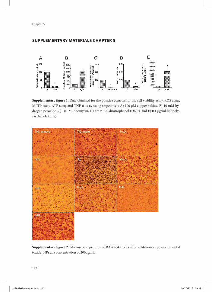
142
Chapter 5
SUPPLEMENTARY MATERIALS CHAPTER 5
Supplementary fi gure 2. Microscopic pictures of RAW264.7 cells aft er a 24-hour exposure to metal (oxide) NPs at a concentration of 200µg/ml.
Supplementary fi gure 1. Data obtained for the positive controls for the cell viability assay, ROS assay, MPTP assay, ATP assay and TNF-α assay using respectively A) 100 µM copper sulfate, B) 10 mM hy-drogen peroxide, C) 10 µM ionomycin, D) 4mM 2,4-dinitrophenol (DNP), and E) 0.1 μg/ml lipopoly-saccharide (LPS).
13937-kloet-layout.indb 142 28/10/2016 09:29

13937-kloet-layout.indb 143 28/10/2016 09:29

12
34
56
78
910
1112
A B C D E F G G H
13937-kloet-layout.indb 144 28/10/2016 09:29

12
34
56
78
910
1112
A B C D E F G G H
Chapter 6
Cellular interactions of silver nanoparticles with systematic variation in size and surface coating with
macrophage RAW 264.7 cells
Sunday MakamaSamantha K. Kloet
Jordi PiellaJohannes H.J. van den Berg
Norbert C. A. de RuijterVictor F. Puntes
Ivonne M.C.M. RietjensNico W. van den Brink
Submitted for publication to Nanotoxicology
13937-kloet-layout.indb 145 28/10/2016 09:29
146
Chapter 6
ABSTRACT
Increasing use of engineered nanoparticles has led to an increased likelihood of their envi-ronmental release and raises concern for ecosystem and health hazard. Th e volume of sil-ver nanoparticle (Ag NP)-based products entering the market is relatively high, making the risk assessment of Ag NPs a priority. In literature, varying and sometimes confl icting reports exists about the physicochemical properties of NPs that are important in infl uencing their uptake and eff ects. To address this discrepancy, small and medium sized (20 and 50 nm) Ag NPs with diff erent surface coating/charges (chitosan/positive: Ag NP_Chit; bovine serum al-bumin/negative: Ag NP_BSA, and; polyvinylpyrrolidone/neutral: Ag NP_PVP) were specifi -cally synthesized and characterized. Macrophage cells (RAW 264.7) were exposed to these Ag NPs at 0 – 200 µg/mL, and uptake dynamics, cell viability, inductions of tumor necrosis factor (TNF)-α and reactive oxygen species (ROS) were assessed. Generally, adverse eff ects from exposure to the tested Ag NPs resulting in reduced overall viability were similar irrespective of Ag NP types or sizes. On adenosine triphosphate production and specifi c mechanisms of toxicity (TNF-α and ROS production) however, the Ag NPs diff ered signifi cantly. Tests targeting specifi c mechanisms of action indicated the highest induction of TNF-α in cells exposed to both sizes of the more potent negatively charged Ag NP_BSA (80x higher than control). Signifi cant ROS induction was only observed with the 20 nm positively charged Ag NP_Chit. Additionally, live microscopic imaging of exposed cells allowed the monitoring of the uptake dynamics and subcellular cytoplasmic accumulation of Ag NPs. Th e present study provides further evidence of the infl uence of NP properties in driving their toxicity in an in vitro model.
13937-kloet-layout.indb 146 28/10/2016 09:29
147
Cellular interacti ons of silver nanoparti cles with systemati c variati on in size and surface coati ng with macrophage
RAW 264.7 cells
6
INTRODUCTION
Nanotechnology can be considered as one of the fastest growing technologies which has revolutionized the industrial sector (Peterson, 2000, Gwinn and Vallyathan, 2006, Kennell, 2009), bringing about possibilities only imagined a few decades ago. Th is has led to increased production and applications of engineered nanoparticles (NPs) for consumer products (Wi-jnhoven et al., 2009, Wijnhoven et al., 2010). Presently, NPs continue to fi nd applications in the design and manufacture of household products, electronics and medical devices, food and transport systems, to mention a few (Ahamed et al., 2010, Haider and Kang, 2015). Con-sequently, the increase in production and use of NPs will likely result in an increased release into the environment as industrial waste (or by-product), sewage or waste water, (Geranio et al., 2009, Wijnhoven et al., 2009, Benn et al., 2010, Reidy et al., 2013). Such environmental release could potentially have harmful impacts.
Currently, silver nanoparticles (Ag NPs) represent the nanomaterial most frequently used in products on the market (Wijnhoven et al., 2010, Vance et al., 2015) owing to their well demon-strated antimicrobial activity (Silver and Phung, 1996, Sondi and Salopek-Sondi, 2004, Lara et al., 2010, Hwang et al., 2012, Prabhu and Poulose, 2012, Franci et al., 2015). At the nanoscale (1 – 100 nm), the small size and increased surface area of Ag NPs result in novel properties. Th ese can be enhanced by stabilization and/or functionalization using biocompatible conju-gates. Essentially, the type of surface coating and process used in stabilizing engineered NPs during synthesis determines their surface charges, solubility and/or hydrophobicity (Evanoff and Chumanov, 2005, Abou El-Nour et al., 2010, Bastus et al., 2014, Shenashen et al., 2014). Th is in turn infl uences the behavior and environmental fate of NPs, as well as their eff ects on organisms (Kreuter, 2004, Roohani-Esfahani et al., 2010, Shoults-Wilson et al., 2011, Cha-nana and Liz-Marzán Luis, 2012).
Over the last two decades, researchers have attempted to elucidate the fate and eff ect of vari-ous NPs using both in vivo and in vitro models (Foldbjerg et al., 2012, Yu et al., 2013, van der Ploeg et al., 2014a, van der Ploeg et al., 2014b). Although the importance of the physicochem-ical properties of NPs in infl uencing their fate and toxicity has increasingly been investigated, reports in available literature have not been consistent. Important physicochemical properties in this regard include size and NPs dispersion (both mono- and polydispersity), shape, zeta potential, and agglomeration and dissolution rates (Reidy et al., 2013). While some studies have reported eff ects of size (Powers et al., 2011b) or charge (Suresh et al., 2012), others fail to detect these (Yang et al., 2012). Additional perspectives on the infl uence of physicochemical properties of Ag NPs on their interactions in both in vivo and in vitro models, has been re-viewed earlier (Johnston et al., 2010, Reidy et al., 2013). From the reviews cited and examples mentioned above, one can identify the challenge associated with the regulation of Ag NPs and
13937-kloet-layout.indb 147 28/10/2016 09:29
148
Chapter 6
other NPs in general. Th is is especially so, given the limited data base and the limitations of generating in vivo data for all the diff erent forms and types of NPs.
A better understanding of the properties that infl uence the fate and eff ects of Ag NPs in or-ganisms therefore, will facilitate appropriate risk assessment which in turn will assist the reg-ulation of nanomaterials. Using in vitro models in risk assessments of chemicals facilitates the setting of priorities for in vivo testing, thereby reducing the number of animals required (Braakhuis et al., 2015). By fi rst investigating with in vitro models, insights in the toxicity and possible mode of action of NPs are obtained. Such information could be used in a weight of evidence approach (Connolly et al., 2015), furthering our understanding of observations made under in vivo situations, as well as defi ning priorities for in vivo testing and facilitating read-across.
In this study, we systematically investigated the physicochemical properties of Ag NPs infl u-encing their interactions at cellular level, using an in vitro mammalian cell line model: the mouse monocyte macrophage (RAW 264.7). Macrophages are involved in the defense system of an organism against infections, and the role of Ag NP properties in initiating infl ammatory (TNF-α) and oxidative stress responses in these cells were investigated. Since it appears that how a NP is produced infl uences both its fate and eff ect following exposures, specifi cally prepared Ag NPs were used in the current study. Th ese were synthesized to diff er in size and surface chemistry (charge), two important properties infl uencing NP uptake and eff ect (Reidy et al., 2013). Using imaging techniques, we also showed the uptake dynamics of Ag NPs at a cellular level. Th e outcome of this study provides further insight into how Ag NP properties determine their fate and eff ects at cellular level, thus indirectly shedding light on what the outcome may likely be in intact organisms (Landsiedel et al., 2012, Cornelis et al., 2014).
MATERIALS AND METHODS
Experimental designTh e mouse monocyte macrophage cell line (RAW 264.7) was used as the in vitro model, and cells were treated with Ag NPs. Experiments were conducted in three independent exposures for each type of Ag NP (n=3) and the data generated were processed with Microsoft Excel. Where appropriate, the data were subjected to one-way analysis of variance (ANOVA) with the aid of GraphPad Prism 5.04 for Windows (GraphPad Soft ware, San Diego California US, www.graphpad.com), and logistic regression was done using GenStat 17th ed. (17.1.0.14713; VSN International, Hemel Hempstead, UK, GenStat.co.uk). A p value of <0.05 was consid-ered signifi cant.
13937-kloet-layout.indb 148 28/10/2016 09:29
149
Cellular interacti ons of silver nanoparti cles with systemati c variati on in size and surface coati ng with macrophage
RAW 264.7 cells
6
Reagents and InstrumentsAll chemicals, enzymes and reagents were analytical grade and purchased from Sigma-Al-drich® (Zwijndrecht, NL), unless where stated otherwise. Apart from one commercially ob-tained Ag NP (Ag NP_NC), all other Ag NPs were synthesized at the Catalan Institute of Nanoscience and Nanotechnology (ICN2, Barcelona, ES) by methods described below. End-point measurements were carried out using SpectraMax M2 Microplate Reader (Molecular Devices LLC, Sunnyvale, CA US) for fl uorescence and absorbance, or Luminoskan Ascent (Th ermo Scientifi c, 5300172, MA, US) for luminescence.
Synthesis of Ag NPsColloidal, dispersed Ag NPs of two diff erent sizes (20 and 50 nm) were prepared separate-ly, following a kinetically controlled seeded-growth method previously reported (Bastus et al., 2014) with slight modifi cations. Th e approach is based on the reduction of silver nitrate (AgNO3) in the presence of two competing reducing agents, tannic acid (TA) and trisodium citrate hexahydrate (SC) at 100°C. Six diff erent Ag NPs were synthesized by surface-coating both sizes with bovine serum albumin (BSA), chitosan (Chit) or polyvinylpyrrolidone (PVP) to generate negative Ag NP_BSA, positive Ag NP_Chit and neutral Ag NP_PVP, respectively. Details of these procedures are elaborated upon in the electronic supplementary material.
An aqueous suspension of a negatively charged 50 nm PVP-coated Ag NP (Ag NP_NC) was obtained from NanoComposix® (San Diego, US) and included in this study for comparison with the synthesized Ag NPs. Based on the manufacturer’s information, the Ag NP_NC is reported to have mean core- and hydrodynamic diameters of 54.8 ± 10 nm and 72 ± 14.4 nm respectively. Th e stock suspension of the pristine Ag NP has a mass concentration of 5.0 mg Ag/mL, and a particle number concentration of 5.2 x 10¹² particles/mL. Th e surface area was 9.8 m2/g while surface charge (zeta-potential) was -37.8 mV. Th e DLS and zeta potential in both milliQ and DMEM were confi rmed in our laboratory, and details previously provided (Makama et al., 2015).
Ag NP CharacterizationPrior to exposure experiments, pristine Ag NPs were dispersed in Dulbecco’s modifi ed Eagle’s medium, DMEM (Invitrogen Breda, NL), as well as milliQ water and characterized by trans-mission electron microscopy (TEM), UV-Vis spectroscopy, dynamic light scattering (DLS), and zeta-potential measurements (ζ-potential). A combination of diff erent techniques was used to characterize the Ag NPs and to monitor their proper coating and stability in the diff erent media. Characterization details are given in the electronic supplementary material.
13937-kloet-layout.indb 149 28/10/2016 09:29
150
Chapter 6
NPs dispersionsAg NPs stock dispersions were prepared by fi rst suspending the respective powders in milliQ water except for Ag NP_Chit, where 50 mM acetic acid (Merck, Darmstadt, DE) was used instead. Ag NP_Chit suspensions were initially prepared in 50 mM acetic acid in order to prevent agglomeration while ensuring monodispersity (Zhao et al., 2011, Nadesh et al., 2013). Where used, the fi nal acetic acid concentration in the medium at which the cells were exposed to was 4 mM (0.02%), and determined to be non-cytotoxic (data not shown). For all Ag NPs, the stock dispersions were prepared at 2.5 mg Ag/mL and sonicated in a Sonorex RK100 (Berlin, DE) water-bath over ice for 15 min. Further dilutions to the desired exposure con-centration ranges (0.1, 1, 5, 10, 20, 50, 100, and 200 µg Ag/mL) were made in DMEM fortifi ed with heat inactivated fetal calf serum (FCS) at 10%.
Ag NPs exposure and Uptake dynamicsA mouse monocyte macrophage cell line, RAW 264.7 (ATCC® TIB-71™; Manassas, VA US) was used in this study at passages 16-22. Cells were cultured in DMEM, enriched with FCS at 10% (v/v). Sub-culturing and passaging of cells was done every 3 – 4 days when reaching 80% confl uency by trypsinization aft er rinsing with phosphate buff ered saline (PBS). Before exposure to Ag NPs, cells were grown for 24 h in 96-well plates at a seeding density of 105 cells/well for all assays, and incubated at 37°C in humidifi ed air (plus 5% CO2) to attain above 80% adherent confl uency. Aft er this initial 24 h of stabilization and growth, Ag NPs exposures for all assays with the exception of the Dichlorofl uorescein (DCF) assay for ROS production, were conducted over a 24 h duration. For the ROS production assay, exposure to Ag NPs was for 6 h, based on an adaptation of a similar method described earlier (Carlson et al., 2008). Unless stated otherwise, general incubation conditions were as described above.
Macrophages were imaged by means of confocal microscopy to follow cellular uptake, lo-calization and accumulation behavior of the diff erent Ag NPs over time. Imaging conditions were chosen such as to achieve low photon stress, without any visible loss of vitality aft er 24 h of exposure and imaging. Th is was performed for the 50 nm size class only, assuming that this large size is generally taken up less rapidly compared to 20 nm (Shang et al., 2014a, Shang et al., 2014b). Details on methodology and other results are provided in the electronic supplementary material.
Eff ect assessmentCytotoxicity assays were performed to evaluate the viability of the macrophages aft er expo-sure to the diff erent Ag NPs. 3-(4,5-dimethylthiazol-2-yl)-2,5-diphenyltetrazolium bromide (MTT) assay (Kloet et al., 2015); ATP content was determined using a bioluminescent somat-ic cell assay kit (Sigma-Aldrich®, St Louis, MO, US), and; mitochondrial permeability tran-
13937-kloet-layout.indb 150 28/10/2016 09:29
151
Cellular interacti ons of silver nanoparti cles with systemati c variati on in size and surface coati ng with macrophage
RAW 264.7 cells
6
sition-pore (MPTP) assays were performed using a commercial kit from Molecular Probes (Leiden, NL). Th e productions of ROS as well as the induction of TNF-α were also assessed to elucidate specifi c modes of action involved. A 2’,7’-dichlorodihydrofl uorescein diacetate (H2DCF-DA) dye from Molecular probes (Carlsbad, CA, US) was used for ROS, while TNF-α induction was assessed by a solid phase sandwich Enzyme-Linked Immuno-Sorbent Assay (ELISA) using a Mouse TNF-α ELISA kit (InvitrogenTM, Paisley, UK). Protocols for these as-sessments were based on the manufacturers’ instructions, and details are provided in the elec-tronic supplementary material.
ControlsMacrophage cells exposed to DMEM, containing equal proportions of milliQ H2O, but no NPs served as blanks. For exposures with Ag NP_Chit group, 0.02 % acetic acid (4 mM fi -nal exposure concentration) was used. Positive controls were prepared in exposure medium without NPs and included for each assay. All controls were incubated according to the respec-tive protocols and results can be found in the supplementary Fig. S1. To check the likelihood of particle interference with spectroscopic measurements, diff erent Ag NP concentrations were included (in the absence of cells) and measured for all assays. Readings confi rmed no interference of the particles with the fl uorescence, absorbance or luminescence readings, and all controls were as expected (Supplementary Fig. S1).
RESULTS
Ag NPs Characterization Fig. 1 a – c presents representative TEM images and UV-Vis spectra of 50 nm Ag NPs charac-terization, while information on the 20 nm size group is provided in supplementary Table S1 and Fig. S2. TEM images below (Fig. 1 a – c) indicate visually that the primary particle sizes targeted by the synthesis were achieved and that for all coatings, the morphology of the parti-cles were preserved aft er coating and lyophilization processes. Average particle sizes obtained by analysis of over 250 NPs by TEM were 51.1 ± 5.7, 51.9 ± 6.4 and 51.0 ± 6.1 for Ag NP_BSA, Ag NP_Chit and Ag NP_PVP 50 nm size group, respectively (Fig. 2 a – c). For the 20 nm group, these values were respectively 19.5 ± 5.4, 18.2 ± 5.1 and 23.8 ± 4.6 nm.
Th e UV-Vis specter of pre-coated Ag NPs showed SPR peaks centered at 405 nm (Supplemen-tary Fig. S2 d-f) for the 20 nm size group, and 441 nm (Fig. 1 d-f dashed line) for the 50 nm sizes. Also, the hydrodynamic sizes of the 20 nm and 50 nm Ag NPs were determined to be 36 nm and 55 nm, respectively. Coating the Ag NPs with BSA, PVP and Chitosan led to a slight red-shift in the positions of the SPR peaks, indicating an affi nity of these molecules for the silver surfaces and their spontaneous association. Th e SPR peaks shift ed 6-8 nm in the case of Ag NP_BSA, 2-3 nm for the Ag NPs-PVP and 9-10 nm for the Ag NPs-Chit.
13937-kloet-layout.indb 151 28/10/2016 09:29

152
Chapter 6
DLS measurements in milliQ water and DMEM showed larger particles sizes (Supplementary Table S1) than those obtained from TEM. Th e hydrodynamic sizes for 20 nm/50 nm NPs were 41.5 ± 1.3/69.9 ± 3.2 and 46.8 ± 0.1/71.3 ± 2.0 for Ag NP_BSA and Ag NP_PVP respectively. In accordance with these results, the hydrodynamic sizes of the Ag NPs aft er their surfaces were coated also increased by 10-12 nm for the Ag NPs_BSA, 15-20 nm for the Ag NPs_PVP and 50 – 100 nm for Ag NP_Chit. Ag NP_Chit formed agglomerates during re-suspension, consequently overestimating the average diameter to be around 240 nm for both sizes. Fig. 2 shows the particle size distribution of Ag NPs as determined by TEM. Th e frequency curves shift s from left to right with increasing particle sizes. Th e surface charges of the 20 nm Ag NPs measured in water (and DMEM) showed ζ-potentials (meV) of –37.0 ± 2.0 (–23.0 ± 2.0), +7.0 ± 1.0 (–21.3 ± 1.4) and –22 ± 2.0 (–19.3 ± 0.7) for Ag NP_BSA, Ag NP_Chit and Ag NP_PVP, respectively. Th e surface charges for the 20 nm particles were similar to their 50 nm counterparts (see supplementary material Table S1). Aft er coating, the ζ-potential measured indicated changes in the surface charges from –40 to –50 meV corresponding to citrate ions on the surface of the pre-coated intermediate product of Ag NP synthesis. Th ese changes were between –29 and –37 meV (Ag NP_BSA), –16 and –22 meV (Ag NP_PVP) and +7 to +8 meV (Ag NP_Chit). Th e ζ-potential values in DMEM were all negative, although the negatively charged Ag NP_BSA had a slightly more negative potential in all measurements.
13937-kloet-layout.indb 152 28/10/2016 09:29
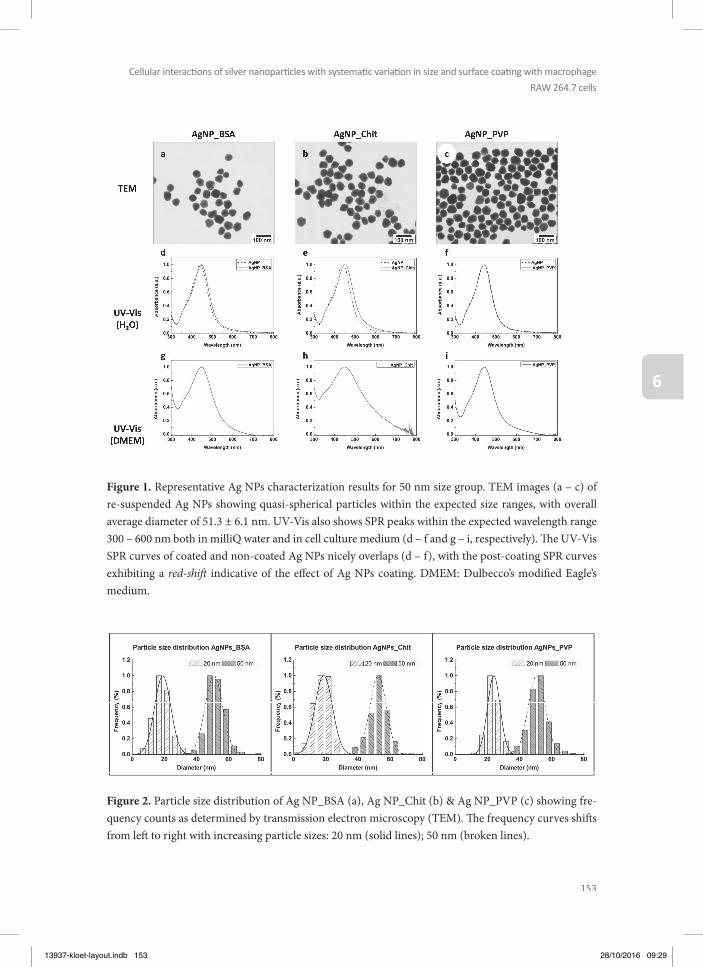
153
Cellular interacti ons of silver nanoparti cles with systemati c variati on in size and surface coati ng with macrophage
RAW 264.7 cells
6
Figure 1. Representative Ag NPs characterization results for 50 nm size group. TEM images (a – c) of re-suspended Ag NPs showing quasi-spherical particles within the expected size ranges, with overall average diameter of 51.3 ± 6.1 nm. UV-Vis also shows SPR peaks within the expected wavelength range 300 – 600 nm both in milliQ water and in cell culture medium (d – f and g – i, respectively). Th e UV-Vis SPR curves of coated and non-coated Ag NPs nicely overlaps (d – f), with the post-coating SPR curves exhibiting a red-shift indicative of the eff ect of Ag NPs coating. DMEM: Dulbecco’s modifi ed Eagle’s medium.
Figure 2. Particle size distribution of Ag NP_BSA (a), Ag NP_Chit (b) & Ag NP_PVP (c) showing fre-quency counts as determined by transmission electron microscopy (TEM). Th e frequency curves shift s from left to right with increasing particle sizes: 20 nm (solid lines); 50 nm (broken lines).
13937-kloet-layout.indb 153 28/10/2016 09:29
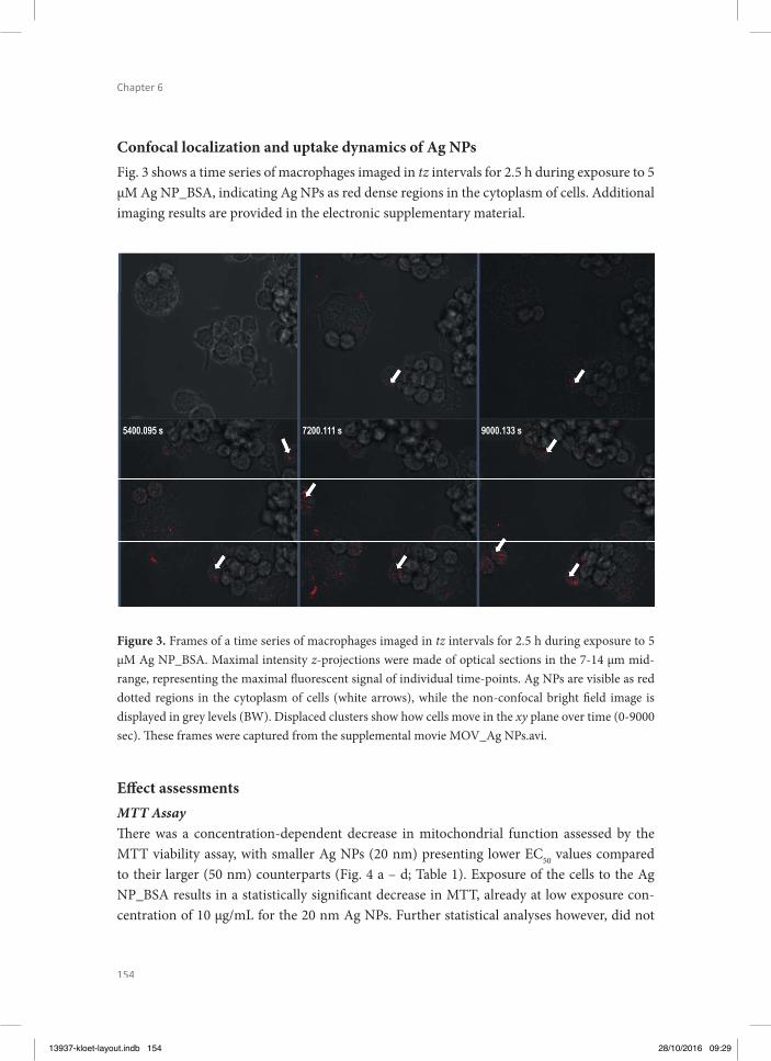
154
Chapter 6
Confocal localization and uptake dynamics of Ag NPsFig. 3 shows a time series of macrophages imaged in tz intervals for 2.5 h during exposure to 5 µM Ag NP_BSA, indicating Ag NPs as red dense regions in the cytoplasm of cells. Additional imaging results are provided in the electronic supplementary material.
Figure 3. Frames of a time series of macrophages imaged in tz intervals for 2.5 h during exposure to 5 µM Ag NP_BSA. Maximal intensity z-projections were made of optical sections in the 7-14 µm mid-range, representing the maximal fl uorescent signal of individual time-points. Ag NPs are visible as red dotted regions in the cytoplasm of cells (white arrows), while the non-confocal bright fi eld image is displayed in grey levels (BW). Displaced clusters show how cells move in the xy plane over time (0-9000 sec). Th ese frames were captured from the supplemental movie MOV_Ag NPs.avi.
Eff ect assessmentsMTT AssayTh ere was a concentration-dependent decrease in mitochondrial function assessed by the MTT viability assay, with smaller Ag NPs (20 nm) presenting lower EC50 values compared to their larger (50 nm) counterparts (Fig. 4 a – d; Table 1). Exposure of the cells to the Ag NP_BSA results in a statistically signifi cant decrease in MTT, already at low exposure con-centration of 10 µg/mL for the 20 nm Ag NPs. Further statistical analyses however, did not
13937-kloet-layout.indb 154 28/10/2016 09:29

155
Cellular interacti ons of silver nanoparti cles with systemati c variati on in size and surface coati ng with macrophage
RAW 264.7 cells
6
present any signifi cant diff erence between the EC50 values obtained for the diff erent sizes (20 nm vs 50 nm) nor types (surface-coating/charge) of Ag NPs. Except for Ag NP_Chit 50 nm for which the EC50 value exceeded the highest exposure concentration (Table 1).
Table 1. Table of EC50s (µg/mL) for assays, representing the eff ective concentration of Ag NPs required to produce the eff ect (increase/decrease) by 50%.
Ag NP_BSA Ag NP_Chit Ag NP_PVP Ag NP_NC
Assay 20nm 50nm P-value 20nm 50nm P-value 20nm 50nm P-value 50nm
MTT 11.7 58.9 0.224 10.3 125.9 0.553 14.8 37.6 0.438 40.7
ATP 28.4 114.7 0.005 93.0 >200 0.006 66.8 104.2 0.333 182.0
MPTP 23.3 114.3 0.197 14.5 24.3 0.112 56.3 109.2 0.301 137.3
ROS 77.6 31.6 0.073 >200 >200 <0.001 80.2 >200 0.588 89.5
TNF-α 1.1 5.0 0.156 49.9 15.1 0.002 nc 14.2 0.038 93.6
Signifi cant when p<0.05; nc, not converged.
Mitochondrial permeability transition pore (MPTP) AssayResults indicated a signifi cant opening of the MPTP with increasing Ag NP exposure concen-tration. A slight right-shift of the Ag NPs 50 nm dose-response curves was observed when compared to the curves obtained aft er exposure to the 20 nm Ag NPs (Fig. 4 e – h). Data showed that higher exposure concentrations of the 50 nm Ag NPs were needed to elicit the same loss in MPTP integrity as was obtained with the 20 nm Ag NPs, indicating a more ef-fi cient induction of opening of the MPTP by the smaller Ag NPs. Th ese diff erences however were not statistically signifi cant. Th e EC50s for the 20 and 50 nm sizes were only within 1-fold diff erence for chitosan- and PVP-coated NPs (Table 1). Th e EC50 for the 50 nm Ag NP_BSA exceeded the highest exposure concentration of 200 µg/mL.
Adenosine Triphosphate (ATP) AssayWith increasing exposure concentration, the amount of ATP measured in the cells decreased in a concentration dependent manner, suggesting a loss in viability since ATP is rapidly de-graded or lost from the cells as they die (Eguchi et al., 1997). For Ag NP_Chit and Ag NP_BSA, there was a signifi cant eff ect of size in addition to exposure concentration, with p values of 0.006 and 0.005 respectively when comparing the EC50s for the respective 20 and 50 nm particles. Th ere was no eff ect of size for the Ag NP_PVP. Similar to the 50 nm Ag NP_PVP, Ag NP_NC (also 50 nm, PVP-coated, but negatively charged), ATP was signifi cantly depleted at the highest exposure concentrations, and EC50 values for both types of Ag NPs exceeded the highest concentration of exposure (Fig. 4 i – l; Table 1).
13937-kloet-layout.indb 155 28/10/2016 09:29

156
Chapter 6
Figure 4. Representative Cytotoxicity of 20 and 50 nm NPs (Ag NP_BSA, Ag NP_Chit, Ag NP_PVP) and 50 nm Ag NP_NC to mouse monocyte macrophage cells (RAW 264.7 cell line) assessed by MTT (a – d), MPTP (e – h) and ATP (i – l) assays; and by assays for reactive oxygen species (ROS) production (m – p) and TNF-α production (q – t). Results are expressed as percentages (mean ± standard deviation, n=3) relative to the blank, set at 100%. Dotted lines and fi lled diamond (20 nm); solid lines and empty circle (50 nm). Statistical signifi cance compared to control is indicated by * (p ≤ 0.05); ‡ (p ≤ 0.01); # (p ≤ 0.001). Cytotoxicity (MTT, ATP and MPTP assays) showed a similar pattern for all tested Ag NPs, with viability decreasing with increasing exposure concentrations and minimal diff erences between NPs of diff erent sizes. On mechanisms of action (ROS production and TNF-α induction) however, there were eff ects of NP type and size (ROS production, 3n), while surface coating of Ag NPs with BSA appeared to result in a potent TNF-α induction (3q).
13937-kloet-layout.indb 156 28/10/2016 09:29
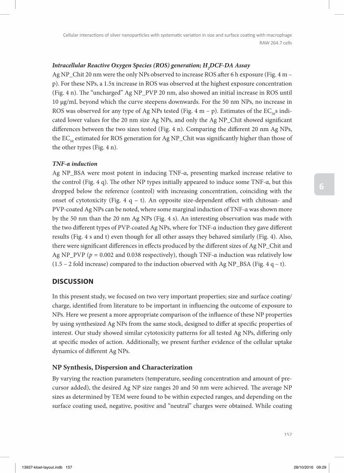
157
Cellular interacti ons of silver nanoparti cles with systemati c variati on in size and surface coati ng with macrophage
RAW 264.7 cells
6
Intracellular Reactive Oxygen Species (ROS) generation; H2DCF-DA AssayAg NP_Chit 20 nm were the only NPs observed to increase ROS aft er 6 h exposure (Fig. 4 m – p). For these NPs, a 1.5x increase in ROS was observed at the highest exposure concentration (Fig. 4 n). Th e “uncharged” Ag NP_PVP 20 nm, also showed an initial increase in ROS until 10 µg/mL beyond which the curve steepens downwards. For the 50 nm NPs, no increase in ROS was observed for any type of Ag NPs tested (Fig. 4 m – p). Estimates of the EC50s indi-cated lower values for the 20 nm size Ag NPs, and only the Ag NP_Chit showed signifi cant diff erences between the two sizes tested (Fig. 4 n). Comparing the diff erent 20 nm Ag NPs, the EC50 estimated for ROS generation for Ag NP_Chit was signifi cantly higher than those of the other types (Fig. 4 n).
TNF-α inductionAg NP_BSA were most potent in inducing TNF-α, presenting marked increase relative to the control (Fig. 4 q). Th e other NP types initially appeared to induce some TNF-α, but this dropped below the reference (control) with increasing concentration, coinciding with the onset of cytotoxicity (Fig. 4 q – t). An opposite size-dependent eff ect with chitosan- and PVP-coated Ag NPs can be noted, where some marginal induction of TNF-α was shown more by the 50 nm than the 20 nm Ag NPs (Fig. 4 s). An interesting observation was made with the two diff erent types of PVP-coated Ag NPs, where for TNF-α induction they gave diff erent results (Fig. 4 s and t) even though for all other assays they behaved similarly (Fig. 4). Also, there were signifi cant diff erences in eff ects produced by the diff erent sizes of Ag NP_Chit and Ag NP_PVP (p = 0.002 and 0.038 respectively), though TNF-α induction was relatively low (1.5 – 2 fold increase) compared to the induction observed with Ag NP_BSA (Fig. 4 q – t).
DISCUSSION
In this present study, we focused on two very important properties; size and surface coating/charge, identifi ed from literature to be important in infl uencing the outcome of exposure to NPs. Here we present a more appropriate comparison of the infl uence of these NP properties by using synthesized Ag NPs from the same stock, designed to diff er at specifi c properties of interest. Our study showed similar cytotoxicity patterns for all tested Ag NPs, diff ering only at specifi c modes of action. Additionally, we present further evidence of the cellular uptake dynamics of diff erent Ag NPs.
NP Synthesis, Dispersion and CharacterizationBy varying the reaction parameters (temperature, seeding concentration and amount of pre-cursor added), the desired Ag NP size ranges 20 and 50 nm were achieved. Th e average NP sizes as determined by TEM were found to be within expected ranges, and depending on the surface coating used, negative, positive and “neutral” charges were obtained. While coating
13937-kloet-layout.indb 157 28/10/2016 09:29
158
Chapter 6
with BSA and PVP is spontaneous and easy to monitor by the characterization techniques described above, that with chitosan resulted in Ag NPs aggregation. Th is may likely be due to the interaction of the negatively charged citrate Ag NP, an intermediate/pre-coated product of Ag NPs synthesis, with the positively charged chitosan (Gnanadhas et al., 2013, Bastus et al., 2014). To overcome this eff ect, a fast mixing of relatively small volumes of Ag NPs and an excess amounts of chitosan was used, resulting in a fast coating of the NPs.
Before coating, the particles showed SPR peaks and hydrodynamic sizes in accordance with expected results for the three diff erent sizes (Bastus et al., 2014). A slight shift in the SPR peak was observed by UV-Vis spectroscopy aft er incubating the NPs for 24 h with the respective coating molecules. Th is shift is related to modifi cations in the close environment of the NPs and proves the affi nity of the BSA, PVP and chitosan molecules for the silver surfaces and their spontaneous association (Bastus et al., 2014). Th e shape of the SPR peaks were pre-served in both milliQ water and DMEM (Fig. 1 d – f and g – i, respectively), which means that the particles were dispersed and stable under these conditions. Th e DLS measurements in both milliQ water and DMEM however, systematically resulted in larger sizes than those obtained before lyophilization. Th is could be indicative of a few agglomerates formed during the re-suspension of the powdered Ag NPs. Despite the large hydrodynamic diameter seen in the particles coated with chitosan, we observe well dispersed particles by TEM (Fig. 1 b).
Dispersed Ag NP_Chit formed some aggregates in the suspensions media, and these may have led to an overestimation of the hydrodynamic sizes during the DLS measurements, re-sulting in rather high NP diameters (Supplementary Table S1). Because the DLS operates on Rayleigh’s approximation principle of light scattering by particles where the intensity of scattering is proportional to the sixth power of the particle’s radius, DLS technique is very sensitive to particle agglomerates and/or any aggregation in NP suspensions (Hagendorfer et al., 2012). Hence, the occurrence of even minute amounts of agglomerates may skew the results. Th is aggregation observed with Ag NP_Chit for instance, represents a small fraction of the NPs, otherwise the SPR peaks would not be preserved (Fig. 1 d – i), and it would also be noticeable in the TEM images. Hence, the results indicate the formation of some aggregates but the majority of the Ag NPs appear to be single NPs, even for Ag NP_Chit.
Following dispersion in DMEM, the surface charges changed to negative values in all Ag NPs. Positively charged particles were likely coated by negatively charged proteins in the media, and may explain the negative ζ-potentials measured for all Ag NPs. A Z-potential measure-ment in a medium like DMEM containing an abundance of charged protein molecules is greatly infl uenced by the electrostatic interactions in the matrix. Th is was demonstrated by others (Pavlin and Bregar, 2012, Shannahan et al., 2013). Similarly, we observed more nega-tive ζ-potentials values (Supplementary Table S1) when Ag NPs were re-suspended in water,
13937-kloet-layout.indb 158 28/10/2016 09:29
159
Cellular interacti ons of silver nanoparti cles with systemati c variati on in size and surface coati ng with macrophage
RAW 264.7 cells
6
with minimal eff ect on hydrodynamic sizes regardless of the dispersant used. Lyophilization of the samples did not result in particle aggregation or surface modifi cations, and except for Ag NP_Chit, they could be easily dispersed in water as TEM images above show (Fig. 1 a – c) and DMEM. Th e SPR peaks and the surface charges were preserved as well.
Ag NPs localization and Uptake dynamicsUptake of the negatively charged Ag NP_BSA was rapid and most signifi cant based on an arbitrary unit (AU) of quantifi cation. Due to agglomeration in the suspension and growth medium, it was not possible to assess uptake of Ag NP_Chit by the cells. Supplementary Table S2 shows that the mean cellular uptake of Ag NP_BSA was twice that of Ag NP_PVP at sim-ilar exposure concentrations and time, though the uptake effi ciency of individual cells does vary a lot. Several cells showed fast uptake, with up to 80% of the Ag NPs observed to be taken up within the fi rst 30 min of exposure (not shown). Aft er 2.5 h however, cytoplasmic fl uores-cence intensity levels reached a plateau for the diff erent Ag NPs tested (Table S2). In fact, there was no signifi cant increase in the mean fl uorescent intensity in the macrophages aft er 24 h exposure (Supplementary Table S2). Th is rapid Ag NP uptake indicates that the 6 h exposure duration used for the assessment of ROS production is suffi cient for cells to internalize the Ag NPs. With increasing exposure concentrations, the mean AU plateau value increased nonlin-early for all Ag NPs. Fresh re-exposure at the same concentration and for an additional 24 h did not signifi cantly increase the plateau value either. Even at the lower exposure ranges, it can be seen that the uptake aft er 2 h is already reaching a plateau for the diff erent Ag NPs (Table S2) and this may suggest that cells loose their uptake capacity over time.
Cellular vitality was monitored during uptake assays by intercellular dynamics and presence of membrane protrusions, the latter being typical for functional macrophages (Vonna et al., 2007, Venter et al., 2014). Over a period of 48 h, no apparent cell death or abnormalities were observed while visualizing the subcellular bio-distribution of the Ag NPs. Subcellular dynam-ics remained visible during standardized imaging, as well as aft er 48 h exposure to 5 µg/mL of 50 nm Ag NP_BSA (Supplementary Fig. S4). Th e density of membrane protrusions however, slowly decreased over time (2.5 – 24 h, not quantifi ed). Only aft er prolonged local imaging were loss of membrane protrusions, amorphic membrane blebbing and cell fusion events trig-gered, indicating photon stress. We also demonstrated the uptake dynamics of Ag NP_BSA sticking to a macrophage surface membrane protrusion, and consequently being transported to the cell body within 30 seconds. Th e time series shows the dynamics of individual vesicles of various sizes in the cytoplasm (Fig. 3 and MOV_Ag NPs.avi). Both dispersed small vesicles as well as vesicles with clustered accumulated Ag NPs, occurred for all Ag NPs. We cannot exclude the likelihood that free Ag NPs were also present in the cytoplasm (Supplementary Fig. S4). Over time, more clustered vesicles appeared, indicating vesicle fusion. Multiple large
13937-kloet-layout.indb 159 28/10/2016 09:29

160
Chapter 6
endosomes or lysosomes with many Ag NPs clustered together were observed, suggesting that uptake was not by diff usion. Th ere are diff erent types of active endocytotic pathways such as receptor mediated endocytosis (clathrin or caveolin mediated) and macropinocytosis, but the current study did not discriminate between those.
Vesicle density varied over the cytoplasmic domain, but Ag NPs were never observed in the nucleoplasms (Fig. 3 and Supplementary Fig. S4). Th e absence of 50 nm Ag NPs in the nuclei was expected since nuclear pores have an eff ective diameter of 9 – 10 nm. Aft er 24 h loading, cells were still viable and healthy as judged by the dynamics from the cells themselves as well as from the organelles within. Th e dynamic movements of the Ag NPs within membrane do-mains indicate that accumulation does not imply clustering (MOV_Ag NPs.avi).
Eff ect AssessmentTh e smaller 20 nm Ag NPs tend to induce earlier eff ects than the 50 nm sizes in almost all as-says reported here (Fig. 4 and Table 1). However, the diff erences were minimal and for all Ag NPs tested, MTT reduction and MPTP opening did not diff er signifi cantly (p<0.05) between diff erent sizes (20 nm vs 50 nm) and surface-coatings or charges. A concentration dependent decrease in MTT reduction was observed, with the 20 nm NPs showing lower EC50 values than the 50 nm NPs (Fig. 4 and Table 1). Ag NP_BSA NPs appear to elicit the most eff ect. With the exception of Ag NP_Chit 50 nm where the EC50 exceeded the highest exposure con-centration, EC50s were between 10 and 15 µg/mL and 35 and 40 µg/mL for the 20 nm and 50 nm size groups respectively (Table 1). Th e infl uence of NP size on its ability to induce toxic eff ects has been demonstrated previously, (Liu et al., 2010, Park et al., 2011) for instance. Our fi ndings partially agree with the proposition that size does matter as could be seen with ATP production, where the 20 nm Ag NPs were more potent than the 50 nm for both Ag NP_BSA and Ag NP_Chit (Fig. 4 i – l; Table 1).
ATP assay results indicated a signifi cant decrease at low exposure concentrations of 10 µg/mL, at which the mitochondrial respiratory systems were also impaired (Fig. 4). Th is eff ect may likely be due to mitochondrial damage (AshaRani et al., 2009). Interestingly and contrary to the cited report in which ATP depletion in the tested cells was observed only aft er 48 h, ATP was depleted aft er 24 h exposure in our current study. Th e surface coating used for the Ag NPs in the cited study was starch, and may have interacted diff erently with the cells, poten-tially delaying the onset of toxicity. We found an increased opening of the mitochondrial tran-sition pore with increasing exposure concentration, leading to a rapid change in permeability. Th is is likely followed by membrane depolarization, release of intra-mitochondrial ions and metabolic intermediates amongst other eff ects (Lemasters et al., 1998). Normally, the MPTP remains closed unless under conditions of stress as found in for instance hypoxia, oxidative stress, and exposure to a calcium ionophore. Ag NPs of all sizes and surface coating tested
13937-kloet-layout.indb 160 28/10/2016 09:29

161
Cellular interacti ons of silver nanoparti cles with systemati c variati on in size and surface coati ng with macrophage
RAW 264.7 cells
6
demonstrated ability to cause the MPTP to open, leading to the quenching of the signal from the calcein which is now accessible to the CoCl2. In our study however, we could not demon-strate ROS production for most Ag NPs tested (Fig. 4 m-p). Th us, other factors including increased accumulation of intracellular fatty acids and lysophosphatidase, as well as glutathi-one oxidation (Lemasters et al., 1998), may also be responsible for activating MPTP opening.
Intracellular ROS generation is an outcome of normal cellular metabolism and these radicals may be cleared by the cell’s scavenging processes. Increase in ROS production however, is considered to be an early phase response to toxicants (Aranda et al., 2013), and has been reported as one of the likely mechanisms of toxicity following exposure to Ag NPs as well as other NPs (Asharani et al., 2011, Foldbjerg et al., 2011, Jeong et al., 2011). In this current study, ROS production was only slightly increased with 20 nm Ag NP_Chit which showed a 1.5 times increase in ROS relative to the control at the highest exposure concentration (Fig. 4 n). Also, 20 nm Ag NP_PVP showed an initial increase in ROS, then a decrease as exposure concentration increases. Th is decrease in ROS production is associated with the onset of cyto-toxicity (Fig. 4), and perhaps other cytotoxic processes are initiated or progressing faster than ROS generation. For example, the opening of the MPTP may lead to partial mitochondrial depolarization, which depending on Ca2+ concentrations, could result in a decrease in ROS production (low Ca2+) or an increase (high Ca2+) as reported earlier (Akopova et al., 2011).
Th e initial increase in ROS generation at lower exposure concentrations (Fig. 4 m-p) agrees with the fi ndings of others where it was suggested that cytotoxic eff ects were as a result of ROS production, particularly at low exposure concentrations and short incubation times (Carlson et al., 2008, Suresh et al., 2012, Foldbjerg et al., 2015). It may be that the cytotoxic eff ect of the Ag NPs overwhelms the cell’s capacity to generate ROS as a response, and rather progresses to cell death following other routes as suggested above. Again, positively charged NPs tend to interact more readily with cells due to the negative cell membrane charges (Nafee et al., 2009). Th is may enhance the exposure of cells to positively charged NPs, likely explaining the increased ROS production observed with the 20 nm positively charged Ag NP_Chit (Fig. 4n) where there was a signifi cant diff erence between the two sizes tested (p<0.001). Interestingly, ROS was rather decreased in macrophage cells with increasing exposure concentrations of the 50 nm size Ag NP_Chit, similar to the other Ag NP types tested. With increasing exposure concentrations, the resulting distortion in metabolic activity coupled with an open MPTP, may eventually lead to ATP depletion and cell death. Th is process has also been associated with mitochondrial respiratory system impairment following oxidative stress (AshaRani et al., 2009). In this current study, ROS production appears to have been inhibited as Ag NPs exposure concentration increases relative to the control. Considering the early onset of cyto-toxicity based on MTT, MPTP and ATP assay results however, the downward plunge of the ROS curves could result from the inability of injured cells to produce oxidative radicals, since their viability has been impaired.
13937-kloet-layout.indb 161 28/10/2016 09:29

162
Chapter 6
Ag NPs have been reported to cause immunogenic response in cells, characterized by induc-tion of cytokines like TNF-α, macrophage inhibitory protein, interleukins, etc. (Carlson et al., 2008, Park et al., 2011). For the Ag NPs tested in this current study, the negatively charged Ag NP_BSA showed the most induction with both sizes resulting in an 80-fold increase be-tween exposure concentrations 10 and 50 µg/mL. Higher concentrations showed a downward plunge of the curves below the reference (control), coinciding with cytotoxicity (Fig. 4 a, e, i, and q). Th ere were no signifi cant diff erences in the eff ects induced by the diff erent sizes of Ag NP_BSA (Fig. 4 q). Interestingly, even when coated with similar biomolecules, the behav-ior of NPs may still diff er as can be seen with the 50 nm PVP-coated Ag NPs commercially produced (negatively charged) and the synthesized one (uncharged). Although both behaved similarly for all other assays tested, the case was diff erent with TNF-α induction (Figs. 3 s and t) where Ag NP_PVP was a signifi cantly higher TNF-α inducer than Ag NP_NC (p = 0.002). Th is could likely be due to the type of synthesis procedure applied for making not only the NPs (Powers et al., 2011a, Samberg et al., 2011), but also the bioconjugate (Bastus et al., 2014). Th e type or composition of the coating agent infl uences the outcome of exposure, cor-roborating previous fi ndings (Aadil et al., 2016) where the infl uence of lignin concentration, incubation temperature and time in the synthesis of Ag NPs were assessed. Also, the pH of the solution during the synthesis was important in determining the surface charge of the NPs, with acidic pH resulting in negatively charged NPs while basic pH leads to positively charged NPs. Th is phenomenon is important not only during synthesis of NPs, but also during expo-sure (Deng et al., 2016).
Th e greater the NP’s relative surface area, the greater the potential for reactivity. Since the relative surface area is determined by the particle size, smaller NPs may interact more with the macrophages (Johnston et al., 2010, Reidy et al., 2013) when administered on an equal mass-basis as was done in the current study. Moreover, surface coatings and their interactions with biomolecules can infl uence dissolution rates of Ag NPs (Yen et al., 2009, Yang et al., 2012), leading to toxicities. Based on this, one could hypothesize that the synthesized Ag NPs tested in this study were quite stable under the experimental conditions reported here. Longer exposure durations of 48 – 72 h may likely present diff erent outcomes.
Also, some studies have shown an opposite eff ect of size for e.g. with PVP-coated Ag NP (Powers et al., 2011a) where the larger particles produced larger eff ect. In the present study, this was observed with Ag NP_Chit and Ag NP_PVP in TNF-α induction assay, with the 50 nm sizes having lower EC50 values (Table 1). For the tested Ag NPs in this study, cytotoxicity patterns were similar irrespective of types and size of the Ag NPs. Looking at ROS and TNF-α induction on the other hand revealed diff erences between the diff erent types of Ag NPs. Th is is indicative of varying mechanisms of action involved, corroborating what has been opined by others (Johnston et al., 2010, Reidy et al., 2013). Th ese authors suggested that a combina-
13937-kloet-layout.indb 162 28/10/2016 09:29
163
Cellular interacti ons of silver nanoparti cles with systemati c variati on in size and surface coati ng with macrophage
RAW 264.7 cells
6
tion of diff erent physicochemical properties of Ag NPs is more likely responsible in inducing the observed toxicities, rather than only size or particle dissolution for instance, as the release of ions would be expected to be greater for smaller particles.
CONCLUSION
Our study used well established in vitro assays to investigate the infl uence of the physico-chemical properties of a specifi cally designed set of diff erent types of Ag NPs systematically synthesized to vary the properties of interest. Th e role of surface coating/charge in infl uencing TNF-α induction was demonstrated by the negatively charged Ag NP_BSA. Eff ect of size was less prominent under our experimental conditions, showing mostly minimal diff erences that were not statistically signifi cant. Overall, the negatively charged Ag NP_BSA appears to be more potent in inducing adverse eff ects on the macrophages. Th e complexity in determining the fate and toxicity of Ag NPs (also other NPs) using in vitro or in vivo models is as yet a challenge that needs further evaluation. Live confocal imaging of exposed cells allowed the monitoring of Ag NPs uptake dynamics and subcellular cytoplasmic accumulation.
A combination of several factors likely play a role in determining the outcome of exposure. Th ese include the processes involved in the NP syntheses, presence and type of coating agents resulting in various physicochemical properties (size, charge, hydrophobicity, etc.), as well as the toxicity tests models used and end-points studied. Th is has been observed in earlier reviews (Johnston et al., 2010, de Lima et al., 2012, Reidy et al., 2013), and still remains a hindrance for conclusive risk assessment evaluation of Ag NPs. Taken together, the results from MTT, ATP and MPTP assays shows that adverse eff ects from exposure to the tested Ag NPs were similar irrespective of type or size. However, for ROS and TNF-α induction, we do fi nd signifi cant toxicity diff erences with variations in the physicochemical properties of the Ag NPs tested.
ACKNOWLEDGEMENTS
Th is work was fi nancially supported by NanoNextNL, a micro- and nanotechnology con-sortium of the Government of Th e Netherlands and 130 partners; funding was also received from Managing Risks of Nanoparticles, MARINA (EU-FP7, contract CP-FP 263215), and; the Strategic Research Funds titled Novel technologies by the Ministry of Economic Aff airs of Th e Netherlands. Synthesis and characterization of the Ag NPs used in this study received support from the QualityNano Project http://www.qualitynano.eu/ which is fi nanced by the European Community Research Infrastructures under the FP7 Capacities Program (Grant No. INFRA-2010-262163).
13937-kloet-layout.indb 163 28/10/2016 09:29
164
Chapter 6
REFERENCES
Aadil, K.R., Barapatre, A., Meena, A.S. & Jha, H., 2016. Hydrogen peroxide sensing and cytotoxicity activity of Acacia lignin stabilized silver nanoparticles. Int J Biol Macromol, 82, 39-47.
Abou El-Nour, K.M.M., Eft aiha, A.A., Al-Warthan, A. & Ammar, R.a.A., 2010. Synthesis and applications of silver nanoparticles. Arabian Journal of Chemistry, 3, 135-140.
Ahamed, M., Alsalhi, M.S. & Siddiqui, M.K., 2010. Silver nanoparticle applications and human health. Clin Chim Acta, 411, 1841-8.
Akopova, O.V., Kolchynskayia, L.Y., Nosar, V.Y., Smyrnov, A.N., Malisheva, M.K., Man’kovskaia, Y.N. & Sahach, V.F., 2011. Th e eff ect of permeability transition pore opening on reactive oxygen species production in rat brain mitochondria. Ukr Biokhim Zh (1999), 83, 46-55.
Aranda, A., Sequedo, L., Tolosa, L., Quintas, G., Burello, E., Castell, J.V. & Gombau, L., 2013. Dichloro-dihydro-fl u-orescein diacetate (DCFH-DA) assay: a quantitative method for oxidative stress assessment of nanoparti-cle-treated cells. Toxicol In Vitro, 27, 954-63.
Asharani, P.V., Lianwu, Y., Gong, Z. & Valiyaveettil, S., 2011. Comparison of the toxicity of silver, gold and platinum nanoparticles in developing zebrafi sh embryos. Nanotoxicology, 5, 43-54.
Asharani, P.V., Low Kah Mun, G., Hande, M.P. & Valiyaveettil, S., 2009. Cytotoxicity and genotoxicity of silver nanoparticles in human cells. ACS Nano, 3, 279-90.
Bastus, N.G., Merkoci, F., Piella, J. & Puntes, V., 2014. Synthesis of Highly Monodisperse Citrate-Stabilized Silver Nanoparticles of up to 200 nm: Kinetic Control and Catalytic Properties. Chemistry of Materials, 26, 2836-2846.
Benn, T., Cavanagh, B., Hristovski, K., Posner, J.D. & Westerhoff , P., 2010. Th e Release of Nanosilver from Consumer Products Used in the Home. Journal of Environmental Quality, 39, 1875-1882.
Braakhuis, H.M., Kloet, S.K., Kezic, S., Kuper, F., Park, M.V., Bellmann, S., Van Der Zande, M., Le Gac, S., Krystek, P., Peters, R.J., Rietjens, I.M. & Bouwmeester, H., 2015. Progress and future of in vitro models to study translo-cation of nanoparticles. Arch Toxicol, 89, 1469-95.
Carlson, C., Hussain, S.M., Schrand, A.M., Braydich-Stolle, L.K., Hess, K.L., Jones, R.L. & Schlager, J.J., 2008. Unique cellular interaction of silver nanoparticles: size-dependent generation of reactive oxygen species. J Phys Chem B, 112, 13608-19.
Chanana, M. & Liz-Marzán Luis, M., 2012. Coating matters: the infl uence of coating materials on the optical proper-ties of gold nanoparticles. Nanophotonics. 199.
Connolly, M., Fernandez-Cruz, M.L., Quesada-Garcia, A., Alte, L., Segner, H. & Navas, J.M., 2015. Comparative Cytotoxicity Study of Silver Nanoparticles (Ag NPs) in a Variety of Rainbow Trout Cell Lines (RTL-W1, RTH-149, RTG-2) and Primary Hepatocytes. Int J Environ Res Public Health, 12, 5386-405.
Cornelis, G., Hund-Rinke, K., Kuhlbusch, T., Van Den Brink, N. & Nickel, C., 2014. Fate and Bioavailability of En-gineered Nanoparticles in Soils: A Review. Critical Reviews in Environmental Science and Technology, 44, 2720-2764.
De Lima, R., Seabra, A.B. & Duran, N., 2012. Silver nanoparticles: a brief review of cytotoxicity and genotoxicity of chemically and biogenically synthesized nanoparticles. J Appl Toxicol, 32, 867-79.
Deng, X., Wang, Y., Zhang, F., Yin, Z., Hu, Q., Xiao, X., Zhou, Z., Wu, Y., Sheng, W. & Zeng, Y., 2016. Acidic pH-in-duced charge-reversal nanoparticles for accelerated endosomal escape and enhanced microRNA modula-tion in cancer cells. Chem Commun (Camb), 52, 3243-6.
Eguchi, Y., Shimizu, S. & Tsujimoto, Y., 1997. Intracellular ATP levels determine cell death fate by apoptosis or ne-crosis. Cancer Res, 57, 1835-40.
Evanoff , D.D., Jr. & Chumanov, G., 2005. Synthesis and optical properties of silver nanoparticles and arrays. Chemphyschem, 6, 1221-31.
Foldbjerg, R., Dang, D.A. & Autrup, H., 2011. Cytotoxicity and genotoxicity of silver nanoparticles in the human lung cancer cell line, A549. Arch Toxicol, 85, 743-50.
Foldbjerg, R., Irving, E.S., Hayashi, Y., Sutherland, D.S., Th orsen, K., Autrup, H. & Beer, C., 2012. Global gene expres-sion profi ling of human lung epithelial cells aft er exposure to nanosilver. Toxicol Sci, 130, 145-57.
Foldbjerg, R., Jiang, X., Miclăuş, T., Chen, C., Autrup, H. & Beer, C., 2015. Silver nanoparticles – wolves in sheep’s clothing? Toxicol. Res., 4, 563-575.
13937-kloet-layout.indb 164 28/10/2016 09:29

165
Cellular interacti ons of silver nanoparti cles with systemati c variati on in size and surface coati ng with macrophage
RAW 264.7 cells
6
Franci, G., Falanga, A., Galdiero, S., Palomba, L., Rai, M., Morelli, G. & Galdiero, M., 2015. Silver nanoparticles as potential antibacterial agents. Molecules, 20, 8856-74.
Geranio, L., Heuberger, M. & Nowack, B., 2009. Th e behavior of silver nanotextiles during washing. Environ Sci Technol, 43, 8113-8.
Gnanadhas, D.P., Ben Th omas, M., Th omas, R., Raichur, A.M. & Chakravortty, D., 2013. Interaction of silver nanoparticles with serum proteins aff ects their antimicrobial activity in vivo. Antimicrob Agents Chemo-ther, 57, 4945-55.
Gwinn, M.R. & Vallyathan, V., 2006. Nanoparticles: health eff ects--pros and cons. Environ Health Perspect, 114, 1818-25.
Hagendorfer, H., Kaegi, R., Parlinska, M., Sinnet, B., Ludwig, C. & Ulrich, A., 2012. Characterization of silver nanopar-ticle products using asymmetric fl ow fi eld fl ow fractionation with a multidetector approach--a comparison to transmission electron microscopy and batch dynamic light scattering. Anal Chem, 84, 2678-85.
Haider, A. & Kang, I.K., 2015. Preparation of Silver Nanoparticles and Th eir Industrial and Biomedical Applications: A Comprehensive Review. Advances in Materials Science and Engineering, 2015, 16.
Hwang, I.S., Hwang, J.H., Choi, H., Kim, K.J. & Lee, D.G., 2012. Synergistic eff ects between silver nanoparticles and antibiotics and the mechanisms involved. J Med Microbiol, 61, 1719-26.
Jeong, Y.S., Oh, W.K., Kim, S. & Jang, J., 2011. Cellular uptake, cytotoxicity, and ROS generation with silica/conduct-ing polymer core/shell nanospheres. Biomaterials, 32, 7217-25.
Johnston, H.J., Hutchison, G., Christensen, F.M., Peters, S., Hankin, S. & Stone, V., 2010. A review of the in vivo and in vitro toxicity of silver and gold particulates: particle attributes and biological mechanisms responsible for the observed toxicity. Crit Rev Toxicol, 40, 328-46.
Kennell, D., 2009. Nanotechnology: An Industrial Revolution? Monthly Review MRZine. Kloet, S.K., Walczak, A.P., Louisse, J., Van Den Berg, H.H., Bouwmeester, H., Tromp, P., Fokkink, R.G. & Rietjens,
I.M., 2015. Translocation of positively and negatively charged polystyrene nanoparticles in an in vitro pla-cental model. Toxicol In Vitro, 29, 1701-10.
Kreuter, J., 2004. Infl uence of the surface properties on nanoparticle-mediated transport of drugs to the brain. J Nanosci Nanotechnol, 4, 484-8.
Landsiedel, R., Fabian, E., Ma-Hock, L., Van Ravenzwaay, B., Wohlleben, W., Wiench, K. & Oesch, F., 2012. Toxico-/biokinetics of nanomaterials. Arch Toxicol, 86, 1021-60.
Lara, H.H., Ayala-Nunez, N.V., Ixtepan-Turrent, L. & Rodriguez-Padilla, C., 2010. Mode of antiviral action of silver nanoparticles against HIV-1. J Nanobiotechnology, 8, 1.
Lemasters, J.J., Nieminen, A.L., Qian, T., Trost, L.C., Elmore, S.P., Nishimura, Y., Crowe, R.A., Cascio, W.E., Bradham, C.A., Brenner, D.A. & Herman, B., 1998. Th e mitochondrial permeability transition in cell death: a common mechanism in necrosis, apoptosis and autophagy. Biochim Biophys Acta, 1366, 177-96.
Liu, W., Wu, Y., Wang, C., Li, H.C., Wang, T., Liao, C.Y., Cui, L., Zhou, Q.F., Yan, B. & Jiang, G.B., 2010. Impact of silver nanoparticles on human cells: eff ect of particle size. Nanotoxicology, 4, 319-30.
Makama, S., Peters, R., Undas, A. & Van Den Brink, N.W., 2015. A novel method for the quantifi cation, characteri-sation and speciation of silver nanoparticles in earthworms exposed in soil. Environmental Chemistry, 12, 643-651.
Nadesh, R., Narayanan, D., P, R.S., Vadakumpully, S., Mony, U., Koyakkutty, M., Nair, S.V. & Menon, D., 2013. He-matotoxicological analysis of surface-modifi ed and -unmodifi ed chitosan nanoparticles. J Biomed Mater Res A, 101, 2957-66.
Nafee, N., Schneider, M., Schaefer, U.F. & Lehr, C.M., 2009. Relevance of the colloidal stability of chitosan/PLGA nanoparticles on their cytotoxicity profi le. Int J Pharm, 381, 130-9.
Park, M.V., Neigh, A.M., Vermeulen, J.P., De La Fonteyne, L.J., Verharen, H.W., Briede, J.J., Van Loveren, H. & De Jong, W.H., 2011. Th e eff ect of particle size on the cytotoxicity, infl ammation, developmental toxicity and genotoxicity of silver nanoparticles. Biomaterials, 32, 9810-7.
Pavlin, M. & Bregar, V., 2012. Stability of nanoparticle suspension in diff erent biologically relevant media. Digest Journal of Nanomaterials and Biostructures, 7, 1389-1400.
Peterson, C., 2000. Taking technology to the molecular level. Computer, 33, 46-+. Powers, C.M., Badireddy, A.R., Ryde, I.T., Seidler, F.J. & Slotkin, T.A., 2011a. Silver Nanoparticles Compromise Neu-
rodevelopment in PC12 Cells: Critical Contributions of Silver Ion, Particle Size, Coating, and Composition. Environmental Health Perspectives, 119, 37-44.
13937-kloet-layout.indb 165 28/10/2016 09:29

166
Chapter 6
Powers, C.M., Badireddy, A.R., Ryde, I.T., Seidler, F.J. & Slotkin, T.A., 2011b. Silver nanoparticles compromise neu-rodevelopment in PC12 cells: critical contributions of silver ion, particle size, coating, and composition. Environ Health Perspect, 119, 37-44.
Prabhu, S. & Poulose, E.K., 2012. Silver nanoparticles: mechanism of antimicrobial action, synthesis, medical appli-cations, and toxicity eff ects. International Nano Letters, 2, 32.
Reidy, B., Haase, A., Luch, A., Dawson, K.A. & Lynch, I., 2013. Mechanisms of Silver Nanoparticle Release, Trans-formation and Toxicity: A Critical Review of Current Knowledge and Recommendations for Future Studies and Applications. Materials, 6, 2295-2350.
Roohani-Esfahani, S.I., Nouri-Khorasani, S., Lu, Z., Appleyard, R. & Zreiqat, H., 2010. Th e infl uence hydroxyapatite nanoparticle shape and size on the properties of biphasic calcium phosphate scaff olds coated with hydroxy-apatite-PCL composites. Biomaterials, 31, 5498-509.
Samberg, M.E., Orndorff , P.E. & Monteiro-Riviere, N.A., 2011. Antibacterial effi cacy of silver nanoparticles of diff er-ent sizes, surface conditions and synthesis methods. Nanotoxicology, 5, 244-53.
Shang, L., Nienhaus, K., Jiang, X., Yang, L., Landfester, K., Mailander, V., Simmet, T. & Nienhaus, G.U., 2014a. Nanoparticle interactions with live cells: Quantitative fl uorescence microscopy of nanoparticle size eff ects. Beilstein J Nanotechnol, 5, 2388-97.
Shang, L., Nienhaus, K. & Nienhaus, G.U., 2014b. Engineered nanoparticles interacting with cells: size matters. J Nanobiotechnology, 12, 5.
Shannahan, J.H., Lai, X., Ke, P.C., Podila, R., Brown, J.M. & Witzmann, F.A., 2013. Silver nanoparticle protein corona composition in cell culture media. PLoS One, 8, e74001.
Shenashen, M.A., El-Saft y, S.A. & Elshehy, E.A., 2014. Synthesis, Morphological Control, and Properties of Silver Nanoparticles in Potential Applications. Particle & Particle Systems Characterization, 31, 293-316.
Shoults-Wilson, W.A., Reinsch, B.C., Tsyusko, O.V., Bertsch, P.M., Lowry, G.V. & Unrine, J.M., 2011. Eff ect of silver nanoparticle surface coating on bioaccumulation and reproductive toxicity in earthworms (Eisenia fetida). Nanotoxicology, 5, 432-44.
Silver, S. & Phung, L.T., 1996. Bacterial heavy metal resistance: new surprises. Annu Rev Microbiol, 50, 753-89. Sondi, I. & Salopek-Sondi, B., 2004. Silver nanoparticles as antimicrobial agent: a case study on E. coli as a model for
Gram-negative bacteria. J Colloid Interface Sci, 275, 177-82. Suresh, A.K., Pelletier, D.A., Wang, W., Morrell-Falvey, J.L., Gu, B. & Doktycz, M.J., 2012. Cytotoxicity induced by
engineered silver nanocrystallites is dependent on surface coatings and cell types. Langmuir, 28, 2727-35. Van Der Ploeg, M.J., Handy, R.D., Waalewijn-Kool, P.L., Van Den Berg, J.H., Herrera Rivera, Z.E., Bovenschen, J.,
Molleman, B., Baveco, J.M., Tromp, P., Peters, R.J., Koopmans, G.F., Rietjens, I.M. & Van Den Brink, N.W., 2014a. Eff ects of silver nanoparticles (NM-300K) on Lumbricus rubellus earthworms and particle charac-terization in relevant test matrices including soil. Environ Toxicol Chem, 33, 743-52.
Van Der Ploeg, M.J., Van Den Berg, J.H., Bhattacharjee, S., De Haan, L.H., Ershov, D.S., Fokkink, R.G., Zuilhof, H., Rietjens, I.M. & Van Den Brink, N.W., 2014b. In vitro nanoparticle toxicity to rat alveolar cells and coelo-mocytes from the earthworm Lumbricus rubellus. Nanotoxicology, 8, 28-37.
Vance, M.E., Kuiken, T., Vejerano, E.P., Mcginnis, S.P., Hochella, M.F., Jr., Rejeski, D. & Hull, M.S., 2015. Nanotech-nology in the real world: Redeveloping the nanomaterial consumer products inventory. Beilstein J Nano-technol, 6, 1769-80.
Venter, G., Oerlemans, F.T.J.J., Wijers, M., Willemse, M., Fransen, J.a.M. & Wieringa, B., 2014. Glucose Controls Morphodynamics of LPS-Stimulated Macrophages. PLoS ONE, 9, e96786.
Vonna, L., Wiedemann, A., Aepfelbacher, M. & Sackmann, E., 2007. Micromechanics of fi lopodia mediated capture of pathogens by macrophages. Eur Biophys J, 36, 145-51.
Wijnhoven, S.W.P., Oomen, A.G., Sips, A.J.a.M., Bourgeois, F.C., Te Dorsthorst, G.J.P.M., Kooi, M.W. & Bakker, M.I., 2010. Development of an inventory for consumer products containing nanomaterials Final Report RIVM Report 000201006, 103.
Wijnhoven, S.W.P., Peijnenburg, W.J.G.M., Herberts, C.A., Hagens, W.I., Oomen, A.G., Heugens, E.H.W., Roszek, B., Bisschops, J., Gosens, I., Van De Meent, D., Dekkers, S., De Jong, W.H., Van Zijverden, M., Sips, A.J.a.M. & Geertsma, R.E., 2009. Nano-silver - a review of available data and knowledge gaps in human and environ-mental risk assessment. Nanotoxicology, 3, 109-U78.
13937-kloet-layout.indb 166 28/10/2016 09:29
167
Cellular interacti ons of silver nanoparti cles with systemati c variati on in size and surface coati ng with macrophage
RAW 264.7 cells
6
Yang, X., Gondikas, A.P., Marinakos, S.M., Auff an, M., Liu, J., Hsu-Kim, H. & Meyer, J.N., 2012. Mechanism of silver nanoparticle toxicity is dependent on dissolved silver and surface coating in Caenorhabditis elegans. Envi-ron Sci Technol, 46, 1119-27.
Yen, H.J., Hsu, S.H. & Tsai, C.L., 2009. Cytotoxicity and immunological response of gold and silver nanoparticles of diff erent sizes. Small, 5, 1553-61.
Yu, S.J., Yin, Y.G. & Liu, J.F., 2013. Silver nanoparticles in the environment. Environ Sci Process Impacts, 15, 78-92. Zhao, L.M., Shi, L.E., Zhang, Z.L., Chen, J.M., Shi, D.D., Yang, J. & Tang, Z.X., 2011. Preparation and Application of
Chitosan Nanoparticles and Nanofi bers. Brazilian Journal of Chemical Engineering, 28, 353-362.
13937-kloet-layout.indb 167 28/10/2016 09:29
168
Chapter 6
SUPPLEMENTARY MATERIALS CHAPTER 6
MATERIALS AND METHODS
Synthesis of Ag NPsMilliQ water (18.2 MΩ/cm) was used for all Ag NPs syntheses. Colloidal, dispersed Ag NPs of two diff erent sizes (20 and 50 nm) were prepared separately, following a kinetically controlled seeded-growth method previously reported (Bastus et al. 2014) with slight modifi cations. Th e approach is based on the reduction of silver nitrate (AgNO3) in the presence of two competing reducing agents, tannic acid (TA) and trisodium citrate hexahydrate (SC) at 100°C. In order to produce large amounts (grams) of Ag NPs in the present study, a 10 L batch reactor was used and the concentration of the AgNO3 (Ag NP precursor) was increased by a factor of 10, while all other parameters remained the same with that reported previously (Bastus et al. 2014).
First, early nucleated Ag NP seeds were formed by injecting 5 mL of 2 M AgNO3 into a boil-ing solution containing SC (5 mM) and TA (0.025-0.075 mM), under continuous vigorous stirring. Th ese seeds were further grown to the desired sizes by injecting more of the silver precursor dropwise to avoid formation of new seeds. During the growth phase of the seeds, the temperature of the solution was reduced to 90ºC, while slowly adding up to 15 mL of the 2 M AgNO3 into the reactor. Under these conditions, 10-20 L of a stable, highly monodisperse and colloidal Ag NP solutions (0.25-0.5 g/L or 2.3 – 4.6 mM) were produced for each size.
Th e solutions containing the synthesized Ag NPs were concentrated to about 6% of their original volume by ultrafi ltration using a tangential fl ow fractionation (TFF) system (KrosFlo® Research II TFF Systems, Spectrum® Laboratories Inc.). Negative, positive, and neutral Ag NPs were obtained by subsequently mixing about 200 mL each of the concentrated solutions of Ag NPs with either 0.01 mM bovine serum albumin (BSA), 0.08 mM chitosan (Chit) or 0.05 mM polyvinylpyrrolidone (PVP) to generate negative Ag NP_BSA, positive Ag NP_Chit and neutral Ag NP_PVP, respectively. To prevent corrosion and facilitate easy transport, the Ag NPs were lyophilized, layered over with argon and kept under dark conditions until use. Th us, six diff erent Ag NPs were synthesized consisting of three types based on surface coat-ing/charge, each at two diff erent sizes.
An aqueous suspension of a negatively charged 50 nm PVP-coated Ag NP (Ag NP_NC) was obtained from NanoComposix® (San Diego, US) and included in this study for comparison with the synthesized Ag NPs. Based on the manufacturer’s information, the Ag NP_NC is reported to have mean core- and hydrodynamic diameters of 54.8 ± 10 nm and 72 ± 14.4 nm respectively. Th e stock suspension of the pristine Ag NP has a mass concentration of 5.0 mg
13937-kloet-layout.indb 168 28/10/2016 09:29
169
Cellular interacti ons of silver nanoparti cles with systemati c variati on in size and surface coati ng with macrophage
RAW 264.7 cells
6
Ag/mL, and a particle number concentration of 5.2 x 10¹² particles/mL. Th e surface area was 9.8 m2/g while surface charge (zeta-potential) was -37.8 mV. Th e DLS and zeta potential in both milliQ and DMEM were confi rmed in our laboratory, and details have been presented earlier (Makama et al. 2015).
Ag NP CharacterizationPrior to exposure experiments, pristine Ag NPs were dispersed in Dulbecco’s modifi ed Ea-gle’s medium, DMEM (Invitrogen Breda, Th e Netherlands), as well as milliQ water and char-acterized by transmission electron microscopy (TEM), UV-Vis spectroscopy, dynamic light scattering (DLS), and zeta-potential measurements (ζ-potential). A combination of diff erent techniques was used to characterize the Ag NPs and to monitor proper coating of NPs surfac-es and their stability in the diff erent media.
TEM images were acquired with a FEI Magellane 400L SEM electron microscope operating at scanning TEM (STEM) mode and low accelerating voltage (20 kV) and a JEOL 1010 electron microscope operating at an accelerating voltage of 80 kV. Samples for TEM were prepared by drop casting onto carbon-coated TEM grids. Th e grids were left to dry at room temperature. Images were acquired in diff erent parts of the grid at diff erent magnifi cations. More than 250 particles were computer-analyzed and measured to calculate the size distribution of each type of Ag NPs.
UV-Vis spectroscopy is a very common and well-known analytical technique (Evanoff and Chumanov 2005; Pan 2015), and a Shimadzu UV-2400 spectrophotometer was used to mea-sure the localized surface Plasmon resonance (SPR) peak. Here, 1 mL of the Ag NPs suspen-sion was placed in a cuvette and the spectrum (UV-Vis) acquired in the 300-800 nm range. Several metallic NPs, such as gold and silver, exhibit a characteristic absorbance maximum in the visible range (SPR peak). Th is characteristic wavelength is highly sensitive to the ENP size, surface modifi cations and aggregation and thus a useful tool to characterize the state of the NPs in solution.
Hydrodynamic sizes of the Ag NPs were estimated by DLS, while particle surface charges (ζ-potentials) were measured using Malvern Zetasizer Nano ZS (Malvern Instruments, UK). A light source wavelength of 532 nm and a fi xed scattering angle of 173° were set for DLS measurement. Ag NPs suspensions (1 mL) were dispensed in a cuvette and the instrument was set with the specifi c parameters of refractive index and absorption coeffi cient of the ma-terial and the viscosity of the solvent. All measurements were conducted at least three times. Ag NPs characterization was done in both milliQ water and DMEM since the exposure was carried out in the cell culture medium.
13937-kloet-layout.indb 169 28/10/2016 09:29
170
Chapter 6
Eff ect assessment: MTT AssayAt the end of the 24hr exposure, 10 µL of the tetrazolium salt 3-(4,5-dimethylthi-azol-2-yl)-2,5-diphenyltetrazolium bromide (MTT) solution at 5 mg/mL in PBS was added to each well and incubated under the same conditions as described above for 90 min. Th e exposure medium containing MTT was then removed and 100 µL pure dimethyl sulfoxide, DMSO (Acros Organics, Geel, BE) was added to solubilize the crystals of formazan salts. Th is was followed by shaking the plates in the dark for 10 min. Th e dissolved formazan produces a purple color and its absorbance was measured at 562 nm spectrophotometrically (Kloet et al. 2015).
Adenosine Triphosphate (ATP) AssayA bioluminescent somatic cell assay kit (Sigma-Aldrich®, St Louis, MO, US), was used to de-termine the ATP content of mouse macrophage cells aft er exposure. In this assay, ATP is the limiting reagent and the light emitted and measured is proportional to the amount of ATP released from the cells, which is in turn determined by the number of viable cells present. Ex-posure to Ag NPs for this assay was conducted in white 96-well plates (Packard White plates, Perkin-Elmer®), using DMEM without phenol red, but with penicillin/streptomycin (Gibco®) 1% (v/v). Aft er exposure, cell culture media was removed and replaced with 25 µL each of fresh medium and somatic cell ATP releasing agent from the kit. Th e plates were placed on a shaker for 10 mins in the dark, then transferred to the luminometer where the ATP assay working mix (containing luciferin, luciferase and MgSO4) was added automatically to each well before measuring the luminescence.
Mitochondrial permeability Transition-Pore (MPTP) AssayA commercial kit from Molecular Probes (Leiden, NL), MitoProbeTM Transition Pore (MPTP) Assay kit (M34153) was used for this assay. Aft er exposure, the culture medium in all wells was replaced by Hank’s balanced salt solution, HBSS (Invitrogen, Breda NL), containing cal-cium but without serum, phenol red or NaHCO3. Cells in all except the positive control wells were loaded with a mixture of cobalt chloride (CoCl2) and an acetoxymethyl ester form of a calcein dye (Calcein AM) at 1 mM and 5 µM respectively, followed by a 15 min incubation. Calcein AM passively diff uses into cells and accumulates in the mitochondria, where it is de-esterifi ed by intracellular esterases to liberate the highly fl uorescent calcein. Subsequently, calcein does not cross the mitochondrial or plasma membrane in appreciable amounts over short periods of time, making it conducive for selective marking of the mitochondria (Martel et al. 2012). Th is selectivity is achieved by adding CoCl2 which quenches the cytosolic fl uores-cence, while mitochondrial fl uorescence is maintained in intact mitochondria since CoCl2 is incapable of entering this organelle.
13937-kloet-layout.indb 170 28/10/2016 09:29
171
Cellular interacti ons of silver nanoparti cles with systemati c variati on in size and surface coati ng with macrophage
RAW 264.7 cells
6
Th e cells in positive control wells were treated during this loading step with an ionophore (ionomycin) at 10µM, which was dissolved in the Calcein AM/CoCl2 mixture above. Iono-mycin allows the entry of Ca2+ into the mitochondria in excess, triggering the opening of the mitochondrial permeability transition pore (MPTP). Th is results in the loss of calcein fl uo-rescence into the cytosolic compartment where it is quenched. Fluorescence was measured at λ 485/530 nm. Th e relative intensity of fl uorescence compared to the blank control (100%) is indicative of the integrity of mitochondrial permeability transition pore aft er exposure to Ag NPs.
Intracellular Reactive Oxygen Species (ROS) generation; DCF-AssayTh e generation of intracellular ROS was determined using a slightly modifi ed method earlier described (Hussain et al. 2006). Cells were preloaded with 20 µM 2’,7’-dichlorodihydrofl uo-rescein diacetate (H2DCF-DA) dye from Molecular probes (Carlsbad, CA, US) for 45 min. Aft er this, the medium containing the dye was removed and the cells were exposed to the var-ious concentrations of Ag NPs for 6h. Fluorescence was measured at λ 485/520 nm at the end of the exposure and expressed as percentages relative to the blank control (optimal healthy cell ROS value set at 100%).
TNF-α inductionAt the end of the 24h exposure period, 80 µL supernatants containing cytokines were collect-ed from each well and stored at -80 °C until analyzed for secreted levels of TNF-α by a solid phase sandwich Enzyme-Linked Immuno-Sorbent Assay (ELISA) using a Mouse TNF-α ELI-SA kit (InvitrogenTM, Paisley, UK). Analysis was conducted according to the manufacturer’s instruction, running calibration and other standards for every 12 x 8 strip-well plates. Briefl y, samples were added onto microtiter strip wells pre-coated with specifi c monoclonal TNF-α antibodies. Next, a polyclonal biotinylated conjugate was added as a second antibody and the plates were incubated at room temperature for 90 min. During incubation, any secreted TNF-α in the samples will form a sandwich with these antibodies. Aft er a washing step to get rid of excess antibodies, streptavidin-peroxidase was added to complete the four-member sandwich by binding to the biotinylated antibody during another 30 min incubation. A wash step gets rid of all unbound enzymes before a substrate solution is added to be acted upon by the bound enzymes to produce a color whose optical density at 450 nm is directly proportion-al to the concentration of TNF-α in the sample.
13937-kloet-layout.indb 171 28/10/2016 09:29

172
Chapter 6
Controls of AssaysE ect assessment controls
Figure S1. Controls for eff ect assessment experiments – MTT (cell viability), MPTP opening, ATP pro-duction, ROS generation, and TNF-α induction assays. Results are expressed as percentages (mean ± standard deviation, n=3) relative to the blank, set at 100%. Statistical signifi cance compared to control is indicated by *** (p ≤ 0.001). Positive controls showed clear eff ects, while exposure to acetic acid (AA) at 4 mM was similar to the blank control. DNP, dinitrophenol; IMCN, ionomycin; LPS, lipopolysaccha-ride.
Ag NPs Characterization
Table S1. Ag NP characterization showing the hydrodynamic sizes of Ag NPs measured by dynamic light scattering (DLS), surface Plasmon Resonance (SPR) peaks and ζ-potentials. Measurements were performed before and aft er surface coating of NPs
BSA (negative) Chitosan (positive) PVP (neutral)
20 nm 50 nm 20 nm 50 nm 20 nm 50 nm
TEM Size nm 19.5 ± 5.4 51.1 ± 5.7 18.2 ± 5.1 51.9 ± 6.4 24.0 ± 4.6 51.0 ± 6.1
Sizea nm 41.5 ± 1.3 65.3 ± 0.1 247.8 ± 2.1 241.8 ± 5.1 46.8 ±0.1 68.4 ± 0.6
Sizeb nm 69.9 ± 3.2 86.3 ± 1.0 209.1 ± 1.4 234.5 ± 1.2 63.0 ± 0.2 71.3 ± 2.0
ζ-potentiala meV -29.0 ± 2.0 -37.0 ± 2.0 +8.0 ± 2.0 +7.0 ± 1.0 -16 ± 2.0 -25.0 ± 2.0
ζ-potentialb meV -25.5 ± 2.3 -23.0 ± 2.0 - 16.4 ± 2.3 - 21.3 ± 1.4 -16.2 ± 2.3 -19.3 ± 0.7
UV-vis SPR peaksc nm 405 441 405 441 405 441
UV-vis SPR peaksa nm 413 448 413 449 408 443
UV-vis SPR peaksb Nm 412 447 414 450 408 444
meV, milli-electron volts; c, before coating; b and c, aft er coating in H2O or DMEM respectively.
13937-kloet-layout.indb 172 28/10/2016 09:29
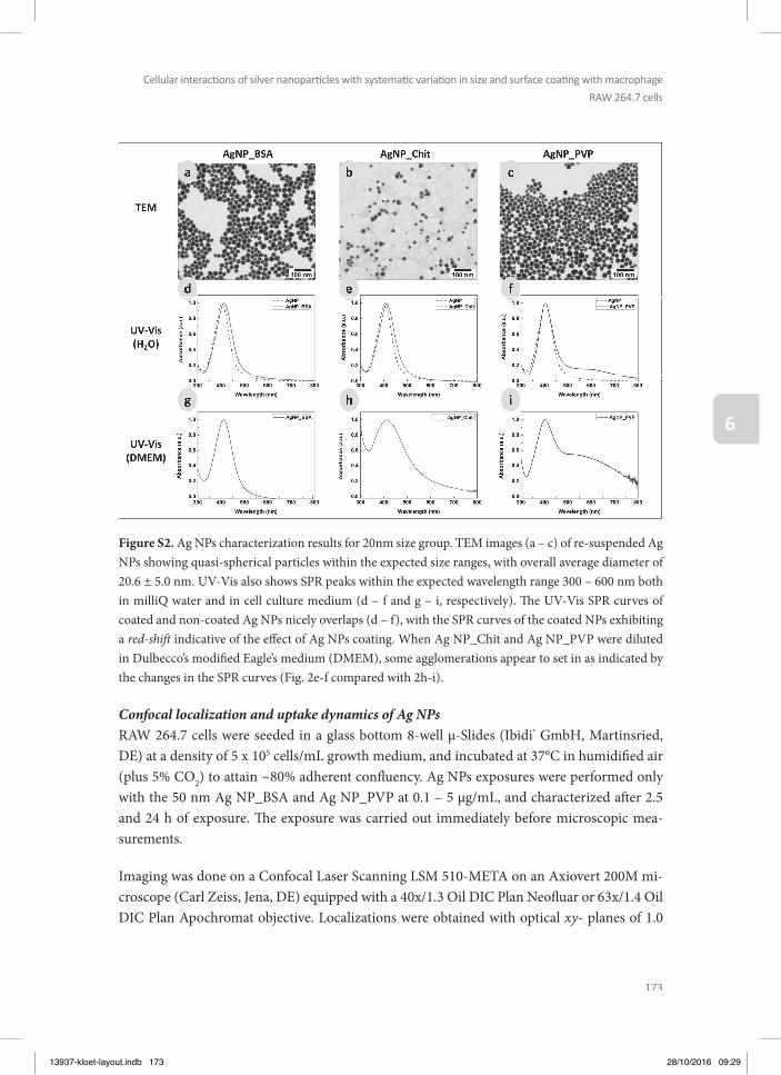
173
Cellular interacti ons of silver nanoparti cles with systemati c variati on in size and surface coati ng with macrophage
RAW 264.7 cells
6
Figure S2. Ag NPs characterization results for 20nm size group. TEM images (a – c) of re-suspended Ag NPs showing quasi-spherical particles within the expected size ranges, with overall average diameter of 20.6 ± 5.0 nm. UV-Vis also shows SPR peaks within the expected wavelength range 300 – 600 nm both in milliQ water and in cell culture medium (d – f and g – i, respectively). Th e UV-Vis SPR curves of coated and non-coated Ag NPs nicely overlaps (d – f), with the SPR curves of the coated NPs exhibiting a red-shift indicative of the eff ect of Ag NPs coating. When Ag NP_Chit and Ag NP_PVP were diluted in Dulbecco’s modifi ed Eagle’s medium (DMEM), some agglomerations appear to set in as indicated by the changes in the SPR curves (Fig. 2e-f compared with 2h-i).
Confocal localization and uptake dynamics of Ag NPsRAW 264.7 cells were seeded in a glass bottom 8-well µ-Slides (Ibidi® GmbH, Martinsried, DE) at a density of 5 x 10⁵ cells/mL growth medium, and incubated at 37°C in humidifi ed air (plus 5% CO2) to attain ~80% adherent confl uency. Ag NPs exposures were performed only with the 50 nm Ag NP_BSA and Ag NP_PVP at 0.1 – 5 µg/mL, and characterized aft er 2.5 and 24 h of exposure. Th e exposure was carried out immediately before microscopic mea-surements.
Imaging was done on a Confocal Laser Scanning LSM 510-META on an Axiovert 200M mi-croscope (Carl Zeiss, Jena, DE) equipped with a 40x/1.3 Oil DIC Plan Neofl uar or 63x/1.4 Oil DIC Plan Apochromat objective. Localizations were obtained with optical xy- planes of 1.0
13937-kloet-layout.indb 173 28/10/2016 09:29
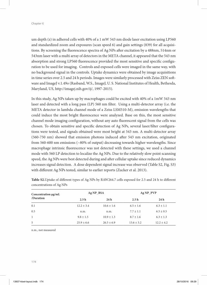
174
Chapter 6
um depth (z) in adhered cells with 40% of a 1 mW 543 nm diode laser excitation using LP560 and standardized zoom and exposures (scan speed 6) and gain settings (839) for all acquisi-tions. By screening the fl uorescence spectra of Ag NPs aft er excitation by a 488nm, 514nm or 543nm laser with a multi array of detectors in the META channel, it appeared that the 543 nm absorption and strong LP560 fl uorescence provided the most sensitive and specifi c confi gu-ration to be used for imaging. Controls and exposed cells were imaged in the same way, with no background signal in the controls. Uptake dynamics were obtained by image acquisitions in time series over 2.5 and 24 h periods. Images were similarly processed with Zeiss ZEN soft -ware and ImageJ v.1.49o (Rasband, W.S., ImageJ, U. S. National Institutes of Health, Bethesda, Maryland, US, http://imagej.nih.gov/ij/, 1997-2015).
In this study, Ag NPs taken up by macrophages could be excited with 40% of a 1mW 543 nm laser and detected with a long pass (LP) 560 nm fi lter. Using a multi-detector array (i.e. the META detector in lambda channel mode of a Zeiss LSM510-M), emission wavelengths that could induce the most bright fl uorescence were analyzed. Base on this, the most sensitive channel mode imaging confi guration, without any auto fl uorescent signal from the cells was chosen. To obtain sensitive and specifi c detection of Ag NPs, several laser/fi lter confi gura-tions were tested, and signals obtained were most bright at 543 nm. A multi-detector array (560-750 nm) showed that emission photons induced aft er 543 nm excitation, originated from 560-600 nm emissions (~80% of output) decreasing towards higher wavelengths. Since macrophage intrinsic fl uorescence was not detected with these settings, we used a channel mode with 560 LP detection to localize the Ag NPs. Due to the relatively slow point scanning speed, the Ag NPs were best detected during and aft er cellular uptake since reduced dynamics increases signal detection. A dose dependent signal increase was observed (Table S2, Fig. S3) with diff erent Ag NPs tested, similar to earlier reports (Zucker et al. 2013).
Table S2.Uptake of diff erent types of Ag NPs by RAW264.7 cells exposed for 2.5 and 24 h to diff erent concentrations of Ag NPs
Concentration µg/mL/Duration
Ag NP_BSA Ag NP_PVP
2.5 h 24 h 2.5 h 24 h
0.1 12.2 ± 3.4 10.6 ± 1.6 6.5 ± 1.6 6.3 ± 1.1
0.5 n.m. n.m. 7.7 ± 1.1 6.5 ± 0.5
1 9.8 ± 1.5 10.9 ± 1.3 8.7 ± 1.6 6.3 ± 1.3
5 25.9 ± 6.6 26.5 ± 6.9 13.6 ± 3.2 12.2 ± 4.2
n.m., not measured
13937-kloet-layout.indb 174 28/10/2016 09:29

175
Cellular interacti ons of silver nanoparti cles with systemati c variati on in size and surface coati ng with macrophage
RAW 264.7 cells
6
Th e fl uorescence signals correlating to the Ag NPs were quantifi ed based on mean pixel in-tensity of fi elds of view of representative cytoplasmic domains (Fig. S3 and S4), and were expressed as arbitrary units (AU). At identical exposures as well as imaging and processing conditions, these fl uorescence intensity values refl ect uptake of the diff erent Ag NPs by mac-rophage cells. To allow Ag NP uptake comparisons, quantifi cations of cytoplasmic dense re-gions from randomly selected cells with similar cellular confl uency (70 – 80%) and z-depths were made (Fig. S3). Th e selection of representative cytoplasm dense regions was based on the visual images. Th is selection was then applied to the fl uorescence signal, and the AU quantifi ed. In this way, regions analyzed were selected based on the occurrence of cytoplasm, without the bias of occurrence of silver signal. Based on the AUs estimated, the uptake of the negatively charged Ag NP_BSA was rapid and most signifi cant. Due to agglomeration in the suspension and growth medium, it was not possible to assess the uptake of Ag NP_Chit and will not be discussed further. Th e cellular uptake of Ag NP_BSA was twice that of Ag NP_PVP at similar exposure concentrations and time (Table S2), although the uptake effi ciency of individual cells does vary a lot.
13937-kloet-layout.indb 175 28/10/2016 09:29
176
Chapter 6
REFERENCES
Bastus NG, Merkoci F, Piella J, Puntes V (2014) Synthesis of Highly Monodisperse Citrate-Stabilized Silver Nanopar-ticles of up to 200 nm: Kinetic Control and Catalytic Properties Chem Mater 26:2836-2846 doi:10.1021/cm500316k
Evanoff DD, Jr., Chumanov G (2005) Synthesis and optical properties of silver nanoparticles and arrays Chemphy-schem 6:1221-1231 doi:10.1002/cphc.200500113
Hussain SM, Javorina AK, Schrand AM, Duhart HM, Ali SF, Schlager JJ (2006) Th e interaction of manganese nanoparticles with PC-12 cells induces dopamine depletion Toxicol Sci 92:456-463 doi:10.1093/toxsci/kfl 020
Kloet SK et al. (2015) Translocation of positively and negatively charged polystyrene nanoparticles in an in vitro placental model Toxicol In Vitro 29:1701-1710 doi:10.1016/j.tiv.2015.07.003
Makama S, Peters R, Undas A, van den Brink NW (2015) A novel method for the quantifi cation, characterisation and speciation of silver nanoparticles in earthworms exposed in soil Environmental Chemistry 12:643-651 doi:10.1071/EN15006
Martel C, Huynh le H, Garnier A, Ventura-Clapier R, Brenner C (2012) Inhibition of the Mitochondrial Perme-ability Transition for Cytoprotection: Direct versus Indirect Mechanisms Biochem Res Int 2012:213403 doi:10.1155/2012/213403
Pan YL (2015) Detection and characterization of biological and other organic-carbon aerosol particles in atmosphere using fl uorescence J Quant Spectrosc Ra 150:12-35 doi:10.1016/j.jqsrt.2014.06.007
Zucker RM, Daniel KM, Massaro EJ, Karafas SJ, Degn LL, Boyes WK (2013) Detection of silver nanoparticles in cells by fl ow cytometry using light scatter and far-red fl uorescence Cytometry Part A 83:962-972 doi:10.1002/cyto.a.22342
MOV_Ag NPs.avi: Uptake dynamics (speeded up 1600x) showing diff erent densities of Ag NP uptake by macro-phages during 5 h exposure to 5 µM Ag NP_BSA. Maximal intensity z-projections were made of sections in the 7-14 µm midrange subset. Th e red moving dots visible in the cytoplasm of cells indicate Ag NPs (also some extracellular).
13937-kloet-layout.indb 176 28/10/2016 09:29
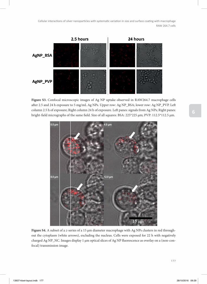
177
Cellular interacti ons of silver nanoparti cles with systemati c variati on in size and surface coati ng with macrophage
RAW 264.7 cells
6
Figure S3. Confocal microscopic images of Ag NP uptake observed in RAW264.7 macrophage cells aft er 2.5 and 24 h exposure to 5 mg/mL Ag NPs. Upper row: Ag NP_BSA; lower row: Ag NP_PVP. Left column 2.5 h of exposure; Right column 24 h of exposure. Left panes: signals from Ag NPs; Right panes: bright-fi eld micrographs of the same fi eld. Size of all squares: BSA: 225*225 µm; PVP: 112.5*112.5 µm.
Figure S4. A subset of a z-series of a 15 µm diameter macrophage with Ag NPs clusters in red through-out the cytoplasm (white arrows), excluding the nucleus. Cells were exposed for 22 h with negatively charged Ag NP_NC. Images display 1 µm optical slices of Ag NP fl uorescence as overlay on a (non-con-focal) transmission image.
13937-kloet-layout.indb 177 28/10/2016 09:29

12
34
56
78
910
1112
A B C D E F G G H
13937-kloet-layout.indb 178 28/10/2016 09:29
12
34
56
78
910
1112
A B C D E F G G H
Chapter 7
General discussion, future perspectives and conclusions
13937-kloet-layout.indb 179 28/10/2016 09:29
180
Chapter 7
GENERAL DISCUSSION
Th e production of nanoparticles (NPs) has increased in the last decades and the number of products in which NPs are being incorporated is still growing. Th e rapid increase of nano-technology has several benefi ts for society, yet there is an increasing concern that exposure to NPs may result in signifi cant adverse health eff ects (Oberdorster et al. 2007, Hussain et al. 2015). Insight into the potential human health risks posed by NPs is essential for sustainable development and safe use of innovative products based on these materials. In the food sec-tor NPs are used in food packaging materials and in food itself, including food supplements (Athinarayanan et al. 2015). Since NPs are incorporated in a variety of consumer products, it is likely that the general population gets exposed to NPs, via NP-containing products such as food and food packaging materials but also personal care products, textiles and medicines. In order to assess whether exposure to these NPs may pose a human health risk, NPs should be assessed for their safety. Testing all these NPs using in vivo animal experiments is undesir-able for scientifi c, ethical and societal reasons. In order to reduce in vivo testing, alternative methods for testing of chemicals are currently being developed and validated (Eisenbrand et al. 2002, Van der Jagt et al. 2004, Stokes 2015). It would be desirable that the safety and risk assessment of NPs could be largely based on studies using in vitro models instead of in vivo models as this would reduce costs and time required to test the large numbers of NPs. Use of in vitro models may also help to defi ne structure activity relationships and facilitate read-across within classes of NPs. Previous research has indicated that NP toxicity can be related to a certain extent to their physicochemical properties (Fourches et al. 2010). It would be of great value to get more insight in such relationships, to be able to set priorities for safety testing.
Th e aim of the present thesis was to investigate the potential of in vitro testing strategies to detect hazards of NPs, focusing on toxicokinetic as well as toxicodynamic endpoints. Toxi-cokinetic studies focused on translocation of NPs in in vitro models of the placental barrier, while toxicodynamic studies were directed at two endpoints that represent potential hazards of NPs that have not yet been well characterized including developmental toxicity and immu-notoxicity. In the present thesis diff erent types of NPs were used. Polystyrene nanoparticles (PS-NPs) were selected because of their commercial availability, with high quality and a wide variety of available physicochemical properties like surface charge, and fl uorescent labeling enabling easy detection in toxicokinetic (translocation) studies. Several metal (oxide) NPs were selected as well, of which some are possible constituents of food additives like TiO2, Fe2O3, SiO2 and Ag. Other metal oxide NPs that were selected were Mn2O3, CuO, Cr2O3, CoO and NiO to which we may be exposed via products like paints, catalysts, construction mate-rials, coatings and batteries.
Chapter 1 introduced the aim of the thesis and the various in vitro models and NPs used in the studies performed. Literature data indicate that translocation of NPs is possible across
13937-kloet-layout.indb 180 28/10/2016 09:29

181
General discussion, future perspecti ves and conclusions
7
the gut, lung and skin barriers, related to oral, inhalation and dermal exposure, respectively (Landsiedel et al. 2012). In order to estimate the in vivo internal exposure, several in vitro models have been developed to study NP translocation across the lung, gut, skin and placental barrier (Mahler et al. 2012, Prow et al. 2012, George et al. 2015, Poulsen et al. 2015). Chap-ter 2 of the present thesis provided a review about the performance of such in vitro models for studies on toxicokinetics of NPs. Th e chapter described models that mimic the diff erent barriers of the human body, with a focus on the lung, gut, skin and placental barrier, thus providing an overview of the state-of-the-art of using in vitro model systems, ranging from single cell-type monolayer to multi-cell (3D) models, to characterize the translocation of NPs across these diff erent barriers. In this chapter it was concluded that at the current state-of-the-art, the in vitro models do not yet allow to predict the absolute transfer rates but they do support the defi nition of relative transfer rates and can thus help to reduce animal testing by setting priorities for subsequent in vivo testing. Th e use of in vitro models is very promising since they are currently further improved to mimic the in vivo situation more closely by, for example, using co-cultures of diff erent cell types and implementing them in a microfl uidic chip format. When these models are further validated by testing exactly the same NPs in an in vivo set-up and in the in vitro model, they could be even used to determine the internal expo-sure (bioavailability) of NPs in an absolute way, besides to set priorities for NP toxicity testing.
Since part of the toxicodynamic studies of the present thesis were directed at developmen-tal toxicity testing of NPs, an important toxicokinetic aspect studied in the present thesis in chapter 3 was placental translocation of NPs. In order to obtain insight in toxicity and trans-location of NPs across the placental barrier, cytotoxicity and translocation was studied for one positively and two negatively charged PS-NPs of 50 nm in an in vitro model of the placenta. Th e model consisted of BeWo b30 cells, derived from a human choriocarcinoma grown on a transwell insert forming a cell layer that separates an apical from a basolateral compartment. PS-NPs were characterized with respect to size, surface charge, morphology and protein co-rona. In this study it appeared that in spite of similar size, surface charge and type of proteins in the protein corona, the diff erently charged NPs displayed a remarkable diff erence in cyto-toxicity, with only the PS-NPs with an original positive charge inducing cytotoxicity. Th e rel-atively higher cytotoxicity of positively as compared to negatively charged NPs was reported before for tri-block copolymer NPs (Bhattacharjee et al. 2012) but also for other type of NPs like gold NPs (Liu et al. 2013) and silicon NPs (Bhattacharjee et al. 2013).
Translocation of PS-NPs appeared not to be related to PS-NP charge alone. A remarkable diff erence in translocation was found between the two 50 nm negatively charged PS-NPs that were obtained from diff erent manufacturers. For the PS-NPs from Polysciences, the appar-ent permeability coeffi cient (Papp) was slightly higher than that of amoxicillin, a compound that is known to be translocated over the in vitro BeWo b30 placental barrier and the ex vivo
13937-kloet-layout.indb 181 28/10/2016 09:29

182
Chapter 7
placental barrier to only a limited extent (Li et al. 2013), with a Papp value of 13 x 10-6 cm/s. When testing the negatively charged PS-NPs from Magsphere, no PS-NPs could be detect-ed in the basolateral compartment upon adding a concentration of 10 µg/ml to the apical compartment and 24 hours incubation. Th is remarkable diff erence in translocation of the two similar sized negatively charged PS-NPs across the BeWo b30 placental barrier is in line with the 30-fold diff erence that was found in translocation of these two negatively charged PS-NPs across a Caco-2 monolayer in a similar transwell model system (Walczak et al. 2014). Since none of the characterized parameters, including size, surface charge and protein corona revealed remarkable diff erences between the two negatively charged NPs, the diff erence may originate from the chemical groups on the surface of the NPs generating the negative charge. Th e general conclusion from this study was that the in vitro BeWo b30 model can be used as a fast method to get an initial qualitative impression about the capacity of NPs to translocate across the placental barrier and to set priorities for further in vivo studies on translocation of NPs to the fetus.
Chapter 4 investigated whether the same PS-NPs as tested in chapter 3 for placental translo-cation, are able to cause in vitro developmental toxicity in the ES-D3 cell diff erentiation assay of the embryonic stem cell test (EST) focusing also on the eff ect that charge may have. Th e study showed that the two negatively charged PS-NPs did not show any eff ect in the ES-D3 cell diff erentiation assay up to the highest concentration tested while the positively charged PS-NP showed a concentration-dependent inhibition of ES-D3 cell diff erentiation. However, eff ect concentrations in the ES-D3 cell diff erentiation assay are close to cytotoxic concentra-tions, which indicated that the inhibition of the ES-D3 cell diff erentiation may be due to cyto-toxic eff ects of the positively charged PS-NPs. Th is indicated that the inhibition of the ES-D3 cell diff erentiation by the positively charged PS-NPs may be caused by non-specifi c eff ects.
Th e ES-D3 cell diff erentiation assay is a useful tool to determine whether chemicals and NPs can pose a hazard for the embryonic development. However, to possibly become a risk for embryonic development, NPs need to be able to reach the developing embryo in the in vivo situation. Th erefore, upon oral exposure, NPs would need to translocate across the intesti-nal barrier as well as the placental barrier. Th e bioavailability of many NPs upon oral ex-posure has been reported to be low, because of a limited translocation across the intestinalbarrier (Walczak et al. 2014). However, in vivo studies using intravenous injection of NPs have shown that NPs can cause adverse eff ects to the developing embryo, indicating that NPs can translocate across the placenta in the in vivo situation (Hougaard et al. 2015). As indicat-ed above in chapter 3, the potential of the same PS-NPs as tested in the EST was assessed to translocate across an in vitro placental barrier of human placental (BeWo) cells. Th e results of these studies indicated that the positively charged PS-NPs were hardly translocated across the BeWo cell layer, which could indicate that fetal exposure of these PS-NPs is expected to be
13937-kloet-layout.indb 182 28/10/2016 09:29
183
General discussion, future perspecti ves and conclusions
7
low. Th erefore, although the EST data indicated the potential of positively charged PS-NPs to cause developmental toxicity, such concentrations may not be reached in the in vivo situation because of the low placental transfer. On the other hand, the negatively Polysciences PS-NPs which did translocate across the BeWo barrier albeit to a limited extent, appeared not to cause an adverse eff ect in the in vitro developmental toxicity assay, also pointing to a limited hazard all together. Th ese examples show that in order to predict which NPs may cause developmen-tal toxicity in the in vivo situation, data obtained in the in vitro ES-D3 cell diff erentiation assay could be combined with placental translocation data from the BeWo model in order to select the NPs that are expected to have the highest potency in vivo. Th ese estimations may then be used to select NPs that could be assessed for potential developmental toxicity in vivo. Combining the results from Chapter 3 and 4 allows the combination of kinetic considerations with the in vitro developmental toxicity assay to improve the prediction of the in vivo hazards as done previously by Li et al. (Li et al. 2015, Li et al. 2015) for a series of azoles, for which the correlation of the prediction of in vivo developmental toxicity improved from R2=0.57 to R2=0.95 when the ES-D3 cell diff erentiation data were combined with the relative transport rates obtained in the BeWo model.
A literature search was performed on the reported potencies of other NPs in the ES-D3 cell diff erentiation assay, in order to assess whether eff ects of NPs in the ES-D3 cell diff erentiation assay may also be related to physicochemical properties other than surface charge, such as size or coating. Although the experiments in chapter 3 of the present thesis showed that positively charged PS-NPs are more toxic than negatively charged PS-NPs, it appeared that this may not be generalizable to other NPs. Th is follows from the fact that SiO2, Ag and TiO2 NPs that were reported to inhibit ES-D3 cell diff erentiation were negatively charged, while the negatively charged PS-NPs of the present study did not aff ect ES-D3 cell diff erentiation. Th is implies that the negative charge did not arise from serum proteins which may aff ect the NP charge, as observed in the present study when NP charge was measured in medium with FCS. To obtain better insight in the role of NP surface charge on developmental toxicity more studies are required that assess the eff ects of NPs with diff erent charge that are made of the same core material. Th e analysis of the available literature also indicated that the in vitro developmen-tal toxicity potency of NPs may also largely depend on the coating applied. However, from the available data it cannot be concluded whether the toxicity potential is dependent on the coating alone, or whether it is also dependent on the combination of the core material and the coating. Th erefore, although the limited data available indicate that charge, size and coating of NPs may be important characteristics that determine the developmental toxicity potential of NPs, more (systematic) studies are needed to assess how physicochemical characteristics of NPs relate to their developmental toxicity. Th is information may help to prioritize NPs for in vitro and in vivo developmental toxicity testing.
13937-kloet-layout.indb 183 28/10/2016 09:29

184
Chapter 7
In chapter 5, toxic eff ects of a series of metal (oxide) NPs were tested in macrophage RAW264.7 cells in order to obtain insight in eff ect of these NPs on cells of the innate immune response. In these macrophage RAW264.7 cells the eff ects of the metal (oxide) NPs were characterized on cell viability, TNF-α production and mitochondria-related parameters like production of reactive oxygen species (ROS), mitochondrial permeability transition pore (MPTP) opening, and intracellular ATP levels, since previous research indicated that mitochondria play an im-portant role in NP-induced toxicity (Park et al. 2008, Sharma et al. 2011, Shvedova et al. 2012, Bhattacharjee et al. 2013). Th e results obtained indicated that TiO2, Fe2O3, Cr2O3, CoO and NiO NPs showed no eff ects on the selected endpoints up to the highest concentration tested (200 µg/ml), and that SiO2 and Ag NPs caused concentration-dependent adverse eff ects in the RAW264.7 cells.
Th e results showed that SiO2 NPs were the most toxic ones among the tested food-related NPs. Exposure to food-related NPs is expected to be mainly via the gastrointestinal tract. When food products containing SiO2 were tested in an in vitro digestion model, simulating the conditions of the human gastrointestinal tract, it was found that SiO2 NPs (5-200 nm) were still present in the intestinal content (Peters et al. 2012). Th is indicates that it is most like-ly that upon consumption of foods containing particulate SiO2, the gut epithelium is exposed to SiO2 NPs (5-200 nm) (Peters et al. 2012). To enter the systemic circulation, NPs need to be absorbed in the gastrointestinal tract. In an in vivo study in rats it was shown that 6.6-9.7% of both SiO2 NPs of 20 and 100 nm were absorbed into the systemic circulation, with no signifi -cant eff ect of the particle size, when orally administered at a single dose of 500 and 1000 mg/kg (Lee et al. 2014), indicating that SiO2 NPs can get translocated across the intestinal barrier.
With an estimated daily intake of SiO2 in the nano-size range of 1.8 mg/kg bw/day (van der Zande et al. 2014, van Kesteren et al. 2014), the daily intake of a 70 kg person would be 126 mg. In the adult human the gastrointestinal tract represents a considerable surface area of about 200 m2 which has the potential to interact with ingested NPs (Bergin et al. 2013). In ad-dition the gastrointestinal tract has localised representations of the adaptive immune system in the form of gut-associated lymphoid tissue (GALT) aggregates (Peyer’s Patches) (Bergin and Witzmann 2013). Th ese Peyer’s patches account for 1% of the total surface area of the gastrointestinal tract (des Rieux et al. 2006). About 9 litres of fl uid pass through the gastro-intestinal tract each day (Sherwood L. 2014). Th us, upon oral intake of 126 mg SiO2 per day
this would lead to a concentration of 14 µg/ml, assuming full distribution of the NPs over the entire intestinal content, which is lower than the lowest observed adverse eff ect concentration of 100 µg/mL in the present study. Only when using a worst case assumption of only limited dilution of an oral dose of 126 mg SiO2 NPs in the human intestine, local concentrations can be expected to reach the eff ect levels observed in vitro.
13937-kloet-layout.indb 184 28/10/2016 09:29
185
General discussion, future perspecti ves and conclusions
7
Furthermore, if 100% of the estimated daily intake of SiO2 NPs (126 mg) would be transported from the intestines into the blood, assuming 5 liters of blood and a 100% clearance per day, a maximal concentration of 25 µg/ml would be reached if NPs would not distribute to tissues. Also this concentration is lower than the lowest observed adverse eff ect concentration of 100 µg/mL defi ned in the present thesis. Considering that the oral bioavailability is lower than 10% and that NPs also distribute to tissues (Lee et al. 2014), concentrations higher than 2.5 µg/mL are not likely to be reached in a real-life exposure scenario. Th is concentration is far below the concentrations of SiO2 causing in vitro toxicity in the present study, suggesting that in vivo toxic eff ects of food additive related SiO2 NPs on macrophages, are expected to be limited. Th is is in line with the results from an oral repeated-dose toxicity study in which male Wistar rats were exposed to SiO2 NPs via the diet for 28 or 84 days, showing no toxicity even at the highest dose level tested (2500 mg/kg bw/day) (van der Zande et al. 2014).
It has to be kept in mind that NPs may not only aff ect the gastrointestinal cells directly but may also infl uence the microbiome although the evaluation of eff ects of NPs on the micro-biome is in its early infancy (Pietroiusti et al. 2015). For example, the cytotoxicity of Ag NPs on Escherichia coli as representative for intestinal bacteria was compared with the cytotoxicity of Ag NPs on gastrointestinal cells. Th is revealed that Ag NPs aff ected bacteria at lower con-centrations than the gastrointestinal cells indicating that Ag NPs may cause adverse eff ects by selectively aff ecting the gut microbiota (Frohlich et al. 2016). In a study using an in vitro colon model (Taylor et al. 2015), exposure to TiO2 , ZnO and CeO2 NPs induced signifi cant phenotype changes of the gut microbial community, including eff ects on the production of short chain fatty acids which are the key metabolites in the control of energy homeostasis and have been implicated in the development of obesity (Schwiertz et al. 2010). For SiO2 and other food-related NPs, however, the occurrence of such eff ects remains to be investigated.
Altogether, results obtained in chapter 5 showed no or limited eff ects of the NP formulations of metal (oxide) food additives on cell viability, ROS production, MPTP opening, ATP levels and TNF-α production in RAW264.7 macrophages. Eff ects were only observed at high con-centrations that may not be physiologically relevant, indicating that related adverse eff ects upon exposure to the respective NPs in vivo may be limited.
Of the NPs tested in chapter 5, Ag NPs were among the NPs showing most eff ects. Th e vol-ume of Ag NPs-based products entering the market is relatively high, making the risk as-sessment of Ag NPs a priority. Th erefore to elucidate the factors that drive Ag NPs’ potential to pose health risks, chapter 6 assessed the eff ects of specifi cally synthetized Ag NPs with two diff erent sizes (20 and 50 nm) and with three diff erent surface coatings (chitosan: Ag NP_Chit, bovine serum albumin: Ag NP_BSA, polyvinylpyrrolidone: Ag NP_PVP) in the same in vitro RAW264.7 model. Th e eff ect of surface charge and size of NPs was character-
13937-kloet-layout.indb 185 28/10/2016 09:29
186
Chapter 7
ized on cell viability, TNF-α production, ROS production, MPTP opening, and intracellular ATP levels. Decreased cell viability was observed for all Ag NPs tested. Th e results from MTT, ATP and MPTP opening assays showed that adverse eff ects from exposure to the tested Ag NPs on these endpoints were similar irrespective of type or size. However, the role of surface coating/charge in infl uencing TNF-α induction was demonstrated by the fact that only neg-atively charged Ag NP_BSA induced TNF-α production (80x higher than control). Further, signifi cant ROS induction was only observed with the 20 nm positively charged Ag NP_Chit. Eff ect of size was less prominent under the experimental conditions, showing only minimal diff erences that were not statistically signifi cant. Overall, the negatively charged Ag NP_BSA appeared to be most potent in inducing adverse eff ects on the macrophages. Th is toxicity of the negatively charged Ag NPs is the opposite of what was found in chapter 3 for the PS-NPs, since the positively charged PS-NPs appeared to be the most cytotoxic to BeWo cells. So this points again to the direction that the eff ect of surface charge may vary with the type of NPs investigated, indicating that more studies with other NPs with diff erent core material and with diff erent charges are required. One has to keep in mind that Ag NPs are known to release ions which can also contribute to toxicity even if the Ag NPs has a negative surface charge, and in general the smaller the Ag NP the higher the ion release (Johnston et al. 2010, Reidy et al. 2013). Given this ion release, the PS-NPs used in chapter 3 and 4 seem to be a better model to evaluate the eff ect of charge or size since they do not present these additional factors leading to toxicity.
Taken together, the present thesis provided further evidence of the infl uence of physicochem-ical properties of NPs in driving toxicity in an in vitro model. However, the determination of the fate and toxicity of NPs using in vitro or in vivo models is a challenge that needs further evaluation. A combination of several factors likely play a role in determining the outcome of exposure including factors like NP core material and presence and type of coating agents resulting in various physicochemical properties (size, charge, etc.), as well as the toxicity test models used and end-points studied. Th is has been observed in earlier reviews (Johnston et al. 2010, Reidy et al. 2013) and appears to hamper conclusive evaluation of the role of physi-cochemical characteristics of NPs in the modes of action underlying their potential hazards and risks so far.
Th e results obtained do show however that in vitro assays can detect diff erences in potential hazards posed by NPs. Th erefore it is concluded that the results of the work presented in this thesis will contribute to the further development and use of non-animal based testing strat-egies for safety testing of NPs providing insight into selected potential hazards of the tested NPs.
13937-kloet-layout.indb 186 28/10/2016 09:29

187
General discussion, future perspecti ves and conclusions
7
FUTURE PERSPECTIVES
Although the present thesis has extended the understanding and prediction of potential haz-ards of diff erent NPs, several issues remain to be elucidated to fully understand the hazards of NPs to human health and the environment. Th e current section presents some considerations on future topics of importance in this fi eld. Th ese topics include:
- Use of alternative testing strategies and especially the EST to detect developmental toxicity.
- Physicochemical characterization of NPs
- Predictive toxicology: Nano-QSAR models
- Dosimetry
- Th e role of the microbiome
Use of alternative testing strategies and especially the EST to detect developmental toxicity. Given the high number of NPs for which at present in vivo toxicity data are missing the po-tential to use alternative in vitro testing methods to detect possible health hazards of NPs, is of high importance to set priorities in future safety testing and also to support development of safer NPs. An important endpoint to include in these activities is developmental toxicity since this is an endpoint requiring relatively large numbers of experimental animals (Rovida et al. 2009). In the present thesis the ES-D3 cell diff erentiation assay of the EST was used. Th e main advantage of this assay over other alternative assays for developmental toxicity testing is that it does not require the use of any animals because it uses an immortal cell line isolated from a mouse blastocyst. Th e EST is also relatively easy to carry out and the endpoints used in the assay do not require extensive knowledge on morphologic development (de Jong et al. 2011).
However, the diff erentiation of the stem cells into beating cardiomyocytes as only endpoint might not be suffi cient for determination of all possible embryotoxic eff ects (zur Nieden et al. 2004). With the EST, only the eff ects on mesodermal development are recorded. But some substances and also NPs might have embryotoxic eff ects on specifi c cell-types other than cardiomyocytes. Th is may lead to false negative results if the test compound aff ects cell types other than the myocardium. Th erefore, also the diff erentiation into neural and endothelial cells, chondrocytes and osteoblasts should be investigated (Buesen et al. 2004).
Other alternative assays for in vivo developmental toxicity are the rat Whole Embryo Culture (WEC) and the limb bud micromass (MM) test (Genschow et al. 2000, Augustine-Rauch et al. 2010). Th e WEC is used to assess the eff ects of chemicals on the development of the com-plete embryo within its intact visceral yolk sac outside the uterus, during a critical phase in organogenesis (de Jong et al. 2011). Th e test makes use of mammalian embryos, but both the
13937-kloet-layout.indb 187 28/10/2016 09:29

188
Chapter 7
exposure and the assessment of eff ects occur in vitro (Piersma et al. 2008). Th erefore, WEC systems off er the most complete in vitro alternative for animal testing for developmental tox-icity. Th e test does require animal material, but has the advantage of representing embryo-genesis in full complexity from cellular proliferation and diff erentiation to pattern formation (Piersma 2004).
In the MM test, chondrogenesis of mesenchymal cells of limb buds of rat embryos is studied (Kistler 1987). Th e test involves exposing cultured chick, mouse or rat embryo midbrain or limb cells to test agents for varying times and estimating the inhibition of cell diff erentiation in these cultures. Th e MM assay reported that when cells from the undiff erentiated mesen-chyme of early chick embryo limbs are cultured in small volumes at high density, they formed numerous small aggregates (foci) of cells of diff erentiating chondrocytes within a background of apparently undiff erentiated cells (Flint 1993). In principle, the MM developmental toxicity test is based on detecting the ability of a particular compound to inhibit the formation of foci. Embryotoxic compounds will reduce the number of foci or the number of cells within foci (Spezia et al. 2013). Both the WEC and MM assay rely on a constant source of embryonic tissues from pregnant animals. However the MM assay is considered most useful for iden-tifying strongly embrytotoxic compounds (Genschow et al. 2004) since the predictivity for non-embryotoxic compounds and the precision for weakly embryotoxic compounds were judged to be insuffi cient.
Another in vitro test for embryotoxicity is the so-called chick embryotoxicity screening test (CHEST) assay. Th e main endpoints assessed using the CHEST are mortality, malformations, embryo development, blood vessel development and blood vessel coloration. Th e test com-pounds can easily be administered to the eggs and eff ects on the developing embryo can be investigated. However, the CHEST has been criticised for not being able to distinguish general toxicity from specifi c developmental eff ects and the absence of mammalian maternal-foetal relations (Jelinek et al. 1985).
A fi nal possible method to study in vitro developmental toxicity is the Zebrafi sh Embryo-toxicity Test (ZET). Th is test uses non-mammalian embryos. Th e eff ects of compounds are tested on the development of embryos of the zebrafi sh. In principle, the fertilized fi sh eggs are exposed to diff erent concentrations of a test substance. At diff erent time points, the exposed developing fi sh embryos are observed and scored for lethal and embryotoxic eff ects. Th e ZET has been used for testing embryotoxiciy of Ag NPs (van Aerle et al. 2013, Yoo et al. 2016), ZnO, SiO2, Al2O3, Pt and Au NPs (George et al. 2011) and TiO2 NPs (Brundo et al. 2016, Shih et al. 2016). Yoo et al. 2016 investigated embryotoxicity eff ects of 10 nm Ag NPs during zebrafi sh development, using the following parameters: abnormal morphology, mortality, hatching rate and heart rate. Th e hatching rate diff ered not signifi cantly between the exposure
13937-kloet-layout.indb 188 28/10/2016 09:29
189
General discussion, future perspecti ves and conclusions
7
groups while the Ag NP exposed group showed increased mortality (11%) and teratogenicity (15%) at 72 h compared to the control group for which these values amounted to 2% and 0% respectively. Th e heart rate of Ag NP exposed embryos tended to be lower than that of the control group but diff erences were not statistically signifi cant (Yoo et al. 2016). Van Aerle et al. (2013) investigated the eff ects of exposure to bulk Ag, 10 nm Ag NPs or ionic Ag to ze-brafi sh embyros using a Next Generation Sequencing approach. All treatments aff ected gene expression and those genes associated with oxidative phosphorylation and protein synthesis overlapped strongly between the three treatments indicating similar mechanisms of toxicity for the three forms of silver studied (van Aerle et al. 2013).
One study investigated a batch of commercial metal/metal oxide NPs both for their cytotoxic potential in vitro using automated epifl uorescence microscopy on mammalian BEAS-2B and RAW 264.7 cells and in vivo using the zebrafi sh (George et al. 2011). Th e automated epifl uo-rescence procedure measures sub-lethal (e.g. intracellular Ca2+fl ux and mitochondrial pertur-bations) and lethal (cell death with increased membrane permeability) responses. Among the NPs analyzed, Pt, Ag, SiO2, Al2O3, and Au NPs triggered sub-lethal eff ects without cytotoxic-ity. Quantum Dots (QDs) and ZnO NPs triggered sub-lethal eff ects and cytotoxicity. In order to compare the in vitro with the in vivo response outcomes in zebrafi sh embryos, NPs were tested for their impact on mortality rate, hatching rate, cardiac rate, and morphological de-fects. While QDs, ZnO and Ag NPs induced morphological abnormalities or interfered with embryo hatching, Pt and Ag NPs exerted inhibitory eff ects on cardiac rate at the sub-lethal dose of 15 μg/mL. Ag NP-treated embryos developed bent spines, short body sizes, pericar-dial edema, submandibular edema and bent tails at 120 hrs, while the majority of ZnO-treat-ed embryos remained unhatched and embryos treated with QDs died early and underwent complete dissolution within 24 hrs. Ag NP was clearly more toxic in zebrafi sh embryos than towards mammalian cells in vitro (George et al. 2011).
Physicochemical characterization of NPsTh e ability to create NPs with a wide variety of characteristics with respect to chemistry, shape, size and surface properties led to attention for the nanotoxicological aspects regarding of the physicochemical characteristics of NPs (Westmeier et al. 2016). It is well known that the biological activity of NPs depends on their physicochemical parameters including size, shape, surface area, surface charge, crystal structure, chemical composition and surface chemistry (Fadeel et al. 2015). Generally, by using the currently available methods, it is only possible to measure a single or several characteristic(s) of NPs but not all (Bouwmeester et al. 2011). Investigations of the NPs behaviour in physiological media are very important to interpret the toxicological results. As an example the formation of a protein corona formation can cause changes in charge, size and surface chemistry of a certain NP thereby impacting on its uptake and cellular fate (Corbo et al. 2016). Th erefore, physicochemical characteristics and biological
13937-kloet-layout.indb 189 28/10/2016 09:29
190
Chapter 7
eff ects should preferably be measured under the same experimental conditions (Meissner et al. 2009).
In this thesis NPs were characterized in the medium used for the respective assay using SEM and dynamic light scattering (DLS) for size and zeta potential (for surface charge). Chapter 3 pointed out that only size and charge characterization of PS-NPs were not enough for inter-preting the results. Translocation of the diff erent PS-NPs across the BeWo cell layer was not found to be related to PS-NP charge and a remarkable diff erence in translocation was found between the two 50 nm negatively charged PS-NPs from diff erent manufacturers, where one did not translocate at all to the basolateral compartment. Th erefore the protein corona was also assessed using SDS-PAGE. Biomolecules rapidly adsorb to NPs as they have a high sur-face energy, leading to the formation of the protein corona which critically aff ects the NPs biological identity (Cedervall et al. 2007). Th e protein corona formation can infl uence the delivery properties of NPs, as well as their toxicity (Tenzer et al. 2013) and plays a critical role in cellular uptake (Pelaz et al.,2015), accumulation, biodegradation and clearance of NPs (Saptarshiet al., 2013). Protein corona analyses of the 3 diff erent PS-NPs in chapter 3 re-vealed diff erences in the amount but not the nature of the proteins in the corona of the three diff erent PS-NPs. Since none of the characterized parameters, including size, zeta potential and protein corona revealed remarkable diff erences between the two negatively charged NPs, it was argued that the diff erence may originate from the chemical groups on the surface of the NPs generating the negative charge. Previously it was demonstrated that three diff erent positively charged tri-block copolymer nanoparticles showed diff erent cellular uptake and toxicity in spite of a similar overall size and zeta potential, and that only a diff erent shielding of the charge due to subtle diff erences in the charge generating surface moiety infl uenced the cellular uptake and toxicity (Bhattacharjee et al. 2011). Such observations indicate that by characterising size, zeta potential and protein corona, even when done in the relevant medium or body fl uid, may prove insuffi cient for full characterization of the NP parameters that direct the biological eff ects and potential toxicity. Obviously this also hampers defi ni-tion of (Q)SARs and read-across and suggests that at the current state-of-the-art parameters generally used to characterise NPs physicochemical characteristics may prove insuffi cient to understand the diff erences in their toxicity.
Although techniques for characterization of NPs have progressed greatly over the past 10 years, further improvements are highly recommended. For example it would be advantageous to modify current standards such as DLS and TEM/SEM in order to perform them with a higher sensitivity and in a more relevant biological environment (Hussain et al. 2015). Limita-tions of the DLS method include that the particle size is correlated with the light scattered and the presence of a few large particles, for example from the culture medium or relevant bio-logical fl uid, could mask the presence of a high number of small particles (Handy et al. 2008).
13937-kloet-layout.indb 190 28/10/2016 09:29
191
General discussion, future perspecti ves and conclusions
7
Establishing nano-QSARs linking NP/corona properties to (patho)physiological responses remains a still distant goal despite the achievements of the past years. It is clear that charac-terizing the nano-cellular interactions is a critical step in the development of a fundamental understanding of nanotoxicology. International guidelines for physicochemical characteriza-tion are needed for regulatory purposes, and the methods used for characterization need to be standardized and validated (Fadeel et al. 2015).
Predictive toxicology: Nano-(Q)SAR models Because of the high costs and time involved in toxicological in vivo and in vitro studies, there has been considerable interest in computer based strategies defi ning alternative computa-tional methods, like the application of (quantitative) structure-activity relationship ((Q)SAR) models. (Q)SAR models are theoretical models that relate the structure or physicochemical characteristics of compounds to their biological activities. (Q)SAR models could be a useful screening tool, eliminating the need to test every single NP on an individual basis (Oksel et al. 2015). Considering the large number of factors that diff er from conventional chemicals and that are likely to infl uence the biological activity of NPs, it might also be concluded that tradi-tional (Q)SAR approaches need serious reconsiderations in order to be applied to NPs (Oksel et al. 2015). Th e simple application of classic (Q)SAR approaches based on the representation of various substances by the structure of their molecules is impossible for the majority of NPs (Toropova et al. 2016). Because of the high structural complexity and diversity of NPs, it is diffi cult to develop quantitative parameters capable of characterizing the structural and chemical properties of NPs. In addition, systematic studies that assess physicochemical and structural properties in relation to biological eff ects of NPs are scarce, making the develop-ment of predictive computational models and their validation diffi cult, since these procedures require relatively large amounts of data (Fourches et al. 2010). Moreover, due to the fact that large datasets are not available for a large part of the NPs one might need to develop a meth-odology that provides predictions based on smaller datasets. Few studies have reported on the development of nano-(Q)SARs. Recently, an approach was published to develop a (Q)SAR for fullerene NPs based on a small dataset (Toropova et al. 2016). In this study a QSAR was developed based on several data (conditions) related to (i) dose (g/plate); (ii) metabolic activation (with or without mix S9); and (iii) illumination (dark or irradiation) as a tool to predict mutagenicity of fullerene NPs in bacterial strains. Th ese computational experiments have shown that the described approach, based on a list of several conditions and not on mo-lecular structure as is usually done in the classical approach, can be a tool to build up models for the prediction of mutagenicity of fullerene NPs under diff erent conditions, based on a small dataset.
Burello et al. 2011, have proposed a theoretical framework to predict the oxidative stress po-tential of oxide NPs based on the available electronic energy levels in the NPs (Burello et al.
13937-kloet-layout.indb 191 28/10/2016 09:29
192
Chapter 7
2011). Fourches et al. 2010 investigated the potential of (Q)SAR modelling to establish statis-tically signifi cant relationships between measured biological activity profi les of NPs and their physical, chemical, and geometrical properties, either measured experimentally or computed from the structure of NPs (Fourches et al. 2010). Th ey studied a fi rst dataset of 51 NPs with diff erent metal cores which were tested in four cell lines (monocytes, hepatocytes, endothelial and smooth muscle cells) in four diff erent assays (ATP levels, reducing equivalents, caspase mediated apoptosis and mitochondrial membrane potential). It was demonstrated that NPs with similar biological activity can be also recognized as similar by their type of metal core, since NPs in one cluster featured the same metal core, independently of their surface modi-fi ers. In a second dataset they studied 109 NPs with the same core but with diff erent surface modifi cations for their cell uptake activity. Here the structure of organic small molecules con-jugated to the NP surface was the only diff erence between the 109 NPs. Factors that correlated with cellular uptake were: lipophilicity, molecular refractivity, specifi c Van der Waals surface area and electrostatic descriptors. Th ese fi ndings imply that the cellular behaviour of a NP dataset based on a common core can be predicted using (Q)SAR analysis of the surface mod-ifying ligands (Fourches et al. 2010).
At the moment the research for nano-(Q)SAR models continues. Th ere remains the need for thorough testing and validation of (Q)SAR methodologies, and there are still barriers that need to be overcome to establish predictive, reliable, and legally acceptable nano-(Q)SAR models. A main issue is the scarcity of consistent and high-quality experimental data which are needed to develop a robust and predictive nano-(Q)SAR model (Oksel et al. 2015). Th e scarcity of such data is mainly caused by diffi culties in standardizing nanotoxicity testing methodologies and characterization conditions. High quality data are required, which are consistent and systematically obtained for NPs that have been comprehensively character-ised under relevant exposure conditions prior to toxicological testing (Oksel et al. 2015). It is therefore suggested that, due to high variability in the molecular structures and diff erent mechanisms of toxicity, individual classes of NPs should be modelled separately (Puzyn et al. 2009). Th e establishment of standard protocols is essential to enable accurate measurements of physicochemical and biological properties of NPs (Oksel et al. 2015).
DosimetryAnother challenge is to arrive at a consensus about which dose metric is best to be used in nanotoxicological studies, for example mass, particle number or surface area (Hussain et al. 2015). Dosimetry is the accurate description of the dose or the amount of NPs that comes into contact with the biological target. It is an important aspect to take into account when predict-ing risks but it should be measurable and it should be possible to incorporate it in regulations. Currently the dose is oft en defi ned as the nominal dose, so the amount of NPs introduced into the culture medium expressed in mass per volume (µg/ml). However, it is also possible
13937-kloet-layout.indb 192 28/10/2016 09:29
193
General discussion, future perspecti ves and conclusions
7
to measure the intracellular or deposited doses (Fadeel et al. 2015). It has been suggested that the NP surface area could be more relevant in nanotoxicology studies than the mass unit (Oberdorster et al. 2007). Braakhuis et al. have demonstrated that for short-term inhalation of Ag NPs, the total surface area of Ag NPs that reach the alveoli seems the most suitable dose descriptor for the acute eff ects of Ag NPs in a range of sizes (Braakhuis et al. 2016). Th is is because the alveoli are the site of the observed toxicity and Ag NPs can release Ag ions, which possibly cause the observed eff ects. Th e release of Ag ions increases with decreasing particle size due to their relative larger surface area (Braakhuis et al. 2016). Th is indicates that for oth-er types of NPs that do not release ions the suitable dose descriptor could be diff erent. Th ere is probably no one-size-fi ts-all dose metric for NPs and various approaches depending on the type of NP and type of toxicological testing system might be needed to apply.
So dosimetry in nanotoxicological studies is complicated by additional topics which are unique for NPs. For example NPs such as Cu and Ag release ions over time thereby introduc-ing a second factor of interaction with the cells. In addition, NPs that are insoluble may have an infl uence on biokinetic behavior, since there is an increase potential for bioaccumulation, which makes it diffi cult to estimate a delivered dose at specifi c tissues or cells. Insoluble NPs may cause a diverse range of chronic eff ects and have the potential to reside for years in the respiratory tract (Bakand et al. 2016). Also the fact that the properties of NPs may vary sig-nifi cantly between cell culture media and physiological fl uids may infl uence the dose metric that most adequately describes the exposure. For example, the protein corona that forms on NP surface when in it comes in contact with biological fl uids such as serum, mediates the in-teractions with cells and could aff ect the cellular uptake of NPs. A study found that SiO2 NPs exposed to cells in the absence of serum have a stronger adhesion to the cell membrane and a higher uptake in cells, compared to SiO2 NPs exposed to cells in medium containing serum, when a preformed corona is present on their surface (Lesniak et al. 2012). Th is indicates the importance of proper performance and description of the physicochemical characterization of nanoparticles in every experiment.
e role of the microbiome Th e gut microbiome consists of trillions of microorganisms located in our gastrointestinal tract (Tsai et al. 2009). Th e gut microbiome plays a major role in infl uencing the health status of the host. Th ere is a complex interaction between the gut microbiome, diet, genetics, envi-ronmental infl uence, host health and neurotransmitters and metabolites (Ghaisas et al. 2015, Pietroiusti et al. 2015). Gut bacteria play a major role in immune modulation and develop-ment of the nervous system and are the main source of vitamin K and B complex. Alterations in the microbiome have been suggested to play a role in several diseases like irritable bowel syndrome (IBS), ulcerative colitis, Alzheimer’s disease, Parkinson’s disease, type-2 diabetes, multiple sclerosis and autism (Althani et al. 2015, Ghaisas et al. 2015).
13937-kloet-layout.indb 193 28/10/2016 09:29

194
Chapter 7
Th e evaluation of eff ects of NPs on the microbiome is in its early infancy. Altough data are limited, available experimental data suggest that diff erent types of NPs including carbon nanotubes (CNTs), TiO2, CeO2, ZnO, SiO2 and Ag NPs, may aff ect the microbiome in a way that may result in adverse health eff ects. It is speculated that this may then result in clinical disorders such as colitis, obesity and immunological dysfunctions (Pietroiusti et al. 2015). For example in a study using an in vitro colon model (Taylor et al. 2015) which included the microbial community taken from a young healthy donor, exposure to TiO2 and ZnO NPs in-duced changes in various phenotypic traits of the colonic bacteria, including the production of short chain fatty acid which are the key metablolites in the control of energy homeostasis and have been implicated in the development of obesity (Schwiertz et al. 2010). Development of colitis associated to gut microbiota alterations was reported in mice which were exposed to ambient particulate matter containing carbon based NPs (Kish et al. 2013). Given the few studies done so far on possible microbiome-NP interactions, there is a need for more in vitro and in vivo studies. In these studies the potential degradation of ingested NPs by the microbi-ome could be investigated as well (Pietroiusti, Magrini et al. 2015).
OVERALL CONCLUSION
Overall, the work presented in this thesis showed that in vitro assays can contribute to the development and use of non-animal based testing strategies for safety testing of NPs and provide insight into selected potential hazards of the tested NPs and a means to set priorities in future in vivo tetsing. Use of in vitro models may also help to defi ne nano-(Q)SAR meth-odologies. Th e work presented in this thesis also showed that the role of surface charge varies with the type of NP and the endpoint of interest. So this would mean that case-by-case testing is needed to which in vitro assays will form an important contribution.
13937-kloet-layout.indb 194 28/10/2016 09:29
195
General discussion, future perspecti ves and conclusions
7
REFERENCES
Althani, A., H. E. Marei, W. S. Hamdi, G. K. Nasrallah, M. E. El Zowalaty, S. Al Khdor, M. Al-Asmakh and H. Ab-del-Aziz (2015). “Human Microbiome and Its Association With Health and Diseases.” Journal of cellular physiology.
Athinarayanan, J., A. A. Alshatwi, V. S. Periasamy and A. A. Al-Warthan (2015). “Identifi cation of Nanoscale Ingre-dients in Commercial Food Products and their Induction of Mitochondrially Mediated Cytotoxic Eff ects on Human Mesenchymal Stem Cells.” J Food Sci 80(2): N459-464.
Augustine-Rauch, K., C. X. Zhang and J. M. Panzica-Kelly (2010). “In vitro developmental toxicology assays: A review of the state of the science of rodent and zebrafi sh whole embryo culture and embryonic stem cell assays.” Birth defects research. Part C, Embryo today : reviews 90(2): 87-98.
Bakand, S. and A. Hayes (2016). “Toxicological Considerations, Toxicity Assessment, and Risk Management of In-haled Nanoparticles.” International journal of molecular sciences 17(6).
Bergin, I. L. and F. A. Witzmann (2013). “Nanoparticle toxicity by the gastrointestinal route: evidence and knowledge gaps.” International journal of biomedical nanoscience and nanotechnology 3(1-2).
Bhattacharjee, S., D. Ershov, K. Fytianos, J. Gucht, G. Alink, I. Rietjens, A. Marcelis and H. Zuilhof (2012). “Cytotox-icity and cellular uptake of tri-block copolymer nanoparticles with diff erent size and surface characteristics.” Particle and Fibre Toxicology 9(1): 11.
Bhattacharjee, S., D. Ershov, J. V. Gucht, G. M. Alink, I. M. Rietjens, H. Zuilhof and A. T. Marcelis (2011). “Surface charge-specifi c cytotoxicity and cellular uptake of tri-block copolymer nanoparticles.” Nanotoxicology.
Bhattacharjee, S., I. M. Rietjens, M. P. Singh, T. M. Atkins, T. K. Purkait, Z. Xu, S. Regli, A. Shukaliak, R. J. Clark, B. S. Mitchell, G. M. Alink, A. T. Marcelis, M. J. Fink, J. G. Veinot, S. M. Kauzlarich and H. Zuilhof (2013). “Cytotoxicity of surface-functionalized silicon and germanium nanoparticles: the dominant role of surface charges.” Nanoscale 5(11): 4870-4883.
Bouwmeester, H., I. Lynch, H. J. Marvin, K. A. Dawson, M. Berges, D. Braguer, H. J. Byrne, A. Casey, G. Chambers, M. J. Clift , G. Elia, T. F. Fernandes, L. B. Fjellsbo, P. Hatto, L. Juillerat, C. Klein, W. G. Kreyling, C. Nickel, M. Riediker and V. Stone (2011). “Minimal analytical characterization of engineered nanomaterials needed for hazard assessment in biological matrices.” Nanotoxicology 5(1): 1-11.
Braakhuis, H. M., F. R. Cassee, P. H. Fokkens, L. J. de la Fonteyne, A. G. Oomen, P. Krystek, W. H. de Jong, H. van Loveren and M. V. Park (2016). “Identifi cation of the appropriate dose metric for pulmonary infl ammation of silver nanoparticles in an inhalation toxicity study.” Nanotoxicology 10(1): 63-73.
Brundo, M. V., R. Pecoraro, F. Marino, A. Salvaggio, D. Tibullo, S. Saccone, V. Bramanti, M. A. Buccheri, G. Impel-lizzeri, V. Scuderi, M. Zimbone and V. Privitera (2016). “Toxicity Evaluation of New Engineered Nanomate-rials in Zebrafi sh.” Frontiers in physiology 7: 130.
Buesen, R., A. Visan, E. Genschow, B. Slawik, H. Spielmann and A. Seiler (2004). “Trends in improving the embry-onic stem cell test (EST): an overview.” Altex 21(1): 15-22.
Burello, E. and A. P. Worth (2011). “A theoretical framework for predicting the oxidative stress potential of oxide nanoparticles.” Nanotoxicology 5(2): 228-235.
Cedervall, T., I. Lynch, M. Foy, T. Berggard, S. C. Donnelly, G. Cagney, S. Linse and K. A. Dawson (2007). “Detailed identifi cation of plasma proteins adsorbed on copolymer nanoparticles.” Angew Chem Int Ed Engl 46(30): 5754-5756.
Corbo, C., R. Molinaro, A. Parodi, N. E. Toledano Furman, F. Salvatore and E. Tasciotti (2016). “Th e impact of nanoparticle protein corona on cytotoxicity, immunotoxicity and target drug delivery.” Nanomedicine (Lond) 11(1): 81-100.
de Jong, E., M. Barenys, S. A. B. Hermsen, A. Verhoef, B. C. Ossendorp, J. G. M. Bessems and A. H. Piersma (2011). “Comparison of the mouse Embryonic Stem cell Test, the rat Whole Embryo Culture and the Zebrafi sh Em-bryotoxicity Test as alternative methods for developmental toxicity testing of six 1,2,4-triazoles.” Toxicology and Applied Pharmacology 253(2): 103-111.
des Rieux, A., V. Fievez, M. Garinot, Y. J. Schneider and V. Preat (2006). “Nanoparticles as potential oral delivery systems of proteins and vaccines: a mechanistic approach.” J Control Release 116(1): 1-27.
Eisenbrand, G., B. Pool-Zobel, V. Baker, M. Balls, B. J. Blaauboer, A. Boobis, A. Carere, S. Kevekordes, J. C. Lhugue-not, R. Pieters and J. Kleiner (2002). “Methods of in vitro toxicology.” Food Chem Toxicol 40(2-3): 193-236.
13937-kloet-layout.indb 195 28/10/2016 09:29
196
Chapter 7
Fadeel, B., A. Fornara, M. S. Toprak and K. Bhattacharya (2015). “Keeping it real: Th e importance of material char-acterization in nanotoxicology.” Biochem Biophys Res Commun 468(3): 498-503.
Flint, O. P. (1993). “In vitro tests for teratogens: desirable endpoints, test batteries and current status of the micromass teratogen test.” Reprod Toxicol 7 Suppl 1: 103-111.
Fourches, D., D. Pu, C. Tassa, R. Weissleder, S. Y. Shaw, R. J. Mumper and A. Tropsha (2010). “Quantitative nano-structure-activity relationship modeling.” ACS Nano 4(10): 5703-5712.
Frohlich, E. E. and E. Frohlich (2016). “Cytotoxicity of Nanoparticles Contained in Food on Intestinal Cells and the Gut Microbiota.” International journal of molecular sciences 17(4).
Genschow, E., G. Scholz, N. Brown, A. Piersma, M. Brady, N. Clemann, H. Huuskonen, F. Paillard, S. Bremer, K. Becker and H. Spielmann (2000). “Development of prediction models for three in vitro embryotoxicity tests in an ECVAM validation study.” In vitro & molecular toxicology 13(1): 51-66.
Genschow, E., H. Spielmann, G. Scholz, I. Pohl, A. Seiler, N. Clemann, S. Bremer and K. Becker (2004). “Validation of the embryonic stem cell test in the international ECVAM validation study on three in vitro embryotoxicity tests.” Alternatives to laboratory animals : ATLA 32(3): 209-244.
George, I., S. Vranic, S. Boland, A. Courtois and A. Baeza-Squiban (2015). “Development of an in vitro model of human bronchial epithelial barrier to study nanoparticle translocation.” Toxicol In Vitro 29(1): 51-58.
George, S., T. Xia, R. Rallo, Y. Zhao, Z. Ji, S. Lin, X. Wang, H. Zhang, B. France, D. Schoenfeld, R. Damoiseaux, R. Liu, S. Lin, K. A. Bradley, Y. Cohen and A. E. Nel (2011). “Use of a high-throughput screening approach coupled with in vivo zebrafi sh embryo screening to develop hazard ranking for engineered nanomaterials.” ACS Nano 5(3): 1805-1817.
Ghaisas, S., J. Maher and A. Kanthasamy (2015). “Gut microbiome in health and disease: Linking the microbi-ome-gut-brain axis and environmental factors in the pathogenesis of systemic and neurodegenerative dis-eases.” Pharmacology & therapeutics.
Handy, R. D., F. von der Kammer, J. R. Lead, M. Hassellov, R. Owen and M. Crane (2008). “Th e ecotoxicology and chemistry of manufactured nanoparticles.” Ecotoxicology 17(4): 287-314.
Hougaard, K. S., L. Campagnolo, P. Chavatte-Palmer, A. Tarrade, D. Rousseau-Ralliard, S. Valentino, M. V. Park, W. H. de Jong, G. Wolterink, A. H. Piersma, B. L. Ross, G. R. Hutchison, J. S. Hansen, U. Vogel, P. Jackson, R. Slama, A. Pietroiusti and F. R. Cassee (2015). “A perspective on the developmental toxicity of inhaled nanoparticles.” Reprod Toxicol 56: 118-140.
Hussain, S. M., D. B. Warheit, S. P. Ng, K. K. Comfort, C. M. Grabinski and L. K. Braydich-Stolle (2015). “At the Crossroads of Nanotoxicology in vitro: Past Achievements and Current Challenges.” Toxicol Sci 147(1): 5-16.
Jelinek, R., M. Peterka and Z. Rychter (1985). “Chick embryotoxicity screening test--130 substances tested.” Indian J Exp Biol 23(10): 588-595.
Johnston, H. J., G. Hutchison, F. M. Christensen, S. Peters, S. Hankin and V. Stone (2010). “A review of the in vivo and in vitro toxicity of silver and gold particulates: particle attributes and biological mechanisms responsible for the observed toxicity.” Crit Rev Toxicol 40(4): 328-346.
Kish, L., N. Hotte, G. G. Kaplan, R. Vincent, R. Tso, M. Ganzle, K. P. Rioux, A. Th iesen, H. W. Barkema, E. Wine and K. L. Madsen (2013). “Environmental particulate matter induces murine intestinal infl ammatory responses and alters the gut microbiome.” PLoS One 8(4): e62220.
Kistler, A. (1987). “Limb bud cell cultures for estimating the teratogenic potential of compounds. Validation of the test system with retinoids.” Arch Toxicol 60(6): 403-414.
Landsiedel, R., E. Fabian, L. Ma-Hock, W. Wohlleben, K. Wiench, F. Oesch and B. van Ravenzwaay (2012). “Toxico-/biokinetics of nanomaterials.” Arch Toxicol.
Lee, J. A., M. K. Kim, H. J. Paek, Y. R. Kim, M. K. Kim, J. K. Lee, J. Jeong and S. J. Choi (2014). “Tissue distribution and excretion kinetics of orally administered silica nanoparticles in rats.” Int J Nanomedicine 9 Suppl 2: 251-260.
Lesniak, A., F. Fenaroli, M. P. Monopoli, C. Aberg, K. A. Dawson and A. Salvati (2012). “Eff ects of the presence or absence of a protein corona on silica nanoparticle uptake and impact on cells.” ACS Nano 6(7): 5845-5857.
Li, H., B. Flick, I. M. Rietjens, J. Louisse, S. Schneider and B. van Ravenzwaay (2015). “Extended evaluation on the ES-D3 cell diff erentiation assay combined with the BeWo transport model, to predict relative developmental toxicity of triazole compounds.” Arch Toxicol.
13937-kloet-layout.indb 196 28/10/2016 09:29

197
General discussion, future perspecti ves and conclusions
7
Li, H., I. M. Rietjens, J. Louisse, M. Blok, X. Wang, L. Snijders and B. van Ravenzwaay (2015). “Use of the ES-D3 cell diff erentiation assay, combined with the BeWo transport model, to predict relative in vivo developmental toxicity of antifungal compounds.” Toxicol In Vitro 29(2): 320-328.
Li, H., B. van Ravenzwaay, I. M. Rietjens and J. Louisse (2013). “Assessment of an in vitro transport model using BeWo b30 cells to predict placental transfer of compounds.” Arch Toxicol 87(9): 1661-1669.
Liu, X., N. Huang, H. Li, Q. Jin and J. Ji (2013). “Surface and Size Eff ects on Cell Interaction of Gold Nanoparticles with Both Phagocytic and Nonphagocytic Cells.” Langmuir 29(29): 9138-9148.
Mahler, G. J., M. B. Esch, E. Tako, T. L. Southard, S. D. Archer, R. P. Glahn and M. L. Shuler (2012). “Oral exposure to polystyrene nanoparticles aff ects iron absorption.” Nat Nanotechnol 7(4): 264-271.
Meissner, T., A. Potthoff and V. Richter (2009). “Physico-chemical characterization in the light of toxicological ef-fects.” Inhal Toxicol 21 Suppl 1: 35-39.
Oberdorster, G., E. Oberdorster and J. Oberdorster (2007). “Concepts of nanoparticle dose metric and response metric.” Environ Health Perspect 115(6): A290.
Oberdorster, G., V. Stone and K. Donaldson (2007). “Toxicology of nanoparticles: A historical perspective.” Nano-toxicology 1(1): 2-25.
Oksel, C., C. Y. Ma, J. J. Liu, W. T. and W. X. Z. (2015). “(Q)SAR modelling of nanomaterial toxicity: A critical review.” Particuology 21: 1-19.
Oksel, C., C. Y. Ma and X. Z. Wang (2015). “Current situation on the availability of nanostructure-biological activity data.” SAR and QSAR in environmental research 26(2): 79-94.
Park, E. J., J. Yi, K. H. Chung, D. Y. Ryu, J. Choi and K. Park (2008). “Oxidative stress and apoptosis induced by tita-nium dioxide nanoparticles in cultured BEAS-2B cells.” Toxicol Lett 180(3): 222-229.
Peters, R., E. Kramer, A. G. Oomen, Z. E. Rivera, G. Oegema, P. C. Tromp, R. Fokkink, A. Rietveld, H. J. Marvin, S. Weigel, A. A. Peijnenburg and H. Bouwmeester (2012). “Presence of nano-sized silica during in vitro diges-tion of foods containing silica as a food additive.” ACS Nano 6(3): 2441-2451.
Piersma, A. H. (2004). “Validation of alternative methods for developmental toxicity testing.” Toxicology letters 149(1): 147-153.
Piersma, A. H., G. Janer, G. Wolterink, J. G. Bessems, B. C. Hakkert and W. Slob (2008). “Quantitative extrapolation of in vitro whole embryo culture embryotoxicity data to developmental toxicity in vivo using the benchmark dose approach.” Toxicol Sci 101(1): 91-100.
Pietroiusti, A., A. Magrini and L. Campagnolo (2015). “New frontiers in nanotoxicology: Gut microbiota/microbi-ome-mediated eff ects of engineered nanomaterials.” Toxicology and applied pharmacology.
Poulsen, M. S., T. Mose, L. L. Maroun, L. Mathiesen, L. E. Knudsen and E. Rytting (2015). “Kinetics of silica nanopar-ticles in the human placenta.” Nanotoxicology 9 Suppl 1: 79-86.
Prow, T. W., N. A. Monteiro-Riviere, A. O. Inman, J. E. Grice, X. Chen, X. Zhao, W. H. Sanchez, A. Gierden, M. A. Kendall, A. V. Zvyagin, D. Erdmann, J. E. Riviere and M. S. Roberts (2012). “Quantum dot penetration into viable human skin.” Nanotoxicology 6(2): 173-185.
Puzyn, T., D. Leszczynska and J. Leszczynski (2009). “Toward the development of “nano-QSARs”: advances and challenges.” Small 5(22): 2494-2509.
Reidy, B., A. Haase, A. Luch, K. Dawson and I. Lynch (2013). “Mechanisms of Silver Nanoparticle Release, Transfor-mation and Toxicity: A Critical Review of Current Knowledge and Recommendations for Future.” Studies and Applications Materials 6: 2295-2350
Rovida, C. and T. Hartung (2009). “Re-evaluation of animal numbers and costs for in vivo tests to accomplish REACH legislation requirements for chemicals - a report by the transatlantic think tank for toxicology (t(4)).” Altex 26(3): 187-208.
Schwiertz, A., D. Taras, K. Schafer, S. Beijer, N. A. Bos, C. Donus and P. D. Hardt (2010). “Microbiota and SCFA in lean and overweight healthy subjects.” Obesity 18(1): 190-195.
Sharma, V., D. Anderson and A. Dhawan (2011). “Zinc oxide nanoparticles induce oxidative stress and genotoxicity in human liver cells (HepG2).” J Biomed Nanotechnol 7(1): 98-99.
Sherwood L. (2014). Human Physiology from cells to systems. Canada, Cengage Learning.Shih, Y. J., C. C. Su, C. W. Chen, C. D. Dong, W. S. Liu and C. P. Huang (2016). “Adsorption characteristics of na-
no-TiO2 onto zebrafi sh embryos and its impacts on egg hatching.” Chemosphere 154: 109-117.Shvedova, A. A., A. Pietroiusti, B. Fadeel and V. E. Kagan (2012). “Mechanisms of carbon nanotube-induced toxicity:
focus on oxidative stress.” Toxicol Appl Pharmacol 261(2): 121-133.
13937-kloet-layout.indb 197 28/10/2016 09:29

198
Chapter 7
Spezia, F. and P. C. Barrow (2013). “Th e teratology testing of cosmetics.” Methods Mol Biol 947: 91-94.Stokes, W. S. (2015). “Animals and the 3Rs in toxicology research and testing: Th e way forward.” Human & experi-
mental toxicology 34(12): 1297-1303.Taylor, A. A., I. M. Marcus, R. L. Guysi and S. L. Walker (2015). “Metal oxide nanoparticles induce minimal pheno-
typic changes in a model colon gut microbiota.” Environ Eng Sci 32: 602-612.Tenzer, S., D. Docter, J. Kuharev, A. Musyanovych, V. Fetz, R. Hecht, F. Schlenk, D. Fischer, K. Kiouptsi, C. Reinhardt,
K. Landfester, H. Schild, M. Maskos, S. K. Knauer and R. H. Stauber (2013). “Rapid formation of plasma protein corona critically aff ects nanoparticle pathophysiology.” Nat Nanotechnol 8(10): 772-781.
Toropova, A. P., A. A. Toropov, A. M. Veselinovic, J. B. Veselinovic, E. Benfenati, D. Leszczynska and J. Leszczynski (2016). “Nano-QSAR: Model of mutagenicity of fullerene as a mathematical function of diff erent condi-tions.” Ecotoxicol Environ Saf 124: 32-36.
Tsai, F. and W. J. Coyle (2009). “Th e microbiome and obesity: is obesity linked to our gut fl ora?” Current gastroen-terology reports 11(4): 307-313.
van Aerle, R., A. Lange, A. Moorhouse, K. Paszkiewicz, K. Ball, B. D. Johnston, E. de-Bastos, T. Booth, C. R. Tyler and E. M. Santos (2013). “Molecular mechanisms of toxicity of silver nanoparticles in zebrafi sh embryos.” Environ Sci Technol 47(14): 8005-8014.
Van der Jagt, K., S. Munn, J. Torslov and J. de Bruijn (2004). “Alternative Approaches can reduce the use of test animals under REACH. Addendum to the report “assessment of additional testing needs under REACH. Eff ects of (Q)SARs, Risk Based Testing and Volunary Industry Initiatives.” JRC Report EUR: 25.
van der Zande, M., R. J. Vandebriel, M. J. Groot, E. Kramer, Z. E. Herrera Rivera, K. Rasmussen, J. S. Ossenkoppele, P. Tromp, E. R. Gremmer, R. J. Peters, P. J. Hendriksen, H. J. Marvin, R. L. Hoogenboom, A. A. Peijnenburg and H. Bouwmeester (2014). “Sub-chronic toxicity study in rats orally exposed to nanostructured silica.” Part Fibre Toxicol 11: 8.
van Kesteren, P. C., F. Cubadda, H. Bouwmeester, J. C. van Eijkeren, S. Dekkers, W. H. de Jong and A. G. Oomen (2014). “Novel insights into the risk assessment of the nanomaterial synthetic amorphous silica, additive E551, in food.” Nanotoxicology: 1-10.
Walczak, A. P., E. Kramer, P. J. Hendriksen, P. Tromp, J. P. Helsper, M. van der Zande, I. M. Rietjens and H. Bouw-meester (2014). “Translocation of diff erently sized and charged polystyrene nanoparticles in in vitro intesti-nal cell models of increasing complexity.” Nanotoxicology: 1-9.
Westmeier, D., R. H. Stauber and D. Docter (2016). “Th e concept of bio-corona in modulating the toxicity of engi-neered nanomaterials (ENM).” Toxicol Appl Pharmacol 299: 53-57.
Yoo, M. H., Y. C. Rah, J. Choi, S. Park, H. C. Park, K. H. Oh, S. H. Lee and S. Y. Kwon (2016). “Embryotoxicity and hair cell toxicity of silver nanoparticles in zebrafi sh embryos.” International journal of pediatric otorhino-laryngology 83: 168-174.
zur Nieden, N. I., G. Kempka and H. J. Ahr (2004). “Molecular multiple endpoint embryonic stem cell test--a possi-ble approach to test for the teratogenic potential of compounds.” Toxicol Appl Pharmacol 194(3): 257-269.
13937-kloet-layout.indb 198 28/10/2016 09:29

13937-kloet-layout.indb 199 28/10/2016 09:29

12
34
56
78
910
1112
A B C D E F G G H
13937-kloet-layout.indb 200 28/10/2016 09:29

Chapter 8
Summary
12
34
56
78
910
1112
A B C D E F G G H
13937-kloet-layout.indb 201 28/10/2016 09:29

202
Chapter 8
SUMMARY
Th e production of nanoparticles (NPs) has increased in the last decades and the number of products in which NPs are being incorporated is still growing. Th e rapid increase of nano-technology has several benefi ts for society, yet there is an increasing concern that exposure to NPs may result in signifi cant adverse health eff ects. Since NPs are incorporated in a variety of consumer products, it is likely that the general population will be exposed to NPs, via NP-containing products such as personal care products, food and food packaging materials, textiles and medicines. It would be desirable that the safety and risk assessment of NPs could be largely based on studies using in vitro models instead of in vivo models as this would reduce the use of test animals, costs and time required to test the large numbers of NPs. Th e aim of the present thesis was to investigate the potential of in vitro testing strategies to detect hazards of NPs, focusing on toxicokinetic as well as toxicodynamic endpoints. Toxicokinetic studies focused on translocation of NPs in in vitro models of the placental barrier, while toxi-codynamic studies were directed at two endpoints that represent potential hazards of NPs that have not yet been well characterized including: developmental toxicity and immunotoxicity.
Chapter 1 introduced the aim of the thesis and the various in vitro models and NPs used in the studies performed. In the present thesis diff erent types of NPs were used. Polysty-rene nanoparticles (PS-NPs) were selected because of their commercial availability, with high quality and a wide variety of available physicochemical properties like surface charge, and fl uorescent labeling enabling easy detection in toxicokinetic (translocation) studies. Several metal (oxide) NPs were selected as well, of which some are possible constituents of food ad-ditives like TiO2, Fe2O3, SiO2 and Ag. Other metal oxide NPs that were selected were Mn2O3, CuO, Cr2O3, CoO and NiO to which we may be exposed via products like paints, catalysts, construction materials, coatings and batteries.
Chapter 2 of the present thesis provided a review about the performance of in vitro models for studies on toxicokinetics of NPs. Th e chapter described models that mimic the diff erent barriers of the human body, with a focus on the lung, gut, skin and placental barrier, thus providing an overview of the state-of-the-art of using in vitro model systems, ranging from single cell-type monolayer to multi-cell (3D) models, to characterize the translocation of NPs across these diff erent barriers. In this chapter it was concluded that at the current state-of-the-art, the in vitro models do not yet allow prediction of absolute transfer rates but they do support the defi nition of relative transfer rates and can thus help to reduce animal testing by setting priorities for subsequent in vivo testing. Th e use of in vitro models is very promising since they are currently further improved to mimic the in vivo situation more closely by, for example, using co-cultures of diff erent cell types and implementing them in a microfl uidic chip format. When these models are further validated by testing exactly the same NPs in an in
13937-kloet-layout.indb 202 28/10/2016 09:29

203
Summary
8
vivo set-up and in the in vitro model, they could be even used to determine the internal expo-sure (bioavailability) of NPs in an absolute way, besides to set priorities for NP toxicity testing.
Chapter 3 showed the results of placental translocation of NPs as an important toxicokinetic aspect, since part of the toxicodynamic studies of the present thesis were directed at devel-opmental toxicity testing of NPs. In order to obtain insight in toxicity and translocation of NPs across the placental barrier, cytotoxicity and translocation was studied for one positively and two negatively charged PS-NPs of 50 nm in an in vitro model of the placenta. Th e model consisted of BeWo b30 cells, derived from a human choriocarcinoma grown on a transwell insert forming a cell layer that separates an apical from a basolateral compartment. PS-NPs were characterized with respect to size, surface charge, morphology and protein corona. In this study it appeared that in spite of similar size, surface charge and type of proteins in the protein corona, the diff erently charged NPs displayed a remarkable diff erence in cytotoxicity, with only the PS-NPs with an original positive charge inducing cytotoxicity. Translocation of PS-NPs appeared not to be related to PS-NP charge alone. A remarkable diff erence in translo-cation was found between the two 50 nm negatively charged PS-NPs that were obtained from diff erent manufacturers. For the negatively charged PS-NPs from Polysciences, the apparent permeability coeffi cient (Papp) was slightly higher than that of amoxicillin, a compound that is known to be translocated over the in vitro BeWo b30 placental barrier and the ex vivo hu-man placental barrier to only a limited extent. When testing the negatively charged PS-NPs from Magsphere, no PS-NPs could be detected in the basolateral compartment upon adding a concentration of 10 µg/ml to the apical compartment and 24 hours incubation. Since none of the characterized parameters, including size, surface charge and protein corona revealed remarkable diff erences between the two negatively charged NPs, the diff erence may originate from the chemical groups on the surface of the NPs generating the negative charge. Th e gen-eral conclusion from this study was that the in vitro BeWo b30 model can be used as a fast method to get an initial qualitative impression about the capacity of NPs to translocate across the placental barrier and to set priorities for further in vivo studies on translocation of NPs to the fetus.
Chapter 4 investigated whether the same PS-NPs as tested in chapter 3 for placental translo-cation, are able to cause in vitro developmental toxicity in the ES-D3 cell diff erentiation assay of the embryonic stem cell test (EST) focusing also on the eff ect that charge may have. Th e study showed that the two negatively charged PS-NPs did not show any eff ect in the ES-D3 cell diff erentiation assay up to the highest concentration tested while the positively charged PS-NP showed a concentration-dependent inhibition of ES-D3 cell diff erentiation. However, eff ect concentrations in the ES-D3 cell diff erentiation assay were close to cytotoxic concen-trations, which indicated that the inhibition of the ES-D3 cell diff erentiation may be due to cytotoxic eff ects of the positively charged PS-NPs. Th is indicated that the inhibition of the ES-D3 cell diff erentiation by the positively charged PS-NPs may be caused by non-specifi c eff ects.
13937-kloet-layout.indb 203 28/10/2016 09:29
204
Chapter 8
A literature search was done on the reported potencies of other NPs in the ES-D3 cell diff er-entiation assay, in order to assess whether eff ects of NPs in the ES-D3 cell diff erentiation assay may also be related to physicochemical properties other than surface charge, such as size or coating. Although the experiments in chapter 3 of the present thesis showed that positively charged PS-NPs are more toxic than negatively charged PS-NPs, it appeared that this may not be generalizable to other NPs. Th is follows from the fact that SiO2, Ag and TiO2 NPs that were reported to inhibit ES-D3 cell diff erentiation were negatively charged, while the negatively charged PS-NPs of the present study did not aff ect ES-D3 cell diff erentiation. To obtain better insight in the role of NP surface charge on developmental toxicity more studies are required that assess the eff ects of NPs with diff erent charge that are made of the same core material. Although the limited data available indicate that charge, size and coating of NPs may be important characteristics that determine the developmental toxicity potential of NPs, more (systematic) studies are needed to assess how physicochemical characteristics of NPs relate to their developmental toxicity. Th is information may help to prioritize NPs for in vitro and in vivo developmental toxicity testing.
In chapter 5, toxic eff ects of a series of metal (oxide) NPs were tested in macrophage RAW264.7 cells in order to obtain insight in eff ect of these NPs on cells of the innate immune response. In these macrophage RAW264.7 cells the eff ects of the metal (oxide) NPs were characterized on cell viability, TNF-α production and mitochondria-related parameters like production of reactive oxygen species (ROS), mitochondrial permeability transition pore (MPTP) opening, and intracellular ATP levels. Th e results showed that SiO2 and Ag NPs were the most toxic ones among the tested food-related NPs and that TiO2, Fe2O3, Cr2O3, CoO and NiO NPs showed no eff ects on the selected endpoints up to the highest concentration tested (200 µg/ml). Altogether, results obtained in chapter 5 showed no or limited eff ects of the NP formula-tions of metal (oxide) food additives on cell viability, ROS production, MPTP opening, ATP levels and TNF-α production in RAW264.7 macrophages. Eff ects were only observed at high concentrations that may not be physiologically relevant, indicating that related adverse eff ects upon exposure to the respective NPs in vivo may be limited.
Of the NPs tested in chapter 5, Ag NPs were among the NPs showing most eff ects. Th e volume of Ag NPs-based products entering the market is relatively high, making the risk assessment of Ag NPs a priority. Th erefore to elucidate the factors that drive Ag NPs’ potential to pose health risks, chapter 6 assessed the eff ects of specifi cally synthetized Ag NPs with two dif-ferent sizes (20 and 50 nm) and with three diff erent surface coatings (chitosan: Ag NP_Chit, bovine serum albumin: Ag NP_BSA, polyvinylpyrrolidone: Ag NP_PVP) in the same in vitro RAW264.7 model. Th e eff ect of surface coating and size of NPs was characterized on cell viability, TNF-α production, ROS production, MPTP opening, and intracellular ATP levels. Decreased cell viability was observed for all Ag NPs tested. Th e results from MTT, ATP and
13937-kloet-layout.indb 204 28/10/2016 09:29

205
Summary
8
MPTP opening assays showed that adverse eff ects from exposure to the tested Ag NPs on these endpoints were similar irrespective of surface coating or size. However, for ROS pro-duction and TNF-α induction, signifi cant diff erences were found with variations in the phys-icochemical properties of the tested Ag NP. Th e role of surface coating/charge in infl uencing TNF-α induction was demonstrated by the fact that only negatively charged Ag NP_BSA induced TNF-α production (80x higher than control). Further, signifi cant ROS induction was only observed with the 20 nm positively charged Ag NP_Chit. Overall, the negatively charged Ag NP_BSA appeared to be most potent in inducing adverse eff ects on the macrophages. Th is toxicity of the negatively charged Ag NPs is the opposite of what was found in chapter 3 for the PS-NPs, since the positively charged PS-NPs appeared to be the most cytotoxic to BeWo cells. So this points again to the direction that the eff ect of surface charge may vary with the type of NPs investigated, indicating that more studies with other NPs with diff erent core ma-terial and with diff erent charges are required.
Finally chapter 7 of the thesis presented a general discussion and some future perspectives for use of in vitro models in defi ning hazards of NPs. It was concluded that taken together, the present thesis provided further evidence of the infl uence of physicochemical properties of NPs in driving toxicity in in vitro models. However, the determination of the fate and toxicity of NPs using in vitro or in vivo models is a challenge that needs further evaluation. A combination of several factors likely play a role in determining the outcome of exposure including factors like NP core material and presence and type of coating agents resulting in various physicochemical properties (size, charge, etc.). Th is appears to hamper conclusive evaluation of the role of physicochemical characteristics of NPs in their potential hazards and risks so far. Th e results obtained do show however that in vitro assays can detect diff erences in potential hazards posed by NPs. Th erefore it is concluded that the results of the work pre-sented in this thesis will contribute to the further development and use of non-animal based testing strategies for safety testing of NPs providing insight into selected potential hazards of the tested NPs.
13937-kloet-layout.indb 205 28/10/2016 09:29

12
34
56
78
910
1112
A B C D E F G G H
13937-kloet-layout.indb 206 28/10/2016 09:30
12
34
56
78
910
1112
A B C D E F G G H
Appendix
Acknowledgements
About the author
List of publications
Overview of completed training activities
13937-kloet-layout.indb 207 28/10/2016 09:30
208
ACKNOWLEDGEMENTS
Het is zover, na ruim 4 jaar onderzoek doen en schrijven, mijn boekje is af! Er zijn veel mensen die een rol hebben gespeeld bij de totstandkoming van mijn proefschrift . Ik ben daarvoor veel dank verschuldigd aan mijn begeleiders, collega’s, familie en vrienden. In het bijzonder wil ik allereerst mijn supervisors bedanken.
Ivonne, bedankt dat je me de mogelijkheid gaf om dit project te doen en bedankt voor alle begeleiding en support afgelopen jaren. Als promotor heb je me enorm geholpen om de grote lijnen van het proefschrift vast te houden. Ik heb ontzettend veel geleerd van jouw kritische en kundige blik op mijn resultaten. De snelle feedback en constructieve commentaren op abstracts, manuscripten, presentaties en posters die je altijd gaf waren als erg leerzaam en inspirerend ervaren, bedankt! Jochem, als directe begeleider en co-promotor maakte je altijd direct tijd vrij om mijn vragen te beantwoorden en mijn manuscripten te bekritiseren en ad-vies te geven. Bedankt voor de discussies die we hadden en waardoor ik leerde ook vanuit een andere invalshoek naar de dingen te gaan kijken. Ik vond het fi jn dat ik met je samen mocht werken de afgelopen jaren. Ik wil co-promotor Nico bedanken voor de fi jne samenwerking en je waardevolle bijdrages in de latere stadia van het project. Bedankt voor je kritische blik op de manuscripten en fi jne feedback daarop.
Daarnaast wil ik ook NanonextNL bedanken als sponsor van het project. Tevens alle NanonextNL collegas van andere instituten die betrokken waren bij het project bedankt voor de fi jne samenwerking.
Furthermore, I would like to thank all my colleagues at Toxicology. My offi ce mates Henrique, Myrthe, Jonathan and Hequn, thank you for the nice company during the years, lively discus-sions about many topics like food and ‘dutch habbits’ and fun in our rooms. My nano-buddies Sunday, Merel, Sourav, thank you for the nice collaborations and nano talks and other dis-cussions and fun during conferences we have visited sometimes. Agata, thank you for all the nano-wisdom we shared! I enjoyed all the coff ees and lunches we had, both in Wageningen and now in Utrecht which I am sure we will continue. Alexandros, thank you for the fun talks about music. Th e CD compilations with some heavy metal music are still in my car and they have kept me awake for sure, during long traveling hours. Lenny and Ignacio, I am very happy that you are my paranymphs. Th ank you for being great colleagues and friends! Th ank you Irene, Gré and Lidy for helping out with ordering a variety of chemicals and labware and for your help with all kinds of administrative things. My buddy Hans, thank you so much for not only your help in the lab during experimental rush hours, but also for the nice company and talks! Th anks Laura, Bert, Sebas for your technical assistance and other help. Also many thanks to all other (and former) TOX-colleagues Abdul, Agata, Ashraf, Amer, Ans, Arif, Ar-
13937-kloet-layout.indb 208 28/10/2016 09:30
209
Appendix
A
tem, Aziza, Barae, Erryana, Gerrit, Ignacio, Jac, Jia, Justine, Karsten, Letty, Lu, Marcia, Mari-je, Marta, Myrthe, Myrtho, Mengying, Mebrahtom, Nico, Nynke, Reiko, Rozaini, Rungnapa, Ruud, Sophie, Suzanne, Tinka, Wasma et al. It has been a great pleasure working with you. Several students were involved in my project. Vasileios, Leonie, Bernice, Edith, Gang, George, Heike, Aikaterini, Sarah, and Ignacio and Ashraf of which the last two of you are currently doing their own PhD project. Th ank you for the nice collaboration and all the work you did. Miljards of cells, miljons of nanoparticles, in the end we succeeded to know some more about the eff ects of nanoparticles. I enjoyed supervising you all and wish you the best in your future career. I would also like to express my gratitude to all co-authors I have not already men-tioned. Th ank you for your hard work that brought us successful output.
I would also like to express my gratitude to the committee members Prof. Dr A.A. Koelmans, Prof. Dr A.H. Piersma, Dr A.G. Oomen and Dr T. Hamers, for the time and eff orts they took to evaluate this thesis.
Marije, Christa en Noortje, jullie waren daar altijd voor een luisterend oor en hielpen me her-inneren dat er meer in het leven is dan ‘nano’. Bedankt voor al die momenten waar we zoveel plezier hadden, de oldschool high-beers in Nijmegen, de vele etentjes en ‘apekop’ shotjes erna, de leuke uitjes (o.a. York, Londen en het jaarlijkse Zwarte Cross) tijdens de soms stressvolle periodes van mijn PhD. Het was top en ik hoop dat we deze tradities voort gaan zetten!
Vrienden en familie, ouders en broertje, die al mijn verhalen over nanodeeltjes en experi-menten moesten aanhoren, bedankt voor jullie interesse en ondersteuning. Pap en mam, fi jn om te weten dat jullie trots op me zijn. Bedankt voor jullie steun afgelopen jaren en voor alle keren dat er weer een heerlijke (indonesische) maaltijd klaarstond of een logeerplek wanneer ik eens tot laat moest werken.
Als allerlaatste wil ik mijn man Renzo bedanken, de afgelopen jaren was jij mijn praatpaal, mijn rots in de branding. Je hebt altijd veel begrip gehad en bedankt voor je aandacht, geduld, grappen, gezelligheid en vele avondjes ‘rocken’ danwel op een metalconcert/festival ofwel op een rockband-party thuis, die voor ontspanning zorgden. Met name de laatste fase van mijn PhD was roerig en was erg stressvol met allerlei zaken die tegelijk kwamen: de verhuizing, de verbouwingen, de voorbereidingen van de bruiloft , het afronden van het proefschrift etc. Maar we hebben ons er uiteindelijk doorheen geslagen en ik kijk uit naar onze verdere toe-komst samen!
13937-kloet-layout.indb 209 28/10/2016 09:30

210
ABOUT THE AUTHOR
Samantha Kristina Kloet was born on November 9th, 1987 in Arnhem and grew up in Elst. Aft er her graduation from secondary school at the Olympus College (Arnhem, Th e Netherlands) in 2005, she started to study Biomedical Sciences at the Radboud University (Nijmegen, Th e Netherlands). During her bachelor she did an internship at the department of membrane biochemistry at the Nijmegen Centre for Molecular Life Sciences (NCMLS, Nijmegen, Th e Netherlands), for wich she won the ‘Bex price’ for outstanding accomplishment of the bachelor internship. Aft er obtaining her Bachelor’s degree in 2008 she started the MSc programs Pathobiology and Toxicology at the Radboud Univeristy that same year. During her MSc, she performed two internships; one at the Division of Toxicology (Wageningen University and Research Center) and another one at the department of Molecular Ophthalmogenetics (Amsterdam, Netherlands Institute for Neuroscience). Aft er graduation from her MSc degree, she started her PhD research described in this thesis at the Division of Toxicology at Wageningen University. During her PhD, Samantha followed a postgraduate education program and registered as toxicologist-in-training, to obtain a registration as European Registered Toxicologist (ERT) aft er graduation. During the period of her PhD she was a member of the board committee at the Division of Toxicology and she was an editorial board member of the newsmagazine (TCDD) of the Dutch Society of Toxicology (NVT). In 2016, Samantha is working as a toxicological risk assessor at the National Institute for Public Health and the Environment (RIVM, Bilthoven).
13937-kloet-layout.indb 210 28/10/2016 09:30
211
Appendix
A
LIST OF PUBLICATIONS
Kloet SK, Walczak AP, Louisse J, van den Berg HHJ, Bouwmeester H, Tromp P, Fokkink RG, Rietjens IMCM . (2015). “Translocation of positively and negatively charged polystyrene nanoparticles in an in vitro placental model.” Toxicology In Vitro.
Kloet SK, van den Berg HHJ, Fransen LFH, Estruch IM, Vasileiadis V, de Haan L, Rietjens IMCM, Louisse J. (2016). “Assessment of the potential developmental toxicity of positively and negatively charged polystyrene nanoparticles using the embryonic stem cell diff erentia-tion assay.” Submitted to Toxicology Letters.
Kloet SK, Makama S, van den Berg HHJ, Wu G, Saviolakis G, Vogt H, Manolesou A, van den Brink NW, Rietjens IMCM, Louisse J. (2016). “Eff ects of nanoparticle forms of metal (oxide) food additives and related nanoparticles on cell viability, TNF-α production and mitochon-dria-related parameters in RAW264.7 macrophages”. Submitted to Toxicology In Vitro.
Makama S, Kloet SK, Piella J, van den Berg HHJ, de Ruijter NCA, Puntes VF, Rietjens IMCM, van den Brink NW. (2016) “Cellular interactions of silver nanoparticles with systematic vari-ation in size and surface coating with macrophage RAW 264.7 cells”. Submitted to Nanotox-icology.
Braakhuis HM, Kloet SK, Kezic S, Kuper F, Park MVDZ, Bellmann S, van der Zande M, Le Gac S, Krystek P, Peters RJB, Rietjens IMCM, Bouwmeester H. (2015). “Progress and future of in vitro models to study translocation of nanoparticles.” Archives of Toxicology.
Gijsbers L., Man HY, Kloet SK, de Haan LH, Keijer J, Rietjens IMCM, van den Burg B, Aarts JM. (2011). “Stable reporter cell lines for peroxisome proliferator-activated receptor γ-medi-ated modulation of gene expression.” Analytical Biochemistry.
Gijsbers L, van Eekelen HD, de Haan LH, Swier JM, Heijink NL, Kloet SK, Man HY, Bovy AG, Keijer J, Aarts JM, van der Burg B, Rietjens IMCM. (2013). “Induction of peroxisome proliferator-activated receptor γ (PPARγ)-mediated gene expression by tomato (Solanum ly-copersicum L.) extracts.” Journal of Agricultural and Food Chemistry.
13937-kloet-layout.indb 211 28/10/2016 09:30

212
OVERVIEW OF COMPLETED TRAINING ACTIVITIES
Discipline speci c coursesRisk Assessment, Postgraduate Education in Toxicology (PET) (2012)Epidemiology, PET (2012)Medical and Forensic Toxicology, PET (2012)Food Toxicology, PET (2013)Environmental toxicology, Wageningen University (WUR) (2013)Risk Analysis and Technology Assessment, NanoNextNL (2013)
Meetings and ConferencesMicroNanoConference, Ede, Th e Netherlands (2011)First Joint German-Dutch Meeting of SOT, Düsseldorf, Germany (2012) (poster)NanoNextNL RATA meeting, Bilthoven, Th e Netherlands (2012) (poster)NanoNextNL meeting, Wageningen, Th e Netherlands (2013) (oral)7th International Nanotoxicology Congress, Antalya, Turkey (2014) (poster)Nanocity conference, De Fabrique, Utrecht, Th e Netherlands (2014) (poster)Nanocity conference, De Rijtuigenloods, Amersfoort, Th e Netherlands (2015) (poster)
General coursesVLAG PhD week (2012)How to give and receive feedback, Young AFSG (2012)Project and time management, WGS (2012)Competence assessment, WGS (2012)Voice matters – V&PT, WGS (2013)Philosophy and Ethics of Food Science and Technology, WGS (2014)Mobilising your – scientifi c – network, VLAG (2014)Intellectual Property & Valorisation Awareness, NanoNextNL (2014)
Optional activitiesPreparing PhD research proposal (2012)Attending scientifi c presentations (Division of Toxicology)PhD excursion UK (2013)
Approved by the Graduate School VLAG.
13937-kloet-layout.indb 212 28/10/2016 09:30
213
Appendix
A
Th is thesis is part of NanoNextNL, a micro and nanotechnology innovation consortium of the Government of the Netherlands and 130 partners from academia and industry. More information on www.nanonextnl.nl.
Financial support from Wageningen University for printing this thesis is gratefully acknowledged.
Cover and Layout design: Jardine Media | jardinemedia.comPrinting: Ridderprint, Ridderkerk (NL) | ridderprint.nl
Samantha K. Kloet, 2016
13937-kloet-layout.indb 213 28/10/2016 09:30