Isotype-specific detection of ABO blood group antibodies using a novel flow cytometric method
Upload
independentCategory
view
0download
0
Cell Biology and Toxicology, VoL 6, No. 1, 1990 81
FLOW CYTOMETRIC ANALYSIS OF HUMAN BREAST TUMORS AND ASSESSMENT OF IN VITRO
CHEMOSENSITIVITY BY CLONOGENIC ASSAYS
JACQUES HUOT, JOSEE AUBIN, FRANCINE GOULET, and RENE GOYETTE
t . , , v
Centre de Recherche en Caneero|ogle de l~lJnlverslte Laval HStel-Dieu de Quebec, 1 rue de rArsenal, Quebec, G1R 2J6, P.Q.
! ! v .
Canada et Departement de Pharmacologic, Faculte de Medeclne • o r p Unlverslte Laval, Quebec, GIK 7P4, P.Q., Canada
We report a double-agar clonogenic system adapted to human breast cancer. We optimized the conditions for cell growth and clo- nogenicity with respect to hormones (insulin, estradiol, progeste- rone) and components of the extracellular matrix (collagen, laminin and fibronectin). Using our experimental improvements, 67% of the breast tumor samples received were grown successfully. Tests on 21 tumors with three agents: Doxorubicin, Methotrexate and 5-Fluorouracil permit objective discrimination of the in vitro pharmacosensitivity of human breast tumors. Flow cytometric analysis reveal that 64% of the tumors were diploid and 36% were aneuploid. The aneuploid tumors grew better in the double agar layer system used for the clonogenic assay. The diploid tumors were especially rich in estrogen (ER +) and progesterone (PR ÷) receptors whereas the aneuploid tumors were mostly estrogen and progesterone receptors negative (ER--/ PR--). Finally, we noted no difference in drug responsiveness depending on the tumor ploidy and steroid receptor content.
1. Address correspondence to: Jacques Huot, Ph.D., Associate Professor, Centre de Recherche en Canc~rologie de l~Universit~ Laval, H~tel-Dieu de Quebec, 1 rue de l'Arsenal, Quebec, G1R 2J6, P.Q., Canada. Tel.: (418) 691-5281.
2. Abbreviations: DCC, dextran coated charcoal; DI, DNA index; DXB, Doxorubicin; ECM, extracellular matrix component; ER, estrogen receptors; FCM, flow cytometry; 5-FU, 5-Fluorouracil; HTSCA, human tumor stem cell assay; MTX, Methotrexate; PBC, primary breast carcinoma; PI, proliferative index; PR, progesterone receptors; SPF, S phase fraction.
3. Key words: Clonogenie assay, breast cancer chemotherapy, flow cytometry.
Cell Biology and Toxicology, Vol. 6, No. 1, pp. 91-94 Copyright © 1990 Princeton Scientific Publishing Co., Inc.
ISSN: 0742-2091

82 Huot, et al
INTRODUCTION
The selection of the optimal pharmacological treatment is one of the major problems of human antineoplastic chemotherapy. The individual response to a given drug or mixture of drugs can vary from tumors with the same histological type and having the same localization (Kaufman, 1984). This stimulates works to develop short-term tests to predict the individual clinical response of a patient to chemotherapy based on the pharmacological responsiveness of the tumor in vitro. In 1977, Hamburger and Salmon reported the clonogenic assay to evaluate human tumor cell sensitivity to anti-cancer agents. The test was promising but some practical difficulties still limit its systematic use as a predictive therapeutic trial (Selby et al., 1983; Von Hoff et al., 1983; Bergeron et al., 1985; Von Hoff et al., 1986; Bertoncello and Bradley, 1987; Ali-Osman and Beltz, 1988; Nicholson et al., 1988). In this study, we described experimental conditions which improve the cloning of human breast cancer cells in soft agar and the reliability of the clonogenic assay as an objective test to discriminate the in vitro drug sensitivity of human breast tumors. We also report the influence of steroid hormone receptor status and ploidy level on cellular clonogenicity and on in vitro drug sensitivity of the tumors.
METHODS
Human mammary tumor specimens. We used a total of 168 samples from patients with primary breast carcinomas (PBC). In addition 15 samples came from patients with lymph node metastasis. The features of the samples are described in Table I. The samples were obtained as part of therapeutic procedures, e.g. partial or
TABLE 1 Features of the Breast Tumor Samples Studied
Number of samples
Viability of dissociated cells* (Mean values for PBC) Total number of cells per sample Total number of cells/g tissue wet weight/PBC samples (Mean value) % of tumor cells per sample¢ % Diploid tumors (FMF) % Aneuploid tumors (FMF)
Total: 183 Primary Breast Carcinomas (PBC): 168 Metastasis : 15
78.02% -Z-_ 6.4
10.19 X 10⁶ -F 3.3 X 10⁶ 4.68 X 10⁶ -F 1.3 X l06
28.0% 64.0% 36.0%
* Values are the mean ___ standard deviation. The percentage of tumor cells was evaluated by microscopic examination of the samples by a pathologist.

Cell Biology and Toxicology, Vol. 6, No. 1, 1990 83
total mastectomy. Tumor samples were collected aseptically at time of surgery and immediately placed in cold McCoy's medium containing 10% fetal calf serum. They were sent at 4 ° to the clonogenic assay unit where they were processed in the 48 h following their excision.
Preparation of single cell suspension. Tumors were minced with a scalpel and the minces were placed in 50 ml of McCoy's medium containing Collagenase 0.14%, type IV, from Clostridium histolyticum (Boehringer Mannheim). They were incubated 18 h in a gyratory shaker bath at 37 °. DNAse (0.005%) was added for the last 30 min of incubation. The cell suspensions were passed through wire mesh gauze of 40 #m pore size in order to reduce to a minimum the cellular aggregates and cell clumps. The monodispersed state of the cellular preparation was checked under the inverted microscope. The total number of cells was evaluated in a hemocytometer and cell viability was determined with trypan blue. The cell suspensions were washed by three centrifugations (150 g X 10 min) at 4 ° in McCoy's medium. The cells were finally diluted to --~ 2 X 10⁵ viable cells/ml in fully supplemented McCoy's medium. The cell viability assay, drug sensitivity tests and FCM analysis were done on one sample from each tumor.
Culture methods and clonogenic assays. The two-layer soft agar culture system described by Hamburger and Salmon (1977) was used. However, some modifications in basal medium formulation were carried out in an attempt to increase the plating efficiency. Cells were suspended in an overlayer of 0.3% low melting agarose in enriched CMRL 1066 medium supplemented with 15% horse serum, penicillin G (50 U/ml), streptomycin sulfate (50 ~g/ml), CaCI2 (4 mM), glutamine (2 mM) and insulin (2 U/ml). Hormones and other agents studied were added to the medium prior to plating. Optimal clonogenicity was obtained when insulin (2 U/ml final concentration), 17 fl-estradiol (5 ng/ml), progesterone (500 ng/ml), laminin (5 #g/ ml) and fibronectin (10 #g/ml) were added. One ml of this mixture was placed on 2 ml underlayer in 35 mm plastic petri dish (Falcon Plastic). The final concentration of cells in each dish was between 3 and 5 X 10⁵ viable cells in one ml of agar medium. The underlayer contained McCoy's 5A medium plus 10% heat inactivated fetal calf serum and horse serum, Hepes (20 raM), glutamine (2 raM), penicillin G (100 IU/ml) and streptomycin sulfate (50 #g/ml). Immediately before use, agarose (0.5% final concentration) was added to 40 ml of the enriched medium. The underlayers were poured in 35 mm petri dishes.
We examined the plates at day 0 under the inverted microscope in order to localize the eventual presence of cell aggregates. The cultures were incubated at 37 ° in a humidified atmosphere of 7% CO2/93% air.
Drug treatment in vitro. Concentrated (100 X) stock solutions of Doxorubicin, Methotrexate and 5-Fluorouracil in sterile saline were kept at -70 °. Final dilutions were made at the time of drug exposure by direct addition in the incubation medium CMRL 1066. Cells ((=) 1 to 2 X 10⁵ cells/ml in 10 ml) were incubated in the

84 Huot, et al
presence or absence of drug for lh in 15 ml sterile capped plastic conic tubes (1.106 cells/tube) placed at 37 ° . The incubations were terminated by cooling and dilution followed by two centrifugations (200 g for 10' at 4°). The drug concentrations were determined by using the values reported in the literature for the agents studied when they are added to in vitro cell culture systems (Ali-Osman et al., 1983; Safa and Tseng, 1983; Safa et al., 1983; Kanzawa et al., 1984).
Identification and scoring of colonies in vitro. Only colonies containing more than 30 cells were scored. Colonies were counted 15-21 days post-plating under the binocular microscope at 40)<. A decrease of at least 50% in the number of colonies found in the treated cultures as compared to controls was considered as a positive response in vitro (chemosensitivity). Conversely, cultures were considered negative or resistant to drugs when the reduction was less than 50% control values. Experiments were done in triplicate.
Flow cytometric analysis. Cells (0.5 to 1.106) were obtained from cell suspensions prepared as above. They were pelleted by centrifugation, resuspended in 300 #1 of 0.1% solution of sodium citrate containing I mg/ml RNAse (Sigma, St. Louis, MO) and incubated for 10 min. at 37°C. An equal amount of propidium iodide 0.1 mg/ml (Sigma) in 0.1% solution of sodium citrate was added and cells were incubated for another 10 min. in the dark at room temperature. Human peripheral blood lymphocytes stained under the same conditions were used as an external diploid standard. DNA analysis were performed on an EPICS V (Coulter Electronics, Hialiah, FL) flow cytometer. Up to 10,000 cells of each sample investigated were measured for their DNA content by their relative PI fluorescence. The emitted fluorescence was processed with multiparameter data acquisition and display system to give a single-parameter histogram. In a normal sample, a diploid peak appears on the histogram at the same channel as with the standard lymphocyte peak. Any aneuploid cell population would appear as displaced either to the right (hyperdiploid) or to the left (hypodiploid) of the standard diploid peak (G0/G 1 cells) channel. The DNA index (DI) is determined by dividing the Go/G1 peak channel of the sample by the Go/GI peak channel of the lymphocyte diploid cells.
Steroid receptor assay. The estrogen (ER) and progesterone (PR) receptors were assayed on samples from the same specimens as those used for the clonogenic assay. ER and PR levels were measured at 4 ° using the hydroxylapatite exchange assay. The samples were homogenized into an extraction buffer containing: monothioglycerol, 0.1%, EDTA, 1.5 mM and Tris HC1, 40 mM, pH 7.5. The homogenates were centrifuged at 0 ° for 60' at 100.000 g. Proteins were determined on the supernatants. The samples were diluted in the above medium to obtain 0.75 to 2 mg/ml of protein/sample. The steroid receptors were assayed on the cytosolic fractions by direct competition assays using various concentrations of cold and labelled ligands. Estrogen receptors were measured by adding various concentrations (8.4 to 300 fmoles under 10 /A) of 17fl [3H] estradiol (NEN Spec.

Ceil Biology and Toxicology, Vol. 6, No. 1, 1990 85
Act., 143 Ci/mmole) to 500 #1 of each sample. Non-specific binding was determined by parallel incubation with [3H]-estradiol in the presence of 200 fold excess of diethylstilbestrol (0.8 ~M). Progesterone receptors were assayed by using increasing concentrations of [3H]-R5020 (NEN Spec. Act., 86 Ci/mmole). The radioligand was displaced with an excess (200 X) of cold R5020. The receptor-ligand binding equilibrium was obtained by incubating the samples overnight at 0 °-4 ° . Two hundred ~1 of each samples were poured through hydroxylapatite columns in extraction buffer (pH 7.5) for ER and in extraction buffer plus 20% glycerol for progesterone receptors. The counts were collected by recovering the columns which were placed in counting vials to which were added 10 ml of Omnifluor containing 30% Triton X-100. Total counts were obtained by directly adding 200 ~1 of the samples into counting vials. Radioactivity was counted using a Rack/3 liquid scintillation counter. The data were analyzed according to Scatchard after substraction of the non-specific binding. Results were expressed as fmole of steroid specifically bound/mg of cytosolic receptor protein. The samples were considered to be estrogen receptor positive (ER +) and progesterone receptor (PR +) when they bind at least 10 fmoles mg of steroids per mg cytosol protein.
RESULTS
Utilization of the material received. We used a total of 168 primary breast carcinoma (PBC) samples obtained from individual female patients. The majority of them (112/168; 67%) were adequate for our double-agar elonogenic assays. The remaining (56) were unsuitable for clonogenic assays due to insufficient growth. Among samples which were suitable for colonogenic assays; 23 served during the initial steps of setting-up the assay and 54 were used to optimize cell growth and clonogenicity with respect to hormones and extracellular matrix components. Thirty-five samples were used for chemosensitivity testing. The estrogen and progesterone receptor status as well as the ploidy level of the tumors were also determined on 134 human breast tumor samples.
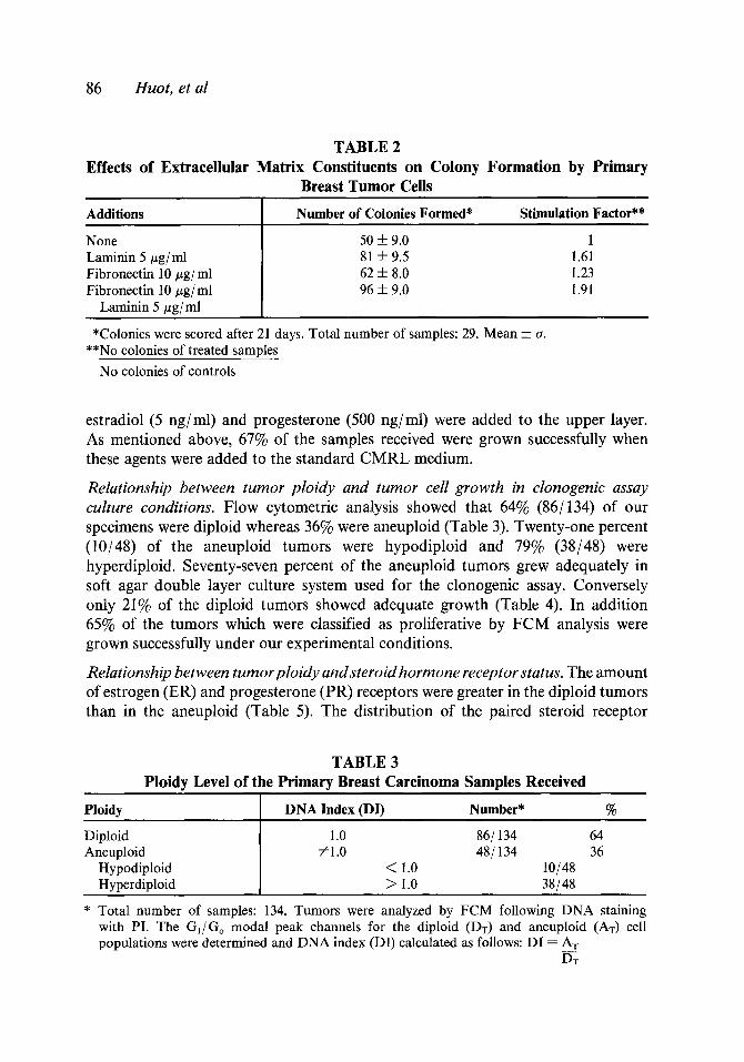
Effects of extracellular matrix constituents and hormones on in vitro colony formation by primary breast carcinoma cells. Immunofluorescence microscopic observations using specific antibodies showed that the human breast tumor specimens contained collagen, fibronectin and laminin. Hence we investigated whether these compounds can influence the formation of colonies by isolated clonogenic cells in double agar layer cultures. Our results showed that fibronectin (10 #g/ml) and laminin (5 #g/ml) added in the upper layer stimulated the colony formation especially when the two were associated (stimulating factor = 1.91). The effect of laminin was more pronounced when the cells were obtained from metastasis (data not shown). Collagen (Type IV) did not exert any significant effect (Table 2). We also studied the effects of various concentrations of hormones on colony formation and found that optimal growth could be obtained when insulin (2 U/ml), 17 /3-
86 Huot, et al
TABLE 2 Effects of Extracellular Matrix Constituents on Colony Formation by Primary
Breast Tumor Cells
Additions Number of Colonies Formed* Stimulation Factor**
None Laminin 5 ~zg/ml Fibronectin 10 #g/ml Fibronectin 10 #g/ml
Laminin 5 #g/ml
50 ± 9.0 1 81 ± 9.5 1.61 62 ± 8.0 1.23 96 ± 9.0 1.91
*Colonies were scored after 21 days. Total number of samples: 29. Mean ± a. **No colonies of treated samples
No colonies of controls
estradiol (5 ng/ml) and progesterone (500 ng/ml) were added to the upper layer. As mentioned above, 67% of the samples received were grown successfully when these agents were added to the standard CMRL medium.
Relationship between tumor ploidy and tumor cell growth in clonogenic assay culture conditions. Flow cytometric analysis showed that 64% (86/134) of our specimens were diploid whereas 36% were aneuploid (Table 3). Twenty-one percent (10/48) of the aneuploid tumors were hypodiploid and 79% (38/48) were hyperdiploid. Seventy-seven percent of the aneuploid tumors grew adequately in soft agar double layer culture system used for the clonogenic assay. Conversely only 21% of the diploid tumors showed adequate growth (Table 4). In addition 65% of the tumors which were classified as proliferative by FCM analysis were grown successfully under our experimental conditions.
Relationship between tumor ploidy and steroid hormone receptor status. The amount of estrogen (ER) and progesterone (PR) receptors were greater in the diploid tumors than in the aneuploid (Table 5). The distribution of the paired steroid receptor
TABLE 3 Ploidy Level of the Primary Breast Carcinoma Samples Received
Ploidy DNA Index (DI) Number* %
Diploid 1.0 86/134 64 Aneuploid #1.0 48/134 36
Hypodiploid < 1.0 10/48 Hyperdiploid > 1.0 38/48
* Total number of samples: 134. Tumors were analyzed by FCM following DNA staining with PI. The GI/Go modal peak channels for the diploid (DT) and aneuploid (AT) cell populations were determined and DNA index (DI) calculated as follows: DI = AT
DT

Cell Biology and Toxicology, Vol. 6, No. 1, 1990 87
Growth in soft agar double layer cultures*
TABLE 4 Relation Between Tumor Cell D N A Content and Growth in Agar
Tumor Cell DNA Content~
Diploid Aneuploid
+ 8 (21%) 10 (77%) -- 30 (79%) 3 (23%)
* Cells cultures were considered to be growth positive when they contained at least 30 colonies of more than 30 cells within 14-21 days. DNA content: relative DNA content of the aneuploid tumor G1/Go cells as determined by FMF after calibration with human lymphocytes (See Table 3).
contents in relation with the ploidy status of the tumor is given in Table 6. Fifty- two percent (45/86) of the diploid tumors were E+/P + whereas only 23% (20/ 86) were E-- /P-- . Conversely 27% (13/48) of the aneuploid tumors were E+/P + and 42% (20/48) were E-- /P-- . The distribution of the E+/P - and E - - /P + were quite similar between the two groups of tumors. The aneuploid E- - /P - - tumors grew better in the clonogenic assay.
Drug sensitivity in vitro. We studied 35 samples for drug responsiveness in our improved double agar clonogenic assay. A decrease of 50% in the number of colony formed by the treated culture as compared with controls was chosen as the end point for drug sensitivity. Twenty-one samples grew adequately to permit objective discrimination of drug sensitivity of the tumors. Seventy-two percent of the samples were sensitive to DXB (20 #M) whereas the remaining were resistant. Only 39% and 11% were sensitive to MTX (10 #M) and 5-FU (50 #M) respectively. Three samples were resistant to each of the three agents (Table 7). Estimations of the ICs0 on one responsive sample were 0.6 #M, 30/~M and 40 #M for DXB, MTX and 5-FU respectively. We also found that the various combinations of two of these agents were supraadditive.
TABLE 5 Relationship Between Ploidy of Human Breast Tumors and Their
Cytoplasmic Steroid Hormone Receptor Contents
Ploidy*
Total population Diploid Aneuploid
fmole/mg Cyt. Prot.
n ER ___ SE PR ± SE
134 73.70 q- 9.0 50.51 i 6.1 86 97.0 -+- 15.2 63.4 ± 12.2 48 32.34± 5.7 27.6 ± 2.8
* Ploidy level was determined by flow cytometry and the amount of steroid receptors as described under Methods.

88 Huot, et al
TABLE 6 Receptor Status and Ploidy Level of Human Breast Tumor Samples*
atustOr E ÷ P+ E + P- E- P÷ E- P-
No (%) No (%) No (%) No (%) Ploidy ~ . ~
Diploid n 86 45 (52) 16 (19) 5 (6) 20 (23) Aneuploid n 48 13 (27) 11 (23) 4 (8) 20 (42) TOTAL n 134 58 (43) 27 (20) 9 (7) 40 (30)
* Distribution of the aired steroid receptor content among diploid and aneuploid tumors. Ploidy was determined by flow cytometry and the steroid receptors were assayed as described in Methods. Samples were considered to be receptor positive (E ÷ and P÷) if they contained at least 10 fmoles of receptor protein/mg of cytoplasmic protein.
DISCUSSION
The clonogenic assays have been proposed for prognostic purposes in choosing pharmacological treatment of cancer patient (Salmon et al., 1978; Von Hoff et al., 1983; Huot et al., 1985; Shoemaker et al., 1985; Von Hoff et al., 1986). However, many authors reported that the routine use of these assays in clinical prospective trials is impaired by various factors of methodological origin (Selby et al., 1983; Bernard and Riou, 1984; Ali-Osman and Beltz, 1988; Nicholson et al., 1988). In this study, we report a double agar clonogenic system in which we have improved the conditions for human breast cancer cell cloning. We also investigated the influence of ploidy level and hormone receptor status of the tumors on their cellular clonogenicity and drug sensitivity.
The preparation of mono-dispersed viable cell suspension is a critical and often limiting step in the analysis of clonogenicity of solid tumors. In our experimental
TABLE 7 In Vitro Drug Sensitivity of Primary Human Breast Cancer
Cells in the Clonogenic Assay*
Drugs % Sensitivityt % Resistant**
Doxorubicin 20 # M 72% 28% Methotrexate 10 # M 11% 89% 5-Fluorouracil 50 # M 39% 61%
* Total number of samples ---- 35 usable =21 = 60% unusable = 14 = 40%
** Three cultures (11%) were resistant to the three agents. Sensitivity is defined as a ~ of 50% in the formation of colonies of at least 30 cells in cultures containing at least 30 colonies.

Cell Biology and Toxicology, VoL 6, No. 1, 1990 89
conditions, we have recovered 4.7 X 10⁶ cells per gram of tissue with an average viability of 80%. This is an important improvement when compared with the value reported in the literature (Sandbach et al., 1982; Maestroni and Lusa, 1984). We have also improved the cell plating by adding estradiol (5 ng/ml), progesterone (500 ng/ml), insulin (2 U/ml), laminin (5 tzg/ml) and fibronectin (10 #g/ml) in the upper layer of our double agar system. Using these conditions, close to 70% of the samples grew successfully (_> 30 colonies/500.000 plated cells) in the clonogenic assay. This is superior to the value of 53% recently reported by Ali-Osman and Beltz (1988). In fact, our growth success rate is close to the 77% obtained by these authors with the capillary human tumor clonogenic cell assay. The effects of extracellular matrix component (laminin and fibronectin) appear especially significant and we propose their systematic addition in the standard clonogenic system assay for breast tumor. We have added these agents since we have found their presence following microscopic observations of sections of breast samples incubated with specific antibodies. The laminin is more effective in increasing the formation of colonies by cells of metastatic origin. This suggests that it might be involved in the metastatic process. Fliegiel et al. (1985) and Kleinman et al. (1985) have also proposed such analogous function for laminin. It is difficult to evaluate whether the addition of hormones and extracellular matrix component favor the formation of colonies by subpopulations of responsive cells.
The proportion of cells which form colonies in our assay was of the order of 0.02% which means that they constitute a subpopulation of the original sample. These clonogenic cells are probably the so-called stem cells. It is well established that stem cells generally account for only a small fraction of the total tumor cell population, that their growth properties are different from those of the other cells forming the tumor and that they are responsible for the repopulation of a tumor. Moreover, the stem cells of tumoral origin are described as cells capable of giving rise to a large family of descendants specific for the tumors within its natural environment (Hamburger, 1981). It is worth mentioning that it is this small proportion of cells that is the desired target for chemotherapeutic agents. At the present time, there is no way in humans in which the descendants of a given tumor cell can be counted in situ.
While the concept of stem cells remains hypothetical for human tumors, there are grounds to believe that the stem ceils are those whose growth in agar is related to the self renewal properties of the cells in vivo (MacKillop et al., 1983). In this regard, Salmon et al. (1978) summarized several identifiable features of human tumor colony forming unit which are consistent with in situ tumor stem cell origin. However, Nicholson et al. (1988) has recently reported that heterogeneity exists in the ability of adenocarcinoma cells of different metastic potential and drug sensitivities to grow in semi solid agar. Furthermore, the assay may fail to detect cells in Go/G1 phases of the cell cycle which might be important in refractoriness to therapy (Clarkson, 1974). Nevertheless, these observations do not invalidate

90 Huot, et al
the test but rather suggest that further work shall be done in an attempt to increase the cloning efficiency of tumor cells in vitro and to develop assays which can discriminate between the various subpopulations of tumor cells.
Tumor heterogeneity is another point that should be addressed. Many tumors are composed of cells of different phenotypes and most of them are mosaics of different somatic karyotypes (Ito and Moore, 1967). Slocum et al. (1981) could not conclude that anatomical segregation within solid tumors did not exist but found that four areas of the same tumor were similar in cell yield, dye exclusion, drug uptake and metabolism. Others reported that differents parts of the same tumor can grow differently in soft agar and react differently to drug (Bergeron et al., 1985). There is evidence indicating that causes of this heterogeneity could include factors such as the host status, tumor grade, nutritive factors and anoxic effects. It is suggested that, whenever possible, clonogenic stem cell assay should be done on different samples from the same specimens.
Multiparametric analysis of tumor ploidy by FCM showed that aneuploid tumor grew better than the diploid tumors in a double agar clonogenic system. This suggests that FCM analysis can be used to select the tumors which have the greater probability to grow in soft agar. Among the diploid tumors which grew in soft agar, we cannot rule out the possibility that some of them contain a small aneuploid population which has not been detected. Comparable data were reported by Verheijen et al. (1985). The amounts of ER and PR (fmoles/mg cell prot.) were greater in the diploid tumors than in the aneuploid. Accordingly, the diploid tumors were mostly E+/P + whereas the aneuploid were mostly E--/P--. These results are similar to those reported in the literature (Olsewski et al., 1981; Chassevent et al., 1984; Coulson et al., 1984). The lesser amount of receptor proteins found in the aneuploid tumor would reflect their less differentiated state. Since the E - / P - were mostly aneuploid,it should not be a surprise to have found them growning better in the double agar clonogenic assay system. This is in agreement with the data previously reported by Barlogie et al. (1982), Raber et al. (1982) and Chassevent et al. (1984).
Thirty-five samples were tested for their drug sensitivity in our improved experimental conditions. Valuable and unambiguous results could be used in 21 of them. As proposed by Von Hoff et al. (1981) and Sandbach et al. (1982), we used the -->50% decrease in tumor colony forming unit of more than 30 cells as the criteria of pharmacological response in vitro. We found that 72% of tumors respond to Doxorubicin (20 #M) whereas only 11% and 39% were sensitive to MTX (10 #M) and 5-FU (50 uM) respectively. Sandbach et al. (1982) used lower concentrations of these agents and showed that the response rates were proportionally weaker. Estimations of the ICs0 for the drugs studied were 0.6 #M for DXB, 30 #M for MTX and 40 #M for 5-FU. These values are comparable to those reported in the literature for these agents when they are added to in vitro cell culture systems (MacKintosh et al., 1981; Ali- Osman et al., 1983; Bernard et al., 1983; Safa et al., 1983; Kanzawa et al., 1984).

Cell Biology and Toxicology, Vol. 6, No. 1, 1990 91
One of the major questions actually debated in breast cancer chemotherapy is whether there are relationships between tumor responsiveness to drug and their hormonal receptor status and ploidy levels. Our results suggest that the tumor sensitivity to drugs seems to be rather independent of their steroid hormone status and ploidy level. Sandbach et al. (1982) also reported that estrogen receptor status did not appear to influence in vitro drug sensitivity. Conversely, Silvestrini et al. (1984) observed that ER ÷ breast cancer were more sensitive in vitro to DXB. This could be explained by the fact that these authors used 5 fmoles/mg protein instead of 10 fmoles/mg protein as the cut-off point for sensitivity.
In summary, this study showed that we have been successful in improving conditions for clonogenic assays. Using these improved experimental conditions, clonogenic assays can permit objective discrimination of the in vitro pharmacosensitivity of human breast tumors. In addition, our study suggests that ploidy measurements by FCM and steroid hormone receptor assays can be useful to assess the capacity of a given tumor to grow in the clonogenic assay. Further work is still required to better refine these tests and increase their potential as predictors of the individual response to cancer treatments and as screening assays of new antineoplastic drugs. Among others, more should be known about the influence of the clonal heterogeneity of the tumors on drug responsiveness in vitro.
ACKNOWLEDGEMENTS
We are deeply grateful to the Department of Health and Welfare Canada (National Health Research and Development Program) which financially supported this research.
The authors are greatly indebted to the following clinicians who participated in this study: Drs. Yvan Drolet, Louis Dionne and Antoine Kibritd, H6tel-Dieu de Qudbec, Luc Desch~nes and Simon Jacob, H6pital St-Sacrement de Qudbec, Paul Fortin, H6pital de l'Enfant-Jdsus de Qudbec, Hubert Falenga, H6tel-Dieu de Ldvis, Pierre Fernet, H6pital de Chicoutimi, Georges Normand, Rend Boyer, Georges Groulx and Robert Perron, H6pital St-Joseph de Trois-Rivi~res, Jean-Louis Ldtourneau, Pierre Matteau and Michel Baribeau, Hgpital Ste-Marie de Trois- Rivi~res, Roger Poisson and Sandra Legault-Poisson, H6pital St-Luc de Montrdal. We also thank Dr. Pritam Singh for having read the manuscript.
They also thank Miss Diane Huot Blais for her excellent assistance in the preparation of this manuscript.
REFERENCES
ALI-OSMAN, F. and BELTZ, P. A. (1988). Optimization and characterization of the capillary human tumor clonogenic cell assay. Cancer Res. 48:715-724.

92 Huot , et al
ALI-OSMAN, F., MAURER, H. R. and BIER, J. (1983). In vitro cytostatic drug sensitivity testing in the human tumor stem cell assay: A model feed method for the determination of the sensitivity index. Tumor Diagnostik Therapie 4:1-6.
BARLOGIE, B., JOHNSTON, D., SMALLWOOD, L., RABER, M., MADDOX, A. M., LATREILLE, J., SWARZENDRUBER, D. and DREWINKO, B. (1982). Prognostic implication of ploidy and proliferative activity in human solid tumor. Cancer Gen. Cytogen. 6:17-28.
BERGERON, M., DROLET, Y. et HUOT, J. (1985). Le test de sensibilit~ in vitro des ceUules clonog~niques tumorales: Une revue de la question. Un. M~. Can. 114:380-382.
BERNARD, J. and RIOU, G. (1984). Culture in vitro des cellules clonog~nes de diff~rentes tumeurs humaines: essais de chimiosensibilit~. Bull. Cancer 71:287-291.
BERNARD, J., DA SILVA, J. and RIOU, G. (1983). Culture of clonogenic cells from various human tumor drug sensitivity assay. Cancer Clin. Oncol. 19:65-72.
BERTONCELLO, I. and BRADLEY, T. R. (1987). Human cell cultures for screening anti-cancer drugs. TIPS 8:249-251.
CHASSEVENT, A., DAVER, G., BERTRAND, F. and GEORGE, L. P. (1984). Analyse d'ADN par cytom&rie de flux dans les cancers du sein. Bull. Cancer 71:494-495.
CLARKSON, B. D. (1974). In: Control of proliferation in animal cells (Clarkson, B. D. and Baserga, R., eds), pp. 945-972. Cold Spring Harbor Laboratories.
COULSON, P. B., THORNTHWAITE, J. T. and WOOLLEY, T. W. (1984). Prognostic indicators including DNA histogram type, receptor content and staging related to human breast cancer survival. Cancer Res. 44:4187-4196.
FLIEGIEL, S. E. G., RODRIGUEZ, A. F., KNIBBS, R. N., MC COY, J. P. and VARAMI, J. (1985). Characterization of Laminin-stimulated adherence and motility in tumor cells. Oncology 42:265-271.
HAMBURGER, A. W. (1981). Use of in vitro tests in predictive cancer chemotherapy. J. Nat. Cancer Inst. 66:981-988.
HAMBURGER, A. W. and SALMON, S. E. (1977). Primary bioassay of human tumor stem cells. Science 197:461-463.
HUOT, J., GOULET, F. and GOYETTE, R. (1985). Test clonog~nique et chimiosensibilite du cancer du sein. Un. M~d. Can. Vol. 114 9:1-2.
ITO, E. and MOORE, G. E. (1967). Characteristics differences in clones isolated from an $37 ascites tumor in vitro. Exp. Cell Res. 48:440-447.
KANZAWA, F., HOSLT, A., SHIMIZO, E., SAIJO, N., MIYAZAMA, W., SHIMABO, A. and KURO, Z. (1984). Clonogenic cell assay for carcinoma of the lung. GANN 75:81-88.
KAUFMAN, M. (1984). Biochemical short-term predictive assay: results of correlative trials in comparison to other assays. Rec. Results Cancer Res. 94:151-160.
KLEINMAN, H. K., CANNON, F. B., LAURIE, G. W., HASSELL, J. R., AUMAILLEY, M., TERRANOVA, V. P., MARTIN, G. R. and DUBOIS-DALCQ, M. (1985). Biological activities of laminin. J. Cell Biochem. 27:317-325.
MAC KILLOP, W. J., CIAMPI, A., TILL, J. E. and BUICK, R. W. (1983). A stem cell model of human tumor growth; implications for tumor cell clonogenic assays. J. Nat. Cancer Inst. 70:9-16.
MAC KINTOSH, F. R., EVANS, T. L. and SIKK, B. I. (1981). Methodologic problems in clonogenic assays of spontaneous human tumors. Cancer Chemother. Pharmacol. 6:205-210.

Cell Biology and Toxicology, Vol. 6, No. 1, 1990 93
MAESTRONI, G. J. M. and LUSA, G. A. (1984). Effect of autologous serum and steroid hormones receptors on direct cloning of human breast cancer. Arch. Pathol. Lab. Med. 108:783-785.
NICHOLSON, G. L., LEMBO, T. M. and WELCH, D. R. (1988). Growth of rat mammary adenocarcinoma cells in semi solid clonogenic medium not correlated with spontaneous metastatic behavior: Heterogeneity in the metastatic antigenic and drug sensitivity properties of cells from different sized colonies. Cancer Res. 48:399-404.
OLSEWSKI, W., DARZYNKIEWICZ, Z. and ROSEN, P. P. (1981). Flow cytometry of breast cancer: I. Relation of DNA ploidy level to histology and estrogen receptors. Cancer 48:980-984.
RABER, M. N., BARLOGIE, B. and LATRAILLE, J. (1982). Ploidy, proliferation activity and estrogen receptor content in human breast cancer. Cytometry 3:36-41.
SAFA, A. R. and TSENG, M. T. (1983). Doxorubicin-induced ultrastructural and growth changes in primary cultures of 7,12-dimethylbenz(a)anthracene-induced mammary tumor. J. Nat. Cancer Inst. 70:535-540.
SAFA, A. R., TSENG, M. T. and BALLEU, R. J. (1983). Sequential study on the influence of adriamycin on cell proliferation and ultrastructure of cultured mammary tumor cells. Proc. Soc. Exp. Biol. Med. 174:276-283.
SALMON, S. E., HAMBURGER, A. W., SOETTNLEN, B., PURIE, R. G. M., ALBERTS, A. S. and MOON, T. E. (1978). Quantitation of differential sensitivity of human tumor stem cell assay. N. Engl. J. Med. 298:1321-1327.
SANDBACH, J., VON HOFF, D. D., CLARK, G., CRUZ, A. B., O'BRIEN, M. and the SOUTH CENTRAL TEXAS HUMAN TUMOR CLONING GROUP (1982). Direct cloning of human breast cancer in soft agar culture. Cancer 50:1315-1321.
SELBY, R. J., BUICK, R. N. and TANNOCK, I. (1983). A critical appraisal of the "human tumor stem-cell assay." N. Engl. J. Med. 308:129-134.
SHOEMAKER, R. H., WOLPERT-DEFILIPPER, M. K., KERN, D. H., LIEBER, M. M., MAKUCH, R. W., MELNICK, R. R., MILLER, W. T., SALMON, S. E., SIMON, R. M., VENDITTI, J. M. and VON HOFF, D. D. (1985). Application of a human tumor colony forming assay to new drug screening. Cancer Res. 45:2145-2153.
SILVESTRINI, R., SANFILLIPO, O., DAIDONE, M. D. and ZAFFARONI, C. (1984). Predictive relevance for clinical outcome of in vitro sensitivity evaluated through antimetabolic assay. Rec. Results Cancer Res. 94:140-150.
SLOCUM, H. K., PAVELIC, Z. P., KANTER, P. M., NOVAK, N. J. and RUSTERM, Y. M. (1981). The soft agar clonogenicity and characterization of cells obtained from human tumors by mechanical and enzymatic means. Cancer Chemother. Pharmacol. 6:219-225.
VERHEIJEN, R. H. M., FEITZ, W. F. J., BECK, J. L. M., DEBRUYNE, F. M. J., VOOKS, G. P., KENEMOUS, P. and HERMAN, C. J. (1985). Cell DNA content correlations with clonogenicity in the human tumor cloning system (HTCS). Int. J. Cancer 35:653-657.
VON HOFF, D. D., CLARK, G. M., FORSETH, B. J. and COWAN, J. P. (1986). Simultaneous in vitro drug sensitivity testing on tumors from different sites in the same patient. Cancer 58:1007-1013.

94 Huot, et al
VON HOFF, D. D., CLARK, G. M., STOGDILL, B. J., SAROSDY, M. F., O'BRIEN, M. T., CASPER, J. F., MATTOX, D. F., PAGE, C. P., CRUZ, A. B. and SANDBACH, J. F. (1983). Prospective clinical trial of a human tumor cloning system. Cancer Res. 43:1925-1931.
VON HOFF, D. D., PAGE, C. P., HANIS, G., CLARK, G., GOWAN, J. and COLTMAN, C. A. (1981). Prospective clinical trial of a human tumor cloning system. Proc. Am. Ass. Cancer Res. 22:154.
Copyright © 2022 FDOKUMEN