Microfluidic integration for automated targeted proteomic assays
457
Review
www.expert-reviews.com ISSN 1478-7210© 2011 Expert Reviews Ltd10.1586/ERI.11.23
TB remains a pervasive, morbid and lethal disease that thrives on neglect. It has been studied for decades and boasts curative treatment, yet to this day, its spread, associated mortality and burden of suffering impacts patients of all ages and races, on all continents [1]. Despite global attempts at TB control, the worldwide disease incidence rate was 139 per 100,000 population in 2008, with the incidence rate in Africa reaching 350 per 100,000 in the same year. This corresponds with the stag-gering estimate of 9.4 million incident cases of TB worldwide in 2008. Only 61% of these cases were reported and of the 440,000 estimated total num-ber of multidrug resistant (MDR) cases in 2008, only 11% were notified and treated according to international guidelines [2]. If infectious cases are quickly identified and treated, TB control improves, as patients’ infectivity decreases rapidly on appropriate treatment. Indeed, the dynamics of the TB epidemic are greatly determined by how soon cases are diagnosed, impacting the duration of infectiousness [3].
In line with the objectives of the WHO Stop TB Strategy and the Global Plan to stop TB, TB diagnostics should lead our endeavor to control the epidemic. These two international programs aim for universal access to quality diagnostics and the detection of 84% of global cases by 2015 [2]. Furthermore, the updated goals for the new diag-nostics component of the Global Plan to Stop TB 2011–2015 include increased diagnosis of active TB at point-of-care level and screening for MDR and extensively drug resistant (XDR) TB [4].
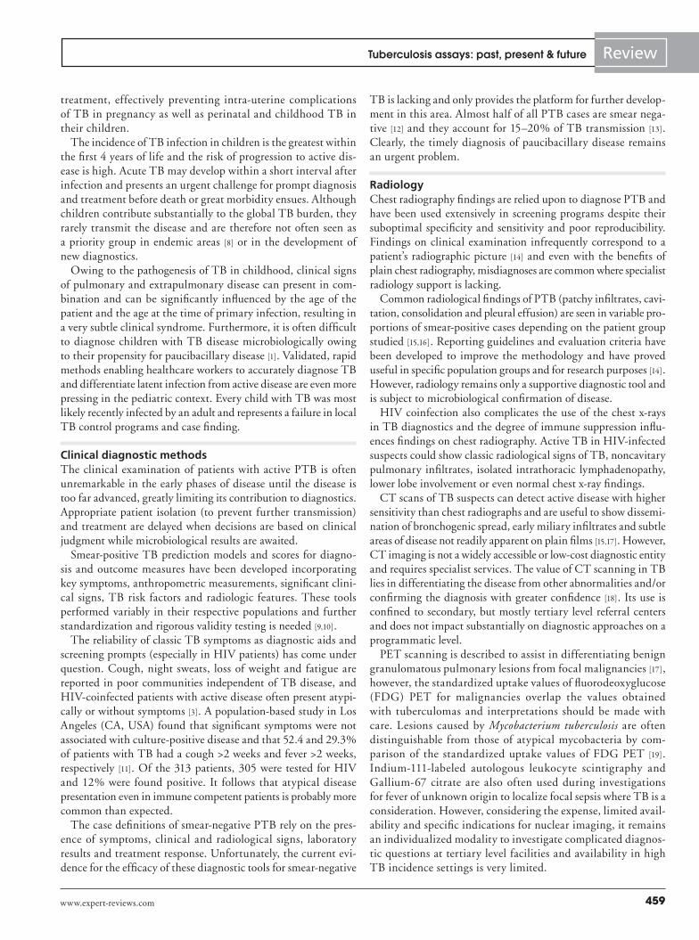
A recent prospective study in South Africa showed that 17% of 367 TB cases diagnosed on smear results never started treatment owing to incomplete sputum sample collection, prob-lems with sample transport and poor record-keeping of the samples taken and their subse-quent results [5]. This illustrates that the key to effectively combat TB remains comprehen-sive case finding and reporting. It has become clear that a rapid, simple and reliable method of diagnosis at the point of care could mean the end of missed opportunities – lowering the incidence of delayed treatment, noso comial infections, drug resistance and treatment failure. Figure 1 presents the current pipeline of diagnostic tests [4]. This article addresses the advances and challenges in the search for effective diagnostics.
Special target groups for improved diagnosticsHIV coinfectionThe undercurrent of the global HIV epidemic drives the resurgence of TB, especially in Sub-Saharan Africa. Unfortunately, many chal-lenges exist in the management of HIV and TB coinfection, including barriers that persist in harmonizing the two traditionally different primary treatment services.
HIV alters the clinical and radiological presentation of TB with worsening immune deficiency. Symptoms are often nonspecific or subclinical and the presence of coinfections
Novel N Chegou1, Kim GP Hoek2, Magdalena Kriel1, Robin M Warren1, Thomas C Victor1 and Gerhard Walzl†1
1DST/NRF Centre of Excellence for Biomedical Tuberculosis Research and MRC Centre for Molecular and Cellular Biology, Division of Molecular Biology and Human Genetics, Department of Biomedical Sciences, University of Stellenbosch, P.O. Box 19063, Francie van Zijl Drive, Tygerberg 7505, South Africa 2Division of Medical Microbiology, Department of Pathology, Faculty of Health Sciences, University of Stellenbosch, P.O. Box 19063, Francie van Zijl Drive, Tygerberg 7505, South Africa †Author for correspondence:Tel.: +27 219 389 124 Fax: +27 219 389 863 [email protected]
Recent developments in the field of TB diagnostics, including the introduction of the Xpert MTB/RIF assay in field testing, raise the hope for faster and more accurate identification of active TB patients. However, there are still many issues that need to be addressed as no point-of-care tests are yet available. Furthermore, no tests are available which are universally applicable to all patients. Improvements in the microbiological and molecular-based approaches are promising and the diagnostic pipeline is encouraging. Host markers associated with active disease may hold promise, especially in situations where sputum diagnostics are problematic, including in children, HIV-infected individuals and in the case of extrapulmonary TB.
Keywords: diagnostic test • latent tuberculosis infection • Mycobacterium tuberculosis • pulmonary tuberculosis • tuberculosis • tuberculosis diagnosis
Tuberculosis assays: past, present and futureExpert Rev. Anti Infect. Ther. 9(4), 457–469 (2011)
For reprint orders, please contact [email protected]

Expert Rev. Anti Infect. Ther. 9(4), (2011)458
Review
could obscure or mimic the symptoms of TB [3,6]. In a recent population-based survey in an African community with high HIV prevalence and increasing TB notification rates, 63% of adult cases with pulmonary TB (PTB) remained undiagnosed in an efficient directly observed treatment shortcourse (DOTS) program and passive case finding in HIV-infected individuals only identified 33% of the smear-positive TB cases [3]. In light of the vulnerability of HIV patients to TB infection and reactiva-tion and the alarming possibility of increased coinfection with MDR and XDR TB strains, the timely diagnosis of TB in HIV patients remains paramount.
Women & childrenOther populations specifically at risk for TB morbidity and mortality are women and children. More women die worldwide from TB than from combined maternal causes every year [7] and the disease morbidity in this group greatly impacts social welfare and disease spread within households. In 2008, an estimated 3.6 million women were diagnosed with TB, with the highest disease burden during the childbearing years – also the age group of highest HIV infection incidence [2]. It follows that pregnant women and especially those with HIV would benefit greatly from timely TB screening, diagnosis and
2007 2008 2009 2010 2011
10–40%
70%
95%
2012 2013 2014 2015 2016
First referral level
Peripheral lab
Community health center
Liquid culture and DST
New SS+ case definition
2-specimen approaches
Rapid speciation
LPA for MDR-TB
Noncommercial culture and DST (MODS, N
RA and CRI)
LPA for XDR-TB
IGRAs for LTBI
LED microscopy and same day diagnosis
Xpert MTB/RIF test fo
r TB and MDR-TB
Commercial serological tests
Manual NAAT 1st generation
Rapid colorimetric
DST
POC test (detection)
Predictive LT
BI
Manual NAAT 2nd generation
Dis
tan
ce f
rom
pat
ien
ts
% A
ccess after 5 years
Technologies or processes endorsed by STAG/WHO Technologies for which WHO review is in process
Figure 1. 10-year diagnostic pipeline. Timeline of diagnostic tests currently endorsed by the WHO, as well as those that are under development and review. CRI: Colorimetric redox assay; DST: Drug-susceptibility testing; IGRA: IFN-g release assay; LED: Light-emitting diode; LPA: Line-probe assay; LTBI: Latent TB infection; MDR-TB; Multidrug-resistant TB; MODS: Microscopic observation of drug susceptibility; NAAT: Nucleic acid amplification test; NRA: Nitrate reductase assay; POC: Point of care; SS+: Sputum smear positive; STAG: Strategic and Technical Advisory Group; XDR-TB: Extensively drug-resistant TB. Figure provided by Richard O’Brien (Foundation for Innovative New Diagnostics). Reproduced with permission from [4].
Chegou, Hoek, Kriel, Warren, Victor & Walzl
www.expert-reviews.com 459
Review
treatment, effectively preventing intra-uterine complications of TB in pregnancy as well as perinatal and childhood TB in their children.
The incidence of TB infection in children is the greatest within the first 4 years of life and the risk of progression to active dis-ease is high. Acute TB may develop within a short interval after infection and presents an urgent challenge for prompt diagnosis and treatment before death or great morbidity ensues. Although children contribute substantially to the global TB burden, they rarely transmit the disease and are therefore not often seen as a priority group in endemic areas [8] or in the development of new diagnostics.
Owing to the pathogenesis of TB in childhood, clinical signs of pulmonary and extrapulmonary disease can present in com-bination and can be significantly influenced by the age of the patient and the age at the time of primary infection, resulting in a very subtle clinical syndrome. Furthermore, it is often difficult to diagnose children with TB disease microbiologically owing to their propensity for paucibacillary disease [1]. Validated, rapid methods enabling healthcare workers to accurately diagnose TB and differentiate latent infection from active disease are even more pressing in the pediatric context. Every child with TB was most likely recently infected by an adult and represents a failure in local TB control programs and case finding.
Clinical diagnostic methodsThe clinical examination of patients with active PTB is often unremarkable in the early phases of disease until the disease is too far advanced, greatly limiting its contribution to diagnostics. Appropriate patient isolation (to prevent further transmission) and treatment are delayed when decisions are based on clinical judgment while microbiological results are awaited.
Smear-positive TB prediction models and scores for diagno-sis and outcome measures have been developed incorporating key symptoms, anthropometric measurements, significant clini-cal signs, TB risk factors and radiologic features. These tools performed variably in their respective populations and further standardization and rigorous validity testing is needed [9,10].
The reliability of classic TB symptoms as diagnostic aids and screening prompts (especially in HIV patients) has come under question. Cough, night sweats, loss of weight and fatigue are reported in poor communities independent of TB disease, and HIV-coinfected patients with active disease often present atypi-cally or without symptoms [3]. A population-based study in Los Angeles (CA, USA) found that significant symptoms were not associated with culture-positive disease and that 52.4 and 29.3% of patients with TB had a cough >2 weeks and fever >2 weeks, respectively [11]. Of the 313 patients, 305 were tested for HIV and 12% were found positive. It follows that atypical disease presentation even in immune competent patients is probably more common than expected.
The case definitions of smear-negative PTB rely on the pres-ence of symptoms, clinical and radiological signs, laboratory results and treatment response. Unfortunately, the current evi-dence for the efficacy of these diagnostic tools for smear-negative
TB is lacking and only provides the platform for further develop-ment in this area. Almost half of all PTB cases are smear nega-tive [12] and they account for 15–20% of TB transmission [13]. Clearly, the timely diagnosis of paucibacillary disease remains an urgent problem.
RadiologyChest radiography findings are relied upon to diagnose PTB and have been used extensively in screening programs despite their suboptimal specificity and sensitivity and poor reproducibility. Findings on clinical examination infrequently correspond to a patient’s radiographic picture [14] and even with the benefits of plain chest radiography, misdiagnoses are common where specialist radiology support is lacking.
Common radiological findings of PTB (patchy infiltrates, cavi-tation, consolidation and pleural effusion) are seen in variable pro-portions of smear-positive cases depending on the patient group studied [15,16]. Reporting guidelines and evaluation criteria have been developed to improve the methodology and have proved useful in specific population groups and for research purposes [14]. However, radiology remains only a supportive diagnostic tool and is subject to microbiological confirmation of disease.
HIV coinfection also complicates the use of the chest x-rays in TB diagnostics and the degree of immune suppression influ-ences findings on chest radiography. Active TB in HIV-infected suspects could show classic radiological signs of TB, noncavitary pulmonary infiltrates, isolated intrathoracic lymphadenopathy, lower lobe involvement or even normal chest x-ray findings.
CT scans of TB suspects can detect active disease with higher sensitivity than chest radiographs and are useful to show dissemi-nation of bronchogenic spread, early miliary infiltrates and subtle areas of disease not readily apparent on plain films [15,17]. However, CT imaging is not a widely accessible or low-cost diagnostic entity and requires specialist services. The value of CT scanning in TB lies in differentiating the disease from other abnormalities and/or confirming the diagnosis with greater confidence [18]. Its use is confined to secondary, but mostly tertiary level referral centers and does not impact substantially on diagnostic approaches on a programmatic level.
PET scanning is described to assist in differentiating benign granulomatous pulmonary lesions from focal malignancies [17], however, the standardized uptake values of fluorodeoxy glucose (FDG) PET for malignancies overlap the values obtained with tuberculomas and interpretations should be made with care. Lesions caused by Mycobacterium tuberculosis are often distinguishable from those of atypical mycobacteria by com-parison of the standardized uptake values of FDG PET [19]. Indium-111-labeled autologous leukocyte scintigraphy and Gallium-67 citrate are also often used during investigations for fever of unknown origin to localize focal sepsis where TB is a consideration. However, considering the expense, limited avail-ability and specific indications for nuclear imaging, it remains an individualized modality to investigate complicated diagnos-tic questions at tertiary level facilities and availability in high TB incidence settings is very limited.
Tuberculosis assays: past, present & future
Expert Rev. Anti Infect. Ther. 9(4), (2011)460
Review
Bacteriological methodsSputum smear microscopyZiehl-Neelsen staining remains the most widely used and cost-effective diagnostic able to identify infectious patients with a relatively high speed and acceptable specificity [20]. However, while the current global detection rate of acid-fast bacilli (AFB) in smear-positive individuals is 60%, the rate is much lower (20–40%) in settings with a high incidence of HIV infection [21,22]. Immunocompromised patients often present with lower bacterial loads, which are easily missed by AFB testing owing to its detection threshold of 104 bacilli per milliliter of specimen [23]. A further concern in HIV-infected individuals is the increased risk of nontuberculous mycobacteria (NTM) infections, since AFB testing is not able to differentiate between M. tuberculosis and NTM disease [24]. In addition, up to 30% of patients are unable to produce sputum [25] (this is especially common in children). Therefore, while sputum smear microscopy remains a particu-larly valuable rapid diagnostic in resource-limited settings, this test should not be used to exclude TB disease. Recent improve-ments to the sensitivity of the method include a concentration step following sputum processing (33% improvement over direct microscopy) [26] and the use of fluorescent auramine-rhodamine staining (10% improvement over Ziehl-Neelsen staining) [27,28] and light-emitting diode fluorescent microscopy [201].
Phenotypic culture & drug susceptibility testingCulture of M. tuberculosis from a clinical specimen is regarded as the gold standard for proving a case of TB. Culture is able to detect 10–100 M. tuberculosis bacilli per milliliter of speci-men [29]. The Mycobacterial Growth Indicator Tube (MGIT; Becton Dickinson) is currently the preferred culture system in high-throughput settings as it is automatable [30], reduces time to a positive result to 2 weeks (compared with 4–8 weeks) and has an increased sensitivity (up to 10% higher) [30–33] over the traditional solid (Löwenstein-Jensen) and agar (7H10 or 7H11 Middlebrook) based culture methods. Furthermore, culture of clinical specimens allows for further investigations, including drug-susceptibility testing (DST), mycobacterial speciation and epidemiological studies (through strain typing). Culture-based DST is considered the most significant determinant of drug sus-ceptibility as it can define resistance irrespective of the molecular mechanism responsible for resistance. DST involves the inocula-tion of a clinical specimen onto/into media containing specific concentrations of anti-TB drugs. This inoculum is either derived from a pure culture (indirect method) or from a decontaminated clinical specimen (direct method). While the direct method does not require preculture and therefore reduces the turnaround time to 1–3 weeks when performed in liquid media (such as in MGIT) [34], there is an increased risk of bacterial contamination and a higher rate of NTM recovery. As a result, the indirect proportion method on agar media remains the gold standard for phenotypic-based DST [35,36]. Although the method is particularly reliable for the first-line drugs isoniazid (INH) and rifampicin (RIF) [35,36], inconsistent results are a common occurrence [37]. This is particularly evident in the case of ethambutol (EMB) resistance [37]. The significant delay
associated with culture-based diagnosis and DST has many impli-cations including that the patients may have higher morbidity and mortality while awaiting diagnosis and appropriate therapy and that there may be transmission of drug-resistant strains of M. tuberculosis to close contacts. Furthermore, in high-incidence countries, phenotypic DST is only requested following treatment failure with first-line anti-TB drugs. This extends the diagnostic delay even further.
Various improvements to reduce the delay associated with phenotypic DST methods have been suggested, which include:
• Microscopic detection of early mycobacterial growth (thin-layer agar) [38,39];
• The microscopic-observation drug-susceptibility assay [40];
• Colorimetric assays (alamar blue/resazurin [41], 3-(4,5-dimeth-ylthiazol-2-yl)-2,5-diphenyltetrazolium bromide [42] and nitrate reductase assays [43]);
• Phage amplification assays [44].
These modifications have not been capable of consistently providing a diagnostic result within 1 week of specimen collec-tion. Furthermore, culture requires expensive reagents, exten-sive biosafety facilities [40] and further speciation to confirm the etiological agent as M. tuberculosis. Therefore, it may be wise to focus on genetic markers that are specific to the M. tuberculosis genome to accelerate the diagnosis of both TB and anti-TB drug resistance.
Genotypic-based methodsDrug-resistant TB may arise through various mechanisms, including chromosomal single nucleotide polymorphisms (SNPs) in specific genes of M. tuberculosis which are targeted by a par-ticular anti-TB drug. Identification of these SNPs forms the basis of genotypic DST, since drug-susceptible isolates lack the cor-responding SNPs. Genotypic-based diagnoses have a number of potential advantages over phenotypic-based methods in that, first, there are more data points (i.e., SNPs, genes) available to analyze than with phenotypic-based testing, second, determination of the phenotype is not dependant on culturing of the bacteria (as genotype leads to a phenotype and if the genotype of a particular phenotype is well-described, the genotype may be used to infer the phenotype without the need to culture the bacteria) and third, the method is more specific and can be performed more rapidly.
Sequencing DNA sequencing remains the gold standard in genetic-based DST methods, since it can reveal the complete genetic profile of the region targeted by the anti-TB drug. Any genotypic-based assay should be validated against sequencing results as well as phenotypic tests to determine diagnostic accuracy. Not only can sequencing be used for species identification (by ana lysis of 16S rRNA) [45] and searching for known resistance-causing mutations, but it can also be used to screen for novel SNPs that may be associ-ated with drug resistance [46]. However, the major drawbacks of sequencing are the costs associated with the test, the technical skill
Chegou, Hoek, Kriel, Warren, Victor & Walzl

www.expert-reviews.com 461
Review
required to operate the expensive equipment and the unavailabil-ity of sequencing facilities in most resource-poor settings where the burden of TB is high.
Commercial genotypic-based assaysAlthough numerous in-house PCR methods have been described and various commercial-based methods are available, this section will focus on the genotypic-based assays currently endorsed by the WHO for use in high-incidence settings, as well as those not yet endorsed that show promise
Line-probe hybridization assaysLine-probe hybridization assays are based on the use of the PCR to amplify regions of interest in the mycobacterial genome followed by reverse hybridization to immobilized sequence-specific probes. These hybridization assays are often designed as DNA-strips that can simultaneously detect M. tuberculosis complex infection and anti-TB drug resistance. In 2008, the WHO released a policy statement in which they recommend that these assays be imple-mented directly on all smear-positive sputum samples to enable rapid detection of RIF resistance (thereby identifying MDR-TB patients) [47,202].
Two commercial line-probe assays are currently available. The INNO-LiPA Rif TB assay (Innogenetics, Ghent, Belgium) can detect M. tuberculosis complex and RIF resistance, with a 2005 systematic review of the method reporting sensitivities and speci-ficities ranging from 82 to 100 and 92 to 100%, respectively [48]. However, this assay is not able to detect additional anti-TB drug resistances [48].
An improved assay, the GenoType® MTBDRplus assay (HAIN Lifescience, GmbH, Nehren, Germany) is able to detect the most common mutations in the rpoB (responsible for RIF resistance), katG and inhA promoter genes (responsible for resistance to INH) [47]. A recent meta-ana lysis reported the sensitivity and specificity for detecting RIF resistance to be 98.1 and 98.7%, respectively, and slightly lower for detecting INH resistance (84.3 and 99.5%, respectively) [47]. The average time to ana lysis was only 2 days when performed directly on sputum-positive samples. The assay also performed well in the highly endemic setting of South Africa with the cost being half of that for conventional phenotypic test-ing [49]. The line-probe assay has recently been adapted to detect second-line drug resistance (the GenoType® MTBDRsl assay) to the fluoroquinolones, injectable drugs (capreomycin/amikacin/kanamycin) and EMB by targeting the gyrA, rrs and embB genes in M. tuberculosis, respectively. Preliminary data shows that the test performs reasonably well for determination of resistance to fluoroquinolones and injectable drugs but poorly for EMB resis-tance [50,51]. Further validation studies are necessary, especially in areas in which rates of acquired resistance and mixed infections are high.
Hybridization assays do, however, have a number of disadvan-tages, including that they focus only on the most prominent SNPs and not all the known SNPs that encode anti-TB drug resistance. Additionally, these assays require well-trained staff, dedicated equipment and specialized laboratory infrastructure (including
dedicated work stations to prevent cross-contamination). Lastly, but perhaps most importantly, the technique has an open-tube format, with the potential for release of amplicons and therefore an increased risk for cross-contamination. This may have serious consequences, including an increased rate of false-positive results and thus, inappropriate treatment of patients.
Molecular beaconsRecent advances in real-time technology have led to the develop-ment of the first automated sputum processing and real-time-based molecular beacon assay, the Xpert MTB/RIF assay (Cepheid Inc., Sunnyvale, CA, USA), which can detect M. tuberculosis complex and associated RIF resistance directly from sputum samples using ultrasensitive hemi-nested PCR.
Initial study results indicated that the method had a detection limit of 4.5 bacilli or 131 CFU/ml (in clinical specimens) per reaction and a turnaround time of less than 2 h [52]. Additional advantages of the system include the use of independent dispos-able plastic cartridges, which allow individual runs without the need for processing samples in batches. The system is also fully automated in that sample decontamination, PCR and real-time ana lysis occurs within the apparatus [53]. The only manual step consists of adding a bactericidal buffer to the specimen and then transferring this mixture to the cartridge. A recent large-scale multicenter study showed that the Xpert MTB/RIF assay was able to detect M. tuberculosis complex with 98.2% sensitivity in smear-positive clinical samples and 72.5% in smear-negative cases [54]. This reduced sensitivity in smear-negative cases was improved to 90% when the assay was repeated three times. The method also showed 99.1% sensitivity and 100% specificity as compared with culture (and sequencing of discrepant results) for detecting the most common mutations occurring in the rpoB gene conferring RIF resistance (thereby rapidly identifying MDR-TB patients) [54]. Advantages of the method include that the assay can be performed in a closed tube system and that the fluores-cent output eliminates the need for post-amplification process-ing and associated amplicon release [55]. While the automated Xpert MTB/RIF assay shows promise, the cost of the equipment and reagents remain high in resource-limited settings. It would be useful to perform cost–benefit ana lysis validation studies at point-of-care settings.
Loop-mediated isothermal amplificationTo reduce the cost of equipment and reagents, the Foundation for Innovative New Diagnostics (FIND) is developing the loop-medi-ated isothermal amplification (LAMP) method to enhance or replace microscopy. The first performance dossier will be submit-ted to the WHO in mid-2011. The method relies on auto-cycling strand displacement DNA amplification which is performed at a uniform temperature by the enzyme Bst polymerase. This implies that no expensive PCR equipment is needed, as the technique requires only a standard laboratory water bath or heating block to maintain uniform temperature. The resulting stem-looped ampli-cons can be visually detected by the addition of calcein (a fluores-cent metal indicator) in a closed-tube format [56]. The increased
Tuberculosis assays: past, present & future

Expert Rev. Anti Infect. Ther. 9(4), (2011)462
Review
specificity of the LAMP technique is due to the incorporation of multiple oligonucleotide primers targeting the same region. The enhanced DNA amplification efficiency ensures amplification of up to 50 times more than by standard PCR [57].
Numerous LAMP-based assays have been designed to detect M. tuberculosis infection. The method targets areas specific to mycobacteria including the gyrB [58], rrs [59] and rimM [56] (encod-ing 16S rRNA-processing protein) genes. A recent study investi-gated the use of the highly repetitive IS6110 insertion sequence and reported enhanced sensitivity in both purified and crude samples, detecting essentially all smear-positive specimens and half of smear-negative specimens [60]. In summary, LAMP is a rapid (<90 min), highly specific and sensitive technique, which requires minimal infrastructure and laboratory skill. However, the technique has only been developed to detect M. tuberculosis and currently provide no indication of drug susceptibility.
Transrenal DNAIn the next 5 years we should see an increase in genotypic-based diagnostics that focus on identifying short fragments of mycobac-terial DNA in alternative specimens, including urine. A particular advantage of targeting urine is that collection is noninvasive and may therefore be particularly useful in the diagnosis of patients from whom sputum samples are difficult to obtain, including children and in people with extrapulmonary disease. Short frag-ments of free DNA arising from the mycobacterial site of infec-tion transverse the renal barrier and are excreted from the body in urine [61]. These transrenal DNA (Tr-DNA) fragments have been detected by nested-PCR from the soluble portion of urine, using modified extraction and amplification conditions to isolate the low-molecular-weight fragments [61]. Previous studies showed promising results amongst HIV-infected individuals for detect-ing M. tuberculosis infection [62]. However, further evaluation is necessary to confirm the increased sensitivity in these patients. Multicenter validation studies are necessary to evaluate the diag-nostic accuracy in HIV-infected individuals and those with extra-pulmonary disease. The further development of Tr-DNA for the detection of M. tuberculosis infection is now underway (FIND).
In summary, PCR-based drug resistance diagnostic tests hold a number of disadvantages in that they carry a high financial cost, require technically skilled staff and there is the potential for cross-contamination among the open-tube based assays, which is espe-cially serious in high-throughput laboratories. The performance of the assay is also affected by the quality of the specimen. A negative result should be interpreted with caution since PCR inhibitors may be present in the sample analyzed. Importantly, in the case of DST results, not all the relevant resistance-causing mutations have been discovered. Although the specificity of the nucleic acid amplifica-tion tests (NAATs) remains high, the variable sensitivities should be addressed. The most promising characteristic of NAATs is the drastic reduction in turnaround times, which are a vast improve-ment over the phenotypic-based assays; however, as NAATs are able to detect nonviable bacteria as well, these assays should be interpreted with caution and empirical treatment undertaken until results can be confirmed by phenotypic and clinical data.
Immunological methodsMicrobiological methods represent the most direct indication of disease due to M. tuberculosis and although culture methods and the genotypic-based assays often perform with high specificity they often have poor sensitivity. This is evident in paucibacilliary dis-ease, including smear negative and extrapulmonary TB [63–66]. This is likely due to the dependence of these methods on high bacillary loads in biological specimens such as sputum and complicates the diagnosis of individuals with difficulty in providing good quality specimens such as pediatric patients. The availability of tools to measure host factors released by immune cells during interaction with M. tuberculosis make immune-based diagnostic tools a very promising option. Immunodiagnostic tools employed in TB diag-nostics measure nonspecific mediators of inflammation secreted by innate and adaptive immune cells, aspects of the T-cell-mediated immune response to M. tuberculosis antigens or the detection of specific antibodies against these antigens by serological tests.
Diagnosis of latent M. tuberculosis infectionIn the absence of microbiological diagnostic tools for latent M. tuberculosis infection (LTBI), which is associated with low numbers of nonreplicating bacteria, LTBI diagnosis is only possible using immunological methods.
Although originally administered by Robert Koch in 1890 as a substance that should cure TB disease [67], the purified protein derivative, a refined variant of Koch’s ‘old tuberculin’, has been used in the diagnosis of M. tuberculosis infection in the tuberculin skin test (TST) for more than a century [68]. Although the TST is used as a diagnostic aid for LTBI and employed as an epidemiologi-cal tool, the test plays a mostly circumstantial role in diagnosing active disease (most often in children) and its limitations, including poor sensitivity and specificity, have been well publicized [1,69,70]. However, skin testing still remains the most widely used method to identify TB infection without active disease. This is due to the considerable evidence associating the size of TST induration to future development of active TB disease [71,72] and the lack of another established and cost-effective test with unequivocally proven diagnostic ability superior to that of the TST.
The discovery of the diagnostic abilities of the TB-specific anti-gens, early secretory antigenic target-6 and culture filtrate protein (CFP)-10 [73], led to the development of the IFN-g-release assays (IGRAs). There are several published studies demonstrating the superior performance of the two commercially available IGRAs (Quantiferon TB Gold tests, Cellestis, Victoria, Australia and T-SPOT.TB, Oxford Immunotec, Abington, UK) over the TST in the diagnosis of M. tuberculosis infection. This large evidence base has been reviewed and/or meta-analyzed by several authors [65,69,74–79]. Despite these studies, the lack of a reference standard test for LTBI makes it difficult to access the true accuracies of these assays. However, it is known that the probability of being infected with M. tuberculosis is increased by factors such as pro-longed contact with a TB index case, as observed in individuals sharing the same household with a smear-positive TB case [80–82]. Many investigators have made use of this knowledge and gener-ated contact scores (based on the gradient of exposure to a TB
Chegou, Hoek, Kriel, Warren, Victor & Walzl

www.expert-reviews.com 463
Review
index case), against which these tests are assessed, considering the exposure gradient as a proxy for LTBI [83–85]. It has been widely acknowledged that the true accuracies of these assays can only be determined by longitudinal studies in which the predictive ability of the tests for progression to active TB is assessed. In two such studies conducted in low TB incidence settings, nondis-eased individuals initially evaluated with the Quantiferon In Tube (QFT) assay were followed-up for 19 months [86] and 2 years [87], respectively, for the development of TB disease. In both studies, all individuals that progressed to active TB were initially QFT positive. These and other studies that evaluated these character-istics of IGRAs [88–90], provide some evidence of the promising positive predictive value of these assays. More studies are needed in order to establish the true predictive abilities of these assays as they are increasingly considered as replacements of the TST [91]. It has been suggested that in high burden settings, these assays may be especially useful in subject groups where M. tuberculosis infection is difficult to diagnose, including immunosuppressed individuals and children [92,93]. However, the TST performed better than the IGRAs in some studies in these subject groups [94,95].
Utility of commercial IGRAs in the diagnosis of TB diseaseThe usefulness of IGRAs in the diagnosis of active TB remains questionable. Most studies that evaluated the performances of these assays in active TB reported high sensitivities [92,96–99]. However, the lower specificities are not surprising owing to the inability of these tests to discriminate between LTBI and active TB disease. The implications of this limitation may be more important for high TB burden settings because of the high prevalence of LTBI and the fact that TB control programs often focus limited resources towards rapid identification and treatment of patients with active TB dis-ease and not LTBI. Notwithstanding the inability of commercial IGRAs to discriminate between active TB and LTBI, adaptations of the assays have been investigated, including differential induction of IFN-g by RD1 peptides and the ratio of IFN-g-secreting cells to CD4 counts [100,101] in the two infection states [102]. There is a considerable amount of evidence suggesting that these assays may be useful in the diagnosis of some forms of active TB, if adapted to use material other than the manufacturers’ recommended whole blood and peripheral blood mononuclear cells.
When pleural fluid instead of whole blood was used in QFT tubes in one study [103], the accuracy of the pleural fluid QFT test did not differ significantly from that of the standard QFT test. A significantly lower accuracy was, however, obtained for the pleural fluid test variant in another study in which 82% of participants were HIV infected [104]. The use of pleural fluid cells has been shown to perform significantly better than the blood-based or whole pleural fluid assays in both the QFT and T-SPOT.TB tests [103,105,106]. Direct measurement of IFN-g in unmanipulated pleural fluid using the QFT ELISA has also been shown to diagnose pleural TB with an accuracy reaching 100% in some studies [103,107–109].
IGRAs have also been applied successfully in smear-negative pul-monary TB and TB meningitis using mononuclear cells isolated from bronchoalveolar lavage [110,111] and cerebrospinal fluid [112],
respectively, in ELISPOT-based tests. In all studies where IGRAs were adapted to use material other than the manufacturers’ rec-ommended samples, the adapted versions employing material obtained from the site of disease performed better than the standard manufacturers’ blood-based tests [103,107,110]. Further assessment of adapted IGRAs using material obtained from the disease site including extrapulmonary forms of TB such as TB lymphadeni-tis might be required. A number of in-house IGRAs making use of other antigens including heparin-binding hemagglutinin adhesin, CFP21 and mitochondrial permeability transition protein (MPT)-64, amongst others, have been described [113–116], but the inadequate specificity of some of these antigens [117] for M. tuberculosis suggests that these assays may not be clinically useful.
Role of host markers other than IFN-g in the diagnosis of TB infection & diseaseUsing the Luminex platform, researchers have been able to iden-tify host markers which, when used alone or in combination with IFN-g, diagnose M. tuberculosis infection with high accu-racy. IFN-g inducible protein (IP)-10 [118–120], IL-2 and monocyte chemotactic protein-3 [118,119] are among the biomarkers that have been suggested for such purposes. Despite the promising nature of these analytes, the utility of diagnostic tests based on such markers in high burden TB settings remains questionable since these markers, like IFN-g, do not discriminate between active TB and LTBI when measured in the commercial QFT supernatants [121–123]. However, measurement of multiple biomarkers includ-ing epidermal growth factor, macrophage inflammatory protein (MIP)-1b and IL-1a in TB-specific antigen-stimulated superna-tants was shown to discriminate reasonably between active TB and LTBI when used in combination [121]. There are still only limited reports on the utility of these markers. More studies are required to validate the diagnostic potential of these proteins, including studies performed in high burden settings, immunosuppressed individuals and children. Furthermore, the potential usefulness and cost–effectiveness of these markers over commercial IGRAs still needs to be assessed.
The diagnosis of smear-negative and extrapulmonary TB remains challenging as it continues to rely on clinical judgment and response to TB therapy, especially in resource-limited settings. Examination of biopsies is the most sensitive method for diagnosing extrapulmonary TB such as pleural TB [124] and TB lymphadeni-tis. Collection of such specimens, however, remains challenging, especially in resource-limited settings. Identification of host mark-ers that are detectable in samples, which are easier to obtain such as serum, urine or exhaled metabolites, may be very valuable in such settings, especially if such markers can be incorporated into an easy-to-use dipstick-like test. In an attempt to identify such biomarkers, investigators have used diverse approaches ranging from conventional immunological to unbiased (‘omic’) approaches.
Pleural fluid adenosine deaminase [125,126] and IFN-g [102,127–129] are amongst the most evaluated markers for diagnosis of TB dis-ease. Published studies including reviews and meta-analyses [130] have highlighted the usefulness of both markers in the diagnosis of pleural TB, but both are not widely used routinely. In an attempt to
Tuberculosis assays: past, present & future

Expert Rev. Anti Infect. Ther. 9(4), (2011)464
Review
identify serum biosignatures that could be characteristic of differ-ent forms of TB, investigators used the Luminex platform to inves-tigate samples from patients with diverse TB infection states [131]. High plasma levels of multiple inflammatory markers were found in pleural TB patients and the levels of IP-10 and MIP-1a could differentiate pleural TB patients from participants with effusions of other etiologies with reasonable accuracy [131]. Other periph-eral blood markers, including soluble ICAM-1 [132] and neopterin [133], have been suggested, but more work still needs to be done in order to identify optimal biomarker signatures. Furthermore, these small exploratory studies have employed case–control designs and validation in larger prospective studies is required. Identification of useful diagnostic biomarkers in easy-to-obtain specimens such as serum and urine [134] will be very valuable in the screening for TB disease, especially if such markers could be subsequently incorporated into easy-to-use, field-friendly strip tests.
Serological tests Serological diagnosis of TB disease has been practiced since 1898 (cited in [135]). While the initial agglutination tests evaluated in the 1890s made use of crude cell preparations of M. tuberculosis or M. bovis BCG, the serological tests that are currently being developed and evaluated often employ highly purified native or recombinant antigens. Antigens that are commonly used in these assays include the 38 kDa antigen, antigen 60, lipoarabinomannan, members of the antigen 85 complex, MPT32 and MPT51, amongst others, and have previously been extensively reviewed [136–138].
Serological tests, especially those in immunochromatographic format, are known to be rapid, easy-to-use and therefore highly suitable as point-of-care tests in resource-limited settings. However, results of meta-ana lysis of these tests have consistently revealed very poor accuracies [65,139,140]. In spite of the superior technical advantages that these tests offer over other diagnostic techniques, the poor accuracies of the currently available serologi-cal tests suggest that these assays do not make any meaningful contribution to the diagnosis of TB disease.
It has been hypothesized that the accuracies of serological tests could be enhanced by the use of combinations of antigens in addi-tion to the detection of multiple classes of antibodies (IgG plus IgA and IgM). The unavailability of useful commercial tests using such approaches suggest that this concept may not be practical. There is
evidence of strong serological recognition of diverse M. tuberculosis infection phase-specific antigens that are traditionally regarded as T-cell-based antigens [141–143]. A number of these antigens, includ-ing heat shock protein 65, have been shown to have some diagnos-tic potential in serodiagnostic assays [142–144], but these results are yet to be confirmed by other investigators.
Expert commentary & five-year view To impact on the growing TB problem in resource-limited settings, a sensitive, specific, inexpensive point-of-care test that does not require advanced training, uses equipment that is easily transport-able and does not require mainline electricity, is needed. Recent advances in real-time technology have led to the development of the first automated sputum processing and real-time-based molec-ular beacon assay, Xpert MTB/RIF assay, which can detect M. tuberculosis complex and associated RIF resistance directly from sputum samples using ultrasensitive hemi-nested PCR. This may represent the biggest advance in TB diagnostics in past decades, as it allows the simultaneous detection of M. tuberculosis and RIF resistance in one assay, requires minimum handling and training, and yields results within 2 h. However, the equipment and assay are expensive and it is currently not clear if the test can be scaled up to be of use in resource-poor settings, where the technology will be most needed. The field will strive to develop NAATs that meet these requirements. Additionally, we need to identify indi-vidual host markers or combinations of host markers that can be measured by point-of-care tests that will enable the accurate diagnosis of active TB in patients with extrapulmonary TB, pauci-bacillary disease or where obtaining sputum samples is difficult, such as in children. Such markers will in all likelihood form part of a diagnostic algorithm that includes clinical and demographic characteristics (i.e., age of patient, HIV infection status, presence of a household TB contact, amongst others).
Financial & competing interests disclosureThe authors have no relevant affiliations or financial involvement with any organization or entity with a financial interest in or financial conflict with the subject matter or materials discussed in the manuscript. This includes employment, consultancies, honoraria, stock ownership or options, expert testimony, grants or patents received or pending, or royalties.
No writing assistance was utilized in the production of this manuscript
Key issues
• Cultivation of Mycobacterium tuberculosis from clinical specimens remains the gold-standard for diagnosis of TB disease.
• Examination of Ziehl-Neelson stained smears for acid-fast bacilli remains the most cost effective and rapid diagnostic available in resource-limited settings.
• The paucibacillary nature of TB disease in HIV-infected individuals and children necessitates the development of diagnostics aimed at using alternate specimen sources (e.g., stool, fine needle aspirates, cerebrospinal fluid, urine and saliva).
• Nucleic acid amplification tests (NAATs) significantly improve sensitivity and turnaround time of diagnosis, but as NAATs are not able to report on the viability of the bacteria, bacterial confirmation is still necessary.
• IFN-g release assays do not differentiate between active TB and latent TB infection.
• Host biosignatures comprised of cytokines should be investigated for a role in diagnostic algorithms.
• Development of promising host markers into rapid, dipstick-like tests will enhance the diagnosis of TB disease at point-of-care settings.
• Serological tests remain disappointing to date.
Chegou, Hoek, Kriel, Warren, Victor & Walzl

www.expert-reviews.com 465
Review
ReferencesPapers of special note have been highlighted as:•ofinterest••ofconsiderableinterest
1 Iseman MD. A Clinician’s Guide to Tuberculosis. Lippincott Williams & Wilkins, Philadelphia, PA, USA (2000).
2 WHO. Global Tuberculosis Control: a Short Update to the 2009 Report. WHO, Geneva, Switzerland (2010).
3 Wood R, Middelkoop K, Myer L et al. Undiagnosed tuberculosis in a community with high HIV prevalence: implications for tuberculosis control. Am. J. Respir. Crit. Care Med. 175(1), 87–93 (2007).
4 Stop TB Partnership, WHO. The Global Plan to Stop TB 2011–2015: Transforming the Fight Towards Elimination of Tuberculosis. WHO, Geneva, Switzerland (2010).
•• ProvidesinformationontheTBdiagnostictestscurrentlyevaluatedatdifferentlevelsofhealthcare,aswellasinformationonpriorityresearchareasinTBdiagnostics.
5 Botha E, den Boon S, Lawrence KA et al. From suspect to patient: tuberculosis diagnosis and treatment initiation in health facilities in South Africa. Int. J. Tuberc. Lung Dis. 12(8), 936–941 (2008).
6 Habib AG. A clinical and epidemiologic update on the interaction between tuberculosis and human immunodeficiency virus infection in adults. Ann. Afr. Med. 8(3), 147–155 (2009).
7 Marais BJ, Gupta A, Starke JR, El Sony A. Tuberculosis in women and children. Lancet 375(9731), 2057–2059 (2010).
8 Marais BJ, Gie RP, Schaaf HS, Beyers N, Donald PR, Starke JR. Childhood pulmonary tuberculosis: old wisdom and new challenges. Am. J. Respir. Crit. Care Med. 173(10), 1078–1090 (2006).
9 Tattevin P, Casalino E, Fleury L, Egmann G, Ruel M, Bouvet E. The validity of medical history, classic symptoms, and chest radiographs in predicting pulmonary tuberculosis: derivation of a pulmonary tuberculosis prediction model. Chest 115(5), 1248–1253 (1999).
10 Wejse C, Gustafson P, Nielsen J et al. TBscore: signs and symptoms from tuberculosis patients in a low-resource setting have predictive value and may be used to assess clinical course. Scand. J. Infect. Dis. 40(2), 111–120 (2008).
11 Miller LG, Asch SM, Yu EI, Knowles L, Gelberg L, Davidson P. A population-based survey of tuberculosis symptoms: how atypical are atypical presentations? Clin. Infect. Dis. 30(2), 293–299 (2000).
12 Siddiqi K, Lambert ML, Walley J. Clinical diagnosis of smear-negative pulmonary tuberculosis in low-income countries: the current evidence. Lancet Infect. Dis. 3(5), 288–296 (2003).
13 Frieden TR, Sterling TR, Munsiff SS, Watt CJ, Dye C. Tuberculosis. Lancet 362(9387), 887–899 (2003).
14 Graham S, Das GK, Hidvegi RJ et al. Chest radiograph abnormalities associated with tuberculosis: reproducibility and yield of active cases. Int. J. Tuberc. Lung Dis. 6(2), 137–142 (2002).
15 Lee KS, Song KS, Lim TH, Kim PN, Kim IY, Lee BH. Adult-onset pulmonary tuberculosis: findings on chest radiographs and CT scans. Am. J. Roentgenol. 160(4), 753–758 (1993).
16 Rathman G, Sillah J, Hill PC et al. Clinical and radiological presentation of 340 adults with smear-positive tuberculosis in The Gambia. Int. J. Tuberc. Lung Dis. 7(10), 942–947 (2003).
17 Goo JM, Im JG, Do KH et al. Pulmonary tuberculoma evaluated by means of FDG PET: findings in 10 cases. Radiology 216(1), 117–121 (2000).
18 Lee KS, Hwang JW, Chung MP, Kim H, Kwon OJ. Utility of CT in the evaluation of pulmonary tuberculosis in patients without AIDS. Chest 110(4), 977–984 (1996).
19 Hara T, Kosaka N, Suzuki T, Kudo K, Niino H. Uptake rates of 18F-fluorodeoxyglucose and 11C-choline in lung cancer and pulmonary tuberculosis: a positron emission tomography study. Chest 124(3), 893–901 (2003).
20 Pai M, Kalantri S, Dheda K. New tools and emerging technologies for the diagnosis of tuberculosis: part II. Active tuberculosis and drug resistance. Expert Rev. Mol. Diagn. 6(3), 423–432 (2006).
21 WHO. Global Tuberculosis Control 2008 – Surveillance, Planning, Financing. WHO, Geneva, Switzerland, 12, 1–304 (2008).
22 Hawken MP, Muhindi DW, Chakaya JM, Bhatt SM, Ng’ang’a LW, Porter JD. Under-diagnosis of smear-positive pulmonary tuberculosis in Nairobi, Kenya. Int. J. Tuberc. Lung Dis. 5(4), 360–363 (2001).
23 Apers L, Mutsvangwa J, Magwenzi J et al. A comparison of direct microscopy, the concentration method and the
Mycobacteria Growth Indicator Tube for the examination of sputum for acid-fast bacilli. Int. J. Tuberc. Lung Dis. 7(4), 376–381 (2003).
24 Conde MB, Figueira CM, Moraes R, Fonseca LS, DeRiemer K, Kritski AL. Predictive value of the acid fast smear for detection of Mycobacterium tuberculosis in respiratory specimens in a reference center of HIV/AIDS in Rio de Janeiro, Brazil. Mem. Inst. Oswaldo Cruz 94(6), 787–790 (1999).
25 Davies PD, Pai M. The diagnosis and misdiagnosis of tuberculosis. Int. J. Tuberc. Lung Dis. 12(11), 1226–1234 (2008).
26 Steingart KR, Ng V, Henry M et al. Sputum processing methods to improve the sensitivity of smear microscopy for tuberculosis: a systematic review. Lancet Infect. Dis. 6(10), 664–674 (2006).
27 Steingart KR, Henry M, Ng V et al. Fluorescence versus conventional sputum smear microscopy for tuberculosis: a systematic review. Lancet Infect. Dis. 6(9), 570–581 (2006).
28 Marais BJ, Brittle W, Painczyk K et al. Use of light-emitting diode fluorescence microscopy to detect acid-fast bacilli in sputum. Clin. Infect. Dis. 47(2), 203–207 (2008).
29 Palomino JC. Newer diagnostics for tuberculosis and multi-drug resistant tuberculosis. Curr. Opin. Pulm. Med. 12(3), 172–178 (2006).
30 Drobniewski FA, Caws M, Gibson A, Young D. Modern laboratory diagnosis of tuberculosis. Lancet Infect. Dis. 3(3), 141–147 (2003).
31 Lee JJ, Suo J, Lin CB, Wang JD, Lin TY, Tsai YC. Comparative evaluation of the BACTEC MGIT 960 system with solid medium for isolation of mycobacteria. Int. J. Tuberc. Lung Dis. 7(6), 569–574 (2003).
32 Somoskovi A, Kodmon C, Lantos A et al. Comparison of recoveries of Mycobacterium tuberculosis using the automated BACTEC MGIT 960 system, the BACTEC 460 TB system, and Lowenstein-Jensen medium. J. Clin. Microbiol. 38(6), 2395–2397 (2000).
33 Tortoli E, Cichero P, Chirillo MG et al. Multicenter comparison of ESP Culture System II with BACTEC 460TB and with Lowenstein-Jensen medium for recovery of mycobacteria from different clinical specimens, including blood. J. Clin. Microbiol. 36(5), 1378–1381 (1998).
34 Perkins MD, Cunningham J. Facing the crisis: improving the diagnosis of tuberculosis in the HIV era. J. Infect. Dis. 196(Suppl. 1), S15–S27 (2007).
Tuberculosis assays: past, present & future

Expert Rev. Anti Infect. Ther. 9(4), (2011)466
Review
35 Kim SJ. Drug-susceptibility testing in tuberculosis: methods and reliability of results. Eur. Respir. J. 25(3), 564–569 (2005).
36 Drobniewski F, Rusch-Gerdes S, Hoffner S. Antimicrobial susceptibility testing of Mycobacterium tuberculosis (EUCAST document E.DEF 8.1)–report of the Subcommittee on Antimicrobial Susceptibility Testing of Mycobacterium tuberculosis of the European Committee for Antimicrobial Susceptibility Testing (EUCAST) of the European Society of Clinical Microbiology and Infectious Diseases (ESCMID). Clin. Microbiol. Infect. 13(12), 1144–1156 (2007).
37 Johnson R, Jordaan AM, Pretorius L et al. Ethambutol resistance testing by mutation detection. Int. J. Tuberc. Lung Dis. 10(1), 68–73 (2006).
38 Robledo JA, Mejia GI, Morcillo N et al. Evaluation of a rapid culture method for tuberculosis diagnosis: a Latin American multi-center study. Int. J. Tuberc. Lung Dis. 10(6), 613–619 (2006).
39 Martin A, Paasch F, Von GA et al. Thin-layer agar for detection of resistance to rifampicin, ofloxacin and kanamycin in Mycobacterium tuberculosis isolates. Int. J. Tuberc. Lung Dis. 13(10), 1301–1304 (2009).
40 Bwanga F, Hoffner S, Haile M, Joloba ML. Direct susceptibility testing for multi drug resistant tuberculosis: a meta-analysis. BMC Infect. Dis. 9, 67 (2009).
41 Yajko DM, Madej JJ, Lancaster MV, et al. Colorimetric method for determining MICs of antimicrobial agents for Mycobacterium tuberculosis. J. Clin. Microbiol. 33, 2324–2327 (1995).
42 Martin A, Morcillo N, Lemus D et al. Multicenter study of MTT and resazurin assays for testing susceptibility to first-line anti-tuberculosis drugs. Int. J. Tuberc. Lung Dis. 9(8), 901–906 (2005).
43 Angeby KA, Klintz L, Hoffner SE. Rapid and inexpensive drug susceptibility testing of Mycobacterium tuberculosis with a nitrate reductase assay. J. Clin. Microbiol. 40(2), 553–555 (2002).
44 Pai M, Kalantri S, Pascopella L, Riley LW, Reingold AL. Bacteriophage-based assays for the rapid detection of rifampicin resistance in Mycobacterium tuberculosis: a meta-analysis. J. Infect. 51(3), 175–187 (2005).
45 Tortoli E. Impact of genotypic studies on mycobacterial taxonomy: the new mycobacteria of the 1990s. Clin. Microbiol. Rev. 16(2), 319–354 (2003).
46 Richter E, Rüsch-Gerdes S, Hillemann D. Drug-susceptibility testing in TB: current status and future prospects. Expert Rev. Respir. Med. 3(5), 497–510 (2009).
47 Ling DI, Zwerling AA, Pai M. GenoType MTBDR assays for the diagnosis of multidrug-resistant tuberculosis: a meta-analysis. Eur. Respir. J. 32(5), 1165–1174 (2008).
48 Morgan M, Kalantri S, Flores L, Pai M. A commercial line probe assay for the rapid detection of rifampicin resistance in Mycobacterium tuberculosis: a systematic review and meta-analysis. BMC Infect. Dis. 5, 62 (2005).
49 Barnard M, Albert H, Coetzee G, O’Brien R, Bosman ME. Rapid molecular screening for multidrug-resistant tuberculosis in a high-volume public health laboratory in South Africa. Am. J. Respir. Crit. Care Med. 177(7), 787–792 (2008).
50 Hillemann D, Rusch-Gerdes S, Richter E. Feasibility of the GenoType MTBDRsl assay for fluoroquinolone, amikacin–capreomycin, and ethambutol resistance testing of Mycobacterium tuberculosis strains and clinical specimens. J. Clin. Microbiol. 47(6), 1767–1772 (2009).
51 Kiet VS, Lan NT, An DD et al. Evaluation of the MTBDRsl test for detection of second-line-drug resistance in Mycobacterium tuberculosis. J. Clin Microbiol. 48(8), 2934–2939 (2010).
52 Helb D, Jones M, Story E et al. Rapid detection of Mycobacterium tuberculosis and rifampin-resistance using on-demand, near patient technology. J. Clin. Microbiol. 48(1), 229–237 (2009).
53 Raja S, Ching J, Xi L et al. Technology for automated, rapid, and quantitative PCR or reverse transcription-PCR clinical testing. Clin. Chem. 51(5), 882–890 (2005).
54 Boehme CC, Nabeta P, Hillemann D et al. Rapid molecular detection of tuberculosis and rifampicin resistance. N. Engl. J. Med. 363(11), 1070–1081 (2010).
• LargemulticenterstudyvalidatingtheaccuracyofXpertMTB/RIFfordiagnosisofTBdiseaseanddetectionofrifampicinresistance.
55 Piatek AS, Telenti A, Murray MR et al. Genotypic analysis of Mycobacterium tuberculosis in two distinct populations using molecular beacons: implications for rapid susceptibility testing. Antimicrob. Agents Chemother. 44(1), 103–110 (2000).
56 Zhu RY, Zhang KX, Zhao MQ et al. Use of visual loop-mediated isotheral amplification of rimM sequence for rapid
detection of Mycobacterium tuberculosis and Mycobacterium bovis. J. Microbiol. Methods 78(3), 339–343 (2009).
57 Tomita N, Mori Y, Kanda H, Notomi T. Loop-mediated isothermal amplification (LAMP) of gene sequences and simple visual detection of products. Nat. Protoc. 3(5), 877–882 (2008).
58 Iwamoto T, Sonobe T, Hayashi K. Loop-mediated isothermal amplification for direct detection of Mycobacterium tuberculosis complex, M. avium, and M. intracellulare in sputum samples. J. Clin. Microbiol. 41(6), 2616–2622 (2003).
59 Pandey BD, Poudel A, Yoda T et al. Development of an in-house loop-mediated isothermal amplification (LAMP) assay for detection of Mycobacterium tuberculosis and evaluation in sputum samples of Nepalese patients. J. Med. Microbiol. 57(Pt 4), 439–443 (2008).
60 Boehme CC, Nabeta P, Henostroza G et al. Operational feasibility of using loop-mediated isothermal amplification for diagnosis of pulmonary tuberculosis in microscopy centers of developing countries. J. Clin. Microbiol. 45(6), 1936–1940 (2007).
61 Cannas A, Goletti D, Girardi E et al. Mycobacterium tuberculosis DNA detection in soluble fraction of urine from pulmonary tuberculosis patients. Int. J. Tuberc. Lung Dis. 12(2), 146–151 (2008).
62 Aceti A, Zanetti S, Mura MS et al. Identification of HIV patients with active pulmonary tuberculosis using urine based polymerase chain reaction assay. Thorax 54(2), 145–146 (1999).
63 Sarmiento OL, Weigle KA, Alexander J, Weber DJ, Miller WC. Assessment by meta-analysis of PCR for diagnosis of smear-negative pulmonary tuberculosis. J. Clin. Microbiol. 41(7), 3233–3240 (2003).
64 Ling DI, Flores LL, Riley LW, Pai M. Commercial nucleic-acid amplification tests for diagnosis of pulmonary tuberculosis in respiratory specimens: meta-analysis and meta-regression. PLoS ONE 3(2), e1536 (2008).
65 Dinnes J, Deeks J, Kunst H et al. A systematic review of rapid diagnostic tests for the detection of tuberculosis infection. Health Technol. Assess. 11(3), 1–196 (2007).
66 Reid MJ, Shah NS. Approaches to tuberculosis screening and diagnosis in people with HIV in resource-limited settings. Lancet Infect. Dis. 9(3), 173–184 (2009).
Chegou, Hoek, Kriel, Warren, Victor & Walzl
www.expert-reviews.com 467
Review
67 Sakula A. Robert Koch: centenary of the discovery of the tubercle bacillus, 1882. Thorax 37(4), 246–251 (1982).
68 Shingadia D, Novelli V. The tuberculin skin test: a hundred, not out? Arch. Dis. Child. 93(3), 189–190 (2008).
69 Menzies D, Pai M, Comstock G. Meta-analysis: new tests for the diagnosis of latent tuberculosis infection: areas of uncertainty and recommendations for research. Ann. Intern. Med. 146(5), 340–354 (2007).
70 Diagnostic Standards and Classification of Tuberculosis in Adults and Children. This official statement of the American Thoracic Society and the Centers for Disease Control and Prevention was adopted by the ATS Board of Directors, July 1999. This statement was endorsed by the Council of the Infectious Disease Society of America, September 1999. Am. J. Respir. Crit. Care Med. 161(4 Pt 1), 1376–1395 (2000).
71 Watkins RE, Brennan R, Plant AJ. Tuberculin reactivity and the risk of tuberculosis: a review. Int. J. Tuberc. Lung Dis. 4(10), 895–903 (2000).
72 Stout JE, Menzies D. Predicting tuberculosis: does the IGRA tell the tale? Am. J. Respir. Crit. Care Med. 177(10), 1055–1057 (2008).
73 van Pinxteren LA, Ravn P, Agger EM, Pollock J, Andersen P. Diagnosis of tuberculosis based on the two specific antigens ESAT-6 and CFP10. Clin. Diagn. Lab. Immunol. 7(2), 155–160 (2000).
74 Lalvani A. Diagnosing tuberculosis infection in the 21st Century: new tools to tackle an old enemy. Chest 131(6), 1898–1906 (2007).
75 Lalvani A, Millington KA. T cell-based diagnosis of childhood tuberculosis infection. Curr. Opin. Infect. Dis. 20(3), 264–271 (2007).
76 Pai M, Riley LW, Colford JM Jr. Interferon-g assays in the immunodiagnosis of tuberculosis: a systematic review. Lancet Infect. Dis. 4(12), 761–776 (2004).
77 Pai M, Kalantri S, Dheda K. New tools and emerging technologies for the diagnosis of tuberculosis: part I. Latent tuberculosis. Expert Rev. Mol. Diagn. 6(3), 413–422 (2006).
78 Pai M, O’Brien R. New diagnostics for latent and active tuberculosis: state of the art and future prospects. Semin. Respir. Crit. Care Med. 29(5), 560–568 (2008).
79 Sester M, Sotgiu G, Lange C et al. Interferon-g release assays for the diagnosis of active tuberculosis: a systematic review
and meta-analysis. Eur. Respir. J. DOI: 10.1183/ 09031936.00114810 (2010) (Epub ahead of print).
80 Richeldi L. An update on the diagnosis of tuberculosis infection. Am. J. Respir. Crit. Care Med. 174(7), 736–742 (2006).
81 Hill PC, Jackson-Sillah D, Donkor SA, Otu J, Adegbola RA, Lienhardt C. Risk factors for pulmonary tuberculosis: a clinic-based case control study in The Gambia. BMC Public Health 6, 156 (2006).
82 Lienhardt C, Sillah J, Fielding K et al. Risk factors for tuberculosis infection in children in contact with infectious tuberculosis cases in the Gambia, West Africa. Pediatrics 111(5 Pt 1), e608–e614 (2003).
83 Hesseling AC, Mandalakas AM, Kirchner HL et al. Highly discordant T cell responses in individuals with recent exposure to household tuberculosis. Thorax 64(10), 840–846 (2009).
84 Mandalakas AM, Hesseling AC, Chegou NN et al. High level of discordant IGRA results in HIV-infected adults and children. Int. J. Tuberc. Lung Dis. 12(4), 417–423 (2008).
85 Shams H, Weis SE, Klucar P et al. Enzyme-linked immunospot and tuberculin skin testing to detect latent tuberculosis infection. Am. J. Respir. Crit. Care Med. 172(9), 1161–1168 (2005).
86 Aichelburg MC, Rieger A, Breitenecker F et al. Detection and prediction of active tuberculosis disease by a whole-blood interferon-g release assay in HIV-1-infected individuals. Clin. Infect. Dis. 48(7), 954–962 (2009).
87 Diel R, Loddenkemper R, Meywald-Walter K, Niemann S, Nienhaus A. Predictive value of a whole blood IFN-g assay for the development of active tuberculosis disease after recent infection with Mycobacterium tuberculosis. Am. J. Respir. Crit. Care Med. 177(10), 1164–1170 (2008).
88 Diel R, Goletti D, Ferrara G et al. Interferon-g release assays for the diagnosis of latent M. tuberculosis infection: a systematic review and meta-analysis. Eur. Respir. J. 37(1), 88–89 (2010).
89 Kik SV, Franken WPJ, Mensen M et al. Predictive value for progression to tuberculosis by IGRA and TST in immigrant contacts. Eur. Respir. J. 35(6), 1346–1353 (2010).
90 Clark SA, Martin SL, Pozniak A et al. Tuberculosis antigen-specific immune responses can be detected using enzyme-linked immunospot technology in human
immunodeficiency virus (HIV)-1 patients with advanced disease. Clin. Exp. Immunol. 150(2), 238–244 (2007).
91 Todd B. The Quantiferon TB Gold Test. A new blood assay offers a promising alternative in tuberculosis testing. Am. J. Nurs. 106(6), 33–37 (2006).
92 Liebeschuetz S, Bamber S, Ewer K, Deeks J, Pathan AA, Lalvani A. Diagnosis of tuberculosis in South African children with a T-cell-based assay: a prospective cohort study. Lancet 364(9452), 2196–2203 (2004).
93 Dheda K, Smit RZ, Badri M, Pai M. T-cell interferon-g release assays for the rapid immunodiagnosis of tuberculosis: clinical utility in high-burden vs. low-burden settings. Curr. Opin. Pulm. Med. 15(3), 188–200 (2009).
94 Hill PC, Brookes RH, Fox A et al. Surprisingly high specificity of the PPD skin test for M. tuberculosis infection from recent exposure in The Gambia. PLoS ONE 1, e68 (2006).
95 Kampmann B, Whittaker E, Williams A et al. Interferon-g release assays do not identify more children with active tuberculosis than the tuberculin skin test. Eur. Respir. J. 33(6), 1374–1382 (2009).
96 Ravn P, Munk ME, Andersen AB et al. Prospective evaluation of a whole-blood test using Mycobacterium tuberculosis-specific antigens ESAT-6 and CFP-10 for diagnosis of active tuberculosis. Clin. Diagn. Lab. Immunol. 12(4), 491–496 (2005).
97 Kobashi Y, Mouri K, Yagi S et al. Clinical evaluation for diagnosing active TB disease and transitional change of two commercial blood tests. Scand. J. Infect. Dis. 40(8), 629–634 (2008).
98 Mori T, Sakatani M, Yamagishi F et al. Specific detection of tuberculosis infection: an interferon-g-based assay using new antigens. Am. J. Respir. Crit. Care Med. 170(1), 59–64 (2004).
99 Wang JY, Chou CH, Lee LN et al. Diagnosis of tuberculosis by an enzyme-linked immunospot assay for interferon-g. Emerg. Infect. Dis. 13(4), 553–558 (2007).
100 Goletti D, Carrara S, Mayanja-Kizza H et al. Response to M. tuberculosis selected RD1 peptides in Ugandan HIV-infected patients with smear positive pulmonary tuberculosis: a pilot study. BMC Infect. Dis. 8, 11 (2008).
101 Rangaka MX, Diwakar L, Seldon R et al. Clinical, immunological, and epidemiological importance of antituberculosis T cell responses in HIV-infected Africans. Clin. Infect. Dis. 44(12), 1639–1646 (2007).
Tuberculosis assays: past, present & future

Expert Rev. Anti Infect. Ther. 9(4), (2011)468
Review
102 Goletti D, Vincenti D, Carrara S et al. Selected RD1 peptides for active tuberculosis diagnosis: comparison of a g interferon whole-blood enzyme-linked immunosorbent assay and an enzyme-linked immunospot assay. Clin. Diagn. Lab. Immunol. 12(11), 1311–1316 (2005).
103 Chegou NN, Walzl G, Bolliger CT, Diacon AH, van den Heuvel MM. Evaluation of adapted whole-blood interferon-g release assays for the diagnosis of pleural tuberculosis. Respiration 76(2), 131–138 (2008).
104 Baba K, Sornes S, Hoosen AA et al. Evaluation of immune responses in HIV infected patients with pleural tuberculosis by the QuantiFERON TB-Gold interferon-g assay. BMC Infect. Dis. 8, 35 (2008).
105 Wilkinson KA, Wilkinson RJ, Pathan A et al. Ex vivo characterization of early secretory antigenic target 6-specific T cells at sites of active disease in pleural tuberculosis. Clin. Infect. Dis. 40(1), 184–187 (2005).
106 Losi M, Bossink A, Codecasa L et al. Use of a T-cell interferon g release assay for the diagnosis of tuberculous pleurisy. Eur. Respir. J. 30(6), 1173–1179 (2007).
107 Dheda K, Van-Zyl Smit RN, Sechi LA et al. Utility of quantitative T cell responses versus unstimulated IFN-g for the diagnosis of pleural tuberculosis. Eur. Respir. J. 34(5), 1118–1126 (2009).
108 Joshi R, Pai M. Can pleural tuberculosis be diagnosed using interferon-g release assays? Respiration 76(2), 128–130 (2008).
109 Ariga H, Kawabe Y, Nagai H et al. Diagnosis of active tuberculous serositis by antigen-specific interferon-g response of cavity fluid cells. Clin. Infect. Dis. 45(12), 1559–1567 (2007).
110 Jafari C, Ernst M, Kalsdorf B et al. Rapid diagnosis of smear-negative tuberculosis by bronchoalveolar lavage enzyme-linked immunospot. Am. J. Respir. Crit. Care Med. 174(9), 1048–1054 (2006).
111 Jafari C, Ernst M, Strassburg A et al. Local immunodiagnosis of pulmonary tuberculosis by enzyme-linked immunospot. Eur. Respir. J. 31(2), 261–265 (2008).
112 Murakami S, Takeno M, Oka H et al. Diagnosis of tuberculous meningitis due to detection of ESAT-6-specific g interferon production in cerebrospinal fluid enzyme-linked immunospot assay. Clin. Vaccine Immunol. 15(5), 897–899 (2008).
113 Masungi C, Temmerman S, Van Vooren JP et al. Differential T and B cell responses against Mycobacterium tuberculosis heparin-binding hemagglutinin adhesin in infected healthy individuals and patients with tuberculosis. J. Infect. Dis. 185(4), 513–520 (2002).
114 Temmerman ST, Place S, Debrie AS, Locht C, Mascart F. Effector functions of heparin-binding hemagglutinin-specific CD8+ T lymphocytes in latent human tuberculosis. J. Infect. Dis. 192(2), 226–232 (2005).
115 Hougardy JM, Schepers K, Place S et al. Heparin-binding-hemagglutinin-induced IFN-g release as a diagnostic tool for latent tuberculosis. PLoS ONE 2(10), e926 (2007).
116 Fu R, Wang C, Shi C et al. An improved whole-blood g interferon assay based on the CFP21-MPT64 fusion protein. Clin. Vaccine Immunol. 16(5), 686–691 (2009).
117 Savolainen L, Pusa L, Kim HJ, Sillanpaa H, Seppala I, Tuuminen T. Pilot study of diagnostic potential of the Mycobacterium tuberculosis recombinant HBHA protein in a vaccinated population in Finland. PLoS ONE 3(9), e3272 (2008).
118 Ruhwald M, Petersen J, Kofoed K et al. Improving T-cell assays for the diagnosis of latent TB infection: potential of a diagnostic test based on IP-10. PLoS ONE 3(8), e2858 (2008).
119 Ruhwald M, Bodmer T, Maier C et al. Evaluating the potential of IP-10 and MCP-2 as biomarkers for the diagnosis of tuberculosis. Eur. Respir. J. 32(6), 1607–1615 (2008).
120 Whittaker E, Gordon A, Kampmann B. Is IP-10 a better biomarker for active and latent tuberculosis in children than IFNg? PLoS ONE 3(12), e3901 (2008).
121 Chegou NN, Black GF, Kidd M, van Helden PD, Walzl G. Host markers in Quantiferon supernatants differentiate active TB from latent TB infection: preliminary report. BMC Pulm. Med. 9(1), 21 (2009).
122 Goletti D, Raja A, Ahamed Kabeer BS et al. IFN-g, but not IP-10, MCP-2 or IL-2 response to RD1 selected peptides associates to active tuberculosis. J. Infect. 61(2), 133–143 (2010).
123 Goletti D, Raja A, Syed Ahamed KB et al. Is IP-10 an accurate marker for detecting M. tuberculosis-specific response in HIV-infected persons? PLoS ONE 5(9), e12577 (2010).
124 Diacon AH, Van de Wal BW, Wyser C et al. Diagnostic tools in tuberculous pleurisy: a direct comparative study. Eur. Respir. J. 22(4), 589–591 (2003).
125 Greco S, Girardi E, Masciangelo R, Capoccetta GB, Saltini C. Adenosine deaminase and interferon g measurements for the diagnosis of tuberculous pleurisy: a meta-analysis. Int. J. Tuberc. Lung Dis. 7(8), 777–786 (2003).
126 Baba K, Hoosen AA, Langeland N, Dyrhol-Riise AM. Adenosine deaminase activity is a sensitive marker for the diagnosis of tuberculous pleuritis in patients with very low CD4 counts. PLoS ONE 3(7), e2788 (2008).
127 Jiang J, Shi HZ, Liang QL, Qin SM, Qin XJ. Diagnostic value of interferon-g in tuberculous pleurisy: a metaanalysis. Chest 131(4), 1133–1141 (2007).
128 Aoe K, Hiraki A, Murakami T et al. Diagnostic significance of interferon-g in tuberculous pleural effusions. Chest 123(3), 740–744 (2003).
129 Hiraki A, Aoe K, Eda R et al. Comparison of six biological markers for the diagnosis of tuberculous pleuritis. Chest 125(3), 987–989 (2004).
130 Krenke R, Korczynski P. Use of pleural fluid levels of adenosine deaminase and interferon g in the diagnosis of tuberculous pleuritis. Curr. Opin. Pulm. Med. 16(4), 367–375 (2010).
131 Djoba Siawaya JF, Chegou NN, van den Heuvel MM et al. Differential cytokine/chemokines and KL-6 profiles in patients with different forms of tuberculosis. Cytokine 47(2), 132–136 (2009).
• Authorsevaluatedlevelsof30hostmarkersinplasmasamplesfromparticipantswithpulmonaryTBandthosepresentingwithpleuraleffusionsofdiverseetiologies.Authorsdescribedcytokinepatternsthatmightbecharacteristicofeachdiseasepresentation.
132 Demir T, Yalcinoz C, Keskinel I, Demiroz F, Yildirim N. sICAM-1 as a serum marker in the diagnosis and follow-up of treatment of pulmonary tuberculosis. Int. J. Tuberc. Lung Dis. 6(2), 155–159 (2002).
133 Fuchs D, Hausen A, Kofler M, Kosanowski H, Reibnegger G, Wachter H. Neopterin as an index of immune response in patients with tuberculosis. Lung 162(1), 337–346 (1984).
134 Cannas A, Calvo L, Chiacchio T et al. IP-10 detection in urine is associated with lung diseases. BMC Infect. Dis. 10, 333 (2010).
135 Singh M, Espitia C. Immunological diagnosis. In: Tuberculosis 2007: From Basic Science to Patient Care. Palomino JC, Leao SC, Ritacco V (Eds). BourcillierKamps.com, 425–440 (2007)
Chegou, Hoek, Kriel, Warren, Victor & Walzl

www.expert-reviews.com 469
Review
136 Abebe F, Holm-Hansen C, Wiker HG, Bjune G. Progress in serodiagnosis of Mycobacterium tuberculosis infection. Scand. J. Immunol. 66(2–3), 176–191 (2007).
137 Steingart KR, Dendukuri N, Henry M et al. Performance of purified antigens for serodiagnosis of pulmonary tuberculosis: a meta-analysis. Clin. Vaccine Immunol. 16(2), 260–276 (2009).
138 Verma RK, Jain A. Antibodies to mycobacterial antigens for diagnosis of tuberculosis. FEMS Immunol. Med. Microbiol. 51(3), 453–461 (2007).
139 Steingart KR, Henry M, Laal S et al. Commercial serological antibody detection tests for the diagnosis of pulmonary tuberculosis: a systematic review. PLoS Med. 4(6), e202 (2007).
140 Steingart KR, Ramsay A, Pai M. Commercial serological tests for the diagnosis of tuberculosis: do they work? Future Microbiol. 2, 355–359 (2007).
141 Ireton GC, Greenwald R, Liang H, Esfandiari J, Lyashchenko KP, Reed SG. Identification of Mycobacterium tuberculosis antigens of high serodiagnostic value. Clin. Vaccine Immunol. 17(10), 1539–1547 (2010).
142 Mudaliar AV, Kashyap RS, Purohit HJ, Taori GM, Daginawala HF. Detection of 65 kD heat shock protein in cerebrospinal fluid of tuberculous meningitis patients. BMC Neurol. 6, 34 (2006).
143 Rajan AN, Kashyap RS, Purohit HJ, Taori GM, Daginawala HF. Serodiagnosis of tuberculosis based on the analysis of the 65 kD heat shock protein of Mycobacterium tuberculosis. Int. J. Tuberc. Lung Dis. 11(7), 792–797 (2007).
144 Weldingh K, Rosenkrands I, Okkels LM, Doherty TM, Andersen P. Assessing the serodiagnostic potential of 35 Mycobacterium tuberculosis proteins and
identification of four novel serological antigens. J. Clin. Microbiol. 43(1), 57–65 (2005).
Websites
201 Foundation for Innovative New Diagnostics. FIND Tuberculosis Product Deliverables 2008–2013 www.finddiagnostics.org/programs/tb/find_activities/index.html
202 WHO. Molecular Line Probe Assays for Rapid Screening of Patients at Risk of Multidrug-resistant Tuberculosis (MDR-TB) www.who.int/tb/features_archive/policy_statement.pdf
Tuberculosis assays: past, present & future
Copyright © 2022 FDOKUMEN