
Fluorescence-based melting assays for studying quadruplex ligands
13
Methods 42 (2007) 183–195 www.elsevier.com/locate/ymeth 1046-2023/$ - see front matter © 2006 Elsevier Inc. All rights reserved. doi:10.1016/j.ymeth.2006.10.004 Fluorescence-based melting assays for studying quadruplex ligands Anne De Cian a , Lionel Guittat a,1 , Markus Kaiser b,2 , Barbara Saccà a,3 , Samir Amrane a , Anne Bourdoncle a , Patrizia Alberti a , Marie-Paule Teulade-Fichou b , Laurent Lacroix a , Jean-Louis Mergny a,¤ a Laboratoire de Biophysique, Muséum National d’Histoire Naturelle, USM 503, INSERM UR 565, CNRS UMR 5153, 43 rue Cuvier, 75231 Paris Cedex 05, France b Laboratoire de Chimie des Interactions Moléculaires, Collège de France, CNRS UPR 285, 11, place Marcelin Berthelot, 75005 Paris, France Accepted 5 October 2006 Abstract The telomeric G-rich single-stranded DNA can adopt in vitro an intramolecular quadruplex structure, which has been shown to directly inhibit telomerase activity. The reactivation of this enzyme in immortalized and most cancer cells suggests that telomeres and tel- omerase are relevant targets in oncology, and telomere ligands and telomerase inhibitors have been proposed as new potential anticancer agents. In this paper, we have analysed the FRET method used to measure the stabilization and selectivity of quadruplex ligands towards the human telomeric G-quadruplex. The stabilization value depends on the nature of the Xuorescent tags, the incubation buVer, and the method chosen for T m calculation, complicating a direct comparison of the results obtained by diVerent laboratories. © 2006 Elsevier Inc. All rights reserved. Keywords: FRET; Telomeres; Telomerase inhibitor; G-quadruplex; G-quartet; DNA ligands 1. Introduction G-quadruplexes are a family of secondary DNA struc- tures which form in the presence of monovalent cations and consist of four-stranded motifs stabilized by G-quartets (Fig. 1A) [1,2]. Researchers demonstrate a renewed interest for G-quadruplex structures due to their putative biological regulatory function at telomeres. Telomeres protect chro- mosomal ends from fusion events and provide means for complete replication of the chromosome. Telomere repeats are added by a specialized enzyme, telomerase, which is overexpressed in most tumour cells. In contrast, most nor- mal cells exert a low level of telomerase activity [3]. The 3- terminal region of the G-rich strand of human telomeres is single-stranded and may adopt a G-quadruplex conforma- tion [4,5]. This structure has been shown to directly inhibit telomerase elongation in vitro [6]. Therefore, ligands that selectively bind to G-quadruplex motifs may interfere with telomere structure, telomere elongation/replication, and proliferation of cancer cells [7,8]. These compounds may be of natural origin (such as cryptolepine [9], berberine [10], and telomestatin [11,12]) or synthetic (such as RHPS4 [13], pyridine dicarboxamides [14] or bi- and trisubstituted acri- dines [15,16]). Several reviews concerning telomerase inhibi- tors in general or quadruplex telomere ligands have been published in the last few years [17–25]. The synthesis or screening of large chemical libraries of potential quadruplex ligands needs an inexpensive, repro- ducible, and fast assay to test quadruplex binding. One way to semi-quantify the interaction between a ligand and a quadruplex structure is to measure the increase in the * Corresponding author. Fax: +33 1 40 79 37 05. E-mail address: [email protected] (J.-L. Mergny). 1 Washington University, Department of Cell Biology and Physiology, 660 S. Euclid Avenue, Box 8228, St. Louis, MO 63110-1093, USA. 2 Chemical Genomics Centre, Otto-Hahn-Str. 15, D-44227 Dortmund, Germany. 3 Fachbereich Chemie, Biologisch-Chemische Mikrostrukturtechnik, Universität Dortmund, Otto-Hahn-Str. 6, D-44227 Dortmund, Germany.
Transcript of Fluorescence-based melting assays for studying quadruplex ligands

Methods 42 (2007) 183–195
www.elsevier.com/locate/ymeth
Fluorescence-based melting assays for studying quadruplex ligands
Anne De Cian a, Lionel Guittat a,1, Markus Kaiser b,2, Barbara Saccà a,3, Samir Amrane a, Anne Bourdoncle a, Patrizia Alberti a, Marie-Paule Teulade-Fichou b,
Laurent Lacroix a, Jean-Louis Mergny a,¤
a Laboratoire de Biophysique, Muséum National d’Histoire Naturelle, USM 503, INSERM UR 565, CNRS UMR 5153, 43 rue Cuvier, 75231 Paris Cedex 05, France
b Laboratoire de Chimie des Interactions Moléculaires, Collège de France, CNRS UPR 285, 11, place Marcelin Berthelot, 75005 Paris, France
Accepted 5 October 2006
Abstract
The telomeric G-rich single-stranded DNA can adopt in vitro an intramolecular quadruplex structure, which has been shown todirectly inhibit telomerase activity. The reactivation of this enzyme in immortalized and most cancer cells suggests that telomeres and tel-omerase are relevant targets in oncology, and telomere ligands and telomerase inhibitors have been proposed as new potential anticanceragents. In this paper, we have analysed the FRET method used to measure the stabilization and selectivity of quadruplex ligands towardsthe human telomeric G-quadruplex. The stabilization value depends on the nature of the Xuorescent tags, the incubation buVer, and themethod chosen for Tm calculation, complicating a direct comparison of the results obtained by diVerent laboratories.© 2006 Elsevier Inc. All rights reserved.
Keywords: FRET; Telomeres; Telomerase inhibitor; G-quadruplex; G-quartet; DNA ligands
1. Introduction
G-quadruplexes are a family of secondary DNA struc-tures which form in the presence of monovalent cations andconsist of four-stranded motifs stabilized by G-quartets(Fig. 1A) [1,2]. Researchers demonstrate a renewed interestfor G-quadruplex structures due to their putative biologicalregulatory function at telomeres. Telomeres protect chro-mosomal ends from fusion events and provide means forcomplete replication of the chromosome. Telomere repeatsare added by a specialized enzyme, telomerase, which is
* Corresponding author. Fax: +33 1 40 79 37 05.E-mail address: [email protected] (J.-L. Mergny).
1 Washington University, Department of Cell Biology and Physiology,660 S. Euclid Avenue, Box 8228, St. Louis, MO 63110-1093, USA.
2 Chemical Genomics Centre, Otto-Hahn-Str. 15, D-44227 Dortmund,Germany.
3 Fachbereich Chemie, Biologisch-Chemische Mikrostrukturtechnik,Universität Dortmund, Otto-Hahn-Str. 6, D-44227 Dortmund, Germany.
1046-2023/$ - see front matter © 2006 Elsevier Inc. All rights reserved.doi:10.1016/j.ymeth.2006.10.004
overexpressed in most tumour cells. In contrast, most nor-mal cells exert a low level of telomerase activity [3]. The 3�-terminal region of the G-rich strand of human telomeres issingle-stranded and may adopt a G-quadruplex conforma-tion [4,5]. This structure has been shown to directly inhibittelomerase elongation in vitro [6]. Therefore, ligands thatselectively bind to G-quadruplex motifs may interfere withtelomere structure, telomere elongation/replication, andproliferation of cancer cells [7,8]. These compounds may beof natural origin (such as cryptolepine [9], berberine [10],and telomestatin [11,12]) or synthetic (such as RHPS4 [13],pyridine dicarboxamides [14] or bi- and trisubstituted acri-dines [15,16]). Several reviews concerning telomerase inhibi-tors in general or quadruplex telomere ligands have beenpublished in the last few years [17–25].
The synthesis or screening of large chemical libraries ofpotential quadruplex ligands needs an inexpensive, repro-ducible, and fast assay to test quadruplex binding. One wayto semi-quantify the interaction between a ligand and aquadruplex structure is to measure the increase in the

184 A. De Cian et al. / Methods 42 (2007) 183–195
melting temperature of a quadruplex induced by the pres-ence of the ligand. In principle, absorbance or circulardichroism may be used to evidence this stabilization, pro-vided a proper wavelength is chosen [26,27]. However, onlyfew compounds have been tested by these methods [28].One explanation is that absorbance and CD require rela-tively high DNA and ligand concentrations. As a result, onemay encounter precipitation/aggregation problems. Fur-thermore, many ligands have a signiWcant absorbance inthe region where DNA absorbs light, which may interferewith the signal resulting from quadruplex dissociation (L.Guittat and J.L. Mergny, unpublished observations).
Since the original reports demonstrating that FRET couldbe used to study G- [29] and C- [30] quadruplexes, FRET sys-tems became very popular, due to a large diVerence betweenthe Xuorescence properties of the folded and unfolded forms[31,32] (for a recent review: [33]). Most quadruplexes studiedby Xuorescence are intramolecular, but reports of tetramolec-ular systems have been made [34]. We will restrict our analysisto the study of quadruplex–ligand interactions. Nevertheless,it should be noted that such Xuorescent quadruplex probesmay Wnd a number of diVerent applications such as the studyof duplex–quadruplex equilibria [35], conformational hetero-geneity [36], quadruplex kinetics [37], and protein–DNA inter-actions [38,39]. They can also be used as potassium sensors[40,41], aptamers, nanodevices [42–44] (for a review: [45]) ormolecular beacons [46]. In this article, we will present a singleapplication of FRET-G4 oligonucleotides, i.e., the identiWca-tion and semi-quantitative analysis of quadruplex ligands.
2. Choice of the oligonucleotide, experimental conditions
2.1. The Xuorescent oligonucleotide
Fluorescence spectroscopy can be used to probe the sec-ondary structure of guanine-rich oligonucleotides, provided
that a Xuorescein/FAM molecule (Xuorescent donor forFRET studies) and a dabcyl (quencher) or tetramethyl-rhodamine (TAMRA, acceptor for FRET) are attached tothe 5� and 3� ends of the oligonucleotide, respectively[31,33]. Most data presented below refer to a sequence thatmimics 3.5 repeats of the guanine-rich strand of vertebratetelomeres. This corresponds to a 21-base-long oligomer:only the guanine track is repeated four times, allowingintramolecular folding into a quadruplex. The sequence is5�-d-GGGTTAGGGTTAGGGTTAGGG3�. The FAM-TAMRA and FAM-Dabcyl dual-labelled oligomers arecalled “F21T” and “F21D”, respectively. Other groupshave chosen identical or diVerent FRET pairs [41,47–50],such as for example Xuorescein-Cy3, Cy3-Cy5, or pairs ofchemically modiWed Xuorescein and rhodamine dyes (i.e.,Texas Red or ROX; for a recent review on FRET pairs, see[51]). These doubly labelled oligonucleotides may be easilypurchased from a number of companies; the results pre-sented here were obtained with oligomers synthesised andpuriWed by Eurogentec (Belgium). All concentrations areexpressed in strand molarity, using the nearest-neighbourapproximation for the absorption coeYcients of theunfolded species [52] and adding the contribution of thedyes at 260 nm. We recommend to purify these oligomers,check their UV-absorbance spectra, and verify their con-centration. A mass spectrometry analysis may also be use-ful.
In most cases, we found that following the emission ofthe “donor” (in our case, Xuorescein or FAM) gives morereproducible results than the sensitized emission of theacceptor (TAMRA). At least three reasons may explain thisobservation: (i) other mechanisms than FRET may accountfor donor quenching at low temperature; in that case, nosensitized emission of the acceptor is obtained; (ii) Xuores-cein gives a signiWcant contribution to the Xuorescencesignal in the acceptor emission range; (iii) Wnally, most
Fig. 1. (A) A G-quartet (left) and schematic drawing of intramolecular G-quadruplexes involving three quartets (right). (B) Formula of the compoundsused for FRET melting experiments: neomycin-capped acridines (left), meridine (right).
A
B

A. De Cian et al. / Methods 42 (2007) 183–195 185
real-time PCR machines are not equipped to measureTAMRA emission with an excitation around 490 nm (i.e.,corresponding to Xuorescein absorbance maximum).
Upon Xuorescein excitation around 490 nm (470 nm withthe SpexFluoromax 3), one should observe a 5-fold orhigher increase in emission around 520 nm between the lowtemperature (folded/quenched) and the high temperatureform (unfolded/brightly Xuorescent). This signal ratio mayin principle be optimized to reach levels approaching classi-cal molecular beacons (a >100-fold increase in Xuorescencemay be observed upon binding of the complementaryprobe [53,54]). For our applications, this consideration isnot critical, and we did not try to use diVerent donor/accep-tor pairs to improve this diVerence.
2.2. BuVer: choice of the monocation
In order to measure a Tm variation upon ligand binding,one obviously needs an accurate value for the melting tem-perature of the oligonucleotide alone (i.e., in the absence ofa ligand). Taking into account the fact that most real-timePCR (RT-PCR) machines work in a temperature rangebetween 25 and 95 °C, one needs a F21T or F21D quadru-plex with a melting temperature between 45 and 55 °C. Alower Tm will lead to incomplete folding at low temperaturewhile a higher Tm will prevent the analysis of extremelypotent ligands. Initial experiments were performed in a0.1 M lithium chloride, 10 mM sodium cacodylate, pH 7.2buVer [32] (i.e., with 10 mM Na+ ions).
For the human telomeric motif, taking into account thefact that folding is intramolecular (meaning that the Tm isconcentration-independent) and that the stability of aquadruplex depends on the nature and concentration of thecation, we selected three diVerent buVer conditions that givesatisfactory melting temperature values. We will deWne T1/2as the temperature for which the emission intensity is mid-value between the minimal (at low temperature) and maxi-mal (at high temperature) emission of Xuorescein. Mostemission vs temperature plots are normalized between 0and 1; in that case T1/2 is the temperature corresponding toa normalized emission of 0.5. As this crude determinationdoes not take into account baseline drifts, it may somewhatdiVer from a true temperature of half-melting (Tm). Wedecided to keep the total monocation concentration around110 mM. LiCl is added in order to approach physiologicalionic strength without stabilizing the quadruplex (lithium isa monocation which does not interact with G-quartets).Each solution contains 10 mM lithium cacodylate, pH 7.2buVer. For F21D (which is more stable than F21T) weadded:
(i) 50 mM sodium chloride + 50 mM lithium chloride(under these conditions, F21D has a T1/2 of 54 °C)
(ii) (ii) 5 mM potassium chloride + 95 mM lithium chlo-ride (T1/2D 55 °C)
(iii) 50 mM ammonium chloride + 50 mM lithium chloride(T1/2D 50 °C)
These buVer conditions are often referred to as “Na+”,“K+”, and “NH4
+” respectively.For F21T we chose:
(i) 100 mM sodium chloride (under these conditions,F21T has a T1/2D50 °C)
(ii) 10 mM potassium chloride + 90 mM lithium chloride(T1/2D 53.5 °C)
(iii) 100 mM ammonium chloride (T1/2D 46 °C)
These buVer conditions are often referred to as “Na+”,“K+”, and “NH4
+” respectively.Concerning the eVect of pH, one should note that the
properties of the Xuorescein-conjugated oligonucleotide arepH-dependent: an important quenching may be observedbelow pH 7.0 [29,30].
2.3. Experimental settings
Initial measurements were performed on a Spex Fluoro-max 3 instrument with 600 �l of solution containing 0.2 �Mof tagged oligonucleotide. In most recent experiments, areal-time PCR apparatus (MX3000P, Stratagene; or RocheLight Cycler or DNA engine Opticon, MJ Research) isused, allowing the simultaneous recording of 32–96independent samples as Wrst proposed by S. Neidle andco-workers. The typical concentration of Xuorescent oligo-nucleotide is 0.2 �M (strand concentration), but acceptablecurves may be obtained with concentrations in the 0.2–0.5 �M range depending on the volume (from 20 to 100 �l),the gain and the type of detection of the quantitative PCRapparatus used. We will present the setting of theMX3000P instrument in more details. All experiments areperformed onto 96 wells. Each condition is tested at least induplicate, in a volume of 25 �L for each sample. The FAMtag is excited at 492 nm, with a 9 nm full width at half maxi-mum (FWHM) Wlter, and the emission is collected at516 nm (10 nm FWHM Wlter) with a 8-fold gain. Thisallows the signal for 0.2 �M of unfolded oligonucleotide tobe under the 35,000 value recommended by the manufac-turer; the saturating level of the apparatus being at 65,536.
Most real-time PCR machines do not incorporate Xuores-cent melting temperature measurements as standard proto-cols. In order to study quadruplex (or other molecularbeacons) denaturation, one needs to create a short protocol.The protocol used for our experiments is the following: a Wrststep of equilibration at the lowest temperature (5 min at25 °C) and a stepwise increase of 1 °C every minute for 71cycles to reach 95 °C. The recording is performed after 1 minstabilization. We avoid heating Wrst the oligonucleotide withthe compound as this could cause its partial degradation.Nevertheless, a Wrst annealing of the oligonucleotide in thebuVer can be performed just before adding the compound.The chosen heating rate allows the recording of the 96 wellsin 75min and is slow enough to ensure the reversibility of thedenaturation curve of the oligonucleotide alone. Much fastertemperature ramps may be selected with a real-time PCR

186 A. De Cian et al. / Methods 42 (2007) 183–195
apparatus, but this may lead to a hysteresis phenomenon[34,55–57]. Smaller steps with shorter stabilization (ex: 0.5 °C/30 s) can also be performed if the maximum number of cyclesallowed by the machine is suYcient.
2.4. Determination of the Tm
Real-time PCR machines generate a lot of data, if onefollows the stabilization of several compounds at diVerentconcentrations and ionic conditions. Manual baselinedetermination, which is normally required for true Tmdetermination [58], is actually impractical when facing sev-eral 96-well experiments per day. Only two feasible automa-tizable alternatives are possible: (i) T1/2 determination and(ii) Wrst derivative analysis. Although both methods pro-vide a less rigorous analysis, they are much faster to imple-ment, and values are obtained in a user-independent way.We will brieXy present these two methods. Although wetend to favour the Wrst one (i), both methods are usuallyvalid.
(i) T1/2 determination. The emission of Xuorescein is nor-malized between 0 and 1, and the T1/2 is deWned as thetemperature for which the normalized emission is 0.5.Such a mathematical treatment may be performed byExcel in a simple way. In order to apply this method,the oligonucleotide must be (quasi)completely foldedat the lowest recording temperature, otherwise theT1/2 will be overestimated.
(ii) First derivative analysis. The Tmax is deWned as themaximum of the Wrst derivative of the Xuorescence vs.temperature curve. Such a mathematical transforma-tion may also be performed by Excel in a simple way.Smoothing the derivative before Tmax determinationis often required.
2.5. Ligand concentration
One may (and should!) test the stabilization of the quad-ruplex at various ligand concentrations, as initially pro-posed by Neidle and co-workers [48]. The lowest ligandconcentration should be higher than the oligonucleotideconcentration (0.2 �M in our tests). The highest ligand con-centration should not lead to a too-high stabilization (a Tmabove 80 °C is experimentally diYcult to determine) and/orprecipitation of the oligonucleotide (evidenced by a Xatemission vs. temperature curve). As a rule of thumb, “poor”ligands may be tested in the 1–10�M concentration range,whereas strong quadruplex ligands may be tested in the0.2–2 �M range. Comparison of the proWles obtained atdiVerent concentrations generates a �Tm vs. concentrationcurve [48,59]. One can then determine, for example, the con-centration required for a �Tm of 20 °C [48,50]. We alsobelieve that the shape of this �Tm vs. concentration curve isinformative; eVorts are currently undertaken to understandimportant diVerences between various ligands (ADC,unpublished observations).
2.6. Competitors
Each compound may be tested alone or in the presenceof a competitor. Each competitor, which is susceptible to“trap” a drug, may lower the stabilization of the quadru-plex. Basically, one may test a number of diVerent nucleicacids, provided that the following conditions are fulWlled:
(i) In the absence of a quadruplex-interacting drug, thiscompetitor must have no eVect on the melting tem-perature of the F21T or F21D quadruplex. A typi-cally inappropriate competitor is a sequencecomplementary, at least in part, to the Xuorescentquadruplex.
(ii) The stability of the competitor structure (duplex, tri-plex, and quadruplex) must be signiWcantly higherthan that of the Xuorescent quadruplex. Otherwise, atthe temperature for which the F21T/F21D unfolds,the competitor will be single-stranded, thus loosing itsbinding capability. We initially underestimated theimportance of this condition, and chose competitorswith moderate stability: in that case, a lower-thanexpected eVect was observed.
The “reference” competitor we chose is a self-comple-mentary, 26 base-long oligodeoxynucleotide “ds26” (sequence5�-d-CAATCGGATCGAATTCGATCCGATTG-3�). Itsmelting temperature is around 70.5 °C, i.e., signiWcantlyhigher than the Tm of F21D/F21T (which is around 50 °C inour experimental conditions). This double-stranded com-petitor may in principle be tested at diVerent concentra-tions: however, particularly, for “highly speciWc”quadruplex ligands, the competitor should be added inexcess as compared to the Xuorescent quadruplex andligand concentration in order to assess weak binding todouble-stranded DNA. The upper limit for competitor con-centration is delimited by solubility/crowding eVects: inother words, one may test a very high molar excess of anunlabelled sequence (1000£ or more). The competitor maybe DNA or RNA, an oligonucleotide of deWned length or apolynucleotide, or even a mixture of sequences. Double-stranded sequences with various GC content may also betested.
As for equilibrium dialysis experiments, one may discussthe relevance of comparing oligonucleotide concentrationsor “motif” (base pair, quartet) concentrations. In general,we tested ds26 at 3 and 10 �M, and compared with single-stranded competitors, such as dT26 (a 26-base-long single-stranded oligonucleotide which is composed of thymidinesonly) at the same oligonucleotide concentration. Double-stranded DNA was generally a stronger competitor thansingle-stranded DNA.
2.7. Controls, limitations, and possible artefacts
The method described here is relatively simple. As manyexperiments may be performed in parallel, reproducibility

A. De Cian et al. / Methods 42 (2007) 183–195 187
may—and should—be tested. DiVerences of more than 2 °Cbetween duplicate measurements are often the sign of atechnical problem. Of importance is the control of theF21T/F21D oligonucleotide alone as an internal reference,which should be performed for each 96-well plate.
However, besides technical artefacts, there are two situa-tions in which the assay will fail:
(i) The ligand molecule is highly Xuorescent (high extinc-tion coeYcient around 490 nm, and strong emission inthe 520–580 nm range). In that case, the “donor” Xuo-rescence will be polluted by a signiWcant contributionfrom the tested molecule. Fortunately, with the nota-ble exception of RHPS4, this situation is exception-ally rare (LG, ADC, unpublished results).
(ii) The ligand molecule may interfere with Xuoresceinand/or TAMRA rather than with the quadruplex.In that case, an increase in melting temperaturereXects an interaction with the Xuorescent dye, notwith the target structure, generating false positives.We have encountered this situation several times(ADC, SA, unpublished data). To exclude this pos-sibility, it is essential to conWrm quadruplex recog-nition with a non-tagged oligonucleotide, usingindependent techniques such as CD, absorbance,equilibrium dialysis, gel electrophoresis, DSC, ITC,SPR, etc. One may also try a competition experi-ment with a very stable but unlabelled quadruplex:if the ligand has a signiWcant aYnity for G4 DNA,this oligonucleotide should be an excellent competi-tor, lowering F21T/F21D stabilization. One mayalso encounter intermediate cases, in which theaYnity for F21T/F21D is diVerent than that for theunlabelled oligonucleotide: the presence of the Xuo-rescent tags may modulate the accessibility of theligand. So far, we have not found any false nega-tives (molecules that bind to quadruplexes but donot stabilize F21T/F21D as their binding site ismasked by the Xuorescent tags).
On the basis of stabilization data only, one cannotdirectly determine the aYnity of a ligand towards a quad-ruplex, because, besides the artefacts mentioned above,another parameter plays a crucial role in ligand–quadru-plex stabilization: the binding stoichiometry. In fact, wesuspect that the Tm of the ligand–quadruplex complexalso strongly depends on the number of ligand-bindingsites per quadruplex, and it is diYcult to extract this con-tribution from the whole enhancement of quadruplex sta-bility. Another parameter that will complicate theanalysis is the aYnity of the molecule for the single-stranded form. In principle, a molecule that has a higheraYnity for the unfolded over the folded form of the Xuo-rescent oligonucleotide should lower its melting tempera-ture. Up to now, we screened several thousands ofcompounds with this method but we never observed thisphenomenon.
3. Interpretation of the melting results
3.1. Examples of quadruplex ligands
To illustrate the FRET method, we have chosen to pres-ent compounds that we recently designed for speciWc quad-ruplex recognition [60]. A series of aminoglycoside-cappedmacrocyclic structures were prepared using intramolecularbis tethering of neomycin on three aromatic platforms(phenanthroline, acridine, and quinacridine). We specu-lated that an appropriate derivatization of aminoglycosideswith an intercalator system could lead to high-aYnityligands through simultaneous targeting of the G-quartetsurface and of the loop phosphates or residues. We assem-bled the two motifs in a cyclic scaVold in the hope to com-promise duplex binding. Hence, capping of variousaromatic platforms with a neomycin moiety was investi-gated as an approach to design cyclic dome-shaped scaV-olds suitable for adapting the topology of loop-containingquadruplexes.
FRET melting stabilization measurements showed thatthe series display moderate to high aYnity for the G4-con-formation of human telomeric repeats [60]. In addition, aFRET competition assay evidenced the poor binding abil-ity of all macrocycles to duplex DNA compared to quadru-plex DNA [60]. In the next paragraphs, we will present adetailed study on two molecules from this family, num-bered 9 and 10, whose formula are shown in Fig. 1B.
We Wrst monitored by Xuorescence the melting tempera-ture of a Xuorescent-tagged G-quadruplex (F21T) in thepresence of various concentrations (0.5–8�M) of 9 and 10.Raw curves are shown in Fig. 2A (for 9) and D (for 10),whereas normalized data are shown in Fig. 2B and E.Finally, Wrst derivative plots are presented in Fig. 2C and F.The black curves represent the melting of F21T alone.When increasing the ligand concentration from 0.5 to 8 �M,the melting curves are shifted toward higher temperatures,showing the concentration eVect of the ligands on the G-quadruplex stabilization. Each melting proWle is fairlyreproducible, as shown by a fair superimposition betweenduplicates, apart from a few diVerences in the amplitudemainly due to pipetting errors. These discrepancies can betotally abolished by normalizing the curves between 0 and 1(for example, see the black curves: F21T without ligand inFig. 2B). The T1/2 can be then determined accurately by tak-ing the temperature at which the normalized Xuorescence is0.5 (Fig. 2B and E). Nevertheless, problems can still beencountered (see below). Finally, Tm can also be approxi-mated by taking the maximum of the Wrst derivative or itsmain peaks, depending on the shape of the transition. Fromour experience the raw derivative is often too noisy todetermine with a good accuracy the maximum of the peak.That is why we perform a slight smoothing on three values(i¡1, i, i + 1) of the derivative. For some curves (blue andred in Fig. 2C or dark green and blue in Fig. 2F) the repro-ducibility is fair and the determination is unambiguous.However, in some cases, the curve may be noisy (even after

188 A. De Cian et al. / Methods 42 (2007) 183–195
smoothing) or bimodal and the determination of the maxi-mum is more questionable (black curve in Fig. 2C and F,green curves in Fig. 2C and light green, orange and redcurves in Fig. 2F). In these cases, duplicates are not super-imposable.
Concentration eVects are displayed in Fig. 3 (panels Aand B for compounds 9 and 10, respectively). In our
hands, the best G4 ligands stabilize F21T or F21D by20 °C or more at 1 �M. The data shown in Fig. 3 suggestthat 9 is a better quadruplex ligand than 10 under all con-ditions tested. In order to obtain a stabilization of t20 °C,one needs to add approx. 3 �M of compound 9 (Fig. 3A)or more than 10 �M of compound 10 (Fig. 3B). We callthis concentration C20 °C. The exact concentration
Fig. 2. Stabilization of F21T in the presence of various concentrations of 9 (panels A–C) or 10 (panels D–F). The thermal denaturation proWle of F21T inK+ conditions (Li cacodylate 10 mM, pH 7.2, KCl 10 mM, and LiCl 90 mM) is recorded alone (black symbols) or in the presence of various concentrationsof quadruplex ligands, 9 or 10 (green, 0.5 �M; light green, 1 �M; orange, 2 �M; red, 4 �M; blue, 8 �M). Panels A, D, raw data; panels B, E, normalized melt-ing curves; panels C, F, Wrst derivative of the melting curves. Stabilization is more pronounced with 9 (compare left and right panels). All experiments wereperformed in duplicate; both curves are shown to illustrate reproducibility. (For interpretation of the references to color in this Wgure legend, the reader isreferred to the web version of this paper.)

A. De Cian et al. / Methods 42 (2007) 183–195 189
required depends on the buVer (lower concentrations arerequired in potassium) and on the choice of the Xuores-cent oligonucleotide. For compounds 9 and 10 C20°C is notvery sensitive to experimental conditions, even if at 1 �Mtheir �T1/2 can vary in a 5 °C range. For most compounds,lower concentrations are required in potassium and forthe F21T oligomer. In an extreme case, we found C20°Cbetween 1.6 and 8 �M for the same molecule (ADC,unpublished data)! It is also worth pointing out that wetry to keep constant the ionic force to 110 mM in all ourexperiments. A drop in salt concentration may also causean increase in non-speciWc electrostatic interactionsbetween the tagged DNA structure and the ligands. Thisillustrates the importance of comparing results obtainedunder identical conditions.
3.2. What is a selective ligand?
The stabilization observed in potassium for a 2 �Msolution of compound 9 was challenged by the additionof a single-stranded or double-stranded competitor(Fig. 4A). Addition of a large excess of ds26, a self-com-plementary 26-base-long oligonucleotide of mixed basecontent, had little eVect on the �T1/2 of the Xuorescentoligo. A stabilization of +10.1 °C was obtained for theF21T–9 complex in presence of 10 �M ds26 competitor(which corresponds to 130 �M in base pairs, or to a 200-fold molar excess of base pairs over base quartets). This�T1/2 value is relatively close to the +12.5 °C stabilizationobserved in absence of a competitor, showing that 9 isweakly binding to double-stranded DNA. For compound10, which is a weaker stabilizer (Fig. 3B), addition of10 �M ds26 led to a relatively similar eVect (data notshown).
Fig. 4B presents a quantitative analysis of the variation ofthe thermal stability of the F21T–9 complex (1�M) upon
addition of diVerent competitors both in sodium (black bars)or potassium (light bars) buVer. In the absence of a competi-tor, the stabilization of the complex (in respect to the F21Talone) is similar in both conditions. Addition of increasingamounts of a single-stranded (dT26) or double-stranded(ds26) competitor leads to a slight decrease in �T1/2 (i.e., todestabilization of the complex). This decrease is more pro-nounced in sodium than in potassium, suggesting that theligand has a better selectivity for the quadruplex motif inpotassium solution. This may indicate a preference of 9 forthe parallel or mixed parallel (potassium) rather than theantiparallel (sodium) form of G-quadruplex (see below,Section 3.2). In contrast, two intramolecular quadruplexesare excellent competitors. Addition of 3�M 27NHEG(sequence 5�-TGGGGAGGGTGGGGAGGGTGGGGAAGG) or 30AG (5� A(G5T2A)3G5) leads to a dramaticdecrease in �T1/2. This decrease is observed both in sodiumand potassium. These competition experiments indicate that9 and 10 have a preference for quadruplexes over duplexes orsingle strands, but do not discriminate between various intra-molecular quadruplexes.
We recently reported similar observations for anotherfamily of ligands, ascididemin and meridine (Fig. 1B) [61].The oligonucleotide used for these experiments was F21D.In the concentration range tested (1–10 �M), meridine wasalways more potent than ascididemin towards quadruplexstabilization. In sodium, the G-quadruplex is only slightlystabilized by ascididemin (+1.2 °C at the highest concentra-tion: 10 �M) and a weak stabilization (T1/2D+3.6 °C at10 �M) is also visible with meridine. By replacing sodiumwith potassium the stabilization induced by ascidideminand meridine was more pronounced. At 10 �M, ascidideminand meridine stabilized the quadruplex by 5.7 and 11.4 °C,respectively. These data indicate that these dyes prefer tobind to the Xuorescent F21D oligonucleotide in potassiumrather than in sodium. Based on our experience, these two
Fig. 3. Concentration dependency of the �T1/2 : eVect of oligomer and salt conditions. The stabilization (�T1/2) induced by 9 (A) or 10 (B) is presented forF21T (full line) or F21D (dotted line) in a sodium (in blue) or potassium (in red) buVer. Each concentration (0.5–8 �M) was tested in duplicate. (For inter-pretation of the references to color in this Wgure legend, the reader is referred to the web version of this paper.)
A B

190 A. De Cian et al. / Methods 42 (2007) 183–195
molecules cannot be considered as strong quadruplexligands.
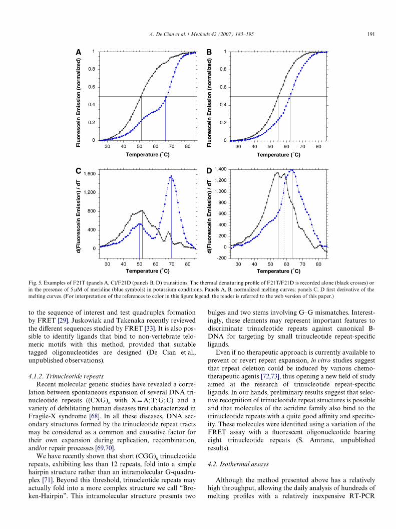
We also used the F21T oligomer to perform these exper-iments. As shown in Fig. 5 for meridine at 5 �M in K+ con-ditions, there is a signiWcant diVerence in Tm and in theshape of the curves when using F21T (Fig. 5A–C) or F21D(Fig. 5B–D). This experiment illustrates again the impor-tance of comparing results obtained under similar condi-tions, but also of analyzing the shape of the curves and notonly the stabilization values (see Section 3.3). We can alsodiscuss here the best method to determine the Tm of such atransition. The normalization and the T1/2 determination
Fig. 4. Competition experiments. (A) Example of a competition curve.Normalized melting proWles of F21T alone (black symbols), in presence of1 �M 9 in a potassium buVer without any competitor (green), with 10 �Mds26 (blue symbols), 3 �M 27NHEG (orange symbols) or 3 �M 30AG (redsymbols) competitors. All experiments were performed in duplicate; bothcurves are shown to illustrate reproducibility. (B) Quantitative analysis ofthe competition experiments. The stabilization in sodium (black bars) andpotassium (grey bars) is indicated for compound 9 (1 �M) in the absence(“none”) or presence of various single-stranded (dT26, mut27NHEG: 5�-d-TGAGTAGCGTGAGCAGAGTGCGTAACG-3�), double-stranded(ds26), and quadruple-stranded (27NHEG, 30AG) competitors at 3 or10 �M. (For interpretation of the references to color in this Wgure legend,the reader is referred to the web version of this paper.)
A
B
will give a biased Tm that does not reXect any of the twotransitions, but somewhat “averages” them. On the otherhand, the Wrst derivative will give a better idea of thebiphasic transition with the evidence of two main peaks(Fig. 5C–D). The reason for this biphasic behaviourremains an enigma. Several explanations may be proposed.For example, one could envision that distinct quadruplexconformations are diVerently stabilized by the ligand. Asthis behaviour depends on the nature of the Xuorescenttags, this implies that the equilibria between quadruplexspecies may be shifted by these labels (a reasonable butunveriWed assumption). The risk of such a determination isto notify only the stabilization values of the second transi-tion. This can result in an overestimation of the stabilizingeVect of the ligand.
3.3. How can the diVerences among diVerent cationic conditions be interpreted?
Quadruplexes are very polymorphic and, for a givensequence, several foldamers may coexist. Recent data dem-onstrated that the crystal [5] and solution structure [62,63]of the human telomeric quadruplex in potassium may bediVerent from the structure in sodium [4]. Even if high-reso-lution structures have been determined in Na+ and K+,some precautions should be taken: for the crystal structuredetermination, a G-quadruplex ligand was used duringcrystallization and for solution structure, sequence variantswere used to overcome the conformational heterogeneity.Wild-type telomeric sequences do form G-quadruplexes,but fail to produce good crystal or NMR data, probablybecause of the existence of multiple foldamers. Further-more, Xuorescent labelling of the oligonucleotide may alsoalter the folding or the foldamer population distribution.
One may propose that most ligands (illustrated here byascididemin and meridine) have a preference for the mixedparallel (potassium form) over antiparallel basket (sodiumform) quadruplexes. This preference for the K+ over theNa+ form seems to be a general rule (ADC, unpublishedobservations). This trend is in line with the higher accessi-bility of the terminal tetrads in the K+ form(s) as seen in thesolid-state structure and in the solution conformation(hybrid form). This is probably an oversimpliWcation: onewould probably need to consider ligand binding to a popu-lation of quadruplexes rather than to a single quadruplexstructure. One can envision that complex melting curves(biphasic) could result from this structural heterogeneity.
4. Variations and conclusion
4.1. Extension to other sequence motifs
4.1.1. Non-telomeric G-quadruplexesSeveral groups are interested in the design of quadruplex
ligands that recognize non-telomeric motifs, such as thosefound in the promoters of oncogenes [64–67]. One may inprinciple design a Xuorescent oligonucleotide corresponding
A. De Cian et al. / Methods 42 (2007) 183–195 191
to the sequence of interest and test quadruplex formationby FRET [29]. Juskowiak and Takenaka recently reviewedthe diVerent sequences studied by FRET [33]. It is also pos-sible to identify ligands that bind to non-vertebrate telo-meric motifs with this method, provided that suitabletagged oligonucleotides are designed (De Cian et al.,unpublished observations).
4.1.2. Trinucleotide repeatsRecent molecular genetic studies have revealed a corre-
lation between spontaneous expansion of several DNA tri-nucleotide repeats ((CXG)n with XDA;T; G;C) and avariety of debilitating human diseases Wrst characterized inFragile-X syndrome [68]. In all these diseases, DNA sec-ondary structures formed by the trinucleotide repeat tractsmay be considered as a common and causative factor fortheir own expansion during replication, recombination,and/or repair processes [69,70].
We have recently shown that short (CGG)n trinucleotiderepeats, exhibiting less than 12 repeats, fold into a simplehairpin structure rather than an intramolecular G-quadru-plex [71]. Beyond this threshold, trinucleotide repeats mayactually fold into a more complex structure we call “Bro-ken-Hairpin”. This intramolecular structure presents two
bulges and two stems involving G–G mismatches. Interest-ingly, these elements may represent important features todiscriminate trinucleotide repeats against canonical B-DNA for targeting by small trinucleotide repeat-speciWcligands.
Even if no therapeutic approach is currently available toprevent or revert repeat expansion, in vitro studies suggestthat repeat deletion could be induced by various chemo-therapeutic agents [72,73], thus opening a new Weld of studyaimed at the research of trinucleotide repeat-speciWcligands. In our hands, preliminary results suggest that selec-tive recognition of trinucleotide repeat structures is possibleand that molecules of the acridine family also bind to thetrinucleotide repeats with a quite good aYnity and speciWc-ity. These molecules were identiWed using a variation of theFRET assay with a Xuorescent oligonucleotide bearingeight trinucleotide repeats (S. Amrane, unpublishedresults).
4.2. Isothermal assays
Although the method presented above has a relativelyhigh throughput, allowing the daily analysis of hundreds ofmelting proWles with a relatively inexpensive RT-PCR
Fig. 5. Examples of F21T (panels A, C)/F21D (panels B, D) transitions. The thermal denaturing proWle of F21T/F21D is recorded alone (black crosses) orin the presence of 5 �M of meridine (blue symbols) in potassium conditions. Panels A, B, normalized melting curves; panels C, D Wrst derivative of themelting curves. (For interpretation of the references to color in this Wgure legend, the reader is referred to the web version of this paper.)
-200
0
200
400
600
800
1,000
1,200
1,400
30 40 50 60 70 80
d(F
luo
resc
ein
Em
issi
on
) / d
T
Temperature (˚C)
0
400
800
1,200
1,600
30 40 50 60 70 80
d(F
luo
resc
ein
Em
issi
on
) / d
T
Temperature (˚C)
0
0.2
0.4
0.6
0.8
1
30 40 50 60 70 80Flu
ore
scei
n E
mis
sio
n (
no
rmal
ized
)
Temperature (˚C)
0
0.2
0.4
0.6
0.8
1
30 40 50 60 70 80Flu
ore
scei
n E
mis
sio
n (
no
rmal
ized
)
Temperature (˚C)
A B
C D

192 A. De Cian et al. / Methods 42 (2007) 183–195
instrument, it is not easily transformed into high-through-put screening (HTS) format. An isothermal assay would berequired.
4.2.1. Hybridization assayA good basis for such an assay is a hybridization test: a
complementary C-rich strand is added to a quadruplex-forming oligonucleotide. In the presence of a quadruplexligand, the folded conformation is locked and hybridizationis impaired or delayed. Duplex formation may then be fol-lowed by FRET using a double-labelled sequence such asF21D or F21T [74]. This approach has to be used with cau-tion. A strong delay in duplex formation has been observedin the presence of ligands not signiWcantly increasing theT1/2 of the quadruplex (P.A. unpublished observations).Indeed, certain ligands may interact with the Xuorescein tagand quench its Xuorescence emission, resulting in an appar-ent delay of duplex formation. In order to avoid false posi-tives, i.e., to discriminate between a real and an apparentdelay, it should be better to label the 3� (or 5�) end of theG-rich sequence with the donor and the 5� (or 3�) end of theC-rich sequence with the acceptor or quencher. Thisalternative approach should permit to verify if the donoremission is quenched by the ligand, prior to the addition ofthe complementary C-rich strand. Isothermal assays can beperformed on standard plate readers. In principle it couldbe possible to test up to 384 and 1536 samples in a singlereading. It could also be envisaged to set up a multiplexedassay permitting to test a ligand on diVerent quadruplex-forming sequences in the same well. Each quadruplexsequence could be discriminated at a speciWc emissionwavelength using a speciWc FRET couple. Moreover, incontrast with thermal melting experiments, an isothermalhybridization assay allows, in principle, performing compe-tition experiments using competitor structures as stable asthe quadruplex to be tested, or even less stable. It is suY-
cient to work at a temperature low enough to ensure thefolding of both structures (quadruplex and competitor).
4.2.2. Quenching eYciencyIsothermal assays become useful when the diVerence in
melting temperatures between the Xuorescent oligonucleo-tide alone (F21T for example) and its ligand-bound form istoo high to allow a complete visualization of the meltingcurve. Another situation in which an alternative methodmay be useful occurs when several ligands show a similarstabilization eVect on the Xuorescent oligonucleotide andthe diVerence in melting temperatures is not evident. Onepossible alternative is to measure the FRET eYciency at agiven temperature (usually around 25 °C) by recording theemission spectra of the free and ligand-bound form in thesame conditions. Surprisingly, stabilization of the dual-labelled oligonucleotide (F21T) by the ligand very oftenresults in a further decrease in emission (B. Saccà, unpub-lished observations). Furthermore, we observed that theextent of quenching is related to the aYnity of the ligandfor the oligonucleotide tested (all precautions must be
taken to avoid measurements of false positives, such as useof the same experimental conditions in absence and pres-ence of ligand, control assay with a labelled non-quadru-plex motif to test the possibility of non-speciWc interactionsof the ligand with the oligonucleotide and/or Xuorophore,and assessment of the quadruplex conformation of theF21T at the chosen temperature). Normalization allows forcomparison of diVerent ligands. Even small variations inligand-binding aYnity may be visualized as a decrease inthe Xuorescence emission of the donor. The rapidity of themethod represents its main advantage although the isother-mal conditions preclude the possibility of a thermodynamicanalysis of the binding reaction.
The usefulness of this method is illustrated by the fol-lowing experiment recently performed in our group. Weanalysed a series of paromomycin–quinacridine derivatives(Fig. 6) to test their binding aYnity towards the G-quadru-plex motif. Recent studies have demonstrated the bindingcapabilities of these aminoglycoside–quinacridine systemsto the stem–loop motif of the P6.1 element, which is criti-cally involved in telomerase assembly [59]. However, theirbinding properties towards telomeric G-quadruplex struc-tures have not yet been elucidated. For this reason, weinvestigated their eVect on the stabilization of F21T, bothin 100 mM sodium or 100 mM potassium in 10 mM caco-dylate buVer at pH 7.0. As the melting temperaturesrecorded by FRET experiments were too high to be accu-rately determined (even in LiCl buVer), we performed anisothermal FRET measurement of a 0.2 �M solution ofF21T in absence and presence of 1 �M of diVerent paromo-mycin conjugates. Upon excitation at 470 nm, emissionspectra were recorded in the 480–650 nm range at 20 °Cafter 12 h equilibration at 4 °C. The FRET eYciencies forall the F21T–ligand pairs, both in sodium and potassiumbuVer, are shown in Fig. 7 and are directly proportional tothe binding aYnities of the ligand towards the G-quadru-plex motif. It appears evident that in sodium buVer (Fig. 7,grey bars) all the ligands show a strong binding aYnitytowards the F21T quadruplex according to the followingorder: PM1 7 PD2 t PD1 > PM3 > PD3. The same rank-ing of binding aYnity was obtained in potassium buVer(Fig. 7, black bars), although in this case the binding condi-tions seem to be more stringent. Only PM1 suYcientlybinds to F21T, resulting in a quenching eYciency of morethan 50% (while in sodium buVer all the ligands led to morethan 70% of quenching). In particular, in potassium buVerthe weakly binding PD3 ligand shows almost no bindingaYnity. This demonstrates that for weakly binding ligandsthe choice of the buVer conditions may be determinant.Additionally, we could easily and rapidly determine theeVect of linker length and number of aminoglycoside moie-ties on the binding aYnities of diVerent paromomycinderivatives to the G-quadruplex motif. Though specula-tions may be drawn to fully explain these results, a cleartendency is however deWned: the binding is favoured byshorter linker lengths and monosubstituted quinacridineplatforms (PM1 and PM3). The isothermal FRET

A. De Cian et al. / Methods 42 (2007) 183–195 193
approach may also be suitable for competition assays, onceall the necessary precautions described above are taken intoconsideration.
In conclusion, although the method allows only semi-quantitative analysis of ligand-binding aYnities, it may beused to rapidly identify the most promising candidatesamong many compounds in order to direct synthetic and/oranalytical eVorts towards the best direction. As a Wnal cau-tion note, one should consider that this method may gener-ate false positives (quenching of donor by the ligand) andfalse negatives (folding with the ligand in such conWgura-tion that the distance/orientation prevents quenching/FRET).
Fig. 7. Relative binding aYnities of the paromomycin derivatives for theF21T oligo. The eYciency of energy transfer between the Xuorescein andTAMRA of the quadruplex-forming F21T oligo (0.2 �M) was measuredin presence of diVerent paromomycin–quinacridine conjugates at 1 �Mconcentration (for the nomenclature, see Fig. 6) in 10 mM sodium caco-dylate buVer at pH 7.0, either in 100 mM NaCl (grey bars) or in 100 mMKCl (black bars). The extent of energy transfer is proportional to thebinding aYnity of the ligand for the G-quadruplex motif.
4.3. Concluding remarks
The number of G4 ligands has grown rapidly over a fewyears: a range of molecules has been shown to recognize thetelomeric quadruplex [7,12,13,15,32,75–88]. One molecule,telomestatin, is exceptionally potent with an IC50 of 5 nM inthe TRAP assay [11]. The success in the generation of largelibraries argues for analytical methods that allow a rapid,simple, inexpensive, and semi-quantitative evaluation ofbinding “strength”. The G4 FRET assay presented here iswell suited for this purpose, especially in 96-well format.Contrary to other methods, it allows a variety of ionic con-ditions. Each molecule may be tested at various concentra-tions, and in the presence of a large excess of competitors.Hence, the G4 FRET not only monitors aYnity, but alsospeciWcity towards quadruplexes, a critical issue for manyligands [89,90].
Acknowledgments
We thank Patrick Mailliet and Eliane Mandine (SanoW-Aventis, Vitry, France) and Jean-François Riou (Universitéde Reims) for helpful discussions. This work was supportedin part by an ARC grant (No. 365), an EU FP6 “MolCan-cerMed” grant (LSHC-CT-2004-502943 to J.-L.M.) and aFondation Jérôme Lejeune Ph.D. fellowship (to S.A.).
References
[1] J.R. Williamson, Annu. Rev. Biophys. Biomol. Struct. 23 (1994) 703–730.
[2] J.T. Davis, Angew. Chem. Int. Ed. 43 (2004) 668–698.[3] K. Masutomi, E.Y. Yu, S. Khurts, I. BenPorath, J.L. Currier, G.B.
Metz, M.W. Brooks, S. Kaneko, S. Murakami, J.A. DeCaprio, R.A.Weinberg, S.A. Stewart, W.C. Hahn, Cell 114 (2003) 241–253.
Fig. 6. Paromomycin–quinacridine derivatives used for isothermal FRET experiments. Combination of the aminoglycoside moiety (paromomycin, P) withthree diVerent linker chains (1–3) and a mono- or dicarboxaldehyde (M and D, respectively) leads to formation of Wve diVerent conjugates (PM1, PM3,PD1, PD2, and PD3).
194 A. De Cian et al. / Methods 42 (2007) 183–195
[4] Y. Wang, D.J. Patel, Structure 1 (1993) 263–282.[5] G.N. Parkinson, M.P.H. Lee, S. Neidle, Nature 417 (2002) 876–880.[6] A.M. Zahler, J.R. Williamson, T.R. Cech, D.M. Prescott, Nature 350
(1991) 718–720.[7] D. Sun, B. Thompson, B.E. Cathers, M. Salazar, S.M. Kerwin, J.O.
Trent, T.C. Jenkins, S. Neidle, L.H. Hurley, J. Med. Chem. 40 (1997)2113–2116.
[8] J.L. Mergny, C. Hélène, Nat. Med. 4 (1998) 1366–1367.[9] L. Guittat, P. Alberti, F. Rosu, S. Van Miert, E. Thetiot, L. Pieters, V.
Gabelica, E. De Pauw, A. Ottaviani, J.F. Riou, J.-L. Mergny, Biochi-mie 85 (2003) 535–547.
[10] M. Franceschin, L. Rossetti, A. Dambrosio, S. Schirripa, A. Bianco,G. Ortaggi, M. Savino, C. Schultes, S. Neidle, Bioorg. Med. Chem.Lett. 16 (2006) 1707–1711.
[11] K. Shin-ya, K. Wierzba, K. Matsuo, T. Ohtani, Y. Yamada, K. Furih-ata, Y. Hayakawa, H. Seto, J. Am. Chem. Soc. 123 (2001) 1262–1263.
[12] M.Y. Kim, H. Vankayalapati, K. Shin-ya, K. Wierzba, L.H. Hurley, J.Am. Chem. Soc. 124 (2002) 2098–2099.
[13] S. Gowan, R. Heald, M. Stevens, L. Kelland, Mol. Pharmacol. 60(2001) 981–988.
[14] G. Pennarun, C. Granotier, L.R. Gauthier, D. Gomez, F. HoVschir, E.Mandine, J.F. Riou, J.L. Mergny, P. Mailliet, F.D. Boussin, Oncogene24 (2005) 2917–2928.
[15] R.J. Harrison, J. Cuesta, G. Chessari, M.A. Read, S.K. Basra, A.P.Reszka, J. Morrell, S.M. Gowan, C.M. Incles, F.A. Tanious, W.D. Wil-son, L.R. Kelland, S. Neidle, J. Med. Chem. 46 (2003) 4463–4476.
[16] A.M. Burger, F.P. Dai, C.M. Schultes, A.P. Reszka, M.J. Moore, J.A.Double, S. Neidle, Cancer Res. 65 (2005) 1489–1496.
[17] J.L. Mergny, J.F. Riou, P. Mailliet, M.P. Teulade-Fichou, E. Gilson,Nucleic Acids Res. 30 (2002) 839–865.
[18] G. Saretzki, Cancer Lett. 194 (2003) 209–219.[19] E.M. Rezler, D.J. Bearss, L.H. Hurley, Annu. Rev. Pharmacol. Toxi-
col. 43 (2003) 359–379.[20] J. Cuesta, M. Read, S. Neidle, Mini Rev. Med. Chem. 3 (2003) 11–21.[21] L.K. White, W.E. Wright, J.W. Shay, Trends Biotechnol. 19 (2001)
114–120.[22] P.T. Rowley, Expert. Opin. Ther. Patents 11 (2001) 1815–1823.[23] S.M. Kerwin, Curr. Pharm. Des. 6 (2000) 441–471.[24] L. Guittat, P. Alberti, D. Gomez, A. de Cian, G. Pennarun, T. Lemar-
teleur, C. Belmokhtar, R. Paterski, H. Morjani, C. Trentesaux, E.Mandine, F. Boussin, P. Mailliet, L. Lacroix, J.-F. Riou, J.-L. Mergny,Cytotechnology 45 (2004) 75–90.
[25] J.W. Shay, W.E. Wright, Nat. Rev. Drug Discov. 5 (2006) 577–584.[26] J.L. Mergny, A.T. Phan, L. Lacroix, FEBS Lett. 435 (1998) 74–78.[27] C.C. Chang, J.Y. Wu, C.W. Chien, W.S. Wu, H. Liu, C.C. Kang, L.J.
Yu, T.C. Chang, Anal. Chem. 75 (2003) 6177–6183.[28] G.S. Minhas, D.S. Pilch, J.E. Kerrigan, E.J. Lavoie, J.E. Rice, Bioorg.
Med. Chem. Lett. 16 (2006) 3891–3895.[29] T. Simonsson, R. Sjoback, J. Biol. Chem. 274 (1999) 17379–17383.[30] J.L. Mergny, Biochemistry 38 (1999) 1573–1581.[31] J.L. Mergny, J.C. Maurizot, ChemBioChem 2 (2001) 124–132.[32] J.L. Mergny, L. Lacroix, M.P. Teulade-Fichou, C. Hounsou, L. Guit-
tat, M. Hoarau, P.B. Arimondo, J.P. Vigneron, J.M. Lehn, J.F. Riou,T. Garestier, C. Hélène, Proc. Natl. Acad. Sci. USA 98 (2001) 3062–3067.
[33] B. Juskowiak, S. Takenaka, Methods Mol. Biol. 335 (2006) 311–341.[34] E.E. Merkina, K.R. Fox, Biophys. J. 89 (2005) 365–373.[35] N. Kumar, S. Maiti, Biochem. Biophys. Res. Commun. 319 (2004)
759–767.[36] J.Y. Lee, B. Okumus, D.S. Kim, T.J. Ha, Proc. Natl. Acad. Sci. USA
102 (2005) 18938–18943.[37] J.J. Green, L.M. Ying, D. Klenerman, S. Balasubramanian, J. Am.
Chem. Soc. 125 (2003) 3763–3767.[38] S. Lyonnais, R.J. Gorelick, J.L. Mergny, E. LeCam, G. Mirambeau,
Nucleic Acids Res. 31 (2003) 5754–5763.[39] T. Romero-Salas, I. Petruseva, O. Lavrik, A. Bourdoncle, J.-L.
Mergny, A. Favre, C. Saintome, Nucleic Acids Res. 34 (2006) 4857–4865.
[40] F. He, Y.L. Tang, S. Wang, Y.L. Li, D.B. Zhu, J. Am. Chem. Soc. 127(2005) 12343–12346.
[41] B. Juskowiak, E. Galezowska, A. Zawadzka, A. Gluszynska, S. Take-naka, Spectrochim. Acta A: Mol. Biomol. Spectrosc. 64 (2006) 835–843.
[42] J.J. Li, W. Tan, Nano Letters 2 (2002) 315–318.[43] P. Alberti, J.L. Mergny, Proc. Natl. Acad. Sci. USA 100 (2003) 1569–
1573.[44] P. Alberti, J.L. Mergny, Cell. Mol. Biol. 50 (2004) 241–253.[45] P. Alberti, A. Bourdoncle, B. Saccà, L. Lacroix, J.L. Mergny, Org. Bio-
mol. Chem. 4 (2006) 3383–3391.[46] A. Bourdoncle, A. Estévez Torres, C. Gosse, L. Lacroix, P. VekhoV, T.
Le Saux, L. Jullien, J.L. Mergny, J. Am. Chem. Soc. 128 (2006) 11094–11105.
[47] R. Darby, M. Sollogoub, C. McKeen, L. Brown, A. Risitano, N.Brown, C. Barton, T. Brown, K. Fox, Nucleic Acids Res. 30 (2002)e39.
[48] C.M. Schultes, W. Guyen, J. Cuesta, S. Neidle, Bioorg. Med. Chem.Lett. 14 (2004) 4347–4351.
[49] D. Gomez, R. Paterski, T. Lemarteleur, K. Shinya, J.L. Mergny, J.F.Riou, J. Biol. Chem. 279 (2004) 41487–41494.
[50] M.J.B. Moore, C.M. Schultes, J. Cuesta, F. Cuenca, M. Gunaratnam,F.A. Tanious, W.D. Wilson, S. Neidle, J. Med. Chem. 49 (2006) 582–599.
[51] K.E. Sapsford, L. Berti, I.L. Medintz, Angew. Chem. Int. Ed. Engl. 45(2006) 4562–4589.
[52] C.R. Cantor, M.M. Warshaw, H. Shapiro, Biopolymers 9 (1970)1059–1077.
[53] S. Tyagi, F.R. Kramer, Nat. Biotechnol. 14 (1996) 303–308.[54] S. Tyagi, D.P. Bratu, F.R. Kramer, Nat. Biotechnol. 16 (1998) 49–53.[55] A. Risitano, K.R. Fox, Biochemistry 42 (2003) 6507–6513.[56] A. Risitano, K.R. Fox, Org. Biomol. Chem. 1 (2003) 1852–1855.[57] A. Risitano, K.R. Fox, Nucleic Acids Res. 32 (2004) 2598–2606.[58] J.L. Mergny, L. Lacroix, Oligonucleotides 13 (2003) 515–537.[59] M. Kaiser, M. Sainlos, J.M. Lehn, S. Bombard, M.P. Teulade-Fichou,
ChemBioChem 7 (2006) 321–329.[60] M. Kaiser, A. De Cian, M. Sainlos, C. Renner, J.L. Mergny, M.P. Teu-
lade-Fichou, Org. Biomol. Chem. 4 (2006) 1049–1057.[61] L. Guittat, A. DeCian, F. Rosu, V. Gabelica, E. DePauw, E. Delfourne,
J.L. Mergny, Bioch. Biophys. Acta Gen. Subjects 1724 (2005) 375–384.[62] A. Ambrus, D. Chen, J. Dai, T. Bialis, R.A. Jones, D. Yang, Nucleic
Acids Res. 34 (2006) 2723–2735.[63] K.N. Luu, A.T. Phan, V. Kuryavyi, L. Lacroix, D.J. Patel, J. Am.
Chem. Soc. 128 (2006) 9963–9970.[64] A. Rangan, O.Y. FedoroV, L.H. Hurley, J. Biol. Chem. 276 (2001)
4640–4646.[65] C.L. Grand, H.Y. Han, R.M. Munoz, S. Weitman, D.D. VonHoV, L.H.
Hurley, D.J. Bearss, Mol. Cancer Ther. 1 (2002) 565–573.[66] A.T. Phan, V. Kuryavyi, H.Y. Gaw, D.J. Patel, Nat. Chem. Biol. 1
(2005) 167–173.[67] E.M. Rezler, J. Seenisamy, S. Bashyam, M.Y. Kim, E. White, W.D.
Wilson, L.H. Hurley, J. Am. Chem. Soc. 127 (2005) 9439–9447.[68] I. Oberle, F. Rousseau, D. Heitz, C. Kretz, D. Devys, A. Hanauer, J.
Boue, M. Bertheas, J. Mandel, Science 252 (1991) 1097–1102.[69] C.T. McMurray, Proc. Natl. Acad. Sci. USA 96 (1999) 1823–1825.[70] J.D. Cleary, C.E. Pearson, Trends Genet. 21 (2005) 272–280.[71] S. Amrane, J.L. Mergny, Biochimie 88 (2006) 4857–4865.[72] V.I. Hashem, R.R. Sinden, Mutat. Res. 508 (2002) 107–119.[73] V.I. Hashem, M.J. Pytlos, E.A. Klysik, K. Tsuji, M. Khajav, T. Ashiz-
awa, R.R. Sinden, Nucleic Acids Res. 32 (2004) 6334–6346.[74] P. Alberti, J. Ren, M.P. Teulade-Fichou, L. Guittat, J.F. Riou, J.B.
Chaires, C. Hélène, J.P. Vigneron, J.M. Lehn, J.L. Mergny, J. Biomol.Struct. Dyn. 19 (2001) 505–513.
[75] R.T. Wheelhouse, D. Sun, H. Han, F.X. Han, L.H. Hurley, J. Am.Chem. Soc. 120 (1998) 3261–3262.
[76] O.Y. FedoroV, M. Salazar, H. Han, V.V. Chemeris, S.M. Kerwin, L.H.Hurley, Biochemistry 37 (1998) 12367–12374.
[77] P.J. Perry, M.A. Read, R.T. Davies, S.M. Gowan, A.P. Reszka, A.A.Wood, L.R. Kelland, S. Neidle, J. Med. Chem. 42 (1999) 2679–2684.

A. De Cian et al. / Methods 42 (2007) 183–195 195
[78] R.J. Harrison, S.M. Gowan, L.R. Kelland, S. Neidle, Bioorg. Med.Chem. Lett. 9 (1999) 2463–2468.
[79] S. Neidle, R.J. Harrison, A.P. Reszka, M.A. Read, Pharmacol. Ther.85 (2000) 133–139.
[80] V. Caprio, B. Guyen, Y. Opoku-Boahen, J. Mann, S.M. Gowan, L.M.Kelland, M.A. Read, S. Neidle, Bioorg. Med. Chem. Lett. 10 (2000)2063–2066.
[81] F. Koeppel, J.F. Riou, A. Laoui, P. Mailliet, P.B. Arimondo, D. Labit,O. Petigenet, C. Hélène, J.L. Mergny, Nucleic Acids Res. 29 (2001)1087–1096.
[82] P. Alberti, P. Schmidt, C.H. Nguyen, M. Hoarau, D. Grierson, J.L.Mergny, Bioorg. Med. Chem. Lett. 12 (2002) 1071–7074.
[83] D.F. Shi, R.T. Wheelhouse, D.Y. Sun, L.H. Hurley, J. Med. Chem. 44(2001) 4509–4523.
[84] R.A. Heald, C. Modi, J.C. Cookson, I. Hutchinson, C.A. Laughton, S.M.Gowan, L.R. Kelland, M.F.G. Stevens, J. Med. Chem. 45 (2002) 590–597.
[85] S.M. Gowan, J.R. Harrison, L. Patterson, M. Valenti, M.A. Read, S.Neidle, L.R. Kelland, Mol. Pharmacol. 61 (2002) 1154–1162.
[86] L. Rossetti, M. Franceschin, A. Bianco, G. Ortaggi, M. Savino, Bio-org. Med. Chem. Lett. 12 (2002) 2527–2533.
[87] M. Read, R.J. Harrison, B. Romagnoli, F.A. Tanious, S.H. Gowan,A.P. Reszka, W.D. Wilson, L.R. Kelland, S. Neidle, Proc. Natl. Acad.Sci. USA 98 (2001) 4844–4849.
[88] A. Maraval, S. Franco, C. Vialas, G. Pratviel, M.A. Blasco, B. Meu-nier, Org. Biomol. Chem. 1 (2003) 921–927.
[89]P. Alberti, M. Hoarau, L. Guittat, M. Takasugi, P.B. Arimondo, L.Lacroix, M. Mills, M.P. Teulade-Fichou, J.P. Vigneron, J.M. Lehn, P.Mailliet, J.L. Mergny, in: C. Bailly, M. Demeunynck, D. Wilson (Eds.),Small molecule DNA and RNA binders: from synthesis to nucleicacid complexes, Wiley VCH, 2002, pp. 315–336.
[90] P. Alberti, L. Lacroix, L. Guittat, C. Hélène, J.L. Mergny, Mini Rev.Med. Chem. 3 (2003) 23–36.


![Solution Structure of the Na+ form of the Dimeric Guanine Quadruplex [d(G3T4G3)]2](https://static.fdokumen.com/doc/165x107/6318f44265e4a6af370f95cf/solution-structure-of-the-na-form-of-the-dimeric-guanine-quadruplex-dg3t4g32.jpg)