Rational Design of Substituted Diarylureas: A Scaffold for Binding to G-Quadruplex Motifs
17
Rational Design of Substituted Diarylureas: A Scaffold for Binding to G-Quadruplex Motifs William C. Drewe, † Rupesh Nanjunda, ‡ Mekala Gunaratnam, † Monica Beltran, † Gary N. Parkinson, † Anthony P. Reszka, † W. David Wilson, ‡ and Stephen Neidle* ,† The Cancer Research UK Biomolecular Structure Group, The School of Pharmacy, UniVersity of London, 29-39 Brunswick Square, London WC1N 1AX, U.K., and Department of Chemistry, Georgia State UniVersity, Atlanta, Georgia 30303-3083 ReceiVed June 13, 2008 The design and synthesis of a series of urea-based nonpolycyclic aromatic ligands with alkylaminoanilino side chains as telomeric and genomic G-quadruplex DNA interacting agents are described. Their interactions with quadruplexes have been examined by means of fluorescent resonance energy transfer melting, circular dichroism, and surface plasmon resonance-based assays. These validate the design concept for such urea- based ligands and also show that they have significant selectivity over duplex DNA, as well as for particular G-quadruplexes. The ligand-quadruplex complexes were investigated by computational molecular modeling, providing further information on structure-activity relationships. Preliminary biological studies using short- term cell growth inhibition assays show that some of the ligands have cancer cell selectivity, although they appear to have low potency for intracellular telomeric G-quadruplex structures, suggesting that their cellular targets may be other, possibly oncogene-related quadruplexes. Introduction Nucleic acids containing repetitive guanine-rich tracts can form higher-order structures, G-quadruplexes, which are held together by the G-quartet motif of four in-plane hydrogen- bonded guanines. 1-4 Quadruplex arrangements were first de- scribed for eukaryotic telomeric DNA sequences, which com- prise long tandem G-tracts of simple repeats such as TTAGGG in mammals. 5,6 The terminal 3′ ends of human and other mammalian telomeres are single-stranded, 7,8 and although these are normally associated with single-stranded binding proteins such as hPOT1 in human telomeres, 9-12 they can have enhanced potential to fold up into quadruplex arrangements compared to such sequences within duplex DNA. Telomeres in normal human cells progressively shorten with successive rounds of replication 13 but are maintained in length in many cancer cells by the reverse transcriptase action of the telomerase enzyme complex. 14-18 Telomerase activity requires a linear 3′ end telomeric DNA template for transcribing, which is inhibited by folding it into a quadruplex, which can be induced by a wide range of quadruplex-stabilizing ligands. 19-36 The potential to form quadruplexes has been established in the human and a number of other genomes and appears to have an elevated frequency of occurrence in eukaryotic promoter regions 37 notably in a number of oncogenes such as c-myc, 38-41 c-kit, 42-44 bcl-2, 45 and K-ras. 46 A quadruplex sequence has also been identified in the 5′-untranslated region of the N-ras oncogene. 47 The recent discovery of a functional interaction between the human nuclear protein poly ADP-ribose poly- merase-1 and several quadruplexes suggests a further role for these structures. 48 Telomere regulation and stability are achieved through a complex capping mechanism, 49-51 required to protect the chromosome ends from being recognized as damaged DNA, and are necessary for cellular viability. 52 Disruption of the capping status of the telomere results in the up-regulation of the DNA damage response system, telomere end-end fusions, and other genomic rearrangements. 51-55 It has been demon- strated that several established telomerase inhibitors, 26,56 including the trisubstituted acridine compound BRACO-19 (3,6-bis(3-pyr- rolidin-1-ylpropionamido)-9-(4-dimethylaminophenylamino) acri- dine), 32 are able to induce rapid cellular responses of senescence and apoptosis in the absence of the characteristic lag period associated with classical telomerase inhibition. 57-60 This more rapid response has been associated with intracellular displacement of the telomeric proteins, a γH2AX/p53/p21 mediated DNA damage response and mitotic abnormalities such as telomere end-end fusions and anaphase bridges. 22,27,61-67 These observations are indicative that these ligands can have a dual action in cells, disrupting the protective capping function of the telomere, as well as acting as classic telomerase inhibitors as seen by progressive telomere shortening. Knowledge of the details of telomeric 68-72 and genomic 38,39,43 G-quadruplex molecular structures, as well as those of G-quadruplex-ligand complexes 39,73-75 such as that involv- ing BRACO-19, 76 can facilitate the rational design not only of new ligand analogues but also of novel G-quadruplex DNA binding scaffolds. Crystallographic studies on several telo- meric quadruplex-ligand complexes have shown (1) that in all structures solved to date the quadruplexes have the all- parallel strand topology, as initially observed in the native structures and (2) the details of loop and groove geometry are variable, dependent on the nature of the individual ligand. 75,76 We have used the general features of the parallel quadruplex to rationally design a new category of nonpoly- cyclic quadruplex ligand that incorporates features of the inherently planar 1,3-diphenyl urea functionality, a “privi- leged” scaffold that has well-established druglike character- istics as well as optimal dimensions in its more stable trans-trans conformation 77,78 for efficient G-tetrad overlap (Figure 1a). This scaffold is present in a number of clinically approved and experimental therapeutic agents. We report here the design, synthesis, and evaluation of a library of molecules based around this diarylurea functionality. 78 Structure-based * To whom correspondence should be addressed. Phone: 44 207 753 5969. Fax: 44 207 753 5970. E-mail: [email protected]. † University of London. ‡ Georgia State University. J. Med. Chem. 2008, 51, 7751–7767 7751 10.1021/jm801245v CCC: $40.75 2008 American Chemical Society Published on Web 12/03/2008
-
Upload
independent -
Category
Documents
-
view
2 -
download
0
Transcript of Rational Design of Substituted Diarylureas: A Scaffold for Binding to G-Quadruplex Motifs

Rational Design of Substituted Diarylureas: A Scaffold for Binding to G-Quadruplex Motifs
William C. Drewe,† Rupesh Nanjunda,‡ Mekala Gunaratnam,† Monica Beltran,† Gary N. Parkinson,† Anthony P. Reszka,†
W. David Wilson,‡ and Stephen Neidle*,†
The Cancer Research UK Biomolecular Structure Group, The School of Pharmacy, UniVersity of London, 29-39 Brunswick Square,London WC1N 1AX, U.K., and Department of Chemistry, Georgia State UniVersity,Atlanta, Georgia 30303-3083
ReceiVed June 13, 2008
The design and synthesis of a series of urea-based nonpolycyclic aromatic ligands with alkylaminoanilinoside chains as telomeric and genomic G-quadruplex DNA interacting agents are described. Their interactionswith quadruplexes have been examined by means of fluorescent resonance energy transfer melting, circulardichroism, and surface plasmon resonance-based assays. These validate the design concept for such urea-based ligands and also show that they have significant selectivity over duplex DNA, as well as for particularG-quadruplexes. The ligand-quadruplex complexes were investigated by computational molecular modeling,providing further information on structure-activity relationships. Preliminary biological studies using short-term cell growth inhibition assays show that some of the ligands have cancer cell selectivity, although theyappear to have low potency for intracellular telomeric G-quadruplex structures, suggesting that their cellulartargets may be other, possibly oncogene-related quadruplexes.
Introduction
Nucleic acids containing repetitive guanine-rich tracts canform higher-order structures, G-quadruplexes, which are heldtogether by the G-quartet motif of four in-plane hydrogen-bonded guanines.1-4 Quadruplex arrangements were first de-scribed for eukaryotic telomeric DNA sequences, which com-prise long tandem G-tracts of simple repeats such as TTAGGGin mammals.5,6 The terminal 3′ ends of human and othermammalian telomeres are single-stranded,7,8 and although theseare normally associated with single-stranded binding proteinssuch as hPOT1 in human telomeres,9-12 they can have enhancedpotential to fold up into quadruplex arrangements compared tosuch sequences within duplex DNA. Telomeres in normalhuman cells progressively shorten with successive rounds ofreplication13 but are maintained in length in many cancer cellsby the reverse transcriptase action of the telomerase enzymecomplex.14-18 Telomerase activity requires a linear 3′ endtelomeric DNA template for transcribing, which is inhibited byfolding it into a quadruplex, which can be induced by a widerange of quadruplex-stabilizing ligands.19-36
The potential to form quadruplexes has been established inthe human and a number of other genomes and appears to havean elevated frequency of occurrence in eukaryotic promoterregions37 notably in a number of oncogenes such as c-myc,38-41
c-kit,42-44 bcl-2,45 and K-ras.46 A quadruplex sequence has alsobeen identified in the 5′-untranslated region of the N-rasoncogene.47 The recent discovery of a functional interactionbetween the human nuclear protein poly ADP-ribose poly-merase-1 and several quadruplexes suggests a further role forthese structures.48
Telomere regulation and stability are achieved through acomplex capping mechanism,49-51 required to protect thechromosome ends from being recognized as damaged DNA,and are necessary for cellular viability.52 Disruption of the
capping status of the telomere results in the up-regulation ofthe DNA damage response system, telomere end-end fusions,and other genomic rearrangements.51-55 It has been demon-strated that several established telomerase inhibitors,26,56includingthe trisubstituted acridine compound BRACO-19 (3,6-bis(3-pyr-rolidin-1-ylpropionamido)-9-(4-dimethylaminophenylamino) acri-dine),32 are able to induce rapid cellular responses of senescenceand apoptosis in the absence of the characteristic lag periodassociated with classical telomerase inhibition.57-60 This more rapidresponse has been associated with intracellular displacement of thetelomeric proteins, a γH2AX/p53/p21 mediated DNA damageresponse and mitotic abnormalities such as telomere end-endfusions and anaphase bridges.22,27,61-67 These observations areindicative that these ligands can have a dual action in cells,disrupting the protective capping function of the telomere, as wellas acting as classic telomerase inhibitors as seen by progressivetelomere shortening.
Knowledge of the details of telomeric68-72 and genomic38,39,43
G-quadruplex molecular structures, as well as those ofG-quadruplex-ligand complexes39,73-75 such as that involv-ing BRACO-19,76 can facilitate the rational design not onlyof new ligand analogues but also of novel G-quadruplex DNAbinding scaffolds. Crystallographic studies on several telo-meric quadruplex-ligand complexes have shown (1) that inall structures solved to date the quadruplexes have the all-parallel strand topology, as initially observed in the nativestructures and (2) the details of loop and groove geometryare variable, dependent on the nature of the individualligand.75,76 We have used the general features of the parallelquadruplex to rationally design a new category of nonpoly-cyclic quadruplex ligand that incorporates features of theinherently planar 1,3-diphenyl urea functionality, a “privi-leged” scaffold that has well-established druglike character-istics as well as optimal dimensions in its more stabletrans-trans conformation77,78 for efficient G-tetrad overlap(Figure 1a). This scaffold is present in a number of clinicallyapproved and experimental therapeutic agents. We report herethe design, synthesis, and evaluation of a library of moleculesbased around this diarylurea functionality.78 Structure-based
* To whom correspondence should be addressed. Phone: 44 207 7535969. Fax: 44 207 753 5970. E-mail: [email protected].
† University of London.‡ Georgia State University.
J. Med. Chem. 2008, 51, 7751–7767 7751
10.1021/jm801245v CCC: $40.75 2008 American Chemical SocietyPublished on Web 12/03/2008
design has indicated that incorporation of additional phe-nylcarbamoyl groups allows all four guanine bases of aG-tetrad to be targeted, with addition of cationic side chainsto enhance G-quadruplex potency and selectivity and to aidaqueous solubility.79 These groups were systematically alteredthroughout the ligand series to gain an insight intostructure-activity relationships (Schemes 1 and 2). Telomericand a small set of promoter (c-kit and c-myc) G-quadruplexinteractions were assessed by biophysical assays. Our ultimateaim is to develop a novel series of ligands with low levelsof short-term toxicity so that cellular and ultimately in vivoadministration can be undertaken at concentrations that affecttelomere maintenance in cancer cells, in the absence ofgeneralized toxic effects.
Results
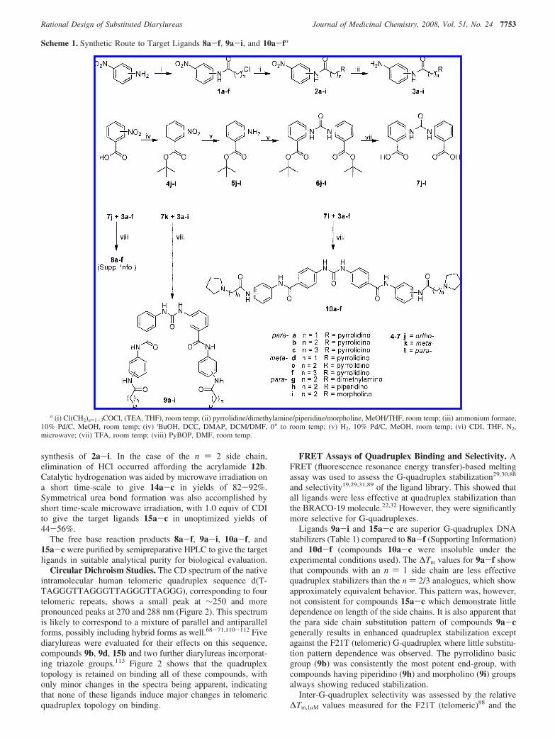
Chemistry. The synthesis of compounds 9a-i and 10a-fwas achieved through two key building blocks: the alkylami-noanilino side chains 3a-i and the urea-bearing dibenzoic acids7j-l (Scheme 1).
The alkylaminoanilino side chains 3a-i were synthesizedfrom 3- and 4-nitroaniline in overall yields (three steps) of19-99%, following methodology adapted from previouslyreported procedures.30,31 Acylation was achieved by reactionwith the required neat acid chloride, or the acid chloride inthe presence of TEA/THF to afford 1a-f in yields of62-100%. Amination of 1a,b/d,e/g-i was achieved byreaction with the required tertiary amine base (pyrrolidine/dimethylamine/piperidine/morpholine) in the presence ofmethanol or THF to yield 2a,b/d,e/g-i in 86-100%. 1c/fwas reacted with neat pyrrolidine to yield 2c/f in 60-88%.Catalytic hydrogenation afforded the alkylaminoanilino sidechains 3a-i in yields of 40-100%.
The synthesis of the diarylurea building blocks 7j-l wasachieved from 2-, 3-, and 4-nitrobenzoic acid in overall yields(four steps) of 33-68%. Protection of the carboxylic acidwas required to avoid amide bond formation upon treatment
with CDIa in the urea forming step80 and was achieved withtert-butanol in the presence of DCC and catalytic DMAP81
to afford 4j-l in yields of 72-86%. Catalytic hydrogenationgave the amines 5j-l in yields of 84-100%, which weresubsequently coupled into symmetrical ureas 6j-l by reactionwith CDI in refluxing anhydrous THF.82-84 6k was isolatedfollowing reaction with 0.6 equiv of CDI in a yield of 93%following workup, whereas 6j/l required an additional 0.6equiv of CDI to consume all of the starting amine. This gave6j/l as crude material that was used without further purifica-tion. 7j-l were isolated by suspension of 6j-l in TFA.85
Isolation of the precipitate afforded 7k/l in yields of95-100%, whereas 7j demonstrated enhanced TFA solubilityand was isolated in 46% yield following ether precipitation.
The target ligands 9a-i and 10a-f were synthesized byreaction of the alkylaminoanilino side chains 3a-i with thediarylurea building blocks 7k/l. Several reagents were evaluatedin order to achieve this transformation, with PyBOP in DMF86
proving the most efficient without requiring the addition oftertiary amine base because of the basic nature of 3a-i. Basicworkup gave the free base of 9a-i and 10a-f in yields of46-99%. The analogous reaction of the ortho-substituted 7jwith the alkylaminoanilino side chains 3a-f led to cyclizationof the reactive intermediate, yielding the nonplanar quinazolinedione ligands 8a-f (Supporting Information).
The target ligands 15a-c were synthesized from 3-amino-5-nitrobenzoic acid in overall yields (five steps) of 18-32%(Scheme 2). Amide bond formation was achieved using DCC/HOBt in DMF87 with an excess of aniline to afford 11 in ayield of 91%. Subsequent acylation to 12a-c in yields of66-92% and amination reactions to 13a-c in yields of 77-91%were achieved following the methodology discussed for the
a Abbreviations: FRET, fluorescent resonance energy transfer; CD, circulardichroism; SPR, surface plasmon resonance; DCC, N,N′-dicyclohexylcarbo-diimide; DMAP, 4-dimethylaminopyridine; CDI, 1,1′-carbonyldiimidazole; TFA,trifluoroacetic acid; PyBOP, benzotriazol-1-yloxytripyrrolidinophosphonium hexaflu-orophosphate; TRAP, telomerase repeat amplification protocol; SRB, sulfor-hodamine B.
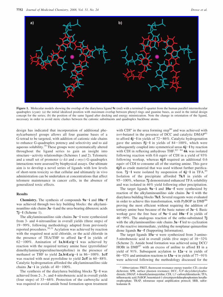
Figure 1. Molecular models showing the overlap of the diarylurea ligand 9c (red) with a terminal G-quartet from the human parallel intermolecularquadruplex (cyan): (a) the initial idealized position with maximum overlap between phenyl rings and guanine bases, as used in the initial designconcept for the series; (b) the position of the same ligand after docking and energy minimization. Note the change in orientation of the ligand,necessary in order to avoid steric clashes between the cationic substituents and quadruplex backbone atoms.
7752 Journal of Medicinal Chemistry, 2008, Vol. 51, No. 24 Drewe et al.
synthesis of 2a-i. In the case of the n ) 2 side chain,elimination of HCl occurred affording the acrylamide 12b.Catalytic hydrogenation was aided by microwave irradiation ona short time-scale to give 14a-c in yields of 82-92%.Symmetrical urea bond formation was also accomplished byshort time-scale microwave irradiation, with 1.0 equiv of CDIto give the target ligands 15a-c in unoptimized yields of44-56%.
The free base reaction products 8a-f, 9a-i, 10a-f, and15a-c were purified by semipreparative HPLC to give the targetligands in suitable analytical purity for biological evaluation.
Circular Dichroism Studies. The CD spectrum of the nativeintramolecular human telomeric quadruplex sequence d(T-TAGGGTTAGGGTTAGGGTTAGGG), corresponding to fourtelomeric repeats, shows a small peak at ∼250 and morepronounced peaks at 270 and 288 nm (Figure 2). This spectrumis likely to correspond to a mixture of parallel and antiparallelforms, possibly including hybrid forms as well.68-71,110-112 Fivediarylureas were evaluated for their effects on this sequence,compounds 9b, 9d, 15b and two further diarylureas incorporat-ing triazole groups.113 Figure 2 shows that the quadruplextopology is retained on binding all of these compounds, withonly minor changes in the spectra being apparent, indicatingthat none of these ligands induce major changes in telomericquadruplex topology on binding.
FRET Assays of Quadruplex Binding and Selectivity. AFRET (fluorescence resonance energy transfer)-based meltingassay was used to assess the G-quadruplex stabilization29,30,88
and selectivity19,29,31,89 of the ligand library. This showed thatall ligands were less effective at quadruplex stabilization thanthe BRACO-19 molecule.22,32 However, they were significantlymore selective for G-quadruplexes.
Ligands 9a-i and 15a-c are superior G-quadruplex DNAstabilizers (Table 1) compared to 8a-f (Supporting Information)and 10d-f (compounds 10a-c were insoluble under theexperimental conditions used). The ∆Tm values for 9a-f showthat compounds with an n ) 1 side chain are less effectivequadruplex stabilizers than the n ) 2/3 analogues, which showapproximately equivalent behavior. This pattern was, however,not consistent for compounds 15a-c which demonstrate littledependence on length of the side chains. It is also apparent thatthe para side chain substitution pattern of compounds 9a-cgenerally results in enhanced quadruplex stabilization exceptagainst the F21T (telomeric) G-quadruplex where little substitu-tion pattern dependence was observed. The pyrrolidino basicgroup (9b) was consistently the most potent end-group, withcompounds having piperidino (9h) and morpholino (9i) groupsalways showing reduced stabilization.
Inter-G-quadruplex selectivity was assessed by the relative∆Tm,1µM values measured for the F21T (telomeric)88 and the
Scheme 1. Synthetic Route to Target Ligands 8a-f, 9a-i, and 10a-fa
a (i) Cl(CH2)n)1-3COCl, (TEA, THF), room temp; (ii) pyrrolidine/dimethylamine/piperidine/morpholine, MeOH/THF, room temp; (iii) ammonium formate,10% Pd/C, MeOH, room temp; (iv) tBuOH, DCC, DMAP, DCM/DMF, 0° to room temp; (v) H2, 10% Pd/C, MeOH, room temp; (vi) CDI, THF, N2,microwave; (vii) TFA, room temp; (viii) PyBOP, DMF, room temp.
Rational Design of Substituted Diarylureas Journal of Medicinal Chemistry, 2008, Vol. 51, No. 24 7753

c-kit143,44 and c-kit242 G-quadruplexes (Table 1). These showa broad correlation for G-quadruplex stabilization, with theselectivity trend being c-kit2 > F21T > c-kit1 (see SupportingInformation). It is noted that the c-kit1 G-quadruplex did notproduce a simple sigmoidal melting curve in the presence ofligand (i.e., there were multiple phases to the G-quadruplex
melting). Hence, the experimental melting curves were fittedto idealized sigmoid curves to enable ∆Tm values to be obtainedby extrapolation (see Supporting Information).
G-Quadruplex vs duplex DNA selectivity was assessed byseveral FRET-based methods. Low ∆Tm,1µM stabilization of aFRET-tagged duplex DNA sequence19,31 indicated high G-quadruplex selectivity (Table 1). Ligands 10d-f, however,
Scheme 2. Synthetic Route to Target Ligands 15a-ca
a (i) Aniline, DCC, HOBt, DMF, room temp; (ii) Cl(CH2)n)1-3COCl, TEA, THF, room temp; (iii) pyrrolidine, THF, room temp; (iv) ammonium formate,10% Pd/C, MeOH, microwave; (v) CDI, THF, N2, microwave.
Figure 2. CD spectra of native sequence d(TTAGGGTTAGGGT-TAGGGTTAGGG), marked as control, in 100 mM potassium chloride/phosphate buffer and in the presence of five different ligands 9b, 9d,15b, and two triazole-containing diarylureas.113
Table 1. Quadruplex and Duplex DNA FRET Assay Data Showing theExtent to Which the Ligand Stabilizes DNA Sequences against Meltinga
FRET ∆Tm at a 1 µM ligand concentration (°C)
F21T c-kit1 c-kit2 duplex
BRACO-19 25.9 ( 0.2 20.1 ( 0.3 25.3 ( 0.4 11.2 ( 0.69a 7.9 ( 0.9 3.6 ( 1.1 11.3 ( 1.4 0.0 ( 0.69b 13.5 ( 0.6 10.2 ( 1.1 18.7 ( 1.2 0.4 ( 0.39c 12.0 ( 1.3 10.1 ( 0.5 17.1 ( 1.3 0.0 ( 0.69d 6.8 ( 0.8 3.4 ( 0.8 10.5 ( 0.6 0.3 ( 0.39e 14.1 ( 0.3 6.7 ( 0.7 15.3 ( 1.1 2.8 ( 0.79f 13.6 ( 1.3 7.8 ( 1.1 16.3 ( 0.7 0.0 ( 0.39g 12.3 ( 0.5 2.9 ( 1.1 13.6 ( 1.0 0.1 ( 0.29h 5.2 ( 0.3 7.0 ( 0.2 7.1 ( 0.6 0.1 ( 0.19i 1.0 ( 0.2 0.0 ( 1.1 1.3 ( 0.3 0.2 ( 0.310a nd nd nd nd10b nd nd nd nd10c nd nd nd nd10d 1.8 ( 0.7 0.7 ( 0.5 2.0 ( 0.4 2.5 ( 0.010e 5.1 ( 0.9 0.5 ( 1.0 7.5 ( 0.8 4.9 ( 0.510f 3.3 ( 0.6 0.0 ( 0.8 6.5 ( 1.0 5.5 ( 0.615a 13.6 ( 0.5 5.3 ( 0.6 8.7 ( 0.3 0.0 ( 0.315b 13.3 ( 0.8 4.9 ( 1.2 12.9 ( 0.9 0.0 ( 0.315c 12.3 ( 0.6 6.3 ( 0.8 11.7 ( 0.9 0.5 ( 0.0
a ∆Tm,1µM is the change in melting temperature (°C) at 1 µM ligandconcentration. nd represents values not determined because of insolubility.
7754 Journal of Medicinal Chemistry, 2008, Vol. 51, No. 24 Drewe et al.

demonstrated comparable G-quadruplex and duplex DNAaffinity. Quadruplex selectivity was further probed for theligands 9a-h and 15a-c by a FRET-based competitionassay19,29,31,89 using calf thymus DNA, which demonstrated thatall ligands had enhanced G-quadruplex DNA selectivity relativeto BRACO-19, with the para side chain substitution pattern ofcompounds 9a-c/g/h being optimal (Figure 3).
SPR Assays of Quadruplex Binding and Selectivity. Theequilibrium binding constants of the potent and highly G-quadruplex selective ligands 9a-c were assessed by surfaceplasmon resonance (SPR)90,91 against quadruplex sequencesoriginating from the human telomere (hTel), as well as fromthe c-kit1/2 and c-myc proto-oncogenes. G-quadruplex selectivitywas further assessed using a duplex DNA sequence (Table 2).Figure 4 shows example sensorgrams for ligands 9a-c withc-myc and hTel quadruplexes, and a DNA duplex.
In terms of G-quadruplex affinity, SPR produced equilibriumbinding constants that fit a single strong binding-site model witha significantly weaker secondary binding observed in most cases.In agreement with the FRET ∆Tm assay, the SPR results forhTel and c-kit1/2 indicate that the strongest binding for eachcompound is to c-kit2, with c-kit1 and hTel having weaker andmore similar affinities (Table 2). The SPR binding constantsare inversely proportional to side chain length, a trend that isnot consistent with the results of the FRET assay. There is noFRET data for interactions with the c-myc quadruplex, but SPRresults indicate inter-G-quadruplex selectivity for the c-mycG-quadruplex-9a interaction. Compound 9a demonstratedslower dissociation kinetics, a stronger interaction and about3-fold selectivity for the c-myc quadruplex (Table 2). Whilethe affinity constants for the hTel telomeric quadruplex arereduced relative to BRACO-19,91 the G-quadruplex/duplexDNA selectivity is significantly enhanced, with no duplex DNAinteraction detected (KA e 1 × 10³) under the experimentalconditions used. This further confirms the G-quadruplex selec-tivity of ligands 9a-c, which is >400- to 14000-fold (ratioKQuadruplex/KDuplex).
Molecular Modeling. Ligand structures were built using theInsight II package92 with the CVFF force field93 prior toassessment of the lowest energy conformations of the corestructure of ligands 9a-i and 15a-c by manual bond rotationand minimization to convergence (root-mean-square differences(rmsd) < 0.01 kcal mol-1 Å-1). Throughout this process theurea and amide bonds were maintained in their more stabletrans-conformations.77,78 This resulted in core ligand structureswith all four phenyl rings coplanar. Each low-energy structurediffered by 1-5 kcal mol-1, suggesting that several rotamerforms may coexist (Figure 5a). The lowest-energy conformationof four in-plane phenyl rings has a square arrangement of similardimensions to that of a G-quartet and was subsequently usedfor ligand building.
Following the identification of a potent single binding-sitemode of G-quadruplex DNA interaction by SPR, a multistageMonte Carlo minimization simulated annealing protocol94,95 wasused to dock the synthesized ligands to the 3′ G-quartet of theparallel crystal form of the human telomeric intramolecularG-quadruplex,68 using the CVFF force field93 in the Dockingmodule of Insight II.96 This structure has been used for severalprevious ligand modeling studies and is supported by theconsistent observation of the parallel quadruplex topology inall X-ray crystal structures of human telomeric quadruplex-ligandcomplexes reported to date.75,76 A number of low-energycomplex structures were predicted by this docking methodology;the urea oxygen atom was consistently predicted to be locatedover the ion channel of the G-quadruplex. This resulted in theoptimal positioning of the diphenylurea moiety for π-stackingon the exposed G-quartet and allowed the charged side chainsto penetrate the G-quadruplex grooves, forming electrostatic andhydrogen-bonding interactions. Two low-energy binding modeswere predicted for these ligands: the first was demonstrated byligands 9a-c, 9h, 9i, and 15a-c where the ligand core adopteda square conformation allowing three or four phenyl rings tosimultaneously π-stack onto the G-quartet (Figures 1b and 5b).The alternative binding mode was predicted for ligands 9d-gwhere a more extended conformation was observed so that thediphenylurea was itself optimally positioned for π-stacking andallowed the side chains to deeply penetrate the grooves (Figure5b). It was also observed that the n ) 2/3 side chain lengthswere optimal, since the n ) 1 side chain appeared to disruptπ-stacking of the ligand core, and that the sterically compactdimethylamino and pyrrolidino basic groups were better ac-commodated within the grooves relative to the more bulkypiperidino group. The ligand with the uncharged morpholinoend-group (9i) did not have electrostatic interactions with theG-quadruplex (Table 3).
The calculated interaction energies of the ligands with theG-quadruplex suggest a dependence upon the electrostaticcontribution to the total energy. This also showed that all ligandshave comparable overall interaction energies, with the exceptionof ligand 9i with uncharged morpholino end-groups and ligands9a and 9d with n ) 1 side chains (Table 3).
The G-quadruplex vs duplex DNA selectivity of ligands 9a-cwas also examined using a multistage Monte Carlo minimizationsimulated-annealing procedure, docking the ligands to both theduplex DNA major and minor grooves, as well as to apseudointercalation site built at the GC-step of a 10-mer B-formduplex DNA of sequence analogous to the FRET-based duplexDNA oligonucleotide.95 It was found that the planar aromaticcore of ligands 9a-c was sterically too large (∼10.6-13.3 Å)relative to the pseudointercalation site in the DNA duplex (∼9.2Å) to allow significant DNA intercalation. The ligands were
Figure 3. FRET-based competition assay data, showing the percentageof retained F21T ∆Tm,1µM stabilization when an increased ratio of calfthymus duplex DNA competitor is added.
Table 2. Quadruplex and Duplex DNA SPR Equilibrium BindingConstants (KA × 106 M-1) for the Strong Binding Sitea
SPR ligand KA (×106 M-1) for a single-site fit
hTel c-kit1 c-kit2 c-myc duplex
BRACO-1991 31.0 na na na 0.59a 1.4 2.1 5.1 14.0 nd9b 1.1 1.4 5.0 2.1 nd9c 0.4 0.4 3.1 0.9 nd
a na, not available. nd, cannot be determined accurately because KA <1.0 × 10³ M-1.
Rational Design of Substituted Diarylureas Journal of Medicinal Chemistry, 2008, Vol. 51, No. 24 7755

well accommodated in the duplex DNA grooves; however,docking into the minor groove resulted in disruption ofWatson-Crick DNA base pairs,97 leading to duplex DNAdestabilization (Supporting Information). Hence, interaction withthe DNA minor groove was discounted on structural stabilitygrounds. Assessment of the interaction energies of these dockedmodels demonstrated that binding to the G-quadruplex wassignificantly favored relative to DNA duplex intercalation andmajor groove binding (Table 4). This is consistent with theproposed G-quadruplex/duplex DNA selectivity demonstratedby these ligands in the biophysical assays.
TRAP Assay of Telomerase Activity. The ability of ligands9a-c, 9f, and 9g to inhibit telomerase activity in vitro wasassessed by a modified TRAP assay, the TRAP-LIG assay.98
This enables the quantification of telomerase inhibition byremoval of the ligand which can inhibit the PCR amplificationstep of the TRAP assay. Ligands 9a-c, 9f, and 9g were allfound to be inactive telomerase inhibitors up to the limit ofsolubility, with EC50 values of >50 µM. This indicates thatneither changing the linkage from para to meta nor altering thelength of the terminal side chain results in telomerase inhibition.
Cell Biology. Effects of ligands on cell growth were assessedusing a panel of cancer cell lines by means of the SRBassay99,100 to give IC50 values. The cancer cell lines screenedinclude the non-small-cell lung carcinoma cell line A549 andthe breast adenocarcinoma cell line MCF7. The fetal lungsomatic cell line WI38 was used as a model for normal humancells to allow assessment of cancer cell selectivity.
The SRB assay results show that the MCF7 cell line wasgenerally the most sensitive to the ligands, with 9b, 9h, and10e having low micromolar IC50 values, comparable to that ofBRACO-1922 (Table 5). A more limited growth inhibitionresponse was observed with the A549 cell line, with ligands9b and 10e again demonstrating behavior comparable to thatof BRACO-19.22 Selective toxicity for cancer cell lines wasassessed by the ratio of the IC50 values for the MCF7 and A549cell lines relative to that of the somatic control line WI38. Thisshowed that compounds 9c, 9h, and 10e are the most cancercell selective agents; however, a potential “therapeutic window”does exist for several other ligands. Compounds 9d-f and15a-c show little discrimination between cancer and normalcell lines.
Longer-term (1 week) exposure of MCF7 cells to subtoxicconcentrations of ligands 9a-c, 9h, and 10e resulted in a0-60% reduction in cellular population doublings relative toan untreated control, which was associated with a 1-4%incidence of cellular senescence as assessed by the �-galac-tosidase assay101 (Table 6). Assessment of chromosomalend-end fusions64 was undertaken for ligand 9b and did notshow significant changes relative to an untreated control (datanot shown).
Discussion
This study has validated the design concept of nonpolycyclicligands based on the diarylurea skeleton as effective quadruplex-binding ligands coupled with a high level of selectivity over
Figure 4. SPR sensorgrams for compounds 9a (top), 9b (middle), and 9c (bottom) with immobilized sequences for the c-myc and hTel quadruplexes,and a DNA hairpin duplex (sequences are given in the Experimental Section).
7756 Journal of Medicinal Chemistry, 2008, Vol. 51, No. 24 Drewe et al.
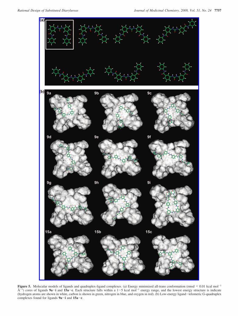
Figure 5. Molecular models of ligands and quadruplex-ligand complexes. (a) Energy minimized all-trans conformation (rmsd < 0.01 kcal mol-1
Å-1) cores of ligands 9a-i and 15a-c. Each structure falls within a 1-5 kcal mol-1 energy range, and the lowest energy structure is indicate(hydrogen atoms are shown in white, carbon is shown in green, nitrogen in blue, and oxygen in red). (b) Low-energy ligand-telomeric G-quadruplexcomplexes found for ligands 9a-i and 15a-c.
Rational Design of Substituted Diarylureas Journal of Medicinal Chemistry, 2008, Vol. 51, No. 24 7757

duplex DNA, as shown by molecular modeling and biophysicaldata. The exceptionally low level of duplex DNA binding isdue to poor shape complementarity and consequent disruptionof duplex DNA structure. Although ∆Tm and affinities are notthe highest reported for quadruplex-binding ligands,19,20 the datapresented here suggest that future analogue development toimprove affinity is a desirable goal, especially since diarylureashave an inherently druglike skeleton.
Chemistry. The synthesis of the target ligands 9a-i, 10a-f,and 15a-c was achieved in reasonable (unoptimized) yieldsand high purity, suitable for biological analysis. The synthesisof the 2-substituted target ligands was, however, not achievedby this methodology, unexpectedly leading instead to thequinazoline dione compounds 8a-f (see the Supporting Infor-mation, structure confirmed by IR spectroscopy102-105). Alter-native synthetic strategies were considered for the synthesis ofthe 2-subtituted targets. However, the cyclization reaction was
thought to be favored in all cases because of the proximity ofthe ortho-substituted phenylcarbamoylamide nitrogen of thealkylaminoanilino side chain to the imidazole-1-carboxamideintermediate in the urea forming step.
Biophysical Assays and Molecular Modeling of DNAInteractions. The ligands were screened by a FRET assay togive an indirect measure of G-quadruplex binding ability throughassessment of thermal stabilizing potential at a specifiedconcentration.34,88,106 The trend in ∆Tm,1µM values for eachligand (the change in melting temperature induced by a ligandconcentration of 1 µM) is in broad accord with the requirementfor ligand planarity; thus, the nonplanar quinazoline dioneligands 8a-f give only low ∆Tm values with all G-quadruplexDNA sequences (Supporting Information). The global confor-mation of the ligand core was also shown to be an importantfactor for efficient G-quadruplex DNA interaction, with thelinear analogues 10a-f having only a very weak stabilizingcapability with all G-quadruplex DNA sequences (some com-pounds were insoluble under the experimental conditions used).These particular ligands also have comparable duplex andG-quadruplex DNA affinities, which may result from thestructural similarity that these ligands have with duplex DNAgroove binding ligands.107
The CD studies show that interactions of the human telomericintramolecular quadruplex with a representative set of com-pounds result in a retention of the solution topologies, whichindicates that both parallel and antiparallel forms may bepresent.110-112 Molecular modeling has been performed on theparallel form, in accord with several ligand-quadruplex crystalstructures75,76,114 and the finding that the parallel form ispreferred in concentrated solution akin to a cellular environ-ment.115 Modeling predicts that the core of ligands 9a-i and15a-c can form a “square planar” conformation of similardimensions to a G-quartet; however, several other core confor-mations of comparable energy were also predicted (Figure 5a).The low-energy square planar core would, however, result inoptimal G-quadruplex DNA affinity due to enhanced π-stackinginteractions between the four phenyl rings and the guanine basesof the G-quartet, in accord with the experimental findings inthe FRET assay of enhanced G-quadruplex DNA stabilizingability relative to ligands 8a-f and 10a-f. This is generally inaccord with SPR binding data for ligands 9a-c, which fit asingle strong binding-site mode of G-quadruplex interaction.Further assessment of the potential molecular interactions wasobtained by a molecular docking protocol, which found severallow-energy binding conformations for these ligands; however,the square planar conformation in which the urea oxygen atom
Table 3. Calculated Ligand-Telomeric Quadruplex DNA DockingInteraction Energies (kcal mol-1)
G-quadruplex interaction energy
total energy van der Waals electrostatic
9a -1103.23 ( 13.61 -101.06 ( 3.21 -1002.17 ( 12.839b -1157.49 ( 7.33 -102.39 ( 3.02 -1055.07 ( 7.429c -1177.01 ( 16.24 -105.31 ( 3.79 -1071.66 ( 18.569d -1102.44 ( 10.98 -94.20 ( 5.04 -1008.24 ( 14.999e -1188.99 ( 14.71 -96.98 ( 3.10 -1092.01 ( 12.969f -1171.68 ( 28.58 -103.69 ( 3.56 -1069.80 ( 27.589g -1140.28 ( 34.06 -89.54 ( 4.37 -1050.74 ( 31.769h -1132.65 ( 25.47 -102.92 ( 2.94 -1029.72 ( 25.309i -134.71 ( 5.52 -92.02 ( 2.39 -42.68 ( 6.4815a -1174.23 ( 17.09 -100.89 ( 3.44 -1073.36 ( 16.1115b -1182.22 ( 19.06 -102.35 ( 2.98 -1079.88 ( 20.4115c -1167.67 ( 16.91 -103.31 ( 2.66 -1064.37 ( 16.35
Table 4. Calculated Ligand-Duplex DNA Docking Interaction Energies(kcal mol-1) for Three Binding Modes
duplex interaction energy
intercalation major groove minor groove
9a -966.99 ( 15.02 -1011.11 ( 18.65 nra
9b -956.87 ( 10.39 -971.71 ( 28.95 nra
9c nrb -1027.70 ( 10.63 nra
a nr: no result due to duplex DNA destabilization. b nr: no result due tono suitably pseudointercalated structure observed.
Table 5. SRB IC50 Data (µM)a
SRB IC50
MCF7 A549 WI38selectivity ratio
MCF7/A549/WI38
BRACO-1922 2.5 2.4 10.7 4.3:4.5:1.09a 14.4 ( 2.8 >50.0 16.4 ( 1.3 1.1:0.3:1.09b 2.6 ( 0.3 5.3 ( 1.0 7.5 ( 2.9 2.9:1.4:1.09c 11.1 ( 0.8 >50.0 >50.0 4.5:1.0:1.09d 34.1 ( 2.0 >50.0 22.8 ( 2.3 0.7:0.5:1.09e 14.4 ( 1.5 >50.0 19.5 ( 1.9 1.4:0.4:1.09f 49.6 ( 6.0 >50.0 26.4 ( 5.4 0.5:0.5:1.09g 12.2 ( 0.6 >50.0 >50.0 4.1:1.0:1.09h 5.1 ( 1.4 >50.0 >50.0 9.8:1.0:1.09i 40.6 ( 8.4 >50.0 >50.0 1.2:1.0:1.010a >25.0 >25.0 >25.0 1.0:1.0:1.010b nd nd nd nd10c nd nd nd nd10d >50.0 >50.0 >50.0 1.0:1.0:1.010e 3.2 ( 1.1 4.3 ( 1.1 >50.0 15.6:11.6:1.010f 19.5 ( 2.0 19.4 ( 1.7 >25.0 1.3:1.3:1.015a 21.9 ( 2.8 >50.0 13.8 ( 1.6 0.6:0.3:1.015b 32.4 ( 2.2 41.5 ( 3.0 16.2 ( 1.1 0.5:0.4:1.015c 24.2 ( 1.9 30.6 ( 5.6 11.0 ( 1.2 0.5:0.4:1.0
a nd represents values not determined because of insolubility.
Table 6. Long-Term (1 Week) Effects on MCF7 Cell Growtha
concentration (µM) PDb % of control (vc)c % senescenced
vc 4.11 ( 0.34 100.0 ( 8.3 0.7 ( 0.29a 5.00 3.79 ( 0.12 92.2 ( 3.0 1.0 ( 0.3
10.00 3.00 ( 0.38 73.1 ( 9.2 1.7 ( 1.69b 1.00 4.53 ( 0.07 110.4 ( 1.8 1.1 ( 1.1
1.75 4.50 ( 0.03 109.6 ( 0.73 3.1 ( 0.22.25 2.73 ( 0.20 66.5 ( 5.0 2.4 ( 0.7
9c 3.50 3.94 ( 0.24 96.0 ( 5.9 1.6 ( 0.97.00 3.35 ( 0.03 81.5 ( 0.8 3.1 ( 0.4
9h 2.00 3.66 ( 0.00 89.2 ( 0.0 1.29 ( 0.03.50 3.46 ( 0.34 84.3 ( 8.2 1.33 ( 0.8
11e 1.00 3.40 ( 0.26 82.9 ( 6.3 3.0 ( 0.42.00 1.81 ( 0.10 44.0 ( 2.4 4.1 ( 1.2
a vc represents experiments performed solely with a vehicle control. b PD,population doublings ( sd for 1 week MCF7. c Percentage of 1 weekpopulation doublings normalized to the control (vc ) 100%) ( sd.d Percentage of senescent cells assessed by the �-galactosidase assay (sd.101
7758 Journal of Medicinal Chemistry, 2008, Vol. 51, No. 24 Drewe et al.

was consistently orientated over the ion channel of the G-quadruplex was often observed. The positioning of the urea bondis possibly the result of a weak electrostatic attraction to thecationic ion channel or a secondary result of the optimalorientation of the π-stacked diphenylurea moiety. It is notedthat a more extended conformation for bound ligands was alsoobserved, which may represent the conformational diversitypredicted for the ligand core. Qualitative assessment of the twopredicted core substituted urea conformations indicated that themore extended core is not optimal for π-stacking interactionson the G-quartet; however, this is not reflected in the calculatedinteraction energies of the complexes, which is possibly theresult of the force field overemphasizing electrostatic contribu-tions to the binding. In general calculated energies in this studycan only provide semiquantitative indications of properties, sinceimplicit solvent is used in the simulations and the effects ofmetal ions in quadruplexes are challenging to model precisely.
The FRET assay results for ligands 9a-i and 15a-c showthat the G-quadruplex stabilization ability of these ligandsdepends to some extent upon the length of the alkylamino sidechains, with the side chain length of 9a and 9d (n ) 1) alwaysbeing inferior to those of 9b/c or 9e/f (n ) 2/3), which areapproximately comparable. This pattern is not, however, shownby ligands 15a-c, which have reduced side chain lengthdependence. The results of the docking study are consistent withthese results with a qualitative assessment of the generated low-energy complexes showing that the side chains are generallyable to be deeply buried in the G-quadruplex DNA grooves,making several electrostatic and hydrogen-bonding contacts;however, the n ) 1 side chain was inferior to the n ) 2/3, sinceforming favorable interactions within the grooves affected theorientation of the aromatic core on the G-tetrad surface, alteringthe π-stacking of the ligand. This effect is shown by the orderof calculated interaction energies for these complexes. The highcorrelation between calculated interaction energies and theFRET-based ∆Tm,1µM values (Supporting Information) (r )-0.833)indicates the predictive value of this docking method for thedesign of further ligands in this series.
The side chain length dependence of G-quadruplex interactionwas also assessed by SPR measurements. The results did notcorrelate with the FRET observations, although this is probablytoo small a sample from which to draw firm conclusions. SPRresults indicate that the n ) 1 side chain of 9a is optimal forgeneral G-quadruplex DNA interaction over 9b (n ) 2) and 9c(n ) 3). It may be that in this instance affinity and thermalstability cannot be directly compared. For the hTel, c-kit1/2,and duplex DNAs where FRET and SPR results can becompared, there is, however, good agreement (Tables 1 and 2).
Variations in the basic end-groups were also assessed by theFRET assay. This showed that for the telomeric and c-kit2G-quadruplexes, the pyrrolidino (9b) and the dimethylaminobasic groups (9g) resulted in superior quadruplex stabilizationcompared to the piperidino group (9h). Qualitative examinationof the predicted molecular interactions of the ligand-telomericG-quadruplex complexes indicated that this may result from thedecreased steric bulk of the pyrrolidino and dimethylamino basicgroups, which the modeling suggests are well accommodatedin the DNA grooves relative to the piperidino group. The FRETassay results also indicate that the introduction of the nonbasicmorpholino group (compound 9i) is deleterious for all G-quadruplex sequences examined here, with docking to thetelomeric quadruplex suggesting that this may result from poorgroove penetration, a direct result of the absence of cationiccharge at physiological pH, thus minimizing electrostatic
contribution to the predicted interaction energy. The dimethy-lamino ligand 9g also has a low ∆Tm,1µM value with the c-kit1G-quadruplex, which has no explanation in the absence of adetailed structural model for this complex.
Examination of potential selectivity between different G-quadruplexes as assessed in the FRET assay (comparison ofthe ∆Tm,1µM values) shows for any one compound a broadcorrelation with G-quadruplex DNA affinity, which may beexpected since the ligands were initially designed to interactgenerally with G-quadruplexes (focusing on G-quartet recogni-tion) and not for a specific G-quadruplex. However selectivity,generally in the order c-kit2 > F21T > c-kit1 G-quadruplexesis apparent. The c-kit1 G-quadruplex was observed to melt in anonsigmoidal manner (Supporting Information), which mayresult from the structural difference between the c-kit1 and otherG-quadruplex folds.38,39,43,68-72 The SPR results indicate adistinct order of selectivity with c-myc > c-kit2 > c-kit1 >F21T, with the interaction of ligand 9a being particularlyenhanced for the c-myc quadruplex, which also has slowerdissociation kinetics. It is noted that the FRET-based assessmentof the interactions of these ligands with the c-myc G-quadruplexis not possible under the experimental conditions used becauseof the exceptional stability of this quadruplex fold.
The best agreement between the FRET and SPR assays isfor G-quadruplex vs duplex DNA selectivity, which shows thatligands 9a-i and 15a-c, and especially the para-substitutedligands 9a-h, are significantly more G-quadruplex selectivethan BRACO-19. The lack of stabilization of a duplex DNAoligonucleotide complex (∆Tm,1µM < 2.8 °C) indicates a highlevel of selectivity for G-quadruplex forming sequences, whichwas confirmed by a competition assay using calf-thymus DNAat a 200-fold excess of duplex DNA. A subset of these ligands(9a-c) were assessed for duplex DNA interactions by SPR,with no significant duplex DNA interaction found (KA < 1.0 ×10³ M-1). This represents at least 400- to 14000-fold selectivityfor G-quadruplex vs duplex DNA. These selectivity results havebeen rationalized by the molecular modeling studies with ligands9a-c and duplex DNA, which show that the core of the ligandsis sterically too large for intercalation. Although the ligands wereobserved to dock tightly into the duplex DNA grooves, thesemodes have lower calculated interaction energies relative to thatfor G-quadruplex interaction.
Examination of the predicted interaction energies for theseligands indicates a dependence upon the electrostatic contribu-tion to the binding and an independence from the predictedbinding mode, with all interaction energies comparable acrossthis series.
Cell Biology. There are few clear patterns in the short termcell growth inhibtion data for the two cancer cell lines usedhere (IC50 values). However, it is apparent that altering the basicgroup from pyrrolidino (9b) as is the case for ligands 9g-iresults in improved MCF7 selectivity albeit with reducedpotency. It is also evident that the linear ligands 10e and 10fhave selectivity for cancer cells over a normal cell line and thatthe n ) 2 side chain of 10e confers enhanced potency. Theseeffects do not correlate with the predicted G-quadruplex orduplex DNA ∆Tm,1µM results from the FRET assay. We havenot measured cellular uptake of these compounds. However thepotency shown by some compounds in the short term SRB cellviability assay (Table 5) indicates that some at least are readilyable to cross cell membranes, although at this stage we cannotdiscount the possibility that the less potent ligands are not ableto do so.
Rational Design of Substituted Diarylureas Journal of Medicinal Chemistry, 2008, Vol. 51, No. 24 7759

Investigation into the longer-term effects (1 week) of the moretoxic ligands (for example, 9b) in the MCF7 cell line usedsubcytotoxic concentrations of ligands. Over this period, theligands had a significant effect on population growth, whichwas associated with a small induction of cellular senescence.No significant levels of telomere end-end fusions wereobserved over this time period. These results, taken with thelack of telomerase inhibitory activity, suggest that these ligandsare at best only modest inducers of telomere uncapping/dysfunction. This further suggests that a minimum thresholdlevel of telomeric quadruplex affinity is required for effectiveinterference with telomere function in cancer cells, which isnot reached by these particular diarylurea ligands but is achievedbycompoundssuchasBRACO-19,22,24,32,61,76telomestatin,20,56,62,63,67
and the more recently developed naphthalene diimides.19
There is a strong suggestion from the IC50 data that thecompounds reported here fall into several distinct biologicalclasses that may reflect distinct modes of action (thoughdifferences in cellular uptake cannot be discounted): (i) the para-substituted set 9a-c/g-i, which are moderately selective forthe cancer cell lines, especially MCF7; (ii) the smaller set 10a-f,with compound 10e showing exceptional selectivity; and (iii)the nonselective meta-substituted sets 9d-f and 15a-c. Thediarylurea skeleton is represented in many kinase inhibitors, forexample, multitargeted tyrosine kinase inhibitors108 and inhibi-tors of insulin-like growth factor I receptor signaling.109 Eventhough the substitution patterns in the present compounds havenot been previously reported for kinase inhibitors, we cannotdiscount at present the possibility that the biologically activecompounds reported here may be acting directly on kinasebinding sites.
Conclusions
The purpose of this study was to design and evaluate a novelseries of nonpolycyclic compounds that would show effectivebinding to quadruplex DNAs. This goal has been achievedthrough the diphenylurea-based scaffold. Even though initialstudies of their cellular effects indicate that the derivativesdiscussed here do not show potent telomerase inhibitory activity,further analogue development is warranted. A number of thepresent compounds did show significant effects on cancer cellgrowth that are not associated with significant telomere uncap-ping/dysfunction and are unlikely to be effects on duplex DNA.We introduce the concept that a threshold level of quadruplexaffinity is required for a compound to show such effects. It isalso possible that interactions with nontelomeric quadruplexesmay be involved, and screening is planned on larger librariesof derivatives with a more extensive set of quadruplexes fromoncogenic promoter sequences,37 as well as use of a larger panelof cancer cell lines.
Experimental Section
Fluorescence Resonance Energy Transfer (FRET) Assays.FRET oligonucleotides (Eurogentec Ltd., U.K.) have the followingsequences: F21T, 5′FAM-d[G3(T2AG3)3]-TAMRA3′; c-kit1, 5′FAM-d[AGAG3AG2GCGCTG3AG2AG3GCT]-TAMRA3′; c-kit2, 5′FAM-d[C3G3CG3CGCGAG3AG4AG2]-TAMRA3′;duplex,5′FAM-d[(TA)2GC-(TA)2T6(TA)2GC(TA)2]-TAMRA3′ where FAM is 6-carboxy-fluoresein and TAMRA 6-carboxytetramethylrhodamine. For theFRET assay, the required oligonucleotide was suspended in FRETbuffer (60 mM KCl, Kcacodylate, pH 7.4; 400 nM DNA) andheated to 85 °C for 10 min prior to cooling to room temperature.DNA was distributed (50 µL) across a 96-well RT-PCR plate (Bio-Rad) to which ligand was added (50 µL; stored as a 20 mM DMSOstock, -20 °C; diluted to 1 mM in HPLC grade DMSO) to afford
the required concentration. FRET buffer was used as a negativecontrol. DNA melting was assessed on a MJ Research Opticon DNAengine continuous fluorescence detector exciting at 450-495nm.Fluorescence values were recorded at 515-545nm at 0.5 °Cintervals as the plate was heated from 30 to 100 °C. The data wereanalyzed with the Origin 7.0 software package (Origin LaboratoryCorp., Northampton, MA). In the case of the c-kit1 oligonucleotide,melting curves were fitted to sigmoid curves prior to analysis. Thechange in melting temperature at 1 µM ligand concentration(∆Tm,1µM) was calculated from four experiments by subtraction ofthe averaged negative control from the averaged 1 µM ligandmelting temperature ( the maximum standard deviation (sd). Forthe competition assay, to F21T DNA (50 µL) was added calf thymusDNA (CT-DNA, 25 µL, 533.3 µM CT-DNA bp stock in 0.5 mMEDTA/30 mM K+ cacodylate buffer) to afford the required CT-DNA bp concentration. To this was added ligand (25 µL, 4 µM) toafford the required ligand concentration (1 µM). FRET bufferrepresented no competitor. The percentage of retained stabilizationwas calculated from three experiments and normalized to the∆Tm,1µM for that ligand with no CT-DNA competitor (100%) (normalized sd. BRACO-19 was prepared in-house30,32 to a HPLCpurity of >98% (data not shown).
Circular Dchroism (CD) Sudies. CD spectra of the 24-mertelomeric DNA sequence d(TTAGGGTTAGGGTTAGGGTTAGGG)both in the absence of ligands and with ligands 9b, 9d, and 15btogether with two triazole-containing diarylurea derivatives syn-thesized in a separate project110 were acquired on an AppliedPhotophysics Ltd. Chirascan spectrometer at King’s CollegeLondon. All samples were prepared at 100 µM in 100 mMpotassium chloride/phosphate, pH 7.4, and heated to 95 °C andslowly annealed overnight to room temperature. The samples werefurther diluted with buffer to 0.8 optical density unit prior to datacollection, and where appropriate, ligand was added to give a 1:1molar ratio. UV absorbance and CD spectra were measured between320 and 220 nm in a 10 mm path length cell. Spectra were recordedwith a 0.5 nm step size, a 1.5 s time-per-point, and a spectralbandwidth of 1 nm. All spectra were acquired at room temperature,and buffer baseline was corrected. The concentration of the aboveoligonucleotide was determined by using the absorbance value at260 nm and the Beer-Lambert law.
Surface Plasmon Resonance (SPR). Biosensor experimentswere conducted in filtered, degassed HEPES buffer (10 mMHEPES, 100 mM KCl, 3 mM EDTA, 0.000 05 v/v of 10% P20BIACORE surfactant, pH 7.3) at 25 °C. The 5′-biotin labeled DNAsequences (Integrated DNA Technologies) were HPLC purified andcomprise the following sequences: hTel, 5′biotin-d[AG3(T2AG3)3]3′;c-kit1, 5′biotin-d[(AG3)2CGCTG3AG2AG3]3′; c-kit2, 5′biotin-d[(CG3)2-CGCGAG3AG4]3′; c-myc, 5′biotin-d[(AG3TG4)2A]3′; duplex, 5′biotin-d[CGA2T2CGTCTC2GA2T2CG]3′. The experiments were conductedupon a BIAcore 2000 optical biosensor instrument (BIAcore Inc.).Flow cell 1 was left blank as a reference, while flow cells 2-4were immobilized with DNA on a streptavidin-derivatized gold chip(SA chip from BIAcore) by manual injection of DNA stocksolutions (flow rate of 1 µL/min) until the desired value of DNAresponse was obtained (350-400 RU). Typically, a series ofdifferent ligand concentrations (1 nM to 10 µM from 20 mM DMSOstock) were injected onto the chip (flow rate of 50 µL/min, 5-10min) until a constant steady-state response was obtained followedby a dissociation period (buffer, 10 min). After every cycle, thechip surface was regenerated (20 s injection of 10 mM glycinesolution, pH 2.0) followed by running buffer flow. The data wereprocessed as previously described,30,91 using BIAevaluation (BIA-core Inc.) and Kaleidagraph (Synergy Software) software fornonlinear least-squares optimization of the binding parameters.
Molecular Modeling. Molecular modeling was performed onan SGI Octane workstation (2 × 225 MHz MIPS R12000 CPUs;IRIX64 6.5 OS; Silicon Graphics Ltd.) with the Insight II softwarepackage (Accelerys Software Inc., 2000) utilizing the Builder,Discover, and Docking modules. G-Quadruplex DNA was preparedfrom the 22-mer crystal structure of the G-quadruplex formed fromthe human telomere of sequence d[AG3(T2AG3)3] (PDB 1KF1),68
7760 Journal of Medicinal Chemistry, 2008, Vol. 51, No. 24 Drewe et al.
and a self-complementary duplex DNA was built in Insight II fromthe sequence d[(TA)2GC(TA)2]. An intercalation site with 6.76 Åbase-pair separation was introduced at the GC step of the DNAduplex. Ligands were built with atomic potentials and partial chargesassigned in the CVFF force field prior to minimization (500 stepssteepest descents; 5000 steps conjugate gradients; rmsd of 0.001kcal mol-1 Å-1) in the Discover module. Basic side chainsconsisting of the pyrrolidino, dimethylamino, and piperidino groupswere built as protonated species by addition of a formal atomiccharge. Morpholino groups were kept uncharged. Core conforma-tional analysis was achieved by manual rotation prior to minimiza-tion (500 steps steepest descent; successive 2000 step conjugategradients until convergence to an rmsd of 0.01 kcal mol-1 Å-1) inthe Discover module of Insight II.
A multistage Monte Carlo minimization simulated-annealingdocking protocol analogous to those previously reported94,95 wasutilized in the Docking module of Insight II to determine thepotential low energy ligand-DNA complexes and their energy,using default program settings unless specified. For G-quadruplexinteraction, the ligand was positioned ∼5 Å above the center ofthe 3′-G-quartet, defining their hydrogen atoms as the binding siteabout which a Monte Carlo minimization methodology wasemployed to generate 200 random orientations of the startingstructure, with van der Waals radii scaled to 10% of their full valuesand charges not considered. The maximum allowable energy changeof successive random structures was 10 000 kcal mol-1 with anenergy range of 40 kcal mol-1. Each conformation was minimized(300 steps of conjugate gradients), and the 75 lowest total systemenergy conformations were filtered by Insight II for further analysis.For duplex DNA interaction, an analogous procedure was useddefining the binding site as all of the hydrogen atoms that projectinto the required DNA groove or by inserting the ligand into theDNA pseudointercalation site in the best perceived conformation,defining the hydrogen atoms of the GC base pairs as the bindingsite. These 75 structures were subjected to 800 steps of conjugategradients minimization in which charges were included, van derWaals radii were set to 100%, and the distant dependent dielectricconstant was 1r, prior to simulated annealing between 500 and 300K over 10 ps to the predefined binding site. The resulting structureswere energy-minimized (800 steps of conjugate gradients) and the25 lowest total system energy conformations filtered by Insight IIfor further manual evaluation. These docked structures wereanalyzed on the basis of the chemical correctness of the ligandensuring trans urea and amide integrity, the lowest total interactionenergy of the complex assessed for all atoms with no cutoff, thenumber of hydrogen bonding interactions observed, the bindingmodes that best represent the populations, and the perceivedcorrectness of the proposed ligand binding mode. This allowed theselection of two lead low energy binding conformations for furtheranalysis. Each low energy binding conformation was used to definea flexible binding site containing all DNA atoms residing 7-8 Åfrom the ligand, fixing all other atoms and tethering the 3′-G-quartetor the terminal duplex DNA TA base pairs. A further 30 rounds of(300 steps of conjugate gradients minimization prior to simulatedannealing between 800 and 200 K over 10 ps) were performed usinga distance-dependent dielectric constant of 4r. Final minimization(1000 steps conjugate gradients) allowed the filtering of the 15lowest total system energy conformations by Insight II for finalanalysis. These bound ligand-DNA complexes were manuallyassessed by the predefined criteria, averaging the total interactionenergy observed for the 10 lowest energy binding conformations( sd, assessed between all atoms with no cutoff. Figures wereconstructed from a docked binding conformation selected to bestrepresent the optimal low energy binding conformations of thepopulation.
Cell Culture. Cell lines were supplied by ATCC-LGC Promo-chem and viability maintained in a Heraeus Hera Cell 240 incubator(37 °C, 5% CO2, 75 cm2 plates, TPP). Cells were removed forexperimentation as required. Sterile work was conducted in aHeraeus Hera Safe hood. Dulbecco’s modified Eagles medium(DMEM, Invitrogen) supplemented with fetal bovine serum (10%
v/v, Invitrogen), hydrocortisone (0.5 µg/mL, Acros Organics),L-glutamine (2 mM, Invitrogen), and nonessential amino acids (1×,Invitrogen) was used for the MCF7 and A549 cell lines, andminimum essential medium (MEM; Sigma-Aldrich) supplementedwith fetal bovine serum (10% v/v, Invitrogen), L-glutamine (2 mM,Invitrogen), and nonessential amino acids (1×, Invitrogen) was usedfor the WI38 cell line.
SRB Cytotoxicity Assay. Short-term growth inhibition wasmeasured using the SRB assay as described previously.22,30,100
Briefly, cells were seeded (4000 cells/wells) into the wells of 96-well plates in appropriate medium and incubated overnight to allowthe cells to attach. Subsequently cells were exposed to freshly madesolutions of drug at increasing concentrations between 0.25 and50 µM in quadruplicate and incubated for a further 96 h. Followingthis the cells were fixed with ice cold trichloacetic acid (10% w/v)for 30 min and stained with 0.4% SRB dissolved in 1% acetic acidfor 15 min. All incubations were carried out at room temperature.The IC50 value, concentration required to inhibit cell growth by50%, was determined from the mean absorbance at 540 nm foreach drug concentration expressed as a percentage of the wellabsorbance in untreated control cells.
Subcytotoxic Induction of Cellular Senescence (�-Galactosi-dase Assay).30,101 MCF7 cells (1 × 10⁵, 10 mL of media, ATCC-LGC Promochem) were exposed to two independent subcytotoxicconcentrations of the required ligand over a 1 week period, with abiweekly treatment. A media negative control was also screened.Cells were counted on a Neybauer hemocytometer (Assistant,Germany) and the number of cellular population doublings assessedby the equation n ) (log Pn - log P0)/log 2 where Pn is the numberof cells collected and P0 the initial seeding density. Cells werestained for senescence using the �-galactosidase staining kit (CellSignaling Technology) according to the manufacturer’s instructions.In short, cells were seeded (1 × 10⁵, 2 mL) in a 6-well plate (Fisher-Scientific) with the required ligand concentration and incubatedovernight. The medium was removed and the well washed withPBS (2 mL) prior to fixing (1× fixative solution, 10 min). Thefixative was removed and the well washed with PBS (2 × 2 mL)prior to the addition of the staining solution (1 mL), and the plateswere incubated overnight. Three independent fields of cells werevisualized (200× magnification) from both repeats, with the meanpercentage of blue senescent cells reported ( sd.
TRAP-LIG Assay. This was performed as recently described.98
Chemistry. All reagents were reagent grade and were used assupplied without further purification (Sigma-Aldrich, Alfa Aesar,Avocado Organics, and Lancaster Synthesis). Palladium catalystwas 10 wt % loading on activated carbon. Ammonia in methanol(NH3/MeOH, ∼7 N; Sigma-Aldrich) was diluted in MeOH asrequired. Solvents (BDH and Fisher Scientific), anhydrous solvents(Sigma-Aldrich), and HPLC grade solvents (Fisher Scientific) wereused as supplied. Microwave reactions were conducted in a Biotageinitiator microwave, software version 1.1, in Biotage vials (0.5-2.0or 2.0-5.0 mL) sealed with Biotage caps with septa. All chemistrywas conducted in clean, oven-dried glassware. 1H and 13C NMRspectra were recorded at 295 K on on a Bruker Avance 400spectrometer using the specified deuterated solvent (GOSS Scientificand Sigma-Aldrich). NMR spectra were analyzed by the MestReC4.5.6.0 program with chemical shifts calibrated to the residual protonand carbon resonance of the solvent. NMR multiplicity and couplingconstants (J) are reported as observed. Melting points (mp) weremeasured on a Bibby Stuart Scientific SMP3 melting pointapparatus, where “dec” indicates decomposition. Mass spectra wererecorded on a ThermoQuest Navigator mass spectrometer usingelectrospray ionization in positive ([M + H]+) or negative ([M -H]-) modes. High resolution accurate mass spectra were recordedon a Micromass Q-TTOF Ultima Global tandem mass spectrometerusing electrospray ionization mode and 50% acetonitrile in waterand 0.1% FA as solvent and processed using the MassLab 3.2software. Infrared spectra were recorded from neat samples on aNicolet Smart Golden Gate spectrometer (Avatar 360 FT-IR E.S.P.)and processed using the software package OMNIC ESP 5.1. Flashchromatography was conducted on silica gel (partial size 33-70
Rational Design of Substituted Diarylureas Journal of Medicinal Chemistry, 2008, Vol. 51, No. 24 7761

µm, BDH). Analytical HPLC was performed on a Gilson chro-matograph with a YMC C18 5 µm (100 mm × 4.6 mm) columnand an Agilent 1100 series photodiode array detector. Spectra wereprocessed in the Unipoint 5.11 software, and compound purity andretention times (tR) were assessed at 254 nm unless stated otherwise.Several HPLC solvent systems and gradients were employed asspecified: method A, 0.1% TFA in MeOH and 0.1% aqueous TFA,25-75% organic over 28 min (1 mL/min); method B, 0.1% TFAin MeOH and 0.1% aqueous TFA, 25-50% organic over 18 min(1 mL/min); method C, 0.1% FA in acetonitrile and 0.1% aqueousFA, 5-50% organic over 28 min (1 mL/min); method D, 0.1%FA in acetonitrile and 0.1% aqueous FA, 5-50% organic over 18min (1 mL/min); method E, 0.1% FA in MeOH and 0.1% aqueousFA, 25-75% organic over 18 min (1 mL/min). Semipreparativereversed-phase HPLC (semiprep HPLC) was performed on a Gilsonchromatograph with a Gilson 215 liquid handler, a Gilson 845Zinjection module coupled to a Gilson UV/visible 155 detector, anda YMC C18 5 µm (100 mm × 20 mm) column. Several HPLCsolvent systems, gradients, and methods of sample isolation wereemployed as specified: method F, 0.1% TFA in MeOH and 0.1%aqueous TFA, 25-75% organic over 25 min (10 mL/min), injectedfrom 25% MeOH in 0.1% aqueous TFA (3 mL); method G, 0.1%TFA in MeOH and 0.1% aqueous TFA, 25-50% organic over 60min, 50-75% organic over 5 min (10 mL/min), injected from 25%MeOH in 0.1% aqueous TFA (3 mL). Purified compounds wereisolated by reduction of fraction volumes (5 mL), precipitation with5% NH3 (aq), and filtration.
Method 1 (1a-f and 12a-c). (a) The required nitroaniline wasdissolved in THF (150 mL) and cooled to 0 °C prior to the additionof TEA (2 equiv) and the required acid chloride (1.5-2 equiv),and the mixture stirred overnight at room temperature. Theprecipitate was filtered and the solvent evaporated in vacuo. Theproduct was either precipitated with saturated NaHCO3 (aq) (200mL) or dissolved in EtOAc (50 mL), washed with 5% NH3 (aq)(50 mL), brine (50 mL), and dried over MgSO4. (b) The requirednitroaniline (15.0 g, 0.109 mol) was added to the appropriate acidchloride (45 mL, 0.402-0.471 mol) in portions at room temperature,followed by stirring at 50 °C overnight. The mixture was cooledto 0 °C and the product isolated by filtration, washing with ether(50 mL).
Method 2 (2a-i and 13a-c). 1a,b/d-e/g-i or 12a-c weredissolved in MeOH/THF (5-100 mL), and the required amine base(2-3.6 equiv) was added. The mixture was either (a) stirredovernight at room temperature prior to the solvent being removed(5% NH3 (aq) (50 mL) was added and the product extracted withEtOAc (50 mL), washed with brine (25 mL), and dried overMgSO4), (b) heated under reflux for 4 h prior to workup as above,or (c) stirred at 30 °C overnight prior to workup as above. 1c/fwas reacted with neat pyrrolidine (20 mL, 0.240 mol) at roomtemperature overnight, followed by evaporation of the excesspyrrolidine and workup as described above.
Method 3 (3a-i and 14a-c). (a) 2a-i were dissolved in MeOH(100 mL), and then ammonium formate (10 equiv) and Pd/C (0.1equiv w/w) were added. The mixture was stirred overnight at roomtemperature prior to filtration through Celite and evaporation ofthe solvent. The residue was dissolved in CHCl3 (100 mL), washedwith 5% NH3 (aq) (2 × 50 mL), brine (50 mL), and dried overMgSO4. (b) 13a-c were suspended in absolute EtOH (4 mL) priorto the addition of ammonium formate (4 equiv) and Pd/C (0.1 equivw/w). The mixture was heated by microwave irradiation (120 °C,10 min, 7 bar) followed by workup as described above.
Method 4 (4j,k,l). The required nitrobenzoic acid (5.0 g, 0.030mol) was dissolved in anhydrous DCM (150 mL), and DMF wasadded as required. DMAP (500 mg, 0.1 equiv w/w) and tBuOH(4.29 mL, 0.045 mol, 1.5 equiv) were added prior to cooling themixture to 0 °C and adding DCC (6.79 g, 0.033 mol, 1.1 equiv) intwo portions. The mixture was warmed to room temperature andstirred for 4 h. The precipitate was filtered and the DCM washedwith 1 M HCl (aq) (2 × 50 mL), brine (50 mL) and dried overMgSO4. The resulting oil was purified by flash chromatography.
Method 5 (5j,k). 4j,k were dissolved in MeOH (100 mL), andPd/C (0.1 equiv w/w) was added prior to purging with H2 (g), andthe mixture was stirred at room temperature until reaction wascomplete, when it was filtered through Celite and the solventevaporated in vacuo.
Method 6 (6j,k and 15a-c). (a) 5j,k were dissolved inanhydrous THF to a concentration of 0.4 M, and CDI (0.6-1.2equiv) was added. The mixture was heated at reflux for 24 h underN2 (g). The THF was evaporated and the residue dissolved in EtOAc(200 mL), washed with 1 M HCl (aq) (2 × 100 mL), brine (50mL), and dried over MgSO4. (b) As above, 14a-c were reactedwith CDI (1.0 equiv) by microwave irradiation (75 °C, 20 min, N2
(g)). The mixture was cooled to 0 °C and the product precipitatedwith EtOAc (10 mL).
Method 7 (7j,k). 6j,k were suspended in TFA (30 mL) andstirred vigorously at room temperature for 1 h. The precipitate wasisolated by filtration, washed with DCM (50 mL), and oven-dried.If no precipitate formed, precipitation was induced by cooling themixture to 0 °C and treatment with ether (50 mL).
Method 8 (8a-f, 9a-i, and 10a-f). 7j,k were dissolved inanhydrous DMF (8 mL), and to this were added the required sidechains 3a-i (4 equiv) and PyBOP (3 equiv). The mixture wasstirred at room temperature under N2 (g) for 22 h. The DMF wasevaporated and the resulting oil suspended in CHCl3 (20 mL) withsonication. The resulting precipitate was isolated by filtration;however, when an oily residue formed, the organic solvent wasdecanted. In either instance, this crude material was dissolved inNH3/MeOH (1 M, 3 mL), and the product was precipitated with5% NH3 (aq) (30 mL) and isolated by filtration.
Detailed experimental procedures and analytical data for the sidechain building blocks 1a-f, 2a-i, 3a-i, 4j-l, 5j-l and thequinazoline dione ligands 8a-f are given in the SupportingInformation.
2,2′-Ureylene-di-(tert-Butylbenzoate) (6j). Following method6a, 5j (3.38 g, 17.488 mmol) was reacted with CDI (3.40 g, 20.986mmol) to yield a yellow solid (3.59 g, 8.703 mmol, quant) whichwas used without further purification. 1H NMR (400 MHz, CDCl3)10.69 (2H, s), 8.46 (2H, dd, J ) 8.5, 0.9 Hz), 7.96 (2H, dd, J )8.0, 1.6 Hz), 7.49 (2H, ddd, J ) 8.7, 7.3, 1.7 Hz), 6.99 (2H, ddd,J ) 8.2, 7.3, 1.1 Hz), 1.64 (18H, s) ppm. 13C NMR (100 MHz,CDCl3) 167.8, 152.4, 142.4, 133.8, 131.0, 121.1, 120.0, 116.4, 82.3,28.3 ppm. HRMS m/z calcd C23H28N2O5 [M + H]+ 413.2071, found413.2065.
3,3′-Ureylene-di-(tert-butyl benzoate) (6k). Following method6a, 5k (4.01 g, 20.744 mmol) was reacted with CDI (2.02 g, 12.458mmol) to yield a white solid (3.97 g, 9.625 mmol, 93%). Mp 296°C dec. 1H NMR (400 MHz, DMSO-d6) δ 8.97 (2H, s), 8.09 (2H,t, J ) 1.8 Hz), 7.75 (2H, ddd, J ) 8.0, 2.1, 1.0 Hz), 7.58 (2H, d,J ) 7.8 Hz), 7.46 (2H, t, J ) 7.9 Hz), 1.61 (18H, s) ppm. 13CNMR (100 MHz, DMSO- d6) δ 164.8, 152.5, 139.8, 131.9, 129.0,122.5, 122.5, 118.7, 80.7, 27.8 ppm. HRMS m/z calcd C23H28N2O5
[M + H]+ 413.2071, found 413.2075.4,4′-Ureylene-di-(tert-butyl benzoate) (6l). Following method
6a, 5l (3.71 g, 19.202 mmol) was reacted with CDI (3.74 g, 23.042mmol) to yield a white solid (3.94 g, 9.552 mmol, 99%) whichwas used without further purification. 1H NMR (400 MHz, CDCl3)δ 9.23 (2H, s), 7.89 (4H, d, J ) 8.8 Hz), 7.63 (4H, d, J ) 8.8 Hz),1.59 (18H, s) ppm. HRMS m/z calcd C23H28N2O5 [M + H]+
413.2071, found 413.2092.2,2′-Ureylenedibenzoic Acid (7j). Following method 7, 6j (3.11
g, 7.546 mmol) was deprotected to yield a white solid (1.05 g,3.483 mmol, 46%). Mp 192-194 °C. 1H NMR (400 MHz, DMSO-d6) 13.48 (2H, s), 10.73 (2H, s), 8.29 (2H, dd, J ) 8.4, 0.8 Hz),7.95 (2H, dd, J ) 7.9, 1.6 Hz), 7.56 (2H, ddd, J ) 8.7, 7.3, 1.7Hz), 7.09 (2H, m) ppm. 13C NMR (100 MHz, DMSO- d6) 169.7,151.7, 141.6, 133.8, 131.0, 121.5, 119.7, 116.4 ppm. HRMS m/zcalcd C15H12N2O5 [M + H]+ 301.0819, found 301.0814.
3,3′-Ureylenedibenzoic Acid (7k). Following method 7, 6k (3.01g, 7.266 mmol) was deprotected to yield a white solid (2.18 g,7.260 mmol, quant). Mp 321-323 °C. 1H NMR (400 MHz, DMSO-d6) δ 12.84 (2H, s), 8.97 (2H, s), 8.15 (2H, t, J ) 1.7 Hz), 7.66
7762 Journal of Medicinal Chemistry, 2008, Vol. 51, No. 24 Drewe et al.
(2H, ddd, J ) 8.1, 2.0, 0.9 Hz), 7.57 (2H, d, J ) 7.8 Hz), 7.41(2H, t, J ) 7.9 Hz) ppm. 13C NMR (100 MHz, DMSO- d6) δ 167.2,152.5, 139.8, 131.3, 129.0, 122.8, 122.5, 119.0 ppm. HRMS m/zcalcd C15H12N2O5 [M + H]+ 301.0819, found 301.0807.
4,4′-Ureylenedibenzoic Acid (7l). Following method 7, 6l (3.12g, 7.558 mmol) was deprotected to yield a fine white powder (2.15g, 7.172 mmol, 95%). Mp >350 °C [lit. >400 °C]. 1H NMR (400MHz, DMSO) δ 12.62 (2H, s), 9.21 (2H, s), 7.88 (4H, d, J ) 8.7Hz), 7.58 (4H, d, J ) 8.7 Hz) ppm. 13C NMR (100 MHz, DMSO-d6) δ 166.9, 151.9, 143.6, 130.5, 123.9, 117.3 ppm. HRMS m/zcalcd C15H12N2O5 [M + H]+ 301.0819, found 301.0817.
1,3-Bis(3-(4-(2-(pyrrolidin-1-yl)acetamido)phenylcarbam-oyl)phenyl)urea (9a). Following method 8, 7k (150.0 mg, 0.500mmol) was reacted with 3a (438.1 mg, 1.998 mmol) to yield apale-brown solid (333.0 mg, 0.474 mmol, 95%), which was purifiedby semiprep HPLC (method F). HPLC (method A) 97%, tR ) 17.69min. Mp 286-288 °C. 1H NMR (400 MHz, DMSO- d6) δ 10.20(2H, s), 9.73 (2H, s), 8.98 (2H, s), 7.98 (2H, s), 7.70 (6H, d, J )8.9 Hz), 7.61 (4H, d, J ) 8.9 Hz), 7.56 (2H, d, J ) 7.8 Hz), 7.44(2H, t, J ) 7.9 Hz), 3.32 (4H, s), 2.67 (8H, m), 1.77 (8H, m) ppm.13C NMR (100 MHz, DMSO- d6) δ 167.9, 165.2, 152.4, 139.7,135.7, 134.6, 134.3, 128.6, 121.1, 120.8, 120.6, 119.6, 117.6, 59.0,53.7, 23.3 ppm. HRMS m/z calcd C39H42N8O5 [M + H]+ 703.3351,found 703.3386.
1,3-Bis(3-(4-(3-(pyrrolidin-1-yl)propanamido)phenylcarbam-oyl)phenyl)urea (9b). Following method 8, 7k (150.0 mg, 0.500mmol) was reacted with 3b (466.2 mg, 1.998 mmol) to yield awhite solid (346.0 mg, 0.473 mmol, 95%). HPLC (method A) 98%,tR ) 19.00 min. Mp 330 °C dec. 1H NMR (400 MHz, DMSO-d6)δ 10.18 (2H, s), 10.05 (2H, s), 8.95 (2H, s), 7.97 (2H, s), 7.70(6H, m), 7.55 (6H, m), 7.44 (2H, t, J ) 7.9 Hz), 2.75 (4H, t, J )7.1 Hz), 2.50 (12H, m), 1.70 (8H, m) ppm. 13C NMR (100 MHz,DMSO- d6) δ 169.6, 165.2, 152.4, 139.6, 135.7, 135.0, 134.3, 128.7,121.1, 120.8, 120.7, 119.1, 117.6, 53.3, 51.4, 35.7, 23.0 ppm.HRMS m/z calcd C41H46N8O5 [M + H]+ 731.3664, found 731.3690.
1,3-Bis(3-(4-(4-(pyrrolidin-1-yl)butanamido)phenylcarbam-oyl)phenyl)urea (9c). Following method 8, 7k (150.0 mg, 0.500mmol) was reacted with 3c (494.2 mg, 1.998 mmol) to yield a whitesolid (344.8 mg, 0.454 mmol, 91%). HPLC (method A) 96%, tR )19.86 min. Mp 239-241 °C. 1H NMR (400 MHz, DMSO- d6) δ10.20 (2H, s), 9.89 (2H, s), 9.00 (2H, s), 7.99 (2H, s), 7.70 (6H,m), 7.57 (6H, m), 7.44 (2H, t, J ) 7.9 Hz), 2.50 (12H, m), 2.35(4H, t, J ) 7.4 Hz), 1.77 (4H, m), 1.70 (8H, m) ppm. 13C NMR(100 MHz, DMSO- d6) δ 170.7, 165.2, 152.4, 139.7, 135.7, 135.1,134.2, 128.6, 121.1, 120.8, 120.6, 119.1, 117.6, 54.9, 53.4, 45.7,34.1, 23.0 ppm. HRMS m/z calcd C43H50N8O5 [M + H]+ 759.3977,found 759.3940.
1,3-Bis(3-(3-(2-(pyrrolidin-1-yl)acetamido)phenylcarbam-oyl)phenyl)urea (9d). Following method 8, 7k (150.0 mg, 0.500mmol) was reacted with 3c (438.1 mg, 1.998 mmol) to yield a pale-yellow solid (310.1 mg, 0.441 mmol, 88%). HPLC (method A)91%, tR ) 18.69 min. Mp 154-157 °C. 1H NMR (400 MHz,DMSO- d6) δ 10.27 (2H, s), 9.78 (2H, s), 8.96 (2H, s), 8.12 (2H,s), 7.79 (2H, s), 7.71 (2H, d, J ) 8.0 Hz), 7.57 (2H, d, J ) 7.7Hz), 7.44 (4H, m), 7.38 (2H, d, J ) 8.3 Hz), 7.26 (2H, t, J ) 8.0Hz), 3.31 (4H, s), 2.65 (8H, m), 1.76 (8H, m) ppm. 13C NMR (100MHz, DMSO- d6) δ 168.3, 165.6, 152.6, 139.7, 139.4, 138.8, 135.8,128.8, 128.7, 121.3, 121.0, 117.8, 115.7, 115.0, 111.8, 59.2, 53.7,23.4 ppm. HRMS m/z calcd C39H42N8O5 [M + H]+ 703.3351, found703.3344.
1,3-Bis(3-(3-(3-(pyrrolidin-1-yl)propanamido)phenylcarbam-oyl)phenyl)urea (9e). Following method 8, 7k (150.0 mg, 0.500mmol) was reacted with 3e (466.2 mg, 1.998 mmol) to yield a pale-yellow solid (292.4 mg, 0.400 mmol, 80%). HPLC (method A)93%, tR ) 19.39 min. Mp 160 - 163 °C. 1H NMR (400 MHz,DMSO- d6) δ 10.26 (2H, s), 10.13 (2H, s), 9.00 (2H, s), 8.10 (2H,s), 7.98 (2H, s), 7.72 (2H, d, J ) 7.9 Hz), 7.57 (2H, d, J ) 7.7Hz), 7.44 (4H, m), 7.38 (2H, d, J ) 7.9 Hz), 7.25 (2H, t, J ) 7.7Hz), 2.72 (4H, t, J ) 6.9 Hz), 2.49 (12H, m), 1.70 (8H, m) ppm.13C NMR (100 MHz, DMSO- d6) δ 170.0, 165.5, 152.6, 139.6,139.3, 139.3, 135.7, 128.6, 128.5, 121.2, 120.9, 117.7, 115.2, 114.5,
111.2, 53.3, 51.4, 35.9, 23.1 ppm. HRMS m/z calcd C41H46N8O5
[M + H]+ 731.3664, found 731.3651.1,3-Bis(3-(3-(4-(pyrrolidin-1-yl)butanamido)phenylcarbam-
oyl)phenyl)urea (9f). Following method 8, 7k (150.0 mg, 0.500mmol) was reacted with 3f (494.2 mg, 1.998 mmol) to yield a whitesolid (189.6 mg, 0.249 mmol, 50%). HPLC (method A) 96%, tR )19.56 min. Mp 234-236 °C. 1H NMR (400 MHz, DMSO- d6) δ10.26 (2H, s), 9.94 (2H, s), 9.01 (2H, s), 8.11 (2H, s), 7.99 (2H, s),7.72 (2H, d, J ) 8.0 Hz), 7.57 (2H, d, J ) 7.6 Hz), 7.43 (4H, m),7.37 (2H, d, J ) 7.9 Hz), 7.25 (2H, t, J ) 8.1 Hz), 2.48 (12H, m),2.37 (4H, t, J ) 7.3 Hz), 1.77 (4H, m), 1.69 (8H, m) ppm. 13CNMR (100 MHz, DMSO- d6) δ 171.0, 165.5, 152.5, 139.6, 139.4,139.3, 135.7, 128.6, 128.5, 121.2, 120.9, 117.7, 115.1, 114.5, 111.3,54.9, 53.4, 34.2, 24.1, 23.0 ppm. HRMS m/z calcd C43H50N8O5 [M+ H]+ 759.3977, found 759.3942.
1,3-Bis(3-(4-(3-(dimethylamino)propanamido)phenylcarbamoyl)-phenyl)urea (9g). Following method 8, 7k (100.0 mg, 0.333 mmol)was reacted with 3g (276.1 mg, 1.333 mmol) to yield a white solid(222.8 mg, 0.328 mmol, 99%), which was purified by filtration fromboiling MeOH (2 mL). HPLC (method C) 95%, tR ) 17.28 min.Mp 322 -324 °C. 1H NMR (400 MHz, DMSO- d6) δ 10.18 (2H,s), 10.00 (2H, s), 8.94 (2H, s), 7.98 (2H, s), 7.70 (6H, m), 7.56(6H, m), 7.44 (2H, t, J ) 7.9 Hz), 2.57 (4H, t, J ) 7.0 Hz), 2.44(4H, t, J ) 6.9 Hz), 2.19 (12H, s) ppm. 13C NMR (100 MHz,DMSO- d6) δ 169.9, 165.3, 152.5, 139.7, 135.8, 135.1, 134.4, 128.8,121.2, 120.9, 120.7, 119.2, 117.7, 55.1, 44.9, 34.7 ppm. HRMSm/z calcd C37H42N8O5 [M + H]+ 679.3351, found 679.3334.
1,3-Bis(3-(4-(3-(piperidin-1-yl)propanamido)phenylcarbam-oyl)phenyl)urea (9h). Following method 8, 7k (100.0 mg, 0.333mmol) was reacted with 3h (329.8 mg, 1.333 mmol) to yield awhite solid (221.1 mg, 0.291 mmol, 87%), which was purified byfiltration from boiling MeOH (2 mL). HPLC (method C) 100%, tR
) 18.71 min. Mp 322-324 °C. 1H NMR (400 MHz, DMSO- d6)δ 10.18 (2H, s), 10.14 (2H, s), 8.95 (2H, s), 7.97 (2H, s), 7.70(6H, m), 7.55 (6H, m), 7.44 (2H, t, J ) 7.9 Hz), 2.60 (4H, t, J )7.0 Hz), 2.45 (4H, t, J ) 7.0 Hz), 2.39 (8H, m), 1.51 (8H, m), 1.40(4H, m) ppm. 13C NMR (100 MHz, DMSO- d6) δ 167.0, 165.3,152.5, 139.7, 135.8, 135.1, 134.4, 128.7, 121.2, 120.9, 120.8, 119.2,117.7, 54.4, 53.6, 34.0, 25.6, 24.0 ppm. HRMS m/z calcdC43H50N8O5 [M + 2H]+2 380.2025, found 380.2018.
1,3-Bis(3-(4-(3-(morpholino)propanamido)phenylcarbamoyl)phenyl)-urea (9i). Following method 8, 7k (100.0 mg, 0.333 mmol) wasreacted with 3i (332.4 mg, 1.333 mmol) to yield a pale-purple solid(239.2 mg, 0.314 mmol, 94%), which was purified by filtration fromboiling MeOH (2 mL). HPLC (method C) 97%, tR ) 17.20 min.Mp 313-315 °C. 1H NMR (400 MHz, DMSO- d6) δ 10.19 (2H,s), 10.00 (2H, s), 8.92 (2H, s), 7.98 (2H, s), 7.70 (6H, m), 7.56(6H, m), 7.44 (2H, t, J ) 7.9 Hz), 3.59 (8H, t, J ) 4.5 Hz), 2.64(4H, t, J ) 7.0 Hz), 2.48 (4H, t, J ) 7.1 Hz), 2.42 (8H, m) ppm.13C NMR (100 MHz, DMSO- d6) δ 169.8, 165.3, 152.5, 139.7,135.8, 135.1, 134.4, 128.8, 121.2, 120.9, 120.8, 119.2, 117.7, 66.2,54.2, 53.0, 33.8 ppm. HRMS m/z calcd C41H46N8O7 [M + H]+
763.3562, found 763.3539.1,3-Bis(4-(4-(2-(pyrrolidin-1-yl)acetamido)phenylcarbam-
oyl)phenyl)urea (10a). Following method 8, 7l (150.0 mg, 0.500mmol) was reacted with 3a (438.1 mg, 1.998 mmol) to yield adark-brown solid (325.3 mg, 0.463 mmol, 93%), which was purifiedby semiprep HPLC (method F). HPLC (method A, 280nm) 98%,tR ) 19.23 min. Mp 296-298 °C. 1H NMR (400 MHz, DMSO-d6) δ 10.04 (2H, s), 9.65 (2H, s), 9.14 (2H, s), 7.94 (4H, d, J ) 8.8Hz), 7.69 (4H, d, J ) 9.0 Hz), 7.61 (4H, d, J ) 8.8 Hz), 7.60 (4H,d, J ) 9.0 Hz), 3.25 (4H, s), 2.61 (8H, m), 1.76 (8H, m) ppm.HRMS m/z calcd C39H42N8O5 [M + H]+ 703.3351, found 703.3353.
1,3-Bis(4-(4-(3-(pyrrolidin-1-yl)propanamido)phenylcarbam-oyl)phenyl)urea (10b). Following method 8, 7l (150.0 mg, 0.500mmol) was reacted with 3b (466.2 mg, 1.998 mmol) to yield anoff-white solid (298.6 mg, 0.409 mmol, 82%). HPLC (method A,280nm) 99%, tR ) 18.65 min. Mp 299-301 °C. 1H NMR (400MHz, DMSO- d6) δ 10.04 (4H, s), 9.15 (2H, s), 7.94 (4H, d, J )8.6 Hz), 7.69 (4H, d, J ) 8.8 Hz), 7.62 (4H, d, J ) 8.6 Hz), 7.55(4H, d, J ) 8.8 Hz), 2.77 (4H, t, J ) 6.4 Hz), 2.50 (12H, m), 1.72
Rational Design of Substituted Diarylureas Journal of Medicinal Chemistry, 2008, Vol. 51, No. 24 7763
(8H, m) ppm. HRMS m/z calcd C41H46N8O5 [M + H]+ 731.3664,found 731.3653.
1,3-Bis(4-(4-(4-(pyrrolidin-1-yl)butanamido)phenylcarbam-oyl)phenyl)urea (10c). Following method 8, 7l (150.0 mg, 0.500mmol) was reacted with 3c (494.2 mg, 1.998 mmol) to yield abrown crystalline solid (332.3 mg, 0.438 mmol, 88%). HPLC(method A, 280nm) 91%, tR ) 20.01 min. Mp 289-291 °C.1HNMR (400 MHz, DMSO-d6) δ 10.02 (2H, s), 9.85 (2H, s), 9.16(2H, s), 7.93 (4H, d, J ) 8.7 Hz), 7.67 (4H, d, J ) 8.9 Hz), 7.61(4H, d, J ) 8.7 Hz), 7.55 (4H, d, J ) 8.9 Hz), 2.5 (12H, m), 2.35(4H, t, J ) 7.3 Hz), 1.77 (4H, m), 1.69 (8H, m) ppm. HRMS m/zcalcd C43H50N8O5 [M + H]+ 759.3977, found 759.3992.
1,3-Bis(4-(3-(2-(pyrrolidin-1-yl)acetamido)phenylcarbam-oyl)phenyl)urea (10d). Following method 8, 7l (150.0 mg, 0.500mmol) was reacted with 3d (438.1 mg, 1.998 mmol) to yield apale-yellow solid (265.0 mg, 0.377 mmol, 75%), which was purifiedby semiprep HPLC (method F). HPLC (method A) 99%, tR ) 18.35min. Mp 232-234 °C. 1H NMR (400 MHz, DMSO-d6) δ 10.10(2H, s), 9.71 (2H, s), 9.15 (2H, s), 8.11 (2H, s), 7.95 (4H, d, J )8.8 Hz), 7.62 (4H, d, J ) 8.8 Hz), 7.46 (2H, d, J ) 8.2 Hz), 7.36(2H, d, J ) 8.5 Hz), 7.25 (2H, t, J ) 8.1 Hz), 3.28 (4H, s), 2.63(8H, m), 1.76 (8H, m) ppm. 13C NMR (100 MHz, DMSO- d6) δ168.5, 164.8, 152.1, 142.6, 139.5, 138.7, 128.8, 128.6, 127.9, 117.2,115.6, 114.7, 111.7, 59.3, 53.6, 23.4 ppm. HRMS m/z calcdC39H42N8O5 [M + H]+ 703.3351, found 703.3375.
1,3-Bis(4-(3-(3-(pyrrolidin-1-yl)propanamido)phenylcarbam-oyl)phenyl)urea (10e). Following method 8, 7l (150.0 mg, 0.500mmol) was reacted with 3e (466.2 mg, 1.998 mmol) to yield a pale-yellow solid (233.1 mg, 0.319 mmol, 64%), which was purified bysemiprep HPLC (method F). HPLC (method A) 99%, tR ) 19.01min. Mp 231-233 °C. 1H NMR (400 MHz, DMSO-d6) δ 10.11(2H, s), 10.10 (2H, s), 9.17 (2H, s), 8.11 (2H, s), 7.96 (4H, d, J )8.8 Hz), 7.62 (4H, d, J ) 8.8 Hz), 7.42 (2H, d, J ) 8.3 Hz), 7.35(2H, d, J ) 8.2 Hz), 7.25 (2H, t, J ) 8.1 Hz), 2.76 (4H, t, J ) 7.0Hz), 2.50 (12H, m), 1.71 (8H, m) ppm. 13C NMR (100 MHz,DMSO-d6) δ 169.9, 164.8, 152.0, 142.5, 139.5, 139.3, 128.7, 128.5,127.9, 117.2, 115.2, 114.3, 111.3, 53.3, 51.4, 35.8, 23.0 ppm.HRMS m/z calcd C41H46N8O5 [M + H]+ 731.3664, found 731.3668.
1,3-Bis(4-(3-(4-(pyrrolidin-1-yl)butanamido)phenylcarbam-oyl)phenyl)urea (10f). Following method 8, 7l (150.0 mg, 0.500mmol) was reacted with 3f (494.2 mg, 1.998 mmol) to yield a whitesolid (173.4 mg, 0.228 mmol, 46%), which was purified bysemiprep HPLC (method G). HPLC (method A) 97%, tR ) 20.40min. Mp 249-252 °C. 1H NMR (400 MHz, DMSO-d6) δ 10.09(2H, s), 9.91 (2H, s), 9.31 (2H, s), 8.11 (2H, s), 7.95 (4H, d, J )8.5 Hz), 7.63 (4H, d, J ) 8.5 Hz), 7.41 (2H, d, J ) 8.1 Hz), 7.34(2H, d, J ) 7.9 Hz), 7.23 (2H, t, J ) 8.0 Hz), 2.52 (12H, m), 2.37(4H, t, J ) 7.2 Hz), 1.78 (4H, m), 1.70 (8H, m) ppm. 13C NMR(100 MHz, DMSO-d6) δ 170.9, 164.7, 152.1, 142.6, 139.4, 139.4,128.7, 128.4, 127.9, 117.2, 115.2, 114.3, 111.4, 54.9, 53.4, 34.1,23.9, 22.9 ppm. HRMS m/z calcd C43H50N8O5 [M + H]+ 759.3977,found 759.3975.
3-Amino-5-nitro-N-phenylbenzamide (11). 3-Amino-5-nitroben-zoic acid (1.00 g, 5.490 mmol) was dissolved in DMF (25 mL),and to this were added aniline (0.55 mL, 6.039 mmol, 1.1 equiv),HOBt (816.1 mg, 6.039 mmol, 1.1 equiv), and DCC (1.25 g, 6.039mmol, 1.1 equiv), and the mixture was stirred under N2 (g) at roomtemperature for 24 h. The precipitate was filtered from the reaction,the solvent was evaporated, and to the residue was added 5% NH3
(aq) (100 mL). The product was extracted into EtOAc (3 × 100mL), washed with brine (50 mL), and dried over MgSO4. Thedesired product was purified by flash chromatography to yield anorange powder (1.29 g, 5.019 mmol, 91%). Mp 202-204 °C. 1HNMR (400 MHz, DMSO-d6) δ10.40 (1H, s), 7.90 (1H, t, J ) 1.8Hz), 7.77 (2H, d, J ) 7.6 Hz), 7.57 (1H, t, J ) 2.2 Hz), 7.51 (1H,t, J ) 1.8 Hz), 7.37 (2H, t, J ) 7.9 Hz), 7.12 (1H, t, J ) 7.4 Hz),6.09 (2H, s) ppm. 13C NMR (100 MHz, DMSO-d6) δ 164.2, 150.2,148.7, 138.8, 136.9, 128.6, 123.9, 120.5, 118.8, 109.5, 108.6 ppm.HRMS m/z calcd C13H11N3O3 [M + H]+ 258.0873, found 258.0867.
3-(2-Chloroacetamido)-5-nitro-N-phenylbenzamide (12a). Fol-lowing method 1a, 11 (150.0 mg, 0.583 mmol) was reacted with
2-chloroacetyl chloride (0.10 mL, 1.255 mmol) to yield a pink solid(127.8 mg, 0.383 mmol, 66%). Mp 187 °C dec. 1H NMR (400MHz, DMSO-d6) δ 11.03 (1H, s), 10.65 (1H, s), 8.82 (1H, t, J )2.0 Hz), 8.59 (1H, t, J ) 2.0 Hz), 8.46 (1H, t, J ) 2.0 Hz), 7.78(2H, dd, J ) 8.6, 1.3 Hz), 7.40 (2H, m), 7.15 (1H, t, J ) 7.3 Hz),4.37 (2H, s) ppm. 13C NMR (100 MHz, DMSO- d6) δ 165.6, 163.1,148.0, 139.7, 138.6, 136.8, 128.7, 124.6, 124.2, 120.6, 117.1, 116.0,43.4 ppm. HRMS m/z calcd C15H12ClN3O4 [M + H]+ 334.0589,found 334.0582.
3-(Acrylamido)-5-nitro-N-phenylbenzamide (12b). Followingmethod 1a, 11 (1.50 g, 5.831 mmol) was reacted with 3-chloro-propanoyl chloride (1.12 mL, 11.732 mmol) to yield a cream solid(1.22 g, 3.928 mmol, 67%). Mp 197-199 °C. 1H NMR (400 MHz,DMSO-d6) δ 10.67 (1H, s), 10.61 (1H, s), 8.93 (1H, t, J ) 2.1Hz), 8.55 (1H, t, J ) 1.8 Hz), 8.51 (1H, t, J ) 1.8 Hz), 7.76 (2H,dd, J ) 8.6, 1.0 Hz), 7.39 (2H, m), 7.15 (1H, t, J ) 7.4 Hz), 6.48(1H, dd, J ) 17.0, 9.9 Hz), 6.36 (1H, dd, J ) 17.0, 2.0 Hz), 5.88(1H, dd, J ) 9.9, 2.0 Hz) ppm. 13C NMR (100 MHz, DMSO-d6) δ163.8, 163.2, 147.9, 140.2, 138.5, 136.7, 131.0, 128.6, 128.4, 124.5,124.1, 120.5, 116.6, 115.9 ppm. HRMS m/z calcd C16H13N3O4 [M+ H]+ 312.0976, found 312.0965.
3-(4-Chlorobutanamido)-5-nitro-N-phenylbenzamide (12c). Fol-lowing method 1a, 11 (1.50 g, 5.831 mmol) was reacted with4-chlorobutanoyl chloride (1.30 mL, 11.617 mmol) to yield a creamsolid (1.94 g, 5.360 mmol, 92%). Mp 213-215 °C. 1H NMR (400MHz, DMSO-d6) 10.69 (1H, s), 10.60 (1H, s), 8.85 (1H, t, J ) 2.1Hz), 8.52 (1H, t, J ) 1.8 Hz), 8.44 (1H, t, J ) 1.8 Hz), 7.78 (2H,dd, J ) 8.6, 1.0 Hz), 7.39 (2H, m), 7.15 (1H, t, J ) 7.4 Hz), 3.74(2H, t, J ) 6.5 Hz), 2.58 (2H, t, J ) 7.3 Hz), 2.09 (2H, m) ppm.13C NMR (100 MHz, DMSO-d6) 171.2, 163.3, 147.9, 140.4, 138.6,136.7, 128.7, 124.3, 124.1, 120.6, 116.3, 115.7, 44.9, 33.4, 27.6ppm. HRMS m/z calcd C17H16ClN3O4 [M + H]+ 362.0908, found362.0900.
3-(2-(Pyrrolidin-1-yl)acetamido)-5-nitro-N-phenylbenzamide (13a).Following method 2a, 12a (102.2 mg, 0.306 mmol) was reactedwith pyrrolidine (0.05 mL, 5.990 mmol) to yield a beige solid (102.9mg, 0.279 mmol, 91%). Mp 181-184 °C. 1H NMR (400 MHz,DMSO-d6) δ 10.57 (1H, s), 10.41 (1H, s), 8.92 (1H, t, J ) 2.0Hz), 8.58 (1H, t, J ) 1.7 Hz), 8.51 (1H, t, J ) 1.8 Hz), 7.77 (2H,m), 7.39 (2H, m), 7.15 (1H, t, J ) 7.3 Hz), 3.33 (2H, s), 2.57 (4H,m), 1.72 (4H, m) ppm. 13C NMR (100 MHz, DMSO-d6) δ 170.0,163.4, 147.9, 140.1, 138.7, 136.7, 128.7, 124.9, 124.1, 120.6, 116.5,116.2, 59.5, 53.7, 23.5 ppm. HRMS m/z calcd C19H20N4O4 [M +H]+ 369.1557, found 369.1539.
3-(3-(Pyrrolidin-1-yl)propanamido)-5-nitro-N-phenylbenza-mide (13b). Following method 2a, 12b (1.00 g, 3.212 mmol) wasreacted with pyrrolidine (0.54 mL, 6.469 mmol) to yield a creamsolid (947.8 mg, 2.478 mmol, 77%). Mp 151-153 °C. 1H NMR(400 MHz, DMSO-d6) δ 10.69 (1H, s), 10.59 (1H, s), 8.85 (1H, t,J ) 1.8 Hz), 8.51 (1H, s), 8.41 (1H, s), 7.77 (2H, d, J ) 8.0 Hz),7.38 (2H, t, J ) 7.8 Hz), 7.14 (1H, t, J ) 7.4 Hz), 2.75 (2H, t, J) 6.9 Hz), 2.55 (2H, t, J ) 6.9 Hz), 2.48 (4H, m), 1.68 (4H, m)ppm. 13C NMR (100 MHz, DMSO- d6) δ 171.0, 163.2, 147.8, 140.4,138.6, 136.6, 128.6, 124.1, 124.0, 120.5, 116.1, 115.5, 53.3, 51.2,36.0, 23.0 ppm. HRMS m/z calcd C20H22N4O4 [M + H]+ 383.1714,found 383.1727.
3-(4-(Pyrrolidin-1-yl)butanamido)-5-nitro-N-phenylbenzamide(13c). Following method 2b, 12c (1.00 g, 2.764 mmol) was reactedwith pyrrolidine (0.69 mL, 8.266 mmol) to yield a pale-yellow solid(861.1 mg, 2.172 mmol, 79%). Mp 157-161 °C. 1H NMR (400MHz, DMSO-d6) δ 10.58 (2H, s), 8.84 (1H, t, J ) 2.0 Hz), 8.50(1H, t, J ) 1.8 Hz), 8.42 (1H, t, J ) 1.7 Hz), 7.77 (2H, d, J ) 8.6Hz), 7.38 (2H, m), 7.14 (1H, t, J ) 7.7 Hz), 2.43 (8H, m), 1.79(2H, m), 1.66 (4H, m) ppm. 13C NMR (100 MHz, DMSO-d6) δ172.1, 163.2, 147.8, 140.5, 138.5, 136.6, 128.6, 124.1, 124.0, 120.5,116.0, 115.5, 54.9, 53.4, 34.4, 24.0, 23.0 ppm. HRMS m/z calcdC21H24N4O4 [M + H]+ 397.1876, found 397.1860.
3-(2-(Pyrrolidin-1-yl)acetamido)-5-amino-N-phenylbenzamide(14a). Following method 3b, 13a (300.0 mg, 0.814 mmol) wasreduced to yield a brown solid (254.1 mg, 0.751 mmol, 92%). Mp88-91 °C. 1H NMR (400 MHz, DMSO-d6) δ10.16 (1H, s), 9.61
7764 Journal of Medicinal Chemistry, 2008, Vol. 51, No. 24 Drewe et al.

(1H, s), 7.80 (2H, d, J ) 7.7 Hz), 7.39 (2H, t, J ) 7.8 Hz), 7.29(1H, s), 7.20 (1H, s), 7.13 (1H, t, J ) 7.4 Hz), 6.82 (1H, s), 5.43(2H, s), 3.28 (2H, s) 2.65 (4H, m), 1.81 (4H, m) ppm. 13C NMR(100 MHz, DMSO-d6) δ 168.6, 166.4, 149.0, 139.4, 139.1, 136.5,128.5, 123.3, 120.1, 108.4, 107.5, 106.7, 59.4, 53.7, 23.5 ppm.HRMS m/z calcd C19H22N4O2 [M + H]+ 339.1815, found 339.1797.
3-(3-(Pyrrolidin-1-yl)propanamido)-5-amino-N-phenylbenza-mide (14b). Following method 3b, 13b (300.0 mg, 0.784 mmol)was reduced to yield a white solid (225.7 mg, 0.640 mmol, 82%).Mp 196-198 °C. 1H NMR (400 MHz, DMSO-d6) δ 10.12 (1H, s),9.97 (1H, s), 7.74 (2H, d, J ) 7.7 Hz), 7.32 (2H, t, J ) 7.9 Hz),7.16 (1H, s), 7.10 (1H, s), 7.06 (1H, t, J ) 7.4 Hz), 6.72 (1H, s),5.35 (2H, s), 2.64 (2H, t, J ) 7.0 Hz), 2.45 (6H, m), 1.68 (4H, m)ppm. 13C NMR (100 MHz, DMSO-d6) δ 170.0, 166.5, 148.9, 139.7,139.3, 136.5, 128.4, 123.2, 119.9, 107.9, 107.1, 106.4, 53.3, 51.5,36.0, 23.1 ppm. HRMS m/z calcd C20H24N4O2 [M + H]+ 353.1972,found 353.1955.
3-(4-(Pyrrolidin-1-yl)butanamido)-5-amino-N-phenylbenza-mide (14c). Following method 3b, 13c (801.0 mg, 2.021 mmol)was reduced to yield a white solid (646.9 mg, 1.765 mmol, 87%).Mp 159-161 °C. 1H NMR (400 MHz, DMSO-d6) δ 10.07 (1H, s),9.75 (1H, s), 7.74 (2H, d, J ) 8.4 Hz), 7.32 (2H, t, J ) 8.0 Hz),7.16 (1H, t, J ) 1.8 Hz), 7.12 (1H, t, J ) 1.6 Hz), 7.06 (1H, t, J) 7.4 Hz), 6.72 (1H, t, J ) 1.7 Hz), 5.30 (2H, s), 2.42 (4H, m),2.40 (2H, t, J ) 7.2 Hz), 2.30 (2H, t, J ) 7.4 Hz), 1.74 (2H, m),1.67 (4H, m) ppm. 13C NMR (100 MHz, DMSO-d6) δ 171.0, 166.6,148.8, 139.8, 139.3, 136.5, 128.4, 123.2, 119.9, 107.9, 107.2, 106.4,55.0, 53.4, 34.4, 24.3, 23.0 ppm. HRMS m/z calcd C21H26N4O2 [M+ H]+ 367.2134, found 367.2088.
1,3-Bis(3-(phenylcarbamoyl)-5-(2-(pyrrolidin-1-yl)acetamido)phe-nyl)urea (15a). Following method 6b, 14a (500.0 mg, 1.478 mmol)was reacted with CDI (239.7 mg, 1.478 mmol) to yield a whitesolid (231.0 mg, 0.329 mmol, 44%), which was purified bysemiprep HPLC (method F). HPLC (method A) 96%, tR ) 21.02min. Mp 179-181 °C. 1H NMR (400 MHz, DMSO-d6) δ 10.28(2H, s), 9.92 (2H, s), 8.94 (2H, s), 8.14 (2H, t, J ) 1.7 Hz), 7.77(4H, d, J ) 7.8 Hz), 7.72 (2H, t, J ) 1.7 Hz), 7.70 (2H, t, J ) 1.7Hz), 7.36 (4H, t, J ) 7.9 Hz), 7.11 (2H, t, J ) 7.4 Hz), 3.33 (4H,s), 2.66 (8H, m), 1.78 (8H, m) ppm. 13C NMR (100 MHz, DMSO-d6) δ 168.7, 165.7, 152.2, 139.8, 139.1, 139.0, 136.4, 128.5, 123.5,120.1, 112.5, 112.5, 111.9, 59.2, 53.6, 23.4 ppm. HRMS m/z calcdC39H42N8O5 [M + H]+ 703.3351, found 703.3369.
1,3-Bis(3-(phenylcarbamoyl)-5-(3-(pyrrolidin-1-yl)propanami-do)phenyl)urea (15b). Following method 6b, 14b (500.0 mg, 1.419mmol) was reacted with CDI (230.1 mg, 1.419 mmol) to yield awhite solid (245.8 mg, 0.336 mmol, 47%), which was purified bysemiprep HPLC (method F). HPLC (method A) 99%, tR ) 21.01min. Mp 167-169 °C. 1H NMR (400 MHz, DMSO-d6) δ 10.27(2H, s), 10.22 (2H, s), 8.90 (2H, s), 8.08 (2H, s), 7.74 (4H, d, J )8.6 Hz), 7.65 (2H, s), 7.62 (2H, s), 7.33 (4H, t, J ) 7.8 Hz), 7.07(2H, t, J ) 7.4 Hz), 2.75 (4H, t, J ) 6.9 Hz), 2.50 (12H, m), 1.68(8H, m) ppm. 13C NMR (100 MHz, DMSO-d6) δ 170.2, 165.8,152.2, 139.8, 139.6, 139.1, 136.5, 128.5, 123.4, 120.1, 112.1, 112.1,111.4, 53.3, 51.3, 35.8, 23.0 ppm. HRMS m/z calcd C41H46N8O5
[M + H]+ 731.3664, found 731.3674.1,3-Bis(3-(phenylcarbamoyl)-5-(4-(pyrrolidin-1-yl)butanami-
do)phenyl)urea (15c). Following method 6b, 14c (500.0 mg, 1.364mmol) was reacted with CDI (221.2 mg, 1.364 mmol) to yield awhite solid (288.9 mg, 0.381 mmol, 56%), which was purified bysemiprep HPLC (method F). HPLC (method A) 97%, tR ) 21.89min. Mp 176-178 °C. 1H NMR (400 MHz, DMSO-d6) δ 10.28(2H, s), 10.09 (2H, s), 8.91 (2H, s), 8.10 (2H, s), 7.76 (4H, d, J )7.7 Hz), 7.69 (2H, s), 7.65 (2H, s), 7.36 (4H, t, J ) 7.4 Hz), 7.10(2H, t, J ) 7.4 Hz), 2.46 (12H, m), 2.39 (4H, t, J ) 7.4 Hz), 1.78(4H, m), 1.69 (8H, m) ppm. 13C NMR (100 MHz, DMSO-d6) δ171.3, 165.9, 152.2, 139.7, 139.7, 139.1, 136.4, 128.5, 123.4, 120.1,112.1, 112.0, 111.5, 55.0, 53.4, 34.3, 24.1, 23.0 ppm. HRMS m/zcalcd C43H50N8O5 [M + H]+ 759.3977, found 759.4016.
Acknowledgment. We are grateful to CRUK (ProgramGrant No. C129/A4489) and the EU (FP6 Molecular Cancer
Medicine) for support (S.N.) and to NIH for Grant GM61587(W.D.W.). We are also grateful to various colleagues for usefuladvice and discussions.
Supporting Information Available: Experimental proceduresand characterization of side chain building blocks, quinazoline dioneligands; HPLC purity analysis results; FRET-based correlations;TRAP-LIG data for 9a-c; MCF7 population growth; senescenceand telomere end-end fusion data; additional modeled ligand-DNAcomplexes. This material is available free of charge via the Internetat http://pubs.acs.org.
References(1) Burge, S.; Parkinson, G. N.; Hazel, P.; Todd, A. K.; Neidle, S.
Quadruplex DNA: sequence, topology and structure. Nucleic AcidsRes. 2006, 34, 5402–5415.
(2) Gellert, M.; Lipsett, M. N.; Davies, D. R. Helix formation by guanylicacid. Proc. Natl. Acad. Sci U.S.A. 1962, 48, 2013–2018.
(3) Patel, D. J.; Phan, A. T.; Kuryavyi, V. Human telomere, oncogenicpromoter and 5′-UTR G-quadruplexes: diverse higher order DNAand RNA targets for cancer therapeutics. Nucleic Acids Res. 2007,35, 7429–7455.
(4) Zimmerman, S. B.; Cohen, G. H.; Davies, D. R. X-ray fiber diffractionand model-building study of polyguanylic acid and polyinosinic acid.J. Mol. Biol. 1975, 92, 181–192.
(5) Meyne, J.; Ratliff, R. L.; Moyzis, R. K. Conservation of the humantelomere sequence (TTAGGG)n among vertebrates. Proc. Natl. Acad.Sci U.S.A. 1989, 86, 7049–7053.
(6) Moyzis, R. K.; Buckingham, J. M.; Cram, L. S.; Dani, M.; Deaven,L. L.; Jones, M. D.; Meyne, J.; Ratliff, R. L.; Wu, J. R. A highlyconserved repetitive DNA sequence, (TTAGGG)n, present at thetelomeres of human chromosomes. Proc. Natl. Acad. Sci U.S.A. 1988,85, 6622–6626.
(7) Makarov, V. L.; Hirose, Y.; Langmore, J. P. Long G tails at bothends of human chromosomes suggest a C strand degradationmechanism for telomere shortening. Cell 1997, 88, 657–666.
(8) Sfeir, A. J.; Chai, W.; Shay, J. W.; Wright, W. E. Telomere-endprocessing the terminal nucleotides of human chromosomes. Mol.Cell 2005, 18, 131–138.
(9) Lei, M.; Podell, E. R.; Cech, T. R. Structure of human POT1 boundto telomeric single-stranded DNA provides a model for chromosomeend-protection. Nat. Struct. Mol. Biol. 2004, 11, 1223–1229.
(10) de Lange., T. Shelterin: the protein complex that shapes andsafeguards human telomeres. Genes DeV. 2005, 19, 2100–2110.
(11) Loayza, D.; de Lange, T. POT1 as a terminal transducer of TRF1telomere length control. Nature 2003, 423, 1013–1018.
(12) Loayza, D.; Parsons, H.; Donigian, J.; Hoke, K.; de Lange, T. DNAbinding features of human POT1: a nonamer 5′-TAGGGTTAG-3′minimal binding site, sequence specificity, and internal binding tomultimeric sites. J. Biol. Chem. 2004, 279, 13241–13248.
(13) Harley, C. B.; Futcher, A. B.; Greider, C. W. Telomeres shortenduring ageing of human fibroblasts. Nature 1990, 345, 458–460.
(14) Morin, G. B. The human telomere terminal transferase enzyme is aribonucleoprotein that synthesizes TTAGGG repeats. Cell 1989, 59,521–529.
(15) Hanahan, D.; Weinberg, R. A. The hallmarks of cancer. Cell 2000,100, 57–70.
(16) Kim, N. W.; Piatyszek, M. A.; Prowse, K. R.; Harley, C. B.; West,M. D.; Ho, P. L.; Coviello, G. M.; Wright, W. E.; Weinrich, S. L.;Shay, J. W. Specific association of human telomerase activity withimmortal cells and cancer. Science 1994, 266, 2011–2015.
(17) Greider, C. W.; Blackburn, E. H. Identification of a specific telomereterminal transferase activity in tetrahymena extracts. Cell 1985, 43,405–413.
(18) Shay, J. W.; Bacchetti, S. A survey of telomerase activity in humancancer. Eur. J. Cancer 1997, 33, 787–791.
(19) Cuenca, F.; Greciano, O.; Gunaratnam, M.; Haider, S.; Munnur, D.;Nanjunda, R.; Wilson, W. D.; Neidle, S. Tri- and tetra-substitutednaphthalene diimides as potent G-quadruplex ligands. Bioorg. Med.Chem. Lett. 2008, 18, 1668–1673.
(20) Kim, M. Y.; Vankayalapati, H.; Shin-Ya, K.; Wierzba, K.; Hurley,L. H. Telomestatin, a potent telomerase inhibitor that interacts quitespecifically with the human telomeric intramolecular G-quadruplex.J. Am. Chem. Soc. 2002, 124, 2098–2099.
(21) Gowan, S. M.; Harrison, J. R.; Patterson, L.; Valenti, M.; Read, M. A.;Neidle, S.; Kelland, L. R. A G-quadruplex-interactive potent small-molecule inhibitor of telomerase exhibiting in vitro and in vivoantitumor activity. Mol. Pharmacol. 2002, 61, 1154–1162.
(22) Gunaratnam, M.; Greciano, O.; Martins, C.; Reszka, A. P.; Schultes,C. M.; Morjani, H.; Riou, J. F.; Neidle, S. Mechanism of acridine-
Rational Design of Substituted Diarylureas Journal of Medicinal Chemistry, 2008, Vol. 51, No. 24 7765
based telomerase inhibition and telomere shortening. Biochem.Pharmacol. 2007, 74, 679–689.
(23) Harrison, R. J.; Gowan, S. M.; Kelland, L. R.; Neidle, S. Humantelomerase inhibition by substituted acridine derivatives. Bioorg. Med.Chem. Lett. 1999, 9, 2463–2468.
(24) Harrison, R. J.; Cuesta, J.; Chessari, G.; Read, M. A.; Basra, S. K.;Reszka, A. P.; Morrell, J.; Gowan, S. M.; Incles, C. M.; Tanious,F. A.; Wilson, W. D.; Kelland, L. R.; Neidle, S. Trisubstitutedacridine derivatives as potent and selective telomerase inhibitors.J. Med. Chem. 2003, 46, 4463–4476.
(25) Harrison, R. J.; Reszka, A. P.; Haider, S. M.; Romagnoli, B.; Morrell,J.; Read, M. A.; Gowan, S. M.; Incles, C. M.; Kelland, L. R.; Neidle,S. Evaluation of by disubstituted acridone derivatives as telomeraseinhibitors: the importance of G-quadruplex binding. Bioorg. Med.Chem. Lett. 2004, 14, 5845–5849.
(26) Heald, R. A.; Modi, C.; Cookson, J. C.; Hutchinson, I.; Laughton,C. A.; Gowan, S. M.; Kelland, L. R.; Stevens, M. F. Antitumorpolycyclic acridines. 8. Synthesis and telomerase-inhibitory activityof methylated pentacyclic acridinium salts. J. Med. Chem. 2002, 45,590–597.
(27) Leonetti, C.; Amodei, S.; D’Angelo, C.; Rizzo, A.; Benassi, B.;Antonelli, A.; Elli, R.; Stevens, M. F.; D’Incalci, M.; Zupi, G.;Biroccio, A. Biological activity of the G-quadruplex ligand RHPS4(3,11-difluoro-6,8,13-trimethyl-8H-quino[4,3,2-kl]acridinium metho-sulfate) is associated with telomere capping alteration. Mol. Phar-macol. 2004, 66, 1138–1146.
(28) Martins, C.; Gunaratnam, M.; Stuart, J.; Makwana, V.; Greciano,O.; Reszka, A. P.; Kelland, L. R.; Neidle, S. Structure-based designof benzylamino-acridine compounds as G-quadruplex DNA telomeretargeting agents. Bioorg. Med. Chem. Lett. 2007, 17, 2293–2298.
(29) Mergny, J. L.; Lacroix, L.; Teulade-Fichou, M. P.; Hounsou, C.;Guittat, L.; Hoarau, M.; Arimondo, P. B.; Vigneron, J. P.; Lehn,J. M.; Riou, J. F.; Garestier, T.; Helene, C. Telomerase inhibitorsbased on quadruplex ligands selected by a fluorescence assay. Proc.Natl. Acad. Sci. U.S.A. 2001, 98, 3062–3067.
(30) Moore, M. J.; Schultes, C. M.; Cuesta, J.; Cuenca, F.; Gunaratnam,M.; Tanious, F. A.; Wilson, W. D.; Neidle, S. Trisubstituted acridinesas G-quadruplex telomere targeting agents. Effects of extensions ofthe 3,6- and 9-side chains on quadruplex binding, telomerase activity,and cell proliferation. J. Med. Chem. 2006, 49, 582–599.
(31) Moorhouse, A. D.; Santos, A. M.; Gunaratnam, M.; Moore, M.;Neidle, S.; Moses, J. E. Stabilization of G-quadruplex DNA by highlyselective ligands via click chemistry. J. Am. Chem. Soc. 2006, 128,15972–15973.
(32) Read, M.; Harrison, R. J.; Romagnoli, B.; Tanious, F. A.; Gowan, S. H.;Reszka, A. P.; Wilson, W. D.; Kelland, L. R.; Neidle, S. Structure-baseddesign of selective and potent G quadruplex-mediated telomerase inhibitors.Proc. Natl. Acad. Sci U.S.A. 2001, 98, 4844–4849.
(33) Read, M. A.; Wood, A. A.; Harrison, J. R.; Gowan, S. M.; Kelland,L. R.; Dosanjh, H. S.; Neidle, S. Molecular modeling studies onG-quadruplex complexes of telomerase inhibitors: structure-activityrelationships. J. Med. Chem. 1999, 42, 4538–4546.
(34) Schultes, C. M.; Guyen, B.; Cuesta, J.; Neidle, S. Synthesis, biophysicaland biological evaluation of 3,6-bis-amidoacridines with extended 9-anilinosubstituents as potent G-quadruplex-binding telomerase inhibitors. Bioorg.Med. Chem. Lett. 2004, 14, 4347–4351.
(35) Sun, D.; Thompson, B.; Cathers, B. E.; Salazar, M.; Kerwin, S. M.;Trent, J. O.; Jenkins, T. C.; Neidle, S.; Hurley, L. H. Inhibition ofhuman telomerase by a G-quadruplex-interactive compound. J. Med.Chem. 1997, 40, 2113–2116.
(36) Wheelhouse, R. T.; Sun, D. K.; Han, H. Y.; Han, F. X. G.; Hurley,L. H. Cationic porphyrins as telomerase inhibitors: the interactionof tetra-(N-methyl-4-pyridyl)porphine with quadruplex DNA. J. Am.Chem. Soc. 1998, 120, 3261–3262.
(37) Huppert, J. L.; Balasubramanian, S. G-quadruplexes in promoters throughoutthe human genome. Nucleic Acids Res. 2007, 35, 406–413.
(38) Phan, A. T.; Modi, Y. S.; Patel, D. J. Propeller-type parallel-strandedG-quadruplexes in the human c-myc promoter. J. Am. Chem. Soc.2004, 126, 8710–8716.
(39) Phan, A. T.; Kuryavyi, V.; Gaw, H. Y.; Patel, D. J. Small-moleculeinteraction with a five-guanine-tract G-quadruplex structure from thehuman MYC promoter. Nat. Chem. Biol. 2005, 1, 167–173.
(40) Siddiqui-Jain, A.; Grand, C. L.; Bearss, D. J.; Hurley, L. H. Directevidence for a G-quadruplex in a promoter region and its targetingwith a small molecule to repress c-MYC transcription. Proc. Natl.Acad. Sci. U.S.A. 2002, 99, 11593–11598.
(41) Simonsson, T.; Pecinka, P.; Kubista, M. DNA tetraplex formation in thecontrol region of c-myc. Nucleic Acids Res. 1998, 26, 1167–1172.
(42) Fernando, H.; Reszka, A. P.; Huppert, J.; Ladame, S.; Rankin, S.;Venkitaraman, A. R.; Neidle, S.; Balasubramanian, S. A conservedquadruplex motif located in a transcription activation site of thehuman c-kit oncogene. Biochemistry 2006, 45, 7854–7860.
(43) Phan, A. T.; Kuryavyi, V.; Burge, S.; Neidle, S.; Patel, D. J. Structureof an unprecedented G-quadruplex scaffold in the human c-kitpromoter. J. Am. Chem. Soc. 2007, 129, 4386–4392.
(44) Rankin, S.; Reszka, A. P.; Huppert, J.; Zloh, M.; Parkinson, G. N.;Todd, A. K.; Ladame, S.; Balasubramanian, S.; Neidle, S. PutativeDNA quadruplex formation within the human c-kit oncogene. J. Am.Chem. Soc. 2005, 127, 10584–10589.
(45) Dexheimer, T. S.; Sun, D.; Hurley, L. H. Deconvoluting the structuraland drug-recognition complexity of the G-quadruplex-forming regionupstream of the bcl-2 P1 promoter. J. Am. Chem. Soc. 2006, 128,5404–5415.
(46) Cogoi, S.; Xodo, L. E. G-quadruplex formation within the promoterof the KRAS proto-oncogene and its effect on transcription. NucleicAcids Res. 2006, 34, 2536–2549.
(47) Kumari, S.; Bugaut, A.; Huppert, J. L.; Balasubramanian, S. An RNAG-quadruplex in the 5′ UTR of the NRAS proto-oncogene modulatestranslation. Nat. Chem. Biol. 2007, 3, 218–221.
(48) Soldatenkov, V. A.; Vetcher, A. A.; Duka, T.; Ladame, S. Firstevidence of a functional interaction between DNA quadruplexes andpoly(ADP-ribose) polymerase-1. ACS Chem. Biol. 2008, 3, 214–219.
(49) Blackburn, E. H. Telomere states and cell fates. Nature 2000, 408,53–56.
(50) Blackburn, E. H. Switching and signaling at the telomere. Cell 2001,106, 661–673.
(51) Smogorzewska, A.; de Lange, T. Regulation of telomerase bytelomeric proteins. Annu. ReV. Biochem. 2004, 73, 177–208.
(52) Li, G. Z.; Eller, M. S.; Hanna, K.; Gilchrest, B. A. Signaling pathwayrequirements for induction of senescence by telomere homologoligonucleotides. Exp. Cell Res. 2004, 301, 189–200.
(53) Herbig, U.; Jobling, W. A.; Chen, B. P.; Chen, D. J.; Sedivy, J. M.Telomere shortening triggers senescence of human cells through apathway involving ATM, p53, and p21(CIP1), but not p16(INK4a).Mol. Cell 2004, 14, 501–513.
(54) Murnane, J. P. Telomeres and chromosome instability. DNA Repair2006, 5, 1082–1092.
(55) Gilley, D.; Tanaka, H.; Herbert, B. S. Telomere dysfunction in agingand cancer. Int. J. Biochem. Cell Biol. 2005, 37, 1000–1013.
(56) Shin-Ya, K.; Wierzba, K.; Matsuo, K.; Ohtani, T.; Yamada, Y.;Furihata, K.; Hayakawa, Y.; Seto, H. Telomestatin, a novel telomeraseinhibitor from Streptomyces anulatus. J. Am. Chem. Soc. 2001, 123,1262–1263.
(57) Hahn, W. C.; Stewart, S. A.; Brooks, M. W.; York, S. G.; Eaton, E.;Kurachi, A.; Beijersbergen, R. L.; Knoll, J. H.; Meyerson, M.;Weinberg, R. A. Inhibition of telomerase limits the growth of humancancer cells. Nat. Med. 1999, 5, 1164–1170.
(58) Harley, C. B. Telomerase and cancer therapeutics. Nat. ReV. Cancer2008, 8, 167–179.
(59) Herbert, B. S.; Gellert, G. C.; Hochreiter, A.; Pongracz, K.; Wright,W. E.; Zielinska, D.; Chin, A. C.; Harley, C. B.; Shay, J. W.;Gryaznov, S. M. Lipid modification of GRN163, an N3′fP5′ thio-phosphoramidate oligonucleotide, enhances the potency of telomeraseinhibition. Oncogene 2005, 24, 5262–5268.
(60) Kelland, L. Targeting the limitless replicative potential of cancer: thetelomerase/telomere pathway. Clin. Cancer Res. 2007, 13, 4960–4963.
(61) Burger, A. M.; Dai, F.; Schultes, C. M.; Reszka, A. P.; Moore, M. J.;Double, J. A.; Neidle, S. The G-quadruplex-interactive moleculeBRACO-19 inhibits tumor growth, consistent with telomere targetingand interference with telomerase function. Cancer Res. 2005, 65,1489–1496.
(62) Gomez, D.; O’Donohue, M. F.; Wenner, T.; Douarre, C.; Macadre,J.; Koebel, P.; Giraud-Panis, M. J.; Kaplan, H.; Kolkes, A.; Shin-Ya, K.; Riou, J. F. The G-quadruplex ligand telomestatin inhibitsPOT1 binding to telomeric sequences in vitro and induces GFP-POT1dissociation from telomeres in human cells. Cancer Res. 2006, 66,6908–6912.
(63) Gomez, D.; Wenner, T.; Brassart, B.; Douarre, C.; O’Donohue, M. F.;El, K. V.; Shin-Ya, K.; Morjani, H.; Trentesaux, C.; Riou, J. F.Telomestatin-induced telomere uncapping is modulated by POT1through G-overhang extension in HT1080 human tumor cells. J. Biol.Chem. 2006, 281, 38721–38729.
(64) Incles, C. M.; Schultes, C. M.; Kempski, H.; Koehler, H.; Kelland,L. R.; Neidle, S. A G-quadruplex telomere targeting agent producesp16-associated senescence and chromosomal fusions in humanprostate cancer cells. Mol. Cancer Ther. 2004, 3, 1201–1206.
(65) Phatak, P.; Cookson, J. C.; Dai, F.; Smith, V.; Gartenhaus, R. B.;Stevens, M. F.; Burger, A. M. Telomere uncapping by the G-quadruplex ligand RHPS4 inhibits clonogenic tumour cell growth invitro and in vivo consistent with a cancer stem cell targetingmechanism. Br. J. Cancer 2007, 96, 1223–1233.
(66) Salvati, E.; Leonetti, C.; Rizzo, A.; Scarsella, M.; Mottolese, M.;Galati, R.; Sperduti, I.; Stevens, M. F.; D’Incalci, M.; Blasco, M.;Chiorino, G.; Bauwens, S.; Horard, B.; Gilson, E.; Stoppacciaro, A.;Zupi, G.; Biroccio, A. Telomere damage induced by the G-quadruplex
7766 Journal of Medicinal Chemistry, 2008, Vol. 51, No. 24 Drewe et al.
ligand RHPS4 has an antitumor effect. J. Clin. InVest. 2007, 117,3236–3247.
(67) Tahara, H.; Shin-Ya, K.; Seimiya, H.; Yamada, H.; Tsuruo, T.; Ide,T. G-Quadruplex stabilization by telomestatin induces TRF2 proteindissociation from telomeres and anaphase bridge formation ac-companied by loss of the 3′ telomeric overhang in cancer cells.Oncogene 2006, 25, 1955–1966.
(68) Parkinson, G. N.; Lee, M. P.; Neidle, S. Crystal structure of parallelquadruplexes from human telomeric DNA. Nature 2002, 417, 876–880.
(69) Phan, A. T.; Patel, D. J. Two-repeat human telomeric d(TAGGGT-TAGGGT) sequence forms interconverting parallel and antiparallelG-quadruplexes in solution: distinct topologies, thermodynamicproperties, and folding/unfolding kinetics. J. Am. Chem. Soc. 2003,125, 15021–15027.
(70) Phan, A. T.; Luu, K. N.; Patel, D. J. Different loop arrangements ofintramolecular human telomeric (3 + 1) G-quadruplexes in K+solution. Nucleic Acids Res. 2006, 34, 5715–5719.
(71) Phan, A. T.; Kuryavyi, V.; Luu, K. N.; Patel, D. J. Structure of twointramolecular G-quadruplexes formed by natural human telomeresequences in K+ solution. Nucleic Acids Res. 2007, 35, 6517–6525.
(72) Wang, Y.; Patel, D. J. Solution structure of the human telomericrepeat d[AG3(T2AG3) 3] G-tetraplex. Structure 1993, 1, 263–282.
(73) Gavathiotis, E.; Heald, R. A.; Stevens, M. F.; Searle, M. S. Drugrecognition and stabilisation of the parallel-stranded DNA quadruplexd(TTAGGGT) 4 containing the human telomeric repeat. J. Mol. Biol.2003, 334, 25–36.
(74) Haider, S. M.; Parkinson, G. N.; Neidle, S. Structure of a G-quadruplex-ligand complex. J. Mol. Biol. 2003, 326, 117–125.
(75) Parkinson, G. N.; Ghosh, R.; Neidle, S. Structural basis for bindingof porphyrin to human telomeres. Biochemistry 2007, 46, 2390–2397.
(76) Campbell, N. H.; Parkinson, G. N.; Reszka, A. P.; Neidle, S.Structural basis of DNA quadruplex recognition by an acridine drug.J. Am. Chem. Soc. 2008, 130, 6722–6724.
(77) Dugave, C. Cis-Trans Isomerization in Biochemistry; Wiley-VCH:Weinheim, Germany, 2006.
(78) Rajnikant; Dinesh; Deshmukh, M. B.; Kamni. Synthesis, X-raystrucutre and N-H · · ·O interactions in 1,3-diphenyl urea. Bull. Mater.Sci. 2006, 29, 239–242.
(79) Monchaud, D.; Teulade-Fichou, M. P. A hitchhiker’s guide toG-quadruplex ligands. Org. Biomol. Chem. 2008, 6, 627–636.
(80) Paul, R.; Anderson, G. W. N,N′-Carbonyldiimidazole, a new peptideforming reagent. J. Am. Chem. Soc. 1960, 82, 4596–4600.
(81) Smirnova, J.; Woll, D.; Pflediderer, W.; Steiner, U. E. Synthesis ofcaged nucleosides with photoremovable protecting groups linked tointramolecular antennae. HelV. Chim. Acta 2005, 88, 891–904.
(82) Batey, R. A.; Shen, M.; Santhakumar, V.; Yoshina-Ishii, C. Parallel synthesisof tri- and tetrasubstituted ureas from carbamoyl imidazolium salts. Comb.Chem. High Throughput Screening 2002, 5, 219–232.
(83) Coyne, W. C.; Cusic, J. W. 3,4-Dihydro-2(1H)-quinazolinones.J. Med. Chem. 1968, 11, 1208–1213.
(84) Curran, D. P.; Kuo, L. H. Altering the stereochemistry of allylationreactions of cyclic R-sulfinyl radicals with diarylureas. J. Org. Chem.1994, 59, 3259–3261.
(85) Wuts, P. G. M.; Greene, T. W. Greene’s ProtectiVe Groups inOrganic Synthesis; Wiley & Sons Inc.: Hoboken, NJ, 2007.
(86) You, S.-L.; Kelly, J. W. Total stnthesis of didomolamides A and B.Tetrahedron Lett. 2005, 46, 2567–2570.
(87) Mas, T.; Pardo, C.; Salort, F.; Elguero, J.; Torres, M. R. A new entryto bis-Troger’s bases. Eur. J. Org. Chem. 2004, 1104.
(88) Mergny, J. L.; Maurizot, J. C. Fluorescence resonance energy transferas a probe for G-quartet formation by a telomeric repeat. ChemBio-Chem 2001, 2, 124–132.
(89) De Cian, A.; Guittat, L.; Shin-Ya, K.; Riou, J. F.; Mergny, J. L.Affinity and selectivity of G4 ligands measured by FRET. NucleicAcids Symp. Ser. 2005, 235–236.
(90) Nguyen, B.; Tanious, F. A.; Wilson, W. D. Biosensor-surface plasmonresonance: quantitative analysis of small molecule-nucleic acidinteractions. Methods 2007, 42, 150–161.
(91) White, E. W.; Tanious, F.; Ismail, M. A.; Reszka, A. P.; Neidle, S.;Boykin, D. W.; Wilson, W. D. Structure-specific recognition ofquadruplex DNA by organic cations: influence of shape, substituentsand charge. Biophys. Chem. 2007, 126, 140–153.
(92) Insight II: Modeling EnVironment; Molecular Simulations Inc.: SanDiego, CA, 2000.
(93) Duber-Osguthorpe, P.; Roberts, V. A.; Osguthorpe, D. J.; Wolff, J.;Genest, M.; Hagler, A. T. Structure and energetics of ligand bindingto proteins: Escherichia coli dihydrofolate reductase-trimethoprim,a drug-receptor system. Proteins 1988, 4, 31–47.
(94) Franceschin, M.; Alvino, A.; Casagrande, V.; Mauriello, C.; Pascucci,E.; Savino, M.; Ortaggi, G.; Bianco, A. Specific interactions withintra- and intermolecular G-quadruplex DNA structures by hydro-soluble coronene derivatives: a new class of telomerase inhibitors.Bioorg. Med. Chem. 2007, 15, 1848–1858.
(95) Ladame, S.; Schouten, J. A.; Stuart, J.; Roldan, J.; Neidle, S.;Balasubramanian, S. Tetrapeptides induce selective recognition forG-quadruplexes when conjugated to a DNA-binding platform. Org.Biomol. Chem. 2004, 2, 2925–2931.
(96) Affinity; Molecular Simulations Inc.: San Diego, CA, 2000.(97) Watson, J. D.; Crick, F. H. Molecular structure of nucleic acids; a
structure for deoxyribose nucleic acid. Nature 1953, 171, 737–738.(98) Reed, J. E.; Gunaratnam, M.; Beltran, M.; Reszka, A. P.; Vilar, R.;
Neidle, S. TRAP-LIG, a modified TRAP assay to quantitate telom-erase inhibition by small molecules. Anal. Biochem., published online2008, http://dx.doi.org/10.1016//j.ab.2008.05.013.
(99) Papazisis, K. T.; Geromichalos, G. D.; Dimitriadis, K. A.; Kortsaris,A. H. Optimization of the sulforhodamine B colorimetric assay.J. Immunol. Methods 1997, 208, 151–158.
(100) Skehan, P.; Storeng, R.; Scudiero, D.; Monks, A.; McMahon, J.;Vistica, D.; Warren, J. T.; Bokesch, H.; Kenney, S.; Boyd, M. R.New colorimetric cytotoxicity assay for anticancer-drug screening.J. Natl. Cancer Inst. 1990, 82, 1107–1112.
(101) Dimri, G. P.; Lee, X.; Basile, G.; Acosta, M.; Scott, G.; Roskelley,C.; Medrano, E. E.; Linskens, M.; Rubelj, I.; Pereira-Smith, O.;Peacocke, M.; Campisi, J. A biomarker that identifies senescenthuman cells in culture and in aging skin in vivo. Proc. Natl. Acad.Sci. U.S.A. 1995, 92, 9363–9367.
(102) Choo, H. Y.; Kim, M.; Lee, S. K.; Kim, S. W.; Chung, I. K. Solid-phase combinatorial synthesis and cytotoxicity of 3-aryl-2,4-quinazolindiones. Bioorg. Med. Chem. 2002, 10, 517–523.
(103) Jo, M. N.; Seo, H. J.; Kim, Y.; Seo, S. H.; Rhim, H.; Cho, Y. S.;Cha, J. H.; Koh, H. Y.; Choo, H.; Pae, A. N. Novel T-type calciumchannel blockers: dioxoquinazoline carboxamide derivatives. Bioorg.Med. Chem. 2007, 15, 365–373.
(104) Kakuta, H.; Tanatani, A.; Nagasawa, K.; Hashimoto, Y. Specificnonpeptide inhibitors of puromycin-sensitive aminopeptidase with a2,4(1H,3H)-quinazolinedione skeleton. Chem. Pharm. Bull. 2003, 51,1273–1282.
(105) Peet, N. P.; Sunder, S. Phosgenation of methyl anthranilate. J. Org.Chem. 1974, 39, 1931–1935.
(106) Rachwal, P. A.; Fox, K. R. Quadruplex melting. Methods 2007, 43,291–301.
(107) Neidle, S. DNA minor-groove recognition by small molecules. Nat.Prod. Rep. 2001, 18, 291–309.
(108) Dai, Y.; Hartandi, K.; Ji, Z.; Ahmed, A. A.; Albert, D. H.; Bauch,J. L.; Bouska, J. J.; Bousquet, P. F.; Cunha, G. A.; Glaser, K. B.;Harris, C. M.; Hickman, D.; Guo, J.; Li, J.; Marcotte, P. A.; Marsh,K. C.; Moskey, M. D.; Martin, R. L.; Olson, A. M.; Osterling, D. J.;Pease, L. J.; Soni, N. B.; Stewart, K. D.; Stoll, V. S.; Tapang, P.;Reuter, D. R.; Davidsen, S. K.; Michaelides, M. R. Discovery ofN-(4-(3-amino-1H-indazol-4-yl)phenyl)-N’-(2-fluoro-5-methylpheny-l)urea (ABT-869), a 3-aminoindazole-based orally active multitar-geted receptor tyrosine kinase inhibitor. J. Med. Chem. 2007, 50,1584–1597.
(109) Gable, K. L.; Maddux, B. A.; Penaranda, C.; Zavodovskaya, M.;Campbell, M. J.; Lobo, M.; Robinson, L.; Schow, S.; Kerner, J. A.;Goldfine, I. D.; Youngren, J. F. Diarylureas are small-moleculeinhibitors of insulin-like growth factor I receptor signaling and breastcancer cell growth. Mol. Cancer Ther. 2006, 5, 1079–1086.
(110) Ambrus, A.; Chen, D.; Dai, J.; Bialis, T.; Jones, R. A.; Yang, D.Human telomeric sequence forms a hybrid-type intramolecularG-quadruplex structure with mixed parallel/antiparallel strands inpotassium solution. Nucleic Acids Res. 2006, 34, 2723–2735.
(111) Phan, A. T.; Kuryavyi, V.; Luu, K. N.; Patel, D. J. Structure of twointramolecular G-quadruplexes formed by natural human telomeresequences in K+ solution. Nucleic Acids Res. 2006, 35, 6517–6525.
(112) Dai, J.; Carver, M.; Punchihewa, C.; Jones, R. A.; Yang, D. Structureof the hybrid-2 type intramolecular human telomeric G-quadruplexin K+ solution: insights into structure polymorphism of the humantelomeric sequence. Nucleic Acids Res. 2007, 35, 4927–4740.
(113) Drewe, W. C.; Neidle, S. Click chemistry assembly of G-quadruplexligands incorporating a diarylurea scaffold and triazole linkers. Chem.Commun., 5295-5297.
(114) Xue, Y.; Kan, Z. Y.; Wang, O.; Yao, Y.; Liu, J.; Hao, Y. H.; Tan,Z. Human telomeric DNA forms parallel-stranded intramolecularG-quadruplex in K+ solution under molecular crowding condition.J. Am. Chem. Soc. 2007, 129, 11185–11191.
(115) Parkinson, G. N.; Cuenca, F.; Neidle, S. Topology conservation andloop flexibility in quadruplex-drug recognition: crystal structuresof inter- and intramolecular telomeric DNA quadruplex-drugcomplexes. J. Mol. Biol. 2008, 381, 1145–1156.
JM801245V
Rational Design of Substituted Diarylureas Journal of Medicinal Chemistry, 2008, Vol. 51, No. 24 7767