
Synthesis of distamycin A polyamides targeting G-quadruplex DNA
10
PAPER www.rsc.org/obc | Organic & Biomolecular Chemistry Synthesis of distamycin A polyamides targeting G-quadruplex DNA Michael J. B. Moore, a Francisco Cuenca, a Mark Searcey b and Stephen Neidle* a Received 31st May 2006, Accepted 11th July 2006 First published as an Advance Article on the web 14th August 2006 DOI: 10.1039/b607707b A number of amide-linked oligopyrroles based on distamycin molecules have been synthesized by solid-state methods, and their interactions with a human intramolecular G-quadruplex have been measured by a melting procedure. Several of these molecules show an enhanced ratio of quadruplex vs. duplex DNA binding compared to distamycin itself, including one with a 2,5-disubstituted pyrrole group. Quadruplex affinity increases with the number of pyrrole groups, and it is suggested that this is consistent with a mixed groove/G-quartet stacking binding mode. Introduction The telomerase enzyme complex, which catalyses the synthesis of telomeric DNA repeats, is responsible for the maintenance of telomere integrity in cancer cells, and plays a major role in their immortalisation. 1–3 Telomerase, which is expressed in 80– 90% of cancer cells, and not significantly up-regulated in normal somatic cells, is therefore a key target for selective therapeutic intervention. 4 One particular strategy involves the targeting not of the enzyme per se, but its substrate, the single-stranded 3 overhang of telomeric DNA. 5 In this approach, small-molecule telomere targeting agents induce this guanine-rich DNA to fold into an intramolecular quadruplex structure, which cannot be recognized by the RNA template domain of telomerase and so cannot act as a substrate for the enzyme itself. 6 In addition quadruplex formation may dissociate telomere ends from physical association with telomerase and other telomere-binding proteins, 7 which in cells then results in the triggering of a DNA damage response and eventual cell death. 8 A large number of such small quadruplex-binding molecules have been studied, 9–16 the majority of which have high affinity for quadruplex DNAs by virtue of their possessing a planar aromatic chromophore such as an acridine, anthraquinone or porphyrin, which can interact with the planar G-quartet surface of a quadruplex by p–p interactions. A lead compound, BRACO-19, from one such series, of trisubstituted acridines, has selectivity for quadruplex vs. duplex DNA, 14 and shows cellular effects consistent with G-quadruplex formation and telomere targeting 17 as well as demonstrating in vivo antitumour activity in a xenograft model. 18 An extended heteroaromatic chromophore is not an essential feature of quadruplex-binding ligands, as shown by molecules such as the cyclic oxazole natural product telomestatin, 13 and by pyridine derivatives 19 and triazines 20 bearing x-aminoalkyl substituents. We report here on another category of ligand, based on the polyamide architecture that has been extensively explored for sequence-selective binding to duplex DNA. We have taken a Cancer Research UK Biomolecular Structure Group, The School of Pharmacy, University of London, 29-39 Brunswick Square, London, UK WC1N 1AX. E-mail: [email protected]; Fax: +44 (0)20 7753 5970; Tel: +44 (0)20 7753 5971 b Department of Pharmaceutical and Biological Chemistry, The School of Pharmacy, University of London, 29-39 Brunswick Square, London, UK WC1N 1AX the tri-N-methyl-pyrrole duplex-DNA binding ligand distamycin A 21 (Fig. 1) as a lead molecule. Derivatives of distamycin A have been previously reported to be inhibitors of the human telomerase enzyme 22 although distamycin A itself lacks activity. Recent NMR studies 23,24 have suggested that distamycin A is also able to interact with G-quadruplex DNA. Two contrasting alternative models have been proposed in which (i) distamycin molecules bind as dimers in two of the four grooves of a quadruplex, 23 and (ii) in which two molecules of distamycin A extend over each of the two G-tetrad planes in a 4 : 1 binding mode. 24 Fig. 1 Distamycin A. We describe here the solid phase synthesis of a number of distamycin A polyamides and assess their ability to selectively bind to telomeric G-quadruplex DNA, in comparison with their binding to duplex DNA. Distamycin A is a classic DNA minor groove binder, 21 with selectivity for the narrow, deep minor groove of B-DNA A/T sequences, derived from its planar, curved isohelical structure. 25 The observation of the side-by-side binding of distamycin A in longer A/T sequences, 26 ultimately led to the design of the sequence-reading oligopyrrole carboxamide (polyamide) hairpin structures. 27 These molecules can be used to target particular DNA sequences and act as inhibitors of DNA– protein interactions. 28 Key to the successful development of distamycin A polyamide molecules as quadruplex binding and stabilizing agents is the ability to selectively target G-quadruplex over duplex DNA. Using qualitative molecular modeling with the human intramolecular G-quadruplex structure, 29 we reasoned that if a 2,5-disubstituted pyrrole-carboxamide were also to adopt the isohelicity required to complement the duplex structure, it would then position the N-methyl group into rather than out of the groove, causing a steric clash that would inhibit binding to duplex DNA but would This journal is © The Royal Society of Chemistry 2006 Org. Biomol. Chem., 2006, 4, 3479–3488 | 3479
Transcript of Synthesis of distamycin A polyamides targeting G-quadruplex DNA
PAPER www.rsc.org/obc | Organic & Biomolecular Chemistry
Synthesis of distamycin A polyamides targeting G-quadruplex DNA
Michael J. B. Moore,a Francisco Cuenca,a Mark Searceyb and Stephen Neidle*a
Received 31st May 2006, Accepted 11th July 2006First published as an Advance Article on the web 14th August 2006DOI: 10.1039/b607707b
A number of amide-linked oligopyrroles based on distamycin molecules have been synthesized bysolid-state methods, and their interactions with a human intramolecular G-quadruplex have beenmeasured by a melting procedure. Several of these molecules show an enhanced ratio of quadruplex vs.duplex DNA binding compared to distamycin itself, including one with a 2,5-disubstituted pyrrolegroup. Quadruplex affinity increases with the number of pyrrole groups, and it is suggested that this isconsistent with a mixed groove/G-quartet stacking binding mode.
Introduction
The telomerase enzyme complex, which catalyses the synthesisof telomeric DNA repeats, is responsible for the maintenanceof telomere integrity in cancer cells, and plays a major role intheir immortalisation.1–3 Telomerase, which is expressed in 80–90% of cancer cells, and not significantly up-regulated in normalsomatic cells, is therefore a key target for selective therapeuticintervention.4 One particular strategy involves the targeting notof the enzyme per se, but its substrate, the single-stranded 3′
overhang of telomeric DNA.5 In this approach, small-moleculetelomere targeting agents induce this guanine-rich DNA to foldinto an intramolecular quadruplex structure, which cannot berecognized by the RNA template domain of telomerase and socannot act as a substrate for the enzyme itself.6 In additionquadruplex formation may dissociate telomere ends from physicalassociation with telomerase and other telomere-binding proteins,7
which in cells then results in the triggering of a DNA damageresponse and eventual cell death.8 A large number of such smallquadruplex-binding molecules have been studied,9–16 the majorityof which have high affinity for quadruplex DNAs by virtue of theirpossessing a planar aromatic chromophore such as an acridine,anthraquinone or porphyrin, which can interact with the planarG-quartet surface of a quadruplex by p–p interactions. A leadcompound, BRACO-19, from one such series, of trisubstitutedacridines, has selectivity for quadruplex vs. duplex DNA,14 andshows cellular effects consistent with G-quadruplex formation andtelomere targeting17 as well as demonstrating in vivo antitumouractivity in a xenograft model.18
An extended heteroaromatic chromophore is not an essentialfeature of quadruplex-binding ligands, as shown by moleculessuch as the cyclic oxazole natural product telomestatin,13 andby pyridine derivatives19 and triazines20 bearing x-aminoalkylsubstituents. We report here on another category of ligand, basedon the polyamide architecture that has been extensively exploredfor sequence-selective binding to duplex DNA. We have taken
aCancer Research UK Biomolecular Structure Group, The School ofPharmacy, University of London, 29-39 Brunswick Square, London, UKWC1N 1AX. E-mail: [email protected]; Fax: +44 (0)207753 5970; Tel: +44 (0)20 7753 5971bDepartment of Pharmaceutical and Biological Chemistry, The School ofPharmacy, University of London, 29-39 Brunswick Square, London, UKWC1N 1AX
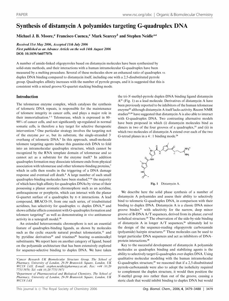
the tri-N-methyl-pyrrole duplex-DNA binding ligand distamycinA21 (Fig. 1) as a lead molecule. Derivatives of distamycin A havebeen previously reported to be inhibitors of the human telomeraseenzyme22 although distamycin A itself lacks activity. Recent NMRstudies23,24 have suggested that distamycin A is also able to interactwith G-quadruplex DNA. Two contrasting alternative modelshave been proposed in which (i) distamycin molecules bind asdimers in two of the four grooves of a quadruplex,23 and (ii) inwhich two molecules of distamycin A extend over each of the twoG-tetrad planes in a 4 : 1 binding mode.24
Fig. 1 Distamycin A.
We describe here the solid phase synthesis of a number ofdistamycin A polyamides and assess their ability to selectivelybind to telomeric G-quadruplex DNA, in comparison with theirbinding to duplex DNA. Distamycin A is a classic DNA minorgroove binder,21 with selectivity for the narrow, deep minorgroove of B-DNA A/T sequences, derived from its planar, curvedisohelical structure.25 The observation of the side-by-side bindingof distamycin A in longer A/T sequences,26 ultimately led tothe design of the sequence-reading oligopyrrole carboxamide(polyamide) hairpin structures.27 These molecules can be used totarget particular DNA sequences and act as inhibitors of DNA–protein interactions.28
Key to the successful development of distamycin A polyamidemolecules as quadruplex binding and stabilizing agents is theability to selectively target G-quadruplex over duplex DNA. Usingqualitative molecular modeling with the human intramolecularG-quadruplex structure,29 we reasoned that if a 2,5-disubstitutedpyrrole-carboxamide were also to adopt the isohelicity requiredto complement the duplex structure, it would then position theN-methyl group into rather than out of the groove, causing asteric clash that would inhibit binding to duplex DNA but would
This journal is © The Royal Society of Chemistry 2006 Org. Biomol. Chem., 2006, 4, 3479–3488 | 3479
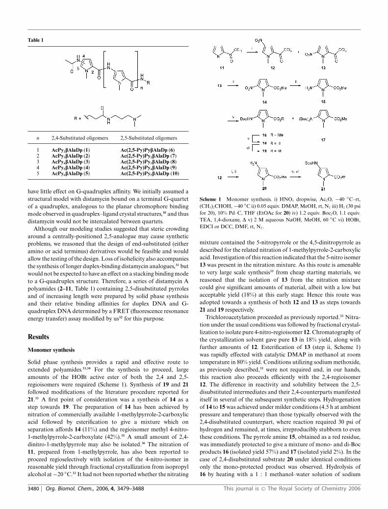
Table 1
n 2,4-Substituted oligomers 2,5-Substituted oligomers
1 AcPy2bAlaDp (1) Ac(2,5-Py)PybAlaDp (6)2 AcPy3bAlaDp (2) Ac(2,5-Py)Py2bAlaDp (7)3 AcPy4bAlaDp (3) Ac(2,5-Py)Py3bAlaDp (8)4 AcPy5bAlaDp (4) Ac(2,5-Py)Py4bAlaDp (9)5 AcPy6bAlaDp (5) Ac(2,5-Py)Py5bAlaDp (10)
have little effect on G-quadruplex affinity. We initially assumed astructural model with distamycin bound on a terminal G-quartetof a quadruplex, analogous to the planar chromophore bindingmode observed in quadruplex–ligand crystal structures,30 and thusdistamycin would not be intercalated between quartets.
Although our modeling studies suggested that steric crowdingaround a centrally-positioned 2,5-analogue may cause syntheticproblems, we reasoned that the design of end-substituted (eitheramino or acid terminus) derivatives would be feasible and wouldallow the testing of the design. Loss of isohelicity also accompaniesthe synthesis of longer duplex-binding distamycin analogues,31 butwould not be expected to have an effect on a stacking binding modeto a G-quadruplex structure. Therefore, a series of distamycin Apolyamides (2–11, Table 1) containing 2,5-disubstituted pyrrolesand of increasing length were prepared by solid phase synthesisand their relative binding affinities for duplex DNA and G-quadruplex DNA determined by a FRET (fluorescence resonanceenergy transfer) assay modified by us32 for this purpose.
Results
Monomer synthesis
Solid phase synthesis provides a rapid and effective route toextended polyamides.33,34 For the synthesis to proceed, largeamounts of the HOBt active ester of both the 2,4 and 2,5-regioisomers were required (Scheme 1). Synthesis of 19 and 21followed modifications of the literature procedure reported for21.33 A first point of consideration was a synthesis of 14 as astep towards 19. The preparation of 14 has been achieved bynitration of commercially available 1-methylpyrrole-2-carboxylicacid followed by esterification to give a mixture which onseparation affords 14 (11%) and the regioisomer methyl 4-nitro-1-methylpyrrole-2-carboxylate (42%).35 A small amount of 2,4-dinitro-1-methylpyrrole may also be isolated.36 The nitration of11, prepared from 1-methylpyrrole, has also been reported toproceed regioselectively with isolation of the 4-nitro-isomer inreasonable yield through fractional crystallization from isopropylalcohol at −20 ◦C.33 It had not been reported whether the nitrating
Scheme 1 Monomer synthesis. i) HNO3 dropwise, Ac2O, −40 ◦C–rt,(CH3)2CHOH, −40 ◦C ii) 0.05 equiv. DMAP, MeOH, rt, N2 iii) H2 (30 psifor 20), 10% Pd–C, THF (EtOAc for 20) iv) 1.2 equiv. Boc2O, 1.1 equiv.TEA, 1,4-dioxane, D v) 2 M aqueous NaOH, MeOH, 60 ◦C vi) HOBt,EDCI or DCC, DMF, rt, N2.
mixture contained the 5-nitropyrrole or the 4,5-dinitropyrrole asdescribed for the related nitration of 1-methylpyrrole-2-carboxylicacid. Investigation of this reaction indicated that the 5-nitro isomer13 was present in the nitration mixture. As this route is amenableto very large scale synthesis33 from cheap starting materials, wereasoned that the isolation of 13 from the nitration mixturecould give significant amounts of material, albeit with a low butacceptable yield (18%) at this early stage. Hence this route wasadopted towards a synthesis of both 12 and 13 as steps towards21 and 19 respectively.
Trichloroacetylation proceeded as previously reported.33 Nitra-tion under the usual conditions was followed by fractional crystal-lization to isolate pure 4-nitro-regioisomer 12. Chromatography ofthe crystallization solvent gave pure 13 in 18% yield, along withfurther amounts of 12. Esterification of 13 (step ii, Scheme 1)was rapidly effected with catalytic DMAP in methanol at roomtemperature in 80% yield. Conditions utilizing sodium methoxide,as previously described,33 were not required and, in our hands,this reaction also proceeds efficiently with the 2,4-regioisomer12. The difference in reactivity and solubility between the 2,5-disubstituted intermediates and their 2,4-counterparts manifesteditself in several of the subsequent synthetic steps. Hydrogenationof 14 to 15 was achieved under milder conditions (4.5 h at ambientpressure and temperature) than those typically observed with the2,4-disubstituted counterpart, where reaction required 30 psi ofhydrogen and remained, at times, irreproducibly stubborn to eventhese conditions. The pyrrole amine 15, obtained as a red residue,was immediately protected to give a mixture of mono- and di-Bocproducts 16 (isolated yield 57%) and 17 (isolated yield 2%). In thecase of 2,4-disubstituted substrate 20 under identical conditionsonly the mono-protected product was observed. Hydrolysis of16 by heating with a 1 : 1 methanol–water solution of sodium
3480 | Org. Biomol. Chem., 2006, 4, 3479–3488 This journal is © The Royal Society of Chemistry 2006
hydroxide (7 equiv.) afforded 18 (79%). DCC or EDCI mediatedesterification of the acid 18 gave 19 in 94% yield (6% overall yieldfrom 11).
Solid phase synthesis
Manual solid phase synthesis of polyamides from 19 and 21 wasundertaken using standard coupling conditions.33,34 Chloranil hasbeen described as an effective colorimetric test for the presenceof aromatic amines37 and in this synthesis we utilized a chloranilsolution to assess coupling of the pyrrole amines. The synthesesof 2,4-pyrrole oligomers 1–5 proceeded as planned with twonotable exceptions. Coupling between 21 and resin bound Py5
amine required an extended overnight reaction time period beyondthe standard 45 minutes. Acetylation of the resin bound Py6 aminealso required treatment beyond the standard 30 minute period.Repeated treatments with a mixture of Ac2O, DIPEA in DMF of1 h, 1.5 h and 2 h proceed with little conversion as indicated by thechloranil test. However, utilization of a freshly prepared mixturecontaining acetyl chloride (12 equiv.) and DIPEA (12 equiv.)in DMF proved successful in reducing the reaction times to30 minutes in subsequent experiments.
The synthesis of the 2,5-pyrrole oligomer series 6–10 provedmore demanding. To date, solid phase synthesis of polyamidespossessing a centrally positioned 2,5-pyrrole has not been dis-closed. The synthesis of trimer 7 was attempted first and achieveddirectly on the solid phase. Coupling of 19 to the resin boundPy2 amine required repeated couplings and longer reaction timesconsisting of three cycles of 45, 90 and 90 minutes duration.However resin-bound Boc(2,5-Py)Py2 deprotection and acetyla-tion proceeded smoothly. Resin cleavage by treatment with 3-dimethylaminopropylamine gave 7 in a reasonable 55% yieldfollowing purification by amine scavenge.
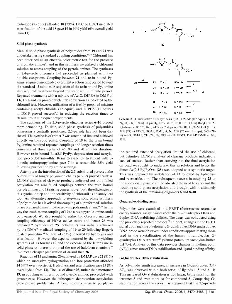
Attempts at the introduction of the 2,5-substituted pyrrole at theN-terminus of longer polyamide chains (n > 2) proved fruitless.LC/MS analysis of cleavage products indicated not only failedacetylation but also failed couplings between the resin boundpyrrole amines and 19 raising concerns over both the efficiencies ofthis synthetic step and the sensitivity of chloranil as an analyticaltool. An alternative approach to step-wise solid phase synthesisof polyamides has involved the coupling of a ‘preformed’ solutionphase prepared dimer into the growing polyamide chain.33,38 In thisway the troublesome coupling of 19 to a resin pyrrole amine couldbe by-passed. We also sought to utilize the observed increasedcoupling efficiency of HOAt active esters and hence 26 wasprepared.34 Synthesis of 25 (Scheme 2) was initially achievedby the DMAP mediated coupling of 19 to 20 following Boger’srelated procedure39 to give 24 (53%) followed by hydrolysis andesterification. However the expense incurred by the low yieldingsynthesis of 13 towards 19 and the expense of the latter’s use insolid phase syntheses prompted the use of haloform chemistry40
to direct a cheaper preparation of 24 and then 26.Reaction of 13 and amine 20 catalysed by DMAP gave 22 (81%)
which on successive hydrogenation and Boc protection afforded24 (44% over two steps). Hydrolysis and esterification gave 25 (8%overall yield from 13). The use of dimer 25, rather than monomer19, in coupling with resin bound pyrrole amines, proceeded withgreater ease. However the subsequent deprotection acetylationcycle proved problematic. A bead colour change to purple on
Scheme 2 Dimer active ester synthesis. i) 20, DMAP (0.2 equiv.), THF,N2, rt, 2 h, 81% ii) 30 psi H2, 10% Pd–C, EtOH, rt, 3 h iii) Boc2O, TEA,1,4-dioxane, 85 ◦C, 16 h, 44% for 2 steps iv) NaOH, H2O–MeOH (1 : 1),39% (27) v) EDCI, HOAt, DMF, rt, N2 21% (25 over 2 steps), 66% (28)vi) Ac2O, DMAP, CH2Cl2, N2, 38% vii) 19, EDCI, DMAP, DMF, rt, N2,53%.
the required extended acetylation limited the use of chloranilbut definitive LC/MS analysis of cleavage products indicated alack of success. Rather than carrying out the final acetylationon bead we sought to undertake this in solution and hence thedimer Ac(2,5-Py)PyOAt (28) was adopted as a synthetic target.This was prepared by acetylation of 23 followed by hydrolysisand re-esterification. The subsequent success in coupling 28 tothe appropriate pyrrole amine obviated the need to carry out thetroubling solid phase acetylation and brought with it ultimatelythe synthesis of the remaining oligomers 6 and 8–10.
Quadruplex-binding assay
Polyamides were examined in a FRET (fluorescence resonanceenergy transfer) assay to assess both their G-quadruplex DNA andduplex DNA stabilising abilities. The assay was conducted usingthe previously reported protocol.32 In brief, changes in the FRETsignal upon melting of telomeric G-quadruplex DNA and a duplexDNA probe were observed under conditions approximating thoseused in the crystallisation of the human intramolecular G-quadruplex DNA structure29 (50 mM potassium cacodylate buffer,pH 7.4). Analysis of this data provides changes in melting point(DTm), a measure of DNA stabilization and ligand binding affinity.
G-Quadruplex DNA stabilization
As polyamide length increases, an increase in G-quadruplex (G4)DTm was observed within both series of ligands 1–5 and 6–10.This increased G4 stabilization is not linear, being small for thetetramer 3 and even more so for compound 8. Comparing G4stabilization across the series it is apparent that the 2,5-pyrrole
This journal is © The Royal Society of Chemistry 2006 Org. Biomol. Chem., 2006, 4, 3479–3488 | 3481
dimer and trimer, compounds 6 and 7, possess greater G4 DNAbinding affinity than the corresponding 2,4-pyrrole oligomers 1and 2. This trend however is reversed when longer polyamides areconsidered. The 2,4-polyamides 4 and 5 are more potent than their2,5-pyrrole counterparts 9 and 10.
Duplex DNA stabilization
A near-perfect linear increase of DTm with polyamide lengthwas observed within the 2,4-pyrrole series 1–5. The binding ofcompounds 6–10 to duplex DNA is not so consistent. Thustetramer 8 has a high DTm (16 ◦C) relative to the trimer 7(1.3 ◦C) and pentamer 9 (8.5 ◦C). Across the two series, 2,5-pyrrole tetramer 8 has increased duplex DNA binding affinityrelative to the 2,4-pyrrole tetramer 3. This relationship exists forthe dimers 6 and 1 but the magnitude of the difference is small. Forthe remaining members, substitution of a 2,4-pyrrole heterocycle(Py) with a 2,5-pyrrole heterocycle (2,5-Py) results in decreasedduplex DNA binding affinity. Distamycin A, as control, has aDTm of 20.5 ◦C, much higher than the stabilization produced bythe synthetic trimers 2 (3.8 ◦C) and 7 (1.3 ◦C).
Discussion
We expected that polyamides beyond five contiguous rings woulddisplay reduced duplex DNA affinity,16 as a consequence of theproblem of helical phasing. The results of this study indicate thata plateau is yet to be reached after which introduction of addi-tional pyrrole carboxamides for compounds 1–5 is penalized andduplex DNA stabilization decreases, at least for the experimentalconditions employed here (Table 2). The DTm values show thatduplex DNA stabilization increases markedly after addition of apyrrole carboxamide to the pentamers 4 (DTm = 16.3 ◦C) and 9(8.5 ◦C) to give hexamers 5 (25.3 ◦C) and 10 (22 ◦C) respectively.In addition the viability of the concept of elongating polyamidesto enhance G-quadruplex binding at the expense of duplex DNAaffinity is demonstrated by the present FRET results. Compounds4, 5 and 9 induce greater G4 stabilization than distamycin itself,and thus show enhanced relative quadruplex affinity.
G-Quadruplex versus duplex DNA stabilization
All compounds (except 5) have decreased G4–duplex stabilizationcompared to distamycin itself. G4 DNA stabilization for the 2,4-pyrrole oligomers 1–5 follows the trend set for duplex DNAstabilization, and all display 2–4 fold selectivity for duplexDNA over G4 DNA. All the 2,5-pyrrole oligomers 6–10 displayselectivity for duplex DNA, with the one notable exception beingthe trimer 7 which displays a preference for G4 DNA. Thispreference is qualified by the modest DTm vales for both G4DNA (3.3 ◦C) and duplex DNA (1.3 ◦C) although the ratio ofmelting temperatures (duplex DNA DTm–G-Quadruplex DNADTm) is 0.4 whereas for the other polyamides reported here thisratio is always >1. The selectivity shown by 7 is a consequencemore of its low duplex DNA stabilization than significant G-quadruplex stabilization. Polyamides 8 and 9 have comparableG4 DNA stabilization to 7 but also have significantly increasedduplex DNA affinity. Here the addition of a pyrrole carboxamideseems advantageous for duplex binding but not for G-quadruplexbinding. There is an increase in G4 stabilization observed for 9relative to 7, however this is accompanied by increased duplexstabilization. Compound 5 is notable in showing a high level ofbinding to both duplex and quadruplex DNA, so that the ratio ofmelting temperatures favours quadruplex some 3-fold more thandistamycin itself.
The concept that incorporation of 2,5-pyrroles will promoteselective G-quadruplex binding is borne out in principle by theseFRET results. Comparison of melting temperature ratios (G-quadruplex DNA DTm–duplex DNA DTm) for dimers 1 (0.3) and6 (0.55) indicates that incorporation of the 2,5-pyrrole into thepolyamide scaffold has resulted in modest selectivity towards G4DNA by decreasing duplex DNA binding affinity. However thisfavourable effect is diluted with considering longer sequences. 2,4-Pyrrole polyamides 4 and 5 have similar selectivity for G4 DNAover duplex DNA compared to their 2,5-pyrrole counterparts9 and 10. The unexpected finding here that the longer lengthpolyamides show the greatest quadruplex stabilization, suggeststhat these may be binding in the grooves of a low-energy formof the human intramolecular quadruplex structure. However theshort length of groove in these structures appears to be insufficient
Table 2 G-Quadruplex DNA and duplex DNA stabilization (FRET) for compounds 1–10, distamycin and the established quadruplex-binding molecule,BRACO-1914
Compound Duplex DNAc/[DTm]D G-Quadruplex DNAb/[DTm]Q Ratio [DTm]D–[DTm]Q
1 1 0.3 3.332 3.8 1.8 4.753 10.8 2.7 4.004 16.3 7.2 2.265 25.3 14 1.816 2 1.1 1.817 1.3 3.3 0.398 16 2.4 6.679 8.5 3.8 2.2410 22 9.1 2.42BRACO-1914 11 31Distamycin A 20.5 3.5 5.86
a Values reported are the average of two determinations at a compound concentration of 10 lM. b Using the labeled G-quadruplex telomeric sequence (5′-FAM-d[GGG(TTAGGG)3]-TAMRA-3′). c Duplex DNA sequence d[TATATATATA] linked by hexaethyleneglycol and labeled with FAM and TAMRAat 5′ and 3′ ends respectively.
3482 | Org. Biomol. Chem., 2006, 4, 3479–3488 This journal is © The Royal Society of Chemistry 2006
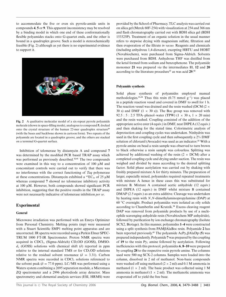
to accommodate the five or even six pyrrole-amide units incompounds 4, 5 or 9. This apparent inconsistency may be resolvedby a binding model in which one end of these conformationallyflexible polyamides stacks onto G-quartet ends, and the other isbound in a quadruplex groove. Such a model is stereochemicallyfeasible (Fig. 2) although as yet there is no experimental evidenceto support it.
Fig. 2 A qualitative molecular model of a six-repeat pyrrole polyamidemolecule (shown in space-filling mode), analogous to compound 5, dockedonto the crystal structure of the human 22-mer quadruplex structure29
(with the bases and backbone shown in cartoon form). Two repeats of thepolyamide are located in a quadruplex groove, and the others are stackedon a terminal G-quartet surface.
Inhibition of telomerase by distamycin A and compound 7was determined by the modified PCR based TRAP assay whichwas performed as previously described.32,41 The two compoundswere examined in this way to a concentration of 100 lM andconcomitant controls were carried out to verify that there wasno interference with the correct functioning of Taq polymeraseat these concentrations. Distamycin exhibited a telEC50 of 25 lMwhereas compound 7 showed no telomerase inhibitory activityat 100 lM. However, both compounds showed significant PCRinhibition, suggesting that the positive results in the TRAP assaywere not necessarily indicative of telomerase inhibition per se.
Experimental
General
Microwave irradiation was performed with an Emrys Optimizerfrom Personal Chemistry. Melting points (mp) were measuredwith a Stuart Scientific SMP1 melting point apparatus and areuncorrected. IR spectra were recorded using a Perkin Elmer SPEC-TRUM 1000 FT-IR Spectrometer. Proton NMR spectra wereacquired in CDCl3, (Sigma-Aldrich) CD3OD (GOSS), DMSO-d6 (GOSS) solutions with chemical shift (d) reported in ppmrelative to the internal standard TMS or for CD3OD solutionsrelative to the residual solvent resonance (d = 3.31). CarbonNMR spectra were recorded in CDCl3 solutions referenced tothe solvent peak (d = 77.0 ppm). LC/MS was performed using aWaters system combining a 2695 separation module, a MicromassZQ spectrometer and a 2996 photodiode array detector. Massspectrometry and elemental analysis services (ESI, HRMS) were
provided by the School of Pharmacy. TLC analysis was carried outon silica gel (Merck 60F-254) with visualization at 254 and 366 nmand flash chromatography carried out with BDH silica gel (BDH153325P). Treatment of an organic solution in the usual mannerrefers to stepwise drying with magnesium sulfate, filtration andthen evaporation of the filtrate in vacuo. Reagents and chemicals(including anhydrous 1,4-dioxane), excepting HBTU and HOBT(Novabiochem), were purchased from Sigma-Aldrich. Solventswere purchased from BDH. Anhydrous THF was distilled fromthe ketal formed from sodium and benzophenone. The polyamidemonomer 21 was prepared via the intermediates 11, 12 and 20according to the literature procedure33 as was acid 29.42
Polyamide synthesis
Solid phase synthesis of polyamides employed manualmethodologies.33,34 Thus this resin (0.75 mmol g−1) was placedin a peptide reaction vessel and covered in DMF to swell for 1 h.The reaction vessel was drained and the resin washed (DCM (2 ×30 s) and DMF (1 × 30 s)). The Boc group was removed with92.5 : 5 : 2.5 TFA–phenol–water (TPW) (1 × 30 s, 1 × 20 min)and the resin washed. Coupling consisted of the addition of theappropriate active ester (4 equiv.) in DMF, neat DIPEA (12 equiv.)and then shaking for the stated time. Colorimetric analysis ofdeprotection and coupling cycles was undertaken. Ninhydrin wasused in the first coupling cycle and then subsequently a 2% DMFsolution of chloranil (Avocado) was used as an indicator.22 With apyrrole amine on bead a resin sample was observed to turn brownto black otherwise a resin sample was colourless. Splitting wasachieved by additional washing of the resin (2 × DCM) after acompleted coupling cycle and drying under suction. The resin wasweighed and divided by mass according to the desired splittingfactor. Solid phase acetylation was carried out by shaking withfreshly prepared mixture A for thirty minutes. The preparation oflarger, especially mixed, polyamides required repeated treatmentswith mixture A hence in these cases this was substituted formixture B. Mixture A contained acetic anhydride (12 equiv.)and DIPEA (12 equiv.) in DMF whilst mixture B containedDMAP (2.5 equiv.) as an extra additive. Cleavage was undertakenby heating resin with N,N-dimethylaminopropylamine (DAP) at60 ◦C overnight. Product polyamides were isolated as oily solidsaccording to Chamberlin and Krutzik.34 Excess cleaving reagentDAP was removed from polyamide products by use of a nucle-ophile scavenging anhydride resin (Novabiochem MP anhydride),followed by purification by ion exchange chromatography (IsoluteSCX2, Biotage). In this manner, polyamides 1–4 were constructedusing a split synthesis from PAMbAlaBoc resin. Polyamide 2 hasbeen reported previously.43 The polyamide AcPy6bAlaDp (5) wasprepared independently. Polyamide 7 was prepared by the couplingof 19 to the resin Py2 amine followed by acetylation. Followinginefficiencies with this protocol, polyamides 6, 8–10 were preparedby coupling 28 to the respective resin pyrrole amine. The columnsused were 500 mg SCX-2 columns. Samples were loaded into thecolumn, dissolved in 2 ml of methanol. Non-basic compoundswere washed off using methanol (2 × 2 ml) and 0.1 M ammonia inmethanol (1 × 2 ml). The basic product was collected using 1 Mammonia in methanol (1 × 2 ml). The methanolic ammonia wasevaporated off to yield the free base.
This journal is © The Royal Society of Chemistry 2006 Org. Biomol. Chem., 2006, 4, 3479–3488 | 3483
Biophysical studies
All oligonucleotides and their fluorescent conjugates were pur-chased from Eurogentec (Southampton, UK). DNA was initiallydissolved as a stock 50 lM solution in purified water; furtherdilutions were carried out in the relevant buffer.
The ability of the compounds to stabilise G-quadruplex DNAwas investigated using a fluorescence resonance energy transfer(FRET) assay modified to be used as a high-throughput screenin a 96-well format. The labelled oligonucleotide F21T (5′-FAM-dGGG(TTAGGG)3-TAMRA-3′; donor fluorophore FAM:6-carboxyfluorescein; acceptor fluorophore TAMRA: 6-carboxy-tetramethylrhodamine) used as the FRET probe was diluted fromstock to the correct concentration (400 nM) in a 50 mM potassiumcacodylate buffer (pH 7.4) and then annealed by heating to 85 ◦Cfor 10 min, followed by cooling to room temperature in the heatingblock.
Compounds were stored as 10 mM stock solutions in DMSO;final solutions (at 2 × concentration) were prepared using 10 mMHCl in the initial 1 : 10 dilution, after which 50 mM potassiumcacodylate buffer (pH 7.4) was used in all subsequent steps. Themaximum HCl concentration in the reaction volume (at a ligandconcentration of 20 lM) is thus 200 lM, well within the rangeof the buffer used. Relevant controls were also performed toascertain a lack of interference with the assay. 96-Well plates (MJResearch, Waltham, MA) were prepared by aliquoting 50 ll of theannealed DNA into each well, followed by 50 ll of the compoundsolutions. Measurements were made on a DNA Engine Opticon(MJ Research) with excitation at 450–495 nm and detection at515–545 nm. Fluorescence readings were taken at intervals of0.5 ◦C over the range 30–100 ◦C, with a constant temperature beingmaintained for 30 s prior to each reading to ensure a stable value.Final analysis of the data was carried out using a script writtenin the program Origin 7.0 (OriginLab Corp., Northampton, MA).The advanced curve-fitting function in Origin 7.0 was used forcalculation of DTm values.
Synthetic chemistry
5-Nitro-2-(trichloroacetyl)-1-methylpyrrole (13). A solution of11 (10.10 g, 0.045 mol) in Ac2O (60 ml) was treated dropwisewith nitric acid (69%, 8 ml) over a period of 0.5 h at −40 ◦C ina CCl4–dry ice bath. The reaction solution was allowed to warmto rt and then stirred for 3 h. The solution was then cooled to−40 ◦C, isopropyl alcohol (53 ml) added and the temperaturemaintained at −40 ◦C for 0.5 h and −22 ◦C overnight. Theresultant brown solution was concentrated, poured into H2O andthe resulting mixture was extracted with CHCl3. The organicextract was worked up in the usual manner to give a brownresidue which solidified on standing. The solid was trituratedwith ice-cold EtOAc upon which a white solid (12), deposited.The solid was collected by filtration and washed with ice-coldEtOAc. The combined filtrate and washings were concentratedand stored at −22 ◦C overnight to provide a second crop of12. The brown residue resulting from evaporation of the filtratewas submitted to flash column chromatography (1 : 4 EtOAc–hexane) to afford, first, 13 as a pale green solid and then a furtheramount of 12. Yield 13 (1.093 g, 18%): mp 67–69 ◦C; IR (cm−1)1692, 1540, 1503, 1353, 1290, 1210, 1183, 1130;1H NMR (CDCl3)
d ppm 4.28 (s, 3H), 7.15 (d, 2H, J = 4.7 Hz), 7.44 (d, 2H, J =4.9 Hz); 13C NMR (CDCl3) d 36.3, 95.2, 111.3, 120.4, 125.2, 142.9,174.1.
Methyl 5-nitro-1-methylpyrrole-2-carboxylic acid ester (14)35.DMAP (38 mg, 0.3 mmol, 0.05 equiv.) was added portionwiseto a stirred suspension of 13 (1.567 g, 5.8 mmol) in anhydrousCH3OH (20 ml) under N2. The suspension was warmed slightlyto aid dissolution. After 15 min, a pale yellow solid precipitatedand the resulting suspension was stirred for a further 1.25 h. Thesolid (14) was collected by filtration and a second crop isolated.Total yield 14 (852 mg, 80%): mp 104–106 ◦C (MeOH) (lit.20 mp114 ◦C); IR (cm−1) 3121, 2363, 1710, 1532, 1495, 1476, 1303,1247; 1H NMR (CDCl3) d ppm 3.90 (s, 3H), 4.32 (s, 3H), 6.90(d, 2H, J = 4.6 Hz), 7.13 (d, 2H, J = 4.5 Hz); 13C NMR(CDCl3) d ppm 35.2, 52.1, 112.0, 115.7, 126.9, 141.3, 160.6.Found C, 45.89; H, 4.30; N, 15.10. Calcd C, 45.66; H, 4.38; N,15.21%.
Methyl 5-amino-1-methylpyrrole-2-carboxylate (15). Methylester 14 (146 mg, 0.8 mmol) in anhydrous THF was stirred underH2 in the presence of 10% Pd–C (30 mg) for 4.5 h. The reactionmixture was filtered through Celite, the Celite was washed withEtOAc and the combined filtrate and washings were concentratedto give 15 as a red residue which was submitted directly to the nextsynthetic step. Yield (123 mg, quantitative).
Methyl 5-[(tert-butoxycarbonyl)amino]-1-methylpyrrole-2-carb-oxylate (16) and methyl 5-[(di-tert-butoxycarbonyl)amino]-1-methylpyrrole-2-carboxylate (17). A solution of the amine 15(618 mg, 4 mmol) and Et3N (0.6 mL, 446 mg, 4.4 mmol,1.1 equiv.) in 1,4-dioxane was treated portionwise with Boc2O(1.050 g, 4.8 mmol, 1.2 equiv.). The solution was then heated underreflux and reaction progress monitored by TLC. On completion,the solution was concentrated to give a mixture of 16 and 17.Purification by flash chromatography (1 : 4 EtOAc–hexane) gave17 (30 mg, 2%) and 16 (577 mg, 57%): 16 mp 87–88 ◦C; IR (cm−1)3300, 2978, 1697, 1561, 1486, 1239, 1154, 1105, 747; 1H NMR(CDCl3) d 1.49 (s, 9H), 3.76 (s, 3H), 3.79 (s, 3H), 6.02 (d, 1H, J =3.6 Hz), 6.50 (bs, 1H), 6.89 (d, 1H, J = 4.2 Hz); 13C NMR (CDCl3)d ppm 161.6, 153.5, 132.2, 119.2, 116.7, 103.0, 81.3, 50.9, 31.4,28.1; HRMS (+ESI) m/z 255.1351 (MH+, C12H19N2O4 requires255.1339). 17 Mp 110–112 ◦C; IR (cm−1): 2978, 1759, 1732, 1702,1367, 1249, 1105; 1H NMR (CDCl3) d ppm 1.35 (s, 18H), 3.63(s, 3H), 3.74 (s, 3H), 5.91 (d, 1H, J = 4.2 Hz), 6.83 (d, 1H, J =4.2 Hz); 13C NMR (CDCl3) d 27.7, 31.1, 50.9, 83.3, 105.6, 116.2,120.2, 132.3, 150.6, 161.5; HRMS (+ESI) m/z 355.1862 (MH+,C17H27N2O6 requires 355.1864).
5-[(tert -Butoxycarbonyl)amino]-1-methylpyrrole-2-carboxylicacid (18). A slurry of 6 (175 mg, 0.69 mmol) in a 1 : 1 solutionof CH3OH–2 M aq. NaOH (2.56 ml) was heated at 60 ◦C for2 h. The resulting pink solution was allowed to cool to rt, washedwith diethyl ether, acidified to pH = 1 (universal indicator paper)with 10% aq. HCl and extracted with EtOAc. The combinedorganic extracts were treated in the usual manner to give a tanresidue which was dissolved in CH2Cl2 and a volume of hexane(4 × volume of CH2Cl2 used) was added. The resulting slurry wasconcentrated. This was repeated three times to give 19 as an orange
3484 | Org. Biomol. Chem., 2006, 4, 3479–3488 This journal is © The Royal Society of Chemistry 2006
solid (130 mg, 79%): mp 110–113 ◦C; IR (cm−1) 3317, 2981, 1697,1651, 1530, 1461, 1365, 1246; 1H NMR (DMSO-d6) d ppm 1.44(s, 9H), 3.63 (s, 3H), 5.90 (d, 1H, J = 4.1 Hz), 6.74 (d, 1H, J =4.1 Hz), 8.99 (s, 1H), 11.97 (s, 1H); 13C NMR (DMSO-d6) d ppm27.9, 31.2, 79.4, 102.2, 116.0, 118.8, 133.2, 153.5, 161.8.
1,2,3-Benzotriazolyl-5-[(tert-butoxycarbonyl)amino]-1-methyl-pyrrole-2-carboxylate (19). The acid 18 (130 mg, 0.5 mmol) wasdissolved in DMF (2 ml) and HOBt (80 mg, 0.5 mmol, 1 equiv.) andDCC (156 mg, 0.76 mmol, 1.4 equiv.) were added. The reactionmixture was microwaved at 80 ◦C for 20 minutes in fixed timemode at which point TLC analysis indicated reaction completion.The reaction mixture was filtered, and then evaporated to give anoil which was dissolved in EtOAc and passed through a silica plugto give 19 as a brown solid (177 mg, 94%): mp 109–113 ◦C, IR(cm−1) 2977, 2931, 1761, 1731, 1558, 1364, 1227, 1149, 1044; 1HNMR (CDCl3) d ppm 1.49 (s, 9H), 3.76 (s, 3H), 6.29 (d, 1H, J =4 Hz), 7.36–7.53 (m, 4H), 7.98 (s, 1H), 8.03 (d, 1H, J = 9 Hz);13C NMR (CDCl3) d ppm 156.6, 152.8, 143.4, 137.3, 129.2, 128.5,124.7, 121.4, 120.3, 112, 108.6, 103.5, 81.9, 31.7, 28.2; HRMS(+ESI) m/z 358.1515 (MH+, C17H20N5O4 requires 358.1510).
Methyl 4-(5-nitro-1-methylpyrrole-2-carboxamido)-1-methyl-pyrrole-2-carboxylate (22). A solution of 13 (1.608 g, 5.9 mmol)in anhydrous THF (5 ml) was added to a stirred solution of 21(902 mg, 5.9 mmol) in anhydrous THF (10 ml) at rt. DMAP(142 mg, 1.2 mmol, 0.2 equiv.) was added and stirring continued.After 2 h a yellow solid (22) had precipitated and analysis ofthe reaction mixture by LC/MS showed the reaction to becomplete. Filtration gave a first crop (953 mg) and concentratingthe filtrate and crystallisation of the resulting residue fromchloroform–hexane afforded a second crop (507 mg). The totalyield of 22 was 1.46 g (81%): mp 166 ◦C (dec.); IR (cm−1) 3342,1691, 1670, 1566, 1439, 1348, 1292, 1239; 1H NMR (DMSO-d6)d ppm 3.75 (s, 3H), 3.86 (s, 3H), 4.15 (s, 3H), 6.88 (d, 1H, J =4.6 Hz), 6.92 (d, 1H, J = 2.0 Hz), 7.28 (d, 1H, J = 4.6 Hz), 7.50 (d,1H, J = 1.9 Hz), 10.53 (bs, 1H); 13C NMR (CDCl3) d ppm 35.4,36.9, 51.2, 106.4, 108.2, 110.1, 112.4, 112.8, 120.4, 120.9, 121.2,131.3, 161.3; HRMS (ESI+) m/z 307.1029 (MH+, C13H15N4O5
requires 307.1037).
Methyl (5-amino-1-methylpyrrole-2-carboxamido)-1-methyl-pyrrole-2-carboxylate (23). A solution of 22 (20 mg, 0.39 mmol)in ethanol (20 ml) containing 10% Pd–C (15 mg) was shakenunder 30 psi of H2 for 3 h. At reaction completion the mixturewas filtered through Celite and concentrated to give 23 as a redresidue, which was submitted directly to the next synthetic step.
Methyl 4-{5-[(tert-butyloxycarbonyl)amino]-1-methylpyrrole-2-carboxamido}-1-methylpyrrole-2-carboxylate (24). Triethylamine(0.059 mL, 43 mg, 0.42 mmol, 1.1 equiv.) and Boc2O (70 mg,0.27 mmol, 0.7 equiv.), were added sequentially and portionwise toa solution of the amine 23 (0.39 mmol) in anhydrous 1,4-dioxane(1.7 ml). The resulting solution was heated at 85 ◦C overnightunder N2. The reaction solution was cooled and concentrated togive a red-brown residue. Purification by flash chromatography(3 : 1 EtOAc–hexane) gave 24 as a pale brown solid (65 mg, 44%):mp 138–139 ◦C; IR (cm−1) 1701, 1636, 1552, 1446, 1242, 1155,1107; 1H NMR (CDCl3) d ppm 1.50 (s, 9H), 3.75 (s, 3H), 3.80 (s,
3H), 3.89 (s, 3H), 5.99 (m, 1H), 6.32 (bs, 1H), 6.56 (d, 1H, J =4.1 Hz), 6.75 (d, 1H, J = 2.0 Hz), 7.40 (d, 1H, J = 1.9 Hz), 7.77(bs, 1H); 13C NMR (CDCl3) d ppm 28.2, 31.4, 36.7, 51.1, 81.3,103.2, 108.3, 110.7, 119.7, 121.0, 121.9, 122.8, 130.9, 153.9, 159.3,161.6; HRMS (+ESI) m/z 377.1819 (MH+, C18H25N4O5 requires377.1826).
Compound 24 was also prepared from the reaction between 19and 20 in 53% yield after purification by flash chromatography.
7-Azabenzo-1,2,3-triazolyl 4-[5-[(tert-butoxycarbonyl)amino-1-methylpyrrole-2-yl]-carbonyl]amino-1-methylpyrrole-2-carboxylate(25). A suspension of the methyl ester 24 (30 mg, 0.08 mmol)in aq. NaOH (0.8 M, 1 mL, 0.8 mmol, 10 equiv.) and CH3OH(1 ml) was heated at 50–60 ◦C overnight. The resulting yellowsolution was washed with diethyl ether and acidified with 10%HCl. Extraction with EtOAc and the usual work up gave a brownresidue. This was dissolved in CH2Cl2 and hexane was addedand the suspension concentrated to dryness. This procedure wasrepeated twice to give the corresponding acid as a pale yellowsolid. A solution of this solid (112 mg, 0.31 mmol) in anhydrousDMF (1.5 ml) was treated with HOAt (42 mg, 0.31 mmol) andEDCI (60 mg, 0.31 mmol). The reaction mixture was stirredunder a N2 atmosphere for 4 h, filtered through Celite intoice–water (15 ml) and the resulting suspension was filtered to givea pale yellow solid. This solid was dissolved in acetone (3 ml) andadded to cold hexane (15 ml). The isolated solid was redissolvedin CH2Cl2 (3 ml) and this solution was added to cold hexanes(15 ml). The pale yellow solid was collected by filtration and driedin vacuo. Yield 25 (75 mg, 21% over two steps): mp 139–143 ◦C;IR (cm−1) 2981, 2358, 1687, 1555, 1447, 1390, 1243, 1157; 1HNMR (CDCl3) d ppm 1.50 (s, 9H), 3.83 (s, 3H), 3.91 (s, 3H), 6.06(d, 1H, J = 3.5 Hz), 6.24 (bs, 1H), 6.64 (d, 1H, J = 4.2 Hz), 7.25(d, 1H, J = 1.9 Hz), 7.45 (dd, 1H, J = 4.5, 8.4 Hz), 7.76 (d, 1H,J = 1.7 Hz), 8.45 (dd, 1H, J = 1.4, 8.4 Hz), 8.74 (dd, 1H, J = 1.4,4.5 Hz); 13C NMR (CDCl3) d ppm 28.2, 31.5, 36.8, 81.4, 103.1,111.0, 111.2, 113.3, 115.9, 120.8, 122.3, 123.4, 125.2, 129.5, 131.4,135.0, 141.0, 151.7, 156.3, 159.2; HRMS (+ESI) m/z 481.1919(MH+, C22H25N8O5 requires 481.1942).
Methyl 4-(5-acetamido-1-methylpyrrole-2-carboxamido)-1-methylpyrrole-2-carboxylate (26). Acetic anhydride (175 lL,190 mg, 1.9 mmol, 1.2 equiv.) and DMAP (484 mg, 4 mmol,2.5 equiv.) were added to a solution of amine 23 (0.44 g, 1.6 mmol)in anhydrous CH2Cl2 (15 ml) at rt under N2. Stirring wascontinued for 3 h at which point LC/MS analysis showed thereaction to be complete. The mixture was concentrated, loadedonto a silica cartridge and eluted with EtOAc to give 26 as ayellow oil (193 mg, 38%). IR (cm−1) 2359, 1700, 1663, 1625, 1540,1444, 1245; 1H NMR (DMSO-d6) d ppm 2.04 (s, 3H), 3.65 (s,3H), 3.73 (s, 3H), 3.83 (s, 3H), 5.95 (d, 1H, J = 4 Hz), 6.84 (d, 1H,J = 4.1 Hz), 6.87 (d, 1H, J = 1.9 Hz), 7.44 (d, 1H, J = 1.9 Hz),9.68 (bs, 1H), 9.81 (bs, 1H); 13C NMR (DMSO-d6) d ppm 22.7,31.5, 36.2, 50.9, 101.7, 108.2, 111.4, 118.5, 120.6, 122.1, 122.9,131.7, 158.6, 160.7, 169.2; HRMS (+ESI) m/z 319.1409 (MH+,C15H19N4O4 requires 319.1401).
4-(5-Acetamido-1-methylpyrrole-2-carboxamido)-1-methylpyr-role-2-carboxylic acid (27). The methyl ester 26 (293 mg,0.92 mmol) was suspended in 1 : 1 NaOH (aq. 2 M)–CH3OH(4.6 ml) and heated at 45 ◦C for 2 h. The resulting solution was
This journal is © The Royal Society of Chemistry 2006 Org. Biomol. Chem., 2006, 4, 3479–3488 | 3485
cooled and then washed with diethyl ether, acidified to pH = 1(universal indicator paper) with 10% HCl and then extracted withEtOAc. The organic extracts were treated in the usual mannerto give 27 as a pale brown solid which was dried in vacuo. Yield(108 mg, 39%): mp 213–215 ◦C; IR (cm−1) 3253, 1666, 1626, 1537,1448, 1250; 1H NMR (acetone-d6) d ppm 1.95 (s, 3H), 3.64 (s,3H), 3.75 (s, 3H), 5.86 (d, 1H, J = 4 Hz), 6.72 (s, 1H), 6.77 (s, 1H),7.31 (s, 1H), 9.20 (bs, 1H), 9.34 (bs, 1H), 11.65 (bs, 1H); 13C NMR(acetone-d6) d ppm 24.0, 33.0, 37.6, 103.3, 110.1, 112.9, 121.7,121.8, 121.9, 125.1, 133.9, 160.7, 164.4, 170.6; HRMS (+ESI) m/z305.1220 (MH+, C14H17N4O4 requires 305.1244).
7-Azabenzo-1,2,3-triazolyl 4-(5-acetamido-1-methylpyrrole-2-carboxamido)-1-methylpyrrole-2-carboxylate (28). A solutionof the acid 27 (80 mg, 0.25 mmol) in DMF (3 ml) was treatedsuccessively with HOAt (34 mg, 0.25 mmol, 1 equiv.) and EDCI(48 mg, 0.25 mmol, 1 equiv.) and the resulting mixture was stirredat rt overnight under N2. The red suspension was filtered intoa stirred ice–water mixture (15 ml) at which point a pale yellowsolid precipitated. The ice–water mixture was allowed to melt andwas then filtered and the isolated solid partially dissolved in asmall volume of CH2Cl2 and added to ice-cold hexanes (15 ml).The resulting suspension was filtered to give 28 as a pale yellowsolid which was dried under suction and then in vacuo. Yield 28(70 mg, 66%): mp 159–162 ◦C; IR (cm−1) 3322, 2361, 1780, 1666,1555, 1392, 957; 1H NMR (CDCl3) d ppm 8.74 (dd, 1H, J = 1.1,4.5 Hz), 8.45 (dd, 1H, J = 1.1, 8.4 Hz), 7.83 (s, 1H), 7.76 (d, 1H,J = 1.3 Hz), 7.45 (dd, 1H, J = 4.5, 8.3 Hz), 7.12 (s, 1H), 6.65 (d,1H, J = 4.1 Hz), 6.09 (d, 1H, J = 4.1 Hz), 3.91 (s, 3H), 3.80 (s,3H), 2.22 (s, 3H); 13CNMR (CDCl3) d ppm 23.1, 31.7, 36.7, 103.2,111.3, 111.7, 112.9, 120.7, 122.6, 123.9, 125.2, 126.0, 129.4, 131.2,134.9, 140.9, 151.6, 156.3, 170.1; HRMS (+ESI) m/z 423.1543(MH+, C19H19N8O4 requires 423.1524)
AcPy2bAlaDp (1)41. Resin PAMbAlaPy2Boc (0.032 mmol)was deprotected and acetylated. Polyamide isolation followed bypurification by ion-exchange chromatography afforded 1 (6 mg,43%). 1H NMR (CD3OD) d ppm 1.66–1.72 (m, 2H), 2.07 (s, 3H),2.20 (s, 6H), 2.36 (m, 2H), 2.44–2.49 (m, 2H), 3.19–3.26 (m, 2H),3.55 (t, 2H, J = 6.7 Hz), 3.87 (s, 3H), 3.89 (s, 3H), 7.18 (d, 1H, J =1.6 Hz), 7.13 (d, 1H, J = 1.6 Hz), 6.81 (d, 1H, J = 1.6 Hz), 6.76 (d,1H, J = 1.7 Hz); HRMS (+ESI) m/z 460.2663 (MH+, C22H34N7O4
requires 460.2667).
AcPy4bAlaDp (3). Resin PAMbAlaPy3Boc (0.033 mmol) wasdeprotected and coupled to 20 (4 equiv.) with addition ofDIPEA (12 equiv.) and shaking for 45 minutes. Deprotectionand acetylation was followed by cleavage, polyamide isolation andpurification by ion-exchange chromatography to give 3 (5 mg,22%). 1H NMR (CD3OD) d ppm 1.69 (m, 2H), 1.92 (s, 3H), 2.07(s, 6H), 2.31–2.42 (m, 2H), 2.47 (t, 2H, J = 6.5 Hz), 3.21 (m,2H), 3.55 (t, 2H, J = 6.7 Hz), 3.87–3.91 (m, 12H), 6.79–6.86(m, 4H), 7.15–7.22 (m, 4H); HRMS (+ESI) m/z 704.3647 (MH+,C34H46N11O6 requires 704.3627).
AcPy5bAlaDp (4). Resin PAMbAlaPy4Boc (0.035 mmol) wasdeprotected and coupled to 20 (4 equiv.) with addition ofDIPEA (12 equiv.) and shaking for 45 minutes. Deprotection andacetylation was followed by polyamide isolation and purification
by ion-exchange chromatography to afford 4 (14 mg, 48%). 1HNMR (CD3OD) d ppm 1.83 (m, 2H), 1.92 (s, 3H), 2.05 (s, 6H),2.50 (t, 2H, J = 6.4 Hz), 2.86–2.90 (m, 2H), 3.27 (m, 2H), 3.58 (t,2H, J = 5.9 Hz), 3.87–3.91 (m, 15H), 6.84 (m, 2H), 6.96 (m, 2H),7.13 (d, 1H, J = 1.2 Hz), 7.17 (d, 1H, J = 1.2 Hz), 7.21–7.20 (m,4H); HRMS (+ESI) m/z 826.4091 (MH+, C40H52N13O7 requires826.4107).
AcPy6bAlaDp (5). PAMbAlaPy6Boc (0.52 mmol) was pre-pared from PAMbAlaBoc resin employing stepwise coupling of29 (twice), activated by stirring with HBTU and HOBt in DMFprior to addition to resin, and then 21 (twice). Deprotectionand acetylation using mixture B (4 × 1 h treatments) followed.Polyamide isolation gave an oily solid of which a sample waspurified by silica column chromatography eluting with 3 : 6 :1 CH3OH–CH2Cl2–Et3N to afford 5 (3 mg, 43%). 1H NMR(CD3OD) d ppm 1.69 (m, 2H), 1.92 (s, 3H), 2.07 (s, 6H), 2.34(m, 2H), 2.48 (t, 2H, J = 6.5 Hz), 3.22 (m, 2H), 3.56 (t, 2H, J =6.7 Hz), 3.88–3.93 (m, 18H), 6.78 (d, 1H, J = 1.8 Hz), 6.84 (d, 1H,J = 1.8 Hz), 6.94–6.95 (m, 4H), 7.13 (d, 1H, J = 1.7 Hz), 7.21–7.19 (m, 5H); HRMS (+ESI) m/z 948.4621 (MH+, C46H58N15O8
requires 948.4588).
2,5-Pyrrole polyamides (AcPyn = 2–6bAlaDp, 6–10)
Ac(2,5-Py)PybAlaDp (6). Resin PAMbAlaBoc (26 mg,0.02 mmol) was deprotected and coupled to 28 (24 mg,0.057 mmol, 3 equiv.) with addition of DIPEA (0.041 mL, 30 mg,0.24 mmol, 12 equiv.) and shaking overnight. The resin was thencapped by shaking with an acetylation mixture containing Ac2O(12 equiv.) and DIPEA (12 equiv.) in DMF for 1 h. Polyamideisolation and then purification by ion-exchange chromatographygave 6 (3 mg, 33%). 1H NMR (CD3OD) d ppm 1.76 (m, 2H), 1.92(s, 3H), 2.16 (s, 6H), 2.60 (m, 2H), 3.33 (m, 2H), 3.42 (t, 2H, J =6.8 Hz), 3.57 (t, 2H, J = 6.7 Hz), 3.73 (s, 3H), 3.87 (s, 3H), 6.01(d, 1H, J = 4.2 Hz), 6.78 (d, 1H, J = 1.9 Hz), 6.81 (d, 1H, J =4.1 Hz), 7.15 (d, 1H, J = 1.7 Hz); HRMS (+ESI) m/z 460.2675(MH+, C22H34N7O4 requires 460.2667).
Ac(2,5-Py)Py2bAlaDp (7). Prepared from PAMbAlaPyBoc(29 mg, 0.019 mmol) by coupling to 20 (two cycles required of45 minutes’ duration) and then 19 (45 minutes). Acetylation,cleavage and polyamide isolation and purification gave 7 (6 mg,55%). 1H NMR (CD3OD) d ppm 1.71 (m, 2H), 1.92 (s, 3H), 2.21(s, 6H), 2.46 (m, 2H), 2.62 (m, 2H), 3.56 (t, 2H, J = 6.8 Hz), 3.75(s, 3H), 3.88 (s, 3H), 3.91 (s, 3H), 6.02 (d, 1H, J = 4.1 Hz), 6.77(d, 1H, J = 1.9 Hz), 6.82 (d, 1H, J = 4.1 Hz), 6.92 (d, 1H, J =1.9 Hz), 7.19 (m, 2H), 7.99 (bs, 1H); HRMS (+ESI) m/z 582.3148(MH+, C28H40N9O5 requires 582.3147).
Ac(2,5-Py)Py3bAlaDp (8). Prepared from the resinPAMbAlaPy2Boc (0.0133 mmol) by coupling to 29 for 90 minutes.Cleavage and polyamide isolation gave 8 (5 mg, 54%). 1H NMR(CD3OD) d ppm 1.73 (m, 2H), 2.14 (s, 3H), 2.35 (s, 6H), 2.46–2.54(m, 4H), 3.21–3.24 (m, 2H), 3.56 (t, 2H, J = 6.6 Hz), 3.73 (s,3H), 3.86 (s, 3H), 3.90 (s, 3H), 3.90 (s, 3H), 6.01 (d, 1H, J =4.1 Hz), 6.78 (d, 1H, J = 1.9 Hz), 6.82 (d, 1H, J = 4.1 Hz),6.928–6.932 (m, 2H), 7.17–7.19 (m, 3H); HRMS m/z 704.3659(MH+, C34H46N11O6 requires 704.3627).
3486 | Org. Biomol. Chem., 2006, 4, 3479–3488 This journal is © The Royal Society of Chemistry 2006

Ac(2,5-Py)Py4bAlaDp (9). Prepared from resinPAMbAlaPy2Boc (0.027 mmol) by coupling to 20, a two-fold split and then coupling to 29 for 90 minutes. Cleavage andpolyamide isolation gave 9 (4 mg, 18%). 1H NMR (CD3OD)d ppm 1.77 (m, 2H), 2.16 (s, 3H), 2.47 (s, 6H), 2.49 (t, 2H, J =6.4 Hz), 2.65 (m, 2H), 3.25 (t, 2H, J = 6.7 Hz), 3.57 (t, 2H, J =6.6 Hz), 3.72 (s, 3H), 3.84 (s, 3H), 3.88 (s, 3H), 3.89 (s, 6H), 6.02(d, 1H, J = 4.1 Hz), 6.81 (d, 1H, J = 1.8 Hz), 6.83 (d, 1H, J =4.1 Hz), 6.95–6.96 (m, 3H), 7.18 (d, 1H, J = 1.8 Hz), 7.20 (d,1H, J = 1.8 Hz), 7.21 (d, 2H, J = 1.8 Hz); HRMS m/z 826.4110(MH+, C40H52N13O7 requires 826.4107).
Ac(2,5-Py)Py5bAlaDp (10). Prepared from resinPAMbAlaPy3Boc (0.0133 mmol) by coupling to 20 andthen 29 for 135 minutes. Cleavage and polyamide isolation gave10 (5 mg, 40%). 1H NMR (CD3OD) d ppm 1.66 (m, 2H), 2.14(s, 3H), 2.19 (s, 6H), 2.31 (t, 2H, J = 7.9 Hz), 2.45 (t, 2H, J =6.7 Hz), 2.71 (m, 2H), 3.53 (t, 2H., J = 6.5 Hz), 3.74 (s, 3H),3.86 (s, 3H), 3.90 (s, 3H), 3.91 (s, 3H), 3.91 (s, 6H), 6.00 (d, 1H,J = 4.1 Hz), 6.75 (d, 1H, J = 1.7 Hz), 6.81 (d, 1H, J = 4.1 Hz),6.92–6.93 (m, 4H), 7.18 (m, 5H); HRMS (+ESI) m/z 948.4636(MH+, C46H58N15O8 requires 948.4587).
Acknowledgements
This work has been supported by project and programme grantsto S. N. from Cancer Research UK.
References
1 E. H. Blackburn, Cell, 2001, 106, 661–673.2 N. W. Kim, M. A. Piatyszek, K. R. Prowse, C. B. Harley, M. D. West,
P. L. C. Ho, G. M. Coviello, W. E. Wright, R. L. Weinrich and J. W.Shay, Science, 1994, 266, 2011–2015; C. M. Counter, H. W. Hirte, S.Bacchetti and C. B. Harley, Proc. Natl. Acad. Sci. U. S. A., 1994, 91,2900–2904; J. W. Shay and S. Bacchetti, Eur. J. Cancer, 1997, 33, 787–791.
3 W. C. Hahn, S. A. Stewart, M. W. Brooks, S. G. York, E. Eaton, A.Kurachi, R. L. Beijersbergen, J. H. M. Knoll, M. Meyerson and R. A.Weinberg, Nat. Med., 1999, 10, 1164–1170; B. S. Herbert, A. E. Pitts,S. I. Baker, S. E. Hamilton, W. E. Wright, J. W. Shay and D. R.Corey, Proc. Natl. Acad. Sci. U. S. A., 1999, 96, 14276–14281; Z.Chen, K. S. Koeneman and D. R. Corey, Cancer Res., 2003, 63, 5917–5925.
4 S. Neidle and G. N. Parkinson, Nat. Rev. Drug Discovery, 2002, 1,383–393; E. M. Rezler, D. J. Bearss and L. H. Hurley, Curr. Opin.Pharmacol., 2002, 2, 415–423; J.-L. Mergny, J.-F. Riou, P. Mailliet,M.-P. Teulade-Fichou and E. Gilson, Nucleic Acids Res., 2002, 30,839–865; K. E. Parkinson, Curr. Opin. Invest. Drugs, 2005, 6, 605–610;L. R. Kelland, Eur. J. Cancer, 2005, 41, 971–979.
5 W. E. Wright, V. M. Tesmer, K. E. Huffman, S. D. Levene and J. W.Shay, Genes Dev., 1997, 11, 2801–2809.
6 A. M. Zahler, J. R. Williamson, T. R. Cech and D. M. Prestcott, Nature,1991, 350, 718–720.
7 C. Leonetti, S. Amodei, C. D’Angelo, A. Rizzo, B. Benassi, A.Antonelli, R. Elli, M. F. G. Stevens, M. D’Incalci, G. Zupi and A.Biroccio, Mol. Pharmacol., 2004, 66, 1138–1146.
8 O. Z. Li, M. S. Eller, K. Hanna and B. A. Gilchrest, Exp. Cell Res.,2004, 301, 189–200.
9 D. Sun, B. Thompson, B. E. Cathers, M. Salazar, S. M. Kerwin, J. O.Trent, T. C. Jenkins, S. Neidle and L. H. Hurley, J. Med. Chem., 1997,40, 2113–2116; P. J. Perry, S. Gowan, A. P. Reszka, P. Polucci, T. C.Jenkins, L. R. Kelland and S. Neidle, J. Med. Chem., 1998, 41, 3253–3260; P. J. Perry, A. P. Reszka, A. A. Wood, M. A. Read, S. M. Gowan,H. S. Dosanjh, J. O. Trent, T. C. Jenkins, L. R. Kelland and S. Neidle,J. Med. Chem., 1998, 41, 4873–4884.
10 R. T. Wheelhouse, D. Sun, H. Han, F. X. Han and L. H. Hurley, J. Am.Chem. Soc., 1998, 120, 3261–3262; F. X. Han, R. T. Wheelhouse andL. H. Hurley, J. Am. Chem. Soc., 1999, 121, 3561–3570; D. F. Shi,R. T. Wheelhouse, D. Sun and L. H. Hurley, J. Med. Chem., 2001, 44,4509–4523.
11 R. A. Heald, C. Modi, J. C. Cookson, I. Hutchinson, C. A. Laughton,S. M. Gowan, L. R. Kelland and M. F. G. Stevens, J. Med. Chem.,2002, 45, 590–597; R. A. Heald and M. F. G. Stevens, Org. Biomol.Chem., 2003, 1, 3377–3389.
12 J.-L. Mergny, L. Lacroix, M.-P. Teulade-Fichou, C. Hounsou, L.Guittal, M. Hoarau, P. B. Arimondo, J.-P. Vigneron, J.-M. Lehn, J.-F. Riou, T. Garestier and C. Helene, Proc. Natl. Acad. Sci. U. S. A.,2001, 98, 3062–3067; J.-F. Riou, L. Guittat, P. Mailliet, A. Laoui, E.Renou, O. Petitgenet, F. Megnin-Chanet, C. Helene and J.-L. Mergny,Proc. Natl. Acad. Sci. U. S. A., 2002, 99, 2672–2677.
13 K. Shin-ya, K. Wierzba, K. Matsuo, T. Ohtani, Y. Yamada, K.Furihata, Y. Hayakawa and H. Seto, J. Am. Chem. Soc., 2001, 123,1262–1263; M. Y. Kim, H. Vankayalapati, K. Shin-ya, K. Wierzba andL. H. Hurley, J. Am. Chem. Soc., 2002, 124, 2098–2099.
14 R. J. Harrison, S. M. Gowan, L. R. Kelland and S. Neidle, Bioorg.Med. Chem. Lett., 1999, 9, 2463–2468; M. A. Read, R. J. Harrison, B.Romagnoli, F. A. Tanious, S. H. Gowan, A. P. Reszka, W. D. Wilson,L. R. Kelland and S. Neidle, Proc. Natl. Acad. Sci. U. S. A., 2001,98, 4844–4849; R. J. Harrison, J. Cuesta, G. Chessari, M. A. Read,S. K. Basra, A. P. Reszka, J. Morrell, S. M. Gowan, C. M. Incles, F. A.Tanious, W. D. Wilson, L. R. Kelland and S. Neidle, J. Med. Chem.,2003, 46, 4463–4476; M. J. B. Moore, C. M. Schultes, J. Cuesta, F.Cuenca, M. Gunaratnam, F. Tanious, W. D. Wilson and S. Neidle,J. Med. Chem., 2006, 49, 582–599.
15 E. S. Baker, J. T. Lee, J. L. Sessler and M. T. Bowers, J. Am. Chem. Soc.,2006, 128, 2641–2648.
16 J. E. Reed, A. A. Arola, S. Neidle and R. Vilar, J. Am. Chem. Soc.,2006, 128, 5992–5993.
17 C. M. Incles, C. M. Schultes, H. Kempski, H. Koehler, L. R. Kellandand S. Neidle, Mol. Cancer Ther., 2004, 3, 1201–1206.
18 A. M. Burger, F. Dai, C. M. Schultes, A. P. Reszka, M. J. B. Moore,J. A. Double and S. Neidle, Cancer Res., 2005, 65, 1489–1496.
19 G. Pennarun, C. Granotier, L. R. Gauthier, D. Gomez, F. Hoffschir,E. Mandine, J.-F. Riou, J.-L. Mergny, P. Mailliet and F. D. Boussin,Oncogene, 2005, 24, 2917–2928.
20 D. Gomez, N. Aouali, A. Renaud, C. Douarre, K. Shin-ya, J. Tazi, S.Martinez, C. Trentesaux, H. Morjani and J.-F. Riou, Cancer Res., 2003,63, 6149–6153.
21 C. Zimmer and U. Wahnert, Prog. Biophys. Mol. Biol., 1986, 47, 31–112;S. Neidle, Nat. Prod. Rep., 2001, 18, 291–309.
22 N. Zaffaroni, S. Lualdi, R. Villa, D. Bellarosa, C. Cermele, P. Felicetti,C. Rossi, L. Orlandi and M. G. Daidone, Eur. J. Cancer, 2002, 38,1792–1801.
23 A. Randazzo, A. Galeone and L. Mayol, Chem. Commun., 2001, 1030–1031; A. Randazzo, A. Galeone, V. Esposito, M. Varra and L. Mayol,Nucleosides, Nucleotides Nucleic Acids, 2002, 21, 535–545.
24 M. J. Cocco, L. A. Hanakhi, M. D. Huber and N. Maizels, NucleicAcids Res., 2003, 31, 2944–2951.
25 D. S. Goodsell, Curr. Med. Chem., 2001, 8, 509–516.26 J. G. Pelton and D. E. Wemmer, Proc. Natl. Acad. Sci. U. S. A., 1989,
86, 5723.27 P. B. Dervan and B. S. Edelson, Curr. Opin. Struct. Biol., 2003, 13,
284–299.28 See for example: M. D. Gearhart, L. Dickinson, J. Ehley, C. Melander,
P. B. Dervan, P. E. Wright and J. M. Gottesfeld, Biochemistry, 2005,44, 4196–4203.
29 G. N. Parkinson, M. H. P. Lee and S. Neidle, Nature, 2002, 417, 876–880.
30 S. M. Haider, G. N. Parkinson and S. Neidle, J. Mol. Biol., 2002, 326,117–125; G. R. Clark, P. D. Pytel, C. J. Squire and S. Neidle, J. Am.Chem. Soc., 2003, 125, 4066–4067.
31 J. J. Kelly, E. E. Baird and P. B. Dervan, Proc. Natl. Acad. Sci. U. S. A.,1996, 93, 6981–6986.
32 B. Guyen, C. M. Schultes, P. Hazel, J. Mann and S. Neidle, Org. Biomol.Chem., 2004, 2, 981–988.
33 E. E. Baird and P. B. Dervan, J. Am. Chem. Soc., 1996, 118, 6141–6146.34 P. O. Krutzik and A. R. Chamberlin, Bioorg. Med. Chem. Lett., 2002,
12, 2129–2132.35 M. Bialer, B. Yagen and R. Mechoulam, Tetrahedron, 1978, 34, 2389–
2391.
This journal is © The Royal Society of Chemistry 2006 Org. Biomol. Chem., 2006, 4, 3479–3488 | 3487
36 F. Debart, C. Perigaud, G. Gosselin, D. Mrani, B. Rayner, P. Le Ber,C. Auclair, J. Balzarini, E. De Clercq, C. Paoletti and J.-L. Imbach,J. Med. Chem., 1989, 32, 1074–1083.
37 J. Marik, A. Song and K. S. Lam, Tetrahedron Lett., 2002, 44, 4319–4320.
38 M. A. Marques, R. M. Doss, A. R. Urbach and P. B. Dervan, Helv.Chim. Acta, 2002, 85, 4485–4517.
39 D. L. Boger, B. E. Fink and M. P. Hedrick, J. Am. Chem. Soc., 2000,122, 6382–6394.
40 See for example: J. Xiao, G. Yuan, W. Huang, A. S. C. Chan andK.-L. D. Lee, J. Org. Chem., 2000, 65, 5506–5513.
41 M. J. Moore, C. M. Schultes, J. Cuesat, F. Cuenca, M. Gunaratnam,F. A. Tanious, W. D. Wilson and S. Neidle, J. Med. Chem., 2006, 49,582–599.
42 C. Bailly, N. Pommery, R. Houssin and J.-P. Henichart, J. Pharm. Sci.,1989, 78, 910–917.
43 J. S. Choi, H.-S. Lee, Y. Lee, N. Jeong, H.-J. Kim, Y.-D. Kin and H.Han, Tetrahedron Lett., 2002, 43, 4295–4299.
3488 | Org. Biomol. Chem., 2006, 4, 3479–3488 This journal is © The Royal Society of Chemistry 2006


![Solution Structure of the Na+ form of the Dimeric Guanine Quadruplex [d(G3T4G3)]2](https://static.fdokumen.com/doc/165x107/6318f44265e4a6af370f95cf/solution-structure-of-the-na-form-of-the-dimeric-guanine-quadruplex-dg3t4g32.jpg)