Optically active polyamides containing 1,3-dioxolane cycles in the backbone
11
Optically active polyamides containing 1,3-dioxolane cycles in the backbone J.I. Iribarren, A. Martı ´nez de Ilarduya, C. Alema ´n, J.M. Oraison, A. Rodrı ´guez-Gala ´n, S. Mun ˜oz-Guerra * Departamento d’Enginyeria Quı ´mica, ETSEIB, Universitat Polite `cnica de Catalunya, Diagonal 647, 08028 Barcelona, Spain Received 25 May 1999; received in revised form 20 August 1999; accepted 29 September 1999 Abstract The structure in the solid state of two optically active polyamides obtained from 2,3-O-methylene-l-tartaric acid and linear a,v-alkanediamines with 9 and 12 carbon atoms, abbreviated P9MLT and P12MLT respectively, was investigated. Two ordered phases, one of smectic liquid crystal-type and another showing crystalline order, were characterized for P12MLT. Crystallization of the former into the second was induced by annealing. Infrared spectroscopy showed that hydrogen bonds are intermolecularly set in both phases and that they are weaker in the crystal phase. Quantum mechanical calculations found the O–C–CyO sequence of the tartaric unit to be in a gauche conformation. CP-MAS 13 C NMR revealed that the dioxolane ring is a mixture of the C 2 -exo and C 2 -endo puckered forms in equal amounts and that the polymethylene segment crystallizes in the all-trans conformation. X-ray diffraction of fibers and electron microscopy of solution grown single crystals afforded data consistent with a crystal lattice with orthorhombic geometry and parameters a 7:8 A; b 5:8 A; c 20:0 A: Experimental data obtained from P9MLT indicated a similar behavior for this system. q 2000 Elsevier Science Ltd. All rights reserved. Keywords: Optically active polyamides; Poly(tartaramide)s; Carbohydrate-based polyamides 1. Introduction Whereas a formidable amount of research on the crystal structure of conventional nylons [1–4] has been performed, the knowledge available on optically active non- polypeptidic polyamides is very limited. Most of the structural studies carried out on these systems are concerned with their conformation in solution [5] and only a few cases dealing with their structure in the solid state have been reported [6–10]. This is an unpleasant situation since optically active polyamides are promising materials where chiroptical effects can be combined with good mechanical and thermal properties. Over the past several years, our group has devoted sustained efforts to investigate the synthesis, structure and properties of polyamides derived from carbohydrates [11,12]. Specific attention has been paid to poly(tartara- mide)s, which are the polyamides made from naturally occurring tartaric acid (2,3-dihydroxy-succinic acid). Several classes of poly(tartaramide)s differing either in the group used for masking the hydroxyl side groups of the diacid unit [13,14] or in the diamine used as comonomer [15–17] have been examined. We found that poly(alkylene- di-O-methyl-l-tartaramide)s, i.e. polyamides made from 1,n-alkanediamines and 2,3-di-O-methyl-l-tartaric acid, crystallize in a triclinic lattice with slightly contracted chains arranged in hydrogen bonded layers which are stacked following a model close to that found in the a-form of nylon 6,6 [18]. Similar structures were also described for poly(tartaramide)s entirely derived from tartaric acid containing four stereocenters in the repeating unit [17]. In contrast with these results, helical structures stabilized by intramolecular hydrogen bonds have been proposed recently for certain glucose deriving polyamides on the basis of conformational data provided by NMR in solution [19,20]. This paper is concerned with the structure in the solid state of two optically active polyamides, namely poly(nonamethylene 2,3-O-methylene-l-tartaramide) and poly(dodecamethylene 2,3-O-methylene-l-tartaramide). They will be called here P9MLT and P12MLT, respectively, for consistency with nomenclature used in previously published work for other related poly(tartaramide)s [13]. Polymer 41 (2000) 4869–4879 0032-3861/00/$ - see front matter q 2000 Elsevier Science Ltd. All rights reserved. PII: S0032-3861(99)00716-8 * Corresponding author. E-mail address: [email protected] (S. Mun ˜oz-Guerra).
-
Upload
independent -
Category
Documents
-
view
0 -
download
0
Transcript of Optically active polyamides containing 1,3-dioxolane cycles in the backbone
Optically active polyamides containing 1,3-dioxolane cycles inthe backbone
J.I. Iribarren, A. Martı´nez de Ilarduya, C. Alema´n, J.M. Oraison,A. Rodrıguez-Gala´n, S. Munoz-Guerra*
Departamento d’Enginyeria Quı´mica, ETSEIB, Universitat Polite`cnica de Catalunya, Diagonal 647, 08028 Barcelona, Spain
Received 25 May 1999; received in revised form 20 August 1999; accepted 29 September 1999
Abstract
The structure in the solid state of two optically active polyamides obtained from 2,3-O-methylene-l-tartaric acid and lineara,v-alkanediamines with 9 and 12 carbon atoms, abbreviated P9MLT and P12MLT respectively, was investigated. Two ordered phases,one of smectic liquid crystal-type and another showing crystalline order, were characterized for P12MLT. Crystallization of the former intothe second was induced by annealing. Infrared spectroscopy showed that hydrogen bonds are intermolecularly set in both phases and that theyare weaker in the crystal phase. Quantum mechanical calculations found the O–C–CyO sequence of the tartaric unit to be in agaucheconformation. CP-MAS13C NMR revealed that the dioxolane ring is a mixture of the C2-exoand C2-endopuckered forms in equal amountsand that the polymethylene segment crystallizes in the all-transconformation. X-ray diffraction of fibers and electron microscopy of solutiongrown single crystals afforded data consistent with a crystal lattice with orthorhombic geometry and parametersa� 7:8 �A; b� 5:8 �A;
c� 20:0 �A: Experimental data obtained from P9MLT indicated a similar behavior for this system.q 2000 Elsevier Science Ltd. Allrights reserved.
Keywords: Optically active polyamides; Poly(tartaramide)s; Carbohydrate-based polyamides
1. Introduction
Whereas a formidable amount of research on the crystalstructure of conventional nylons [1–4] has been performed,the knowledge available on optically active non-polypeptidic polyamides is very limited. Most of thestructural studies carried out on these systems are concernedwith their conformation in solution [5] and only a few casesdealing with their structure in the solid state have beenreported [6–10]. This is an unpleasant situation sinceoptically active polyamides are promising materials wherechiroptical effects can be combined with good mechanicaland thermal properties.
Over the past several years, our group has devotedsustained efforts to investigate the synthesis, structure andproperties of polyamides derived from carbohydrates[11,12]. Specific attention has been paid to poly(tartara-mide)s, which are the polyamides made from naturallyoccurring tartaric acid (2,3-dihydroxy-succinic acid).Several classes of poly(tartaramide)s differing either in the
group used for masking the hydroxyl side groups of thediacid unit [13,14] or in the diamine used as comonomer[15–17] have been examined. We found that poly(alkylene-di-O-methyl-l-tartaramide)s, i.e. polyamides made from1,n-alkanediamines and 2,3-di-O-methyl-l-tartaric acid,crystallize in a triclinic lattice with slightly contractedchains arranged in hydrogen bonded layers which arestacked following a model close to that found in thea-form of nylon 6,6 [18]. Similar structures were alsodescribed for poly(tartaramide)s entirely derived fromtartaric acid containing four stereocenters in the repeatingunit [17]. In contrast with these results, helical structuresstabilized by intramolecular hydrogen bonds have beenproposed recently for certain glucose deriving polyamideson the basis of conformational data provided by NMR insolution [19,20].
This paper is concerned with the structure in the solidstate of two optically active polyamides, namelypoly(nonamethylene 2,3-O-methylene-l-tartaramide) andpoly(dodecamethylene 2,3-O-methylene-l-tartaramide). Theywill be called here P9MLT and P12MLT, respectively,for consistency with nomenclature used in previouslypublished work for other related poly(tartaramide)s [13].
Polymer 41 (2000) 4869–4879
0032-3861/00/$ - see front matterq 2000 Elsevier Science Ltd. All rights reserved.PII: S0032-3861(99)00716-8
* Corresponding author.E-mail address:[email protected] (S. Mun˜oz-Guerra).
These poly(tartaramide)s are made of flexible poly-methylene segments and tartaramide units locked by theacetal ring which visibly protrudes from the backbone.The polymer chain may be then envisaged as composed ofan alternating sequence of thread-like hydrophobic andball-like hydrophilic units.
2. Experimental
The polyamides P9MLT and P12MLT investigated in thiswork were prepared by polycondensation in solution ofbis(pentachlorophenyl) 2,3-O-methylene-l-tartrate with1,9-nonanediamine and 1,12-dodecanediamine activated asN,N0-bis(trimethylsilyl) derivatives. A detailed account ofthis synthesis has been published elsewhere [13] and asurvey of the polymer characteristics more relevant to thepresent work are given in Table 1. (4R,5R)-4,5-bis(dodecyl-carbamoyl)-1,3-dioxolane used as model compound in spec-troscopy studies was prepared by reaction of the abovementioned pentachlorophenyl ester with a large excess of1,12-dodecanediamine and then purified and characterizedby usual methods.
Polymer films were prepared by casting either in formicacid or in chloroform and fibers were obtained by stretchingfrom concentrated solutions of the polymer in such solvents.Optical microscopy was performed with a Nikon Labophotpolarizing microscope fitted with a hot stage Mettler FP-80and a Nikon FX-35DX camera. Melting, crystallization andannealing experiments were carried out on a Perkin–ElmerDSC-4 instrument using 3–5 mg of polymer samples.Thermograms were recorded at heating or cooling rates of^208C min21 and they were calibrated with indium. Den-sities were determined by the flotation technique usingmixtures of water and a 25% (w/w) sodium bromideaqueous solution. X-ray diffraction patterns were registeredin an evacuated flat-film Warhus camera with Ni-filtered
Cu-Ka radiation of wavelength 1.54 A˚ . Calibration wasinternally made with molybdenum sulfide�d002�6:147 �A�: Single crystals were grown by isothermal crystal-lization from very dilute solutions (less than 0.1%) of thepolymer in 2-octanol or ethyleneglycol. Crystals wererecovered by centrifugation and then repeatedly washedwith n-butanol. For electron microscopy observations inthe bright field mode, crystals were deposited on carbon-coated grids and shadowed with Pt–carbon at an angle ofabout 158. Electron diffraction diagrams were recorded inthe selected area mode and internally calibrated with gold�d111� 2:35 �A�: A Philips EM-301 electron microscopeoperating at 80 and 100 kV for bright field and electrondiffraction modes, respectively, was used in this work.
Infrared spectra from both, solutions and solid films, wereregistered on a Perkin–Elmer FT-IR 2000 instrumentequipped with a variable temperature cell Specac P/N21525. NMR experiments were performed on an AMX-300 Bruker spectrometer provided with a CP-MAS solidstate accessory. Solution spectra were recorded in chloro-form using TMS as reference. Solid state CP-MAS13Cspectra were recorded from 100–200 mg weight samplesspun at 3.9–4.1 kHz in a cylindrical ceramic rotor. Thesespectra were acquired with contact and repetition times of1.2 ms and 5 s, respectively. Between 256 and 1024ransients were accumulated using a spectral width of31.2 kHz. Chemical shifts were externally calibrated againstthe higher field peak of adamantane which is 29.5 ppmrelative to TMS.
To analyze the molecular conformation of modelcompounds, semi-empirical quantum mechanical calc-ulations were performed with theMopac 6.0 package [22]following the same methodology used in previous works[18]. Calculating programs were run on an IBM SP2 atthe Centre de Supercomputacio´ de Catalunya (CESCA)and on a Silicon-Graphics station RI-4000 at our laboratory.
3. Results and discussion
3.1. Poly(dodecamethylene 2,3-di-O-methylene-l-tartaramide) (P12MLT)
3.1.1. Optical microscopy and DSC measurementsP12MLT films prepared by different methods were
J.I. Iribarren et al. / Polymer 41 (2000) 4869–48794870
Table 1Data of polyamides examined in this work (data taken from Ref. [13])
Polyamide [h ] (dl g21)a Mvb Mw
c Mw=Mnc [a ]D (deg)d Tm (8C)e
P9MLT 0.60–0.80 8000–18,000 – – 212.2 90P12MLT 0.90–1.30 22,700–47,200 24,500–44,100 2.4–1.6 29.6 130
a Intrinsic viscosity measured in dichloroacetic acid at 258C.b Molecular weights calculated by applying the viscosimetric equation reported for nylon 6,6 [21].c Molecular weights estimated by GPC of trifluoroacetylated samples.d Specific optical rotation measured in chloroform�c� 1 g dl21; 258C�:e Melting peak observed by DSC.
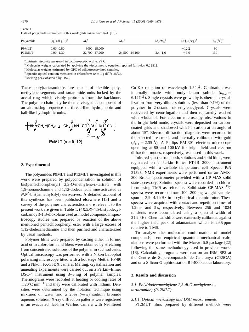
examined under a polarizing optical microscope. Filmsgenerated at high solidification rates either from solutionor from the melt appeared uniformly dark but displayedstrong birefringence upon slightly shearing. Conversely,striking colorful films were obtained by hot-pressingabove 1508C (Fig. 1a). Such optical effects strongly suggestthe existence of a mesophase-like structure. When hot-pressed films were heated at 1008C all signs of birefringencedisappeared and further heating at 110–1158C inducedspherulitic crystallization (Fig. 1b). Spherulites vanishedwhen temperature reached 1308C, which is taken as themelting temperature of the polymer. On the other hand,highly crystalline films composed of impinging spheruliteswere produced by casting in formic acid or chloroform atlow evaporation rates (Fig. 1c).
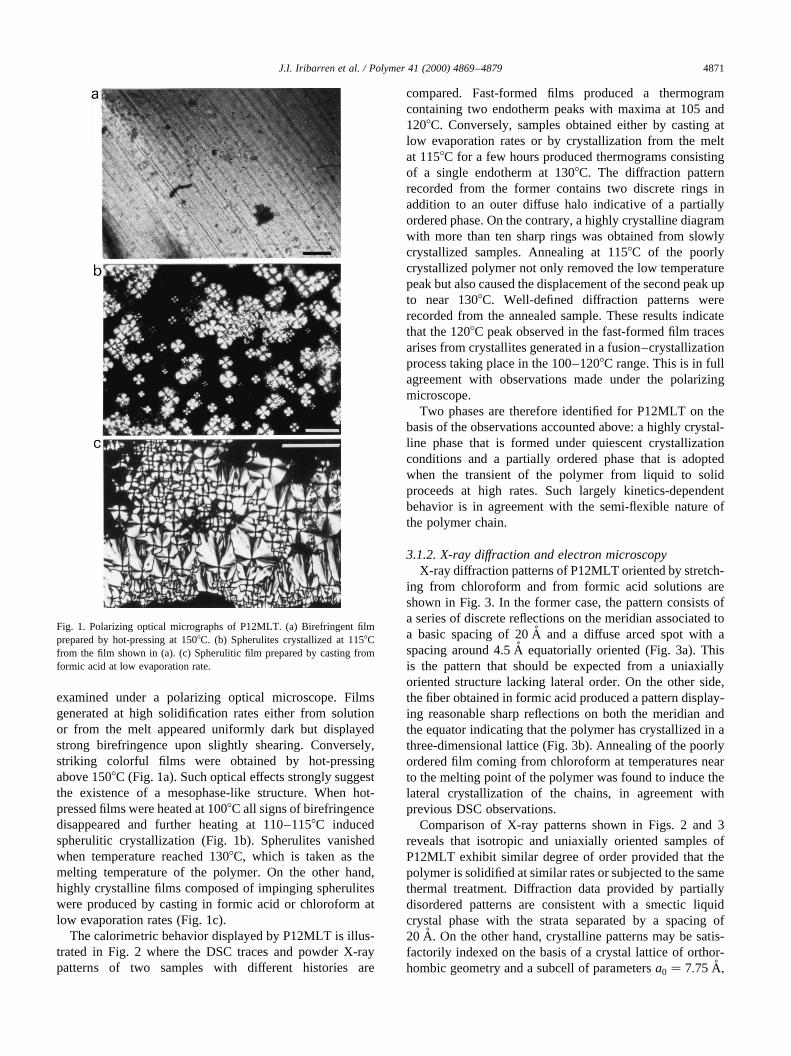
The calorimetric behavior displayed by P12MLT is illus-trated in Fig. 2 where the DSC traces and powder X-raypatterns of two samples with different histories are
compared. Fast-formed films produced a thermogramcontaining two endotherm peaks with maxima at 105 and1208C. Conversely, samples obtained either by casting atlow evaporation rates or by crystallization from the meltat 1158C for a few hours produced thermograms consistingof a single endotherm at 1308C. The diffraction patternrecorded from the former contains two discrete rings inaddition to an outer diffuse halo indicative of a partiallyordered phase. On the contrary, a highly crystalline diagramwith more than ten sharp rings was obtained from slowlycrystallized samples. Annealing at 1158C of the poorlycrystallized polymer not only removed the low temperaturepeak but also caused the displacement of the second peak upto near 1308C. Well-defined diffraction patterns wererecorded from the annealed sample. These results indicatethat the 1208C peak observed in the fast-formed film tracesarises from crystallites generated in a fusion–crystallizationprocess taking place in the 100–1208C range. This is in fullagreement with observations made under the polarizingmicroscope.
Two phases are therefore identified for P12MLT on thebasis of the observations accounted above: a highly crystal-line phase that is formed under quiescent crystallizationconditions and a partially ordered phase that is adoptedwhen the transient of the polymer from liquid to solidproceeds at high rates. Such largely kinetics-dependentbehavior is in agreement with the semi-flexible nature ofthe polymer chain.
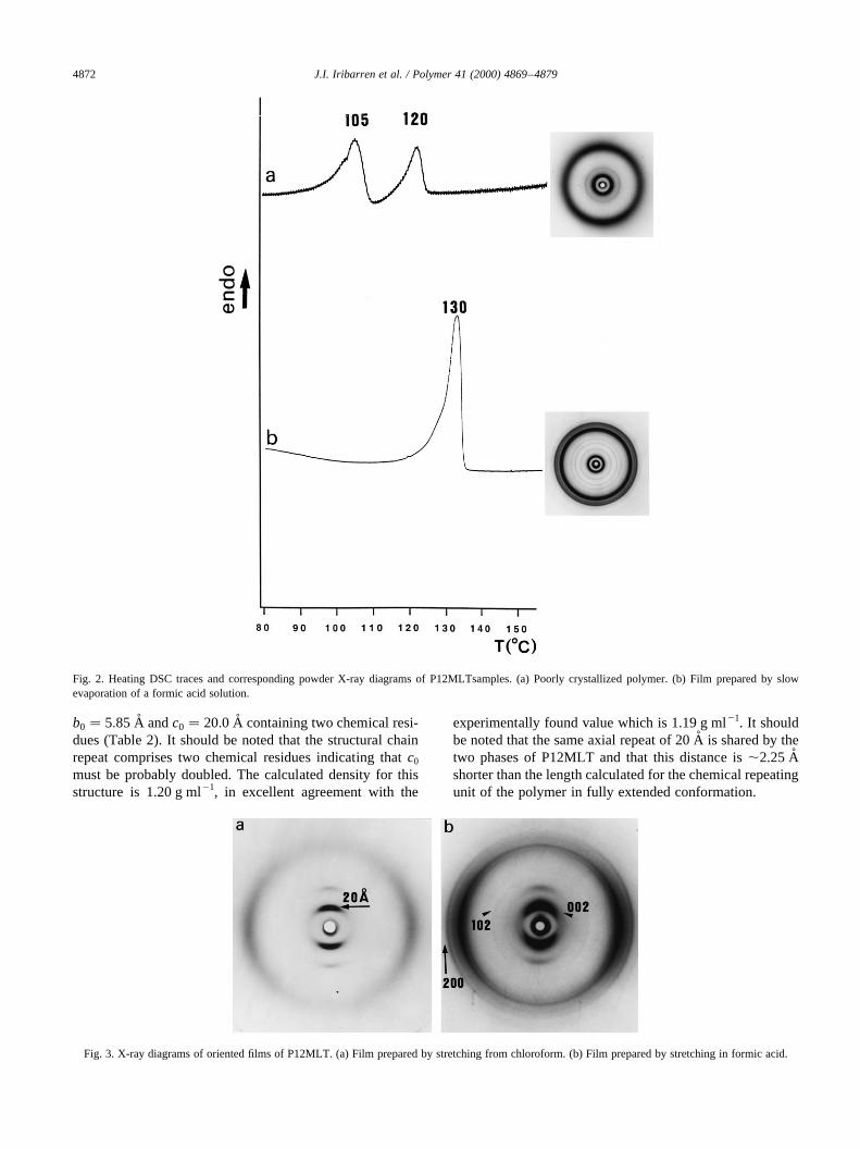
3.1.2. X-ray diffraction and electron microscopyX-ray diffraction patterns of P12MLT oriented by stretch-
ing from chloroform and from formic acid solutions areshown in Fig. 3. In the former case, the pattern consists ofa series of discrete reflections on the meridian associated toa basic spacing of 20 A˚ and a diffuse arced spot with aspacing around 4.5 A˚ equatorially oriented (Fig. 3a). Thisis the pattern that should be expected from a uniaxiallyoriented structure lacking lateral order. On the other side,the fiber obtained in formic acid produced a pattern display-ing reasonable sharp reflections on both the meridian andthe equator indicating that the polymer has crystallized in athree-dimensional lattice (Fig. 3b). Annealing of the poorlyordered film coming from chloroform at temperatures nearto the melting point of the polymer was found to induce thelateral crystallization of the chains, in agreement withprevious DSC observations.
Comparison of X-ray patterns shown in Figs. 2 and 3reveals that isotropic and uniaxially oriented samples ofP12MLT exhibit similar degree of order provided that thepolymer is solidified at similar rates or subjected to the samethermal treatment. Diffraction data provided by partiallydisordered patterns are consistent with a smectic liquidcrystal phase with the strata separated by a spacing of20 A. On the other hand, crystalline patterns may be satis-factorily indexed on the basis of a crystal lattice of orthor-hombic geometry and a subcell of parametersa0 � 7:75 �A;
J.I. Iribarren et al. / Polymer 41 (2000) 4869–4879 4871
Fig. 1. Polarizing optical micrographs of P12MLT. (a) Birefringent filmprepared by hot-pressing at 1508C. (b) Spherulites crystallized at 1158Cfrom the film shown in (a). (c) Spherulitic film prepared by casting fromformic acid at low evaporation rate.
b0 � 5:85 �A andc0 � 20:0 �A containing two chemical resi-dues (Table 2). It should be noted that the structural chainrepeat comprises two chemical residues indicating thatc0
must be probably doubled. The calculated density for thisstructure is 1.20 g ml21, in excellent agreement with the
experimentally found value which is 1.19 g ml21. It shouldbe noted that the same axial repeat of 20 A˚ is shared by thetwo phases of P12MLT and that this distance is,2.25 Ashorter than the length calculated for the chemical repeatingunit of the polymer in fully extended conformation.
J.I. Iribarren et al. / Polymer 41 (2000) 4869–48794872
Fig. 2. Heating DSC traces and corresponding powder X-ray diagrams of P12MLTsamples. (a) Poorly crystallized polymer. (b) Film prepared by slowevaporation of a formic acid solution.
Fig. 3. X-ray diagrams of oriented films of P12MLT. (a) Film prepared by stretching from chloroform. (b) Film prepared by stretching in formic acid.

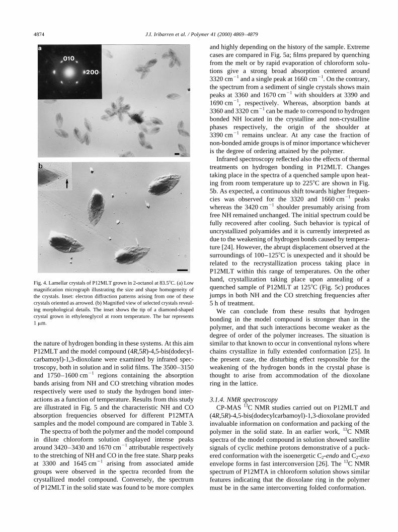
Isothermal crystallization of P12MLT in 2-octanol at83.58C rendered the type of lamellar crystals shown inFig. 4. They appear to be very uniform in both shape andsize with lateral dimensions of about 4mm × 2 mm and athickness of about 120 A˚ as estimated from electron micro-graphs of Pt–carbon shadowed specimens. Although thelenticular habit displayed by these crystals is reminiscentof lozenges, their serrated edges and rounded contoursmake difficult a reliable measurement of their morpho-logical parameters. Crystallization in ethyleneglycol atroom temperature provided crystals with abundant over-growths but displaying sharper outlined contours andrevealing a genuine diamond habit with an acute angleclose to 708 (inset Fig. 4b).
Electron diffraction diagrams obtained from the crystalsgrown in 2-octanol consist of a rectangular array of reflec-tions witha0 � 7:86 �A andb0 � 5:80 �A (inset Fig. 4a). Thepattern may be straightforwardly interpreted as arising fromthe projection down thec-axis of the crystal lattice definedabove. The orientation of the pattern relative to the crystalindicates that the lattice must be aligned with thea-axis
parallel to the long dimension of the lamella. Systematicabsences forh00 reflections withh� 2n 1 1 are detectedalong the reciprocala-axis in agreement with the existencein the crystal of a 21 axis parallel toa. All the independentd-spacings observed in the original photographic filmstogether with their corresponding indexes are listed inTable 2. It should be noticed thathk0 reflections withh� k appear split revealing that more than one reciprocalplane are being intersected by the Ewald sphere. This doesmake sense since we are dealing with thin platelets crystalscomposed of a unit cell with a largec0-parameter.
3.1.3. Infrared spectroscopyX-ray studies carried out on crystallizedN-alkyl
l-gluconamides have shown that these compounds tend tocrystallize in a skew conformation stabilized by bothintermolecular O–H···OyC and intramolecular N–H···Ohydrogen bonds, the latter being of a bifurcated naturewith both carbonyl and hydroxyl groups acting as hydro-gen-accepting groups [23]. The presence of the 1,3-dioxo-lane ring in PnMLT makes advisable to investigate in detail
J.I. Iribarren et al. / Polymer 41 (2000) 4869–4879 4873
Table 2Observed and calculated spacings (A˚ ) for P12MLT
Observeda Calculatedb hklb
Fiberc Crystal matd Single crystale
Meridional120 (m) 120.0 L0 (lamellar height)40 (w) 40.0 3rd order
20.0 (vs) 20.0 (vs) 20.0 6th order and 00115.0 (m) 15.0 8th order12.5 (w) 12.0 10th order
10.0 (m) 10.0 (s) 10.0 12th order and 0028.5 (vw) 8.6 14th order7.5 (vw) 7.5 16th order
6.8 (w) 6.8 (m) 6.7 18th order and 003Equatorial
5.80 (w) 5.80 (w) 5.85 0104.67 (s) 4.65 (vs) 4.60 (vs) 4.67 1103.86 (s) 3.85 (vs) 3.88 (vs) 3.88 200
2.90 (w) 2.92 0201.94 (m) 1.94 4001.30 (vw) 1.29 600
Off-meridional7.24 (m) 7.30 (m) 7.23 1016.25 (w) 6.10 (m) 6.12 102
5.05 (w) 5.05, 5.05 012, 1034.47 (vs) 4.40 (vs) 4.50 (s) 4.55, 4.41 1114.15 (m) 4.10 (s) 4.23, 4.20 112, 104
3.85 (s) 3.82, 3.80 113, 0143.60 (w) 3.60 (m) 3.55 1053.30 (w) 3.30 (w) 3.30 0153.08 (w) 3.10 (m) 3.06, 3.04 106, 115
a Intensities visually estimated and denoted as: vs� very strong, s� strong, m�medium, w� weak, vw� very weak.b Spacings calculated and indexed on the basis of a unit cell witha0 � 7:75 �A; b0 � 5:85 �A andc0 � 20:0 �A; a � b � g � 908:c Fiber obtained by stretching from formic acid.d Mat of single crystals grown at 83.58C in 2-octanol.e Measured on single crystal electron diffraction patterns.
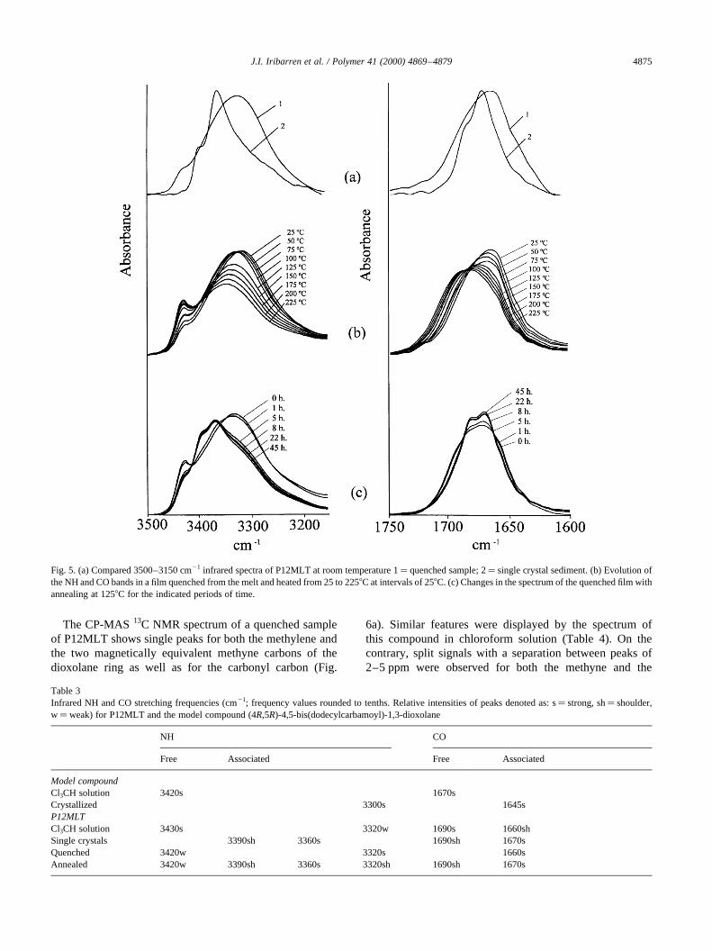
the nature of hydrogen bonding in these systems. At this aimP12MLT and the model compound (4R,5R)-4,5-bis(dodecyl-carbamoyl)-1,3-dioxolane were examined by infrared spec-troscopy, both in solution and in solid films. The 3500–3150and 1750–1600 cm21 regions containing the absorptionbands arising from NH and CO stretching vibration modesrespectively were used to study the hydrogen bond inter-actions as a function of temperature. Results from this studyare illustrated in Fig. 5 and the characteristic NH and COabsorption frequencies observed for different P12MTAsamples and the model compound are compared in Table 3.
The spectra of both the polymer and the model compoundin dilute chloroform solution displayed intense peaksaround 3420–3430 and 1670 cm21 attributable respectivelyto the stretching of NH and CO in the free state. Sharp peaksat 3300 and 1645 cm21 arising from associated amidegroups were observed in the spectra recorded from thecrystallized model compound. Conversely, the spectrumof P12MLT in the solid state was found to be more complex
and highly depending on the history of the sample. Extremecases are compared in Fig. 5a; films prepared by quenchingfrom the melt or by rapid evaporation of chloroform solu-tions give a strong broad absorption centered around3320 cm21 and a single peak at 1660 cm21. On the contrary,the spectrum from a sediment of single crystals shows mainpeaks at 3360 and 1670 cm21 with shoulders at 3390 and1690 cm21, respectively. Whereas, absorption bands at3360 and 3320 cm21 can be made to correspond to hydrogenbonded NH located in the crystalline and non-crystallinephases respectively, the origin of the shoulder at3390 cm21 remains unclear. At any case the fraction ofnon-bonded amide groups is of minor importance whicheveris the degree of ordering attained by the polymer.
Infrared spectroscopy reflected also the effects of thermaltreatments on hydrogen bonding in P12MLT. Changestaking place in the spectra of a quenched sample upon heat-ing from room temperature up to 2258C are shown in Fig.5b. As expected, a continuous shift towards higher frequen-cies was observed for the 3320 and 1660 cm21 peakswhereas the 3420 cm21 shoulder presumably arising fromfree NH remained unchanged. The initial spectrum could befully recovered after cooling. Such behavior is typical ofuncrystallized polyamides and it is currently interpreted asdue to the weakening of hydrogen bonds caused by tempera-ture [24]. However, the abrupt displacement observed at thesurroundings of 100–1258C is unexpected and it should berelated to the recrystallization process taking place inP12MLT within this range of temperatures. On the otherhand, crystallization taking place upon annealing of aquenched sample of P12MLT at 1258C (Fig. 5c) producesjumps in both NH and the CO stretching frequencies after5 h of treatment.
We can conclude from these results that hydrogenbonding in the model compound is stronger than in thepolymer, and that such interactions become weaker as thedegree of order of the polymer increases. The situation issimilar to that known to occur in conventional nylons wherechains crystallize in fully extended conformation [25]. Inthe present case, the disturbing effect responsible for theweakening of the hydrogen bonds in the crystal phase isthought to arise from accommodation of the dioxolanering in the lattice.
3.1.4. NMR spectroscopyCP-MAS 13C NMR studies carried out on P12MLT and
(4R,5R)-4,5-bis(dodecylcarbamoyl)-1,3-dioxolane providedinvaluable information on conformation and packing of thepolymer in the solid state. In an earlier work,13C NMRspectra of the model compound in solution showed satellitesignals of cyclic methine protons demonstrative of a puck-ered conformation with the isoenergetic C2-endoand C2-exoenvelope forms in fast interconversion [26]. The13C NMRspectrum of P12MTA in chloroform solution shows similarfeatures indicating that the dioxolane ring in the polymermust be in the same interconverting folded conformation.
J.I. Iribarren et al. / Polymer 41 (2000) 4869–48794874
Fig. 4. Lamellar crystals of P12MLT grown in 2-octanol at 83.58C. (a) Lowmagnification micrograph illustrating the size and shape homogeneity ofthe crystals. Inset: electron diffraction patterns arising from one of thesecrystals oriented as arrowed. (b) Magnified view of selected crystals reveal-ing morphological details. The inset shows the tip of a diamond-shapedcrystal grown in ethyleneglycol at room temperature. The bar represents1 mm.
The CP-MAS13C NMR spectrum of a quenched sampleof P12MLT shows single peaks for both the methylene andthe two magnetically equivalent methyne carbons of thedioxolane ring as well as for the carbonyl carbon (Fig.
6a). Similar features were displayed by the spectrum ofthis compound in chloroform solution (Table 4). On thecontrary, split signals with a separation between peaks of2–5 ppm were observed for both the methyne and the
J.I. Iribarren et al. / Polymer 41 (2000) 4869–4879 4875
Fig. 5. (a) Compared 3500–3150 cm21 infrared spectra of P12MLT at room temperature 1� quenched sample; 2� single crystal sediment. (b) Evolution ofthe NH and CO bands in a film quenched from the melt and heated from 25 to 2258C at intervals of 258C. (c) Changes in the spectrum of the quenched film withannealing at 1258C for the indicated periods of time.
Table 3Infrared NH and CO stretching frequencies (cm21; frequency values rounded to tenths. Relative intensities of peaks denoted as: s� strong, sh� shoulder,w�weak) for P12MLT and the model compound (4R,5R)-4,5-bis(dodecylcarbamoyl)-1,3-dioxolane
NH CO
Free Associated Free Associated
Model compoundCl3CH solution 3420s 1670sCrystallized 3300s 1645sP12MLTCl3CH solution 3430s 3320w 1690s 1660shSingle crystals 3390sh 3360s 1690sh 1670sQuenched 3420w 3320s 1660sAnnealed 3420w 3390sh 3360s 3320sh 1690sh 1670s
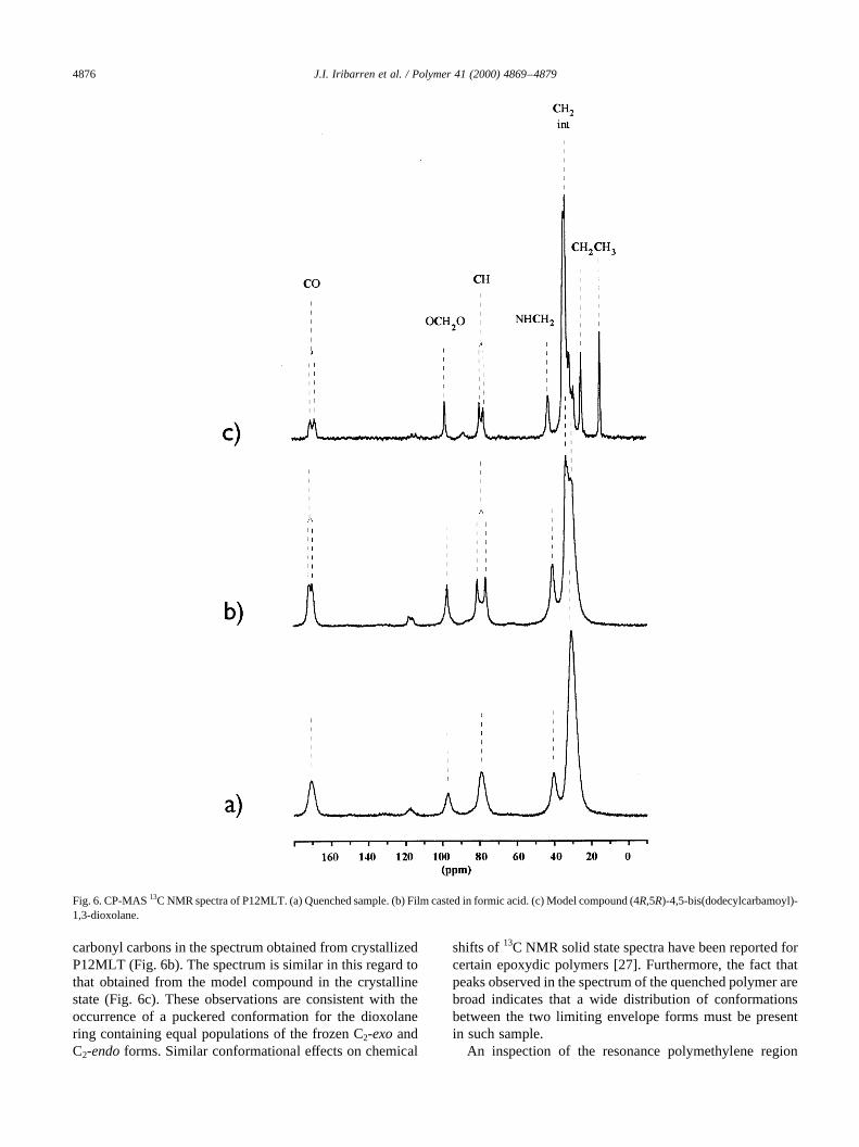
carbonyl carbons in the spectrum obtained from crystallizedP12MLT (Fig. 6b). The spectrum is similar in this regard tothat obtained from the model compound in the crystallinestate (Fig. 6c). These observations are consistent with theoccurrence of a puckered conformation for the dioxolanering containing equal populations of the frozen C2-exoandC2-endoforms. Similar conformational effects on chemical
shifts of 13C NMR solid state spectra have been reported forcertain epoxydic polymers [27]. Furthermore, the fact thatpeaks observed in the spectrum of the quenched polymer arebroad indicates that a wide distribution of conformationsbetween the two limiting envelope forms must be presentin such sample.
An inspection of the resonance polymethylene region
J.I. Iribarren et al. / Polymer 41 (2000) 4869–48794876
Fig. 6. CP-MAS13C NMR spectra of P12MLT. (a) Quenched sample. (b) Film casted in formic acid. (c) Model compound (4R,5R)-4,5-bis(dodecylcarbamoyl)-1,3-dioxolane.
(30–42 ppm) revealed that the signal arising from interiormethylenes (C2–C11) in the crystallized polymer consistsof two peaks at 30.5 and 33.0 ppm which correlate withthose observed for the model compound and the quenched
polymer respectively. The downfield peak arises frommethylenes arranged intrans-gauchewhereas the upfieldone is attributed to methylenes crystallized in the all-transconformation. Differences of 2.5–3.0 ppm between themethylene signals have been reported for nylon 12 in thecrystallized and the amorphous state [25].
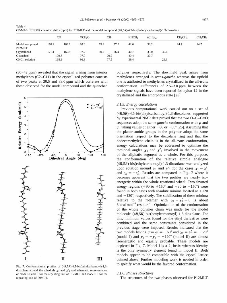
3.1.5. Energy calculationsPrevious computational work carried out on a set of
(4R,5R)-4,5-bis(alkylcarbamoyl)-1,3-dioxolanes supportedby experimental NMR data proved that the two O–C–CyOsequences adopt the samegaucheconformation withw andw 0 taking values of either160 or2608 [26]. Assuming thatthe planar amide groups in the polymer adopt the sameorientation respect to the dioxolane ring and that thedodecamethylene chain is in the all-trans conformation,energy calculations may be addressed to optimize thetorsional anglesx 1 and x 01 involved in the movementof the aliphatic segment as a whole. For this purpose,the conformation of the relative simple analogue(4R,5R)-bis(ethylcarbamoyl)-1,3-dioxolane was analyzedupon rotation aroundx 1 and x 01 for the casesx1 � x 01and x1 � 2x 01: Results are compared in Fig. 7 where itbecomes apparent that the two profiles are nearly iso-energetic within the whole rotational wheel. Two favoredenergy regions (190 to 11508 and 290 to 21508) werefound in both cases with absolute minima located at1120and21208, respectively. The stabilization of these minimarelative to the rotamer withx1 � x 01 � 0 is about6 kcal mol21 residue21. Optimization of the conformationof the whole polymer chain was made for the modelmolecule (4R,5R)-bis(hexylcarbamoyl)-1,3-dioxolane. Forthis, minimum values found for the ethyl derivative werecombined and the same constraints considered in theprevious stage were imposed. Results indicated that thetwo models havingw � w 0 � 2608 andx1 � x 01 � 21208(model I) andx1 � 2x 01 � 11208 (model II) are almostisoenergetic and equally probable. These models aredepicted in Fig. 7. Model I is a 21 helix whereas identityis the only symmetry element found in model II. Bothmodels appear to be compatible with the crystal latticedefined above. Further modeling work is needed in orderto specify what would be the favored conformation.
3.1.6. Phases structuresThe structures of the two phases observed for P12MLT
J.I. Iribarren et al. / Polymer 41 (2000) 4869–4879 4877
Table 4CP-MAS 13C NMR chemical shifts (ppm) for P12MLT and the model compound (4R,5R)-4,5-bis(dodecylcarbamoyl)-1,3-dioxolane
CO OCH2O CH NHCH2 (CH2)int CH2CH3 CH2CH3
Model compound 170.2 168.1 98.0 79.3 77.2 42.6 33.2 24.7 14.7P12MLTCrystallized 171.1 169.9 97.2 80.9 76.4 40.7 33.0 30.6Quenched 170.8 97.0 79.2 40.4 30.7CHCl3 solution 168.9 96.3 77.5 39.4 29.3
Fig. 7. Conformational profiles of (4R,5R)-4,5-bis(ethylcarbamoyl)-1,3-dioxolane around the dihedralsx1 and x 01 and schematic representationof models I and II for the repeating unit of P12MLT and model III for therepeating unit of P9MLT.
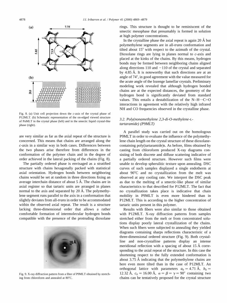
are very similar as far as the axial repeat of the structure isconcerned. This means that chains are arranged along thec-axis in a similar way in both cases. Differences betweenthe two phases arise therefore from differences in theconformation of the polymer chain and in the degree oforder achieved in the lateral packing of the chains (Fig. 8).
The partially ordered phase is envisaged as a stratifiedstructure with chains hexagonally packed with statisticalaxial orientation. Hydrogen bonds between neighboringchains would be set at random in three directions fixing anaverage interchain distance of about 5 A˚ . The chains are inaxial register so that tartaric units are arranged in planesnormal to the axis and separated by 20 A˚ . The polymethy-lene segment runs parallel to the axis in a conformation thatslightly deviates from all-transin order to be accommodatedwithin the observed axial repeat. The result is a structurelacking three-dimensional order that allows a rathercomfortable formation of intermolecular hydrogen bondscompatible with the presence of the protruding dioxolane
rings. This structure is thought to be reminiscent of thesmectic mesophase that presumably is formed in solutionat high polymer concentrations.
In the crystalline phase the axial repeat is again 20 A˚ butpolymethylene segments are in all-trans conformation andtilted about 158 with respect to the azimuth of the crystal.Dioxolane rings are lying in planes normal toc-axis andplaced at the kinks of the chains. By this means, hydrogenbonds may be formed between neighboring chains alignedalong directions 110 and2110 of the crystal and separatedby 4.85 A. It is noteworthy that such directions are at anangle of 748, in good agreement with the value measured forthe acute angle of the lozenge lamellar crystals. Preliminarymodeling work revealed that although hydrogen bondedchains are at the expected distances, the geometry of thehydrogen bond is significantly deviated from standardvalues. This entails a destabilization of the N–H···CyOinteractions in agreement with the relatively high infraredNH and CO frequencies observed in the crystalline phase.
3.2. Poly(nonamethylene 2,3-di-O-methylene-l-tartaramide) (P9MLT)
A parallel study was carried out on the homologousP9MLT in order to evaluate the influence of the polymethy-lene chain length on the crystal structure of these dioxolane-containing poly(tartaramide)s. As before, films obtained bycasting from chloroform produced X-ray diagrams con-sisting of both discrete and diffuse scattering indicative ofa partially ordered structure. However such films wereunable to develop spherulitic texture upon annealing. DSCcurves of such samples displayed a single endotherm atabout 908C and no crystallization from the melt wasobserved at any cooling rate. We interpret the DSC peakas due to the melting of a smectic-like phase of similarcharacteristics to that described for P12MLT. The fact thatno crystallization takes place is indicative that chainmobility in P9MLT is even more hindered than inP12MLT. This is according to the higher concentration oftartaric units present in this polymer.
Results with fibers were also similar to those obtainedwith P12MLT. X-ray diffraction patterns from samplesstretched either from the melt or from concentrated solu-tions display poorly lateral crystallization of the chains.When such fibers were subjected to annealing they yieldeddiagrams containing sharps reflections characteristic of athree-dimensional ordered structure (Fig. 9). Both crystal-line and non-crystalline patterns display an intensemeridional reflection with a spacing of about 15 A˚ corre-sponding to the axial repeat of the structure. In this case theshortening respect to the fully extended conformation isabout 3.75 A˚ indicating that the polymethylene chains arehere even more tilted than in the case of P12MLT. Anorthogonal lattice with parametersa0 � 4:71 �A; b0 �12:32 �A; c0 � 16:00 �A; a � b � g � 908 containing twochains can be tentatively proposed for the crystal structure
J.I. Iribarren et al. / Polymer 41 (2000) 4869–48794878
Fig. 8. (a) Unit cell projection down thec-axis of the crystal phase ofP12MLT. (b) Schematic representation of the on-edged viewed structureof PnMLT in the crystal phase (left) and in the smectic liquid crystal-likephase (right).
Fig. 9. X-ray diffraction pattern from a fiber of P9MLT obtained by stretch-ing from chloroform and annealed at 808C.
in analogy to the structure given for P12MLT. The densitycalculated for such structure is 1.10 g ml21 in excellentconcordance with the observed value which was estimatedto be 1.08 g ml21. Optimization of the conformation byenergy calculations gave model III withw � 2w 0 � 2608andx1 � 2x 01 � 21208 as the most favored arrangementfor the crystallized P9MLT chain (Fig. 7).
4. Concluding remarks
Two crystalline phases differing in the degree of orderingare adopted by polyamides P12MLT and P9MLT dependingon crystallization conditions. At high crystallization rates, asmectic-like phase with defective chain lateral order isdeveloped whilst at low rates a three-dimensional crystalis formed. The favored conformation of these poly(tartara-mide)s entails agauchearrangement for the two O–C–CyOsequences comprised in the tartaric unit which accounts inpart for the reduction observed in the length of the structuralrepeating unit of the two polymers. The dioxolane ring is ina puckered conformation with the isoenergetic C2-endoandC2-exo forms in equal amounts and the polymethylenesegment is arranged in all-trans conformation. Extensiveintermolecular hydrogen-bonding takes place in thesepolyamides both in the ordered and disordered phases.The stability of such interactions decreases with the degreeof ordering because of disturbing effects arising from thepresence of the bulky acetal side group.
Crystallization of the “smectic” phase may be induced inthe solid state by annealing. The process implies arearrangement in both the side-by-side packing of the chainsand the conformation of the polymethylene segment. In theP12MLT crystal lattice the chains describe a wavy trajec-tory with the straight polymethylene segment tilted about168 with respect to the azimuth of the crystal and dioxolanecycles accommodate at the kinks. Preliminary data obtainedfor P9MLT indicate a similar behavior but chains arrangedin the crystal with an even higher inclination.
Acknowledgements
Financial support from the CICYT (Comisio´n Inter-ministerial de Ciencia y Tecnologı´a) for the scientificgrant MAT96-1181-CO3-03 is gratefully acknowledged.The authors are indebted to CESCA for computationalfacilities.
References
[1] Tadokoro H. Structure of crystalline polymers, New York: Wiley,1979.
[2] Kohan MI. Nylon plastics, New York: Wiley, 1971.[3] Aharony SM.n-Nylons: their synthesis, structure and properties, New
York: Wiley, 1997.[4] Wunderlich B. Macromolecular physics, 1. New York: Academic
Press, 1973.[5] Selegny E, Merle-Aubry L. In: Selegny E, editor. Optically active
polymers, Dordrecht: Reidel, 1979. pp. 15–110.[6] Coiro VM, de Santis P, Mazarella L, Picozzi L. J Polym Sci: Part A
1965;3:4001.[7] Schmidt E. Angew Makromol Chem 1970;14:185.[8] Prieto A, Iribarren JI, Mun˜oz-Guerra S, Bui C, Sekiguchi H. Crystal-
lization of polymers. In: Dosiere M, editor. NATO ASI Series, SerieC, 405. Dordrecht: Kluwer, 1993. pp. 613–8.
[9] Munoz-Guerra S, Lo´pez-Carrasquero F, Ferna´ndez-Santı´n JM,Subirana JA. In: Salamone JC, editor. The polymeric materialsencyclopedia, Boca Raton, FL: CRC Press, 1996. pp. 4694–700.
[10] Puiggalı´ J, Munoz-Guerra S, Rodrı´guez-Gala´n A, Subirana JA.Makromol Chem, Macromol Symp 1988;20/21:167.
[11] Bou JJ, Rodrı´guez-Gala´n A, Munoz-Guerra S. In: Salamone JC,editor. The polymeric materials encyclopedia, Boca Raton, FL:CRC Press, 1996. pp. 561–9.
[12] Bou JJ, Iribarren JI, Rodrı´guez-Gala´n A, Munoz-Guerra S. In: Vert M,editor. Biodegradable polymers and plastics, Cambridge, UK: RoyalSociety of Chemistry, 1992. pp. 271–4.
[13] Rodrıguez-Gala´n A, Bou JJ, Mun˜oz-Guerra S. J Polym Sci: PolymChem Ed 1992;30:713.
[14] Bou JJ, Rodrı´guez-Gala´n A, Munoz-Guerra S. Macromolecules1993;26:5664.
[15] Bou JJ, Mun˜oz-Guerra S. Polymer 1984;36:181.[16] Bou JJ, Iribarren JI, Mun˜oz-Guerra S. Macromolecules 1994;27:5263.[17] Bou JJ, Iribarren JI, Martı´nez de Ilarduya A, Mun˜oz-Guerra S. J Polym
Sci: Polym Chem Ed 1999;39:983.[18] Iribarren JI, Alema´n C, Bou JJ, Mun˜oz-Guerra S. Macromolecules
1996;29:4397.[19] Chen L, Kiely DE. Polym Prepr Am Chem Soc, Div Polym Chem
1993;34:550.[20] Chen L, Kiely DE. J Org Chem 1996;61:5847.[21] Bandrup J, Immergut H. Polymer handbook, VII. New York: Wiley,
1989 p. 25.[22] Stewart JJP.Mopac 93 Rev 2, Fujitsu Ltd, 1993.[23] Muller-Fahrnow A, Hilgenfeld R, Hesse H, Saenger W, Pfannemu¨ller
B. Carbohydr Res 1988;176:175.[24] Skrovanek DJ, Howe SE, Painter PC, Coleman MM. Macromolecules
1985;18:1676.[25] Mathias LC, Johnson CG. Macromolecules 1991;24:6114.[26] Aleman C, Martınez de Ilarduya A, Giralt E, Mun˜oz-Guerra S.
Tetrahedron 1996;52:8275.[27] Garroway AN, Ritchey WM, Moniz WB. Macromolecules
1982;15:1051.
J.I. Iribarren et al. / Polymer 41 (2000) 4869–4879 4879